the 8th irish neonatal research symposium -...
TRANSCRIPT

1
The 8th Irish Neonatal Research Symposium Friday 17 November 2017 – Alexander Hotel, Dublin 2
11.45-12.40 Lunch
12.40-12.45 Welcome – Dr Afif El-Khuffash
12.45-14.45 SESSION 1: Chair: Prof. Eleanor Molloy
12.45-13.30 GUEST LECTURE: “OXYGEN: DRUG USE AND MIS-USE IN THE NEONATAL PERIOD” Prof. Maximo Vento, Consultant Neonatologist, Head of Neonatal Services, Hospital La Fe de Valencia, Spain
13.30-14.00 Original Research Session: 3 x 10 minute presentations 13.30-13.40 ASSESSMENT OF NEONATAL HEART RATE IMMEDIATELY AFTER BIRTH USING DIGITAL STETHOSCOPE,
HAND HELD ULTRASOUND AND ELECTROCARDIOGRAPHY Bryony Treston, Semberova J, Kernan R, Crothers E, Branagan A, O’Cathain N, Miletin J, Coombe Women & Infants University Hospital, Dublin
13.40-13.50 NEONATAL ENCEPHALOPATHY: SYSTEMIC INFLAMMASOME ACTIVATION Mary Isabel O’Dea1-3, L Kelly 1-3, Vavasseur C4, O’Leary JJ2,3, EJ Molloy1-5
Paediatrics and Child Health, Trinity College Dublin, National Children’s Hospital, Tallaght, Dublin1; Trinity Translational Medicine Institute, St James Hospital, Dublin2, Neonatology, Coombe Women and Infant’s University Hospital3, Neonatology, National Maternity Hospital4, Neonatology, Our Lady’s Children’s Hospital, Crumlin, Dublin, Ireland5
13.50-14.00 INCIDENTAL FINDINGS ON ROUTINE TARGETED NEONATAL ECHOCARDIOGRAPHY PERFORMED IN PRETERM INFANTS LESS THAN 29 WEEKS GESTATION Aisling Smith1, Breatnach C1, James A1, Franklin O2, El-Khuffash A1
1Department of Neonatology, The Rotunda Hospital, Dublin 2Our Lady’s Children’s Hospital, Crumlin, Dublin
14.00-14.45 GUEST LECTURE:
“MANAGEMENT OF AN INFANT WITH A CONGENITAL DIAPHRAGMATIC HERNIA” Dr Simon Hannam, Head of Neonatal Services/Consultant Neonatologist, Great Ormond Street Hospital for Children, London
14.45-15.15 TEA/COFFEE AND POSTER VIEWING
15.15-16.55 SESSION 2: Chairs – Dr Afif El-Khuffash
15.15-16.30 Original Research Session: 4 X 10 minute presentations
15.15-15.25 MILK ON TIME SAVES MORE THAN AN IV LINE – EVALUATING THE PROVISION OF MATERNAL MILK ON DAY ONE FOR PRETERM VERY LOW BIRTH WEIGHT INFANTS Roberta McCarthy1, Olivia Mason2, Claudine Vavasseur1 1National Maternity Hospital, Holles Street, Dublin 2; 2CSTAR Centre for Support and Training in Analysis & Research, University College Dublin, Dublin 4
15.25-15.35 EXPERIENCE OF INFANTS BORN WITH DOWN SYNDROME: BURDEN OF DISEASE IN THE EARLY NEONATAL PERIOD Aisling Smith1, Therese Martin1, Colm R Breatnach1, Etaoin Kent2, Ita Shanahan2, Michael Boyle1, Philip T Levy3, Orla Franklin4, Afif EL-Khuffash1
1Department of Neonataology, The Rotunda Hospital, Dublin. 2Department of Obstetrics and Gynaecology, RCSI, Dublin. 3Department of Paediatrics, Washington University School of Medicine, St Louis, Missouri. 4 Department of Paediatric Cardiology, Our Lady’s Children’s Hospital, Crumlin, Dublin.
15.35-15.45 LEVELS OF GLUCOSE IN CORD BLOOD SAMPLES (LOGICS) Niamh Ó Catháin1, Stanzelova A1, Treston B1, O'Kelly R2, Killalea A2, Kelly J2, Semberova J1,3, Miletin J1,3,4,5 1 Department of Paediatrics and Newborn Medicine, Coombe Women and Infants University Hospital, Cork St, Dublin 8, 2 Department of Biochemistry, Coombe Women and Infants University Hospital, Cork St, Dublin 8, 3 Institute for the Care of Mother and Child, Prague, Czech Republic, 4 UCD School of Medicine, University College Dublin, Dublin, 5 3rd Faculty of Medicine, Charles University, Prague, Czech Republic

2
The 8th Irish Neonatal Research Symposium Friday 17 November 2017 – Alexander Hotel, Dublin 2 15.45-15.55 A RANDOMISED STUDY OF HEART RATE DISPLAY COMPARING TWO MONITORS (ELECTROCARDIOGRAM
PLUS PULSE OXIMETER VERSUS PULSE OXIMETER ALONE) IN NEWLY-BORN INFANTS: THE SHEEP STUDY (ISRCTN11028739) Madeleine C. Murphy,1-3 Laura de Angelis,4 Lisa K. McCarthy,1,5 Colm P.F. O’Donnell1-3,5
1The National Maternity Hospital, Holles Street, Dublin 2, 2National Children’s Research Centre, OLCHC, Dublin 12, 3School of Medicine and Medical Science, University College Dublin, 4Vittore Buzzi Hospital, University of Milan, Italy, 5Our Lady’s Children’s Hospital, Crumlin, Dublin 12, Ireland
16.00-16.45 GUEST LECTURE: “MANAGEMENT OF INFANTS WITH CDH - CAN ECHOCARDIOGRPAHY BE A USEFUL TOOL?” Dr Neil Patel, Consultant Neonatologist, Royal Hospital for Children, Glasgow
16.55 Announcement of Prizes and Close of Meeting
3
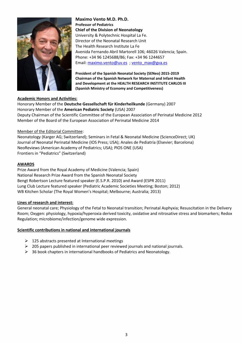
Maximo Vento M.D. Ph.D. Professor of Pediatrics
Chief of the Division of Neonatology University & Polytechnic Hospital La Fe. Director of the Neonatal Research Unit The Health Research Institute La Fe Avenida Fernando Abril Martorell 106; 46026 Valencia; Spain. Phone: +34 96 1245688/86; Fax: +34 96 1244657 Email: [email protected] ; [email protected] President of the Spanish Neonatal Society (SENeo) 2015-2019 Chairman of the Spanish Network for Maternal and Infant Health and Development at the HEALTH RESEARCH INSTITUTE CARLOS III (Spanish Ministry of Economy and Competitiveness)
Academic Honors and Activities: Honorary Member of the Deutsche Gessellschaft für Kinderheilkunde (Germany) 2007 Honorary Member of the American Pediatric Society (USA) 2007 Deputy Chairman of the Scientific Committee of the European Association of Perinatal Medicine 2012 Member of the Board of the European Association of Perinatal Medicine 2014 Member of the Editorial Committee: Neonatology (Karger AG; Switzerland); Seminars in Fetal & Neonatal Medicine (ScienceDirect; UK) Journal of Neonatal Perinatal Medicine (IOS Press; USA); Anales de Pediatría (Elsevier; Barcelona) NeoReviews (American Academy of Pediatrics; USA); PlOS ONE (USA) Frontiers in “Pediatrics” (Switzerland) AWARDS Prize Award from the Royal Academy of Medicine (Valencia; Spain) National Research Prize Award from the Spanish Neonatal Society Bengt Robertson Lecture featured speaker (E.S.P.R. 2010) and Award (ESPR 2011) Lung Club Lecture featured speaker (Pediatric Academic Societies Meeting; Boston; 2012) WB Kitchen Scholar (The Royal Women’s Hospital; Melbourne; Australia; 2013) Lines of research and interest: General neonatal care; Physiology of the Fetal to Neonatal transition; Perinatal Asphyxia; Resuscitation in the Delivery Room; Oxygen: physiology, hypoxia/hyperoxia derived toxicity, oxidative and nitrosative stress and biomarkers; Redox Regulation; microbiome/infection/genome wide expression. Scientific contributions in national and international journals
➢ 125 abstracts presented at International meetings ➢ 205 papers published in international peer reviewed journals and national journals. ➢ 36 book chapters in international handbooks of Pediatrics and Neonatology.

4
Dr Simon Hannam Head of Neonatal Services/Consultant Neonatologist Great Ormond Street Hospital for Children, London
Dr Simon Hannam qualified from University College London in 1988 and passed the MRCP in 1992. He completed his MD on the role of mechanoreceptor reflexes in SIDS at St Thomas’ Hospital under the supervision of Professor Anthony Milner. He completed his training in neonatology at King’s College Hospital and became a consultant neonatologist there in 2000. In 2014, Dr Hannam became the first consultant neonatologist at Great Ormond Street Hospital. His main interests are in neonatal respiratory reflexes and neonatal ventilation techniques.
Dr Neil Patel, Consultant Neonatologist The Royal Hospital for Children, Glasgow Dr Neil Patel Neil Patel is a Consultant Neonatologist at The Royal Hospital for Children in Glasgow. He is a graduate of Edinburgh Medical School. After a flirtation with paediatric cardiology he completed neonatal training in the west of Scotland and Australia in 2009. Neil previously worked as a neonatologist at the Royal Children’s Hospital in Melbourne, but returned to Glasgow in 2014. His clinical research interests include congenital diaphragmatic hernia, innovation in neonatal care, and long-term outcome in congenital anomalies.

5
Assessment of Neonatal Heart Rate Immediately after Birth using Digital Stethoscope, Hand Held Ultrasound and Electrocardiography. Treston B, Semberova J, Kernan R, Crothers E, Branagan A, O’Cathain N, Miletin J, Coombe Women’s & Infants Hospital, Dublin Introduction: The extent and initiation of cardiopulmonary resuscitation of neonates in the delivery room is largely guided by neonatal heart rate (HR). This is usually assessed by stethoscope auscultation. New Neonatal Resuscitation Guidelines have recommended the use of Electrocardiography (ECG) for HR determination during resuscitation as this was found to be the most accurate and efficient method to detect and monitor HR. Our aim was to determine the time to achieve first HR after delivery by handheld ultrasound (HUS), digital stethoscope (DS) and ECG, and compare these to auscultation, and observe the differences in HR achieved. Methods: Women who were planned for elective caesarean sections were recruited prior to delivery. Two physicians attended these deliveries, one assessed the HR by stethoscope auscultation and the second assessed the HR using either HUS (Mortara, Signos RT Personal Ultrasound), DS (Littmann 3200, 3M, US) or ECG (Philips IntelliVue MP5 Portable patient monitor). The time to achieve first HR and the HR recorded was noted, then when both modalities were recording a simultaneous HR was recorded. Results:Sixty participants were recruited in total (twenty in each group). The mean birth weight (±SD) of the cohort was 3.46kg (±0.43) and mean gestational age was 38.8 (±0.83) weeks of gestation. There was no significant difference between group characteristics. Four babies required resuscitation in the ECG group, and one in the DS group. All babies had Apgars ≥8 at 5 minutes of age, no babies required chest compressions or adrenalin The median time from birth to first HR recording by ECG was 98 seconds (compared to 85 seconds by stethoscope; p=0.002), the median time to achieve HR after placing the device was 13 seconds (compared to 13 seconds by stethoscope, p=0.74). The median time from birth to achieve HR by HUS was 113.5 seconds (compared to 90 seconds by stethoscope; p=0.02), the median time to achieve HR after placing the device was 28 seconds (compared to 15 seconds by stethoscope, p=0.007). The median time from birth to achieve HR by DS was 120 seconds (+/- 123) (compared to 110 (+/154) seconds by stethoscope; p=0.19), the median time to achieve HR after placing the device was 45 seconds (+/- 36) (compared to 11 seconds (+/-8) by stethoscope, p=0.0047). There was no reading of HR in seven (35%) patients in digital stethoscope group (no heart rate displayed 3 minutes following device application), these were excluded from statistical analysis. The mean difference between stethoscope and ECG in the HR was -10 bpm (p=0.024), between stethoscope and handheld US was +5 bpm (p=0.4) and between stethoscope and digital stethoscope was +27 bpm (p=0.061). Conclusion: We conclude that ECG was the most reliable and efficient method of recording HR in the delivery room compared to DS and HUS. However, the traditional stethoscope remains the quickest method to obtain a HR when the time delay in applying ECG leads is taken into consideration. We found that the DS used in our study was unreliable at measuring heart rate in the delivery room, frequently not displaying any HR, or displaying a HR significantly lower than the auscultated HR. We would like to further investigate the accuracy of US and Digital stethoscope by directly comparing the HR recorded to ECG.

6
Neonatal Encephalopathy: Systemic Inflammasome Activation M O’Dea1-3, L Kelly 1-3, Vavasseur C4, O’Leary JJ2,3, EJ Molloy1-5
Paediatrics and Child Health, Trinity College Dublin, National Children’s Hospital, Tallaght, Dublin1; Trinity Translational Medicine Institute, St James Hospital, Dublin2, Neonatology, Coombe Women and Infant’s University Hospital3, Neonatology, National Maternity Hospital4, Neonatology, Our Lady’s Children’s Hospital, Crumlin, Dublin, Ireland5
Introduction Systemic inflammation has been demonstrated in both animal and human models of neonatal brain injury. The Inflammasome is a component of the Innate immune system involved in regulating and inducing inflammation in response to infectious microbes and molecules derived from host proteins. It has been implicated in a host of inflammatory disorders and there have been recent developments toward promising therapeutics that target inflammasome activity. The Inflammasome has not been explored in neonatal encephalopathy. Components of the Inflammasome need exploration as potential therapeutic targets in neonatal Brain injury/encephalopathy (NE), as an adjunctive treatment to Therapeutic Hypothermia. We profiled the Inflammasome components, Interleukin (IL)- 1beta and ASC (Apoptosis-associated Speck-like protein containing a carboxy-terminal CARD), and NLR Family Pyrin Domain Containing 3 (NLRP3). Methods Serial blood samples from day 1 to 3 of life were compared to healthy neonatal controls. Inflammasome components IL-1β, NLRP3 and ASC in infants with NE (n=10) were compared to healthy neonatal controls (n=8) in response to endotoxin stimulation (Lipopolysaccharide: LPS). RT-PCR analysis was carried out on the ABI 7900 with analysis using GraphPad Prism Version 7. Results Il1Beta expression was increased on day 1 and day 3 and upregulated with LPS stimulation, day 1 (p=0.009) and day 3 (p=0.01). NLRP3 was increased day 1 and decreasing by day 3 in NE and upregulated in response to LPS on day 3 (p=0.009). ASC was increased day 1 NE and further increased on day 3 life without significant upregulation with LPS stimulation. Conclusion Inflammasome activation was evident in Neonatal Encephalopathy compared to controls and upregulated in response to LPS. The inflammasome and inhibition of systemic inflammation may have a role as a future specific immunomodulatory therapeutic target in NE.

7
INCIDENTAL FINDINGS ON ROUTINE TARGETED NEONATAL ECHOCARDIOGRAPHY PERFORMED IN PRETERM INFANTS LESS THAN 29 WEEKS GESTATION Smith A1, Breatnach C1, James A1, Franklin O2, El-Khuffash A1
1. Department of Neonatology, The Rotunda Hospital, Dublin 2. Our Lady’s Children’s Hospital, Crumlin, Dublin
Background: Targeted neonatal echocardiography (TnECHO) is performed for a wide variety of indications including the assessment of suspected congenital heart disease (CHD), appraisal of myocardial function and evaluation of patent ductus arteriosus (PDA). With the growing use of TnECHO, the discovery of incidental findings is increasing and include umbilical venous catheter (UVC) complications (mal-positioned umbilical catheters, liver haematomas), CHD, pericardial effusions and persistent pulmonary hypertension (PH). Objectives: The aim of this study was to quantify the rate of incidental findings identified on elective research echocardiograms performed on infants less than 29 weeks gestation. Methods: This was a retrospective study of echocardiograms performed within the first 24 hours of age on infants less than 29 weeks gestation over a three year period for research purposes. Infants who had echocardiograms performed for clinical purposes were excluded. Incidental findings identified on echocardiogram and pertinent clinical data were recorded. Results: Echocardiograms performed at a median [IQR] 10 [7 – 13] hours on 145 infants with a gestation and birthweight of 26.9 [25.7 – 28.0] weeks and 940 [750 – 1130] grams respectively were reviewed. Forty three (30%) infants had a total of 54 (37%) unexpected findings. The vast majority comprised of malpositioned UVCs where the tip was identified in the left atrium. The rate of CHD was 10%, the commonest being an atrial septal defect. One infant had an incidental finding of total anomalous pulmonary venous drainage, and another with transposition of the great arteries. The remainder of the findings included liver haematomas, pericardial effusions and unexpected PH [TABLE 1]. The presence of unexpected findings on TnECHO was independently associated with chronic lung disease or death when controlling for gestation [Adjusted OR 3.6 (95%CI 1.4 – 9.6)]. Conclusions: There is a high rate of unexpected findings discovered on screening echocardiograms in preterm infants less than 29 weeks gestation. Malpositioned UVCs when deep are more likely to be found in the left rather than the right atrium contrary to common knowledge. This is likely due to the persistent fetal channels directing inferior vena cava flow across the foramen ovale. Routine TnECHO screening of preterm infants may be warranted to identify the high likelihood of unexpected findings. Table 1: Rate and type of unexpected findings Complication Frequency
Overall unexpected pathology 54 (37%)
Number of infants with pathology 43 (30%)
Total number of infants with a UVC inserted 87 (60%)
Malpositioned UVC identified on TnECHO 24/87 (28%)
Tip in Left atrium 18/24 (75%)
Tip in the Right Atrium 2/24 (8%)
Tip in the Liver 4/24 (17%)
Liver Haematomas 4/87 (17%)
Congenital Heart Disease 15 (10%)
Atrial Septal Defect (number, proportion, median size in mm) 7/15 (47%), 5.2 [4.5-5.2]
Ventricular Septal Defect (number, proportion, median size in mm) 4/15 (27%), 2.3 [1.7-3.1]
Pericardial Effusion 3 (2%)
Unexpected pulmonary hypertension 5 (3%)
Unless stated, the denominator for the values is 145.

8
Milk on Time Saves More than an IV Line – Evaluating the provision of maternal milk on day one for preterm very low birth weight infants Roberta McCarthy1, Olivia Mason2, Claudine Vavasseur1 1National Maternity Hospital, Holles Street, Dublin 2; 2CSTAR Centre for Support and Training in Analysis & Research, University College Dublin, Dublin 4 Introduction: Current recommendations for preterm very low birth weight (VLBW) infants advise the early initiation and establishment of feeds using maternal milk (MM). Unfortunately many preterm infants experience delays initiating feeds while awaiting MM. Our aim was to explore associations with the timing of MM provision. Methods: The population included inborn preterm infants ≤31 weeks gestation or ≤1.5 kg who received MM as their 1st feed in our tertiary level neonatal unit. Comparisons were made between the group of infants who received MM within 24 hours of birth and those who experienced a delay beyond 24 hours. Results: 120/123 (98%) inborn infants who received their 1st feed in the neonatal unit, received this as MM and were included for analysis. 41 (34%) received MM as their 1st feed within 24 hours of birth and 79 (66%) experienced a delay beyond 24 hours. Comparing these groups, the following observations were noted.
1st Maternal Milk Observations
0-24 hours n=41
>24 hours n=79
Time to receive 1st MM (hours): 14.9 (8.7-17.6) 44.5 (35.3-61.6)
Duration of PNa (days): 6.3 (5.7-9.1) 8.6 (6.2-11.1)
Time to establish feeds at 150 ml/kg/db (days): 8.8 (7.4-12.3) 10.4 (8.7-13.1)
Exclusive MM in neonatal unit: 54% (22) 39% (31)
Any donor milk in neonatal unit: 5% (2) 19% (15)
Any formula milk in neonatal unit: 46% (19) 56% (44)
Time to 1st formula milk (days): 26 (11-44) 17 (8-31)
Any breastfeeding at discharge homec: 38% (14) 28% (20)
Corrected gestation at discharge homec (weeks): 35.7 (35.0-37.6) 36.7 (35.6-38.4)
Length of stay in neonatal unitc (days): 35 (27-49) 38 (22-66)
Weight at discharge home <9th centilec: 16% (4) 31% (15)
Demographics
Nationality of mother - Irish: 76% (31) 80% (63)
Twin or higher multiple pregnancy: 22% (9) 32% (25)
Delivery by caesarean section: 44% (18) 67% (53)
Gestation at birth (weeks): 29.6 (28.0-31.1) 29.4 (26.9-31.1)
Birth weight (kg): 1.26 (0.93-1.44) 1.22 (0.99-1.44)
Weight at birth <9th centile: 15% (6) 18% (14)
Values are median (IQR) or percentage (number). aOf those that received PN: 1st MM 0-24 h (n-39); 1st MM >24 h (n=74). bOf those that established feeds at 150 ml/kg/d: 1st MM 0-24 h (n=38); 1st MM >24 h (n=72). cOf those discharged to home: 1st MM 0-24 h (n=25); 1st MM >24 h (n=49).
Discussion: Our results suggest benefits for infants who receive MM earlier compared with those who are delayed. Infants who received MM within 24 hours of birth compared to those who were delayed, had a shorter duration of PN, established feeds earlier, and had a shorter length of stay in the neonatal unit; a greater number also received exclusive breast milk feeds during their stay and were breastfeeding at the time of discharge home. Mothers post caesarean-section and mothers of multiples were identified as being of particular risk of delay. Conclusion: Providing MM to initiate feeds earlier is associated with benefits that suggest a clinical advantage to patients and their families as well as a financial advantage to healthcare providers. This highlights the need appropriate support to enable the early availability of MM for preterm VLBW infants.

9
EXPERIENCE OF INFANTS BORN WITH DOWN SYNDROME: BURDEN OF DISEASE IN THE EARLY NEONATAL PERIOD Aisling Smith1, Therese Martin1, Colm R Breatnach1, Etaoin Kent2, Ita Shanahan2, Michael Boyle1, Philip T Levy3, Orla Franklin4, Afif EL-Khuffash1
Affiliations: 1 Department of Neonataology, The Rotunda Hospital, Dublin. 2 Department of Obstetrics and Gynaecology, RCSI, Dublin. 3 Department of Paediatrics, Washington University School of Medicine, St Louis, Missouri. 4 Department of Paediatric Cardiology, Our Lady’s Children’s Hospital, Crumlin, Dublin.
Background: The incidence of structural abnormalities and early neonatal morbidities in infants with Down syndrome (DS) is under-reported. Many advocate keeping infants with confirmed or suspected DS on the postnatal ward to facilitate early bonding and the establishment of feeding. We hypothesise that the majority of infants with a suspected or confirmed diagnosis of DS require admission due to the high incidence of early neonatal morbidities. Objectives: In a retrospective cohort of infants with a diagnosis of DS, we aimed to examine the rate of admission to the postnatal ward (PNW) versus primary NICU admission, and to present the rate of important morbidities including congenital heart disease (CHD), echocardiography confirmed persistent pulmonary hypertension of the newborn (PPHN) and gastrointestinal disorders. Methods: This was a retrospective cohort study of infants born with DS between January 2011 and June 2016. Relevant clinical demographics, admission details, early neonatal morbidities, NICU related treatments, neonatal outcomes and length of hospital stay were recorded. Results: 121 infants were accessible. Forty nine (41%) were delivered via a caesarean section with a median maternal age and parity of 37 [33 – 39] years and 2 [1 – 3]. Antenatal diagnosis occurred in 31 (26%) who had a higher overall rate of structural anomalies [19/31 (61%) vs. 21/90 (23%), p<0.01]. There was a high incidence of structural anomalies and neonatal morbidities: 84 (69%) CHD; 41 (34%) PPHN; 21 (17%) polycythaemia; 15 (12%) gastrointestinal morbidity; and 60 (49%) neonatal jaundice. 67 (55%) were admitted directly to NICU while 54 (45%) infants were initially cared for on PNW of which 38 were later admitted to NICU; only 16 (13%) remained on the PNW prior to discharge. Table 1 illustrates the morbidities in the three admission groups. PPHN was an independent predictor of death before discharge [adjusted OR 11 (95%CI 2 – 110)]. Conclusion: Infants with Down syndrome have a high rate of neonatal morbidities. The incidence of echocardiography confirmed PPHN in out cohort is much higher than is reported in the literature. The presence of identifiable antenatal anomalies increases the likelihood of an antenatal diagnosis. However, infants initially admitted to the PNW have a high likelihood of requiring NICU admission and have a high rate of neonatal morbidities. Therefore, elective admission of all infants with Down syndrome is recommended to screen for PPHN, CHD and other important morbidities. Table: Demographics and morbidities in the three admission groups. Primary NICU
Admission (n=67) NICU after PNW
(n=38) PNW care only
(n=16)
p
Gestation (weeks) 38.0 [34.4 – 39.0] 38.5 [37.7 – 39.1] 38.9 [37.1 – 39.6] 0.10
< 34 weeks 11 (16) 0 0 <0.01
Birth weight (kg) 2.9 [2.3 – 3.4] 2.9 [2.7 – 3.3] 3.1 [2.7 – 3.7] 0.51
Caesarean Section 35 (52) 10 (26) 4 (25) 0.01
Small for Gestation 17 (25) 7 (8) 0 0.07
Male Gender 29 (43) 23 (61) 10 (63) 0.15
5 minute Apgar Score 9 [8 – 10] 10 [9 – 10] 10 [10 – 10] <0.01
Antenatal Diagnosis 28 (42) 2 (5) 1 (6) <0.01
Any CHD 56 (84) 24 (63) 4 (25) <0.01
AVSD 20 (30) 3 (8) 1 (6) <0.01
PPHN 28 (42) 11 (29) 1 (13) 0.06
GI morbidity 12 (18) 3 (8) 0 0.09
Polycythaemia 13 (19) 8 (21) NA 0.14
Jaundice 32 (48) 19 (50) 9 (56) 0.83
Ventilation Days 4 [2 – 9] 4 [3 – 4] NA 0.62
O2 Days 13 [5 – 26] 13 [8 – 16] NA 0.63
Inotropes 15 (22) 5 (13) 0 0.08
Nitric Oxide 14 (21) 4 (11) 0 0.07
First pH 7.34 [7.28 – 7.37] 7.36 [7.33 – 7.38] NA 0.04
pCO2 (Kpa) 6.0 [5.6 – 7.0] 5.5 [4.8 – 6.2] NA <0.01
HCO3- 23.4 [21.0 – 25.0] 23.1 [21.6 – 25.0] NA 0.91
First haemoglobin 197 [186 – 206] 215 [201 – 208] NA 0.02
Platelets 151 [100 – 201] 169 [102 – 208] NA 0.39
White Blood Cells 18.6 [12.9 – 24.0] 21.1 [16.4 – 26.7] NA 0.02
TAM 5 (8) 1 (3) 0 0.34
Days on TPN 7 [2 – 11] 3 [ 2 – 11] NA 0.69
Hospital Days 26 [11 – 47] 26 [19 – 29] NA 0.20
Death 7 (10) 0 0
Data are presented as medians [IQR] or count (%) and compared using the Kruskal-Wallis test or Chi square/Fisher’s exact test as appropriate. CHD: Congenital Heart Disease; AVSD: atrioventricular septal defect; GI: gastrointestinal; TAM: Transient abnormal myelopoiesis; TPN: total parenteral nutrition.

10
Levels of Glucose in Cord Blood Samples (LOGICS) Ó Catháin N1, Stanzelova A1, Treston B1, O'Kelly R2, Killalea A2, Kelly J2, Semberova J1,3, Miletin J1,3,4,5 1 Department of Paediatrics and Newborn Medicine, Coombe Women and Infants University Hospital, Cork St, Dublin 8, Ireland 2 Department of Biochemistry, Coombe Women and Infants University Hospital, Cork St, Dublin 8, Ireland 3 Institute for the Care of Mother and Child, Prague, Czech Republic 4 UCD School of Medicine, University College Dublin, Dublin, Ireland 5 3rd Faculty of Medicine, Charles University, Prague, Czech Republic Introduction Neonatal hypoglycaemia is a common cause of medical review and admission to the neonatal unit, and a known risk factor for neonatal brain injury. Currently the proposed lower limit for serum glucose level, to diagnose hypoglycaemia after the first hour of life, is 2.6 mmol/l. Maternal and foetal glycaemic control are believed to be closely related, however levels of glucose and fructosamine in umbilical cord blood have rarely been studied. We hypothesise that a proportion of infants may have been hypoglycaemic at delivery. The aim of this study is to determine the incidence of infants born with glucose <2.6mmol/l, and establish the corresponding fructosamine levels. Methods This is a prospective observational study. The inclusion criteria was gestational age ≥37 weeks, irrespective of maternal history. Infants with known congenital or chromosomal anomalies were excluded from participation. Umbilical arterial blood was collected at delivery for cord glucose and lactate, as well as fructosamine which was corrected for total protein. Medical team was notified of glucose levels <2.6 mmol/l and these infants were reviewed at one hour of life and glucose levels were repeated. Our primary outcome was the incidence of hypoglycaemia (defined as glucose <2.6 mmol/l) in cord blood, and the corresponding fructosamine levels. The secondary outcome was to identify a correlation between umbilical cord blood glucose levels and admission to neonatal unit. Results We enrolled 50 patients following maternal consent. Mean gestational age (±SD) was 38.5 weeks of gestation (±0.8) and mean birth weight was 3.5 kg (±0.45). 38% (19) were girls, 8% (4) were small for gestational age (<9th centile), 10% (5) were large for gestational age (>91st centile). All infants were born by caesarean section. Mean cord glucose level was 2.9 mmol/l (±0.5). Ten infants (20%) had glucose level <2.6mmol/l (group 1). Sixty percent (6) of those involved required admission to the neonatal unit compared to 15% (6) in the group with normal cord glucose level (group 2) (p=0.008). Mean fructosamine level was 201 µmol/l (±20.7) for the entire cohort. Fructosamine levels ranged from low to lower limit of normal based on the adult fructosamine reference range. There was no statistically significant difference in the fructosamine level between the group 1 and 2 (p=0.63). Conclusions Umbilical arterial glucose levels were below the neonatal recommended range for normoglycaemia in a significant proportion of our study population. Infants with low cord glucose were significantly more likely to be admitted to the neonatal unit. Corresponding fructosamine levels indicate the possibility of hypoglycaemia in utero, however current reference ranges in practice for corrected fructosamine are adult based.

11
A RANDOMISED STUDY OF HEART RATE DISPLAY COMPARING TWO MONITORS (ELECTROCARDIOGRAM PLUS PULSE OXIMETER VERSUS PULSE OXIMETER ALONE) IN NEWLY-BORN INFANTS: THE SHEEP STUDY (ISRCTN11028739) Madeleine C. Murphy,1-3 Laura de Angelis,4 Lisa K. McCarthy,1,5 Colm P.F. O’Donnell1-3,5
1. The National Maternity Hospital, Holles Street, Dublin 2, Ireland 2. National Children’s Research Centre, OLCHC, Dublin 12, Ireland 3. School of Medicine and Medical Science, University College Dublin, Ireland 4. Vittore Buzzi Hospital, University of Milan, Italy 5. Our Lady’s Children’s Hospital, Crumlin, Dublin 12, Ireland
Introduction: Based on small studies, the International Liaison Committee on Resuscitation suggests that electrocardiogram (ECG) measures newborns’ heart rate (HR) faster than pulse oximetry (PO) in delivery room (DR). Clinical assessment has been shown to underestimate ECG HR. Aim: To determine among newly-born infants whether HR was measured more quickly using the Philips IntelliVue X2 (ECG plus PO) versus the Nellcor portable PO (PO alone). Methods: This unmasked randomised parallel group study was carried out on low-risk infants born at our hospital with ethical approval and parental consent. Infants were randomly assigned to being monitored with the IntelliVue or Nellcor monitor in a 1:1 ratio. Primary outcome was time to first HR from start of monitor application measured in seconds. In addition, we performed a parallel study of clinical assessment of HR. Caregivers auscultated the HR whilst masked to the HR on the monitor. We used video recordings to record our data. Results: We studied 100 infants in the DR; 47 were monitored with the IntelliVue and 53 with the Nellcor. Groups were well matched at study entry. Time to first display of HR was shorter with the IntelliVue (ECG) than the Nellcor PO [median (IQR) 24 (19, 39) vs. 48 (36, 69) seconds, p<0.001]. There was no difference in the total time for either monitor to display both HR and PO readings [median (IQR) 52 (47, 76) vs. 48 (36, 69) seconds, p=0.507]. When comparing the IntelliVue (Massimo PO) with the Nellcor PO, there were more episodes of initial bradycardia [26/47 (55%) vs. 3/53 (6%)]; and intermittent PO readings [15/47 (32%) vs. 0 (0%)]. Infants monitored with the IntelliVue were handled more frequently than infants randomly assigned to the Nellcor [mean (SD) 9.2 (3.8) vs. 6.6 (2.5) seconds, p<0.001]; and for longer [mean (SD) 104 (48.5) vs. 85 (28) seconds, p<0.019]. Clinical assessment underestimated ECG HR with a mean (SD) difference of -9 (22) bpm [mean (SD) IntelliVue (ECG) HR 173 (17) vs. HR auscultated 164 (23) bpm]. Clinical assessment underestimated PO HR with a mean (SD) difference of -5 (22) bpm [mean (SD) Nellcor PO HR 166 (21) vs. HR auscultated 161 (21) bpm]. Median (IQR) time to HR using auscultation was 14 (10, 18) s. Conclusions: Though IntelliVue (ECG) determined initial HR significantly more quickly than the Nellcor PO, overall there was no difference in the total time for both HR and PO to be determined between monitors. There were more episodes of bradycardia and intermittent PO readings with the IntelliVue (PO) monitor, and infants monitored with ECG were handled more often and for longer. Clinical assessment estimated HR within 10 bpm to that on the monitors and significantly more quickly. To better judge whether it is worthwhile, further study of HR assessment using ECG on high-risk infants in the DR is warranted.

12
DISPLAY POSTER BOARD LISTING (Listed alphabetically by presenting author)
Presenting Author Surname
Poster No.
Abstract Title Page in Abstract Book
Aminudin 1 MEASUREMENT OF NEONATAL NOISE LEVELS AND NOISE REDUCTION IN NICU: A CASE STUDY
13
Carey 2 WEIGHT AND OFC GROWTH OF PRETERM BABIES FROM BIRTH TO TWO YEARS CORRECTED AGE COMPARED TO UK-WHO GROWTH STANDARDS
14
Caulfield 3 HYPOGLYCAEMIA IN HIGH RISK NEWBORN INFANTS: AN AUDIT OF THE CURRENT SCREENING PRACTICE
15
Dunworth 4 THE ROLE OF BIOMARKERS IN IDENTIFYING HYPOXIC ISCHAEMIC ENCEPHALOPATHY IN NEONATES AND PREDICTING LONG TERM NEURODEVELOPMENTAL OUTCOMES – A LITERATURE REVIEW
16
Dwyer 5 REDUCING PREVENTABLE HARM: ATTITUDES TOWARD PATIENT SAFETY IN AN IRISH TERTIARY NEONATAL UNIT
17
Fox 6 AUDIT OF THE POSTNATAL MANAGEMENT OF INFANTS AT HIGH RISK OF HAEMOLYTIC DISEASE OF THE NEWBORN
18
Greene 7 ORAL STIMULATION FOR PROMOTING ORAL FEEDING IN PRETERM INFANTS: A COCHRANE SYSTEMATIC REVIEW – NEXT STEPS FOR IRISH NICUS?
19
Greene 8 PERSPECTIVES OF NURSING STAFF ON THE ‘READINESS, ORAL SKILLS, SAFETY, EFFICIENCY’ (ROSE) FEEDING CHECKLIST: TOWARDS ESTABLISHING VALIDITY
20
Halpenny 9 TRANSIENT NEONATAL DIABETES MELLITUS IN A LATE PRETERM INFANT: CASE REPORT
21
Lewis 10 DROP OFF BREAST FEEDING RATES ON POSTNATAL WARDS IN A TERTIARY MATERNITY HOSPITAL
22
Murphy, C 12 REVIEW OF THE NEONATAL FALLS ON THE POSTNATAL WARD IN A LEVEL TWO NEONATAL UNIT 2015-2017
23
Murphy, M 11 ATTITTUDES OF STAFF MEMBERS TOWARDS VIDEO RECORDING IN THE DELIVERY ROOM
24
Smith 13 THE TRIANGULAR SIGN, A USEFUL DIAGNOSTIC MARKER FOR BILIARY ATRESIA: A CASE SERIES OF THREE IRISH INFANTS
25
Smith 14 A REVIEW OF THE PARENTERAL NUTRITION SUPPLY SERVICE IN AN IRISH NEONATAL UNIT
26
Smith 15 AETIOLOGICAL EVALUATION OF 42 INFANTS IDENTIFIED WITH A PERMANENT CHILDHOOD HEARING IMPAIRMENT THROUGH THE IRISH NEWBORN HEARING SCREENING PROGRAMME: FINDINGS AND RECOMMENDATIONS
27
Smith 16 AN AUDIT OF NEONATAL RESUSCITAIRE EQUIPMENT IN THE ROTUNDA HOSPITAL
28

13
MEASUREMENT OF NEONATAL NOISE LEVELS AND NOISE REDUCTION IN NICU: A CASE STUDY Aminudin N1,2 , Corcoran D1, El-khuffash A1, Franta J2, McCallion N1
1. Rotunda Hospital, Dublin. 2. National Neonatal Transport Programme: Rotunda Hospital, Coombe Women & Infants University
Hospital, National Maternity Hospital, Dublin.
Background: Noise is a recognized environmental hazard that predisposes neonates who are critically ill and/ or premature to possible auditory and non-auditory detrimental effects which include noise induced hearing loss, autonomic disturbances, behavioural and cognitive changes. Although the current recommended safe environmental sound pressure levels (SPL) in neonatal intensive care units (NICU) should not exceed 45 dB (decibel) (American Academy of Paediatrics, 1974/1997), reports have shown that levels have ranged between 50-89.5 dB and can peak to 105 dB. This study aims to measure noise levels in a tertiary NICU from 3 positions in relation to the neonate in the incubator and to evaluate the proportion of noise experienced by the neonate if noise reduction is applied to the infant’s ear externally. Methods: We used a 4-channel sound level meter (Svan 958A ®) connected to 3 microphones to measure sound pressure level (SPL) that is quantified in decibel-A weighted (dB-A). A neonatal mannequin was placed in the incubator and noise levels were recorded from 3 positions; neonatal external ear, inside incubator and exterior of the incubator. We applied 3 types of noise reduction apparatus to the neonatal mannequin and recorded noise levels from the 3 positions mentioned. The noise measurement situations that were recorded are: 1) Noise Level recording with no added noise reduction (Standard NICU), 2) Noise Level recording with Neonatal MiniMuffs (Natus®) 3) Noise level recording using Noise Protective Ear Muffs (NPEM) (Em’s4Bubs®) and 4) Noise level recording using Active Noise Cancelling (ANC) Headphones (Bose®). An average of 15 minutes of recording was performed in all 4 situations. All noise level recordings from the meter were transferred into correlative software (Svan PC ++ ®) for further interpretation. Results: Statistical Analysis with SPSS Version 23® was used to analyse data and produce descriptive statistical results. For this study, noise levels are represented as peak sound pressure level (Lpeak) and total sound energy (Leq) that are quantified in units of decibel-A weighted (dB-A). In the NICU the mean peak SPL (dB-A) were; 59.5 (infant ear), 66.7 (inside incubator) and 73.8 (outside incubator). The mean Leq (dB-A) were; 44.1 (infant ear), 52.8 (inside incubator) and 58.9 (outside incubator). The evaluation of the proportion of noise levels experienced by the neonate if noise reduction is applied to the ear externally are represented by the percentage (%) of values detected near the infant’s ear compared to the external incubator noise levels. The mean % peak SPL detected: 80.8 (standard NICU), 83.6 (Natus ®), 78.1 (NPEM) and 74.8 (ANC). Correspondingly, the mean % Leq detected near the neonatal ear over the external sound levels are are: 74.9 (standard NICU), 78.4 (Natus ®), 71.7 (NPEM) and 56.0 (ANC). Paired sample t-test was used to analyse the comparison of standard NICU practise with the 3 noise reduction apparatus demonstrated in the table below:
Peak SPL of standard NICU with Noise reduction apparatus/ interventions
95% Confidence Interval of the Difference
p-Value <0.05 Lower Upper
Standard NICU vs. Natus minimuffs -2.87343 -1.56074 .000
Standard NICU vs. NPEM 2.90474 3.81402 .000
Standard NICU vs. ANC 24.88469 25.80603 .000
Total Sound Energy Standard NICU vs. Noise Reduction apparatus/ intervention
95% Confidence Interval of the Difference
Sig. p Value <0.05
Lower Upper
Standard NICU vs. Natus minimuffs -3.00219 -1.75440 .000
Standard NICU vs. NPEM 4.61103 5.40434 .000
Standard NICU vs. ANC 20.61908 21.42901 .000
Conclusion Our study has shown that noise levels in the NICU detected at the neonatal ear in the incubator can peak at a mean of 60 dB-A (with a mean total sound energy of 44.1dBA). This study demonstrates that the neonate in the incubator is exposed to 80% of the external peak SPL and 75% of Leq during standard care. The application of noise reduction apparatus attenuates peak noise levels and total sound energy that neonates in the incubator are potentially exposed (p value <0.05). As noise exposure is associated with physiological and behavioural stability, we need to consider applicable adjustments in our practise to alleviate this.

14
Weight and OFC Growth of Preterm Babies from Birth to Two Years Corrected Age Compared to UK-WHO Growth Standards Carey R1, Glynn AC2, Reynolds A1,3, McCallion N1,3
1. Royal College of Surgeons in Ireland, Dublin 2. Department of Nutrition and Dietetics, The Rotunda Hospital, Dublin 3. Department of Neonatology, The Rotunda Hospital, Dublin
Aim: To quantify postnatal weight and occipitofrontal circumference (OFC) growth in infants born before 32 weeks gestational age (GA) from birth to 2 years corrected age (CA) and to compare those measurements to the UK-WHO growth standards. Background: The UK-WHO Neonatal and Infant Close Monitoring Charts are the official growth standard for preterm babies born in Ireland.(1) They were not intended to reflect actual growth patterns of babies born prematurely. Methods: This was a retrospective cohort study of babies born before 32 weeks GA in a tertiary maternity hospital between September 2014 and February 2015. The exclusion criteria were death before discharge and major congenital anomalies. Weight and OFC measurements from day of life 1, 7, 14 and 28, discharge and outpatient clinic visits were obtained. Cases were categorised based on the GA at birth (very preterm: 28-31 weeks, extremely preterm: <28 weeks). A standard deviation score (SDS) was calculated for each measurement using the appropriate UK-WHO growth standard LMS values.(2) Localised regression was used to fit curves for the changes in weight and OFC SDS over time based on both days from birth and CA. Results: Sixty-nine babies were eligible. Ten died before discharge. Data on 59 babies was obtained (Male: 58%, Extremely preterm: 39%, Singleton: 59%). At 16-20 months CA, head and weight measurements were available from 28 and 26 children respectively. At birth, the weight regression lines for the very and extremely preterm groups were just below the 50th centile. By day of life 7, both had dropped to between the 9th and 25th centiles. The very preterm line climbed to the 50th centile by 6 weeks CA. The extremely preterm line remained flat between the 9th and 25th centiles until 6 months CA, but then began to climb. At 2 years CA, the very and extremely preterm weight lines were between the 50th and 75th, and 25th and 50th centiles respectively. At birth, the OFC lines for both groups were around the 25th centile. The very preterm line plateaued initially, but climbed to the 75th centile by 6 weeks CA. The extremely preterm line fell to between the 2nd and 9th centiles by four weeks after birth, but rose to the 50th centile by 6 weeks CA. Between six weeks and 18 months CA, the very and extremely preterm OFC lines lay between the 75th and 91st, and around the 50th centiles respectively. Conclusions: Overall, by two years CA, the cohort’s weights and OFCs were in line with the UK-WHO growth standards. Following the initial postnatal fluid loss, very preterm babies experienced early catch-up weight growth, but this process appeared to be relatively delayed in the extremely preterm group. After six weeks CA, the extremely preterm group had OFCs smaller than the very preterm group, but still in line with the growth standard. The small sample size and incomplete follow-up limit the generalisability of these findings, but they agree with existing evidence showing that postnatal weight growth is dependent on gestational age at birth.(3) 1. Cole TJ, Williams AF, Wright CM, Group RGCE. Revised birth centiles for weight, length and head circumference in the UK- WHO growth charts. Ann Hum Biol. 2011;38(1):7-11. 2. Cole TJ, Green PJ. Smoothing reference centile curves: the LMS method and penalized likelihood. Stat Med. 1992;11(10):1305-19. 3. Cole TJ, Statnikov Y, Santhakumaran S, Pan H, Modi N, Neonatal Data Analysis U, et al. Birth weight and longitudinal growth in infants born below 32 weeks' gestation: a UK population study. Arch Dis Child Fetal Neonatal Ed. 2014;99(1):F34- 40.

15
HYPOGLYCAEMIA IN HIGH RISK NEWBORN INFANTS: AN AUDIT OF THE CURRENT SCREENING PRACTICE. Dr Fionnuala Caulfield, Dr Sarah Cronin, Dr Niazy Al-Assaf, Neonatal Department, University Maternity Hospital, Limerick Background: Neonatal hypoglycaemia is associated with abnormal neurological outcomes with the duration of hypoglycaemia having a direct impact on outcome1. Current guidelines for screening of high risk babies on postnatal wards in a secondary maternity hospital are adapted from the national childbirth trust 19972and UNICEF 20083 guidelines. New guidelines were published by the Canadian Paediatric society (CPS) in 20164. Aims: To evaluate whether current screening practices of hypoglycaemia among high risk infants conforms with current local guidelines and how these compare with international standards. Method: Retrospective chart review of infants born in July 2017 identified as high risk for hypoglycaemia under current guidelines and compared to CPS guidelines. Information gathered included baseline infant characteristics, timing of blood sugar measurements (BM) and management of hypoglycaemia. Results: 39 infants born in July 2017 were identified as at high risk of hypoglycaemia under current guidelines. 4 charts were not available for review. 34 infants had BM screening and 1infant did not. The mean time to first BM was 4 hours (Range 1-10). 38% (n=13) infants developed hypoglycaemia, of these 15% (n=2) required NICU admission. There was no significant difference in timing of BM measurements between normoglycaemic and hypoglycaemic infants (p = 0.35). An additional 9 infants were identified as high risk for hypoglycaemia when CPS guidelines were applied. These infants did not have BM screening and 1 infant subsequently became symptomatic with hypoglycaemia. Discussion: Adherence with current local guidelines is satisfactory however these guidelines warrant evidence based modification. 19% of high risk infants were not routinely screened. There was large variation in timing of BM in those infants who were screened. Detection of hypoglycaemia in high risk infants could be optimised if recent international standards were adopted. Recommendation: Implementation of CPS guidelines and a re-audit to evaluate the outcome. References: 1. Singh M, Singhal PK, Paul VK, et al. Neurodevelopmental outcome of asymptomatic & symptomatic babies with neonatal hypoglycaemia. Indian J Med Res 1991;94:6-10. 2. Hypoglycaemia of the breast fed newborn. National Childbirth Trust. Mod Midwife. 1997 Oct;7(10):31-3. 3. UNICEF (2008) Guidance on the development of policies and guidelines for the prevention and management of Hypoglycaemia of the newborn. www.babyfriendly.org.uk. Accessed 8/9/2017. 4. Canadian Paediatric Society, Fetus and Newborn Committee [Principal authors: K Aziz, P Dancey]. Screening guidelines for newborns at risk for low blood glucose Paediatr Child Health 20049723–9.

16
THE ROLE OF BIOMARKERS IN IDENTIFYING HYPOXIC ISCHAEMIC ENCEPHALOPATHY IN NEONATES AND PREDICTING LONG TERM NEURODEVELOPMENTAL OUTCOMES – A LITERATURE REVIEW Niall Dunworth1, Al Assaf N2, Khan R2 1 Graduate Entry Medical School, University of Limerick 2 Department of Neonatology, University Maternity Hospital Limerick
Aims Currently, identification of Hypoxic Ischaemic Encephalopathy (HIE) relies on clinical signs and diagnostic imaging7. These modalities have limitations, prompting the identification of specific biomarkers which are elevated in HIE, thus identifying and risk stratifying HIE patietns. This literature review aims to:
• Review recent work on biomarkers which are associated with HIE
• Determine which biomarkers predict neurodevelopmental outcome Methods A systematic review was conducted using articles from: Pubmed, Embase, Medline, Web of Science, ScienceDirect Search terms included: Hypoxic ischaemic encephalopathy, predictors of outcome, biomarkers and HIE, cord blood biomarkers and HIE. Inclusion criteria: Studies from 2010 - present. Exclusion criteria: Studies performed on animal subjects. Results Searches revealed 4 reviewed neuronal tissue specific markers; Glial fibrillary acidic protein (GFAP), Ubiquitin carboxyl-terminal esterase L1 (UCHL1), Neuronal specific enolase (NSE) and S100β, which are released in HIE. Other markers identified were Lactate and Lactate Dehydrogenase (LDH). Serum UCHL1 was significantly higher in the earlier stages post birth (6-24 hours), in cases of HIE10,14. GFAP levels do not raise until a later stage post birth, with higher levels correlating with poor neurodevelopmental outcome12-15. Umbilical cord blood biomarker levels demonstrated little correlation with neurodevelopmental outcome, suggesting that biomarkers do not raise until hours post birth16,17. S100B also increases with severe presentations of HIE and correlates with poor neurodevelopmental outcome19-21,27. The only significant relationship between NSE and neurodevelopmental outcome was measured at 24 hours post birth18. LDH is a strong predictor of neurodevelopmental outcome suggesting it’s potential in risk stratification of HIE patients. Elevated serum lactate levels also correlate with poor neurodevelopmental outcome24,25. Conclusion UCHL1, GFAP and S100B increase in the presence of HIE. They correlate with severity of HIE and poor neurodevelopmental outcome. LDH levels may also be a useful predictor of outcome. Most studies involved small sample sizes but provide a direction for future work.

17
REDUCING PREVENTABLE HARM: ATTITUDES TOWARD PATIENT SAFETY IN AN IRISH TERTIARY NEONATAL UNIT Dwyer L1. Smith A1 ,McDermott R1, Breatnach C1, Corcoran JD1 Department of Neonatology, The Rotunda Hospital, Dublin Introduction Ensuring patient safety is a vital component of any high quality healthcare service. There is little published research investigating interventions to improve safety culture in neonatal unit. This study aimed to explore the safety culture that exists within a level III Irish neonatal unit. Methods This was a quantitative, cross-sectional study which was performed in the Rotunda Hospital, Dublin, a tertiary neonatal centre with a birth rate of ~8,500 per annum. Data collection took place between July 2016 and March 2017. A 30 item safety attitudes questionnaire (SAQ) was utilized to analyze staff perceptions in six areas namely, the teamwork climate, the safety climate, the perceptions of management, job satisfaction, working conditions and stress recognition, with a 5 point Likert response scale ranging from 1 (disagree strongly) to 5 (agree strongly). The scores were then transformed to a numerical 100 point scale (Positive score >75). Results The ‘Stress Recognition’ domain received the highest score (75.3) followed by ‘Job Satisfaction’ domain with a mean score of 74.4. The ‘Teamwork Climate’ domain showed a mean score of 71.2 followed by the ‘Safety Climate’ domain with a mean score of 68.2. The ‘Working Conditions’ domain had a mean score of 59.6. The lowest mean scale score in the neonatal unit was for ‘Perceptions of Management’, with a mean score of 50.7: 42% agreed that administration supported their daily efforts, with a significantly high percentage agreed that staffing levels were insufficient to handle the number of patients. Internal consistency of the six factors measured via Cronbachs alpha ranged from 0.43 to 0.78 highlighting good consistency within each subscale. Collaboration and Communication scores were high across all disciplines at 76%. Conclusion This SAQ has brought to light a number of important areas for quality improvement and staff satisfaction in our neonatal unit. Importantly, this study highlighted that the Rotunda neonatal unit scored higher for all domains when compared to international benchmarked data, reflecting well on the hospital risk management programme.

18
AUDIT OF THE POSTNATAL MANAGEMENT OF INFANTS AT HIGH RISK OF HAEMOLYTIC DISEASE OF THE NEWBORN Dr Áine Fox, Dr Johannes Letshwiti, Rotunda Hospital, Dublin Introduction Haemolytic disease of the foetus and newborn (HDFN) is caused by the destruction of foetal red blood cells due to red cell antibodies produced by the mother. One of the most clinically significant of these is the Rh antigens and occurs when Rh D-negative mothers have Rh D-positive babies. This group of patients can be identified routinely through antenatal blood group testing of the mother. This is currently the practice worldwide and this facilitates screening of at risk infants by testing cord blood to determine group and Direct Coombs Test (DCT) status of the infant. The DCT aids the detection of this condition. High bilirubin levels have the potential to cause acute bilirubin encephalopathy in the short term and brain damage in the form of kernicterus in the longer term. Early detection of hyperbilirubinemia allows early initiation of treatment, avoiding potential complications. Methods A systematic review of charts was performed for babies born between 01 July 2016 and 31 December 2016 with a positive DCT result. Results There were 116 babies that had a positive DCT result with 20 babies (17%) born at less than 38 weeks gestation. One baby had an attempted intrauterine transfusion which was not successful. Almost 90% of babies (104 of 116) had their DCT status documented in their chart. There were 36 (31%) babies who required phototherapy, 9 (8%) who required immunoglobulin therapy, 8 (7%) who required a red cell transfusion and none who required exchange transfusion. The 63% of babies who required phototherapy had jaundice in the first 24 hours of life. The 27 of the 36 babies (75%) requiring phototherapy required admission to NICU. Nineteen (52.7%) of the 36 babies who required phototherapy had it for 2 or less days. Ninety-four babies (81%) had their haemoglobin (Hb) checked in the outpatient department at 2 weeks of age, of which 6 babies (5.1%) had Hb less than 100mg/dL. All babies with Hb less than 100mg/dL at 2 weeks required phototherapy and 50% had immunoglobulin therapy. Conclusion It is currently standard practice to check the haemoglobin of all DCT positive babies at 2 weeks of age regardless of their clinical course. In this audit, all patients that had low haemoglobin at 2 weeks of age required treatment for jaundice. This may indicate that DCT positive babies that did not require treatment are at very low risk of anaemia. This suggests that checking haemoglobin at 2 weeks of age in this group may be unnecessary. References
1. Victoria Parker and Christopher A. Tormey (2017) The Direct Antiglobulin Test: Indications, Interpretation, and Pitfalls. Archives of Pathology & Laboratory Medicine: February 2017, Vol. 141, No. 2, pp. 305-310
2. Kristinsdottir T et al.(2016)Positive Coomb's test in newborns; causes and clinical consequences Summary of cases diagnosed in the Blood Bank in the years 2005 to 2012. Laeknabladid: July 2016, Vol102 (7-8) 326-31

19
ORAL STIMULATION FOR PROMOTING ORAL FEEDING IN PRETERM INFANTS: A COCHRANE SYSTEMATIC REVIEW – NEXT STEPS FOR IRISH NICUS? Zelda Greene*, Colm O’Donnell**, Margaret Walshe*** *Speech and Language Therapy Department, OLCHC **Department of Neonatology, National Maternity Hospital Dublin **Department of Clinical Speech and Language Studies, Trinity College Dublin Background Preterm infants are often delayed in attaining oral feeding which is an important hospital discharge criteria. Oral stimulation interventions may help infants develop sucking skills, promoting earlier oral feeding and hospital discharge. Irish NICUs have sporadic SLT input and multidisciplinary approaches to managing feeding problems are lacking. This review was required to determine the effectiveness of these stimulation interventions for oral feeding in preterm infants. Methods We used the standard search strategy of the Cochrane Neonatal Review Group. Randomised controlled trials were included. The GRADE system was used to rate quality of evidence. We performed meta-analysis using a fixed-effect model. Results 19 RCTs were included, total 823 participants. Methodological weaknesses were evident across trials. Meta-analysis showed that oral stimulation reduced
• time to transition to oral feeding compared with standard care (mean difference (MD) -4.81, 95% confidence interval (CI) -5.56 to -4.06 days); compared with another non-oral intervention (MD -9.01, 95% CI -10.30 to -7.71 days)
• duration of hospitalisation compared with standard care (MD -5.26, 95% CI -7.34 to -3.19 days); compared with another non-oral intervention (MD -9.01, 95% CI -10.30 to -7.71 days).
• duration of parenteral nutrition for infants compared with standard care (MD -5.30, 95% CI -9.73 to -0.87 days); compared with another non-oral intervention (MD -8.70, 95% CI -15.46 to -1.94 days).
Conclusions Despite poor methodological quality, the review suggests that early intervention in this group shortens hospital stay, days to exclusive oral feeding and duration of parenteral nutrition. The cost of NICU care is high. Reducing bed days and parenteral nutritional costs, should lead to cost savings for Irish NICUS and for tertiary and local hospitals, including OLCHC. These will be discussed.

20
PERSPECTIVES OF NURSING STAFF ON THE ‘READINESS, ORAL SKILLS, SAFETY, EFFICIENCY’ (ROSE) FEEDING CHECKLIST: TOWARDS ESTABLISHING VALIDITY Jessica Sinclair 1, Zelda Greene 2, Christina Cotter 2, Fionnuala O’Neill 3, Laura Brogan 1, Margaret Walshe 1 1 Department of Clinical Speech and Language Studies, Trinity College Dublin 2 Department of Speech and Langauge Therapy, OLCHC 3 Nurse Practice Development Unit, OLCHC Introduction: Two speech and language therapists, in consultation with a Nurse Practice Development Unit, in an acute Irish paediatric hospital devised the ROSE Feeding Checklist; a screening tool for nurses to use with infants, including preterms, to determine readiness for feeding, feeding skill, and need for onward referral to speech and language therapy (Figure 1). The aims of this research are to 1) establish face, content and ecological validity of the ROSE Feeding Checklist from the perspective of nurses, 2) finalise the form prior to ward-based trials, 3) determine the format for training nurses in its use. Materials & Methods A qualitative survey method was adopted in this descriptive, prospective, single-centre research design. Nine nurses were recruited. Structured interviews were conducted individually. Sessions were audio recorded and transcribed verbatim. Content analysis was used to analyse the interview data. Results: The majority of nurses welcomed the ROSE Feeding Checklist and commented on its potential for ease of use. They approved the form's visual appearance. Most were satisfied with the content, but some noted concern that too much information was included, while others had suggestions for additional content. All nurses stated that the tool would be suitable on infant-specific wards and across paediatric hospital settings, but there was disagreement on its suitability on children-specific wards. In terms of training, there was a preference for a formal training programme, delivered through brief, ward-based sessions by a clinical nurse facilitator. Conclusion The ROSE Feeding Checklist represents a key development in the screening and overall management of feeding and swallowing problems in infants. Obtaining user feedback is critical to the validation process of test instruments. This is complete. Further validation, piloting, and design of training are required before it is publically available to nurses working in acute paediatric hospitals.

21
TRANSIENT NEONATAL DIABETES MELLITUS IN A LATE PRETERM INFANT: CASE REPORT L Halpenny1, A Doolan1, M White1, D Cody2
1Department of Neonatal Medicine, Coombe Women & Infant University Hospital, Dublin. 2Department of Endocrinology, Our Lady’s Childrens Hospital, Crumlin, Dublin. Introduction: Neonatal diabetes mellitus is defined as hyperglycaemia occurring within the first month of life requiring exogenous insulin and persists more than 2 weeks.1 Transient neonatal diabetes mellitus (TNDM) presents soon after birth, and remits spontaneously during infancy. It may relapse to a permanent form of DM in later life.2
Case Report: Baby H was born by emergency LSCS at 34+3 weeks gestational age, due to growth restriction on scans and a non-reassuring CTG trace. Maternal history was significant for CMV IgG and IgM positivity, and oligohydramnios.
Baby H was vigorous at delivery (Apgars 9 at 1 minute, 10 at 5 minutes). Birth weight was 1200g (<0.4th centile), HC 28cm (0.4th-2nd centile), length 39cm (0.4th centile). Examination was otherwise unremarkable with no dysmorphic features.
Baby H was admitted to the Neonatal Unit due to prematurity and low birth weight, requiring CPAP for 1 day.
On day 2 of life, blood glucose was raised with laboratory blood glucose measuring 48mmol/L. An IV insulin infusion was commenced. Glucose levels were monitored 6 hourly and the infusion rate was adjusted accordingly. Blood glucose was persistently labile requiring frequent infusion adjustments.
An additional challenge presented when she developed anaemia on day 16, requiring RCC transfusion. Frequent phlebotomy was felt to be a contributing factor. Continuous Glucose Monitoring was considered, however due to a lack of subcutaneous tissue was not feasible.
Genetic testing for TNDM showed paternal uniparental isodisomy of chromosome 6, with complete loss of methylation at the PLAGL1 locus on chromosome 6q24, confirming TNDM. TORCH screen, cranial US and echocardiogram were negative.
Baby H was transferred to a tertiary centre on day 42 for ongoing endocrinology input and establishment of a treatment regime. She required a further RCC transfusion due to anaemia. She commenced on a subcutaneous insulin infusion pump, allowing close control of her glucose levels, which had remained labile. She was discharged on day 59 on a subcutaneous insulin pump.
Discussion: Neonatal diabetes results from insulin deficiency due to insufficient production by beta cells.3 Over expression of paternally expressed imprinted genes including PLAGL1 causes 70% of TNDM cases, either due to paternal uniparental disomy of chr 6q24, paternal duplication of 6q24 or loss of maternal methylation. Growth restriction occurs, likely due to insulin deficiency. Approximately 2/3 of these infants subsequently relapse, however the reasons for this are poorly understood.4
Management is challenging, as seen here, due to a paucity of subcutaneous tissue, and the miniscule doses of insulin required, as well as increased sensitivity to insulin and risk of hypoglycaemia.5 Insulin pump therapy has provided an effective management strategy in these patients. Continuous subcutaneous insulin infusion allows strict glucose control, allowing delivery of smaller infusion volumes, and more precise dose titration and the ability to stop insulin delivery promptly in hypoglycaemia, reducing risk of associated neuronal damage.5
References: 1. Park JH, Kang JH, Lee KH, Kim NH, Yoo HW, Lee DY, Yoo EG. (2013). Insulin pump therapy in transient neonatal diabetes
mellitus. Ann Pediatr Endocrinol Metab 18:148-151. 2. Docherty LE, Kabwama S, Lehmann A, Hawke E, Harrison L, Flanagan SE, Ellard S, Hattersley AT, Shield JPH, Ennis S,
Mackay DJG, Temple IK. (2013). Clinical presentation of 6q24 transient neonatal diabetes mellitus (6q24 TNDM) and genotype-phenotype correlation in an international cohort of patients. Diabetologia. 56:758-762.
3. Bharucha T, Brown J, McDonnell C, Gebert R, McDougall P, Cameron F, Werther G, Zacharin M. (2005). Neonatal diabetes mellitus: Insulin pump as an alternative management strategy. J Paediatr Child Health. 41: 522-526.
4. Greeley SAW, Tucker SE, Naylor RN, Bell GI, Philipson LH. (2010). Neonatal Diabetes Mellitus: A Model for Personalized Medicine. Trends Endocinol Metab. 21(8):464-472.
5. Passanisi S, Timpanaro T, Lo Presti D, Mammi C, Caruso-Nicoletti M. (2014). Treatment of Transient Neonatal Diabetes Mellitus: Insulin Pump or Insulin Glargine? Our Experience. Diabetes Technologies & Therapeutics. 16(12):880-884.

22
DROP OFF BREAST FEEDING RATES ON POSTNATAL WARDS IN A TERTIARY MATERNITY HOSPITAL S Lewis, I Gorman, P O’Connor, Coombe Women’s & Infants Hospital, Dublin Introduction: Breastfeeding is known to have extensive benefits including optimal health and development for newborn babies, prevention of infection and reduction in the rate of obesity and atopy as well as having health benefits for the mother. Despite extensive campaigning, Ireland’s breastfeeding rates remain low. Aim/Methods: The aim of this review was to look at mothers’ intention to breast feed after the birth of their baby and how many women were exclusively breastfeeding on discharge. We performed a retrospective chart review of 200 babies. Results: Following exclusions, 156 infants notes were reviewed. 65% of mothers stated breastfeeding as their intending method of feeding immediately after birth, with 35% intending to formula feed. During inpatient stay, 34% ‘breastfed’ babies had formula ‘top-ups’. On discharge, 31% mothers were exclusively breastfeeding, 39% were formula feeding, and 30% were combined feeding. Conclusion: Among mothers’ intending to breast feed, there was a significant incidence of formula top-up and combined feeding on discharge. This represents an area of need to support mothers in their decision to breastfeed. Education for midwives and doctors, funding for addition lactation support nurses or peer support groups may be of benefit.
23
REVIEW OF THE NEONATAL FALLS ON THE POSTNATAL WARD IN A LEVEL TWO NEONATAL UNIT 2015-2017 Murphy CA, Ryan E. Department of Paediatrics, University Hospital Galway Background: Neonatal falls (NF) occur occasionally on the postnatal ward. However, there is little published data regarding the incidence of in-hospital NF. The postnatal period is a high-risk time for falls due to parental exhaustion, immobility and the use of narcotic analgesia. Falls can lead to serious injury like skull fractures and intracranial haemorrhages. When managing NF, we wish to avoid unnecessary parental stress, disruption to breastfeeding and radiation exposure. There are four published case series of in-hospital neonatal falls (n=9-24) ¹¯⁴. The published incidence of NF ranges from 1.6 to 5.9/10,000 births. ³ʾ¹ There are no national guidelines recommending a particular management strategy for infants’ post fall and there is a large variation in their management. Neonatal falls are also of medico legal importance and claimants typically allege inadequate supervision of fatigued mothers with narcotic analgesia on board who subsequently drop their infants Aim: Our aim was to describe the incidence of NF on the postnatal ward in UHG. We planned to review our management of NF, the incidence of injury post fall and develop a guideline for the management of NF. Methods: This was a retrospective chart review of neonatal and maternal medical notes. Infants were identified from the clinical risk incident reports. The data was collected, input into Excel and analysed. Results: We identified five neonates who fell on the postnatal ward during this period. Incidence of falls on the postnatal ward was 6.3/10,000 live birth All were term singleton infants. Two of the mothers had received epidurals and one received narcotic analgesia postnatally. 4/5 falls occurred between 1 am and 5am. The mechanism of fall was the same in each case, the mother dropped the infant from her bed to the floor, usually after falling asleep while feeding. All infants were admitted to SCBU for neurological observation. The duration of observation ranged from 12 to 48 hours. Two infants had physical findings on exam- one had a temporal bruise and one had a parietal swelling. Two infants had a skull x-ray and one had a CT head. No skull fractures or intracranial haemorrhages were identified. All infants had a normal neurological examination at discharge. Three infants were followed up for their NF. Discussion: In the largest published case series (n=24) two infants sustained skull fractures and two had intracranial haemorrhages. All 58 neonates in the published case series had a normal examination at discharge¹. Each study recommended a different approach to fall management. Monson et al recommended a minimum of skull radiographs, a physical examination and 24 hours of in hospital monitoring³ while others advised a physical examination, 12 to 24 hours neurological observation and only if clinical symptoms were present, admission and imaging. ¹ʾ²ʾ⁵ Conclusion: The incidence of NF in UHG and the disparity in management of such cases is consistent with that identified in the literature. In UHG antenatal classes will specifically educate parents about the risk of NF and we are developing a guideline for their management in our unit. References:
1. Kahn DJ et al.: Variation in Management of in-hospital newborn falls: a single centre experience. J Neurosurg Pediatr 20:176-182, 2017
2. Helsley et al.: Addressing In-Hospital “Falls” of newborn infants. Jt Comm J Qual Patient Saf 36: 327-333, 2010 3. Monson et al.: In- hospital falls of newborn infants: data from a multihospital health care system. Pediatrics 122:e277-
e280, 2008 4. Ruddick et al.: Head trauma outcomes of verifiable falls in newborn babies. Arch Dis Child Fetal Neonatal Ed 95:F144-145,
2010 5. Janieszewski H, Lee L. Guideline for the prevention and management of baby falls. Nottingham University Hospital NHS
Trust 2015

24
ATTITUDES OF STAFF MEMBERS TOWARDS VIDEO RECORDING IN THE DELIVERY ROOM Madeleine C. Murphy1-3, Colm P.F. O’Donnell1-3, Lisa K. McCarthy1
1. National Maternity Hospital, Dublin 2. National Children’s Research Centre, Our Lady’s Children’s Hospital Crumlin, Dublin 3. School of Medicine, University College Dublin
Background: Use of video recording in the delivery room (DR) to evaluate performance at resuscitation was first reported 17 years ago. It has since been used to appraise many aspects of DR care. We wished to study the attitudes of staff to video recording in the DR. Study design: We created an anonymous questionnaire to examine the attitudes of staff to video recording in the DR using a 5-point Likert scale. Findings: We surveyed 25 staff members; 19 (76%) were female and 6 (24%) were male. Most (96%) agreed that video in the DR was important and felt comfortable with its use (Figure). Less than half (44%) were apprehensive about criticism. Most (60%) were not concerned about a potential for recordings to be used for medico-legal purposes. More doctors than nurses reported feeling apprehensive about criticism [8/15 (53%) vs. 3/10 (30%)] and vulnerable [8/15 (53%) vs. 4/10 (40%)]. More females than males reported feeling apprehensive about criticism [9/19 (47%) vs. 2/6 (33.3%)] and being concerned about a potential for recordings to be used medico-legally [7/19 (37%) vs. 1/6 (17%)]. When asked which aspects they wished to include in teaching sessions respondents suggested reviewing; specific scenarios, timeline of interventions and ventilation. Conclusion: We plan to use video recordings to implement a neonatal resuscitation teaching programme that addresses the specific needs and wishes of caregivers at our hospital. Awareness of caregivers’ attitudes will help us to support staff attending the DR and encourage self-evaluation in an open forum. Through the use of video we aim to improve the care of newborns in the DR. Figure: Stacked bar chart displaying attitudes of staff members towards video recording in the delivery room (DR)

25
THE TRIANGULAR SIGN, A USEFUL DIAGNOSTIC MARKER FOR BILIARY ATRESIA: A CASE SERIES OF THREE IRISH INFANTS Smith A 1, Shankar A 2, Collins A 3, Tarrant A4,5, & Boyle M 1
1. Department of Neonatology, The Rotunda Hospital, Dublin 2. The Royal College of Surgeons, Dublin 3. Department of Gastroenterology, Our Lady’s Children’s Hospital, Crumlin, Dublin 4. Department of Radiology, The Rotunda Hospital, Dublin 5. Department of Radiology, Temple Street Children’s University Hospital, Dublin
Background Biliary atresia (BA) is an idiopathic, chronic cholangiopathy leading to progressive obliteration of the intra and extrahepatic biliary tracts. The incidence of BA in the UK and Ireland is estimated at 1/16700 livebirths. The triangular cord (TC) sign is the appearance of a triangular shaped echogenic density visualised immediately cranial to the portal vein bifurcation on ultrasonographic examination. Several studies have reported that this ultrasonographic sign is a reliable and helpful marker in identifying BA. Aims To report the identification of the TC sign in three infants with BA in the Rotunda Hospital, Dublin. Methods A retrospective chart review was performed to evaluate the clinical presentation and imaging of the three patients with positive TC sign and BA. Conclusion Early diagnosis of BA is essential as the condition is fatal unless surgical correction is performed. A 2017 meta-analysis of 17 eligible studies with 1,444 patients reported that the triangular cord sign had a high accuracy for diagnosing BA with a sensitivity and specificity of 85 % (95% confidence interval, 77%-90%) and 97% (95% confidence interval, 94%-99%) respectively1. In conclusion, the TC is a valuable ultrasonographic sign to aid early identification of BA as demonstrated in this case series of three Irish neonates. References
1. Yoon HM, Suh CH, Kim JR, Lee JS, Jung AY, Cho YA. Diagnostic Performance of Sonographic Features in Patients With Biliary Atresia: A Systematic Review and Meta-analysis. Journal of ultrasound in medicine : official journal of the American Institute of Ultrasound in Medicine. 2017.

26
A REVIEW OF THE PARENTERAL NUTRITION SUPPLY SERVICE IN AN IRISH NEONATAL UNIT Smith A1, Glynn AC 2, Shankar A 3, McDermott C 1& McCallion N 1
1. Department of Neonatology, The Rotunda Hospital, Dublin 2. Clinical Nutrition and Dietetics service, The Rotunda Hospital, Dublin 3. The Royal College of Surgeons, Dublin
Background Neonatal Intensive Care (NICU) patients have individual nutritional requirements often requiring Patient Specific Parenteral Nutrition (PSPN). From October 2015, the national PSPN compounding service availability changed from 7 days per week service to 5 days per week (i.e. no weekend and limited bank holiday ordering available). Aims The aim of this study was to examine the introduction of a 5 day only PSPN supply on neonatal patient parenteral nutrition availability in a tertiary NICU. Methods We performed a prospective assessment of the provision of a 5 day rather than 7 day ordering of PSPN over a one month period (June 2017). Results Fifteen neonatal patients received a cumulative 89 days of PN during June 2017. 10 (66%) patients received PSPN during this time period. Side arms of sodium chloride (NaCl) were required in 4 (27%) patients. Additional IV cannulas were necessary in 5 (33%) infants in order to administer their requisite side arm IV infusions. Additional phlebotomy, directly related to monitoring electrolytes while in receipt of side arm infusions, was required in 4 (26%) infants. The same day supply of PSPN was available to the Rotunda on 62 (69%) days of PN during June. Conclusion The provision of PSPN is a national issue affecting all Irish neonatal units. 608 infants were born in Ireland weighing less than 1,500g at birth in 2014, a cohort for whom early and tailored nutrition is vital. The availability of 7 day PSPN ordering would improve the provision of clinically indicated PSPN to premature infants in NICU in Ireland.

27
AETIOLOGICAL EVALUATION OF 42 INFANTS IDENTIFIED WITH A PERMANENT CHILDHOOD HEARING IMPAIRMENT THROUGH THE IRISH NEWBORN HEARING SCREENING PROGRAMME: FINDINGS AND RECOMMENDATIONS Smith A 1, O’Connor A 2, Hennessy S 3, O’ Sullivan P. G. 2 & Gibson L 1
1. Department of Paediatrics, Cork University Hospital, Cork, Ireland 2. Department of Otolaryngology, South Infirmary Victoria University Hospital, Cork, Ireland 3. Epidemiology & Public Health, University College Cork, Cork, Ireland Background & Aim The Newborn Hearing Screening Programme (NHSP) was established in Cork University Maternity Hospital (CUMH) in April 2011. Between April 2011 and July 2014, 42 infants were identified with a Permanent Childhood Hearing Impairment (PCHI). Following this diagnosis infants underwent a paediatric assessment according to recognised guidelines with the intention of identifying the underlying aetiology of the PCHI. The aim of this study was to assess the findings of this aetiological workup via retrospective chart review. Methods 42 infants identified through the NHSP with a PCHI from April 2011 to July 2014 in CUMH were included in this study. PCHI data was obtained from the eSP database. This is a web based information system (eSP) used to track each baby through the screening and referral process All infants diagnosed with a PCHI in CUMH were referred to a designated paediatrician and followed a UK guideline of aetiological investigations. A retrospective chart review of these patients was performed. Results Between April 2011 and July 2014, 27,812 infants were eligible for newborn hearing screening in CUMH and of these 27,451 (98.7%) underwent hearing screening. 42 infants were identified with a PCHI between April 2011 and July 2014 in CUMH, 20 female infants and 22 male infants. 16 (38%) infants were diagnosed with a bilateral sensorineural hearing loss. 8 (19%) babies were born prematurely with gestational age ranging from 29 to 36 weeks. 6 (14%) of infants were classified as Low Birth Weight infants (< 2.5kg) and 3 (7%) as Very Low Birth Weight infants (1.5-2.5kg). 2 infants had congenital CMV infection. There were no cases of congenital rubella, syphillis, toxoplasma or bacterial meningitis. An aminoglycoside was administered to 9 (21%) infants in the neonatal period, the most frequent of which was gentamicin. A Connexin 26 gene mutation was detected in 1 infant. 2 infants were diagnosed with Waardenburg syndrome, 1 with Pendred syndrome and 1 with Pfeiffer syndrome. 19 (45%) of the 42 infants were assessed by Ophthalmology. 12 (26%) assessments were reported as normal and 7 (17%) abnormal. Of the 14 babies (33%) MRI Brain studies performed, 10 (23%) of which were reported as abnormal. 5 babies underwent cochlear implantation. Conclusion Through adherence to the recommended protocol a possible cause of PCHI may be determined. It is vital that a cohesive effort across Ireland with closer cooperation among stakeholders is established to ensure fair treatment and equal access for all with a PCHI to specialised services. This is an opportunity to create such a structured, comprehensive approach to infants diagnosed with a PCHI though the Irish NHSP.

28
AN AUDIT OF NEONATAL RESUSCITAIRE EQUIPMENT IN THE ROTUNDA HOSPITAL Smith A1, McDermott C1, Woolhead E1 and El-Khuffash A1
1. Department of Neonatology, The Rotunda Hospital, Dublin Background A Rotunda Hospital Neonatal Resuscitation Trolley Checklist was implemented in June 2016 in order to improve the consistency of resuscitation equipment available on resuscitaires. The aim of this audit is to assess compliance with this checklist in the delivery suite. Methods This prospective audit was first performed during a two week period in June 2017 (Audit Period 1) and re-audited (Audit Period 2) over a one week period in September 2017. Neonatal resuscitaire equipment was then evaluated for compliance with the Rotunda Hospital Neonatal Resuscitation Trolley Checklist. Results The Rotunda Hospital Neonatal Resuscitation Trolley Checklist is a 25 point checklist. Twenty delivery suites rooms were evaluated over a two week period in June 2017 (Audit Period 1) and then 16 delivery suite rooms were re-audited over a one week period in September 2017 (Audit Period 2) for compliance with the Checklist. Reduced compliance during both audit periods for the neonatal resucitaire drawers was documented for size 3.5 endotracheal tubes, PedicapTM CO2 detectors, suction tubing and SaO2 probes. This is likely a reflection of the equipment which is used most frequently during newborn resuscitation of term infants. Conclusion This audit identified a number of key areas to improve compliance with the Rotunda Hospital Neonatal Resuscitation Trolley Checklist. Ongoing staff education and audit is essential to ensure that vital resuscitaire equipment is readily available when neonatal resuscitation is required.