the 40 th annual international meeting electron spin
TRANSCRIPT

New College, Oxford, 25-29 March 2007
The 40th
Annual International Meeting
Electron Spin Resonance roup
Royal Society of Chemistry
G
������
������
COST P15 action on “Advanced Paramagnetic ResonanceMethods in Molecular Biophysics”

2
Contents
Programme 3
Map of New College 6
Sponsors 7
JEOL student prize talk and reception 8
Bruker lecture and reception 9
Publication of papers in Molecular Physics 10
General information for delegates 10
Next meeting 13
Historical notes 14
Conference committees 16
Abstracts for talks 17
Abstracts for posters 56
Title index 146
Author index 150
List of participants 152

3
Conference programme
All Conference Lectures will be held in the Inorganic Chemistry Laboratory Lecture
Hall. The poster session will take place at the ICL Teaching Labs.
SUNDAY 25th
March
12:00 Check-in desk opens New College Porter’s Lodge
16:00 Registration desk opens New College Conference Office
19:00-20:00 Dinner New College Hall
20:00-20:30 Organ Recital New College Chapel
20:30-01:00 Social Evening and
Introduction to Oxford New College Beer Cellar (sponsored by RSC ESR Group)
MONDAY 26
th March Development of EPR instrumentation and EPR methodology
(with COST P15 Action Working Group 1)
07:30 - 08:40 Breakfast New College
Session Chair: Shirley Fairhurst
08:50 - 08:55 Shirley Fairhurst Conference opening and welcome note
08:55 - 09:45 Graham Smith Keynote Lecture: The HIPER Project – sub-nanosecond pulse ESR
09:45 - 10.05 Edward Reijerse Development of a High Frequency pulsed/CW EPR Spectrometer
operating at 122 and 244 GHz
10:05 - 10:25 Stephen Bingham
Coherent Raman detected electron spin resonance spectroscopy: A
‘double resonance’ technique linking electron spin resonance and
magnetic circular dichroism
10:25 - 10:50 Coffee Inorganic Chemistry Laboratory
Session Chair: Gunnar Jeschke
10:50 - 11:15 Jan Schmidt Invited Lecture: High-Frequency EPR and ENDOR Spectroscopy
on Semiconductor Nanoparticles
11.15 - 11.35 Ilya Kuprov Pattern-matching algorithm for automated symbolic processing of
relaxation theory equations
11:35 - 11:55 Malcolm Forbes Direct Observation of Peroxyl Radical Adducts Formed from
Addition of Oxygen to Amino Acid Radicals
11:55 - 12:15 Ruslan Garipov EPR and Pulsed ENDOR of Cu(II) Centres in Ferroelectric PbTiO3
12:30 - 14:00 Lunch New College
Session Chair: Jonny Woodward
14:00 - 14:25 Sandra Eaton Invited Lecture: Rapid scan EPR imaging
14:25 - 14:45 Olivier Rival
JEOL Student Prize talk: Pulsed ESR study of relaxation times in
molecular magnets : Enough time for quantum information
processing?
14:45 - 15.05 Chris Rodgers JEOL Student Prize talk: Advances in optically-detected low-field
EPR
15:05 - 15:25 Sharon Ruthstein
JEOL Student Prize talk: Characterization of nanostructures at
equilibrium and during the synthesis of mesoporous materials by
Double Electron-Electron Resonance (DEER)
15:25 - 16:00 Coffee Inorganic Chemistry Laboratory
Session Chair: David Keeble
16:00 - 16:25 Dante Gatteschi Invited Lecture: Molecular Nanomagnets: a good gymnasium for
EPR
16:25 - 16.45 Etienne Goovaerts Analysis of the 2nd and 4th order zero-field splitting in a highly
symmetric tetranuclear Fe(III)-oxo single molecule magnet
16:45 - 17:05 Lorenzo Sorace EPR of nitronyl-nitroxide radicals assembled on surfaces
17:05 IES IES Annual Meeting (all welcome to attend)
19:00 Dinner New College
20:15 JEOL Reception New College Beer Cellar

4
TUESDAY 27th
March Advances in theory and novel applications of ESR (with COST P15 action)
07:30- 08:40 Breakfast New College
Session Chair: Eric McInnes
08:50 - 09:40 Frank Neese Keynote Lecture: Theoretical EPR spectroscopy of high-spin
systems - challenges and opportunities
09:40 - 10.00 Graeme Hanson
Application of CW and Pulsed EPR, MoSophe and DFT
Calculations in Unravelling the Electronic Structure of the
Molybdenum(V) Centre in Dimethylsulfoxide Reductase.
10:00 - 10:20 Antal Rockenbauer A new approach to simulate spin labelling spectra: the method of
characteristic jumps
10:20 - 10:50 Coffee Inorganic Chemistry Laboratory
Session Chair: Victor Chechik
10:50 - 11:15 Klaus-Peter Dinse Invited Lecture: Single wall carbon nanotubes and N@C60-based
Peapods
11:15 - 11:35 Solveig Felton The neutral nitrogen vacancy centre in diamond
11:35 - 11:55 Elio Giamello EPR Study of Photoactivity of Nitrogen-doped TiO2 under Visible
Light
11:55 - 12:20 Gareth Eaton Invited Lecture: Electron spin relaxation rates and their
applications
12:30 - 14:00 Lunch New College
14:00 Free time Organized tour of Oxford
19:00 Dinner New College
Session Chair: Shirley Fairhurst
20:30 Daniella Goldfarb Bruker Lecture: High field ENDOR – opportunities and
frustrations
21:30 Bruker Reception New College Beer Cellar
WEDNESDAY 28
th March Studies of biomolecular systems using spin labelling and spin probes
(with COST P15 Action Working Group 3)
07:30 - 08:40 Breakfast New College
Session Chair: Christiane Timmel
08:50 - 09:40 Thomas Prisner British Biophysical Society Lecture: Structural information from
dipolar coupled spin pairs
09:40 - 10.00 Gunnar Jeschke High-resolution EPR structure of the dimer of the Na
+/H
+ antiporter
NhaA of Escherichia coli
10:00 - 10:20 Snorri Sigurdsson Relative Orientation of Nitroxides at a Distance: X-Band PELDOR
Measurements of Duplex DNA Containing Rigid Spin Labels
10:20 - 10:50 Coffee Inorganic Chemistry Laboratory
Session Chair: Heinz-Jürgen Steinhoff
10:50 - 11:15 Jack Freed Invited Lecture: ESR at ACERT (National Biomedical Center for
Advanced ESR Technology)
11:15 - 11:35 Lev Weiner Ru (II) - Nitroxyl Radical as a New Tool for Electron Transfer
Study
11:35 - 11:55 Natalia Lebedeva Location of Spectroscopic Probes in Self-Aggregating Assemblies
11:55 - 12:20 Klaus Möbius
Invited Lecture: New delicacies from the high-field menu:
Orientation resolving 95 GHz PELDOR on transient radical pairs in
photosynthetic reaction center proteins, 360 GHz ENDOR on
nitroxide labels
12:30 - 14:00 Lunch New College
Session Chair: Helen Williams
14:00 - 14:20 Elena
Bagryanskaya
High-field EPR reveals the mobility of nitroxides encapsulated in
calixarenes
14:20 - 14:40 Enrica Bordignon Polarity and proticity details of the nitroxide micro-environment in
spin labeled proteins: a W-band EPR study
14:40 - 17:00 Poster Session Inorganic Chemistry Laboratory (coffee will be served)
17:00 - 18:30 RSC ESR Group Annual General Meeting of the ESR Group (all welcome to attend)
19:00 - 19:30 Pre-dinner drinks New College Founder’s Library
19:30 - 22:00 Banquet New College Hall
22:00 - 01:00 Social Evening New College Beer Cellar

5
THURSDAY 29
th March
Studies of paramagnetic metalloproteins and radical centres in biomolecules
(with COST P15 Action Working Group 2)
07:30 – 08:40 Breakfast New College
by 9:00 Check-out New College Porter’s Lodge (luggage room available)
Session Chair: Sabine Van Doorslaer
09:00 - 09:25 Wolfgang Lubitz Invited Lecture: Advanced EPR studies of metalloenzymes:
Hydrogenase and Water Oxidase
09.25 - 09.45 Stephane Grimaldi
EPR spectroscopy, a useful tool for studying the biogenesis and
the catalytic mechanism of a complex membrane-bound
metalloenzyme
09:45 - 10.05 Anda Ioanitescu The heme-pocket structure of ferric cytoglobin and its E7Q
mutant – an EPR investigation
10:05 - 10:25 Jeffrey Harmer
Understanding the Catalytic Mechanism of Methane Production
by Methyl-coenzyme M Reductase with Labeled Substrates and
Substrate Analogues
10:25 - 10:50 Coffee Inorganic Chemistry Laboratory
Session Chair: Chris Kay
10:50 - 11:15 Jürgen Hüttermann Invited Lecture: Orientation-selection ENDOR spectroscopy of
iron sulphur proteins
11.15 - 11.35 Vasili Petrouleas
Recent X-, Q-band EPR studies of Photosystem II. (A) The
unexpected line shape of the tyrosyl Z• radical. (B) The critical S3
integer-spin state of the Mn cluster
11:35 - 11:55 Fraser MacMillan Resolving Protein Cofactor Interactions in Terminal Oxidases
using Multidimensional EPR Spectroscopy
11:55 - 12:15 Marilena
Di Valentin
Identification by Time-resolved EPR of Chlorophyll – Carotenoid
Pairs involved in Triplet-Triplet Transfer in Photosynthetic
Antenna Systems
12:30 - 14:00 Lunch New College
By 21:00 Departure
Technical and organizational meetings (for the information of the conference staff):
Sunday 14:00-16:00 RSC ESR Committee Meeting (Conduit Room, New College)
Tuesday 14:00-16:00 Manchester MAP Meeting (Conduit Room, New college)
Tuesday 16:00-18:00 Oxford SAC Meeting (Conduit Room, New College)
Tuesday 18:00-19:00 COST P15 WG meeting (McGregor-Matthews Room, New College)
Thursday 14:00-17:00 COST Management meeting (Lecture Hall, New College)

6

7
Sponsors
We are grateful to our sponsors: The British Biophysical Society, Bruker Biospin,
The Inorganic Chemistry Laboratory, JEOL UK, The Royal Society of Chemistry and Taylor & Francis. We encourage participants to view the sponsor displays during
the meeting.

8
JEOL student prize talk and reception
The JEOL student lecture competition started at the Lancaster University meeting and is
now in its 10th
year. The competition is open to postgraduates in their 2nd
or 3rd
year,
and postdoctoral fellows in their 1st year. The 20-minute lectures are judged by the ESR
Group committee on the basis of their scientific content and delivery. An engraved
medal and monetary prize are kindly provided by JEOL for the winner of the
competition, to be presented at the conference banquet.
This year the competition will take place in the ICL lecture theatre during the
Monday afternoon session. The 2007 lectures, selected on the basis of the abstracts
submitted, will be:
Pulsed ESR study of relaxation times in molecular magnets : Enough time for quantum information
processing?
Olivier Rival, Oxford University
Advances in optically-detected low-field EPR
Christopher T. Rodgers, Oxford University
Characterization of nanostructures at equilibrium and during the synthesis of mesoporous materials by
Double Electron-Electron Resonance (DEER)
Sharon Ruthstein, Weizmann Institute of Science
There will be a reception at 20:15, sponsored by JEOL, which will take place in the
Beer Cellar, New College.

9
Bruker lecture and reception
Since 1986 Bruker BioSpin have generously sponsored an annual lectureship and prize,
given to a scientist who has made major contributions to the application of ESR
spectroscopy in chemical or biological systems.
The Bruker Lectureship for 2007 has been awarded to:
Professor Daniella Goldfarb
Department of Chemical Physics,
Weizmann Institute of Science
Rehovot, Israel
her lecture is titled
High field ENDOR – opportunities and frustrations
The lecture will take place in ICL Lecture Theatre, 20:30 Tuesday 27th March.
Participants should return swiftly to the ICL from dinner in New College, in order that
the lecture can start promptly. The lecture will be followed at 21:30 by a reception
sponsored by Bruker, which will take place in the Beer Cellar, New College.
Previous winners of the Bruker Lectureship:
1986 M. C. R. Symons 1987 K. Möbius 1988 H. Fischer
1989 J. S. Hyde 1990 J. H. Freed 1991 E. de Boer
1992 G. Feher 1993 N. M. Atherton 1994 A. Schweiger
1995 H. McConnell 1996 B. M.Hoffman 1997 K. A. McLauchlan
1998 J. Pilbrow 1999 J. Schmidt 2000 D. Gatteschi
2001 J. Hutterman 2002 G. & S. Eaton (Joint) 2003 W. Lubitz
2004 W. Hubbell 2005 K.-P. Dinse 2006 Yu. D. Tsvetkov

10
Publication of papers in Molecular Physics Delegates are invited to contribute to a special issue of Molecular Physics dedicated to
the memory of Arthur Schweiger. The deadline for submission of manuscripts is 31st
May 2007. Contributions should be full papers containing work that has not been
previously published and will be refereed according to the usual high standards of
Molecular Physics. Papers should be submitted via the online manuscript central system
as described in http://www.tandf.co.uk/journals/authors/tmphauth.asp. Authors must
indicate in the appropriate box that their manuscript is a contribution for the ESR
special issue.
General Information
Lectures All lectures will be held in the main Lecture Theatre of the Inorganic Chemistry
Laboratory (ICL), in South Parks Road (see maps). Morning sessions start at 8.50 a.m.
Monday to Wednesday, and afternoon sessions at 2 p.m. To allow extra time to check
out of New College the Thursday morning session will begin at the later time of 9 a.m.
Information for speakers Please allow 5 minutes for discussion at the end of all lectures. Excluding discussion
time, keynote lectures will be 45 minutes, invited lectures 20 minutes, and contributed
lectures and JEOL Student Prize talks 15 minutes. A data projector (“beamer”,
1024×768 resolution) and overhead projector (for transparencies) will be available.
Speakers may use their own laptops or the PC provided (Pentium4 3.0 GHz with 512
MB RAM running PowerPoint XP under Windows XP Pro).
Please arrive at least 20 minutes before the start of your session to upload, set up and
test your presentation. Members of the local organising team will be available to open
the lecture theatre and provide limited assistance. If you are planning to use the PC
provided, please enable the “Embed all fonts” option when saving your presentation to
avoid font and figure corruption.
Please would all speakers ensure they keep strictly to the time schedule for their talk.
Poster sessions The main poster session will take place on Wednesday, from 2.40 p.m. until 5 p.m., in
the teaching laboratory. This area can be found on the first floor of the Inorganic
Chemistry Laboratory, and will be signposted from the main lecture theatre. Posters
should be set up during Monday morning, and removed on Thursday morning. Posters
are identified by their page number in this booklet. Please affix your poster to the
corresponding numbered board only. Coffee will also be served.

11
Common room The New College Junior Common Room (JCR), found in the corner of Garden
Quadrangle (see map), will be available throughout the conference for use of all
participants. Facilities include TV and internet access (see below). The JCR door entry
code is: *******
Tea and coffee breaks Tea and coffee will be served during the morning and afternoon breaks, and the poster
session, from the Oxford ESR office (F20) on the 1st floor of the ICL.
Meals All meals provided during the conference will be in New College Hall:
Breakfast: Mon-Wed: 7.30 a.m. – 8.40 a.m.
Lunch: Mon-Thu: 12.30 p.m. – 2.00 p.m. (Buffet)
Dinner: Sun-Tue: 7.00 p.m.
Conference Banquet: Wed: 7.00 for 7.30 p.m.
Note that those staying an extra night before or after the conference, do so on the basis
of Bed & Breakfast: No lunch or dinner will be provided.
Bar New College Beer Cellar will be open every evening after dinner until 1am.
Conference banquet Wednesday 28th, 7.00 p.m. Pre-dinner drinks in the Founder’s library, New College
7.30 p.m. Conference Banquet in Hall
Accompanying persons We do not offer a specific programme for accompanying persons, but there are many
museums and sites of interest to visit in Oxford and the surrounding countryside. One of
the best is Blenheim Palace at Woodstock, the birth-place of Winston Churchill. For
more information, please ask any member of the local organising committee.
Telephones Rooms within New College are fitted with telephones connected to the university
network. These provide internal calls free of charge by dialling the required extension
number without prefix, and free calls to 0800 numbers. Outgoing calls may be made
using most telephone cards, including BT’s Chargecard plus, Dog&Bone etc. available
from many newsagents. Dial 9 for an outside line. The Porters’ Lodge has a limited
supply of phone cards which are usable with your credit card. The telephone number for
callers outside the university is 0870 120 0870, which will prompt the caller for the
required extension number. Calls will be charged at national rates. Faults should be
reported to the Porter’s Lodge.

12
Internet access Internet access will be available either via college computers located in the JCR or
through Ethernet to connection of personal laptops to the college network. The
organisers regret that Internet access will not be available within the Inorganic
Chemistry Laboratory.
The computers in the New College JCR are password protected, and require the
following logon details.
Username: *******
Password: *******
To use the College network in your room you must have:
• Up to date anti-virus software (updated within a week of your arrival)
• An Ethernet/Network card or socket – Please note modems do not work on the
university telephone exchange
• An Ethernet lead or cable – wireless access is not available
A college firewall exists, which will block unregistered machines. To register your
machine please follow the steps described on the additional sheet provided in your
welcome pack. The final stage of registration requires a registration form to be signed in
the college IT office (see map), from which assistance is also available.
Walking tour The historic centre of Oxford has many medieval to pre-Victorian colleges as well as
the great Gothic and Restoration buildings of the University and Bodleian Library.
Professional guides will take 5 groups of 19 for a 1.5 hour tour of the main sites. Please
gather at the junction of Catte Street and Broad Street (opposite the Kings Arms pub,
the first junction on turning left out of New College). The tours will leave promptly at
2 p.m. on Tuesday 27th
March. Spaces are strictly limited, and in the first instance are
only open to those who signed up on the conference web site. Please check that your
name is on the list during the conference registration. If you no longer wish to attend the
tour, please indicate this on the list. Those who did not sign up will have the opportunity
to do so on the same lists on Sunday only, with a limited number of remaining spaces
being allocated on a first-come-first-served basis.
Assistance With the exception of IT related issues, any queries relating to the facilities in New
College should in the first instance be addressed to the Porters who are available in the
porters’ lodge 24 hours a day. For problems relating to IT, please attend the IT office.
For matters relating to the conference, please ask any of the Oxford participants or
organisers, who will be wearing green name badges.
Departure Participants should vacate their rooms and check out at the porters’ lodge by 9.30 a.m.
on Thursday 29th March, making sure to return all keys. A luggage room will be
available until 9.00 p.m., details from the lodge.

13
Next Meeting
University College London
6th
-10th
April 2008
We are pleased to announce that next ESR group meeting will take place
from Sunday 6th
– Thursday 10th April 2008 and will be hosted by
University College London

14
History of New College New College is one of the oldest and largest Oxford colleges. It is part of the University
of Oxford, which consists of 39 colleges and 7 permanent private halls. The college was
founded by William of Wykeham, Bishop of Winchester, in 1379, begging the question
of why an institution more than 625 years old is called “New”. The origin is in the
“official” name of New – the College of St Mary. This being the same as that of Oriel
College (founded 55 years previously), the college became “The New College of St
Mary”. The College was built as Wykeham’s solution to the problem of finding talented
young men of humble origins and good education to serve the church and state,
following the plague that had ravaged England in the 14th Century. Within a few years
of the College’s foundation the front quadrangle, dining hall, chapel and cloisters were
built, forming a model for many of the later colleges. The site on which the college
stands, by the north-east corner of the city walls, was the red light district of the city, so
with Oxford’s population reduced by the plague the land was redeveloped without
obstruction. The college was successful in producing good scholars, along with several
bishops and archbishops. This is said to have prompted Henry VI to create King’s
College (Cambridge) in imitation of New, along with Eton College to act as a feeder
school, in the same way that Winchester College served New at that time. Although the
college survived the Reformation it lost its way academically during this time, before
eventually reacting to reforms during the 1850s. The college raised intellectual
standards and expanded massively, moving beyond the city walls with the buildings on
Holywell Street being constructed around 1873. Becoming one of the top few colleges
academically by the First World War, New College has been the home of intellectually
eminent figures as students and fellows, H.A.L. Fisher succeeding W.A. Spooner
(famous for his curious verbal traits) as College Warden. Well-known recent members
of New College include figures as diverse as Tony Benn, Rageh Omaar, Richard
Dawkins and Hugh Grant.
History of the Inorganic Chemistry Lab Chemistry in Oxford developed simultaneously with the schools of Physics and
Biochemistry, and was first seen to be a separate discipline upon construction of a
dedicated laboratory as an appendix to the University Science Museum. This laboratory,
one of the first purpose-built Chemistry Laboratories anywhere in the world, opened in
1860 and remains to this day. The distinctive small octagonal structure was built in the
Victorian Gothic style and designed based upon the Abbot’s Kitchen at Glastonbury,
from which it takes its name. The 1878 extension of the original building, still in Gothic
Style, provided the space today used as undergraduate teaching laboratories. A further
major extension to give three wings was completed in 1957, giving the building we
know as the Inorganic Chemistry Laboratory (ICL) today. The school of inorganic
chemistry is the largest in the UK, and one of the biggest in the world. Distinguished
chemists associated with the ICL during the course of its history include Soddy,
Hinshelwood and Hodgkin. Prior to the construction of the £60 million Chemistry
Research Laboratory (CRL), opened by Her Majesty The Queen on February 20th 2004,
the department comprised of five floors of laboratories, workshops, offices and seminar
rooms on the main ICL site, in addition to the Chemical Crystallography building and a
large part of the Central Chemistry Laboratory. Many of these facilities were moved to
the CRL, allowing refurbishment of the main ICL site prior to the recent opening of the
CÆSR, A Multidisciplinary Research Centre for Advanced ESR.

15
Magnetic Resonance in Oxford In the mid 1940s Brebis Bleaney, David Whiffen and Rex Richards, having all
graduated in chemistry, worked towards their doctorates in the Physical Chemistry
Laboratory (PCL) under the supervision of H. W. Thompson, an infra-red spectroscopy
pioneer. Bleaney and Whiffen started to use the microwave techniques invented for
war-time radar whilst Richards turned to NMR, becoming the first in Europe to apply it
to chemical studies. Bleaney soon moved to the Clarendon Laboratory and was ever
afterwards considered a physicist. In 1945, contemporary with but completely
independent of Zavoisky, he performed the first electron magnetic resonance
experiments in condensed phases. EPR has continued in that Laboratory until the
present day.
The interpretation of Bleaney’s work on transition metal and rare earth compounds led
M.H.L. Pryce to introduce the Spin Hamiltonian in the Clarendon and in 1947 Anatole
Abragam joined his group to study for his doctorate. Abragam and Bleaney started a
working and close personal friendship which led to their major monograph together and
lasted until Bleaney’s death in December 2006.
Amongst Richards’ research students was Ray Freeman who returned to the PCL as a
Lecturer after periods at the National Physical Laboratory (NPL) and Varian Associates.
Many of the present international experts in NMR graduated from his laboratory in turn
in a period which saw the introduction of a number of pulse sequences in everyday use
throughout the world.
Keith McLauchlan started his ESR studies only after he joined the PCL, having
previously worked with Whiffen and Freeman on NMR at the NPL. It led, together with
Peter Atkins, to the first successful high-resolution spectra of transient free radicals and
to the study of chemically-induced dynamic electron polarisation (CIDEP). Peter Hore,
Chris Kay and Jonny Woodward were, in turn, research students of his, whilst in yet
another scientific generation, Christiane Timmel did her doctorate in Hore’s group.
In NMR Richards and McLauchlan collaborated in the design of the first successful
high-resolution spectrometer with a super-conducting magnet, the Bruker 270MHz
machine, which opened a whole new field of biochemical studies. Iain Campbell, a
post-doctoral Fellow in the PCL at the time was appointed to oversee it, and has
contributed hugely to the field.

16
RSC ESR group committee
Dr Shirley Fairhurst (Chair)
Dr Chris Kay (Secretary)
Dr Victor Chechik (Treasurer)
Prof Brian Hoffman
Dr Philip James
Dr David Keeble
Prof David Lurie
Prof Eric McInnes
Dr John Maher
Dr Peter Meadows (Companies Representative, JEOL)
Dr Mark Newton
Dr Christiane Timmel (Local Organiser, Oxford)
Dr Helen Williams
Dr Jonny Woodward
Dr Sean McWinnie (Royal Society of Chemistry)
Local organising committee
Dr Christiane Timmel (Chair)
Mr Christopher T. Rodgers (Registration system, database and abstracts)
Dr Ilya Kuprov (Web site, graphics and conference book)
Dr Janet Banham
Mrs Valerie De Newtown
Ms Emma Dell
Mr James Lillington
Mr Alexander Robinson
Mrs Nicola Wagner-Rundell
Mr Christopher Wedge

Mon 08.55
The HIPER Project – sub-nanosecond pulse ESR
Dr Graham Smith1, Dr Paul Cruickshank1, Dr David Bolton1, Dr Duncan Robertson1,
Dr Hassane El Mkami1, Dr John Ingledew
2, Prof Malcolm White
2,
Dr Richard Wylde3, Dr David Keeble
4, Dr Ruslan Garipov
4,
Dr Richard Ward5, Dr David Norman
5
1 School of Physics and Astronomy, St.Andrews University,
2 School of Biomolecular
Science, St.Andrews University3, Thomas Keating Ltd. Billingshurst, England,
4 School of
Physics and Engineering, Dundee University, 5 School of Biology, Dundee University
The HIPER project is a UK Basic Technology Grant, which is seeking to significantly
advance state-of-the-art in pulse ESR instrumentation by constructing a W-band pulse
spectrometer that will permit sub-nanosecond deadtimes and sub-nanosecond π/2 pulses,
whilst allowing arbitrary pulse sequences with amplitude and phase changes of pulses on
single nanosecond time-scales. A variety of new microwave technologies have been
developed to make this possible, and final integration of a full system is almost complete.
Initial low power tests (200mW) have already experimentally demonstrated pulse
sequences at 94GHz with resolution of a few hundred picoseconds combined with single
nanosecond deadtimes, and a high power kW amplifier is about to be integrated into the
system. Such a system has the potential to make Fourier Transform detection viable for
many systems of interest for the first time (analogous to methods routinely used in NMR),
whilst significantly increasing sensitivity through larger excitation bandwidths at high
fields (sufficient to excite a full nitroxide spectrum at 94GHz).
In this paper I will describe initial experiments with the new equipment, outline
some of the new potential opportunities, as well as describe new results from a parallel
SDSL applications program at St.Andrews and Dundee.
Photograph: Front-end of the new W-band
spectrometer. One important parameter in
reducing deadtime is to eliminate system
reflections. This is achieved by using high
performance, low loss, quasi-optical
techniques. The black cones shown in the
picture are high performance loads associated
with new state-of-the-art high power isolators
and circulators. Return loss is approximately
6 orders of magnitude better than equivalent
waveguide systems.
17

Mon 09.45
Development of a High Frequency pulsed/CW EPR Spectrometer operating at 122 and 244 GHz
Eduard Reijerse, Peter Paul Schmidt, Gudrun Klihm, and Wolfgang Lubitz
Max-Planck Institute of Bioinorganic Chemistry,
Muelheim a.d. Ruhr, 45470, Germany, [email protected]
In this contribution we describe the design and construction of a high field high frequency CW and pulsed EPR instrument operating at 122 and 244 GHz using a maximum magnetic field of 12T. This instrument is intended to exploit the advantages of high field high frequency EPR in Bioinorganic Chemistry:
1. Study of high spin systems with large zero-field interactions 2. Extending the range of motional dynamics 3. Sensitive study of very small single crystals (proteins) 4. Orientation selection in double resonance (ENDOR and ELDOR) studies 5. Disentangling overlapping EPR and ENDOR spectra
The instrument is based on a mm-wave bridge built from quasi-optical components. To improve the sensitivity a cryo-cooled detector/mixer is used. The magnetic field is generated using a cryogen-free superconducting 12 Tesla magnet (warm bore 88 mm) equipped with a helium flow cryostat for sample cooling. The advantages of this spectrometer are described and first results (obtained in CW mode) on different types of samples at 122 and 244 GHz are presented. The extensions to pulse operation as well as double resonance techniques (ELDOR and ENDOR) are briefly discussed.
18

Mon 10.05
Coherent Raman detected electron spin resonance spectroscopy: A “double resonance” technique linking electron spin resonance and magnetic circular dichroism
S.J. Bingham*, D. Wolverson, J.J. DaviesDepartment of Physics, University of Bath, Bath, BA2 7AY, EnglandThe simultaneous excitation of a paramagnetic sample with optical (laser) and microwave radiation can cause an amplitude or phase modulation of the transmitted or reflected light at the microwave frequency. The detection of this modulation indicates the presence of coupled optical and electron spin resonance (ESR) transitions. Physically, the phenomenon can be viewed as a coherent Raman scattering effect or, in most cases, as a microwave frequency modulation of the magnetic circular dichroism by the precessing magnetisation. Although commonly referred to as coherent Raman detected ESR, this name is unsatisfactory in that it does not emphasise the “double resonance” nature of the experiment. Double resonance techniques provide much more information than the individual application of the “single resonance” component experiments. Coherent Raman detected ESR is no exception. Two especially powerful capabilities are:
Chemical selectivity: The ability to correlate, unambiguously, the ESR and optical spectra of a given chemical species, independent of the presence of other similar species. Samples containing a mixture of species are very common in many fields of research.
Orientational selectivity: The ability to measure the relative orientation of magneticand optical anisotropies in a sample containing only randomly orientated molecules. This is especially important in biological applications. It is not always possible to crystallise unstable enzyme intermediates, for example. Anisotropy measurements provide insights into the chemical and electronic structures of such species.
Illustrative data taken from applications in metallobiology and semiconductor physics will be presented.References:
Bingham, S.J., J. Gutschank, B. Borger, D. Suter, and A.J. Thomson, Magnetic circular dichroism anisotropy from coherent Raman detected electron paramagnetic resonance spectroscopy: Application to spin-1/2 transition metal ion centers in proteins. Journal of Chemical Physics, 2000. 113(10): p. 4331-4339.
Bingham, S.J., J.J. Davies, and D. Wolverson, High-resolution optical detection of electron spin resonance in epitaxial semiconductor layers by coherent Raman spectroscopy. Physical Review B, 2002. 65(15): art. no.-155301.
Smith, L.C., S.J. Bingham, J.J. Davies, and D. Wolverson, Electron paramagnetic resonance of manganese ions in CdTe detected by coherent Raman spectroscopy. Applied Physics Letters, 2005. 87(20): art. no. 202101.
19

Mon 10.50
High-frequency EPR and ENDOR Spectroscopy on Semiconductor Nanocrystals
Serguei Orlinskii*, Hubert Blok, Jan Schmidt*, Pavel G. Baranov+, Celso de Mello
Donegá#, Andries Meijerink# *Department of Molecular Physics, Huygens Laboratory, Leiden University, P.O. Box
9504, 2300 RA Leiden, The Netherlands +A.F. Ioffe Physiceo-Chemical Institute, Russian Academy of Sciences, 194021
St Petersburg, Russia #Debye Institute, Utrecht University, Utrecht, The Netherlands
We have recently shown that ZnO nanocrystals can be doped with shallow donors by the introduction of interstitial Li and Na atoms [1]. This finding opens up the possibility to study the effect of quantum confinement on the electronic structure of these donors. In this presentation we will show that high-frequency EPR and ENDOR spectroscopy at 95 GHz and 275 GHz is the method of choice to identify the atomic structure of these donors and to probe for the first time the effect of confinement on their electronic wave function. For instance, for Li-doped ZnO nanoparticles, an increase of the hyperfine interaction with the 7Li nucleus and the 1H nuclei of the Zn(OH)2 capping layer is observed proportional to R-3 (R is the radius of the ZnO core) when reducing the radius of the nanoparticles from 2.2 to 1.6 nm [2].
The 67Zn nuclear spins in the ZnO nanoparticles become dynamically polarized . This Overhauser effect is thought to be caused by the zero-point vibrations in the phonon system [3]. This effect allows to probe the phonon spectrum in the nanoparticles. Deep Na-related surface acceptors have been identified and are shown to form exchange-coupled pairs with the shallow Li donors in particles with a radius smaller than 1.5 nm [4].
References. 1. S.B. Orlinskii, J. Schmidt, P.G. Baranov, C. de Mello Donegá, and A. Meijerink,
Phys. Rev. Lett. 92, 047603 (2004). 2. S. B. Orlinskii, J. Schmidt, E.J.J. Groenen, P.G. Baranov, C. de Mello Donegá and
A. Meijerink, Phys. Rev. Lett. (2005) 94 097602 (2005). 3. H. Blok, S.B. Orlinskii, J. Schmidt and P.G. Baranov, Phys. Rev. Lett. 92 047602 (2004). 4. S.B. Orlinskii, H. Blok, J. Schmidt, P.G. Baranov, C. de Mello Donegá, A.
Meijerink. Phys. Rev. B 74, 045204 (2006).
20

Mon 11.15
Pattern-matching algorithm for automated symbolic processing of relaxation theory equations
Ilya Kuprov, Nicola Wagner-Rundell, P.J. Hore
Department of Chemistry, University of Oxford, Physical and Theoretical Chemistry Laboratory,
South Parks Road, Oxford, OX1 3QZ, UK.
We describe a general method for the automated symbolic processing of Bloch-
Redfield-Wangsness relaxation theory equations for liquid-phase spin dynamics in the
algebraically challenging case of rotationally modulated interactions.
The approach relies on automated pattern matching within the irreducible
spherical tensor formalism. The underlying idea is to identify and supply the pattern-
matching engine (e.g. Mathematica) with such a bare minimum of information (in terms
of Wigner function upvalues and integration rules) as would ensure successful
processing, but avoid becoming stuck in irrelevant simplification and transformation
attempts.
The processing typically takes no more than a few seconds (on a contemporary
single-processor workstation) and yields relaxation rate expressions that are completely
general with respect to the spectral density functions, relative orientations and
magnitudes of the interaction tensors, with all cross-correlations accounted for. The
algorithm easily deals with fully rhombic interaction tensors, and is able, with little if
any modification, to treat a large variety of the relaxation mechanisms encountered in
NMR, EPR and spin dynamics in general.1
1 Kuprov, I.; Wagner-Rundell, N.; Hore, P.J. J. Magn. Reson. 2007, 184, 196–206.
21

Mon 11.35
Direct Observation of Peroxyl Radical Adducts Formed from Addition of Oxygen to Amino Acid Radicals
Ryan C. White and Malcolm D. E. Forbes*
Caudill Laboratories
Department of Chemistry, CB #3290 University of North Carolina
Chapel Hill, NC 27599 Peroxyl radical adducts from reaction of molecular oxygen with peptide radicals are suspected as primary participants in protein degradation by radical mechanisms. In this study, peroxyl radicals formed from addition of oxygen to carbon radicals of N-acetyl glycine, serine, and diglycine are directly observed using time–resolved (CW) EPR spectroscopy at X–band.1 The parent carbon radicals are initially created through H-abstraction by hydroxyl radical formed via 248 nm photolysis of H2O2. Chemically induced electron psin polarization (CIDEP) is transferred from the amino acid radicals to the peroxyl radicals, leading to much more intense signals than would normally be expected for such species. Isotopic labeling confirms the identity of the peroxyl radicals formed from the N-acetyl glycine and diglycine radicals. In the case of serine, the α-hydroxyl radical is observed along with its oxygen adduct. No peroxyl radicals are observed with alanine under similar reaction conditions. This is the first report of peroxyl radical detection in aqueous solution at room temperature.
*[email protected] 1White, R. C.; Forbes, M. D. E. Org. Lett. 2006, 8, 6027.
22

Mon 11.55
EPR and Pulsed ENDOR of Cu(II) Centres in Ferroelectric PbTiO3 Ruslan R. Garipov and David J. Keeble University of Dundee, Carnegie Laboratory of Physics, Dundee DD1 4HN, UK Electron paramagnetic resonance and pulsed ENDOR measurements are reported on Cu(II) centres in single crystal PbTiO3. Three Cu(II) centres are observed, g1|| < g2|| <g3||, and A1||>A2||>A3||. Centre 1 is the most intense, and is assigned as Cu(II) substituted at the perovskite B-site with a nearest neighbour oxygen vacancy. Centre 3 is only observed at low temperatures. Various pulsed EPR and ENDOR techniques, including HYEND, HYSCORE, and Davies ENDOR with enhanced polarisation transfer have been applied. Transitions due to the 22.1% abundant I = 1/2 207Pb neighbour nuclei are observed. For centre 1 the two nearest neighbour sub-shells, containing 4 Pb atoms each, are identified. The two sub-shells are quite different. Sub-shell 1a shows a weak angular dependence and a large isotropic hyperfine constant, ~25 MHz. Sub-shell 1b shows a marked angular dependence, and a smaller isotropic component. For centres 2 and 3 a single nearest neighbour shell is observed showing a weak angular dependence. Copper(II) is expected to substitute at either cation site. The small anisotropic components suggest centres 2 and 3 do not involve an oxygen vacancy in the nearest neighbour environment. It is probable that centre 2 is due to Cu(II) substituted at the B-site with distant charge compensation, and centre 3 is Cu(II) substituted at the A-site, again without the presence of a nearest neighbour charge compensating defect.
207Pb ENDOR spectrum of centre 1 in PbTiO3
23

Mon 14.00
Rapid Scan EPR Imaging
Sandra S. Eaton, Gareth R. Eaton, Mark Tseitlin, Richard W. Quine, George A. Rinard, and Tomasz Czechowski, Department of Chemistry and Biochemistry and Department of
Engineering, University of Denver
Rapid-scan EPR encompasses the regime in which the magnetic field sweep time is short relative to relaxation times, which is a newly-developed intermediate time-scale regime (microseconds to tens of seconds) between CW and pulsed EPR. Direct-detection rapid-scan EPR experiments produce the absorption lineshape directly instead of the first-derivative signal that results from the customary phase-sensitive detection. The amplitude of the absorption signal decreases linearly with gradient amplitude which is a major advantage for imaging relative to the first-derivative signal, which decreases quadratically with gradient. Rapid scan spectra also reveal electron spin relaxation times without requiring high incident power. We predict that rapid-scan EPR will become the third major subdivision of EPR (along with CW and pulse).
We are developing hardware and software for rapid-scan EPR imaging. To permit in vivo studies current work is focused primarily on 250 MHz with an active volume large enough to image a mouse. Sweep coils and a driver circuit were designed to generate triangular scans with scan frequencies of 1 to 20 kHz and scan widths up to 60 G. Resonator design is focused on minimizing eddy currents created by the rapidly changing magnetic field while maintaining adequate shielding from environmental signal pickup.
As the scan rate increases there are oscillations in signal intensity on the trailing edge of the signal. The period for the oscillations is determined by the scan rate and can be simulated using the Bloch equations. The slow-scan lineshape can be recovered by Fourier deconvolution with an analytic function that is calculated from the scan rate.
Reconstruction of two-dimensional images by filtered back-projection (FBP) and by the maximum entropy method (MEM) was compared for spectral-spatial EPR images with differing signal-to-noise ratios. The Cambridge MEM algorithm, with allowance for imperfections in experimental data, produced images with more linear intensity scales and more accurate linewidths for weak signals than was obtained with other MEM methods. The more effective elimination of noise in baseline regions by MEM made it possible to detect weak trityl 13C trityl hyperfine lines that could not be distinguished from noise in images reconstructed by FBP. Another advantage of MEM is that projections do not need to be equally spaced.
24

Mon 14.25
Pulsed ESR study of relaxation times in molecular mag-nets : Enough time for quantum information processing?Arzhang Ardavan*, Olivier Rival*, John J.L. Morton*, Stephen J. Blundell*, Alexei M.Tyryshkin**, Grigore A. Timco*** and Richard E. P. Winpenny***
*Clarendon Laboratory, Department of Physics, University of Oxford, OX1 3PU, UnitedKingdom**Department of Electrical Engineering, Princeton University, Princeton, New Jersey08544, United States, USA*** Department of Chemistry, University of Manchester, Oxford Road, Manchester, M139PL, United Kingdom
Certain computational tasks can be efficiently implemented using quantum logic, in whichthe information-carrying elements are permitted to exist in quantum superpositions. Toachieve this in practice, a physical system that is suitable for embodying quantum bits(qubits) must be identified. Some proposed scenarios employ electron spins in the solidstate, for example phosphorous donors in silicon, quantum dots, heterostructures and en-dohedral fullerenes, motivated by the long electron-spin relaxation times exhibited bythese systems. An alternative electron-spin based proposal exploits the large number ofquantum states and the non-degenerate transitions available in high spin molecular mag-nets. Although these advantages have stimulated vigorous research in molecular magnets,the key question of whether the intrinsic spin relaxation times are long enough has hithertoremained unaddressed.
Using X-band pulsed electron spin resonance, we measure the intrinsic spin-lattice(T1) and phase coherence (T2) relaxation times in molecular nanomagnets for the firsttime. In Cr7M heterometallic wheels, with M = Ni and Mn, spin-lattice relaxation ex-hibits a strong temperature dependence suggesting a relaxation via coupling to phonons.On the other hand, phase coherence relaxation shows little variation with temperature andis dominated by the coupling of the electron spin to protons within the molecule. Nosignificant difference in the relaxation times can be found between the isotropic Cr7Nimolecule and the anisotropic Cr7Mn, showing that the relaxation processes at work hereare probably common to the large family of metallic rings.
In deuterated samples T2 reaches 3 µs at low temperatures, which is several orders ofmagnitude longer than the duration of spin manipulations, satisfying a prerequisite for thedeployment of molecular nanomagnets in quantum information applications.
25

Mon 14.45
Advances in optically-detected low-field EPRChristopher T. Rodgers∗, S.A. Norman∗, C.J. Wedge†, P.J. Hore∗ and C.R. Timmel†
∗ Physical & Theoretical Chemistry Lab, University of Oxford, OX1 3QZ, UK.† Inorganic Chemistry Lab, University of Oxford, OX1 3QR, UK.
Radical pairs (RPs) are important reaction intermediates in a wide range of photolytic,thermal and radiolytic processes [1]. RPs also form the basis of the only well establishedmechanism by which magnetic fields are known to influence the rates and yields of chem-ical reactions.
Optically-detected EPR provides a sensitive and selective probe of such RP interme-diates, which are challenging to study by time-resolved EPR because of their transientnature and the limited sensitivity in detecting microwave photons. OD EPR overcomesmany of these limitations by detecting the magnetic field induced changes in absorptionor fluorescence during the course of a RP reaction. The selectivity of OD EPR arises sinceother radical species cannot mask the RP signal because they do not contribute to the re-action yield. OD EPR is also sensitive to fast processes which occur on the timescale ofRP lifetimes (10–100 ns).
We present a series of proof-of-concept studies carried out here in Oxford on small-molecule chemical systems such as the RPs generated in the photochemical reaction ofdicyanobenzene with pyrene or chrysene [2, 3, 4]. These systems are convenient fordeveloping experiments since they produce fluorescent exciplexes with an efficiency thatdepends strongly on the applied magnetic fields. These RPs are expedient for theoreticalwork because they contain relatively few magnetic nuclei.
We focus particularly on a series of measurements and accompanying calculationsshowing the effect of simultaneous RF and static magnetic fields. Such OD EPR signalsare sensitive to the frequency and polarisation of the RF field and to its relative orientationwith the static field. These measurements explore the breakdown of high-field EPR selec-tion rules and of the rotating frame approximation at field strengths or radio frequenciesthat are comparable to the hyperfine interaction. The good agreement between theory andexperiment throughout a wide range of parameters validates our theoretical model.
We conclude by showing how these low-field OD EPR techniques might be developedfor application in biologically-relevant systems. For example, low-field OD EPR must becentral in settling questions about the putative health effects of electromagnetic fieldsemanating from mobile phones or power lines. It also makes a critical contribution to thedebate on the origins of the magnetic sense of migratory birds.
[1] H. HAYASHI, Introduction to Dynamic Spin Chemistry, World Scientific, 2004.
[2] C. T. RODGERS, Magnetic Field Effects in Chemical Systems, DPhil, University of Oxford, 2007.
[3] C. T. RODGERS, HENBEST, KUKURA, TIMMEL, and HORE, J. Phys. Chem. A 109, 5035 (2005).
[4] HENBEST, KUKURA, C. T. RODGERS, HORE, and TIMMEL, JACS 126, 8102 (2004).
26

Mon 15.05
0 500 1000 1500 2000 2500 3000 3500
0.1
0.2
0.3
0.4
0.5
0.6
0.7
0.8
0.9
1.0
1.1
0 min
100 min30 min
20 min
10 min
Intensity
τ [ns]
0 50 100 150 200 250 300 350 400
0.0
0.2
0.4
0.6
0.8
1.0
1.2
1.4
1.6
1.8
decay rate constant
reaction time [min]
a b
Characterization of nanostructures at equilibrium and during the synthesis of
mesoporous materials by Double Electron-Electron Resonance (DEER)
Sharon Ruthstein*, Arnold M. Raitsimring
+, Daniella Goldfarb
*
* Department of Chemical Physics, Weizmann Institute of Science, Rehovot, 76100, Israel.
+ Department of Chemistry, University of Arizona, Tuscon, AZ 85721.
Double Electron Electron Resonance (DEER) is an experimental technique to determine
distances between electron-spins. In this work we show that it can be used to study the
properties of various nanostructures in solution, specifically their volume. The feasibility of
the method was initially tested on micelles of Pluronic block copolymers, PEOx-PPOy-PEOx,
consisting of chains of polyethylene oxide (PEO), comprising the hydrophilic corona, and
polypropylene oxide (PPO) constituting the hydrophobic core. The aggregation number and
the radius of the hydrophobic core of micelles of Pluronic L64 (x=13, y=30), P123 (x=20,
y=70) and F127 (x=106, y=70) were evaluated using the spin-probe 4-hydroxy-tempo-
benzoate (4HTB), which is hydrophobic and is located in the core region. The corona of the
micelles was characterized using spin-labels which are located at different positions in the
corona, such as 5-doxyl stearic acid (5DSA) and Pluronic spin-probes. Once we established
that using DEER we can determine the size of the core and the corona, we have applied
DEER as a tool to follow the evolution of the solution nanostructures during the formation of
silica mesoporous materials. The starting point of the reaction is a micellar solution and long
range order is acquired during the reaction as consequence of the interaction with the silica
precursors. We preformed DEER measurements on samples that were freeze-quenched at
different reaction times to follow changes in the micelles size and their aggregation.
Figure 1a shows how the decay rate of the DEER signal changes at different times of the
reaction. For easy comparison, we have analyzed the data by assuming a homogeneous
distribution of spins, with a first order exponential decay (this will be replaced in the future
with a more realistic model). Figure 1b presents the decay rate values at different times of the
reaction for different experiments. Three main stages in the reaction mechanism were
observed, corresponding to swelling of spheroidal micelles (0-20 min), transition from
spheroidal to thread-like micelles (TLMs) (20-150 min) and aggregation of TLMs (t >150
min).
Figure 1: (a) DEER decay of 4HTB spin probe in the reaction mixture of KIT-6, at different reaction times.
(b) The decay rate constants at different reaction times, done at three different experiments.
27

Mon 16.00
Molecular Nanomagnets: a good gymnasium for EPR Anne Laure Barra*, Andrea Cornia**, Dante Gatteschi***, Roberta Sessoli***, Lorenzo Sorace*** *LCMI-CNRS, BP 166, 25 Avenue des Martyrs, Grenoble, France **University of Modena and Reggio Emilia, INSTM, via G. Campi 183, Modena, Italy ***University of Florence, INSTM, via della Lastruccia 3, Sesto Fiorentino, Italy Molecular nanomagnets [1] are a class of magnetic materials in which individual molecules, comprising a large number of magnetic building blocks, display exotic magnetic behaviour. The whole field started when it was discovered that the magnetization of a cluster comprising twelve manganese ions, characterized by a ground spin state S= 10 relaxes slowly at low temperature. The origin of the slow relaxation was early put in relation with the large zero field splitting of the ground state and High Frequency EPR was the decisive experiment for understanding this crucial point. After that many so called Single Molecule Magnets, SMM, were reported and in many cases EPR spectra were extensively used for their characterization. Since SMM behaviour is associated with large spin S they provide a unique ground for observing fine structures. In particular EPR spectra require the inclusion of high order terms in the spin Hamiltonian describing the spin-spin interaction:
.......44
44
04
04
02
02 +++=− OBOBOBH SS (1)
where Ok
q are Stevens operators of rank k,Bkq are parameters and the sum is extended
up to k= 2S for an integer spin S (Hamiltonian (1) is appropriate for tetragonal symmetry).. Since one of the key features of SMM is the observation of magnetization tunnelling the determination of transverse anisotropy terms characterized by q>0 is of paramount importance. A mini review will be made of the use of EPR for the determination of the spin-spin Hamiltonian in SMM, trying to establish useful correlations between the molecular spin Hamiltonian parameters and those of the individual magnetic building blocks. [1] D. Gatteschi, R. Sessoli, J. Villain, Molecular Nanomagnets, Oxford University Press, Oxford 2005.
28

Mon 16.25
Analysis of the 2nd and 4th order zero-field splitting in a highly symmetric tetranuclear Fe(III)-oxo single molecule magnet
E. Goovaerts*, P. ter Heerdt*, A. Bouwen*, A. Cornia**
*Physics Department, University of Antwerp, Universiteitsplein 1, B-2610 Antwerp, Belgium, e-mail: [email protected]; **Department of Chemistry, INSTM and University of Modena and Reggio Emilia, Via G. Campi 183, I-41100 Modena, Italy.
In the family of Fe(III) single molecule magnets (SMM), in particular the tetra- and octanuclear Fe4 and Fe8 compounds, the oxo-bridged pair of iron ions is a basic pattern. For the understanding and optimization of the SMM behavior, it is necessary to clarify the contributions of single-ion and spin-spin interactions to the cluster properties. These interactions were accurately determined in the antiferromagnetically coupled Fe(III)-dimer, Fe2(OCH3)2(dbm)4] (Fe2), with Hdbm = dibenzoylmethane by single-crystal W-band (95 GHz) electron paramagnetic resonance (EPR).[1] This was taken as a starting point for the interpretation of the properties of the S=5 tetrairon(III) SMM compound [Fe4(thme)2(dpm)6] (Fe4), with Hdpm = dipivaloylmethane, and H3thme = 1,1,1-tris(hydroxymethyl)ethane), possessing D3 molecular symmetry (in space-group cR3 ). [2] As the octahedral environment of the Fe(III)-ions in Fe2 resembles very closely that in Fe4, and because of the similar anti-ferromagnetic couplings in both compounds, the dimer can be considered as a building block for the SMM cluster. The angular variation of the W-band EPR spectra was extensively analyzed. An appreciable D-strain as well as a small, but non-negligible average value of the rhombic ZFS component E = 0.006(1) had to be included in order to simulate the spectral lineshapes and the sixfold modulation of the resonance positions for rotation around the trigonal axis. Finally, the 4th order ZFS parameters B40 = 1.5(3)×10-5 cm-1 and B43 = 2.3(8)×10-5 cm-1 were determined. These results are shown to be consistent with the earlier powder EPR [2] and inelastic neutron scattering (INS) data.[3] The 2nd and 4th order ZFS of the Fe4 SMM was analyzed in a model including S-mixing in terms of (i) the experimental ZFS and spin-spin interaction tensors in the Fe2 dimer [1], and (ii) the results of a recent DFT calculation [4] of the single-ion ZFS in the central Fe(III) ion. This accurately reproduces the experimentally determined 2nd and 4th order ZFS parameters, and demonstrates that anisotropic exchange interactions, usually neglected for S-state ions, are significant. The magnetic behaviour of the complete Fe4 cluster can be accounted for by the magnetic properties of Fe2, considered here as building block for the SMM cluster. [1] P. ter Heerdt et al., J. Magn. Reson., 179, 29-37 (2006); [2] A. Cornia et al., Angew. Chem. Int. Ed., 43, 1136-1139 (2004); [3] S. Carretta et al., Phys. Rev. B, 70 (21), 214403 (2004); [4] Ribas-Arino J. et al., J. Chem. Phys., 123, 044303 (2005).
29

Mon 16.45
EPR of nitronyl-nitroxide radicals assembled on surfaces Lorenzo Sorace*, Matteo Mannini*, Lapo Gorini*, Andrea Caneschi*, S. Menichetti**, F. M. Piras*** and Dante Gatteschi* * University of Florence, Department of Chemistry and INSTM RU, Via della Lastruccia 3, 50019 Sesto Fiorentino (FI) Italy. ** University of Florence, Department of Organic Chemistry, Via della Lastruccia 13, 50019 Sesto Fiorentino (FI) Italy. *** University of Siena, Department of Chemical and Biosystems Science and Technology, Via A. Moro 2, 53100 Siena, Italy.
The scientific appeal of thin films of magnetic materials is mainly related to the development of new data storage materials and to the miniaturization of data storage devices. Bottom-up procedures involved in these miniaturization processes suggest that the ultimate solution for the scaling-down is related to the use of single molecules as functional objects. A necessary preliminary step to address individual molecules is to anchor them on a clean and flat surface as single monolayer or submonolayer. Herein, we wish to describe our studies for the realization of ordered nitronyl-nitroxide radicals monolayers on gold surface and how EPR can complement Scanning Probe Microscopy (SPM) and advanced mass spectrometry techniques in characterization of these systems.
We studied a family of Self-Assembled Monolayers (SAMs) of chemi-sorbed aromatic Nitronyl-Nitroxide radicals appropriately derivatized, with a methyl-thio- group in order to allow their self-assembly on the Au(111) surface. The strong supramolecular interactions between adjacent aromatic groups causes the assembly of monolayers or sub-monolayers on gold surface and creates large area of an ordered system as evidenced by STM measurements. Parallel Tof-SIMS experiments allow to demonstrate the presence of intact molecules on surface.
EPR spectroscopy has been used to verify the effect of the deposition procedure, the persistence of the paramagnetic character of the molecules and surface-adsorbate interaction on radicals’ electronic properties. EPR measurements at different temperatures and different orientations showed that the SAMs have an intermediate behavior between that of fluid solution and the one observed in the solid state. These results confirm the presence of areas with ordered domains of radicals on gold surfaces, originated by the supramolecular interactions between different organic radical molecules.[1] On the basis of these results the organized monolayers of nitronyl-nitroxide derivatives are supposed to be good candidates for ESN-STM characterization, which would allow exploring spin properties of single molecules.[2]
[1] M. Mannini, L. Sorace, L. Gorini, F. M. Piras, A. Caneschi, A. Magnani, S. Menichetti, and D. Gatteschi, Langmuir, 2007 (doi: 10.1021/la062028f) [2] P. Messina, M. Mannini, A. Caneschi, D. Gatteschi, L. Sorace, P. Sigalotti, C. Sandrin, P. Pittana, Y. Manassen, J. Appl. Phys. 2007
30

Tue 08.50
Theoretical EPR spectroscopy of high-spin systems - challenges and
opportunities
Frank Neese
Department of Physical and Theoretical Chemistry, University of Bonn, Wegelerstrasse
12, Bonn 53115, Germany
EPR Spectroscopy plays a key role in the investigation of metal-sites and radicals in
biochemistry. Very often, either the reactants, products or - most importantly - the
reaction intermediates exist in open-shell configurations. For such systems, EPR
spectroscopy and its recent high-resolution variants are the methods of choice.
However, concomitant with the increase in resolution there is an increasing need for the
interpretation of the geometric and electronic structure contents of the EPR spectra. In
this area, quantum chemical methods can play a very important role. The theory of spin-
Hamiltonian parameters has been revived in recent years and a number of first-
principles approaches now exist for all parameters that can be measured by EPR
spectroscopists. In this lecture, I will provide an overview of the activities of our
research group in the development and application of quantum chemical methods for the
prediction of EPR spectral parameters. One particularly challenging area of
investigation is the field of zero-field splitting in transition metal complexes, diradicals,
triplets and carbenes. We have recently made significant progress in the prediction of
ZFSs and therefore this subject will receive special attention.
References
1. (a) Berry, J.F.; Bill, E.; Bothe, E.; DeBeer-George, S.; Mienert, B.; Neese, F.; Wieghardt,
K. (2006) Science, 312, 1937-1941 (b) Neese, F. (2006) J. Inorg. Biochem., 100, 716-726 (c)
Schöneboom, J.; Neese, F.; Thiel, W. (2005) J. Am. Chem. Soc., 127, 5840-5853 (d) Aliaga-Alcade, N.;
DeBeer George, S.; Bill, E.; Wieghardt, K.; Neese, F. (2005) Angewandte Chemie Int. Ed., 44, 2908-2912
2. (a) Neese, F. (2006) J. Am. Chem. Soc., 128, 10213-10222 (b) Ganyushin, D.; Neese, F. (2006) J.
Chem. Phys., 125, 024103 (c) Ray, K.; Begum, A; Weyhermüller, T.;
Piligkos, S.; van Slageren, J.;
Neese, F.; Wieghardt, K. (2005) J. Am. Chem. Soc., 127, 4403-4415 (d) Sinnecker, S.; Neese, F. (2006) J.
Phys. Chem. A, 110, 12267-12275 .
3. (a) Sinnecker, S.; Neese, F. (2006) J. Comp. Chem. 27, 1463-1475 (b) Sinnecker, S.; Rajendran, A.;
Klamt, A.; Diedenhofen, M.; Neese, F. (2006) J. Phys. Chem. A, 110, 2235-2245 (c) Kababya, S.;
Nelson. J.; Calle, C.; Neese, F.; Goldfarb, D. (2006) J. Am. Chem. Soc., 128, 2017-2029 (d) Astashkin,
A.V.; Neese, F.; Raitsimaring, A.M.; Cooney, J.J.A.; Bultman, E.; Enemark, J.H. (2005) J. Am. Chem.
Soc., 127, 16713-16722 (e) Ray, K.; Weyhermüller, T.; Neese, F.; Wieghardt, K. (2005) Inorg. Chem.,
44, 5345-5360 (f) Benisvy, L.; Bittl, R.; Bothe, E.; Garner, C.D.; McMaster, J.; Ross, S.; Teutloff, C.;
Neese, F. (2005) Angew. Chemie Int. Ed., 44, 5314-5317 (g) Neese, F.; Wolf, A.; Reiher, M.; Fleig, T.;
Hess, B.A. (2005) J. Chem. Phys., 122, 204107/1-10 (h) Sinnecker, S.; Neese, F.; Lubitz, W. (2005) J.
Biol. Inorg. Chem., 10, 231-238 (i) Sinnecker, S.; Slep, L.; Bill, E.; Neese, F. (2005) Inorg. Chem., 44,
2245-2254 (j) Neese, F. (2005) J. Chem. Phys., 122, 034107/1-13
31

Tue 09.40
Application of CW and Pulsed EPR, MoSophe and DFT Calculations in Unravelling the Electronic Structure of the Molybdenum(V) Centre
in Dimethylsulfoxide Reductase.
Simon C. Drewa, Ian Lanea, Charles G. Youngb, Graeme R. Hanson a aCentre for Magnetic Resonance and Centre for Metals in Biology, The University of Queensland, St Lucia, Queensland, 4072, Australia, bSchool of Chemistry, The University of Melbourne, Parkville, Victoria 3052, Australia
Dimethylsulfoxide reductase, a bacterial molybdenum oxotransferase, belongs to the
TypeIII Clade of the dimethylsulfoxide (DMSO) reductase family of molybdenum
enzymes and catalyses the conversion of DMSO to dimethylsulfide (DMS) with an
accompanying two electron transfer. Continuous wave (CW) and pulsed EPR spectra of
the Lowg split and Highg unsplit Mo(V) (naturally abundant Mo and 95Mo substituted)
species and a sulfur centered pterin based radical generated upon dithionite reduction of
dimethylsulfoxide reductase from the photosynthetic bacterium Rhodobacter capsulatus
have been measured and the g and Mo and N hyperfine couplings determined through
computer simulation employing Molecular Sophe.[1] In conjunction with the results
obtained from a multifrequency CW EPR and density functional theory (ORCA) of a
series of thiomolybdenyl complexes,[2] the electronic and geometric structures of the
Mo(V) centres in DMSO reductase have been elucidated and their relevance to the
catalytic cycle determined. [1]
1. (a) Lane, I.; Hanson, G.R.; McEwan, A.G.; Noble, C.J.; Pilbrow, J.R. J. Amer. Chem. Soc, 2003, Submitted. (b) Lane, I.; Noble, C.J.; Ridge, J.; Benson, N.; McEwan, A.G.; Hanson, G.R. J. Amer. Chem. Soc., 2007, Submitted.
2. (a) Drew, S.C; Hill, J.P.; Lane, I.; Hanson, G.R.; Gable, R.W.; Young, C.G. Inorg. Chem., 2006, Submitted. (b) Drew, S.C; Young, C.G.; Hanson, G.R. Inorg. Chem., 2007, In Press. (b) Drew, S.C; Young, C.G.; Hanson, G.R. Inorg. Chem., 2007, In Press.
32

Tue 10.00
A new approach to simulate spin labelling spectra: the method of
characteristic jumps
Antal Rockenbauer
Chemical Research Center of the Hungarian Academy of Sciences, Budapest, H-1025,
Pusztaszeri út 69
The construction of line shape function in the slow motion domain needs enhanced
computation time, when it is based on the solution of the stochastic Liouville
equation, since in the time-dependent perturbation procedure a great number of terms
should be considered1. We suggest a new approach to calculate the dynamic line
shape function that can greatly reduce the computation time at any rate of rotational
diffusion starting from the fast rotation up to the rigid limiting case. This method
combines the integration technique used for the build-up of powder pattern and the
exchange line shape of molecular jumps between two sites. The explicit solution of
modified Bloch equations is applied2 and the dynamic line shape function is built up
as superimposed spectra of the characteristic jumps. In the case of fast orientation
diffusion, this method automatically produces the usual relaxation broadening given
by the secular terms in the time-dependent perturbation theory, while for extremely
slow rotation the powder pattern is asymptotically obtained. The rapid computation
allows carrying out the automatic fitting for all EPR parameters (rhombic g- and two
independent hyperfine coupling tensors) and diffusion parameters in reasonable time
by applying standard PC-s. We demonstrate the high quality of fits for spectra
representing various rates of rotational diffusion. The anisotropy of orientation
diffusion and the impact of hindered internal rotation is also considered, when the line
shape of reorienting molecules is computed. The technique can be also used to
simulate the dynamic line shape of transitional metal complexes.
References:
1) D. E. Budil, S. Lee, S. Saxena, J. H. Freed: J. Magnetic Resonance, Series A,
1996, 120, 155-189
2) A. Rockenbauer, L. Korecz: Applied Magnetic Resonance, 1996, 10, 29-43
33

Tue 10.50
Single Wall Carbon Nanotubes and N@C60-based Peapods
K.-P. Dinse*, B. Corzilius*, K. Hata** * Phys. Chem. III., Darmstadt University of Technology; Petersenstr. 20, D-64287 Darmstadt, Germany ** AIST, Tsukuba, 305-8565, Japan
Depending on the synthesis method used for the preparation of single wall carbon nanotubes (SWNT), the EPR signal of empty tubes can differ considerably. Quite often, signals from remaining catalysts and amorphous carbon dominate the spectra. Only recently, SWNT prepared by the “super growth” method developed by Hata et al. /1/ exhibit signals which can be attributed to itinerant spins. EPR results indicate very low defect and catalyst concentrations in this superior material. Under these conditions EPR can be used to study details of charge transport over a wide temperature range, although the material is still “heterogeneous” with respect to tube diameter and helicity. We will present data about unconventional microwave absorption and magnetization behavior in the temperature range below 10 K, indicating the opening of a small gap at the Fermi energy for tubes of metallic character. SWNT filled partially or completely with fullerenes are called “peapods”. Using endohedral fullerenes like N@C60 and N@C70 instead of “empty” fullerenes, SWNT can be investigated “from the inside”, invoking these compounds as spin probes /2/. Because of the instability of nitrogen-based Endofullerenes at elevated temperatures, one is restricted to prepare peapods by a liquid phase filling procedure, which is less efficient than the high temperature gas phase method. Continuous-wave (c. w.) EPR measurements were performed using spin counting techniques to determine the efficiency of peapod preparation. Pulsed EPR was used to determine spin relaxation rates as function of temperature to investigate localization dynamics of Endofullerenes within the tubes or to check for interaction with itinerant electrons. Using SWNT grown by different methods, the dominant influence of tube diameter on fullerene dynamics was revealed by temperature dependent pulsed EPR experiments. These differences can be correlated with the varying interaction between the fullerenes and the nanotube wall. /1/ K. Hata, D. N. Futaba, K. Mizuno, T. Namai, M. Yumura, and S. Iijima, Science 304 (2004) 1362 /2/ B. Corzilius, A. Gembus, N. Weiden, K.-P. Dinse, and K. Hata, phys. stat. sol. (b) 234 (2006) 3273
34

Tue 11.15
The neutral nitrogen vacancy centre in diamond S. Felton1, A. M. Edmonds1, B. L. Cann1, M. E. Newton1, P. M. Martineau2, D. Fisher2, and D. J. Twitchen3 1Department of Physics, University of Warwick, Coventry, CV4 7AL, UK, 2DTC Research Centre, Belmont Road, Maidenhead, Berkshire, SL6 6JW, UK, 3Element Six Ltd, King’s Ride Park, Ascot, Berkshire, SL5 8BP, UK The nitrogen-vacancy centre (NV) has been observed in two forms in diamond; NV− and NV0, with these defects usually dominating the luminescence spectra of nitrogen doped CVD diamond. NV− has been observed using a variety of experimental techniques, including EPR, optical absorption and photoluminescence (PL) and, although questions remain about the excited state structure, it is believed to be relatively well understood. NV0 has been observed using techniques such as optical absorption, cathodoluminescence and photoluminescence, but not EPR. The lack of observation of the neutral charge state by EPR was a serious problem since, if our understanding of NV− and NV0 is correct, both defects should have paramagnetic ground states. The NV− defect has attracted attention for use in quantum information processing, either as a qubit in a quantum computer or single photon sources [1,2,3,4], but for computational applications NV0 has not because of the lack of observation of a ground state EPR signal. The NV0 centre gives rise to a zero phonon line at 2.156 eV (575 nm), and was shown by uniaxial stress to originate from a transition between an E ground state and an A excited state of a trigonal centre [5]. In this paper we report on a series of EPR, optically excited-EPR and optical spectroscopic measurements which for the first time enable us to identify EPR from the NV0 centre. We also provide more accurate data for NV−, including 13C hyperfine structure. These studies provide an explanation as to why EPR from NV0 has been so elusive and highlight what needs to be done in order to measure the NV0 defect concentration directly, quantify the 575 nm absorption strength and potentially exploit this centre in opto-electronic applications. References 1 A. Beveratos, R. Brouri, T. Gacoin, A. Villing, J.-P. Poizat and P. Grangier, Phys. Rev. Lett. 89, 187901 (2002). 2 A. Dräbenstedt, L. Fleury, C. Tietz, F. Jelezko, S. Kilin, A. Nizovtzev and J. Wratchup, Phys. Rev. B 60, 11503 (1999). 3 S. Kühn, C. Hettich, C. Schmitt, J-Ph. Poizat and V. Sandoghdar, Jnl. Micros. 202, 2 (2000). 4 F.T. Charnock and T.A. Kennedy, Phys. Rev. B 64, 041201 (2001). 5 G. Davies, J. Phys. C. Solid. State Phys. 12, 2551 (1979).
35

Tue 11.35
EPR Study of Photoactivity of Nitrogen-doped TiO2 under Visible Light Maria Cristina Paganini, Stefano Livraghi, Elio Giamello. Dipartimento di Chimica IFM, Università di Torino and NIS, Nanostructured Interfaces and Surfaces, Centre of Excellence ,via P. Giuria 7, 10125, Torino, Italy . An intense research activity has been recently devoted to the preparation and
characterisation of titanium dioxide (TiO2) materials doped with non metal impurities.
The goal is to produce an active photocatalyst which can work under visible light, rather
than UV irradiation1. Different chemical species like NOx, substitutional N, or NHx have
been proposed as responsible for the photoactivity in visible light.
Nitrogen doped Titanium dioxide (N-TiO2) was prepared in our laboratory and
investigated by a combined experimental and theoretical approach. The material
contains single atomic nitrogen impurities that form either diamagnetic (Nb−) or
paramagnetic (Nb•) centres (b = bulk). Both types of Nb centres give rise to localized
states in the band gap of the oxide 2. The relative abundance of these species depends on
the oxidation state of the solid, as, upon reduction, electron transfer from Ti3+ ions to
Nb• results in the formation of Ti4+ and Nb
−. A significant advance toward a clarification
of the above issues has been recently achieved by EPR. EPR spectra measured under
irradiation with visible light show that Nb centres are responsible for visible light
absorption with promotion of electrons from the band gap localized states to the
conduction band or to surface adsorbed electron scavengers 3. These results provide a
characterization of the electronic states associated with N impurities in TiO2 and, for
the first time, a picture of the processes occurring in the solid under irradiation with
visible light. 1 Asahi, R.; Morikawa, T.; Ohwaki, T.; Aoki, K.; Taga, Y. Science 2001, 293, 269-271. 2C. Di Valentin, G. Pacchioni, A. Selloni, S. Livraghi, E. Giamello, J. Phys. Chem. B, 109 (2005) 11414-11419. 3 S. Livraghi, M. C. Paganini, E. Giamello, A. Selloni, C. Di Valentin, G. Pacchioni, J. Am. Chem. Soc. 2006, 128, 15666-15671.
36

Tue 11.55
Electron Spin Relaxation Rates and Their Applications
Gareth R. Eaton, Sandra S. Eaton, Velavan Kathirvelu, Alistair Fielding, and Hideo Sato Department of Chemistry and Biochemistry, University of Denver,
Denver, CO 80208
Our long-term goal is to develop and test methods to use changes in electron spin relaxation rates arising from dipolar coupling between rapidly and slowly-relaxing paramagnetic centers to determine interspin distances. As part of that endeavor we seek to understand factors that impact electron spin relaxation rates in the absence of electron-electron spin-spin interaction. To characterize relaxation mechanisms, relaxation rates were measured directly by long-pulse saturation recovery and by electron spin echo as a function of temperature. Extensive studies have been performed for nitroxyl radicals, semiquinone radicals, lithium phthalocyanine, copper(II) complexes, vanadyl complexes, and [4Fe-4S]+
clusters in glassy solutions. For the organic radicals measurements were performed between about 20 and 298 K. For the transition metals relaxation rates were examined from about 10 K to the highest temperature at which T1 was longer than about 0.5 s.
For the paramagnetic metal centers a distribution of relaxation rates gave a better fit to the saturation recovery curves than a single exponential, over a wide range of temperatures. Distributions in g and A values have been described as g-strain and A-strain. We propose that distributions in relaxation rates are similarly characteristic of these systems and arise from distributions in molecular structure and electronic properties. It was necessary to include more than one process to fit the experimental data over the range of temperatures studied. Typically there was a small contribution from the direct process at low temperature. The Raman process was the dominant contribution to relaxation above about 20 K. At higher temperatures up to the softening point, there is an additional process that could be modeled as a local mode or a thermally-activated process if data at a single microwave frequency were analyzed. For samples that have been studied at more than one microwave frequency, a weak frequency dependence of T1 is characteristic of a local mode rather than a thermally-activatedprocess. For the organic radicals it was possible to measure relaxation rates in the temperature regime where the glassy solvent softens and melts. In this motional regime, modulation of g and A anisotropy by molecular tumbling contributes to relaxation.
Relaxation rates for copper(II) are consistently faster than for V(IV). Within the series of complexes studied for each metal ion, faster relaxation is observed for more flexible molecules. For both the organic radicals and the metal complexes relaxation rates increase with increasing g value deviations from the free electron value consistent with modulation of spin-orbit coupling as a significant contribution to relaxation.
Examples of contributions to the relaxation mechanisms for metal ion and organic radicals will be discussed.
37

Tue 20.30
High Field ENDOR – Opportunities and Frustrations Daniella Goldfarb, Department of Chemical Physics, Weizmann Institute of Science, Rehovot, Israel. Following a decade of developments and applications of high field (W-band, 95 GHz, ~3.5 T) pulse ENDOR, concentrating primarily on the coordination environment of paramagnetic transition metal centers in a variety of systems, we have learnt to appreciate its virtues and face some of its limitations. In this talk I will present some its highlights and "lowlights", starting from studies of size limited single crystals, specifically Mn2+ in a aluminophosphate zeotype and the in proteins concanvalin A and enolase. Recording the ENDOR spectra at a high field also allows to observe old phenomena, such as the processes leading to nuclear relaxation and their effect on the ENDOR lineshape through new glasses and to revisit the theory of paramagnetic relaxation to account for the anisotropic behavior of nuclear relaxation times. The "lowlights" are connected in the study of enzyme cd1 nitrite reductase, which contains two low spin hemes, where the large g-anisotropy leads to low S/N. In contrast – studying the NO-bound hemes at high field offers some advantages in terms of resolution and orientation selection measurements. Here, the different types of directly coordinated 14N nuclei span a large range of hyperfine couplings, 4-60 MHz, thus requiring a combination of techniques, carried out at different fields, to determine all of them – demonstrating the importance of the multi-frequency approach.
38

Wed 08.50
Structural information from dipolar coupled spin pairs Thomas F. Prisner, Olav Schiemann, Bela Bode, Dominik Margraf, Jörn Plackmeyer,
Marina Bennati, Vasyl Denysenkov
Institute of Physical and Theoretical Chemistry and Center of Biomolecular Magnetic
Resonance, Goethe University Frankfurt am Main
Dipolar coupling can be used to obtain distance information between two paramagnetic
centres in the nm range. This is especially valuable for macromolecular systems, where
only few other methods exist to obtain structural constrains in this distance regime. The
potential of this EPR method has been widely exploited [1], especially for nitroxide
spin labels covalently attached to proteins. Additional information, as the number of
coupled paramagnetic centres and their relative orientation with respect to each other, is
also encoded in the dipolar spectra. We will show pulse electron double resonance
(PELDOR) experiments at 9 GHz (X-band) [2-4] and 180 GHz (G-band) [5-7]
frequencies and discuss together with quantitative simulations the necessary conditions
to disentangle these additional parameters.
1. BERLINER, L. J., EATON, S. S. & EATON, G. R. (eds.) (2000). Distance measurements in
biological systems by EPR. Biological Magnetic Resonance, vol. 19. New York:
Kluwer Academic/Plenum Publishers.
2. WEBER, A., SCHIEMANN, O., BODE, B. & PRISNER, T. (2002). PELDOR at S- and
X-band frequencies and the separation of exchange coupling form dipolar
coupling. Journal of Magnetic Resonance 157, 277-285.
3. SCHIEMANN, O., WEBER, A., EDWARDS, T. E., PRISNER, T. F. & SIGURDSSON, S.
T. (2003). Nanometer distance measurements on RNA using PELDOR. Journal
of the American Chemical Society 125, 3434-3435.
4. SCHIEMANN, O., PITON, N., MU, Y., STOCK, G., ENGELS, J. W. & PRISNER, T. F.
(2004). A PELDOR-based nanometer distance ruler for oligonucleotides.
Journal of the American Chemical Society 126, 5722-5729.
5. HERTEL, M. M., DENYSENKOV, V. P., BENNATI, M. & PRISNER, T. F. (2005).
Pulsed 180-GHz EPR/ENDOR/PELDOR spectroscopy. Magnetic Resonance in
Chemistry 43, S248-S255.
6. DENYSENKOV, V. P., PRISNER, T. F., STUBBE, J. & BENNATI, M. (2005). High-
frequency 180 GHz PELDOR. Applied Magnetic Resonance 29, 375-384.
7. DENYSENKOV, V. P., PRISNER, T. F., STUBBE, J. & BENNATI, M. (2006). High-
field pulsed electron-electron double resonance spectroscopy to determine the
orientation of the tyrosyl radicals in ribonucleotide reductase. Proceedings of the
National Academy of Science USA 103, 13386-13390.
39

Wed 09.40
High-resolution EPR structure of the dimer of the Na+/H+
antiporter NhaA of Escherichia coli
Daniel Hilger*, Yevhen Polyhach**, Etana Padan***, Heinrich Jung*, Gunnar Jeschke**
*LMU München, Department Biologie I, D-80638 München, Germany**Universität Konstanz, Fachbereich Chemie, D-78457 Konstanz, Germany***Hebrew University of Jerusalem, Alexander Silberman Institute of Life Sciences,Jerusalem, Israel
Many processes in living cells depend on the transient or partial formation of complexesbetween biomacromolecules. The structure of such complexes may be difficult to eluci-date by crystallographic techniques, as packing interactions in the crystal compete withthe weak interactions holding together a complex that is in equilibrium with its compo-nents. EPR spectroscopy combined with site-directed spin-labelling (SDSL) can over-come this problem in cases where the structures of the monomeric biomacromoleculesare known. This is demonstrated here on the example of a membrane protein dimer re-constituted into liposomes. The sodium proton antiporter NhaA of E. coli is involved inthe regulation of intracellular pH, cellular Na+ content, and cell volume and functions asa dimer.
The general problem of docking two biomacromolecules involves three rotational andthree translational degrees of freedom. Thus, a minimum of six distances between sitesin the two monomers has to be known to obtain a unique structure. For a symmetric ho-modimer, the number of free parameters reduces to four. We have prepared nine singlyspin-labelled mutants of the monomer and performed four-pulse DEER distance measure-ments on the corresponding dimers to determine these four parameters. Eight distancesare in the distance range between 1.6 and 6 nm that is accessible with DEER on proteins,with four of them in the range between 2 and 5 nm, where very precise measurements arepossible. Data from these four mutants were used in the first step of structure determina-tion, all data were used for structure refinement.
The main limitation of SDSL EPR is the size of the labels which is close to 1 nm. Ex-plicit modelling of the conformational distribution of the labels, based on a rotamer libraryfor the methanethiosulfonate spin label (MTSSL), drastically improves the resolution ofthe structure. In fact, the final resolution of the EPR dimer structure of NhaA is limitedby the resolution of 3.45 Angstroem of the x-ray structure of the monomer. The EPRstructure suggests that ion translocation depends on relative motion of the translocationdomain with respect to the interfacial domain.
40

Wed 10.00
Relative Orientation of Nitroxides at a Distance: X-Band PELDOR Measurements of Duplex DNA Containing Rigid Spin Labels Olav Schiemann*, Pavol Cekan**, Thomas Prisner*, Snorri Th. Sigurdsson** *Institute of Physical and Theoretical Chemistry, Max-von Laue-Str. 7, J. W. Goethe-University, Frankfurt am Main, Germany **University of Iceland, Science Institute, Dunhaga 3, 107 Reykjavik, Iceland Electron Paramagnetic Resonance (EPR) is a useful technique for the study of biopolymer structure and conformational changes through distance measurements. Continuous wave (cw) EPR can be used to determine distances from 5 to 25 Å and with Pulsed Electron Double Resonance (PELDOR, also referred to as Double Electron-Electron Resonance or DEER) distances of up to 80 Å can be determined. Furthermore, it has been shown that in addition to distance measurements, relative orientation of spin labels can be determined by EPR, which gives substantially more structural information. This has been demonstrated using continous wave (CW) experiments (Hustedt EJ, et. al. Biophys. J., 1997, 74, 1861) or orientation selective high-field PELDOR measurements at 180 GHz (Denysenkov VP, PNAS, 2006, 103, 13386) and at 94 GHz Polyhach Ye, et. al., J. Mag. Res., 2007, in press) .We show here that it is possible to resolve orientations of rigid spin labels with PELDOR measurements at X-band frequencies (9.8 GHz). We have incorporated the rigid spin label Ç (C-spin, Figure 1), which is an analog of deoxycytidine, into two positions of DNA duplexes and systematically changed the distance (from 20 to 50 Å) and orientation (from 0° to 180°) between the spin label pairs. PELDOR measurements, in which the frequency offset between detection and inversion frequencies was varied, revealed a strong dependence on both the modulation frequency and the modulation depth. These results show that information about relative orientations of two spin labels in biopolymes can be obtained over nanometer distances with X-band PELDOR spectroscopy.
N
N
ON
O
H
N N
N
N
O
NHH
H
N
O
Ç G
Figure 1. Rigid spin label Ç, shown base-paired with G.
41

Wed 10.50
Modern ESR Methods for Studying Proteins and Membranes at ACERT
(Center for Advanced ESR Technology)*
Jack H. Freed
Department of Chemistry and Chemical Biology & National Biomedical Center for Advanced ESR
Technology (ACERT), Cornell University, Ithaca, NY 14853, [email protected]
The National Biomedical Center for Advanced ESR Technology(1) (ACERT) is an
outgrowth of the extensive developments of modern ESR at Cornell University, some of
which were presented by Jack Freed on the occasion of his Bruker Lecture 17 years ago (J.
Chem. Soc. Faraday Trans. 86 , 3173 [1990]). They are currently utilized to study proteins
and membranes. The technologies upon which the Center is based include the following.
Pulsed two-dimensional Fourier-Transform ESR, in particular 2-D ELDOR, provides the
capability to address both dynamics and local ordering in membranes and proteins. This
technology involves intense non-selective nanosecond pulses and very short spectrometer
dead times. At ACERT, this is performed over a range of frequencies (9, 17, 35, and 95
GHz) enabling multi-frequency studies. The structure and function of proteins is studied by
pulsed dipolar spectroscopy (PDS) which includes Double Quantum Coherence (DQC)
ESR that is also based on intense non-selective nanosecond pulses, and also DEER, which
requires selective excitation. Again, multi-frequencies are useful. The extension of ESR
to sub-terahertz frequencies (the millimeter wavelength-end of the far-infrared region) has
been one of the most important instrumental advances at ACERT; it is best performed
using modern quasi-optical methods, and is currently performed at (95, 170, and 240 GHz).
The higher ESR frequency acts as a faster “snapshot” of molecular motional dynamics.
This “snapshot” feature enables a multi-frequency ESR approach to study the complex
modes of motion of proteins, DNA, and other polymers, which leads to decomposition of
the motion into modes according to their different time scales. At ACERT, we are
developing micron resolution ESR microscopy for biological and medical applications and
currently we have resolution up to 3x3x8 microns at high spin concentrations in minutes of
acquisition at room temperature. Sensitivity at lower concentrations corresponds to ~2x10⁷
spins/voxel. This technology involves special miniature ESR resonators and rapidly pulsed
gradient coils providing more than 25 T/(m.A). In addition, we are developing the
computational methods needed to interpret these experiments, especially those based on the
stochastic-Liouville equation for studying molecular dynamics. Recent applications of
these technologies will be presented. (1)
Website: www.acert.cornell.edu
*Supported by Grant Number P41RR16292 from the National Center for Research Resources
(NCRR) a component of the National Institutes of Health (NIH).
42

Wed 11.15
Ru (II) - Nitroxyl Radical as a New Tool for Electron Transfer Study* Barbon1, M. Brustolon1, T. Eliash2, M.Sheves2, I. Bilkis3 and L.Weiner 2
1University of Padova, Padova Italy, 2Weizmann Institute of Science, 3Hebrew University of Jerusalem, Rehovot, Israel
Stable nitroxyl radicals (SNR) have been used actively for 40 years and have become a powerful tool for studying the structure, dynamic and conformation of biopolymers and membranes. Paradoxically, SNRs are rarely used to study electron transfer reactions. This is due to the absence of convenient redox active pairs incorporated into a designated location of biopolymers and biological membrane systems. The emergence of site-directed mutagenesis as a routine technique now permits one to substitute any given amino acid residue in a protein by cysteine, which can then be labeled with SNR. It means that it is possible to insert spin label into any desired location in protein of known 3D structure.
We have proposed and implemented a novel approach, which allows investigation of kinetics of electron transfer using SNRs and ESR: Ruthenium (II) – SNR pair. The fast photo oxidation of an SNR (with concomitant production of ESR silent oxoammonium ion) by the Ru (III)-bipyridyl complex was observed. CW and time resolved ESR were used. The kinetic scheme describing electron transfer in solution was proposed.
Recovery of the ESR signal (when light was switched off) was also studied by CW and time resolved ESR. The rate of recovery reaction depends strongly on the presence of organic electron donors, including some amino acids, and on pH. We used site-directed spin-labeled mutants of bacteriorhodopsin (E74C, A103C, M163C), to which the Ru (III) complex was also bound at a well-defined locus, to show the applicability of the proposed method for studying the electron transfer and effects of local neighborhood and water penetration into proteins. Rates of electron transfer and recovery reactions (in darkness) were significantly lower for spin labeled analogue of pigment than for spin labeled mutant E74 of bacteriorhodopsin.
*This research was conducted within the frame of the COST P15 program
43

Wed 11.35
Figure 3. Schematic of the zones through which 5DSE, 16DSE, and pyrene diffuse.
Location of Spectroscopic Probes in Self-Aggregating Assemblies Natalia V. Lebedeva* and Barney L. Bales**
*University of North Carolina at Chapel Hill, NC, USA **California State University, Northridge, CA, USA
A strategy to locate spectroscopic probes in micelles is presented which involves establishing a “benchmark” probe; i.e., one whose position is well known and against which other probe positions may be established1. Theoretically calculated values of the fraction of the micelle polar shell occupied by water, Hshell, are compared with experimental values measured with the spin probe 5-doxylstearic acid methyl ester (5DSE) for a series of sodium n-alkyl sulfate micelles as functions of both the aggregation numbers and the alkyl chain length. Under the hypothesis that the thickness of the polar shell (5Å) remains constant as either the aggregation number or the chain length is varied, we find excellent agreement between the theoretical predictions and the experimental results for chain lengths 8–12 and for aggregation numbers varying from approximately 38 to 130. We argue that these are compelling reasons that 5DSE follows the zero-order model (ZOM) of probe location. Thus, 5DSE will serve as a benchmark in the sodium alkyl sulfate micelles. In contrast, results are also presented for a similar spin probe 16DSE which is found not to adhere to the ZOM in any of the micelles. A simple first-order correction to the ZOM in which 16DSE is displaced slightly from the polar shell is shown to account for the results well. The necessary displacements are from about 0.7 Å outside the polar shell to 1.3 Å inside.
The location of pyrene in sodium dodecyl sulfate (SDS) micelles is determined as a function of the aggregation number by exploiting the fact that spin probes 5DSE and 16DSE are effective quenchers of pyrene fluorescence2. The hydrodynamic theory requires the microviscosity of the regions through which the probe and pyrene diffuse. The same spin probe that serves as quencher provides a measure of the microviscosity; thus, all the information needed to locate pyrene is available from each spin probe. Pyrene diffuses through a zone that is displaced one Ångstrom toward the interior of the micelle, changing position slightly as the micelles grow. All of the spectroscopic probes employed in this study are largely located near the polar shell of the micelles, the largest departure being about 7% of the radius of the micelle.
1) Nataly Lebedeva and Barney L. Bales, J. Phys. Chem. B 2006, 110, 9791-9799. 2) Nataly Lebedeva, Radha Ranganathan, and Barney L. Bales, J. Phys. Chem. B, in press.
44

Wed 11.55
New delicacies from the high-field EPR menue: Orientation resolving95 GHz PELDOR on transient radical pairs in photosynthetic reactioncenter proteins, 360 GHz ENDOR on nitroxide spin labels
Klaus Möbius
Free University Berlin, Dept. of Physics, Arnimallee 14, 14195 Berlin, Germany
To understand the structure-dynamics-function relation of reacting proteins, the spatialand electronic structures of the initial, intermediate and final states are of primaryconcern. Distance and relative orientation of functional groups within protein domainsand their conformational changes during reaction determine the process efficiency. Fordisordered paramagnetic systems, high-field EPR and its extensions PELDOR (pulsedelectron-electron double resonance) and ENDOR (electron-nuclear double resonance)offer powerful tools for obtaining the desired structural and electronic information forsystems with largely varying inter-spin distances (0.1 - 5 nm). To solve the 3D structureof weakly coupled radical-pair systems by PELDOR, the Zeeman field has to be highenough for sufficient orientational selection of the interacting radicals. High fields arealso required for ENDOR on molecules containing several nuclei with only slightlydifferent magnetic moments.We describe recent 95 GHz/3.4 T PELDOR experiments as well as their graphical dataanalysis which ultimately solve the three-dimensional structure of weakly coupledradical-pair systems in disordered samples with a minimum of independent additionalstructural information [1]. This novel approach is applied to the transient laser-flashgenerated radical pairs P865
+●QA -● in frozen-solution reaction centers from Rb.
sphaeroides. Positions and orientations of the spin-correlated ion radicals are comparedwith those of the precursor cofactors P865 and QA known from X-ray crystallography. Asmall but significant reorientation of the reduced QA ubiquinone is revealed andinterpreted as a consequence of the light-induced electron transfer. Such reorientationsmay be of functional importance for optimizing the electron coupling of the redoxpartners in photosynthetic charge separation and wasteful charge recombination.As a second example, we briefly describe the recent realization of 360 GHz/12.9 TENDOR with a proton Larmor frequency of 550 MHz [2]. At such high Zeeman fieldsthe nuclear Larmor frequencies of nuclei occurring in biological systems are completelyseparated. Hence, the nuclei can be immediately identified, and the ENDOR lines, oftenoverlapping at X-band, become disentangled. Moreover, the orientation selectivity ofdisordered radicals becomes so high that single-crystal type ENDOR lines with narrowline widths can be recorded. This will be exploited to measure hyperfine and/orquadrupole tensor components of 1H, 2H and 14N nuclei in cofactor or spin-labelmolecules as sensitive local probes for reaction-induced changes of the protein. Financial support by the DFG (SFB 498, SPP 1051, MO 132/19-2) is gratefullyacknowledged.[1] Savitsky, A.; Dubinskii, A.A.; Flores, M.; Lubitz, W.; Möbius, K., J. Phys. Chem. B, 2007,submitted for publication.[2] Schnegg, A.; Dubinskii, A. A.; Fuchs, M. R.; Grishin, Y. A.; Kirilina, E. P.; Lubitz, W.;Plato, M.; Savitsky, A.; Möbius, K., Appl. Magn.Reson., 2006, 31, in print.
45

Wed 14.00
High-field EPR reveals the mobility of nitroxides
encapsulated in calixarenes
Elena Bagryanskaya, Dmitrii Polovyanenko, Gennady Ananchenko*, Konstatin Udachin,* John Ripmeester*, Anthony Coleman**, Alexander Schnegg*** and Klaus Möbius***
International Tomography Center SB RAS, Institutskaya 3a, 630090 Novosibirsk, Russia
* Steacie Institute for Molecular Sciences, National Research Council Canada, Ottawa, ON, Canada,
** Institut de Chimie et Biologie des Proteins, Lyon Cedex 07, France *** Department of Physics, Free University Berlin, Arnimallee 14, 14195 Berlin, Germany
Encapsulation of stable free radicals in molecular nanocontainers can enhance the
stability of the radicals against enzymes and, hence, has a good potential for application in the MRI technique and oxidative stress studies. Special attention is paid to water insoluble nanocontainers which, however, can form stable colloid systems in water.1
This report concerns the application of X-band and high-field EPR at 360 GHz to study the dynamics of radicals inside the van der Waals nanocapsules of p-hexanoyl calixarene. Two types of radicals have been chosen: 4-methoxy-TEMPO and SG1. The structures of inclusion complexes of calixarene with nitroxides have been studied by single crystal X-ray diffraction prior to EPR experiments with the aim to obtain detailed information about the structures of the complexes. Both complexes crystallize in tetragonal symmetry and P4/nnc space group, in analogy with previously published complexes with other molecules.2,3 Each nanocapsule contains one nitroxide and, due to the crystallographic symmetry, the nitroxide can occupy 8 positions inside the nanocapsule. Therefore, the distances between the N-O groups of radicals in neighboring capsules vary within 7 – 15 Å. Application of high-field EPR (360 GHz) in a wide range of temperatures allowed us to obtain magnetic resonance parameters of the encapsulated radicals, as well as information about their mobility in the calixarene cavities. At ambient temperatures the nitroxides can be considered as jumping between different positions in the capsule, and the changing dipole-dipole interaction determines the main characteristics of the EPR spectra.
We were able to eliminate the inter-capsule dipole-dipole interaction of nitroxides by preparing “diluted” inclusion complexes, where the capsules with incorporated nitroxides were separated from each other in the crystal lattice by the capsules containing diamagnetic guest molecules – dibenzylketone. The polycrystalline and single-crystal samples with different concentration of paramagnetic and diamagnetic guests were examined. The analysis of the EPR spectra of “pure” and “diluted” complexes allowed us to derive anisotropy parameters for the g-factor and HFI constants, as well as to estimate the contribution of the dipole-dipole interaction. The EPR spectra at 373 K are very similar to those in liquid solution. Hence, the calixarene capsule can be considered as a solvent cage for the nitroxide radicals. This work was supported by RFBR-DFG-06-03-04002 and the Deutsche Forschungsgemeinschaft (MO 132/19-2). 1.P. Shahgaldian, E. Da Silva, A. W. Coleman, B. Rather, M. J. Zaworotko. Int. J. Pharm. 2003, 253, 23. 2. M. Pojarova, G. S. Ananchenko, K. A. Udachin, M. Daroszewska, F. Perret, A. W. Coleman, J. A. Ripmeester. Chem. Mater., 2006, 18, 5817. 3. G. S. Ananchenko, K. A. Udachin, M. Pojarova, A. Dubes, J. A. Ripmeester, S. Jebors, A. W. Coleman. Cryst. Growth Des., 2006, 6, 2141.
46

Wed 14.20
Polarity and proticity details of the nitroxide micro-environment in spin labeled proteins: a W-band EPR study E. Bordignon, H. Brutlach, L. Urban, J.-P. Klare and H.-J. Steinhoff
Fachbereich Physik, Universität Osnabrück, Barbarastrasse 7, 49076 Osnabrück, Germany Abstract Taking advantage of the improved spectral resolution of high-field EPR at 95 GHz/3.4 T as compared to conventional X-band EPR (9.5 GHz/0.34 T), detailed information on the nitroxide micro-environment in spin labeled proteins can be obtained. Nitroxide spin label side chains are introduced at positions 88 to 94 in the AS-1 sequence of the membrane adjacent HAMP domain of the transducer protein, NpHtrII, which is reconstituted in complex with sensory rhodopsin, NpSRII. Position dependent variations of the values of the nitroxide magnetic tensor components gxx and Azz suggest that the spin label side chains at positions 88 to 93 of AS-1 are located between a hydrophobic and a hydrophilic micro-environment. The observed periodicity of the polarity properties of the respective spin label micro-environment agrees with an α-helical secondary structure of this part of AS-1 and validates a recently published molecular model which locates residues 88 and 91 in the interface between helices F and G of NpSRII and AS-1 of NpHtrII close to the cytoplasmic lipid-water interface(1,2). This study also aims to investigate the gxx heterogeneity resolved in the W-band spectra of specific spin labeled positions in proteins in terms of heterogeneity of nitroxide populations characterized by different degree of H-bonding to the NO group. The spectra of different nitroxide spin label side chains introduced at selected positions in sensory rhodopsin II (NpSRII) from N. pharaonis, its cognate transducer molecule (NpHtrII), bacteriorhodopsin (BR) from H. salinarum, and colicin A (ColA) from E. coli are analyzed and compared with the spectra obtained from homogeneous solutions of the spin label moiety in apolar aprotic and polar protic solvents. The combined use of the “classical” MTS spin label (1-Oxyl-2,2,5,5,-tetramethyl-3-pyrroline-3-methyl) methanethiosulfonate and the deuterated variant carrying the 15N isotope allows a clear determination of the different contributions of each spin population in the gxx spectral region. The data are compared with theoretical calculations available in literature. The information obtainable on the protein micro-environment in terms of side chains polarity and water molecules accessibility is addressed. 1. Brutlach, H., Bordignon, E., Urban, L., Klare, J. P., Reyer, H.-J., Engelhard, M.,
and Steinhoff, H. J. (2006) Applied Magnetic Resonance 30, 359-372 2. Bordignon, E., Klare, J. P., Doebber, M., Wegener, A. A., Martell, S.,
Engelhard, M., and Steinhoff, H. J. (2005) J. Biol. Chem. 280(46), 38767-38775
47

Thu 09.00
Advanced EPR Studies of Metalloenzymes: “Hydrogenase and Water Oxidase ” Wolfgang Lubitz Max-Planck-Institut für Bioanorganische Chemie, Stiftstraße 34-36, D-45470 Mülheim an der Ruhr, Germany About one-third of all proteins contain metals, and many of the reactions most critical to life on earth are catalyzed by metalloenzymes. In many cases the metals are present in the active sites and catalyze fundamental transformations like oxygen to water, water to oxygen, nitrogen to ammonia, methane to methanol or just protons to hydrogen and vice versa. In these reactions frequently transition metals are involved. These are often paramagnetic – at least in some steps of the catalytic process - and are amenable to EPR studies. The extension of standard EPR methods to pulsed techniques, higher fields and multiple resonance experiments like ENDOR or ELDOR has opened the door to investigate many metalloproteins in greater detail. The obtained data sets can be compared with theoretical data, e.g derived from DFT calculations, to learn more about their geometrical and electronic structures. In this contribution applications from our laboratory are reported to study the light-induced water splitting in photosynthesis, which takes place at a Mn4CaOx-cluster in the so-called photosystem II [1]. Further examples are taken from the field of hydrogenase, the enzyme involved in biological hydrogen conversion. Here results on two classes of enzymes are presented, the first one containing a binuclear NiFe-center [2,3], the second a FeFe-center [4] in their active sites. Conclusions are drawn concerning the mechanisms of these enzymes that are based on the spectroscopic results and theoretical calculations. 1. L.V. Kulik, B. Epel, W. Lubitz, J. Messinger, J. Am. Chem. Soc. 127, 2892 (2005); L.V.
Kulik, B. Epel, W. Lubitz, J. Messinger, J. Am. Chem. Soc., (2007), submitted. 2. W. Lubitz, M. v. Gastel, W. Gärtner, Met. Ions Life. Sci. 2, 279 (2007). 3. M. v. Gastel, W. Lubitz, Biol. Magn. Res., (2007), in press. 5. W. Lubitz, E. Reijerse, M. v. Gastel, Chem. Rev., (2007), submitted.
48

Thu 09.25
EPR spectroscopy, a useful tool for studying the biogenesis and the catalytic mechanism of a complex membrane-bound metalloenzyme Pascal Lanciano*, Stéphane Grimaldi*, Patrick Bertrand*, Adrien Savoyant*, Alexandra Vergnes** , Francis Blasco** , Axel Magalon** , Bruno Guigliarelli* *CNRS and Aix-Marseille University, Bioénergétique et Ingénierie des Protéines ** CNRS, Laboratoire de Chimie Bactérienne 31 chemin Joseph Aiguier, 13402 Marseille cedex 20, France Quinol:nitrate oxidoreductase (NarGHI) is the first enzyme involved in respiratory denitrification in prokaryotes. In this heterotrimeric complex, electron transfer occurs from the membrane quinols to the nitrate ion, involving successively two b-type hemes, five FeS clusters and a Mo-bisMGD cofactor (Moco). By using a combination of mutagenesis, potentiometry, EPR and ENDOR techniques, we have addressed several points regarding both the biogenesis and the catalytic mechanism of the enzyme: (i) The resolution of the three-dimensional structure of NarGHI revealed the existence of a FeS cluster (FeS0) located close to the Moco, which had escaped detection in previous EPR studies. More recently, low intensity EPR lines typical of a S = 3/2 species were observed around g = 5 and attributed to this FeS0 cluster (1). We have carried out a detailed analysis of the magnetic properties of this S = 3/2 cluster, and we have taken advantage of its peculiar characteristics to show that the FeS0 cluster and the Moco are strongly related during the metal insertion processes which occur in the biogenesis pathway of the enzyme. In particular, we have demonstrated that the sequential insertion of these two metal centers is orchestrated by a single protein (2). (ii) We have studied the spectroscopic and thermodynamic properties of a highly stable semiquinone radical recently detected in E. coli membranes overexpressing wild-type NarGHI (3). By using site-directed mutagenesis in combination with EPR techniques, we were able to locate the radical in a quinol oxidation site of the enzyme and to point out aminoacids critical for its stabilization (4). This work provides new insights into the mechanism of quinol oxidation during enzymatic mechanism. 1. Rothery, R. A., Bertero, M. G., Cammack, R., Palak, M., Blasco, F., Strynadka, N. C., and Weiner, J. H.
(2004) The catalytic subunit of Escherichia coli nitrate reductase A contains a novel [4Fe-4S] cluster with a high-spin ground state, Biochemistry 43, 5324-5333.
2. Lanciano, P., Vergnes, A., Grimaldi, S., Guigliarelli, B., and Magalon, A. (2007) Manuscript submitted. 3. Grimaldi, S., Lanciano, P., Bertrand, P., Blasco, F., and Guigliarelli, B. (2005) Evidence for an EPR-
detectable semiquinone intermediate stabilized in the membrane-bound subunit NarI of nitrate reductase A (NarGHI) from Escherichia coli, Biochemistry 44, 1300-1308.
4. Lanciano, P., Magalon, A., Bertrand, P., Guigliarelli, B., and Grimaldi, S. (2007) Manuscript submitted.
49

Thu 09.45
The heme-pocket structure of ferric cytoglobin and its E7Q mutant – an EPR investigation A. Iulia Ioanitescu*, S. Van Doorslaer*, B. Endeward**, S. Dewilde***, L. Moens***
*Departments of Physics, University of Antwerp, Antwerp, Belgium, **Institute for Physical and Theoretical Chemistry, JW Goethe University, Frankfurt am Main, Germany *** Departments of Biomedical Sciences, University of Antwerp, Antwerp, Belgium Cytoglobin, the latest addition to the globin family, is present in all tissue types. Although it has already been studied by means of different biophysical techniques, its function is still debated. Moreover, there is an inconsistency in the X-ray data concerning the percentage of penta-coordination versus hexa-coordination (bis-histidine coordination) and the detailed structure of the heme pocket of ferric human cytoglobin (CYGB)1,2. Multi-frequency continuous wave (CW) and pulse EPR techniques were used to study frozen solutions of wild-type CYGB. The study revealed clearly that the ferric form of the protein shows predominantly bis-histidine coordination of the heme iron and that only a minor amount of the protein is penta-coordinated. This confirms one of the crystallographic results2. This corroboration is even strengthened by the analysis of the HYSCORE data, from which geometric and electronic information about the heme pocket can be derived.
Figure 1. X-band HYSCORE spectrum of ferric wt CYGB.
For a better understanding of the CYGB heme-pocket structure, the ferric form of its E7Q-mutant is analysed using HYSCORE spectroscopy and the results are compared with aquometmyoglobin. In a last step, the possibility of NO-binding to CYGB is investigated by analysis of the CW-EPR and absorption spectra of anaerobically grown E. coli cell cultures over-expressing wt CYGB. 1De Sanctis D., Dewilde S., Pesce A., Moens L., Ascenzi P., Hankeln T., Burmester T., and
Bolognesi M. (2004) J. Mol. Biol. 336, 917-927 2Sugimoto H., Makino M., Sawai H., Kawada N., Yoshizato K., Shiro Y. (2004) J. Mol.
Biol. 339, 873-885
50

Thu 10.05
1
Understanding the Catalytic Mechanism of Methane Production by Methyl-coenzyme M Reductase with Labeled Substrates and Substrate
Analogues Sieglinde Ebner#, Bernhard Jaun#, Meike Goenrich$, Rudolf K. Thauer$, Dariush Hinderberger°’, Jeffrey Harmer° ° Laboratory of Physical Chemistry, ETH Zurich, 8093 Zurich, Switzerland # Laboratory of Organic Chemistry, ETH Zurich, 8093 Zurich, Switzerland $ Max Planck Institute for Terrestrial Microbiology, Department of Biochemistry, Karl-von-Frisch Straße, 35043 Marburg, Germany ’ Current address: Max Planck Institute for Polymer Research, Ackermannweg 10, 55128 Mainz, Germany Methyl-coenzyme M reductase (MCR) catalyzes the key step of methanogenesis in archaea, namely the reduction of methyl-coenzyme M (CH3-S-CoM) with coenzyme B (HS-CoB) to methane and the heterodisulfide CoM-S-S-CoB:
CH3-S-CoM + HS-CoB → CH4 + CoM-S-S-CoB ∆G0’ = -30 kJ mol-1 (1)
Methanogenic archaea are found in strictly anoxic habitats, e.g. wetlands, or the rumen and guts of animals, and they gain the energy necessary for ATP synthesis by producing methane from substrates such as H2/CO2, acetate, formate or methanol. This process is responsible for the largest part of the annual emission (estimated 5·108 tons/y) of this very effective greenhouse gas into the atmosphere. The catalytic mechanism of the reduction (Eq. 1) at the nickel center (NiIF430) is widely disputed. In this presentation, we will present our latest CW and pulse EPR measurements at X-, Q- and W-band on a number of labeled substrates (2H/13C/33S/61Ni) and MCR preparations with the aim of identifying and characterizing paramagnetic intermediates and potential analogues of intermediates in the catalytic cycle. These new data will be discussed in terms of our current knowledge of the catalytic cycle. [1] R. K. Thauer, Microbiology 1998, 144, 2377 [2] J. Harmer, C. Finazzo, R. Piskorski, C. Bauer, B. Jaun, E. C. Duin, M. Goenrich, R. K. Thauer, S. Van Doorslaer, A. Schweiger, J. Am. Chem. Soc. 2005, 127, 17744 [3] D. Hinderberger, R.P. Piskorski, M. Goenrich, R.K.. Thauer, A. Schweiger, J. Harmer, and B. Jaun, Angew. Chem. Int. Ed. 2006, 45, 3602
51

Thu 10.50
Orientation-selected ENDOR spectroscopy of iron sulfur proteins
Reinhard Kappl, Gerhard Bracic and Jürgen Hüttermann
Fachrichtung Biophysik, Universität des Saarlandes,
Klinikum Bau 76, 66421 Homburg, Germany
Electron nuclear double resonance (ENDOR) spectroscopy in its continuous wave (cw-)
and pulsed operational modes is now widely used to characterize the functional, structural
and electronic properties particularly of paramagnetic centers found in metalloproteins. Its
essential advantage is based on the intrinsic high resolution which allows to analyze small
interactions of the metal nucleus or of nuclei in the vicinity of the metal center with the
electron spin not observable by standard EPR techniques. Because most of the protein
samples are available only as frozen solutions, the essential concept introduced for the
analysis of such “powder”-type ENDOR spectra was a method for calculation of
“orientation selected” ENDOR spectra. In this approach the EPR resonance condition is
solved for a set of g-, hyperfine-tensors of the paramagnetic center(s) yielding the
orientational distribution of the subset of molecules contributing to the ENDOR spectrum
recorded at a certain magnetic field value. For these orientations the ENDOR transitions
can be calculated and the ENDOR powder spectrum composed. Typically, the resonance
lines arise from cumulations (turning points) of the selected orientations in defined
frequency ranges. By recording several ENDOR spectra across an EPR spectrum, the
tensors of the interactions, not resolvable in EPR, are probed. Their evaluation provides
important information of the relative orientation of the g-tensor with respect to relevant
hyperfine tensors or, when transformed to a x-ray crystal structure, to the molecular frame.
In addition the distribution of the spin population to several spin centers of the
paramagnetic metal center becomes accessible as well as its delocalisation to amino acid
ligands via the isotropic hyperfine contribution. In this report we summarize the
fundamental theoretical background for the calculation of orientation selected ENDOR
spectra and discuss several approaches for the realisation of algortihms particularly with
respect to fast and efficient interactive simulation of artefact free ENDOR spectra. As
examples of this type of ENDOR analysis iron–sulfur clusters in proteins are chosen.
Reduced [2Fe2S]-clusters in ferredoxins with all-cysteine coordination, the Rieske-type
centers and oxidized [4Fe4S]-clusters in HiPIP proteins are presented. The evaluation of
their proton and 57Fe hyperfine interactions is discussed and it is shown that from these data
not only the g-tensor position in the molecular frame but also the site of reduction or
oxidation within the cluster as well as valence delocalization of the iron ions can be
deduced.
52

Thu 11.15
Recent X-, Q-band EPR studies of Photosystem II. (A) The line shape of the tyrosyl Z• radical in the intact system. (B) The critical S3 integer-spin state of the Mn cluster N. Ioannidis, G Zahariou, J Sarrou, C Goussias, G Sioros, Y. Sanakis, and V. Petrouleas Institute of material science, NCSR Demokritos, 15310 Aghia Paraskevi Attikis, Greece The oxygen evolving complex (OEC) of Photosystem II (PSII) undergoes periodically 4 one-electron oxidation steps, S0 – S1, ..., S3 – (S4)S0 driven by the photooxidizable chlorophyll species P680. The oxidizing equivalents are stored at or in the immediate vicinity of a Mn cluster (the site of water oxidation, believed to be tetranuclear). Oxygen evolves during the critical S3 – (S4)S0 step. Electron transfer between P680
+ and the Mn cluster is mediated by tyrosine Z (D1-Tyr161). (A) Significant interest has been shown on the properties of Tyr Z and the possibility that it takes part in the water-splitting reactions, but the EPR characterization of tyr Z• has been restricted so far to inhibited (unable to evolve O2) preparations. An important recent advance is the trapping at liquid helium temperatures, and EPR characterization, of metalloradical intermediates of the S-state transitions, assigned to tyr Z• magnetically interacting with the Mn cluster. These studies have implied a special arrangement of Tyr Z, its base partner (probably His 190), and the Mn cluster assembly, that may be crucial for the proton translocation reactions accompanying water oxidation. We have studied the spectrum of tyr Z as a function of temperature in various S states and by employing rapid scans at elevated temperatures. At liquid helium temperatures the metalloradical intermediates are dominated by the magnetic interaction with the Mn cluster. The magnetic interaction narrows above about 30 - 35 K, due to the rapid enhancement of the relaxation rate of Mn with increasing temperature, and averages out completely above about 100 K, revealing the unperturbed spectrum of Tyr Z•. These experiments provide for the first time information about the conformation of tyr Z in the functional system. The spectral parameters of the Tyr Z• signal can be related to the strength of the hydrogen bonding with the base partner of the tyrosine and potentially to the deprotonation reactions accompanying the S-state transitions. (B) An ongoing debate exists on the location of the positive hole during the S2 to S3 transition (oxidation of Mn vs ligand oxidation). As formation of S3 is the last step preceding O2 evolution, knowledge of the spin configuration of the Mn in S3 is very important in understanding the molecular mechanism of O2 formation. S3 (an integer-spin state) gives perpendicular and parallel mode signals at X band. Furthermore it has the unusual property to undergo light-induced (near IR) spin transitions at l. helium temperatures. We have recently extended the S3 studies to Q-band. As a first step to providing EPR-based information on S3 we will present simulations of the various EPR spectra yielding the ground spin state of the Mn cluster.
53

Thu 11.35
1
Resolving Protein Cofactor Interactions in Terminal Oxidases using Multidimensional EPR Spectroscopy Fraser MacMillan,$ Heike Angerer,# Gaye White,$ Andrew Thomson,$ Bernd Ludwig§ & Hartmut Michel# $School of Chemical Sciences and Pharmacy, University of East Anglia, Norwich, U.K., §Institut für Biochemie, Johann Wolfgang Goethe-Universität, Frankfurt am Main, Germany, #Max-Planck-Institut für Biophysik, Frankfurt am Main, Germany Cytochrome c Oxidase (CcO) is the terminal enzyme of the respiratory chain. Using cytochrome c as an electron donor it catalyses the reduction of dioxygen to water and additionally pumps protons across the mitochondrial or bacterial membrane. This reaction contributes to the generation of an electrochemical proton gradient across the membrane that is used by the ATP-synthase for the formation of ATP. There are several important questions relating to the mechanism of electron coupled proton transfer in these terminal oxidases that is, however, still poorly understood. (i) the characterisation of specific intermediates within the catalytic cycle
that require an additional electron, which cannot be provided by the two metal centres and which has been suggested to be donated to this binuclear site by an amino acid residue.
(ii) the location and function of the primary electron donor in the related terminal oxidase, the bo3 quinol oxidase from E. coli., which has yet to be observed in the crystal structure model
(iii) the observation of potential structural changes within the proton channels as a function of the redox state of the binuclear centre
These questions have been investigated in detail by applying a wide range of multi-dimensional, multi-frequency EPR spectroscopies (ESEEM, ENDOR, HYSCORE, DONUT, & PELDOR) in combination with isotope labelling, site-directed mutagenesis and more recently with site directed spin-labelling. The significance of these findings for the catalytic function of this important enzyme is discussed.
54

Thu 11.55
Identification by Time-resolved EPR of Chlorophyll – CarotenoidPairs involved in Triplet-Triplet Transfer in Photosynthetic AntennaSystems
M. Di Valentin*, G. Agostini**, D. Carbonera*
*Università di Padova, Dipartimento di Scienze Chimiche, Padova, Italy**CNR, Istituto di Chimica Biomolecolare, Padova, Italy
A temperature-dependent electron paramagnetic resonance study has been performed ontwo major light harvesting complexes: LHCII of higher plants and PCP ofdinoflagellates. Triplet-triplet energy transfer from chlorophyll molecules to carotenoidsis the main photoprotective function of carotenoids close to the chlorophyll moyeties.The two complexes examined differ in molecular chlorophyll/carotenoid ratio. LHCIIhas a ratio of Chlorophyll-a to Lutein of 8/2 while in PCP the opposite is true and wehave a ratio of 2 Chlorophyll-a to 8 Peridinin molecules.Time-resolved EPR measurements allow the observation of the initial spin polarizationof the carotenoid triplet state, that conserves the memory of the spin polarization of thedonor pigment. From the detailed structural information offered by X-ray analysis forthe two protein complexes, the orientation of all possible donor (chlorophyll) andacceptor (carotenoid) pairs is known and the simulation of the resulting spin polarisedcarotenoid triplet spectra is possible. The comparison with the experimental spectrawould then allow the identification of the specific pairs of pigments involved in actualtriplet-triplet transfer.Multifrequency TR-EPR and echo-detected EPR spectra obtained on LHCII fromspinach and on PCP from Amphidinium carterae were recorded immediately after alaser flash in a wide temperature range (10 K – 278 K). The triplet spin polarisationpattern obtained is essentially conserved over the whole temperature range investigated.Since 278 K can be considered in the range of physiologically relevant temperatures, itcan then be concluded that triplet-triplet transfer takes place between the same donor-acceptor partners, independently from temperature. Moreover, most of the considerationpreviously derived from the study of carotenoid triplet, normally observed at cryogenictemperature, are relevant to physiological conditions.The spectra have been simulated in order to identify the donor-acceptor pairs involvedin the triplet-triplet energy transfer. For the LHCII complex, a specific chlorophyll-lutein pair has been recognized as the active one with its quasi-symmetrical omologue.The pair coincides with the one involving the most red-absorbing chlorophyll-a asindicated by optical spectroscopy. Also for the PCP complex, a specific chlorophyll-peridinin pair has been identified. This pair coincides with the one having the shortestcenter to center distance between the two pigments, although all peridinins are in vander Waals contact with the chlorophyll ring.
55

Poster #56
Electron Magnetic Resonance Study of Stable Radicals in X-irradiated β-D-Fructose Single Crystals A. Tarpan*, H. De Cooman*,#,a, H. Vrielinck*,b, E. Pauwels#,b, F. Callens* and M. Waroquier#
*Department of Solid State Sciences, Ghent University, Krijgslaan 281-S1, B-9000 Gent, Belgium #Center for Molecular Modeling, Ghent University, Proeftuinstraat 86, B-9000 Gent, Belgium aResearch Assistant of the Research Foundation - Flanders (FWO - Vlaanderen) bPostdoctoral Fellow of the Research Foundation - Flanders (FWO - Vlaanderen) The study of radiation-induced radicals in sugars gained considerable interest for a number of reasons. For example, recent studies showed that radiation of DNA induces the formation of sugar radicals that almost invariably lead to strand breaks [1]. Another aspect is the radiation treatment of some foodstuffs with the aim of increasing the food safety and hygienic quality. Many foodstuffs contain sugars and after irradiation they exhibit characteristic EPR spectra which can be very useful in demonstrating the irradiation and possibly in determining the radiation dose . The electron paramagnetic resonance (EPR) spectrum of room temperature (RT) X-irradiated β-D-fructose single crystals is very complex due to the presence of several radical species. In the past, RT radiation-induced radicals in fructose have been investigated by means of EMR techniques [2], yielding five proton hyperfine coupling tensors originating from two radicals. Although some radical models were proposed, none could be convincingly validated. New electron nuclear double resonance (ENDOR) and ENDOR-induced EPR (EI-EPR) measurements were carried out at 55K. ENDOR angular variations in the three principal crystallographic planes allowed to determine unambiguously ten proton hyperfine coupling tensors. Five of these tensors match those reported by Vanhaelewyn et al. [2], the other five have never been reported before. From an extensive EI-EPR study it appears that these new hyperfine interactions can be assign to four different radicals. For the dominant radicals, several structural models were assessed by performing ab-initio DFT (density functional theory) calculations. A number of trial structures were proposed, by considering hydrogen/hydroxyl group removal or fructose ring breakage in terms of the measured EPR data. Geometry optimization of these tentative structures was performed in a cluster approach, in which the radical model is surrounded by part of the solid-state environment, in accordance with X-ray crystallographic data. EPR properties were then calculated on these geometries and compared with the measured data. In some cases, this resulted in a fairly good agreement, suggesting the validity of the proposed model.
1. Sevilla, M.D.; Becker, D. ESR Studies of Radiation Damage to DNA and Related Biomolecules. Chapter 6 in
Electron Paramagnetic Resonance: Volume 19; Series: Specialist Periodical Reports 2004. 2. Vanhaelewyn, G.C.A.M.; Lahorte, P.G.A.; De Proft, F.J.A.; Geerlings, P.F.C.; Mondelaers, W.K.P.G.; Callens,
F.J. Phys. Chem. Chem. Phys. 2001, 3 (9), 1709.
56

Poster #57
A radical approach to non natural sugars
Angelo Alberti,a Dante Macciantelli,a Giangiacomo Torri,b Elena Vismarac
aCNR-ISOF, Area della Ricerca, 40129 Bologna, Italy * [email protected] di Chimica e Biochimica “G. Ronzoni”, 20133 Milano, Italy
cDipartimento “G. Natta”, Politecnico, 20131 Milano, Italy * [email protected]
Following voltammetric investigations, the preparative potentiostatic electroreduction of several iodosugars was performed on silver cathode with the aim to obtain non natural sugars via a one-pot radical polymerisation. In this way 1,2:3,4-di-O-isopropylidene-6-deoxy-6-(α-D-1,2:3,4-di-O-isopropylidene-6-deoxy-galactopyranos-6-yl)-α-D-galactopyranose was obtained in a 70% yield from 1,2:3,4-di-O-isopropylidene-6-deoxy-6-iodo-α-D-galactopyranose, the remaining 30% consisting of the reduction product.1
OO
O
OO
I
OO
O
OO
O
OO
O
O
O
O O
O
O OO
O
OO
+
The electrolysis of either 1,2,3-tri-O-acetyl-6-deoxo-6-iodo-4-O-(2,3,4,6-tetra-O-acetyl-α-D-glucopyranosyl)-β-D-glucopyranose or 1,2,3-tri-O-acetyl-6-deoxo-6-iodo-4-O-(hepta-O-acetyl-α-D-maltopyranosyl)-β-D-glucopyranose gave rise to a more complex reaction mixture consisting of reduction products (ca. 50%) and dimers (ca. 50%).2 With both compounds the dimeric fraction consisted of two products of equal molecular weigth in roughly equal amount: one was the expected dimer (tetra or hexasaccharides) whereas the other was a dimer probably originating from a rearranged radical. At variance with the above, electrolysis of 1,2,3,6-tetra-O-acetyl-4-O-(2,3,4-tri-O-acetyl-6-deoxo-6-iodo-α-D-glucopyranosyl)-β-D-glucopyranose only afforded 1,2,3,6-tetra-O-acetyl-β-D-glucopyranose. Halogen abstraction from the iodosugars led to the direct ESR detection of the initial primary radical only in one case, and of rearranged radicals in the others. The occurrence of rearrangement was also supported by spin trapping studies. 1 S. Rondinini, P.R. Mussini, G. Sello, E. Vismara, Journal of the Electrochemical Society 145, 1108 (1998). 2 E. Vismara, A. Naggi, R. Santarsiero, C. Carpanese, Acts on Glycobiology 15, 1200 (2005).
57

Poster #58
Structural Characterisation of Weak Protein Complexes Using DEER
J. E. Banham1, G. Jeschke
3, R. J. M. Abbott
1, S. M. Lea
1, S. Bell
2, I. Forward
2,
D. Caprotti2, L. L. Wong
2, C. R. Timmel
2.
1
The Sir William Dunn School of Pathology, South Parks Road, Oxford, UK 2 Chemistry Department, South Parks Road, Oxford, UK
3 Universität Konstanz, Fachbereich Chemie, 78457 Konstanz, Germany
Living organisms are essentially formed from proteins. Thus protein functions
determine the processes of life. Fundamental components of these processes are protein-
protein interactions. Characterisation of these interactions is essential for understanding
protein function and may lead to the ability to mimic the extraordinary properties of
such complexes. This will lead to advances in technology such as targeted drug design.
The field of investigating the structure of the protein complexes formed is rapidly
expanding and involves a wide range of biophysical techniques.
We have been developing the application of Double Electron Electron Resonance
(DEER) to solving the structures of the transient complexes formed by the interacting
proteins. The protein samples are:
1. human complement system proteins von Willebrand Factor-A, vWF-A and decay
accelerating factor, DAF (also called CD55). vWF-A is a protein domain of Factor B in
the complement immune system within blood serum. The larger protein will lyse (break
open) cells. In order for pathogenic (invading) cells to be specifically targeted, the cell-
membrane-tethered DAF regulates the immune system response and prevents host cell
lysis. The regulation is carried out through the interaction of DAF and vWF-A;
2. P450 (CYP199A2) system from Rhodopseudomonas palustris. The Type I P450
enzymes require two further redox proteins in order to carry out their catalytic activity.
The three electron transfer proteins contain a reducible FAD centre, 2Fe2S cluster and
heme system and thus lend themselves well to investigation by many different EPR
techniques.
The dipolar interaction between strategically placed pairs of radicals is measured by
DEER. For the vWF-A and DAF system both radicals are nitroxide spin labels and the
distance between the labels can be elucidated using the inverse cube relation to inter-
spin separation (see figure below). The results of the study along with the method of
computer modelling using the distance constraints will be presented.
We have chosen to investigate the P450 electron transfer proteins by using the
naturally occurring 2Fe2S (EPR active in its reduced state) and nitroxide-spin-labelling
the second protein. The method of analysing the orientation dependent DEER data shall
be outlined and the results of the study given.
Figure: Method for finding
distances in the DAF/vWF-A
complex. The nitroxide spin labels
on the DAF (left) and vWF-A
(right) proteins are circled. The
corresponding time domain DEER
signal is shown. The distance and
distance distribution are found
using DeerAnalysis2006.
V(t
)/V
(0)
0.9
1.0
Time / µsDistance / nm
2 4 6 8
Main peak @
4.37 ± 0.33 nm
1 2 3 4 5
58

Poster #59
Paramagnetic Carbon impurities in silica prepared by sol-gel methods:an EPR study
Antonio Barbon*, Silvia Gross**, Marina Brustolon*
* University of Padova, Dept. of Chemical Sciences, Via Marzolo 1, 35131 Padova,Italy**CNR–ISTM, University of Padova, via Marzolo 1, 35131 Padova,Italy
Sol-gel methods are widely used to produce mixed and composite materials, in whichthe matrix can act as simple support or active material [1]. The materials are subject todifferent processes, either chemical or thermal. The final result is the generation of anumber of defects in the silica network, some of which are paramagnetic.We present the result of a cw-and a pulse-EPR study on the carbon paramagneticextrinsic centers present in a silica matrix produced by sol-gel methods starting fromdifferent precursors, and annealed at different temperatures.By simulating the cw spectra of the samples calcinated at temperatures of 700 C orhigher, we found a superposition of more components with the same g-value ofg=2.0037, typical of graphitic paramagnetic centers.The most intense components are nicely simulated as sum of two or three lorentzianswith different widths of 0.5, 1.5 and 5 G ca . The different widths indicate that thecorresponding unpaired electrons belong to finite π systems of different extent [2].We have also detected a spin echo signal, weak with respect to the FID, showing thatalso a contribution from inhomogeneous lines is present. The EchoEPR spectrumobtained by recording the integrated echo during the sweeping of the magnetic fieldgives the profile of the inhomogeneous spectrum. The simulation of this spectrumshows the presence of two gaussians, with linewidths of 2.6 and 5.2 gauss,approximately in the same percentage, that are attributed to localized unpaired electronssitting on carbon clusters. The presence of carbon atoms surrounding the paramagneticcenter is confirmed by ESEEM showing a hyperfine interaction with 13C whereas nomodulation due to 29Si can be detected.
References
[1] L. Armelao, H. Bertagnolli, S. Gross, V. Krishnan, U. Lavrencic-Stangar, K.Mueller, B. Orel, G. Srinivasan, E. Tondello, A. Zattin, J. Mater. Chem., 15 (2005)1954.[2] C. Brosseau, P Molinié, F. Bpulic, F. Carmona, J. Appl. Phys., 89 (2001) 8297.
59

Poster #60
���������� ������������ �����������������������
������������������ �
��������������
����������� � ���
�������������
�������������
����
���������������� ��!��"���#���$��������%����������
��&� ��� �$��'���"������"����� ��(# � �)��*�� ���#+ ���"����)���$�
���&� ��� �$��'����������)��*�� ��"����������!�� ��,�������� ����� ���)���$�
�
������ �����!������-� ���#����� ��� �������.������-�$������-���#+����!����� ����������"�
�������'� ��������"!�������� ��%��� �!��/! ���� # ����������������'��!+��+���#������������
�� ������������$��� ��%���������"�� ����0#����!���#�������1���2��*���#������� 3 ���
+��+���$� ��������-���#+����!������ #�� ����������$�� ��� �������� �����/!���!#�� 4��
�''�������+�� ���$�5!���!#�*!����� ����'����������� ��� ���15*�2��*���' ������+�������
��� �����#����!���#������-���#�"�����6��7�87�1�,882791��82:;���7������ ����
�� ��' �����%��� �� ����� �����������#����� �����!�������� ��%�������-������.� % ������
��#��%��� �!����� ���!�"������" ��������� � ���'���� ��#����� ���� �����+$� ������� ���
������ ��#����!�������!��!���"���� �����-���!" �������� ���� �����+$�+��$���3�$������ ��
�������<��+��+��� ����
�
�����7����+��������" ���"���"���� ����#����!������"� ����!������%�����! ��"�'����� ��
3 �"��'���!"$�-��+��'��#�"�����#+������ ��0� ��"=� ��0���/!���$�(���1��0(��2�
��!"$��'�����+������"�� �� ��6��7�87�1�0�!��8��2791��8�2:;���7���!���
-� ���+���������. ����$##���$���"������� ����" ���"����*���� ����0��$�������!"$�
����-�"��%�� � ���'�������' ����� #���� �����+$����#��!+��������� .�����"�����"������ ���
���#��������#����!�������!��!����
�
�
60

Poster #61
Monitoring the opening of the human pancreatic lipase lid by site-directed spin labeling Ranaldi Sébastien*, Valérie Belle*, Mireille Woudstra*, Bruno Guigliarelli*, Frédéric Carrière ** , André Fournel* * Laboratoire de Bioénergétique et Ingénierie des Protéines ** Laboratoire d’Enzymologie Interfaciale et Physiologie de la Lipolyse CNRS - 31 ch. J. Aiguier, 13009 MARSEILLE Human pancreatic lipase (HPL) is a key enzyme in the process of digesting dietary lipids since this enzyme is responsible for the hydrolysis of alimentary triglycerides. HPL is a water soluble enzyme whereas its natural substrates are insoluble, thus carrying out catalysis in heterogeneous media. The crystallographic structures of HPL have shown two different conformations: a closed one and an open one, differing from the position of a loop, called the lid, which covers the active site in the closed form of the enzyme [1] suggesting that HPL undergoes conformational changes at the water-oil interface. In order to shed some light on the molecular mechanisms of this enzyme under physiological conditions we used the technique of site-directed spin labeling. The methanethiosulfonate (MTSL) was used as a nitroxide reagent and grafted on the lid of the HPL. The study consisted first in the attribution of two EPR spectral shapes, differing from their very different mobilities of the spin probe (see figure below), to each conformation of the enzyme. We showed that the open and closed conformations of HPL in solution were in equilibrium and that it can be shifted according to the experimental conditions [2]. While enzymatic studies only provide global information on the catalytic reaction which is a combination of various processes (opening of the lid, binding to the interface), site-directed spin labeling constitutes a unique tool to investigate the conformational change independently from the other processes. Recent results on the effect of the pH on the opening of the lid will be presented.
[1] Van Tilbeurgh H.et al, (1993), Nature, 362, 814-820. [2] Belle V. et al, (2006), Biochemistry, in press.
61

Poster #62
ESR spectroscopy on Cu(II)-bis(oxamato) building blocks for molecule based magnetic materials Björn Bräuer*, Tobias Rüffer*, Georgeta Salvan*, Maria Fittipaldi**, Dante Gatteschi**, Andrea Caneschi**, Federico Totti**, Reinhard Kirmse***, Jan Griebel*** *Chemnitz University of Technology, Faculty of Natural Sciences, Reichenhainer Straße 70, 09126 Chemnitz, Germany. **University of Florence, Department of Chemistry, Via della Lastruccia 3, 50019 Florence, Italy. ***University Leipzig, Faculty of Chemistry and Mineralogy, Johannisallee 29, 04103 Leipzig, Germany. Knowledge of the electronic and structural details of building blocks used for the synthesis of multimetallic transition metal complexes is a prerequisite for understanding the magnetic properties of multimetallic systems. Cu(II)-bis(oxamato) complexes are prominent representatives for basic research studies of magnetic exchange phenomena within coordination compounds [1]. Type I complexes (cf. figure 1) of this kind which where studied until now in literature posses a planar coordination geometry around the Cu centres. In order to study the influence of the deviations from the square planar coordination geometry on the spin density we have synthesized a Cu(II)-(R)-(+)-1,1’-binaphthalene-2,2’-bis(oxamato) complex (I-3) with a N2O2 donor set that imposes a strong distortion from the square planar geometry due to steric reasons. The EPR parameters show a strong dependence on this structural feature.
NN O
OOO
O
OCu
2-
I IINNNNNN = ,
NN O
OOO
O
OCu
N
NCuCu N
N
NN
2+
2 NO3-
2 [Cu(pmdta)](NO3)2
, N N1 2 3
- 2 nBu4N(NO3)
2 nBu4N+ Figure 1. Cu(II)-bis(oxamato) type complexes investigated in this work. The influence of the N,N’-bridge (cf. figure 1) on the magnetic superexchange interaction within the corresponding type II complexes (II-1 and II-2) was investigated using a SQUID magnetometer. The superexchange interactions between the Cu(II) ions were found to be antiferromagnetic and to vary significantly for the different N,N’-bridges of type II complexes. The different values of the J parameter of 1 and 2 stem mostly from packing effects in the molecular crystal. The trends were reinforced by density functional theory studies.
[1] En-Qing Gao, Qi-Hua Zhao, Jin-Kui Tang, Dai-Zheng Liao, Zong-Hui Jiang, Shi-Ping Yan, J. Chem. Soc., Dalton Trans., 2001, 1537. B. B. acknowledges the support of „Molmag“ project, MEST-CT-2004-504204.
62

Poster #63
The triplet of DCM observed for the first time: a TR-EPRstudy of the dye inclusion in a KH Phthalate single crystal.
Marina Brustolon#, Roberto Zanré#, Antonio Barbon#, Kristin L.Wustholz§, Bart Kahr§
#Department of Chemical Sciences, Via Marzolo 1, 35131-I, Padova,(Italy)
§ Department of Chemistry, University of Washington, Box 351700,Seattle, WA 98195-1700 (USA)
Colourless crystals of simple salts grown fromsolution in presence of a dye can be stained bymolecules many times larger than those of thehost. They are called Dyes Inclusion Crystals(DIC) [1] . Time Resolved EPR is a convenienttechnique to study some of their singular
properties, as e.g. the orientations of the included dyes, breaking often thebulk symmetry of the host [2]. 4-dicyanomethylene-2-methyl-6-(p-(dimethylamino)styryl)-4H-pyran (DCM) is a laser dye with a push-pullstructure. It has many interesting properties arising from the chargetransfer that occurs in the excited state. Its photophysical behaviour hasbeen investigated by numerous authors, with different spectroscopies. Itstriplet state had never been observed till the present study. We haveinvestigated DCM in a single crystal of KAP (Potassium Acid Phthalate)by Time-Resolved EPR. In this rigid matrix we have been able to observefor the first time the photoexcited triplet of DCM. Its ZFS parametershave been compared with computations sensitive to the conformation ofthe excited state.Moreover, we have obtained interesting results on the orientations of thedye giving rise to the triplet state, which is included only in {11-1}growth sector. We have isolated two sub-volumes from this sector,cutting the fragment along the c axis. We have found that the DCMmolecules giving the triplet spectrum are present in each sub-sector in anunique orientation, and with slightly different ZFS parameters. The twoorientations are not symmetry related, despite the fact that the two sub-sectors are mirror images one of the other. The breaking of the crystalsymmetry may be explained as a consequence of intrasectoral zoning viathe vicinal faces of growth hillocks. Intrasectoral zoning is here observedfor the first time by EPR.
1. B. Kahr, R. W. Gurney, Chem. Rev. 2001, 101, 893.2. A. Barbon, M. Bellinazzi, J. B. Benedict, M. Brustolon, S. D. Fleming, S.-H. Jang, B.Kahr, A. L. Rohl, , Angew. Chem. Int. Ed. Engl., 2004, 43, 5278-5286; M. Bellinazzi, A.Barbon, B. Kahr, J. B. Benedict, M. Brustolon , 2006, PCCP, 8, 379-385.
O
N (CH3)2
N
N
DCM
CH3
63

Poster #64
EPR study of the synthesis and characterization of micelle templated
materials
Michela Cangiotti*, Federica Sartori*, M.Francesca Ottaviani*, Anne Galarneau§,
Francesco di Renzo§
*Istituto di Scienze Chimiche,Università diUrbino, Italy
§Laboratoire de Matériaux Catalytiques et Catalyse, ENSCM-CNRS, Montpellier, France
In the applicative fields of catalysis, adsorption, and environmental problems, the
porous solids, like zeolites, characterized by controlled porosities and surface reactivity,
gained worldwide interest in the past decades. Among them, micelle-templated silica
and silico-alumina (MTS and MTSA, respectively) materials possess the outstanding
property of having pores with a uniform size between 20 and 150 Å. These materials are
build from a cooperative assembly of micelles and silicates or alumino-silicates. EPR
combined with XRD, sorption isotherms, and TEM has been shown to be a powerful
tool for following the mechanism of MTS formation.1-5 Here we present a computer
aided EPR study of the synthesis and characterization of MTS and MTSA in different
experimental conditions. Radical-surfactants were dissolved in surfactant micelles and
provide in-situ information about MTS and MTSA synthesis (kinetics) at different
temperatures, at different Si/Al ratios and in the absence and in the presence of micelle
swelling agents. Then, these materials were characterized in respect of the surface
properties and interacting ability by using different spin probes.
Finally, the adsorption ability towards different pollutant molecules and biomolecules
were tested by means of labeling the solid surface with a nitroxide probe.
1. Zang, J; Carl, P.J.; Zimmermann, H.; Goldfarb, D. J. Phys. Chem. 2002, 106, 5382 2. Baute, D.; Frydman, V.; Zimmermann, H.; Kababya, S.; Goldfarb, D. J. Phys. Chem. B 2005,
109, 7807. 3. Ottaviani, M.F., Moscatelli, A.; Desplandier-Giscard, D.; Di Renzo, F.; Kooyman, P.J.;
Alonso , B; Galarneau, A. J.Phys. Chem: B 2004, 108, 12123. 4. Ottaviani, M.F.; Galarneau, A; Desplandier-Giscard, D.; Di Renzo, F.; Fajula, F. Microp.
Mesop. Mater. 2001, 44, 1. 5. Galarneau, A.; Cangiotti, M.; di Renzo, F.; Fajula, F.; Ottaviani, M. F.. J. Phys. Chem. B 2006,
110, 4058.
64

Poster #65
EPR study of the stereoselective binding of methylbenzylamine enantiomers to a chiral Cu-Salen complex
I.Caretti*, D.M. Murphy**, I.A. Fallis** and S. Van Doorslaer* *University of Antwerp, Department of Physics, SIBAC laboratory, Universiteitsplein 1, B-2610 Wilrijk, Belgium. **School of Chemistry, Cardiff University, Main Building, Park Place, Cardiff CF10 3AT, UK. The salen-type Schiff base complex, N,N’-bis(3,5-di-tert-butylsalicylidene)-1,2-cyclohexane-diamine, has recently attracted enormous attention as one of the most outstanding chelating agents in asymmetric homogeneous catalysis. Coordination of
transition metal ions into this tetradentate binding motif produces a rich and varied chemistry depending on the exchanged metal (eg., ranging from alkene epoxidation using Cr and Mn to epoxide ring opening using cobalt). The interest in these complexes is based on their ability to act as mediators in the asymmetric epoxidation of several unfunctionalised alkenes with a very high enantiomeric excess. Despite several efforts to probe the mechanism by which the chiral information at the active site is actually transferred to the substrate, many questions still remain unanswered concerning the catalytic cycle and the
interacting forces responsible for the high enantioselectivities. In this work, we present an Electron Paramagnetic Resonance (EPR) study performed on the copper exchanged form of the N,N’-bis(3,5-di-tert-butylsalicylidene)-1,2-cyclohexane-diamine ligand [1]. Although the copper form is non-catalytically active, it serves as a useful model system to explore the weak outer sphere interactions that determine the substrate selectivity of these chiral salen ligands. This work is also important within the context of chiral chromatography since these supported complexes may be used for separation of chiral amines. A combination of pulse and continuous wave EPR & ENDOR measurements at X-, Q- and W-band frequencies have been used to investigate the observed enantio-discrimination between enantiomers of R,-S-methlybenzylamine (MBA) by the RR-, S,S-Cu-[1] in solution. Detailed analysis of the ELDOR-detected NMR, HYSCORE (Hyperfine Sublevel Correlation) and CW-ENDOR experiments is presented. For comparison, and to explore the steric role played by the bulky tert-butyl groups in controlling the final stereointeraction with MBA, a comparative study was also carried out using the RR- and SS-enantiomers of a Cu-[2] complex in which the tert-butyl groups have been removed.
N
N
R
RO
OCu
H
H
NH2
π − π
R
R
65

Poster #66
Site specific anisotropic reactivity of oxygen centred radicals at the
surface of polycrystalline TiO2: An EPR investigation
E. Carter, D.M. Murphy
School of Chemistry, Cardiff University, Main Building, Park Place,
Cardiff CF10 3AT, UK
The photooxidation of a series of aliphatic and cyclic ketones was performed over clean,
oxidised polycrystalline TiO2 and studied via X-band cw-EPR spectroscopy. UV
irradiation at 77K in the case of acetone/oxygen coadsorption, results in an EPR signal
with well-defined spin Hamiltonian parameters (g1 = 2.0345, g2 = 2.0070, g3 = 2.0010).
This transient intermediate decays irreversibly at higher temperatures (>200K) but can
be regenerated by further irradiation at low temperature. The unstable radical has been
identified as a peroxy type species (ROO•
) through the use of isotopically labelled
oxygen 17
O2. This species is formed by the hole mediated attack on adsorbed acetone,
followed by molecular oxygen attack on the acetone radical.
Reduced Ti3+
centres (g = 1.97) were directly and indirectly involved in the oxidative
decomposition pathways of adsorbed ketones, depending on the adopted experimental
conditions. Ti3+
involvement occurs either through the generation of stable inorganic
(O2-) radicals or through the participation of unstable organic intermediates
({CH3COCH3•-
}*) which disproportionate to form methyl (CH3•
) and acyl (CH3CO•
)
radicals. These latter radicals were indirectly identified through their reactivity with
molecular oxygen forming the corresponding organoperoxy radicals (CH3OO•
and
CH3CO3•
). Superoxide (O2-) radicals stabilised at different sites on the TiO2 surface
were also seen to selectively react with adsorbed acetone to form an associated
[acetone-O2-](a) surface complex, with a new signal at g3 = 2.034.
This work demonstrates that free radical pathways, involving both organoperoxy
radicals and superoxide radicals, can be responsible for the thermal and
photodecomposition of ketones over polycrystalline TiO2 (P25). The type of
organoperoxy radical observed depends on the adopted experimental conditions,
particularly the oxygen pressure and temperature. Despite the general importance of
organoperoxy radicals in the oxidation of aldehydes and ketones in solution, their role
in heterogeneous photocatalysis has not been seriously considered or demonstrated until
now.
References:
[1] Attwood, A.L.; Edwards, J.L.; Rowlands, C.C.; Murphy, D.M. J. Phys. Chem. A 2003, 107,
1779
[2] Carter, E.; Murphy, D.M.; Carley, A.F. ChemPhysChem, 2007, 8, 113.
66

Poster #67
Solvated Electrons Localized at the Surface of an Ionic Insulator. A CW and Pulsed EPR Study.
Mario Chiesa,* Elio Giamello,* Sabine Van Doorslaer **
* Dipartimento di Chimica IFM, Università di Torino and NIS, Nanostructured Interfaces and Surfaces Centre of Excellence ,via P. Giuria 7, 10125, Torino, Italy . ** Laboratory for Spectroscopy in Biophysics and Catalysis, Department of Physics, University of Antwerp, Universiteitsplein 1, B-2610 Wilrijk, Belgium. Solvated electrons are remarkable chemical entities which provide great potential for understanding reaction mechanisms in chemistry and biology as well as in the synthesis of new solid state compounds with precisely tuned electronic and magnetic properties 1-
3. Due to their potential role in affecting properties such as charge injection and transport at interfaces, solvated electrons are becoming an important topic among surface scientists. Electron solvation at oxide surfaces is of particular interest as charge localization and transport at metal oxides interfaces are crucial to physical and chemical phenomena in many fields of science and technology including photocatalysis, electrochemistry, sensors and electronic devices.4 Here we report on the magnetic properties of solvated electrons localized at the surface of magnesium oxide and obtained by reaction of ammonia with surface excess electrons. Excess electrons can be stabilized at the surface of insulating ionic oxides by strong local potentials of positive and negative ions in particular arrays and in some circumstances by single cations.5 Exposure of these centres to NH3 leads to ammoniated electrons analogous to those obtained by dissolution of alkali metals in liquid ammonia. 6 CW and pulsed EPR spectra are observed, which display well resolved 14N hyperfine coupling constants to three equivalent nitrogen nuclei. The hyperfine interaction with 1H and weakly interacting, distant 14N are resolved by means of HYSCORE.
1 P. P. Edwards, J. Supercond. 2000, 13, 933. 2 S. Matsuishi, Y. Toda, M. Miyakawa, K. Hayashi, T. Kamiya, M. Hirano, I. Tanaka, H. Hosono, Science, 2003, 301, 626. 3 D. P. Cernette, A.S. Ichimura, S. A Urbin, J.L. Dye, Chem. Mater., 2003, 15, 1441. 4 K. Onda, B. Li, J. Zhao, K. D. Jordan, J. L. Yang, H. Petek, Science, 2005, 308, 1154. 5 M. Chiesa, M. C. Paganini, E. Giamello, C. Di Valentin, G. Pacchioni, D. M. Murphy, Acc. Chem. Res. 2006, 39, 861. 6 P.P. Edwards, Adv. Inorg. Chem. Radiochem. 1982, 25, 135.
67

Poster #68
EPR Study of a Novel Hybrid [Fe3+ -Porphyrin]–[Radical] Catalyst K. C. Christoforidis a, M. Louloudi b E. R. Milaevac & Y. Deligiannakis a* a Dept of Environmental Natural Resources Management, Univ. of Ioannina, 30100 Agrinio, Greece. b Dept of Chemistry, University of Ioannina, 45110 Ioannina, Greece. cDept of Organic Chemistry, Moscow State Lomonosov Univ. Moscow, 119992, Russia
In this work we present an EPR study of a novel iron(III)porphyrin catalyst used for the catalytic decomposition of chlorophenols. The porphyrin complex (FeR4P) bears 2,6-di-tert-butylphenols (di-ButP = R) at each meso-aryl position of the porphyrin ring.
Halogenated aromatic compounds are common environmental pollutants because of their halogen content. Chlorinated compounds are extremely persistent in the environment because of their slow degradation and so efficient chemical treatment processes are needed. The FeR4P catalyst was found to be very efficient for the degradation of pentachlorophenol when NaIO4 or KHSO5 were used as oxidants. The catalytic activity as well as the EPR characteristics were studied for homogeneous FeR4P as well as for supported i.e. catalyst immobilized on silica, FeR4P-SiO2. The immobilization was accomplished though a chain (IGOPS) attached to the complex with an imidazole molecule. In the homogeneous phase the Fe3+ gave a typical high spin S=5/2 axial EPR signal with g = (gxy~6, gz~2). The isolated di-ButPhenol radicals are resolved with no linewidth splittings, in contrary the Fe3+(S=5/2) exerts a strong relaxation enhancement on the di-ButPhenol radicals, evidenced by a shift of the microwave saturation (P1/2=0.054mW, ∆H=5.9G for R4PH2 radical without metal, versus P1/2=0.28mW, ∆H=6.4G for FeR4P-SiO2 at T=77K). In the FeR4P-SiO2, Fe3+ is converted to the low spin state S=1/2. This is attributed to the presence of imidazole as axial ligand. Moreover in FeR4P-SiO2 the sterically hindered phenolic fragments lead to the significant increase of the stability of phenoxyl radicals. Thus a strong coupling between the Fe3+(S=1/2) and the di-ButPhenol radicals results in a new broad signal centered at 3000G. The broad spectrum can be explained by the interaction of the radical with the low-spin iron of the complex according to Fielding et al.1. 1 Fielding L., More K.M., Eaton G.R., Eaton S.S. J. Am. Chem. Soc. 1986, 108, 618-625. This work has been supported by Operational Program for Educational and Vocational Training II (EPEAAEK II) particularly the Program PYTHAGORAS II.
68

Poster #69
Oxidation and halogen abstraction reactions catalyzed by protected gold based nanoparticles
Marco Conte, Petre Ionita, Bruce Gilbert and Victor Chechik
Department of Chemistry, University of York, York, YO10 5DD, UK
Gold nanoparticles are well known to catalyze a wide range of reactions such as CO oxidation [1], selective oxidation of propene to propene oxide [2] and selective hydrogenation of acetylene [3]. Gold catalyzed oxidation reactions are often quite attractive to industrial applications, as gold is able to catalyze such reactions in milder conditions compared with other metals, and the high selectivity is usually observed. In addition, Au(I) and Au(III) also display interesting catalytic properties [4]. However, few studies are available in literature concerning the use of protected gold nanoparticles in catalysis and the mechanism of these reactions. We have recently discovered that phosphine-protected gold nanoparticles can catalyze oxidation reactions via activation of molecular oxygen on the metal surface [5]; in this context the use of spin traps and EPR spectroscopy can provide crucial information on the reaction mechanism and so to help design improved catalysts. Using different protecting ligands such phosphines, amines and thiols on metal nanoparticles, we found that only phosphine-protected particles catalyze oxidation of substrates such as butylamine and diphenylphosphine, although some activity was detected when amine-protected particles were used. We have also found that the nanoparticles can generate free radicals by abstracting halogen atoms from halogenated alkanes, e.g., CHCl3, CH3I, CBrCl3, C4H9Br. These reactions are probably driven by the high affinity of halogens for gold. References [1] M. Haruta, T. Kobayashi, H. Sano and N. Yamada Chem Lett., 1987, 405 [2] T. Hayashi, K. Tanaka and M. Haruta, J. Catal., 1998, 178, 566 [3] G.C. Bond, P.A. Sermon, G. Webb, D. Buchanan and P.B. Wells, J. Chem. Comm., 1973, 444 [4] A. S. K. Hashmi, Gold Bull., 2004, 36(1), 3 [5] P. Ionita, B. Gilbert and V. Chechik, Angew. Chemie Int. Ed. 2005, 44, 372
69

Poster #70
EPR spectroscopy of multiply labelled fullerene-nitroxide molecules
C. Corvajaa, L. Franco
a, I. Nuretdinov
b, M. Ruzzi
a
aDepartment of Chemical Sciences, University of Padova, via Marzolo,1, 35131
Padova-Italy, bArbuzov Institute of Organic and Physical Chemistry. Kazan Scientific Center of the
Russian Academy of Sciences, Arbuzov Street, 8, 420088 Kazan, Russian Federation
C60 could act as a suitable template for placing stable free radicals in different
relative positions and studying their interaction. In this work, mono- bis- and tris-
adducts with as many as six nitroxide radical centres are considered. They are shown in
the figure below. The EPR spectra and their temperature variation depend on the type of
coupling between the radicals.
Moreover, C60 is characterised by a relatively long lived triplet state, which is
produced by a high yield inter system crossing (ISC) process from the first excited
singlet state. This property is preserved in the adducts. Pulsed laser excitation of spin
labelled C60 derivatives generates triplet electron spins whose interaction with the
nitroxide spins give rise to transient high spin systems, revealed by Time Resolved
EPR. Spin polarisation effects due to selective population and decay of the spin levels
are observed as EPR lines in enhanced absorption and in emission.
In the tetraradicals bisadducts b and c, laser pulses ( τ=5 ns; λ=532 nm) generate
TR-EPR spectra showing the typical absorption/emission A/E pattern of spin correlated
radical pairs, as the transient ones extensively studied in the past by pulse photolysis. In
our case the pair is formed by stable radicals (nitroxides) whose triplet and singlet
populations are changed by their interaction with the transient excited triplet spins. Only
the radicals bound to the same addend are coupled. The analysis of the spin polarisation
pattern allows to determine the value of the electron exchange interaction between the
nitroxide spins, including the sign.
Isomer a gives a TR-EPR spectrum consisting of a single line in enhanced
absorption.
N OO NO O
O ON
O
O
N
O
O
O
Oa
b
c
R
R =
3280 3300 3320 3340 3360 3380
E
A
magnetic field (Gauss)
TR-EPR of
C60
bis-adduct c
70

Poster #71
EPR and optical study of Ln3+-doped β-PbF2 single-crystals
(Ln3+=Er
3+, Yb
3+)
Geraldine Dantelle*, Michel Mortier
**, Daniel Vivien
**
*University of Oxford, Department of Materials, QIP IRC, Parks Road, Oxford, OX1
3PH, UK **ENSCP, Laboratoire de Chimie de la Matière Condensée de Paris, 11 rue Curie, F-
75005 Paris, France
Over the last decades, fluoride single-crystals doped with trivalent rare-earth ions have
been studied for laser applications. Among them, fluorine-type crystals such as
Yb:CaF21 or Yb:SrF2
2 appeared very promising for optical amplification in the near
infrared. We now examine the spectroscopic properties of β-PbF2 single-crystals doped
with YbF3 or ErF33. As the intensity of the optical transitions is directly correlated to the
rare-earth symmetry via the selection rules, we are mainly concerned with the
determination of the symmetry of Er3+ and Yb
3+ in β-PbF2 single-crystals.
β-PbF2 crystals doped with ErF3 or YbF3 were characterized by CW-Electron
Paramagnetic Resonance (CW-EPR) at the X-band. Moreover, site-selective laser
spectroscopy of these materials was undertaken.
The EPR spectra of paramagnetic rare-earth ions Er3+ and Yb
3+ were recorded at liquid
helium temperature for different Ln3+ concentrations. They evidenced the presence of
Ln3+ ions in cubic symmetry sites (Yb
3+: g=3.435, A(
171Yb)=901.10
-4 cm
-1,
A(173Yb)=245.10
-4 cm
-1 and Er
3+: g=6.829, A(
167Er)=234.10
-4 cm
-1). At very low Ln
3+
concentration (around 2 ppm) the superhyperfine interaction between Ln3+ and F
- was
observed. The presence of Ln3+ clusters, probably Ln6F37, has also been demonstrated.
Hence, in β-PbF2 single-crystals, there are two types of Ln3+ (Ln
3+=Er
3+, Yb
3+) ions: one
type corresponding to Ln3+ in cubic symmetry sites and the second type corresponding
to Ln3+ forming clusters.
Through the quantification of the EPR signals, it has been demonstrated that the relative
proportion of the two species varies according to the total Ln concentration: Ln3+ in
cubic sites is the major species at very low concentration, while Ln3+ clusters dominate
at higher doping levels. The absorption cross-section of the clusters was calculated to be
ten times higher than the one of cubic Ln3+. It comes primarily from the centro-
symmetric character of the cubic sites, which forbids ED optical transitions. Hence, the
clusters may be responsible for the interesting optical properties evidenced in other Ln3+
activated fluorite crystals, like Yb:CaF2 for instance.
1 Kirton J and McLaughlan S D 1967 Phys. Rev. 155 279–84 2 Falin M L, Gerasimov K I, Latypov V A and Leushin A M 2003 J. Phys.: Condens. Matter 15 2833–47 3 Dantelle G, Mortier M, Goldner Ph, Vivien D, 2006 J. Phys.: Condens. Matter 18 7905-22
71

Poster #72
Electron Paramagnetic Relaxation Enhancement Produced
by Rhombic g-tensors in Rigid Systems.
Yiannis Deligiannakis
Dept of Environmental & Natural Resources Management, University of Ioannina,
30100 Agrinio Greece.
Paramagnetic relaxation enhancement is a fundamental molecular physical phenomenon,
manifested as an acceleration of the relaxation times T1 and T2 of a slowly relaxing spin Sslow due
to the magnetic interaction1,2, dipolar or exchange, with a fast relaxing spin Sfast. The effect of the dipolar interaction on the enhancement of T1
slow is of particular interest since it allows the estimation of interspin distances up to 30Å. This has been fruitfully exploited in model systems2 and more importantly in complicated biological systems where crystal structure was not-available 3, 4, 5. Here, we examine the general case of the enhancement of T1
slow due to a fast relaxer Sfast with rhombic g-tensor. By using the formalism of Kubo and Tomita6 we
derive the following analytical expression for the dipolar effect on T1
slow.
where A ≡ ½(gf1+gf2), B ≡ ½(gf1-g
f2) i.e. B is the g-rhombicity factor.
and f1, f2 are angular functions of the angles θ, φ the Euler angles (α, β, γ). ____________ (1 ) A. V. Kulikov, G. I. Likhtenstein., Adv. Mol. Relax. Interac. Processes (1977), 10, 47. (2 ) M. H. Rakowsky, M. More, A. V. Kulikov, G. R. Eaton, S. Eaton, J. Am. Chem. Soc. (1995) , 117, 2049. (3) Hung, S.-C.; Grant, C. V.; Peloquin, J. M.; Waldeck, A. R.; Britt, R. D.; Chan, S. I. J. Phys. Chem. A. (2000), 104, 4402. (4) Y. Deligiannakis, A. W. Rutherford (1996) Biochemistry, 35, 11239. (5) D. Koulougliotis, J. B. Innes, G. W. Brudvig (1994) Biochemistry 33, 11814. (6) R. Kubo Tomita, K. Tomita, J. Phys. Soc. Japan (1954), 9, 888 .
g2 Y ϑϑϑϑ
Z
φ
B g1
Sf
X
SS
g3
[ ]
−
++
+
−
−+
++
×+
=
12
22
]),([(1
2
22
12
]),([(1
2
)(1
2
3
))(1(
4
1
21
2
21
2
0
2
321
1
2
6
2
,1
)
)
fBfAT
T
fBfAT
T
gT
T
gSS
RT
Ssf
S
Ssf
S
Ss
S
sff
B
dip
f
ωφθω
ωφθω
ω
µπµο
72

Poster #73
Protein-surface interactions investigated via their magnetic field effects
E. J. Dell*, K. Maeda*, K. Henbest**, P. J. Hore** and C. R. Timmel*
*Inorganic Chemistry Laboratory, Oxford, OX1 3QR, U.K.
**Physical & Theoretical Chemistry Laboratory, Oxford, OX1 3QZ, U.K.
Radical pairs generated in protein environments often have increased lifetimes and
therefore magnetic field effects can be observed and enhanced in these systems due to
augmented singlet-triplet spin mixing. The radical pair retains its geminate character in
proteins for approximately 0.5-1µs1. The
ability of a magnetic field to increase or
decrease the average free radical concentration
in a microenvironment allows the possibility
that magnetic fields could modulate the
concentration of free radicals available for
reaction with proteins2,3
. Therefore it is of
interest to establish to what extent a protein
environment can provide the necessary
conditions for the manifestation of magnetic field
effects.
Our work has centred on the study of bovine
serum albumin and lysozyme binding with a
variety of anthraquinone derivatives. In these
systems one radical in the radical pair is
derived from the protein while its geminate
radical partner is an anthraquinone radical.
Flash photolysis of these aqueous solutions
and the resulting transient absorption spectra
have provided evidence for possible radical
intermediates generated from the
anthraquinone and tryptophan radicals. Further
experiments have explored effects of pH, salt, and different Coulomb interactions in
order to gain a more detailed picture of the protein-surface structure.
1
N. Mohtat, F. L. Cozens, T. Hancock-Chen, J. C. Scaiano, J. McLean, and J.
Kim, Photochemistry and Photobiology, 1998, 67, 111. 2
Y. Kobori and J. R. Norris, Journal of the American Chemical Society, 2006,
128, 4. 3 T. Miura, K. Maeda, and T. Arai, Journal of Physical Chemistry B, 2003, 107,
6474.
Figure 1 Transient absorption spectrum of
0.1mM BSA-AQDS2- solution showing the
tryptophan radical at around 520nm, and
the anthraquinone triplet at 390nm.
Figure 2 Graph showing the dramatic decrease
in the magnetic field effect of the lysozyme-
AQDS2- system upon addition of NaCl compared
with the equivalent BSA system.
1.0
0.8
0.6
0.4
0.2
0.0
Norm
aliz
ed m
agn
etic fie
ld e
ffe
ct
0.50.40.30.20.10.0
NaCl concentration / moldm-3
0.1mM BSA
0.1mM Lysozyme
60x10-3
50
40
30
20
10
∆ A
bs
650600550500450400
Wavelength / nm
Time resolved spectrawindow 200ns
t = 0.3 µs
t = 0.6 µs
t = 1.5 µs
t = 2.7 µs
73

Poster #74
Theoretical study of EPR parameters for Dy3+ centre in ZrSiO4 Hui-Ning Dong,1,2 Ruslan R. Garipov,2 and David J. Keeble2 1University of Posts and Telecommunications, Chongqing 400065, P. R. China 2University of Dundee, Carnegie Laboratory of Physics, Dundee DD1 4HN
There is currently an increasing interest in zircon (ZrSiO4), for applications a diverse as radioactive isotope disposal to optical devices. Dysprosium doping has been investigated for possible laser, luminescence dating, and phosphor material applications. The g-values and hyperfine constants for Dy3+ in zircon have been reported[1]. Here we report a theoretical analysis of these spin Hamiltonian parameters using a perturbation approach applied to the ground 6H15/2, the first excited 6H13/2 and second excited 6H11/2 states of the 4f9 ion in tetragonal symmetry. In the resulting expressions for the g-values and hyperfine constants include several contributions. The largest describes J-mixing among the states identified above via crystal-field interaction. Admixtures among the states with the same J value via a spin-orbit coupling interaction are also contribute and are included. Finally, interactions between the lowest Kramers doublet Γg and other 20 doublets ΓX within the states 6HJ (J = 15/2,13/2 and 11/2 ) via crystal-field and orbital angular momentum interactions are considered. These expressions are used to calculate the EPR parameters and fitted to the experimental values by self-consistently adjusting the parameters in the crystal-field superposition model Hamiltonian and the covalence reduction factor. Reasonable fits to the experimental values were obtained with plausible crystal-field parameters. [1] M. A. Laruhin, H J van Es, G R Bulka, A A Turkin, D I Vainshtein and H W den Hartog, J. Phys.: Condens. Matter 14, 3813 (2002)
74

Poster #75
Effects of the exchange and dipolar interactions on singlet yield anisotropy in model radical pairs
Olga Efimova, P. J. Hore
Department of Chemistry, University of Oxford, Physical and Theoretical Chemistry Laboratory, South Parks Road, Oxford, OX1 3QZ, UK
It has been known for some time that weak magnetic fields can influence the rates and yields of radical pair reactions [1, 2]. Although the origin of the chemical effects of strong magnetic fields is well established, there are many questions about so-called low field effects. In particular, it was suggested that the mechanism by which birds detect the Earth’s magnetic field as a navigational aid may rely on a magnetosensitive radical pair reaction [3].
We report a theoretical investigation of an animal magnetoreception model based on photochemical radical pair processes influenced by anisotropic hyperfine interactions, inter-electron dipolar and exchange interactions.
The aim of the calculations is to predict and analyze the singlet yield anisotropy in model radical pairs (1-4 nuclei), with exchange and dipolar interactions between the two electron spins included in the model.
It was demonstrated that high singlet yield anisotropies can be observed for inter-radical distances smaller than 3 nm, if certain conditions are met for the relative orientation of the two radicals in a pair. By judicious choice of interaction magnitudes, it is possible to make the dipolar interaction cancel out some of the effect of the exchange interaction, thus retaining significant yield anisotropy and significant magnetic field effects even at very close distances. This means that the search for magnetosensitive systems should include radical pairs with much smaller inter-radical separations than previously thought.
__________________________ 1. A.J. Hoff, Q. Rev. Biophys. 14 (1981) 599. 2. U. Steiner, T. Ulrich, Chem. Rev. 89 (1989) 51. 3. T. Ritz, S. Adem, K. Schulten, Biophys. J. 78 (2000) 707.
75

Poster #76
Structure determination of neuroglobin using EPR E. Vinck*, M. A. Ezhevskaya*, S. Dewilde**, L. Moens**, S. Van Doorslaer*
*University of Antwerp, Department of Physics, Universiteitsplein 1, B-2610 Wilrijk, Belgium ** University of Antwerp, Department of Biomedical Sciences, Universiteitsplein 1, B-2610 Wilrijk, Belgium 7 years after the discovery of vertebrate neuroglobins, their function and structure-function relationship is still not known. Spectroscopic techniques evidently play an important role in the strategies that are being developed to shed light on this surprising globin. In this contribution, we show how EPR can be used to extract structural information about neuroglobin. In a first part, we focus on the analysis of the heme-pocket structure of ferric wild-type neuroglobin using advanced pulsed EPR techniques. The heme iron shows an unusual bis-histidine coordination, unparalleled in vertebrate myoglobin and hemoglobin. Both the technical aspects of determining the structural parameters and the interpretation of the heme-pocket structure will be highlighted. In a second part, we will show how additional structural aspects of the full Ngb protein can be studied by introduction of a nitrosyl spin-label in the protein. The problems related to changing the spin state of the heme side in the presence of the spin-label will be highlighted. The present study is focussed to reveal information on the CD corner of the neuroglobin protein, a region where the X-ray data show low resolution. However, it will also be shown that the present work also serves as a model study for elucidating structural aspects of globin-coupled sensors.
76

Poster #77
Electron Spin Relaxation Enhancement Measurements of Interspin
Distances in Human, Porcine, and Rhodobacter Electron Transfer
Flavoprotein-ubiquinone Oxidoreductase (ETF-QO) Alistair J. Fielding,1,* Robert J. Usselman,1 Nicholas Watmough,2 Martin Simkovic,3,+ Frank E. Frerman,3 Gareth R. Eaton1 and Sandra S. Eaton1 1Department of Chemistry and Biochemistry, University of Denver, Denver, CO 80208 2Center for Metalloprotein Spectroscopy and Biology and School of Biological Sciences, University of East Anglia, Norwich, NR4 7TJ 3Department of Pediatrics, University of Colorado School of Medicine, Denver, CO 80262 *Permanent address: Centre d' Etudes de Saclay, SBBSM, Bat 532, 91191 Gif-sur-Yvette, FRANCE
+Permanent address: Department of Biochemistry and Microbiology, Faculty of Chemical and Food Technology, Slovak University of Technology, Radlinskeho 9, 81237 Bratislava, Slovak Republic Electron transfer flavoprotein-ubiquinone oxidoreductase (ETF-QO) serves as the membrane-bound electron transport link for the electrons derived from a variety of oxidative processes to the main mitochondrial respiratory chain. There is 67 to 98% sequence homology between the porcine, human, and Rhodobacter proteins so similar structures are expected. Each protein contains a single [4Fe-4S]2+,1+ cluster and one equivalent of FAD, which are diamagnetic in the isolated enzyme and become paramagnetic on reduction with the enzymatic electron donor or with dithionite. The anionic flavin semiquinone can be reduced further to diamagnetic hydroquinone. The redox potentials for the three redox processes are so similar that it is not possible to poise the proteins in a state where both the [4Fe-4S] cluster and the flavoquinone are fully in the paramagnetic form. Inversion recovery was used to measure the relaxation times. Although mixtures of the various redox states are present, it was possible to analyze the enhancement of the electron spin relaxation of the semiquinone signal due to dipolar interaction with the more rapidly relaxing [4Fe-4S] and obtain point dipole interspin distances of 18.6 ± 1 Å for the three proteins. Comparison of the point-dipole distances with calculations based on the X-ray data for porcine ETF-QO and literature ENDOR (electron nuclear double resonance) data on spin distributions are in good agreement. Differences in g-values and electron spin relaxation rates for [4Fe-4S]+ show that the electronic structure is slightly different for the Rhodobacter protein than for the mammalian proteins.
77

Poster #78
Adaptation of a W-band spectrometer to UHV conditions Esther Fischbach, O. Bondarchuk, W. Hänsel-Ziegler, T. Risse, H.-J. Freund Fritz-Haber-Institute of the Max-Planck-Society, Department of Chemical Physics, Faradayweg 4-6, 14195 Berlin, Germany So far only X-band ESR spectroscopy has been used to characterize para- or ferromagnetic species on well defined surfaces in ultrahigh vacuum (UHV). At this frequency the experiments are limited by the absolute sensitivity as well as relaxation properties of the paramagnetic surface species. The latter confines the spectral resolution and prevents the application of pulse spectroscopic techniques. In order to address these issues we have adapted a commercial high field ESR spectrometer operating at 94 GHz such as to allow the investigation of paramagnetic species on single crystalline surfaces under UHV conditions. The main challenge in adapting the high field ESR spectrometer to UHV conditions is the design of an appropriate resonator. We decided to use a Fabry-Perot resonator with one concave and one planar mirror due to easier sample handling compared to monomodal resonators in the 3 mm wavelength regime. The planar mirror is also the single crystal under investigation. In order to characterize the well defined surfaces under UHV conditions an appropriate seal between the microwave bridge operating at ambient pressures and the sample being in UHV had to be found. In the present set up the sample is sealed by a 0.1 mm thick quartz window separating the UHV chamber from a fine vacuum compartment containing the concave mirror. Besides discussing the technical realization of the high field ESR spectrometer working under UHV conditions and presenting first test measurements the overall design of the machine including a scanning tunneling microscope (STM), the possibility to perform infrared reflection absorption spectroscopy (IRAS), and standard UHV spectroscopic techniques such as thermal programmed desorption (TPD), low energy electron diffraction (LEED), and Auger spectroscopy will be introduced.
78

Poster #79
Time-resolved EPR of radical-chromophore interactions in solution
L. Franco, C. Corvaja
Department of Chemical Sciences, University of Padova, via Marzolo,1, 35131 Padova-
Italy
The interaction of a stable radical with a photoexcited chromophore molecule in
liquid solution can give rise to several photochemical and photophysical processes. In
absence of chemical reactivity, the normal process observed is the quenching of excited
state and the generation of a transient spin polarization of the radical. Time-resolved
EPR (TREPR) in the continuous wave, direct-detected mode, is a suitable technique to
detect this phenomenon, being able to reveal the polarised EPR spectrum of the radical
obtained after irradiation with nanosecond laser pulses. Depending on the photophysical
properties of the chromophore, the polarization may be generated by the triplet excited
state but also, in some cases, by the excited singlet state. The two cases are clearly
distinguishable from the sign of the radical polarization and of the TREPR spectrum,
which can be in enhanced absorption or in emission. In many instances, the excited state
of the chromophore molecule is chemically reactive, giving rise to a number of
photochemical pathways, such as electron transfer, hydrogen abstraction and others.
In this work we studied a series of chromophore molecules which include some
of the most used dyes and fluorescent probes, mainly derived from fluorescein. The
chromophores were photoexcited in water solutions in the presence of several stable
radicals such as nitroxides and galvinoxyl, at different conditions (pH, temperature). By
examination of the TREPR spectra, different photochemical and photophysical
pathways are identified, depending on the triplet quantum yield.
For the dyes with high triplet quantum yield, we observed the polarized radical
TREPR spectra due to the radical polarization mechanism by radical-triplet interaction
(RTPM mechanism) and polarization transfer from the triplet.
In case of reactive dyes, additional EPR lines from the photogenerated radicals
of the dyes are visible, whose spin polarization can be ascribed to radical-pair
mechanism (RPM) and to polarization transfer from the stable polarized radical.
The results presented here can be applied to the interpretation of TREPR spectra
in molecular systems labeled with a chromophore in the presence of a nitroxide probe
freely diffusing in the surrounding solvent or the reverse case of a spin labeled molecule
in a chromophore molecule solution.
79

Poster #80
PULSED EPR TECHNIQUES IN Q-BAND APPLIED TO
LOW-SPIN FERRIHEME CENTERS I. García-Rubio and A. Schweiger
Institut für Physikalische Chemie, ETH Zurich, 8093 Zürich, Switzerland
CW-EPR has been used to characterize the ferric form of many high- and low-spin heme complexes and heme proteins. More recently, pulse EPR methods have proved to be a useful tool in studying heme centers. In particular, the study of the hyperfine interactions with the neighboring nuclei by ESEEM techniques can be used to probe the geometrical and electronic structure of the heme site. In the case of low-spin heme complexes most of this kind of work has been done at X-band frequencies. Here, we present the study of two bis-imidazole low-spin heme complexes at Q-band frequencies. In this range of frequencies, hyperfine interactions of ferric iron with directly coordinated nitrogen nuclei are close to the exact cancellation condition, which ensures a strong modulation of the spin echo. Also the presence of combination peaks is reduced subsequently simplifying the spectra. The combination of the two dimensional HYSCORE and Hyperfine Decoupling experiments made possible the accurate determination of A and Q tensors. This allows a much more reliable determination of the axial ligand orientation in the Fe(III)-protoporphyrin IX complex and the identification of two different conformations in the one of Fe(III)-tetraphenylporphin differing in the orientation of the axial ligands.
Figure: HYSCORE (left) and Hyperfine Decoupling (right) of PPIX(15N-Im)2 at a magnetic field position corresponding to gx.
80

Poster #81
EPR study on tyrosyl radical formation mechanism and metal binding
in Mycobacterium tuberculosis RNR
Elka R. Georgieva, Ana Julia Narvaez and Astrid Gräslund
University of Stockholm, Arrhenius Laboratories for Natural Sciences, Department of
Biochemistry and Biophysics, Svante Arrhenius väg 10 – 12, S- 10691 Stockholm,
Sweden
RNRs catalyze the reduction of ribonucleotides to the corresponding
deoxyribonucleotides and thereby provide the building blocks for DNA synthesis. Three
separate classes of reductases are known: Class I – O2 dependent, Class II and Class III.
Class I enzymes, composed of proteins R1 and R2, have been divided in two subclasses
after the discovery of new genes for R1 and R2 proteins. In particular Mycobacterium
tuberculosis (Mtb) RNR belongs to subclass Ib. Protein R1 has the substrate binding site,
whereas R2 has a stable free radical on a tyrosine residue, neighboring an
antiferromagnetically coupled high spin Fe(III) – Fe(III) site.
Here we report EPR results about the mechanism of the tyrosyl radical formation in Mtb
R2 – it is related to X-intermediate characterization with kinetic measurements as well as
metal binding properties of the R2 protein. We found that R2 obtained from bacteria cells
grown in manganese rich medium exhibits EPR spectrum typical for Mn(II) dimers as
well as Mn(IV) with g – values in the range 5 – 2 at 5 K. Also for R2 from cells grown in
a medium with high magnesium concentration an EPR spectrum due to manganese was
observed. Treatment of “normal” R2 (grown in LB medium without metal ion addition)
with HCl and boiling at 100 0C leads to the appearance of a manganese EPR spectrum,
which is due to a particular manganese coordination different from that of free ions of
Mn(II). In the “normal” R2 protein no EPR spectrum of manganese could be recorded,
most probably due to an EPR silent state of this metal. On the basis of these results we
propose that Mtb R2 contains an additional metal site and the preferred metal is
manganese. This site may be monomeric or dimeric. The role of this metal site is not
clear yet, but one of the possibilities is that it functions as an electron donor (acceptor) in
the catalysis turnover, or it has a direct role for the tyrosyl radical formation.
81

Poster #82
An Investigation of Oxygen Centred Radicals by
Electron Spin Resonance
M. Ghosh∗ , J. McAughey*, Petre Ionita**
*British American Tobacco, GR&D Centre, Southampton SO15 8TL, United Kingdom **Institute of Physical Chemistry, 202 Spl. Independentei, Bucharest 060021, Romania
Tobacco smoke is a complex carbon-based, dynamic liquid droplet aerosol, suspended in an equally complex organic vapour mixture. Over 4800 chemical species have been identified in cigarette smoke and many are possible participants in radical-generating reactions. Smoke is generated from a combination of combustion (at up to 950 oC), with pyrolysis and distillation (at less than 600 oC) of tobacco. It has been shown that tobacco smoke can contain high concentrations of free radicals in both particle and vapour phases (ca.1016 spins in the total cigarette smoke). Inhaled radicals may produce adducts with DNA that lead to mutations and/or cancer and are potential contributors to negative health effects of tobacco smoke in lungs. The highly reactive radicals include inorganic (NO2, .OH, .OOH), carbon-centred organic (.R), or oxygen centred (.OR, .OOR) species. It is presumed the initial organic radicals formed are carbon-centred and they react with O2 to generate a myriad of oxygen-centred radical compounds. The high reactivity of most free radicals makes their detection difficult. Thus, detection of oxygen centred radicals present in smoke is a critical step toward understanding radical chemistry occurring in the system. In this work we have identified and quantified oxygen centred radicals from the combustion of tobacco by Electron Spin Resonance spectroscopy (JEOL-FR30EX) using spin trapping methodology. The ESR spectrum obtained after smoking ten 2R4F reference cigarettes through DMPO (5,5-dimethyl-1-pyrroline-N-oxide) dissolved in benzene (0.01 M ) was assigned by computer simulation. The simulation parameters calculated for a mixture of 3 species correspond to two oxygen centered radicals (90%), with hyperfine coupling constants a) aN =12.95G, aH = 6.68 G and aH = 1.76G b) aN =13.2 G, aH =7.5G, and aH = 1.8G c) an unidentified radical (10%) with aN =13.50G, the latter probably resulting from the decomposition of the spin-adducts. It is noted that the literature values for the hyperfine couplings for methoxy radical (CH3O.) in benzene are 13.6G, 7.6G and 1.85 G and are very close to the value of the oxygen radical trapped by DMPO in the present study. The oxygen centred radical concentration in the gas phase was in the range of 1014-1015 spins per cigarette smoked under ISO standard smoking conditions.
∗ Corresponding author: [email protected] Associated Web site: http://www.bat.com Regents Park Road, Southampton SO15 8TL, United Kingdom
82

Poster #83
Distance determinations on oligonucleotides and other biological systems by the DEER method G. Sicoli,* G. Mathis,** D. Gasparutto,** Y. Boulard,*** S. Gambarelli*
DRFMC/SCIB/LRM Département de Recherche Fondamentale sur la Matière Condensée/Service de Chimie Inorganique et Biologique/(* )Laboratoire de Résonance Magnétique; (** )Laboratoire Lésions des Acides Nucléiques. CEA (Commissariat à l’Energie Atomique) – Grenoble 17, Avenue des Martyrs, F-38054 Grenoble Cedex 9, France. (*** ) Département de Biologie Joliot Curie/Service de Biochimie et de Génétique Moléculaire; CEA Saclay, F-91191, Gif-sur-Yvette Cedex , France. Tel. +33 4 38 784031 Fax: +33 4 38 785090 E-mail: [email protected] Double Electron-Electron Resonance (DEER) represents a promising and powerful tool for the elucidation of geometric and dynamic aspects of structurally complex molecules. Recently the application of this technique led to the measurement of significant distances in different systems.1-4 Combined with site-directed spin labeling, it is possible to foresee the application of this method to a broader spectrum of molecules; moreover, aspects concerning noncovalent interactions may be analysed by distance measurements. Our project involves studies on oligonucletide conformation and their conformational changes in response to different conditions (structural modifications caused by selected lesions, protein-DNA interactions, drug-DNA interactions). In addition, the DEER method was applied to study other systems subject to conformational changes, such as copper containing metalloprotein dimer, TEMPO biradicals, comprising aliphatic and aromatic moieties, in addition to poly(ethylene) chain compounds, confirming the versatility of this technique and its utility elucidating structural information. [1] G. Jeschke et al. Lect. Notes Phys. 2006, 684, 21-63. [2] Q. Cai et al. Nucleic Acids Res. 2006, 34, 4722-4730. [3] W. M. Oldham et al. Nature Struct. Biol. 2006, 13, 772-777. [4] J. E. Banham et al. Angew. Chem. Int. Ed. 2006, 45, 1058-1061.
83

Poster #84
Studying peptide-membrane interactions using combined DEER and
ESEEM techniques.
Michal Gordon-Grossman*, Raanan Carmieli
*, Niv Papo
**, Batya Zarmi
**, Herbert
Zimmermann+, Yechiel Shai
** and Daniella Goldfarb
*.
*Departments of Chemical Physics and
**Biological Chemistry, the Weizmann Institute
of science, Rehovot, Israel 76100. +Max-Planck Institute for Medical Research, Heidelberg, Germany.
Determining conformation changes occurring during peptide-membrane interactions is
an essential step for the understanding of the biological function of the peptide on the
cell membrane. The details of the interaction can be studied using model membranes
such as large unilamellar vesicles (LUVs). The orientation of the peptide in the lipid
bilayer depends on the nature of the peptide and the lipids. Here we introduce an
integrated approach to study peptide membrane interactions, based on electron spin
echo envelope modulation (ESEEM) to probe the location of the peptide with respect to
the membrane, as well as double electron-electron resonance (DEER) measurements to
determine the peptide conformation. This approach is demonstrated on melittin (26
residues), which is the major component of the venom of the honey bee and serves as a
model for non cell-selective peptide. Mellitin was singly labelled on Cys added at three
locations – the N- and C-terminus and in the middle of the chain (Ala12 to Cys12). The
model membrane used was dipalmitoyl phosphatidylcholine and phosphatidylglycerol
(DPPC/PG (7:3 w/w)), specifically deuterated at the choline (DPPC-d9/PG (7:3 w/w) or
DPPC-d13/PG (7:3 w/w)). ESEEM measurements carried out on these systems exhibited 2 H modulation. The depth of which was compared with a series of standard spin probes
such as 4-trimethylam monium-2, 2, 6, 6-tetramethylpiperidine-1-oxyl iodide (CAT1),
3-(carboxy)-2, 2, 5, 5-tetramethyl-1-pyrrolidinyloxy (proxy), n-doxyl-stearic acid
(n=5,7,16) and nitroxide labeled DPPC that used as a reference ruler. In addition,
ESEEM measurements were carried out in D2 O solutions to probe the water presence in
the probe region and to study the effect of the peptide on the water penetration profile in
the membrane.
DEER measurements were carried out on a LUVs solution of doubly labeled melittin at
the C-terminus and in position 12, with the spin probe (1-oxyl-2, 2, 5, 5-tetramethyl-3-
pyrroline-3-methyl) methanethiosulfonate (MTSSL), the results show a distribution of
distances in the range of 2.0-2.4nm. Based on the ESEEM and DEER results a crude
model of the peptide relative to the membrane surface was obtained.
84

Poster #85
EPR investigation of the intramolecular charge exchange in organic mixed valence
compounds G. Grampp, D. Kattnig, B. Mladenova Institute of Physical and Theoretical Chemistry Graz University of Technology, Austria We have investigated the radical of a series of bistriarylamine derivatives which have been classified as purely organic mixed valance compounds belonging to the Robin/Day class II (localized redox centres). The compounds investigated follow the general structural principle shown below, with the π-conjugated bridges comprising among others benzene, anthracene and [2,2]paracyclophane. In particular, the distance between the amine groups amounts to up to 28.7 Å corresponding to 25 unsaturated C-C bonds.
N N
O
O
O
O
bridge
The species are known to show rather strong intervalence charge-transfer (IV-CT) bands in the NIR, which have previously been used to extract the electron coupling matrix elements, V, and the reorganization ener-gies, λ, in the framework of Marcus-Hush theory [1-3]. As opposed to the optical charge transfer, the ul-trafast thermal electron transfer has not yet been investigated. Thus, we have studied the latter process by temperature dependent EPR spectroscopy in methylene chloride, acetonitrile and o-dichlorobenzene as sol-vents. Although only three unresolved hyperfine components are apparent from the EPR spectra in the slow exchange region, a simulation assuming merely two exchanging nitrogens yields only a scarce agreement with experimental data. In fact, it was therefore necessary to investigate the hyperfine structure due to the surrounding protons in more detail. On the basis of model compounds that comprise only a single tri-arylamine centre we could extract several hyperfine components by ENDOR spectroscopy, which aided the simulation of the exchange spectra. A simulation program based on the density matrix formalism similar to Heinzer’s approach [4] has been devised to extract the kinetic information by simultaneously fitting all spectra of a temperature series. Experimental activation energies are compared to those extracted from the IV-CT transitions and ab initio simulations. Effects of the structural peculiarities of the bridge on the elec-tron exchange rate are discussed. [1] S. Amthor and C. Lambert, J. Phys. Chem. A, 110, 1177 (2006). [2] C. Lambert, S. Amthor, J. Schelter, J. Phys. Chem. A, 108, 6474 (2004). [3] A. Heckmann, S. Amthor and C. Lambert, Chem. Comm. 28, 2959 (2006). [4] J. Heinzer, J. Magn. Reson., 13 (1974) 124.
85

Poster #86
Extremely wide conformational distributions of the metal sites in metalloproteins and their models
measured by high-spin EPR spectroscopy
Wilfred R Hagen
Delft University of Technology, Department of Biotechnology, Julianalaan 67, 2628 BC Delft, The Netherlands
[email protected] The spectra of distributed high-spin transition ion complexes are frequently found to be rather broad presumably reflecting a distribution in electronic structure as a consequence of relative geometrical flexibility of the complex. This leads to a distribution in the zero-field splitting (ZFS) parameters, which has been named D-strain [1]. The resulting EPR spectra can be simulated by summing a large number of spectra calculated for different ZFS values (e.g. [2]). However, multi-frequency EPR studies of S = 5/2 models and metalloproteins have pointed to the difficulty of establishing the quantitative nature of their ZFS distributions [3]. Recently, an analysis was presented of X-band EPR spectra of iron-catecholato complexes of intermediate rhombicity by simulation as ZFS distributed systems with the unexpected result that a g = 4.3 signal, commonly present in the spectra, is not an S = 5/2 contaminant of high rhombicity (η = E/D ≈1/3), but rather is an intrinsic part of the iron catecholato spectrum whose ZFS distribution is very broad to the extent that even extreme values of rhombicity are sampled with finite intensity by the distribution [4]. However, the analysis used a complex distribution model with seven adjustable parameters, including the absolute value of the axial ZFS parameter, D, which is unsually not unequivocally determinable from data recorded in the weak-field limit at a singe frequency and a single temperature [5]. Here, I present fits of similar quality to data of similar S = 5/2 Fe(III) model compounds based on a drastically simplified model that only uses the rhombicity, η, and its distribution width, Δη, as adjustable parameters. This approach is subsequently applied to metalloprotein spectra, e.g., the S = 9/2 spectrum of Fe/S in sulfite reductase where the new procedure leads to a correction in the quantification: the spin count is now stoichiometric with enzyme concentration. [1] WR Hagen (1981) J. Magn. Reson. 44, 447. [2] WR Hagen, WR Dunham, RH Sands, RW Shaw, H Beinert (1984) Biochim. Biophys.
Acta 765, 339. [3] A Priem, PJM van Bentum, WR Hagen, EJ Reijerse (2001) Appl. Magn. Reson. 21,
535; A Priem, Exprorations in high-frequency EPR, PhD thesis (2002) http://webdoc.ubn.kun.nl/mono/p/priem_a/explinhif.pdf
[4] JT Weissner, MJ Nilges, MJ Sever, JJ Wilker (2006) J. Inorg. Chem. 45, 7736. [5] WR Hagen (2006) Dalton Trans. 2006, 4415.
86

Poster #87
PC
Compressor Water cooling
Water cooling
12 T Magnet
DVM/Thermometers
Sweep coil power supply
Main coil power supply
PC
Compressor Water cooling
Water cooling
12 T Magnet
DVM/Thermometers
Sweep coil power supply
Main coil power supply
Magnet protection/ heat sinks
PC
Compressor Water cooling
Water cooling
12 T Magnet
DVM/Thermometers
Sweep coil power supply
Main coil power supply
PC
Compressor Water cooling
Water cooling
12 T Magnet
DVM/Thermometers
Sweep coil power supply
Main coil power supply
Magnet protection/ heat sinks
A CRYOGEN FREE 12 TESLA SUPERCONDUCTING MAGNET FOR USE IN MULTIFREQUENCY AND HIGH FIELD EPR
Jeremy A. Good,1 Renny Hall,1 and Alex I. Smirnov2
1Cryogenic Ltd, The Vale, Acton, London W3 7QE, (United Kingdom) 2Department of Chemistry, North Carolina State University, Raleigh, NC 27695-8204 (USA)
Magnetic Field Requirements for EPR.High field/high frequency (HF) EPR is a powerful spectroscopy for studying paramagnetic spin systems ranging from organic free radicals to catalytic paramagnetic metal ion centers in metalloproteins. Magnets for EPR experiments are required to have a high homogeneity (1-10 ppm/1 cm3 sphere) over a wide range of magnetic fields. In continuous wave (CW) EPR the experiments are performed at a fixed frequency and the spectrum is obtained by sweeping the magnetic field. This requires the field to be stable in spatial homogeneity and change steadily and repeatedly under computer control. As the EPR spectra vary considerably in width it is essential to be able to control the magnet current and therefore the field accurately over a wide range of field with sufficient precision to detect narrow resonance lines.
Liquid Helium Costs. In liquid helium filled systems it is common to operate the magnet in persistent mode and to perform magnetic field changes using a second sweep coil in the range up to ±0.1Tesla. One of the main advantages of this approach lays in reducing He consumption. Operating superconducting magnets in a non-persistent (i.e. field–sweep) mode is known to be associated with large liquid He losses. The first reason for this is that during the field sweep the current leads should be attached to the magnet and this increases the thermal load to the cryostat. For magnets equipped with a persistent mode switch, the heating of the switch would also result in an additional He boil-off. However, the main losses of liquid He in these magnets occur via heat dissipation in the main coil(s) during the sweep due to joule heating. Such HF EPR magnets require very frequent refilling of liquid helium. This necessitates day-to-day maintenance and annual cost of cryogens to be up to 20,000 Euros. Cryogen Free EPR Magnet And Refrigeration. With actively cryocooled magnets the cryogen costs are eliminated and, thus, there is no need to reduce He consumption, as there is no liquid to evaporate. Instead it is merely required to have accurate and smooth ramping of the magnet current. With power supplies made by CRYOGENIC we can set and measure the current accurately and perform sweeps by maintaining and regulating a small voltage across the magnet. In non-persistent mode of operation such a magnet can be stabilized to better than 0.3 ppm/hr over 15 hour period by employing a transducer controlled power supply. Such stability is sufficient for many High Frequency EPR experiments. The cooling for the magnet is provided by a single two-stage Gifford-McMahon (GM) cycle cryocooler supplied by Sumitomo Heavy Industries (Japan). The cryocooler is capable of providing in excess of 10,000 hours of continuous operation without any service. The base temperature of the first stage is specified to be 35 K and the second stage is about 4 K. Alternatively, the magnet could be outfitted with a pulse tube refrigerator, which has lower maintenance costs and requires service attention only every 2.5 years. We believe that actively cooled superconducting magnets are ideally suited for a wide range HF EPR experiments including studies of spin labeled nucleic acidsand proteins, single-molecule magnets, and metalloproteins. [1] Smirnov, A. I.; Smirnova, T. I.; MacArthur, R. L., Good J. A.; Hall, R. Rev. Sci. Instrum. 2006, 77, 035108
87

Poster #88
Distinct protein and lipid radicals as markers for UVA skin ageing and carcinogenesis and the protection in Afro-Caribbean skin Rachel Haywood, Martin Lee, Fabrice Rogge RAFT Institute of Plastic Surgery, Mount Vernon Hospital, Northwood, Middlesex, HA6 2RN, UK: email:[email protected] Afro-Caribbeans have a lower incidence of skin cancer than Caucasians. UVA damage to Caucasian skin is attributed to free radicals; but whether pigmented skin is protected by melanin against radical damage is not established. We investigated using electron spin resonance/spin-trapping (using 5,5-dimethyl-1-pyrroline N-oxide) the UVA-irradiation of ex-vivo human skin of different pigmentation. Radical formation in Caucasian skin was accelerated at high (≈ twice UK sunlight) compared to low irradiance (UK sunlight). Initial ascorbate radical detection was superseded by lipid and/or protein radical-adducts distinguished by isotropic (a(H) = 1.8 mT) and anisotropic ESR spectra. Lipid radicals were detected in irradiated pig fat (a(H) = 1.9 mT) and protein radicals in bovine serum albumin with comparable spectra to skin radicals; DNA carbon-centred radical-adducts (a(H) = 2.3 mT) and a broad singlet were detected in genomic DNA/melanin but not distinguished in irradiated skin. Protein and lipid radicals were detected in Caucasian skin (n = 6) but not (or in low levels) in Afro-Caribbean/Asian skin (n = 4) and intermediate skin pigmentations were variable (n = 3). In irradiated Afro-Caribbean/Asian skin spectral changes characteristic of UV-irradiated pigmented melanoma cells, genomic DNA/melanin and phaeomelanin could be observed, suggesting possible alternate radical damage mechanisms. In this sample group, protein radical damage (ageing) decreased with pigmentation but protection against lipid radicals (carcinogenesis) was achieved only at high pigmentation. ESR/spin trapping methodology has potential for screening skin susceptibility to ageing and cancer-related radical damage and for measuring free-radical protection afforded by melanin, sunscreens and anti-ageing creams.
88

Poster #89
Extremely low magnetic field effects towards a photochemical compass, K. B. Henbesta,b, K. Maedaa,b F. Cintolesia,b, D. Gustc, P. J. Horeb, C. R. Timmela
a Department of Chemistry, University of Oxford, Inorganic Chemistry Laboratory, South Parks Road, Oxford OX1 3QR, UK b Department of Chemistry, University of Oxford, Physical and Theoretical Chemistry Laboratory, Oxford OX1 3QZ, UK c Department of Chemistry and Biochemistry Center for the study of Early events in photosynthesis, Arizona State University, Tempe, Arizona 85287, USA.
Many animals are responsive to changes in the Earth’s magnetic field strength and use this
sensitivity for navigation. Two possible explanations for this behaviour are the radical pair
mechanism (RPM) and a magnetite-based mechanism [1]. Since Ritz et al. proposed the
action of the RPM in the retina of bird’s eyes as the mechanism of the bird navigation, [2-3] a
lot of attention has been devoted to the study of magnetic field effects on radical pair dynamics
in very weak magnetic fields. The hypothesis of a RPM driven navigation mechanism was
furthermore strengthened by Cintolesi et al. whose theoretical data show that the anisotropy of
the hyperfine interaction in the system of flavoproteins could potentially serve as a magnetic
compass [4]. The observations of so-called low field effects (LFEs, typically at fields smaller
than the radical pair hyperfine coupling) as first predicted by Brocklehurst [5] have been
exceptional and indeed, investigating effects of Earth-strength magnetic fields on radical
recombination reactions is technically very demanding. In the present poster, we demonstrate
clear experimental evidence for the existence of such very weak magnetic field (~40 T) effects
on radical pair dynamics. To optimize the observation of the LFE, we have incorporated three
factors: (1). A long-lived charge separated state of the radical pair. (2). Triplet and singlet
radical recombination with similar rate constants. (3) A rigid structure avoiding the fluctuation
of the spin interactions. Concerning these factors, we have chosen to use
carotenoid-porphyrin-fullerene molecular triads.
The photo-irradiation of this molecule produces a radical ion pair in a singlet spin manifold
through stepwise intramolecular photo induced electron transfer reactions.[6] In this system
we could observe a strong LFE and, at higher magnetic fields, a “normal” magnetic field effect
as measured via the transient absorption signal of the radical pair.
References:[1]Wiltschko, W.;Wiltschko, R. J. Comp. Physiol. A 2005, 191, 675. [2]Ritz, T.;
Adem, Schulten, K. Biophys. J. 2000, 78, 707. [3] Ritz, T.; Thalau, P.; Philips, J. B.; Wiltschko,
W. Nature 2004, 429, 177. [4]Cintolesi, F.; Ritz, T., Kay, C.W.M.; Timmel, C. R.; Hore, P.J.
Chem. Phys. 2003, 294, 385.[5] Brocklehurst, B., J. Chem. Soc. Faraday Trans., 1976, 72, 1869.
[6] Kodis, G.; Liddel, P. A.; Moore, A. L.; Moore, T. A.; Gust, D. J. Phys. Org. Chem. 2004, 17,
724.
89

Poster #90
Is the [(μ-pdt)Fe2(I,I)(CO)5-(H2NPr)] complex truly diamagnetic? ⎯ An EPR and magnetism combined study
Ping Huang, Philipp Kurz, Pradyumna Singh, Lennart Schwartz, Reiner Lomoth, Sascha Othor Department of Photochemistry and Molecular Science, Uppsala University, Box 523, 751 20 Uppsala Sweden Complex 1, [(μ-pdt)Fe2(CO)5-(H2NPr)], (pdt = propyldithiolate, Npr = n-propyl- amine), as a model compound mimicking the active diiron site of Fe-hydrogenase, has been synthesized and characterized [1]. It is the first model compound of this type with a loosely coordinated amine ligand to an Fe(I) center. EPR examination showed that 1 in its Fe(I)Fe(I) valence state is EPR silent. Temperature dependent susceptibility measurements of solid state of 1 confirmed this conclusion at temperature above 5K, but showed a rapid increase of susceptibility when T is below 5K. The nature of this magnetism behavior at low temperature is discussed. When 1 is exposed to coordinating solvents, such as MeCN, the amine ligand can be replaced by solvent MeCN molecule to form 2, as shown in scheme below. The ligand substitution at this site seemed not affect the valence state as both 1 and 2 showed similar EPR inactivity. Upon electrochemical reduction both 1 and 2 are susceptible to one electron to form a mix valence [(μ-pdt)Fe2(I,0)(CO)5], 3-. The mix valence Fe(I)Fe(0) state showed an EPR spectrum. To our knowledge, this is the first reported Fe(I)Fe(0) EPR signal in this type of model complexes. Detailed EPR studies are also discussed.
[1]. L Schwartz, J Ekström, R Lomoth, S Ott . Chem. Commun, 2006, 4206-8.
90

Poster #91
Bis-spin labelled cyclodextrins - synthesis and EPR studies Gabriela Ionita
1, Victor Chechik
2
1. Institute of Physical Chemistry “Ilie Murgulescu”, Splaiul Independentei 202,
Bucharest, 060021, Romania, e-mail: [email protected]
2.University of York, Dept Chemistry, Heslington, York, YO10 5DD, UK e-mail
Cyclodextrins (CDs) are used as cyclic hosts for supramolecular assemblies, thanks to
their rigid structure and ability to bind various low-molecular weight compounds and
polymeric molecules. By attaching a paramagnetic moiety to the CD cavity, it is
possible to obtain information about the complexes by EPR spectroscopy. We earlier
reported the synthesis of some mono spin-labelled CDs and the EPR studies revealed
that they can be used as reporters only for supramolecular complexes with large
molecules.
We describe here the synthesis of two bis spin-labelled CDs by reaction of 6A6B- and
6A6D- dihydroxy permethylated-β-CDs with carboxy-TEMPO in the presence of DCC
(Fig. 1). In case of biradicals, spin-spin interactions may be evidenced in EPR spectra
by appearance of additional lines, which depends on temperature, solvent and
interactions with other species.
Fig.1: Schematic representation of bis spin-labelled CDs and their EPR spectra in
DCM at r.t.
The behaviour of AB and AD isomers was investigated in different solvents (DCM,
ACN, water) at different temperatures (in the range 120 K- 350 K) and in the presence
of various guests (poly ethers, adamantane derivatives) by EPR spectroscopy.
91

Poster #92
Investigation of the protein-based radical intermediates in mono- and
bi-functional peroxidases using multifrequency (9-285 GHz) EPR
spectroscopy, isotope labelling and site-directed mutagenesis.
Anabella Ivancich a, Sun Un
a, Andrew T. Smith
b ,Peter C. Loewen
c
a Service de Bioénergétique, URA 2096 CNRS, CEA Saclay, France ;
b Biochemistry
Department, School of Life Sciences, University of Sussex. UK; cDepartment of
Microbiology, University of Manitoba, Canada.
An increasing number of protein-based (tryptophanyl and tyrosyl) radicals involved in
the enzyme catalysis of metalloproteins have been identified in recent years1. In
particular, certain mono- and bi-functional heme peroxidises form protein-based
radical(s) as the alternative oxidizing intermediate(s) to the [(Fe(IV)=O) por•+] species,
well-characterized for the haem-edge substrate oxidation. Defining accurately the
number and identity of radical species associated with the intermediate states in heme
enzymes is an important step for understanding their different reactivity towards
substrates. A surface Trp radical is associated with the alternative oxidation site in
lignin peroxidase2 and an isozyme-dependent formation of a tyrosyl radical intermediate
has been detected in turnip peroxidase3. Catalase-peroxidases (KatG), the bifunctional
peroxidases, constitute an appealing system to understand better alternative electron
pathways in peroxidases and catalases, based on their being ancestors to peroxidases.
We have demonstrated that the [Fe(IV)=O por•+] species and two subsequent Trp
• and
Tyr• intermediates are formed in Synechocystis KatG, as a result of intramolecular
electron transfer4,5. Trp 106 was identified as the unique site for the Trp
• 6. The presence
of an extensive H-bonding network on the haem distal side is required for the Trp•
formation4 and also for the catalase activity of the enzyme
6. We have also characterized
the intermediates in M. tuberculosis and B. pseudomallei KatGs and demonstrated that
the radical sites are different: one being an exchange-coupled Trp radical on the heme
distal side (as in cytochrome c peroxidase), the other oxoferryl-Trp radical most
possibly being the oxidation site for the isoniazide, the prodrug for the treatment of
tuberculosis7. Accordingly, the different radical sites in KatGs appear to correlate with
the difference in substrate specificity of the KatGs. We have also engineered lignin-
peroxidase activity into the monofunctional Coprinus Cinereus peroxidase, that is
unable to oxidize vertryl alcohol (VA), by creating a Trp radical site. Multifrequency
EPR spectroscopy was used to characterize the [(Fe(IV)=O) Trp•] and its reactivity
towards VA8. Inclusion of an H-bond donated by as Asp residue to mimic the Trp
environment in the LiP enzyme was required to form the Trp radical. 1) Stubbe, J.; van der Donk, W. A. Chem. Rev. 1998, 98, 705-762. 2) A. T. Smith, N. C. Veitch, Curr
Opin Chem Biol 1998, 2, 269-278. 3) A . Ivancich, G . Mazza, A . Desbois., Biochemistry 2001, 40, 6860.
4) A. Ivancich, C. Jakopitsch, M. Auer, S. Un, C. Obinger, J.Amer.Chem.Soc. 2003, 125, 14093-14102.
5) C. Jakopitsch, A. Ivancich, F. Schmuckenschlager, A. Wanasinghe, G. Poltl, P. G. Furtmuller, F.
Ruker, C.Obinger, J. Biol. Chem. 2004 279, 46082-46095.6) C. Jakopitsch, C. Obinger, S. Un, A.
Ivancich, J. Inorg. Biochem. 2006, 100, 1091-1099. 7) R. Singh, J. Switala, S. Un, P. C. Loewen, A.
Ivancich (submitted). 8) W. Doyle, D. Rose, S.Un, A. T. Smith, A. Ivancich (submitted).
92

Poster #93
Electron paramagnetic resonance of photoinduced charge separation in poly(1,4-phenylenevinylene) derivative polymers Elizabeth A. Thomsen, 1 David J. Keeble,2 Bimlesh Lochab3, Paul L. Burn3, and Ifor D.W. Samuel1 1University of St Andrews, School of Physics & Astronomy, St Andrews KY16 9SS 2University of Dundee, Carnegie Laboratory of Physics, Dundee DD1 4HN 3University of Oxford, Department of Chemistry, Mansfield Road, Oxford, OX1 3TA Organic solar cells rely on charge carrier generation from exciton dissociation, and on the efficient collection of these separated carriers at the electrodes. The dominant approach is to blend an electron donating conjugated polymer with fullerenes. Here we present light-induced electron paramagnetic resonance (LEPR) measurements on novel poly(1,4-phenylenevinylene) (PPV) derivative polymers designed to enhance photoinduced charge separation by an intramolecular process. An electron accepting 7-nitrofluorenyl group has been introduced into the side chain; two different lengths of chain, along with control materials without the nitro group, have been studied. The LEPR shows that in addition to the polaron a localized radical anion centre is generated in the nitro group containing materials. Only a weak polaron centre is observed for the materials without this group. The simulation of the nitro group modified PPV materials used ~50% contribution from the broad anion component consistent with groups acting as an electron trap for exciton dissociation generating positive polaron carriers.
(a) (b)
EPR spectra from modified PPV (a) with nitro group, (b) without.
93

Poster #94
EPR studies on the kinase-interacting domain of the phototransducer HtrII from Natronomonas pharaonis Johann P. Klare*, Nadine Mennes**, Enrica Bordignon*, Martin Engelhard**, and Heinz-Jürgen Steinhoff*
*University of Osnabrück, Physics Dep., Barbarastr. 7, 49069 Osnabrück, Germany **Max-Planck-Institute for Molecular Physiology, Otto-Hahn-Str. 11, 44227 Dortmund, Germany The microbial phototaxis receptor sensory rhodopsin II (NpSRII) mediates the photophobic response of the haloarchaeon Natronomas pharaonis to blue-green light by modulating the swimming behaviour of the cell1. After excitation by light NpSRII triggers a signal transduction chain homologous with the two-component system of eubacterial chemotaxis by means of the tighlty bound transducer protein NpHtrII. A site-directed spin labelling (SDSL) EPR study performed on the complex between NpSRII and a C-terminal truncated transducer (NpHtrII-(1-157)), reconstituted in purple membrane lipids showed that light activation induces structural changes in the receptor which are transferred to the transmembrane part of the transducer. The receptor structural rearrangement is suggested to be a slight outward movement of the cytoplasmic edge of helix F2. This rearrangement appears within the transition from the M1 to the M2 state1 and is sustained until the re-formation of the receptor ground state. The receptor structural modification induces a rotation of the second transmembrane helix TM2 in NpHtrII (~25°). A small piston-like movement accompanying the rotation could not be excluded, which would result in a screw-type motion of TM23. While the structural changes in the receptor and in the transducer helix appear simultaneously, the recovery of the receptor and the transducer OFF states are decoupled (TM2 being delayed by about 200 ms with respect to the receptor). In an unknown mechanism this rotary/screw motion is then transmitted from the transmembrane part to the cytoplasmic domain of the transducer which harbors the binding site for the adaptor protein CheW and the His-kinase CheA. In this study, residues in the kinase-interacting domain at the cytoplasmic tip of the complete NpHtrII protein, heterologously expressed in E. coli (MPI Dortmund, mansucript in preparation), are subjected to SDSL and EPR analysis to reveal insights into the structural and dynamic properties of the transducer region directly involved in signal transfer to the His-kinase CheA. EPR data obtained on solubelized samples of the transducer and on the receptor/transducer complex reveal information about the structural and dynamic properties of this domain.
94

Poster #95
PH SENSITIVE SPIN PROBES. MONITORING OF PH AT THE RED CELL
MEMBRANE SURFACE.
Edyta Głogowska1, Lev Weiner
2, Małgorzata Komorowska
1
1. Wrocław University of Technology,
Department of Biomedical Engineering and Instrumentation
Institute of Physics
2. Weizmann Institute, Rehovot
Cell functioning depends on the pH inside cells, on the transmembrane pH gradient or on the
pH at the surface of membranes. A very promising development in the application area of
EPR was synthesis of pH-sensitive probes. They enable the accurate determination of pH
values. Spectroscopic features of the probe, in particular the constant of hyperfine coupling
and g value are sensitive to the protonation of the nitrogen in the position 3. The ratio between
the protonated and unprotonated form depends on the chemical structure of the nitroxide, the
pH value and on the local environment of the spin probe. It is known from literature that
vibrational activation of dissociative ionization reaction by single photon excitation of the
O−H groups stretch overtones (near infrared region) of liquid water producing an excess of
hydronium and hydroxide ions. The quantum yield at 283 ± 1 K varies from 2x10⁻⁹
to 4x10⁻⁵
for wavelengths from 540 nm to 1250 nm [1]. The higher concentration of Hydronium ions
after NIR irradiation should influence on the concentration of protons at the red cell
membrane surface. We tested the usefulness of pH-sensitive spin probe, an imidazoline salt
[2], for monitoring of pH at the surface of red cells. The hydrocarbon chain diffuses into
hydrophobic part of the membrane bilayer but nitroxide group and heterocyclic group
localizes at the interface of the red cell membrane. PKa of used spin probe was close to 8. The
intend of presented study was to monitor the changes of pH at the erythrocytes surface at
different pH outside the membrane and effect of the Near Infrared Radiation on that
parameter.
[1] W. C. Natzle, C. B. Moore, D. M. Goodall, W. Frisch, J. F. Holzwarth, Dissociative
ionization of water induced by single-photon vibrational excitation, .J. Phys. Chem 85 (1981)
2882 - 2884
[2] V.V. Khramtsov, L.M. Weiner: (1988) Proton exhange in stable nitroxyl redicals: pH-
sensitive spin probes. In: L.B. Volodarsky (ed.), Imidazoline Nitroxydes, Vol. II, CRC Press,
Boca Raton, pp. 37-80
95

Poster #96
96
Studies of the reversible oxidation – reduction effects in new copper based radiopharmaceuticals by electrochemistry in-situ Electron Paramagnetic Resonance Radoslaw M. Kowalczyk a, Sofia I. Pascu b, David Collison a, Philip A. Waghornb and Michael Wardb aEPSRC National Service for EPR Spectroscopy, School of Chemistry, University of Manchester, Oxford Road, Manchester M13 9PL, UK bChemistry Research Laboratory, University of Oxford, Mansfield Road, OX1 3TA Oxford, UK, [email protected]
Despite remarkable progress in understanding primary mechanisms cancer still remains one of the most challenging diagnostically and therapeutically illness. Therefore, there is a great interest in developing new target-specific, copper based radiopharmaceuticals which can offer simultaneous imaging and therapy.
In this work we characterised newly synthesized bis(thiosemicarbazone) complexes of Cu(II) by electrochemistry in-situ Electron Paramagnetic Resonance (EPR) spectroscopy. Our studies demonstrated fast and reversible reduction of studied complexes at biologically compatible potentials, which possibly renders them as useful hypoxic imaging agents (Fig.). We also studied oxidation – reduction switching of copper in cancer cells containing studied complexes.
Fig. Electrochemistry in-situ X-band (9.435GHz) EPR spectra of copper bis(4-methyl-3-thiosemicarbazone) acenaphthenequinone taken at room temperature upon applying different potentials between CV electrodes after given equilibration time. Acknowledgements: The authors thank The Royal Society for a URF (SIP), Professors Jon Dilworth and Malcolm Green for support and Drs Simon Llewellyn and Gerard Tobias for useful discussions, Dr Joanna Wolowska and Mr Daniel Sells for help during the experiment.

Poster #97
EPR STUDY OF THE CONVERSION OF N-ARYL KETONITRONES INTO NITROXIDE RADICALS Inas El Hassan, Robert Lauricella, and Béatrice Tuccio
Laboratoire TRACES, JE 2421, Universités Aix-Marseille 1 et 3, Case 512, St Jérôme, 13397 Marseille cedex 20. France . Ketonitrones are essentially used in organic synthesis in 1,3-dipolar cycloadditions, while these compounds are rarely used to obtain nitroxides. On the other hand, stable nitroxides are being used for an increasing number of applications, in particular in polymer chemistry and in biology. Recently, we described the synthesis of a new series of ketonitrones, the N-aryl-C,C-dialkoxycarbonylnitrones 1. Seven compounds of this series were converted into nitroxide radicals 2 according to three different ways:
a) by spin trapping of a free radical b) by a Forrester-Hepburn mechanism (oxidation of the nitrone into a radical
cation followed by a nucleophilic addition) c) by inverted spin trapping (nucleophilic addidtion followed by an
autoxidation) All these transformations were followed by EPR spectroscopy. EPR spectra of the various nitroxides formed were fully analysed, and their hyperfine splitting constants were determined after computer simulation of experimental spectra.
XeF2
N+
R1
R2
R3
R2
R4 O-
CO2R4
CO2R4
+ Y.
N
R1
R2
R3
R2
R4 O.
CO2R4
CO2R4
Y
1 2a
N
R1
R2
R3
R2
R4 O.
CO2R4
CO2R4
F
2b
1°/ Nu-
2°/ [O]
N
R1
R2
R3
R2
R4 O.
CO2R4
CO2R4
Nu
2c
[1] I. El Hassan, R. Lauricella and B. Tuccio, Centr. Eur. J. Chem., 2006, 4 (2), 338-350. [2] I. El Hassan, R. Lauricella and B. Tuccio, Mendeleev Commun., 2006, 16 (3) 149-151.
97

Poster #98
An Investigation into the Magnetic Field Dependence of Carbazoles
using MARY Spectroscopy.
J. E. Lillington*, K. B. Henbest*#, K. Maeda*, T. Ikoma
+, C. R. Timmel*, P. J. Hore
#
*Inorganic Chemistry Laboratory, South Parks Road, Oxford, OX1 3QR,
#Physical and Theoretical Laboratory, South Parks Road, OX1 3QZ +IMRAM, Tohuku University, Sendai 980-8577, Japan
MARY Spectroscopy (Magnetic field effects on Reaction Yields) is a complimentary
technique to EPR spectroscopy and allows measurement of the properties of radicals
even in very low concentrations.
A survey of the Magnetic Field Effect in doped poly(N-vinylcarbazole) films has been
carried out. Radicals in a photochemically generated ion pair diffuse apart with holes
moving between carbazole molecules in a film via a stepwise hop mechanism before
recombining1. Singlet to triplet interconversion may occur by the hyperfine mechanism,
which can be modulated under a magnetic field. This is studied using fluorescence
detected MARY spectroscopy. The results show magnetic field effects. A preliminary
investigation into the effects of oscillating magnetic fields has also been undertaken.
To extend our understanding of the
polymer we have studied monomeric
ethylcarbazole (ECz) since the polymer
field effects can be observed similarly to
those previously seen in a solution phase.
ECz doped with dicyanobenzene
embedded in a thin film and in solution
phase have been investigated. A
significant low field effect was observed
in solution upon manipulation of the
concentrations of both donor (ECz) and
acceptor (DCB isomers) species using
the fluorescence detected MARY setup.
Three isomers of DCB with ECz all
showed similar behaviour to that observed
in Figure 1. subject to the variation in
hyperfine coupling constants for the
different isomers. That is, the trend is more pronounced for those DCB isomers with a
more dissimilar average hyperfine constant to ECz than those with a similar value.
MARY and ν spectroscopy has yielded initial results for ECz solutions doped with
DCB. This will be discussed as a further diagnostic test for the radical pair mechanism. 1
F. Ito, T. Ikoma, K. Akiyama, Y. Kobori, and S. Tero-Kubota, Journal of the
American Chemical Society, 2003, 125, 4722.
0 50
0.00
0.02
0.04
0.06
Applied Field/G
0.2M
0.1M
0.05M
0.005M
Integrated Concentration dependence of 1,4DCB in
EthylCarbazole and THF
Inte
gra
ted S
ignal
Figure 1- MARY spectra of ethylcarbazole
with Varying Concentrations of 1,4DCB in
THF.
98

Poster #99
Reactivity of Aged Gold Nanoparticles in Ligand Exchange Reactions
Yun Ma and Victor Chechik
Department of Chemistry, University of York, York, YO10 5DD, UK
Ligand exchange reactions are extremely useful in the preparation of metal nanoparticles (NPs) [1]. This is a very simple and fast method to introduce functional groups to the NPs. Since these reactions are very important, their mechanism has been studied by many groups [2-4]. Spin-labelled disulfides, which partially exchange with thiol-capped gold NPs, can be
employed in the place exchange reactions to carry out an EPR study [4]. Recently, we reported a dramatically reduced reactivity of the aged gold NPs in the place exchange reaction with disulfides [5]. However, the origin and scope of this effect were not clear.
In this work, we studied the exchange reaction between n-butanethiol stabilized gold NPs I and disulfide II (with gold NPs/disulfide ratio 2:1), and found that the reaction rate of fresh gold NPs was almost 10 times as fast as that of gold NPs aged for 79h. We will report on the effect of different gold NPs/disulfide ratio, different Au particle sizes and different protecting ligands on the Au surfaces on the nanoparticle ageing. References [1] Hosterler, M. J.; Templeton, A.C.; Murray, R. W. Langmuir 1999, 15, 3782 [2] Song, Y.; Huang, T.; Murray, R. W. J. Am. Chem. Soc. 2003, 125, 11694 [3] Woehrle, G. H.; Brown, L. O.; Hutchison, J. E. J. Am. Chem. Soc. 2005, 127, 2172 [4] [a] Ionita, P.; Caragheorgheopol, A.; Gilbert, B. C.; Chechik, V. J. Am. Chem. Soc. 2002,
124, 9048; [b] Chechik, V.; Wellsted, H. J.; Korte, A.; Gilbert, B. C.; Caldararu, H.; Ionita, P.; Caragheorgheopol, A. Faraday Discuss. 2004, 125, 279.
[5] Chechik, V. J. Am. Chem. Soc. 2004, 126, 7780.
No aging2h 8h
21h51h 79h
0.00
10.00
20.00
30.00
40.00
50.00
60.00
70.00
80.00
0 50 100 150 200 250
Reaction Time/min
[Disulfide]/arbitrary unit
99

Poster #100
Observation of effects of oscillating magnetic fields on radical pair dynamics using transient absorption detection. K. Maedaa,b, K. B. Henbesta,b, C. J. Wedgea,b, A. Whitaker a,b, T. Miurac, P. J. Horeb, C. R. Timmel a,b
aDepartment of Chemistry, University of Oxford, Inorganic Chemistry Laboratory, South Parks Road, Oxford OX1 3QR, UK bDepartment of Chemistry, University of Oxford, Physical and Theoretical Chemistry Laboratory, Oxford OX1 3QZ, UK cDepartment of Chemistry, Faculty of Science, Shizuoka University, Ohya 836, Shizuoka City 422-8529, Japan. The combination of an oscillating magnetic field with a static magnetic field is the key component of the magnetic resonance phenomenon. However, in low magnetic fields the situation is complicated by the breakdown of both the high field and rotating frame approximations. Even though the details of the spin dynamics of radical pairs in weak magnetic fields are complex, investigating the effects on biological systems of fields similar in magnitude to that of the Earth is a topic of great interest with relevance, for example, to the magnetic orientation of migratory birds [1-2]. Recently, Timmel, Hore et al. [3-4] have developed a technique to study the radiofrequency magnetic field effects on a radical pair recombination reaction in the 5-50 MHz radiofrequency range. The experiment is performed by monitoring the exciplex emission produced during the recombination reaction of the radical pair under continuous UV irradiation. The number of systems that can be studied is limited to emissive species, and technical improvements will be needed to extend the range of applicable systems. In this report, we modify this experiment by producing the radicals via laser flash photolysis enabling us to obtain time-resolved information on the properties of the radical pair and to study radical pair systems using transient absorption techniques. The following experiments will be described: 1) Time resolved Low Field RYDMR (reaction yield detected magnetic resonance) experiments on the radical pair generated by the hydrogen abstraction reaction of xanthone and di-t-butylphenol in sodium dodecyl sulphate (SDS) micelles. 2) Comparison of the time-resolved spectra obtained with parallel and perpendicular configurations of the applied oscillating and static fields. 3) Observation of spin-locking signals and/or (oscillating) magnetic field effects. 4) Attempts to observe the effect of phase-lock of RF field to laser flash. References [1] T. Ritz, S. Adem, K. Schulten, Biophys. J. 78 (2000) 707. [2] T. Ritz, P. Thalau, J. B. Philips, W. Wiltschko, R. Wiltschko, Nature 429 (2004) 177. [3] K. B. Henbest, P. Kukura, C. T. Rodgers, P. J. Hore, C. R. Timmel, J. Am. Chem. Soc. 126 (2004) 8102. [4] C. R. Rodgers, K. B. Henbest, P. Kukura, C. R. Timmel, P. J. Hore, J. Phys. Chem. 109 (2005) 5035.
100

Poster #101
Excess Electron Transfer in DNA Probed by Muon Spin Relaxation
Iain McKenzie*, Julea N. Butt**, Stephen P. Cottrell*, Penny Hubbard**, Akiko Tani**,and Upali Jayasooriya**
*ISIS Pulsed Muon Facility, Rutherford Appleton Laboratory, Chilton, Didcot,Oxfordshire, OX11 0QX**School of Chemical Sciences and Pharmacy, University of East Anglia, EarlhamRoad, Norwich, NR4 7TJ
Muon spin rotation, resonance, and relaxation (µSR) are a collection of magneticresonance techniques that are based on the radioactive decay of spin-polarized positivemuons and are used to study the structure and dynamics of paramagnetic species. Theinjection of positive muons into condensed matter can produce muonium (Mu), a lightisotope of hydrogen, which can add to unsaturated molecules to give a radical, with themuon acting as a polarized spin label.
The addition of Mu to the bases of DNA introduces an excess electron into the π-systemof the bases and the relaxation rate of the muon’s spin in a longitudinal field providesinformation about the motion of the excess electron. The magnetic field dependence ofthe longitudinal muon spin relaxation rate (Γ) suggests that the excess electron israpidly hopping between neighbouring bases along the DNA chain. The diffusion ratealong the DNA chain (D||) was determined from the magnetic field dependence of Γ andwas found to decrease with increasing temperature.
101

Poster #102
ONE-ELECTRON Ag·C BOND IN ORGANOSILVER RADICALSJ. Turek and J. Michalik
Institute of Nuclear Chemistry and TechnologyDorodna 16, 03-195 Warsaw, Poland
It was postulated that organosilver radicals are formed by reaction between Ag+ and·CH2OH radicals1. However, an open question remains whether the radical structure isAg·OCH3
+ or Ag·CH2OH+. In order to solve that problem we carried out the EPRexperiment with 109Ag1-NaA zeolite exposed to 13CH3OH and γ-irradiated at 77 K. Theanalysis of the complex spectrum recorded at 200 K univocally proved the formation ofsilver hydroxymethyl radical with one-electron Ag·C bond2. The hyperfine splittings forAg·CH2OH+ recorded in various molecular sieves are different very much changing from10.4 mT in zeolite A to 13.7 mT in SAPO-42 isostructural with zeolite A. In ferrieritewith cages smaller than β-cages of A zeolite hyperfine splitting A(Ag·C) = 19.5 mTand 18.0 mT in open structure of SAPO-11 with channel dimensions of 6.4x4.0 Å. So,it looks like that the size of zeolite cages and channels does not affect the value of Ag·Chyperfine splitting in a predicted way. The A(Ag·C) value decreases with zeolite cationcapacity but only for Si/Al ratios smaller than 10.
To find what type of interactions can influence Ag·C hyperfine splitting most wecarried out DFT calculations. They confirmed that the formation of Ag·CH2OH+ radicalwith one-electron bond between silver and carbon caused the substantial shift of spindensity from carbon to silver which stabilized the adduct. An EPR doublet cannotoriginate from Ag·O one-electron bond because such configuration gives very low spindensity on silver.
The results of DFT calculations show that A(Ag·C) of Ag·CH2OH+ radicaldepends strongly on the interaction of Ag+ with the surroundings especially withelectronegative atoms. Two types of interactions are possible – ion-dipole and viahydrogen bond. Theoretical value of hyperfine splitting of free silver hydroxymethylradical is equal 16.4 mT. If one takes into consideration the ion-dipole interaction withone CH3OH molecule the A(Ag·C) value calculated by DFT method decreases to14.2 mT which is very close to experimental value observed in γ-irradiated frozensolutions of AgClO4 in methanol. In contrast, when silver hydroxymethyl radical formshydrogen bond with other molecule the Ag·C splitting increases.
Comparing theoretical calculations with experimental results we set forth ahypothesis that Ag·CH2OH+ radical interacts with small fragment of silicaalumminaframework. In zeolites with high cation capacity (zeolite A, ZK-4) the ion-dipoleinteraction predominates resulting with decrease of spin density on Ag atom. In zeoliteswith small cation capacity the formation of hydrogen bond seems to play major role.
The theoretical calculations showed also the substantial influence of functionalgroups on the Ag·C hyperfine splitting. It depends on the ability of functional group toattract or repulse electrons. Electrondonor functional groups increase spin density on Agnucleus which results with large values of hyperfine splitting ranging from 13 to 20 mT.In contrast, A(Ag·C) for organosilver radicals with electronacceptor groups are in therange 5 - 8.5 mT because they decrease spin density on silver atom.
[1] M. C. R. Symons, R. Janes, A. D. Stevens, Chem. Phys. Lett. 1989, 160, 386[2] J. Michalik, J. Sadlo, A. van der Pol, E. Reijerse, A. Chem. Scan. 1997, 51, 330
102

Poster #103
A multi-frequency EPR study of Co(II)S4 coordination S. Milikisiyantsa, D. Maganasa, H. Bloka, P. Gasta, A. Grigoropoulosb, P. Kyritsisb, G. Pneumatikakisb, J.M.A. Rijnbeeka, S. Sottinia, E.J.J. Groenena
a Department of Molecular Physics, Leiden University, The Netherlands b Department of Chemistry, University of Athens, Greece Extensive crystallographic studies during the last few years have revealed the existence of M-S bonds in the active site of many metalloenzymes, most prominently with the transition metals M=Fe, Ni, Cu, Zn, Mo, W. Besides these metals, one should also consider Co, since it has recently been shown that the enzyme ATP sulfurylase is a metalloenzyme containing Co(II) and Zn(II) in its active site. Moreover, to enable spectroscopic and magnetic investigations, Zn(II)- or Cd(II)-containing active sites of enzymes are frequently reconstituted with Co(II). The relevance of cobalt-sulfur coordination for biological systems has prompted us to synthesize and investigate by EPR and ENDOR model complexes of cobalt with sulfur-containing bidentate ligands LH of the type R2PSNHPSR2' where R, R' = phenyl, isopropyl. The large zero-field splitting of divalent cobalt ( )2
3S= calls for a multi-frequency approach. We report cw and pulsed EPR experiments at X-, W-, and J-band, from room temperature down to liquid-helium temperatures. We have investigated solutions, powders and crystals of complexes in (pseudo-)tetrahedral geometry. In order to decrease the linewidth (increase the resolution), use has been made of diluted samples in which cobalt is largely replaced by zinc, which is EPR silent. Based on the zero-field splitting and the anisotropy of both the g matrix and the cobalt hyperfine interaction, we will discuss the electronic structure of the cobalt coordination. In addition, we will indicate the possibilities of future ENDOR experiments, which aim at the quantitative analysis of the covalent character of the cobalt-ligand interaction.
103

Poster #104
Probing the protonation state of the remote nitrogen in the copper(II) complex of N-confused tetraphenylporphyrin by hyperfine decoupling techniques. George Mitrikas* and Carlos Calle** *Institute of Materials Science, NCSR ‘Demokritos’, Agia Paraskevi Attikis, 15310 Athens, Greece. **Laboratory of Physical Chemistry, Department of Chemistry and Applied Biosciences, ETH-Zurich, CH-8093 Zurich, Switzerland. One of today’s challenges in electron spin echo envelope modulation (ESEEM) spectroscopy is the development of new pulse EPR methods aimed at resolution enhancement.1 This is particularly important for disordered systems containing quadrupolar nuclei (I>1/2) because the anisotropic hyperfine (hf) and nuclear quadrupole (nq) interactions cause line-broadening effects that often make the interpretation of the spectrum possible only via numerical simulations. The interpretation of ESEEM spectra can be simplified if the hf interaction can be eliminated by a decoupling procedure. Hyperfine-decoupling methods are particularly useful when the hyperfine-decoupled spectrum, containing only nuclear Zeeman and nq interactions, is correlated to the original ESEEM spectrum in a 2D experiment.2 Recently,3 we proposed a new pulse scheme which is distinguished by a remarkable reduction of residual hf couplings along the decoupling dimension. Here we present its application on the copper(II) complex of NCTPP 4,5 in order to elucidate the magnetic interactions between the unpaired electron of the complex and the remote nitrogen of the ligand. The obtained high-resolution data allow for an accurate determination of the nq parameters which, in turn, can shed light into the question whether the remote nitrogen is protonated or not.
0 2 4 6 8 10 124
6
8
10
12
14
16
�d
ec/M
Hz
�ESEEM /MHz
2�SQ1
2�SQ2
2�DQ
(b)
(c)
SQ1( )� SQ1( )�
SQ2( )� SQ2( )�
DQ( )� DQ( )�
N
C
HN
N
N
Ph Ph
PhPh
Cu
(a)
1 Schweiger A., Jeschke G. Principles of Pulse Electron Paramagnetic Resonance; Oxford University Press: Oxford, U.K., 2001. 2 Van Doorslaer S., Schweiger A., Chem. Phys. Lett. 308, 187-194 (1999). 3 Mitrikas G., Schweiger A., J. Magn. Reson. 168, 88-96 (2004). 4 Mitrikas G., Calle C., Schweiger A., Angew. Chem., Int. Ed. 44, 3301-3303 (2005). 5 Calle C., Schweiger A., Mitrikas, G., Inorg. Chem. in press (2007)
104

Poster #105
Pulsed EPR Spectrometer Operating at 110-334 GHz
Gavin Morley*, Johan van Tol
**
*London Centre for Nanotechnology, London WC1H 0AH, UK **National High Magnetic Field Laboratory, Florida State University, Tallahassee,
Florida 32310, USA
We upgraded our multi-high-frequency CW EPR spectrometer[1] to run in pulsed
mode also, using a Fabry-Perot resonator. The solid-state microwave source was
purchased from VDI and consists of a synthesizer with multipliers, to which we added a
switch. This provides around 3 mW (30 mW, 100 mW) at 334 GHz (221 GHz, 112
GHz); the π/2-pulse duration is always less than 750 ns.
We present multi-frequency measurements of the spin-relaxation times T1 and T2
for phosphorous donors in silicon as well as the organic radicals BDPA and TEMPOL.
For a single crystal of K3CrO8 in K3NbO8 [2] the Mims ENDOR spectrum was recorded
in just a few minutes.
[1] J van Tol, L C Brunel and R J Wylde, Rev. Sci. Inst. 76, 074101 (2005).
[2] B Cage, A Weekley, L-C Brunel and N S Dalal, Anal. Chem. 71, 1951 (1999).
105

Poster #106
Direct approach towards lifetime analysis of power cable insulation materials using electron spin resonance spectroscopy M. A. Morsy*, M. H. Shwehdi** *Departments of Chemistry, King Fahd University of Petroleum & Minerals, Dhahran 31261, Saudi Arabia. **Electrical Engineering Department, King Fahd University of Petroleum & Minerals, Dhahran 31261, Saudi Arabia. During insulation manufacturing, plastics are exposed to heat that lead to degradation. The degradation can not be eliminated completely, but it must be held to a minimum to keep the quality of the final products high. The low thermal stability of PVC has been an area of intensive research, however, the relationship between microstructure and degradation behavior is still not fully understood. Many analytical methods for thermal endurance characterization of insulating materials are conducted including thermo-gravimetric or relaxation measurements. Unfortunately, electron spin resonance (ESR) method, which has special potential in material degradation analysis, is never used for characterization of insulating materials in cable industries yet. The presented work provides the first ESR investigation of thermal aging on widely used power cable insulation materials, namely, polyvinyl chloride (PVC) and crosslinked polyethylene (XLPE), to determine their endurance characteristics. The results are used to evaluate the quality and main properties of raw and manufactured insulation materials. The outcome confirms the fact that PVC is rapidly degrades than XLPE. It also indicates that additives, like colorant and/or stabilizer enhance the thermal resistance of PVC and verifies the powerfulness and the importance of the ESR-testing of insulation materials compared to other standard procedure methods (Std. 216 procedure) that are assumed by International Electrotechnology Commission (IEC) including weight loss (WL), electrical strength (ES) or tensile strength (TS) methods. On the other hand, the estimated thermal endurance parameters by ESR-method show that the other standard methods overestimate these parameters and produce less accurate thermal lifetime curves of cable insulation materials. The results of PVC or XLPE polymeric insulation materials can be extended from life tests to the application of these materials for higher quality and improvement of cable materials.
106

Poster #107
Selective binding of structural epoxide isomers by a chiral vanadyl Salen complex
D.M. Murphy(a), I.A. Fallis(a), S. Van Doorslaer(b)
, Evi Vinck(b). (a) School of Chemistry, Cardiff University, Main Building, Park Place, Cardiff CF10 3AT, UK. (b) University of Antwerp, Department of Physics, SIBAC laboratory, Universiteitsplein 1, B-2610 Wilrijk, Belgium.
The ability to observe and quantify the weak diastereomeric interactions between
chiral species in solution is fundamentally important for the mechanistic understanding of enantioselective separations and chemical transformations. While a number of techniques can detect the presence of different diastereomeric states, few techniques can characterise the structural differences in the spatial orientation of the chiral complex, particularly in solution and in cases where the chiral interaction is very weak. This is especially true for paramagnetic systems.
In this study we have demonstrated how ENDOR spectroscopy can be used to structurally characterise the very weak interaction between a chiral paramagnetic metal complex and a chiral epoxide. While DFT calculations revealed that the binding energy between the epoxide and the metal complex is less than 10 kJ mol-1, this means that the difference in energy between the enantiomeric pairs must be extremely small indeed. Nevertheless, ENDOR can readily detect these small changes between the two enantiomers. Furthermore we have unequivocal evidence of the higher stability of one diastereomer over the other.
By combining these results with DFT calculation we have begun to explore the possibility of generating three dimensional visualisations of diastereomeric interaction in solution. This result demonstrates how even the weakest chiral interactions in solution, between a (paramagnetic) metal complex and a bound molecule, can in general be studied by ENDOR spectroscopy.
107

Poster #108
Investigating the Point Defects in Superhard Cubic Boron Nitride
Crystalline Powders by Multifrequency ESR
S. V. Nistor*, M. Stefan*, D. Ghica*, E. Goovaerts** and A. Bouwen**
* National Institute for Materials Physics, P.O. Box MG -7 Magurele-Bucuresti, RO-077125 Romania
** Department of Physics-CDE, University of Antwerp, Universiteitsplein 1, BE-2610 Antwerpen(Wilrijk), Belgium
Cubic boron nitride (c-BN) is a synthetic, superhard wide band-gap semiconductor with exceptional physical properties. Because of the small (submillimeter) sizes of the c-BN crystals which can be obtained by synthesis at temperatures and pressures higher than for diamond, until recently the only available information concerning its point defects properties was based on low frequency (X-band) ESR investigations of such micron sized crystalline powders [1].
We shall present here a multifrequency approach of investigating by X(9.4GHz)- and W(95GHz)-band ESR the properties of the paramagnetic point defects in undoped, amber coloured c-BN powders. Thus, powder X-band ESR spectra were firstly recorded in a broad range of temperatures (2.5-295K) and microwave powers (10-1 to 10-6 W). Because different paramagnetic species, which might be present in the c-BN crystal lattice, exhibit different spectra properties as a function of temperature and measuring microwave power , it is possible to identify from the analysis of the resulting ESR spectra, using numerical deconvolution and simulation procedures, the presence of component lines from distinct paramagnetic species. In the case of amber coloured c-BN powders such approach has resulted in the identification of three distinct paramagnetic species called A1, A2 and A3. Moreover, it was found that the A1 and A3 component lines were due to transitions within an S=1/2 ground state of different corresponding paramagnetic species and the A2 component line from transitions within an S ≥ 1/2 excited state of a paramagnetic specie with S=0 ground state.
Further on, high frequency W (95GHz)-band investigations on well faceted, oriented c-BN single crystallites of up to 250microns the largest size, selected from the same large grained superabrasive powders investigated by X-band ESR, resulted in the identification at T < 100K of two paramagnetic species, called A1W and A3W, the first one with axial <100> symmetry, the last one isotropic. The corresponding spin Hamiltonian parameters, which were obtained from the angular dependence of the ESR spectra in the main (111), (100) and (110) crystal planes were further employed in numerical simulations of the X-band powder spectra. Such simulations have confirmed the conclusions of the X-band investigations, by confirming as the source of the A1 and A3 component lines, the A1W and A3 W paramagnetic centres, respectively . References
[1] L. C. Nistor, S. V. Nistor, G. Dinca, P. Georgioni, J. Van Landuyt, C. Manfredotti and E. Vittone, J. Phys. : Condens. Matter. 14, 10983 (2002).
108

Poster #109
Interactive Simulation of CW and Pulsed EPR Spectra
ChristopherJ Noble, Graeme R Hanson
Centre for Magnetic Resonance, University of Queensland, Brisbane, Australia.
A general program for the computer simulation of cw electron paramagnetic reso-nance spectra has previously been developed [1]. This simulation package is currentlybeing extended to include the simulation of pulsed EPR spectra. A number of differentstrategies have been necessary to both accurately and efficiently simulate different pulsedEPR experiments and different spin systems. For spin systems that have little anisotropytransformation to the rotating frame is effective in removing the time dependent compo-nent of the Hamiltonian allowing efficient simulation of time domain EPR spectra. Withspin systems with S>1/2 and large zero field parameters this is no longer valid and animplementation of Floquet theory is necessary. The calculation time for many simula-tions can be greatly reduced by using ‘frequency domain’ methods.[2] A computationallyefficient implementation of Floquet theory has been developed that is valid for all spinsystems but converges to the rotating frame solution for simple cases.
A novel method for the interactive fitting of cw EPR spectra is demonstrated. Thismethod allows a user to directly manipulate the simulated spectra in an intuitive mannerin order to achieve a fit to experimental data. Peaks in the simulated spectrum are simplydragged and dropped to align with corresponding peaks in the experimental spectrum.As the features in the spectrum itself are manipulated rather than the spin Hamiltonianparameters a detailed understanding of the relationship between two is not required bythe user. The approach can also be extended to the fitting of pulsed EPR spectra usingthe ‘frequency domain’ approach and Floquet theory. Automated fitting of experimentalspectra using this methodology is also possible
References
[1] M. Griffin, A. Muys, C.J. Noble, D. Wang, C. Eldershaw, K.E. Gates, K. Burrage,G.R. Hanson,Molecular Physics Reports, 26, 60-84, (2000).
[2] D. Goldfarb,Molecular Physics, 95, 1295-1308, (1998).
109

Poster #110
Distance measurements in a resolvase complex Richard Ward †, David G. Norman† *, David J. Keeble§, David M.J. Lilley†, Alasdair Freeman† † University of Dundee, Cancer Research UK Nucleic Acid Structure Group, School of Life Sciences, MSI/WTB Complex, Dundee DD1 5EH §University of Dundee, Carnegie Laboratory of Physics, , Dundee DD1 4HN, UK … T7 Endonuclease I (E1) is a 149 amino acid residues enzyme involved in the process of the phage recombination. This enzyme recognises specific DNA structures known as four-way (or Holliday) junctions (4WJ) and cleaves the phosphodiester backbone at structurally specific sites. The crystal structure of this protein revealed that it forms an intimately associated symmetrical homo-dimer comprising of two globular domains, the two domains being connected by a bridge that forms part of an extended b-sheet. We have used site directed spin labelling to place nitroxide based spin labels at various positions on a catalytically deficient mutant form of the E1 protein (D55A) and also on its substrate 4WJ. A crystal structure exists for the free protein but not for the complex with DNA. Using a 4-pulse DEER experiment we have measured distances between spin labels situated on either domain of the protein, both on its own and in complex with a DNA 4WJ, demonstrating that the E1 protein does not significantly alter its structure on DNA binding. DNA 4WJs have been shown to exist in dynamic equilibria between different stacking isoforms and, to a minor extent, in different cross-over forms. Cleavage and electrophoretic studies have indicated two major binding modes based on stacking isoforms and, two possible orientations of the symmetrical protein. Recent published work has demonstrated the utility of a DNA junction made up of two long arms and two short arms in the form of hairpin structures. This truncated-hairpin 4-way (hp4WJ) junction restricts the system to a single cleavage competent form but retains a Kd between this modified junction and E1 of 680 pM. We have used this truncated 4WJ to probe the structure of the protein-DNA complex. Spin labels have been incorporated on the long arms of the hp4WJ and DEER experiments used to extract distance distributions between spin pairs on the free DNA junction and the development of clean distances (4 and 5.5 nm) arising from a complex mixture of hp4WJ and E1. Taking advantage of the high stability of the folded protein dimer we have been able to assemble singly labelled E1 dimers and measure the dipolar coupling between a spin label on the protein and either one or two spin labels on the DNA junction. The collection and analysis of results from EPR, footprinting and cleavage measurements have resulted in the development of a more comprehensive and detailed picture of the E1-4WJ complex as well as illustrating some strategies for the investigation of structurally heterogeneous biological systems by EPR.
110

Poster #111
Towards an Identification of Chemically Different Flavin Radicals by Means of Their g-Tensor Asako Okafuji1, Alexander Schnegg1, Robert Bittl1, Martin Fuchs1,§, Christopher W.M. Kay1,†, Erik Schleicher1, and Stefan Weber1* 1Freie Universität Berlin, Fachbereich Physik, Berlin, Germany §Present address: Freie Universität Berlin, Institut für Kristallographie c/o BESSY, Berlin, Germany †Present address: University College London, Department of Biology, London, United Kingdom The g-tensors of two chemically different flavin mononucleotide (FMN) radicals, one of which is covalently bound via N(5) of its 7,8-dimethyl isoalloxazine moiety, and the other one non-covalently bound to mutant LOV domains of phototropin, LOV1 C57M and LOV2 C450A, respectively, have been determined by very high-microwave-frequency/highmagnetic-field electron-paramagnetic resonance (EPR) performed at 360 GHz/12.8 T. Due to the high spectral resolution of the frozen-solution continuous-wave EPR spectra, the anisotropy of the g-tensors could be fully resolved. By least-squares fittings of spectral simulations to experimental data, the principal values of g have been established: gX = 2.00554(5), gY = 2.00391(5), and gZ = 2.00247(7) for the N(5)-alkyl-chain-linked FMN radical in LOV1 C57M-675, and gX = 2.00427(5), gY = 2.00360(5), and gZ = 2.00220(7) for the non-covalently bound FMN radical in LOV2 C450A-605. By a comparison of these values to the ones from the flavin adenine dinucleotide radicals in two photolyases, the radical in LOV2 C450A-605 could be clearly identified as a neutral FMN radical, FMNH•. In contrast, LOV1 C57M-675 exhibits significantly shifted g-principal components, the differences being caused by spin–orbit coupling of the nearby sulfur from the reactive methionine residue, and the modified chemical structure due to the covalent attachment at N(5) of the radical to the apoprotein. The results clearly show the potential of using the gtensor as probe of the global electronic and chemical structure of protein-bound flavin radicals.
Fig 1: Comparison of the FMN radicals in LOV1 C57M-675 and LOV2 C450A-605.
111

Poster #112
An EPR investigation on Pseudogap in SmBa2Cu3-xAlxO6+δ superconductors Cesare Oliva, Marco Scavini, Serena Cappelli Department of Physical Chemistry and Electrochemistry, University of Milan, and CNR-ISTM, Via Golgi 19 20133 Milano (Italy) [email protected] The energy gap is the energy needed to break a Cooper pair into two free electrons and it is one of the defining characteristics of a superconductor. However, in the mid 1990s a similar gap (called pseudogap) has been discovered at the temperature T* > Tc in the so-called underdoped materials, i.e. with carrier concentration not high enough to give the best superconducting properties to the oxide [1, 2]. Many theories have been proposed on pseudogap and superconductivity mechanism, among which that of preformed pairing. Following this theory, electrons would bind into pairs at T*> Tc, but would remain unable to superconduct because they move incoherently, being unable to establish “phase coherence” with all the other pairs. Phase coherence and superconduction would occur only at T = Tc. Therefore, the spin pairing at T* could be only the first step towards superconductivity. However, other researchers have claimed the presence of a pseudogap in a non-superconducting material such as a strontium doped lanthanum manganite, which became ferromagnetic at enough low temperature and displayed colossal magnetoresistance [3]. Therefore, pseudogap formation at T = T* could be a general phenomenon not necessarily involving superconductivity at any T < T*. Indeed, in the present investigation we attribute a broadening at T* ≅ 180 K of the exchange-narrowed Lorentzian-shaped EPR line of SmBa2Cu3-xAlxO6.98 (x = 0.33) to pseudogap formation in this non-superconducting material. An analogous line-broadening is observed with the x = 0.15 sample characterised by Tc ≅ 47 K [4] but not with the x = 0 sample, which is the best-performing superconductor of this series, having Tc ≅ 90 K. References [1] T.Morinari, Superconductor Science and Technology, 16 (2003) 624. [2] M.Vershinin, S.Misra, S.Ono, Y.Abe, Y.Ando, A.Yazdani, Science, 303 (2004)
1995. [3] N.Mannella, W.L.Yang, X.J.Zhou, H.Zheng, J.F.Mitchell, J.Zaanen, T.P.Devereaux,
N.Nagaosa, Z.Hussain, Z.-X.Shen, Nature, 438 (2005) 474. [4] M.Scavini, M.Daldosso, S.Cappelli, C.Oliva, M.Brunelli, C.Ferrero, A.Lascialfari,
Europhysics Lett., 76 (2006) 443.
112

Poster #113
A Study of Electronic and Magnetic Properties of Iron-Sulfur Clusters
Orio, M. and Mouesca, J-M.
CEA Grenoble- DRFMC/SCIB/LRM
17, Avenue des Martyrs, 38054 Grenoble Cedex 9,France
In a first theoretical study by Density Functionnal Theory of the active site of “Plant-
type ferredoxins” (2 Cysteinyl ligands on each iron site), a direct correlation was found
between the spectroscopic parameters (the three principal g-tensor components with an
average value close to 1.96) on the one hand, and the structural parameters (the Fe-Fe-
S-C dihedral angles), on the other hand1.
In this study, we have focused our interest in rationalizing first the difference between
the average values of g-tensors for two families of proteins: the “Plant type ferredoxins”
and the “Rieske type proteins”. Then, we have extended our analysis to other related
systems i.e the (ligand) mutants of the two previous protein types, the synthetic
analogues of protein active sites, the deprotonated Rieske and the Xanthine Oxidases
families. All these additional systems have average g values between 1.91 and 2.01.
To explain these range of average g values, we rely on the competition between
electronic delocalization - due to the double exchange term - and trapping forces – at the
origin of electronic localization in the [2Fe-2S*] clusters.
In order to quantify these competitive terms, we have tried to take account into the
respective influences of the ligand chemical nature and conformations as well as the
vibronic coupling and the solvation effects.
Gambarelli S., Mouesca J-M., Inorg.Chem. 2004, 43 (4), 1441-1451 1
113

Poster #114
Self-assembling copolymer architectures provide different coordination ways for metals C.G. Palivan, A. Napoli, W. Meier Department of Chemistry, University of Basel, Klingelbergstrase 80 Basel, Switzerland One of the most powerful approaches in obtaining well-defined scaffolds for three-dimensional (3D) cell culture, DNA-based structures and metal nanostructured materials is the bottom-up design using self-assembly mechanisms. In this way the molecular building blocks undergo spontaneous organization into well-defined and stable nanostructure morphologies (colloidal size particles, core-shell micelles, nanotubes, etc)1. The presence of metals in these systems changes dramatically their physical and chemical properties leading to composite materials with new behavior and therefore larger technological applications. In this study we were interested to establish if the metal coordination in self-assembling systems is depending on the resulting morphologies and how far this is related to the molecular parameters and environment, such as block lengths, hydrophilic/hydrophobic ratio, or solvents. Using EPR and UV-Vis spectroscopy we investigated the coordination sites for Cu(II) in PEG/PPS block copolymers2, which form either micelles or nanovesicles, when diluted in polar solvents. The first step was to establish if and how copper is coordinating the polymer block chains PEG and PPS, respectively. The second step was to identify and characterize the paramagnetic species formed when copper is added to different polymeric morphologies formed in specific molecular and environmental conditions. In water, where the PEG/PPS copolymer system is forming nanovesicles, the Cu(II) paramagnetic species indicates that the metal is directly coordinating PEG, either as mononuclear or as dimeric species. By changing the solvent to chloroform, PEG/PPS system do not form any more nanovesicles, but is freely moving as polymeric chains. In this situation, the paramagnetic species indicate as major coordination sites the sulfur atoms of PPS and only as minor ones those related to PEG. In the case when the hydrophobic block is very small, the system auto-assembles in spherical micelles in water. The EPR analysis indicates a different metal coordination, the major paramagnetic species being related to PEG coordination points, while the minor species are specific for PPS coordination. The detailed analysis of the spin Hamiltonian parameters together with the electronic spectra let us to establish the various Cu(II) species formed when PEG/PPS block copolymers were assembled in different morphologies and to have an insight into the role of these architectures in forming metal composite nanosystems.
(1) Antonietti, M.; Foerster, S., Adv. Materials, 2003, 15(16), 1323-1333. (2) Napoli, A.; Bermudez, H.; Hubbell, J.A., Langmuir 2005, 21(20), 9149-9153.
114

Poster #115
Field Sweep Versus Frequency Sweep in CW-EPR – an unresolved matter
regarding homogeneously broadened lines
John R Pilbrow*, John F Boas*, Simon C Drew*,**
*School of Physics, Monash University, Victoria, Australia 3800
**Department of Pathology,University of Melbourne, Victoria, Australia 3010
An unresolved matter in field-swept CW-EPR spectroscopy concerning homogeneously
broadened lines will be discussed for the simplest case where S = ½.
The detected signal in Continuous Wave Electron Paramagnetic Resonance, usually
carried out at a fixed microwave frequency by sweeping the applied magnetic field, is
given by the product of a frequency factor and a lineshape function.
For inhomogeneously broadened lines consisting of many overlapping spin packets, the
frequency factor is equal to the applied (fixed) microwave frequency.
The situation for homogeneously broadened lines is unresolved and requires that an
experimental solution should first be sought, to determine whether the appropriate
frequency factor is the applied microwave frequency or the resonance frequency that
varies across a field swept line. The former would lead to symmetric homogeneous
lineshapes, the latter to asymmetric homogeneous lineshapes and, what is more
significant, predictable positive field shifts. These are illustrated by computer
simulations. An experimental resolution of the puzzle posed has not yet been found.
115

Poster #116
Identification of the sources of free radicals generated during the oxidation of Green tea and Bitter tea K. F. Pirker*, J. Ferreira Severino*, K. Stolze**, T. G. Reichenauer*, B. A. Goodman*
*Austrian Research Centers GmbH-ARC, Biogenetics Natural Resources, A-2444 Seibersdorf, Austria **Veterinary University of Vienna, Research Institute for Biochemical Pharmacology and Molecular Toxicology, A-1210 Vienna, Austria Beverages derived from unfermented leaves are considered to have positive health benefits. Two examples are Green tea (Camellia sinensis) and Bitter tea (Ilex kudincha or Ilex latifolia), whose medicinal properties have been recognised in Asia for thousands of years. The beneficial effects of these beverages are now commonly associated with their “antioxidant” properties, and in the case of Green tea these have been assigned to the presence of catechin polyphenols, especially epigallocatechin gallate (EGCG). In order to improve our understanding of the chemistry of these beverages, and possibly also to shed some light on the chemical basis for their medicinal properties, we have investigated free radical generation during their oxidation. As oxidation systems, alkaline autoxidation, reactions with superoxide (from xanthine/xanthine oxidase or KO2) or hydroxyl radicals (generated by a Fenton reaction system), and with horseradish peroxidase/H2O2, have been used. The resulting free radical products have been characterised by EPR spectroscopy. The results with Green tea show that generated free radicals are derived almost exclusively from the catechinic compounds, but with Bitter tea they correspond to oxidation of hydroxycinnamic acids.
116

Poster #117
EPR Spin Trapping of Thiyl Radicals by PBN and PBN attached to cyclodextrin D. Polovyanenko
*, V. Reznikov
**, V. Khramtzov
***, J.-P. Finet
****, S. Marque
****,
H. Karoui****
, D. Bardelang****
, S. Lambertas****,
A. Savitsky*****
, K. Möbius*****
, and E. Bagryanskaya
*
*International Tomography Center SB RAS, Novosibirsk, 630090, Russia,
**Institute of
Organic Chemistry SB RAS, Novosibirsk, 630090, Russia, ***
Dorothy M. Davis Heart & Lung Research Institute, The OSU, Columbus, Ohio, USA,
**** University of
Provence, 13397 Marseille, France, *****
Free University Berlin, 14 195 Berlin, Ger-many PBN (alpha-phenyl-N-tert-butyl nitrone) is a commonly used spin trap. It exhibits ex-cellent neuroprotective, anti-inflammatory and anti-ageing effects. However, the pri-mary radical-trapping mechanism in nitrone therapeutics is still doubtful because of the low trapping efficiency of PBN for superoxide radical adducts. We have measured the
rate constant of the glutathiyl radical (GS•
) trapping by PBN (6.7·107 M
-1s
-1) using EPR
and laser flash photolysis. The PBN spin adduct (PBN/GS•
) was found to be unstable
towards monomolecular decomposition (rate constant 1.8 s-1
) to parent nitrone and GS•
. The mechanism of this spin-trapping reaction is similar to the previously observed re-versible addition of thiols and thiyl radicals to the spin traps DMPO and DEPMPO [1].
It is known that cyclodextrins (CDs) play an important role in stabilizing the PBN/GS•
spin adduct similar to the case of PBN/superoxide adduct [2]. Therefore, we studied the
influence of methyl-β-cyclodextrins on the lifetime of spin adduct PBN/GS•
using X-band and W-band EPR. An increase of lifetime of the spin adducts was observed for high concentrations of CD. It was suggested that the attachment of spin traps to cyclo-dextrins (PBN-CD) would increase the lifetime of spin adducts and, thereby, overcome the requirement of high cyclodextrin concentration. On the other hand, one expects changes of the scavenging rate constant for PBN-CD in comparison with unattached
PBN. Hence, we studied the mechanism of degradation of the GS•
spin adduct with the PBN-CD trap. A reversibility of the thiyl radical addition to PBN-CD was found. The spin adduct decays monoexponentially with a rate constant of 0.25 s
-1. The scavenging
rate constant of thiyl radicals by PBN-CD, as measured by EPR competitive technique, is 1.4·10
7 M
-1s
-1. Thus, the attachment of spin traps to CDs increases the stability of spin
adduct but, on the other hand, the scavenging rate constant decreases. The mechanism of stabilization of the PBN-CD spin adduct as well as its structure are still under discus-sion. Therefore, we performed X- and W- band EPR experiments at different tempera-tures on a stable alkoxyamine nitroxide (TIPNO) radical (paramagnetic analog of dia-magnetic PBN) attached to cyclodextrin. High-field EPR allowed us to reveal two dif-ferent conformations of the nitroxide radical when it is attached to cyclodextrin. The financial support by grants of the RFBResearch 05-04-48632, the RFBR-DFG-06-03-04002 and the DFG (MO 132/19-2) is gratefully acknowledged. [1] D.I. Potapenko et al., J. Phys. Chem. B 108, 9315-9324 (2004) [2] H. Karoui and P. Tordo, Tetrahedron Letters 45, 1043–1045 (2004)
117

Poster #118
Temperature Dependence of the structure of the first stable L-alanine radical: An ENDOR study B. Rakvin*, N. Maltar-Strmečki**, D. Katting*** and G Grampp***
* Ruđer Bošković Institute, P.O. Box 180, 10002, Zagreb, Croatia **Faculty of Veterinary Medicine, University of Zagreb, P.O. Box 466, 10002, Zagreb, Croatia ***Institute of Physical and Theoretical Chemistry, Graz University of Technology, Technikerstrasse 4/I, A-8010 Graz, Austria The first stable L-alanine radical, •CHCH3COOH, henceforth, SAR1, has been studied over many years and remains the most highly studied paramagnetic center of a simple amino acid in the solid form. It has been examined by various electron paramagnetic resonance techniques (ESR, FT ESR, ENDOR, ELDOR). Despite of these investigations, molecular environment of the radical regarding the unusual large shift of the central carbon atom, Cα, containing 0.75 unpaired spin density as well as the dynamic processes associated with the radical motion are still not fully understood. Theoretical modelling of SAR1 center was found restricted only on the center itself without any interaction with surrounding atoms. Moreover, internal rotations of the CH3 and NH3
+ groups accompanied with molecular zwitterionic form in the crystalline state produce even more complex local environment of SAR1 for theoretical description at room temperature. Thus, for complete descriptions of the center one expects that additional improvement on more accurate estimation of the local position of the center and on more detailed evaluation of dynamical properties are essentials. The ENDOR study of α-proton linewidth and line intensities as well as hyperfine coupling of SAR1 as a function of temperature was undertaken in order to bring new experimental parameters for additional more detailed description of SAR1 dynamics. Temperature dependence of the α-proton hyperfine coupling constants for the magnetic field parallel to the a, b, and c axes are measured in the temperature interval 200-325 K. The obtained results are interpreted in terms of statistical average of the stable and unstable radical structures. Presented results are in accord with earlier ESR measurement of α-proton splitting at significantly higher temperatures (289 - 423 K). The ENDOR data clearly confirmed presence of additional dynamic properties of SAR1 center at room temperature besides well known internal rotation of CH3 group.
118

Poster #119
Electronic structure of the H-cluster in [FeFe] hydrogenase as monitored by advanced EPR spectroscopy.
A. Silakov, E.J. Reijerse, W. Lubitz
Max-Planck Institute of Bioinorganic Chemistry,
Muelheim a.d. Ruhr, 45470, Germany, [email protected]
Hydrogen plays an important role in the metabolism of certain microorganisms, where the reaction H2 ���� 2H+ + 2e- is catalyzed by metalloenzymes called hydrogenases. The active site of the W e ] hydrogenase (the so called H-cluster) contains a classical [4Fe-4S] cluster connected via the sulphur of a cysteine residue to a bi-nuclear cluster. The [2Fe]H subcluster is coordinated by CO and CN ligands, which stabilize metals in low oxidation states. Several states of the active site can be detected using EPR. The oxidized form of the H-cluster (Hox) shows a very characteristic rhombic EPR spectrum. The distal iron of the [2Fe] subcluster has an exchangeable coordination site. Upon inhibition of the protein by CO, this coordination site is occupied by the CO ligand (the so-called Hox-CO state). In this case an almost axial EPR signal is observed. The electronic structure of the H-cluster was studied using CW EPR and advanced pulse EPR techniques including Davies-ENDOR, and HYSCORE. The nuclear spin interactions of the 1H, 13C, 14N and 57Fe nuclei were determined. It was found that the Hox-CO state is characterized by a rather strong [2Fe]H-[4Fe4S] exchange interaction which induces strong singlet-triplet mixing in the [4Fe4S] sub-cluster leading to large oberved 57Fe couplings from the cubane. In contrast, the exchange interaction in the Hox state is much weaker. The unpaired spin density was found to be almost equally distributed over the iron atoms of the bi-nuclear subcluster. These facts point to large changes in the electronic structure of the H-cluster upon inhibition by CO. 14N-HYSCORE experiments at X- and Q-band frequencies indicate that in addition to the nitrogens from the CN ligands a third signal is oberved probably originating from the bridging thiol ligand identifying it as di-thiol amine (DTN) unit.
119

Poster #120
120
Molecular dynamics of N@C60 and its derivatives using Pulsed EPR
John J. L. Morton1,2, Jinying Zhang1, Mark R. Sambrook1,Kyriakos Porfyrakis1, Alexei M. Tyryshkin3, Arzhang Ardavan2, S. A. Lyon3,G. Andrew D. Briggs1
1 Department of Materials, Oxford University, Oxford OX1 3PH, UnitedKingdom2 Clarendon Laboratory, Department of Physics, Oxford University, OxfordOX1 3PU, United Kingdom3 Department of Electrical Engineering, Princeton University, Princeton, NJ08544, USA
Using N@C60 and related species we observe electron spin relaxation driven bydifferent kinds of molecular dynamics: vibrational modes of the fullerene cage,in the case of N@C60, compared to molecular rotation in the case ofN@C69H10N2O2. N@C70 demonstrates both modes of relaxation, dependingon the viscosity and therefore the temperature of the CS2 solution.
A pyrrolidine functionalized fullerene derivative, N@C69H10N2O2, wassynthesized and purified by HPLC. CW EPR spectra indicate that thefunctional group introduces a permanent ZFS D = 17.0 MHz and ananisotropy in the hyperfine interaction. By measuring T1 and T2, it is possibleto deduce the molecular dynamics of this fullerene derivatives in solution. Thepredicted ZFS from these measurements matches closely that measured byCW EPR. The nitro functional group was found to slow down the moleculartumbling in solution, with a correlation time of of 31.4± 2.5 ps at roomtemperature.
1

Poster #121
Radical Pairs in Cryptochromes: Investigation of a possible avian compass A. J. Robinson*, K. Maeda*, K. B. Henbest**, P. J. Hore** and C. R. Timmel* *Inorganic Chemistry Laboratory, University of Oxford, South Parks Road, Oxford, OX1 3QR, UK **Physical & Theoretical Chemistry Laboratory, University of Oxford, South Parks Road, Oxford, OX1 3QZ, UK The origin of the magnetic “compass sense” in animals including migratory birds such as the European robin has long been debated. Recent studies by Ritz et al. [1] suggest that, in certain bird species, this sense is driven by a magnetosensitive photoreceptor located within the retina. The cryptochrome (CRY) family of proteins have been identified as strong candidates for this role. CRYs are photoreceptors involved in multiple light-dependent responses in both plants and animals such as stem growth and circadian clock regulation [2]. Upon illumination by blue (< 500nm) light, CRYs undergo a photoinduced sequence of intraprotein electron transfers from tyrosine and tryptophan residues to an excited FAD* cofactor to form a radical pair (RP) [3]. The formation of RPs and their subsequent reaction via the radical pair mechanism are among the key requirements for the observation of magnetic field effects on reaction rates and yields in chemical systems [4].
We have used transient absorption spectroscopy to study the effect, across a timescale of milliseconds, of static magnetic fields (~ 5 mT) on the intraprotein electron transfer in CRY samples of both plant (A. thaliana cry1a) and bird (gw cry1a) origin. Current results are presented and discussed in the context of the proposed reaction mechanism, along with details of future work.
This technique is complementary to more conventional ESR methods, and will provide useful information not only on the origin and sensitivity of the biological magnetic sense, but also on the kinetics of the CRY photocycle itself. [1] T. Ritz et al., Nature 2004, 429(6988), 177-180 [2] M. Ahmad et al., Planta 2006, in press; published online (DOI 10.1007/s00425-006-0383-0) [3] B. Giovani et al., Nature Structural Biology 2003, 10 (6), 489-490 [4] C. R. Timmel and K. B. Henbest, Phil. Trans. R. Soc. Lond. A 2004, 362(1825), 2573-2589
121

Poster #122
Singlet oxygen generated in gas phase by photosensitization withnaphthalene derivatives: a time resolved EPR study.
Marco Ruzzi,1
Elena Sartori,1
Alberto Moscatelli,2
and Nicholas J. Turro2
1Department of Chemical Sciences, University of Padova, Via Marzolo 1, I-35131Padova, Italy2Columbia University in the City of New York , 3000 Broadway, New York 10027
The lowest electronically excited singlet state of oxygen molecule O2(1∆g) has been a
subject of study for many years, mainly for its role in many photochemical andbiological problems. O2(
1∆g) is paramagnetic by virtue of its orbital angular momentumand can be efficiently detected by EPR spectroscopy in the gas phase.
The EPR spectrum of O2(1∆g) was observed and characterized for the first time by
Falick, Mahan & Myers in 1965 after passage of O2 through a microwave discharge.[1]The gas phase EPR technique was subsequently successfully used some years later todetect the formation of O2(
1∆g) generated by photosensitization and chemicalreaction.[2,3]
In this work we present an EPR study of O2(1∆g) in gas phase generated both by
microwave discharge and by photosensitization involving naphthalene,octafluoronaphthalene and their deuterated analogs. Experiments have been carried outflowing oxygen at different pressures, in the range 50 mTorr-500 mTorr. Both steadystate measurements and time resolved measurements (TR-EPR) in the range of secondshave been made in the case of photosensitization experiments. In the TR-EPRexperiments photoexcitation was obtained by flashing a lamp. TR-EPR spectra havebeen detected keeping the magnetic field modulation turned on and using the standardlock-in system of the spectrometer. In our experimental conditions O2(
1∆g) generationand decay kinetics proved to be extremely long, in the range of seconds.
Both the spectral linewidths and time evolutions of the four typical EPR lines (J=2,∆mJ=±1 transitions) of O2(
1∆g) were analyzed as function of both oxygen pressure andsensitizer concentration (vapor pressure). An attempt of interpretation was given takinginto account both the relaxation mechanisms and the whole possible O2(
1∆g) quenchingprocesses in the gas phase O2-naphthalene mixture systems.
O2 pressure and naphthalene concentration were found to be very critical parametersin both photosensitization and decay processes. Using O2(
3Σg) as a quantitativestandard, the highest O2(
1∆g) yield measured was 10%.At our knowledge this is the first work in which the time evolution of O2(
1∆g) EPRlines have been recorded and lifetime of O2(
1∆g) have been measured using gas phaseEPR technique.
[1] A. M. Falick, B. H. Mahan, R. J. Myers, J. Chem. Phys., 42 (1965) 1837.[2] E. Wasserman, R. W. Murray, M. L. Kaplan, W. A. Yager, JACS, 90 (1968) 4160.[3] D. R. Kearns, A. U. Khan, C. K. Duncan, A. H. Maki, JACS, 91 (1969) 1039.
122

Poster #123
Dual mode X-band and High Field/ High Frequency EPR Studies of a Mononuclear Mn2+(S = 5/2) compound. Yiannis Sanakis *, George Sioros*, Demetra Kovala-Demertzi**, Carole Duboc***
*Institute of Materials Science, NCSR “Demokritos”, 15310 Ag. Paraskevi, Attiki, Greece **Department of Chemistry, University of Ioannina, 45110 Ioannina, Greece ***Grenoble High Field Magnetic Laboratory, Grenoble, France We study by EPR spectroscopy the mononuclear Mn2+(S=5/2) compound [Mn(Ac4Ph)2] (HAc4Ph: 4-phenyl-2-acetylpyridine thiosemicarbazone) in frozen dimethylsulfoxide (DMSO) and dimethylformamide (DMF) solutions. In both cases the X-band EPR spectra recorded in the conventional, perpendicular mode configuration (B1⊥B) are characterized by a multitude of resonances spanning several thousand Gauss. Such complicated X-band EPR spectra, recorded in the conventional perpendicular mode, are often found in mononuclear Mn2+ complexes. This is due to the fact that the Zeeman and zero field splitting terms are of comparable magnitude at this microwave frequency (0.3 cm-1). The complication of the perpendicular mode X-band EPR spectra does not allow for a straightforward determination of the zero field splitting terms (D, E/D) as well as the hyperfine interaction tensor. In the present work we have also applied X-band EPR measurements conducted in parallel mode (B1||B). Under these conditions the number of allowed transitions is smaller resulting in less complicated spectra at X-band. The parameters D, E/D and A are obtained by theoretical simulations of the spectra. In order to overcome the complexity of the X-band EPR spectra studies at High frequencies (>95 GHz) and High magnetic fields have also been carried out. Under these conditions the Zeeman term dominates and much simpler spectra are recorded. The analysis of such spectra is almost straightforward and this leads to the determination of the zero field and hyperfine parameters with high accuracy.
123

Poster #124
AN EPR INVESTIGATION ON THE OXIDATION OF GREEN TEA POLYPHENOLS J. Ferreira Severino*, K. F. Pirker*, K. Stolze**, K. Stich***, T. G. Reichenauer* and B. A. Goodman*. *Biogenetics – Natural Resources. Austrian Research Centers GmbH - ARC. 2444 – Seibersdorf (Austria) **Research Institute for Biochemical Pharmacology and Molecular Toxicology, Veterinary University of Vienna. 1210 – Vienna (Austria) ***Institute of Chemical Engineering. Vienna University of Technology. 1060 – Vienna (Austria)
Flavan-3-ol monomers such as (-)-epicatechin (EC), (-)-epicatechin gallate (ECG), (-)-epigallocatechin (EGC) and (-)-epigallocatechin gallate (EGCG) are low-molecular-weight polyphenols, which are commonly known as catechins and are also called green tea polyphenols (GTP). They occur in substantial amounts in several foods and beverages, including, for example, herbs, wines and teas. In recent years, there has been a growing interest in chemistry of green tea and its main components (GTP), as a result of many reports of beneficial health effects accruing from regular consumption of this beverage.
There have been many reports of beneficial health effects of Green tea, but there is still some debate as to which extent these properties are the consequence of the presence of GTP. However, it is a popular view that the “antioxidant” properties of these polyphenols, are responsible in a major way for the health properties of the beverage. Nevertheless, the chemical assays that are frequently used to determine antioxidant (reduction) properties give little insight into the chemistry of the molecules responsible, especially that of their oxidation products.
Alkaline autoxidation of catechins has been investigated previously using the EPR technique, but there are still some ambiguities in the interpretation of the spectra [1-3]. Our present investigation aims to clarify the catechinic free radical chemistry that occurs during autoxidation, and also in response to treatment with various oxidising agents such as horseradish peroxidase/hydrogen peroxide (HRP/H2O2), xanthine/xanthine oxidase (X/XO), and the Fenton reaction at neutral pH. The measurements show that there is more than one site for oxidation in EGCG, and that fragmentation of the molecule follows when oxidation occurs on the gallate moiety. A key part of this work has been to determine the experimental parameters (microwave power, modulation frequency and modulation amplitude) that optimise the quality and resolution of the EPR spectra. A comparison of spectra obtained under various experimental conditions will be presented in order to explain some of the apparently conflicting results in the previous literature.
[1] Oniki, T.; Takahama U. J. Wood Sci., 2004, 50, 545. [2] Guo, Q. N.; Zhao, B. L.; Li, M. F.; Shen, S.R., Xin, W. J. Biochem. Biophys. Acta,
1996, 1304, 210. [3] Jensen, O. N.; Petersen, J. A. Tetrahedron, 1983, 39, 1609.
124

Poster #125
Magnetism and EPR studies of [Fe(III)6(OH)2(O)2{(C4N2H2SCH3}2CO2H}2(C5H9O2)10]
Muralidharan Shanmugam, Paul Cooper, Floriana Tuna, David Collison,
Eric. J. L. McInnes and Richard. E. P. Winpenny
School of Chemistry, University of Manchester, Oxford Road, Manchester M13 9PL, UK
During the last decade a new class of magnetic materials has emerged, based on the observation
that individual molecules can display slow relaxation of magnetisation at low temperature.1,2
For
a molecule to act as a single molecule magnet (SMM), it should fulfill the following two
conditions: (i) Large spin ground state and (ii) negative zero field splitting of this ground state.
There are many reports of SMMs using organic ligands such as phosphonates3 oximates
4 and
tripodal alcohols.5 Di-2-pyridyl ketones (DPKs) are excellent ligands for making polynuclear
transition metal clusters because they can form very active gem-diols in solution which can be
deprotonated easily using alkaline bases.6 We are investigating di-2-(4-methylthio)-pyrimidyl
ketone (DTPK) in order to introduce further coordinating sites therefore increase the nuclearity of
the metal clusters. Using DTPK we have isolated the hexanuclear iron(III) cluster
[Fe(III)6(OH)2(O)2{(C4N2H2SCH3}2CO2H}2(C5H9O2)10]. This poster reports the structure,
magnetic and EPR properties of [Fe(III)6(OH)2(O)2{(C4N2H2SCH3}2CO2H}2(C5H9O2)10] which
has an S = 5 ground state.
Variable Temperature EPR of Fe6 at W-band
Reference:
1. Sessoli, R.; Tsai, H. L.; Schake, A. R.; Wang, S.; Vincent, J. B.; Folting, K.; Gatteschi,
D.; Christou, G.; and Hendrickson, D. N.; J. Am. Chem. Soc., 1993, 115, 1804. 2) Sessoli,
R.; Gatteschi, D.; Caneschi, A.; and Novak, M. A.; Nature, 1993, 365, 141. 3)
Maheswaran, S.; Chastanet, G.; Teat, S. J.; Mallah, T.; Sessoli, R.; Wernsdorfer, W.; and
Winpenny, R. E. P.; Angew. Chem. Int. Ed. 2005, 44, 5044-5048. 4) Milios, C. J.;
Vinslava, A.; Wood, P. A.; Parsons, S.; Wernsdorfer, W.; Christou, G.; Perlepes, S. P.;
and Brechin, E. K.; J. Am. Chem. Soc, 2007, 129, 8-9. 5) Milios, C. J.; Manoli, M.;
Rajaraman, G.; Mishra, A.; Budd, L. E.; White, F.; Parsons, S.; Wernsdorfer, W.;
Christou, G.; and Brechin, E. K.; Inorganic Chemistry, 2006, 45(17), 6782. 6) Tosoho,
A.; Dionyssopoulou, A. S.; Raptopoulou, C. P.; Terzis, A.; Bakalbassis, E. G.; Perlepes,
S. P.; Angew. Chem. Int. Ed., 1999, 38, 983.
0 1 0 0 0 0 2 0 0 0 0 3 0 0 0 0 4 0 0 0 0 5 0 0 0 0 6 0 0 0 0
e x p e r im e n t
s im - v e D
B / T
125

Poster #126
Characterization of the light-induced species of the active center
of the [FeFe] hydrogenase.
A. Silakov, E. Reijerse, W. Lubitz
*Max-Planck Institute of Bioinorganic Chemistry, Muelheim a.d. Ruhr, 45470, Germany.
Hydrogen plays an important role in the metabolism of certain microorganisms, where the
reaction H2 �2H++2e
– is catalyzed by metalloenzymes called hydrogenases. The active site of
the [FeFe] hydrogenase (the so called H-cluster) contains a classical [4Fe-4S] cluster
connected via the sulfur of a cysteine residue to a bi-nuclear cluster [1, 2]. The [2Fe]H
subcluster is coordinated by CO and CN ligands, which stabilize metals in low-oxidation
states [3, 4].
Several states of the active site can be detected using EPR. The oxidized form of the H-cluster
(Hox) shows a very characteristic rhombic EPR spectrum. In this state, the distal iron of the
[2Fe] subcluster has a vacant coordination site. Upon inhibition of the protein by CO, this
coordination site is occupied by the CO ligand (the so-called Hox-CO state). In this case an
axial EPR signal is observed.
The light induced conversion of the HoxCO state has been studied as a function of the
excitation wavelength at low temperature (40 K). Two additional species were detected by
EPR during illumination.
In order to characterize the light-induced species, they were examined for the first time by
advanced EPR spectroscopy, i.e. Davies-ENDOR, and HYSCORE. One of these species has
been identified as the Hox state.
According to earlier FT-IR studies, the second species most probably has lost the bridging CO
ligand. However, our study suggests that the former bridging CO ligand is in the terminal
position of one of the irons of the binuclear subcluster.
The investigation of the electronic structure of the second light induced species (L2) has been
performed via the study of the nuclear spin interactions of the 1H,
14N and
57Fe nuclei.
This study reveals that in terms of the distribution of the unpaired spin this state is
intermediate between the Hox and the Hox-CO state.
1. Y. Nicolet, et al.// Structure with Folding & Design, 7(1):13-23,1999
2. J. W. Peters, et al.// Science, 282(5395):1853-1858,1998
3. A. J. Pierik, et al.// Eur.J.Biochem., 258(2):572-578,1998
4. Z. J. Chen, et al.// Biochemistry, 41(6):2036-2043,2002
126

Poster #127
ESR reveals unforeseen effects of two novel NO donors on nitric oxide production
N. Sjakste*, V.G. Andrianov*, J.-L. Boucher*, I. Shestakova*, L. Baumane*, M. Dzintare*, D. Meirena*, and I. Kalvinsh* *Latvian Institute of Organic Synthesis, Aizkraukles Street 21, Riga LV1006, Latvia; ** Laboratoire de Chimie et Biochimie Pharmacologiques et Toxicologiques, UMR 8601 CNRS, Université Paris V, Rue des Saints Pères 45, 75270 Paris, Cedex 06, France. Demand for both NO donors and NOS inhibitors in pharmaceutics is constantly growing. The expected activity of novel drugs is usually predicted from its chemical structure, however unforeseen effects are observed from time to time. We have studied impact of two novel compounds (TO-85 and TO-133) synthesised in the Latvian Institute of Organic Synthesis and designed as NO donors on nitrite production by cell cultures, NO production in rat tissues and ability to inhibit purified NO synthases (NOS). Both substances induced considerable increase of nitrite production as compared to control cultures in HT-1080 cells, MG-22A, B16 and Neuro 2A cell lines. However when NO production was assayed in rat organs by means of ESR using Fe(DETC) as a spin trap the anticipated NO-increasing activity of TO-85 was observed only in kidneys, the NO level increased almost ten-fold. In other organs of TO-85-treated animals the average NO concentration remained unchanged. Amazingly, treatment of rats with TO-133, decreased NO concentration in brain cortex, cerebellum and liver. Thus both compounds that manifested NO donor effects in cell culture produced opposite effects in animals. When drugs were administered on the background of LPS-induced induction of iNOS and consequent outburst of NO synthesis TO-85 did not modify significantly the LPS-induced NO production. In contrast, administration of the TO-133 during one hour prior to decapitation of the animals caused significant decrease of NO production in brain cortex and cerebellum. Decrease of the NO content in blood was the most significant. Opposite effects of the drugs on the NO production in animals raised the question about ability of TO-85 and TO-133 to modify activity of purified NOSs in vitro using the standard arginine-citrulline conversion assay with. Only high concentrations of TO-85 were capable to inhibit iNOS (IC50 = 7 mM), the substance inhibited eNOS at lower concentrations (IC50 = 250 µM). Inhibitory activities of TO-85 on nNOS were dependent on BH4 concentrations (IC50 = 250 µM in presence of 1 µM, IC50 =1.5 mM in presence of 100 µM of BH4). These data suggest eventual competition of TO-85 with BH4 when the substance interacts with nNOS. TO-133 reduced eNOS activity with IC50 = 200 µM, nNOS activity with IC50 = 200 µM, iNOS activity was not much affected by this substance. Thus both compounds manifest opposite effects on NO production by purified enzymes and in cell culture, pattern of the NO synthesis modification in the living animal appears to be even more complex. Our results stress the importance of direct measurements of NO in the tissues using ESR method.
127

Poster #128
Modifications of NO synthesis by flavonoids in rat organs studied by means of ESR spectroscopy
N. Sjakste*, J. Sharipova*, L. Baumane*, T. Sjakste**, M. Dzintare*, S. Isajevs***, V. Gordjushina***, E. Salna*, D. Meirena*, and I. Kalvinsh* *Latvian Institute of Organic Synthesis, Aizkraukles Street 21, Riga LV1006, Latvia; **Institute of Biology of the University of Latvia, Miera Street 3, Salaspils, LV 2169, Latvia; ***Faculty of Medicine, University of Latvia, Sharlotes Street 1a, Riga LV1001, Latvia
Flavonoids are polyphenolic compounds synthesized in plants. The substance group attracts intensive interest of pharmacologists as many of flavonoids are biologically active. Substances manifest anti-viral, anti-allergic, anti-aggregative, anti-inflammatory, cytostatic and antioxidant activities. The later activity can be due to ability of the substance to interact directly with free radicals or by forming chelates with copper and iron ions. Flavonoids can also interfere with intracellular signal transduction pathways. Some of them can induce or inhibit expression of NO synthases. The goal of our work was to study ability of several flavonoids and their combinations to modify NO production in rat tissues monitored by ESR spectroscopy of Fe(DETC)2-NO complexes.
Baicalein and luteolin. These two substances are known to be NOS inhibitors. In our experiments baicalein and luteolin decreased NO concentration in several organs of both intact and LPS-treated animals. ESR data were supported by other methods. We observed decrease of iNOS gene expression in liver after baicalein administration by means of RT-PCR, histochemistry revealed decrease of NFkappa B protein expression. Surprisingly, combination of the two substances enhances NO synthesis triggered by LPS.
Quercetin decreased NO production in kidneys and blood of intact rats, but enhanced the LPS effect in testes, blood and lungs. Our data reflect multiple effects of the drug on NO production; it inhibits iNOS expression and triggers NO-dependent vasorelaxation in the same time.
Kaemferol stimulated NO production in liver and lungs of our experimental animals, although it is considered to be an iNOS inhibitor.
Indolcarbinol, known as a weak iNOS inhibitor, manifested multiple effects on NO level; it decreased the radical concentration in blood and muscles of intact rats, but increased it in the liver. The drug enhanced the LPS-induced outburst of NO synthesis. Combination of indolcarbinol with quercetin (50/50) decreased NO concentration in many tissues.
Taken together our results suggest that modifications of NO level in tissues by a flavonoid cannot be predicted from data about its effects on NOS expression or activity.
This stresses importance of direct measurements of NO in the tissues using ESR method.
128

Poster #129
SDSL EPR – GHOST – SICS approach to protein structures Aleh Kavalenka*, Janez Štrancar**, David Stopar***, Marcus A. Hemminga**** * Belarusian State University, Minsk, Belarus ** Jožef Stefan Institute, Ljubljana, Slovenia *** Biotechnical faculty at University of Ljubljana, Ljubljana, Slovenia **** Wageningen University, Wageningen , Netherlands Recent developments in biological EPR and in the field of membrane protein structure determination methodology based on site-directed spin labeling EPR experiment, EPR spectra simulations, dHEO inverse-problem-solving, GHOST condensation and simulations of local rotational conformational spaces (SICS), enable us to tackle several problems in structural biology which cannot be directly addressed by high-resolution methods like X-ray crystallography and high-resolution NMR. Although this approach is recently developing for membrane protein structure determination, systems like very flexible water soluble proteins can also be addressed. We will present basic ideas of the approach based on SDSL experimental data and simulations of local rotation conformational spaces. Furthermore we will discuss the effect of primary, secondary structure and lipids on sensitivity of this approach. Finally we will apply this methodology to tackle structural properties of some smaller proteins.
129
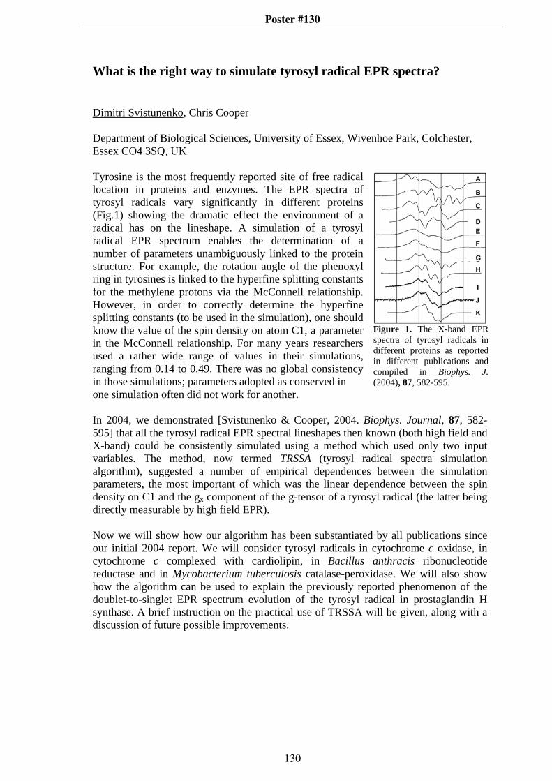
Poster #130
What is the right way to simulate tyrosyl radical EPR spectra? Dimitri Svistunenko, Chris Cooper Department of Biological Sciences, University of Essex, Wivenhoe Park, Colchester, Essex CO4 3SQ, UK Tyrosine is the most frequently reported site of free radical location in proteins and enzymes. The EPR spectra of tyrosyl radicals vary significantly in different proteins (Fig.1) showing the dramatic effect the environment of a radical has on the lineshape. A simulation of a tyrosyl radical EPR spectrum enables the determination of a number of parameters unambiguously linked to the protein structure. For example, the rotation angle of the phenoxyl ring in tyrosines is linked to the hyperfine splitting constants for the methylene protons via the McConnell relationship. However, in order to correctly determine the hyperfine splitting constants (to be used in the simulation), one should know the value of the spin density on atom C1, a parameter in the McConnell relationship. For many years researchers used a rather wide range of values in their simulations, ranging from 0.14 to 0.49. There was no global consistency in those simulations; parameters adopted as conserved in
Figure 1. The X-band EPR spectra of tyrosyl radicals in different proteins as reported in different publications and compiled in Biophys. J. (2004), 87, 582-595.
one simulation often did not work for another. In 2004, we demonstrated [Svistunenko & Cooper, 2004. Biophys. Journal, 87, 582-595] that all the tyrosyl radical EPR spectral lineshapes then known (both high field and X-band) could be consistently simulated using a method which used only two input variables. The method, now termed TRSSA (tyrosyl radical spectra simulation algorithm), suggested a number of empirical dependences between the simulation parameters, the most important of which was the linear dependence between the spin density on C1 and the gx component of the g-tensor of a tyrosyl radical (the latter being directly measurable by high field EPR). Now we will show how our algorithm has been substantiated by all publications since our initial 2004 report. We will consider tyrosyl radicals in cytochrome c oxidase, in cytochrome c complexed with cardiolipin, in Bacillus anthracis ribonucleotide reductase and in Mycobacterium tuberculosis catalase-peroxidase. We will also show how the algorithm can be used to explain the previously reported phenomenon of the doublet-to-singlet EPR spectrum evolution of the tyrosyl radical in prostaglandin H synthase. A brief instruction on the practical use of TRSSA will be given, along with a discussion of future possible improvements.
130

Poster #131
EPR and ENDOR Spectroscopy at 94 GHz on Photosystem II
Christian Teutloff 1 , Sven Keßen1, Jan Kern2, Athina Zouni2, Robert Bittl1
1 Freie Universität Berlin, Fachbereich Physik, Arnimallee 14, D-14195 Berlin, Germany2 Technische Universität Berlin, Institut für Chemie, Straße d. 17. Juni 135, D-10623
Berlin, Germany
The process of lightdriven water splitting and concomitant oxygen evolution takes place in Photosystem II (PS II). Its function is based on a chain of cofactors which transfer electron trough the photosynthetic membrane and a metal cluster, the oxygenevolving complex (OEC) which gets oxidized by the electron transfer process.The metal cluster consists of four manganese ions and a calcium ion which are connected by oxo and carboylato bridges [1]. During the redox cycle the OEC passes through several socalled Sstates, S0S4. To understand the mechanism of water splitting it is essential to have information about the structural changes occuring during the redox cycle. Unfortunately it is not possible only by Xray crystallography to obtain these structural details because af photoreduction of the Mncluster [2].Two of the Sstates, S0 and S2 have a noninteger spin state and are therefore accessible to EPR spectroscopy. The width of their EPR spectra is dominated by the large manganese hyperfine couplings and give rise to the socalled multiline signals at Xband.This contribution will demonstrate that EPR spectroscopy at Wband on the Mncluster enables higher spectral resolution due to higher gresolution [3]. This opens the possibility of orientation selection in Wband ENDOR experiments. Furthermore it opens the opportunity of investigations on small singlecrystals due to the higher sensitivity at 94 GHz. This has the advantage of more orientational information and will ultimately lead to the elucidation of the oxidation state of the individual Mn ions.Another presented example of the application highfield EPR on singlecrystals is the study of the tyrosine YD
radical with EPR and ENDOR spectroscopy at 94 GHz. High
resolved 1HENDOR spectra yield a complete set of proton hyperfine tensors along with their orientation with respect to the molecular frame. 2HENDOR spectroscopy on D2O exchanged samples gives furthermore direct information about the strength and orientation of hydrogen bonds to the tyrosine radical.
[1] B. Loll, J. Kern, W. Saenger, A. Zouni & J. Biesiadka; Nature 438(15), 10401044 (2005)
[2] J. Yano, J. Kern, K.D. Irrgang, M. J. Latimer, U. Bergmann, P. Glatzel, Y. Pushkar, J. Biesiadka, B. Loll, K. Sauer, J. Messinger, A. Zouni, V. K. Yachandra; Proc. Nat. Acad. Sci. 102(34), 1204712052 (2005)
[3] Ch. Teutloff, S. Keßen, J. Kern, A. Zouni, R. Bittl; FEBS Lett. 580(15), 36053609 (2006)
131

Poster #132
EPR and TR-EPR of modified trichogins containingthe amino acid TOAC and the chromophore antAib
Antonio Toffolettia, Tommaso Sandrona, Carlo Corvajaa,Jean-Paul Mazaleyratb, Chiara Baldinia, Fernando Formaggioa, Claudio Tonioloa
aDipartimento di Scienze Chimiche, Università di Padova, ItalybSIRCOB, UMR CNRS 8086, Bât. Lavoisier, Université de Versailles, France
Trichogin GA IV possesses marked membrane-modifying properties despite its shortlength (only ten amino acids). The natural peptide contains three helicogenic Aibresidues (α-aminoisobutyric acid) at positions 1, 4 and 8 of the chain: nOct-Aib1-Gly-Leu-Aib4-Gly-Gly-Leu-Aib8-Gly-Ile-Lol ( nOct = n-octanoyl; Lol = leucinol ).
Two modified trichogins (1 and 2) doubly labelled with a stable free radical (TOAC)and a triplet precursor (the amino acid antAib [1]) have been synthesized. The aminoacid sequences of 1 and 2 are the following (Boc = tert-butyloxycarbonyl):
1: Boc-antAib1-Gly-Leu-TOAC4-Gly-Gly-Leu-Aib-Gly-Ile-Leu-OMe2: nOct-antAib1-Gly-Leu-TOAC4-Gly-Gly-Leu-Aib-Gly-Ile-Leu-OMe
NH
H
CO
OH antAib
N CH3
CH3
CH3
CH3
N C OHH
O
OH
TOAC
It is expected that the 310-helical conformation of trichogin would force the twolabelled residues to accommodate on the same helix face, one on the top of the otherafter a complete turn. EPR spectra of 1 and 2 in solvents of different polarity and in amembrane simulating environment (SDS micelles in water) have been recorded.Rotational correlation times for 1 and 2 have been extracted from the analysis of thespectra. TR-EPR spectra with laser excitation at 355 nm have been recorded for bothfrozen (120 K) and liquid (250-300 K) solutions in toluene at different peptideconcentrations. In glassy toluene the spin-polarized EPR spectrum of triplet anthraceneis observed. At high temperatures the nitroxide TR-EPR signal has not been detected,contrary to other examples in which an intra-molecular interaction between triplet andradical linked to peptide templates produced spin-polarized EPR spectra 2,3. The EPRresults in different solvents and environments are presented and discussed.
[1] J.-F. Lohier, K. Wright, C. Peggion, F. Formaggio, C. Toniolo, M. Wakselman, J.-P.Mazaleyrat, Tetrahedron. 2006, 62, 6203-6213.
[2] E. Sartori, A. Toffoletti, F. Rastrelli, C. Corvaja, A. Bettio, F. Formaggio, S.Oancea, C. Toniolo, J. Phys. Chem. A, 2003, 107, 6905-6912.
[3] C. Corvaja, E. Sartori, A. Toffoletti, F. Formaggio, M. Crisma, C. Toniolo, J.-P.Mazaleyrat, M. Wakselman, Chem. Eur. J., 2000, 6, 2775-2782.
132

Poster #133
Investigation of the Solvent Effects on the IntramolecularSpin Exchange in Nitroxide Biradicals
Van Anh Tran*, K. Rasmussen*, A.I. Kokorin** and G. Grampp*
*Graz University of Technology , Institute of Physical and Theoretical Chemistry, Tech-nikerstrasse 4/1. 8010 - Graz, Austria**Institute of Chemical Physics, RAS, Moscow, Russian Federation
In recent years, biradicals have been established as a good model for studying the mecha-nism of electron transfer in organic and biological molecules. Therefore, understandingthe electron spin exchange in biradicals is of major importance.In solutions containing biradicals both intermolecular and intramolecular spin exchangecan be observed. However, in dilute solutions the former may be neglected and only theintramolecular spin exchange exists. This takes place via direct collisions of the radicalfragments in the same molecule or through the bridge that connects them and dependson the radical structure together with the solvent nature [1, 2, 3]. In nitroxide biradicalshaving two identical radical fragments, each containing only one nucleus with nonzerospin, the Hamiltonian, H , becomes [4]:
H = gβeHo(S(1 )z + S (2 )
z ) + aN (S (1 )z I (1 )
z + S (2 )z )I (1 )
z + J (S(1)z + S(2)
z )
Experimentally, the 14N isotropic hyperfine coupling constant, aN , and the exchange in-tegral, J, can be determined by Electron Spin Resonance (ESR) spectroscopy. In caseswhere more than one conformation of the biradical exists, an effective life time, τeff , maybe calculated as well.ESR experiments have been made on nitroxide biradicals with short rigid as well as withlong flexible bridges in various solvents. A detailed study on aN , J and τeff as func-tions of temperature and solvent properties has been made and very different behavioursobserved, depending on the length and structure of the bridge. The influence of solventviscosity was confirmed and also an indication of the presence of hydrogen bonds beingan important factor in the intramolecular spin exchange process was obtained [5].
[1] G. Grampp, S. Landgraf, I. Grigor’ev, A.B. Shapiro, A.I. Kokorin. Appl. Magn. Re-son. 19, 187 (2004).[2] A.I. Kokorin, V. A. Tran, K. Rasmussen, G. Grampp. Appl. Magn. Reson. 30, 35(2006).[3] A.I. Kokorin. Appl. Magn. Reson. 26, 253 (2005).[4] H. Lemaire. J. Chem. Phys. 64, 559 (1967).[5] A.I. Kokorin, V. A. Tran, K. Rasmussen, G. Grampp. Appl. Magn. Reson. In press(2006).
133

Poster #134
Studying high-spin ferric heme proteins and their low-spin cyanide adducts using pulsed EPR – An application to neuroglobin
F. Trandafir*, M. Fittipaldi*,**, J. Harmer***, S. Dewilde****, L. Moens****, S. Van
Doorslaer*
*Department of Physics, University of Antwerp, B-2610 Wilrijk- Antwerp, Belgium **Department of Chemistry, University of Florence, I-50019 Sesto Fiorentino, Italy
***Laboratory of Physical Chemistry, ETH Zurich, 8093 Zurich, Switzerland ****Department of Biomedical Science, University of Antwerp, B-2610 Wilrijk- Antwerp,
Belgium
Although human neuroglobin (NGB) was discovered almost 7 years ago, its function is still an enigma. This creates a challenging environment for different biophysical and biochemical studies focused on the determination of the function and structure of this protein. In our search to unravel the inner working of neuroglobin, we were confronted with problems concerning the use and spectral interpretation of different multi-frequency pulsed EPR experiments to study high-spin ferric heme proteins and their corresponding low-spin cyanide-binding forms. The latter systems are characterized with a very large g anisotropy. Using the example of neuroglobin, we highlight these problems and present possible solutions. Different X-band HYSCORE schemes in combination with W-band ELDOR-detected NMR are used to study the hyperfine and nuclear-quadrupole couplings of the heme and the histidine nitrogens of ferric E7-Gln NGB in comparison to aquometmyoglobin (Fig. 1). The simulation and interpretation of HYSCORE spectra of these types of S=5/2, I=1 systems will be elucidated. Comparative HYSCORE experiments combined with deuterium exchange experiments for aquometmyoglobin and ferric E7Q-NGB clearly show that the heme iron of the latter protein is pentacoordinated, lacking the distal water. For the low-spin cyanide adducts of wild-type and E7-Gln NGB, the identification of the different cross peaks in the X-band HYSCORE spectra is difficult due to a high degree of spectral overlap. The possibility to overcome some of these problems by selective isotope labelling and by the use of additional pulse experiments at different microwave frequencies will be discussed.
Figure 1. X-band protonHYSCORE spectrum of (a)aquometmyoglobin and (b)ferric E7-Gln NGB
134

Poster #135
USE OF SVD IN EPR SPECTROSCOPY: EFFECT OF pH ON THE TRAPPING OF SUPEROXIDE/HYDROPEROXYL RADICAL BY NITRONES. Ahmad Allouch, Béatrice Tuccio and Robert Lauricella Laboratoire TRACES, JE 2421, Universités Aix-Marseille 1 et 3, Case 512, St Jérôme, 13397 Marseille cedex 20. France . The singular value decomposition method (SVD) is frequently applied in signal processing but very rarely used in EPR spectroscopy, which is certainly regrettable. Though this procedure is deeply rooted in linear algebra, its application to the analysis of a series of EPR spectra evolving with time will be simply described. Then, this method will be employed to determine spin trapping rate constants. The disparity in the results given in literature for the superoxide trapping kinetics using nitrones is noteworthy. Recently, we have demonstrated that the Stern-Volmer-like method frequently used in these studies yielded important overestimation of rate constants for the superoxide trapping. This led us to elaborate a new kinetic approach to the evaluation of rate constants for the spin trapping of hydroperoxyl / superoxide radical by nitrones in buffered media. This method is based on a competition between the superoxide trapping by the nitrone and the spontaneous dismutation of this radical in aqueous media. EPR spectra are recorded as a function of time at various nitrone concentrations, and kinetic curves are obtained after treatment of these spectra using both SVD and pseudo-inverse deconvolution methods. Modelling these curves allowed us to determine the rate constants kT and kD for the superoxide trapping and the adduct decay reactions, respectively. Analysis of the pH dependence of kT permitted the determination of the rate constant of spin trapping of both O2
-. And HO2..
N CO2Et
CO2Et
O-
+N
CO2Et
O-
+EMPO
a) 10 mMb) 30 mMc) 200 mM
DEPO
a) 15 mMb) 10 mMc) 5 mM
R. Lauricella, J.C. Bouteiller and B. Tuccio, Phys. Chem. Chem. Phys., 2005, 7, 399-404. R. Lauricella, A. Allouch, V. Roubaud, J.C. Bouteiller, B. Tuccio, Org. Biomol. Chem. 2004, 2, 1304-1309 ; A. Allouch, V. Roubaud, R. Lauricella, J.C. Bouteiller, B. Tuccio Org. Biomol. Chem. 2005, 3, 2458-2462.
135

Poster #136
Revealing the inner workings of a Cobalt-salen based hydrolytic kinetic resolution catalyst using multi-frequency CW and pulsed EPR
Evi Vinck*, Sabine Van Doorslaer*, Damien M. Murphy**, Ian A. Fallis**
*University of Antwerp, Department of Physics, Universiteitsplein 1, 2610 Wilrijk, Belgium ** Cardiff University, School of Chemistry, Main Building, Park Place, Cardiff CF10 3AT, Wales, UK. Recently, E. Jacobsen et al. developed a new Cobalt salen-type catalyst [1], N,N'-bis(3,5-di-tert-butylsalicylidene)-1,2-cyclohexane-diamino cobalt(II), which can be used for the hydrolytic kinetic resolution (HKR) of terminal epoxides and the enantioselective catalytic ring opening of meso epoxides[1]. Although extensively used in practice, a molecular basis for the catalytic working of [1] remain uncertain. Here, we show how insights into this mechanism can be gained from a detailed EPR study. Using
pulsed EPR techniques, such as HYSCORE, and CW ENDOR at microwave frequencies ranging from 9.5 to 95 GHz, we determined the different EPR parameters of both as synthesized [1]
and of its activated form [2]. Activation of [1] by acetic acid under oxygen is a necessary step in the catalytic process. Where the principal g and cobalt hyperfine values are typical of a low-spin Co(II) complex with a |yz,2A2> ground state, the parameters of [2] bear the characteristics of an organic radical interacting with a cobalt nucleus (Fig. 1), best described as a phenoxyl radical. This assignment is also supported by resonance Raman studies. By combining selective isotope labelling with a diversity of EPR and ENDOR techniques, the hyperfine and nuclear-quadrupole interactions of the surrounding 1H, 13C and 14N nuclei could be mapped out in detail. The large changes observed in the nitrogen and hyperfine proton parameters upon activation of the complex can be directly linked to the electronic structure and reveal interesting aspects of the catalytic mechanism.
O
N N
OCo
R
RR
R
HH
AcOH
air
[1]
O
N N
OCo
OAcR
RR
R
HH
[2]
Fig. 1 Experimental (1) and simulated (2) X-band (a) and W-band (b) CW-EPR spectra of [2]
[1]Tokunaga; M.; Larrow, J.F.; Kakiuchi, F.; Jacobsen, E.N., Science, 277, 936 (1997)
136

Poster #137
EPR STUDY OF THE INTRAMOLECULAR ELECTRON
TRANSFER IN ORGANIC MIXED-VALENCE NANOWIRES.
INFLUENCE OF THE INCREASING MOLECULAR LENGTH.
J. Vidal-Gancedo,* V. Lloveras,
* J. F. Nierengarten,
** J. Veciana
* and C.
Rovira.*
* Institut de Ciència de Materials de Barcelona (CSIC). Campus Universitari de
Bellaterra, E-08193, Bellaterra (Spain).
** CNRS. Route Narbonne, Toulouse, F-31077 4 France.
One of the challenges in the molecular electronics area is to design and produce
molecular wires (conducting wires constituted by only one molecule). One way to study
the electronic conduction through a molecular wire is inducing an intramolecular
electron transfer (IET). Mixed-valence species in which is possible to thermally or
optically induce an IET are ideal to realize these studies. The interest is the potential use
of such systems as molecular wires on integrated molecular-sized devices and, from a
theoretical point of view, to understand the role of the parameters governing the
intramolecular electron transfer rate.
Although many organometallic compounds of this type are known, only few
purely organic, mixed-valence compounds have been synthesized and studied up to now
owing to their high instability.[1]
However, polychlorinated triphenilmethyl (PTM)
radicals present high thermal and chemical stability, they are electro active species and
also the corresponding anions are quite stable.[2]
We synthesized a novel series of diradicals (with two PTM moieties) that
incorporate oligo-PPVs with increasing length as bridges, in order to study in their
corresponding anion-radical mixed-valence systems (see the Figure below) the effect of
the distance in the IET rate. By Electron Paramagnetic Resonance it is possible to
determine the electron transfer rate constant at each temperature which permits to
calculate the attenuation factor β, a part from the thermal electron transfer barrier for
each species, ∆G*.[3]
[1] a) N. Gautier, F. Dumur, V. Lloveras, J. Vidal-Gancedo, J. Veciana, C. Rovira, P. Hudhomme,
Angew. Chem. Int. Ed. 2003, 42, 2765 and cites therein. b) F. Dumur N. Gautier, N. Gallego-Planas,
Y. Sahin, E. Levillain, N. Mercier, P. Hudhomme, M. Masino, A. Girlando, V. Lloveras, J. Vidal-
Gancedo, J. Veciana, C. Rovira, J. Org. Chem. 2004, 69, 2164 and cites therein.
[2] C. Rovira, D. Ruiz-Molina, O. Elsner, J. Vidal-Gancedo, J. Bonvoisin, J.-P. Launay, J. Veciana,
Chem. Eur. J. 2001, 1, 240 and cites therein.
[3] V. Lloveras, J. Vidal-Gancedo, T. Duarte, J.-F. Nierengarten, J. Veciana and C. Rovira, submitted.
R
Rn
Cl
ClCl
ClCl
ClCl
ClClCl
Cl
Cl
Cl
Cl
Cl
Cl Cl
ClCl
Cl Cl
Cl ClCl
Cl
Cl
Cl
Cl
R = -OC8H17
n = 1, 2, 3, 4, 5.
R
Rn
Cl
ClCl
ClCl
ClCl
ClClCl
Cl
Cl
Cl
Cl
Cl
Cl Cl
ClCl
Cl Cl
Cl ClCl
Cl
Cl
Cl
Cl
R
Rn
Cl
ClCl
ClCl
ClCl
ClClCl
Cl
Cl
Cl
Cl
Cl
Cl Cl
ClCl
Cl Cl
Cl ClCl
Cl
Cl
Cl
Cl
R = -OC8H17
n = 1, 2, 3, 4, 5.
-R
Rn
Cl
ClCl
ClCl
ClCl
ClClCl
Cl
Cl
Cl
Cl
Cl
Cl Cl
ClCl
Cl Cl
Cl ClCl
Cl
Cl
Cl
Cl
R = -OC8H17
n = 1, 2, 3, 4, 5.
R
Rn
Cl
ClCl
ClCl
ClCl
ClClCl
Cl
Cl
Cl
Cl
Cl
Cl Cl
ClCl
Cl Cl
Cl ClCl
Cl
Cl
Cl
Cl
R
Rn
Cl
ClCl
ClCl
ClCl
ClClCl
Cl
Cl
Cl
Cl
Cl
Cl Cl
ClCl
Cl Cl
Cl ClCl
Cl
Cl
Cl
Cl
R = -OC8H17
n = 1, 2, 3, 4, 5.
--
137

Poster #138
Low Field RYDMR: Effects of Radiofrequency Polarisa-tion on Radical Recombination ReactionsC. J. Wedge1,2, S. Norman2, C. T. Rodgers1,2, P. J. Hore2 & C. R. Timmel1.
1Inorganic Chemistry Laboratory, University of Oxford, OX1 3QR, UK2Physical & Theoretical Chemistry Laboratory, University of Oxford, OX1 3QZ, UK
It is known that reactions involving spin correlated radical pairs as intermediates aresensitive to applied static and oscillating magnetic fields [1,2], whether these fields areapplied in isolation, or simultaneously in a reaction yield detected magnetic resonance(RYDMR) experiment [3]. Conventional RYDMR studies have used a static field that isstrong with respect to the oscillating magnetic field (often X-band conditions have beenemployed). Under such conditions a well defined quantisation axis is provided by thestatic field which allows theoretical discussion to be conducted within the rotating frameapproximation.
Our recent experiments have applied a weak (≤ 150 µT) oscillating field at a constantradiofrequency whilst simultaneously sweeping a static field across the 0–4 mT range.As reported [4] this technique provides the possibility of a diagnostic test for reactionsoccurring via the Radical Pair Mechanism. The frequency and strength of the oscillatingfield, along with its orientation relative to the static field, were found to strongly influencethe radical recombination yield. Simulation of these results was possible through use ofa modified form of the γ-COMPUTE algorithm in the treatment of the linearly polarisedoscillating field applied [5]. A second form of low field RYDMR experiment detailedpreviously [6] applied a constant static field orthogonal to a linearly-polarised oscillatingfield, whilst the radiofrequency was swept across the range 1–80 MHz.
The data now presented are for both the static-field swept and frequency swept varia-tions of low field RYDMR, using a static field orthogonal to the oscillating field, with bothcircularly and linearly-polarised radiofrequency fields employed. Comparing the effectsobserved for linear and circular polarisations of the oscillating magnetic field providesinsight into the applicability of the rotating frame approximation to magnetic resonanceexperiments performed at low static field strengths.
[1] U. E. Steiner & T. Ulrich, Chemical Reviews, 89, 51–147, (1989).[2] C. R. Timmel & K. B. Henbest, Phil. Trans. A, 362, 2573–2589, (2004).[3] S. N. Batchelor, K. A. McLauchlan & I. A. Shkrob, Molec. Phys., 75, 501–529 (1992)[4] K. B. Henbest, P. Kukura, C. T. Rodgers, P. J. Hore & C. R. Timmel, J. Am. Chem.Soc. 126, 8102–8103 (2004)[5] C. T. Rodgers, K. B. Henbest, P. Kukura, C. R. Timmel, & P. J. Hore, J. Phys. Chem. A,109, 5035–5041, (2005)[6] J. R. Woodward, C. R. Timmel, P. J. Hore & K. A. McLauchlan, Mol. Phys. 100,1181–1186, (2002)
138

Poster #139
Fitting of EPR spectra:Extracting electronic and nuclear g factors, fine-, hyperfine-,and superhyperfine structure parameters as well as nuclear
quadrupole parameters directly from powder spectra.
Høgni Weihe and Stergios PiligkosDepartment of Chemistry, University of Copenhagen, Denmark.
E-mail: [email protected] and [email protected].
Abstract
In this talk we demonstrate the possibility of extracting all or some ofthe parameter types mentioned in the title directly from EPR spectra ofpowdered samples. This is done by fitting the full spectral trace to theoreticalspectrum, usually, based on a standard spin Hamiltonian. The method isillustrated on some spin systems recently synthesised in our laboratories.The figure below illustrates the the result when fitting the EPR spectrumof Mn2+ as an impurity in calcite. In the Figure, “b” and “c” designatedifference curves pertinent to nuclear terms being excluded and included,respectively, in the spin Hamiltonian.
B/G3600340032003000
a
b
c
×15
×15
f f f f f
139

Poster #140
Site-Directed Spin Labelling of TatA Which Forms Part of the E.coli Twin-Arginine Protein Transport System
Gaye F. White*, Sonya Schermann** , Justin Bradley*, Nick Greene** , Ben C. Berks** ,
Fraser MacMillan*, Andrew J. Thomson*
*School of Chemical Sciences and Pharmacy, UEA, Norwich, Norfolk NR4 7TJ
**Department of Biochemistry, University of Oxford, Oxford OX1 3QU
We report on site-directed spin labelling studies of TatA which forms part of the E.coli Twin-Arginine Protein Transport System, known as the Tat ABC translocase. This protein system is known to transport folded proteins of varying dimensions across the cytoplasmic membrane of bacteria.1 The Tat mechanism is distinct from the better known general secretory (Sec) pathway associated with the transport of unfolded proteins across the membrane. Proteins transported by the Tat pathway share a conserved sequence near their N-terminal, S-R-R-x-F-L-K, known as the ‘Tat signal sequence’. The figure opposite shows the scheme proposed for the Tat ABC translocase.1 Isolated TatA is known to form a homo-oligomeric complex made up of a bundle of TatA monomers.1 A number of variants of the TatA protein have been prepared with single cysteine mutations at different positions in its transmembrane helix. Each of these has been labelled with the spin label reagent MTSL. Room temperature CW X-band EPR has been used to study how the dynamics of the spin labels varies along the TatA helix. This gives information on the location of the binding interface between adjoining helices. Low temperature CW X-band EPR studies have shown evidence for dipolar coupling between single spin labels on the transmembrane helix in the TatA complex. Within a restricted range, 10 – 25 Å, this can be used to give information on the distance between neighbouring spin labels. In order to increase the distance range detected, a PELDOR study is also planned. The spin labelled TatA complex provides an interesting example of a system that could show dipolar coupling interactions between nitroxide radicals separated by a series of discrete inter-spin distances.
Acknowledgements: Financial support from BBSRC (grant – BBS/B/03637) and research fellowship for Sonya Schermann from the Natural Sciences and Engineering Research Council of Canada (NSERC)
References: 1. B. C. Berks, F. Sargent, T. Palmer, (2000) Mol. Microbiol. 35, 260-274.
140

Poster #141
EPR Spectroscopy Studies of the Photodegradation of Active
Pharmaceutical Ingredients and Formulations
Helen Williams*
*AstraZeneca, Milford 2-29, Silk Road Business Park, Macclesfield, Cheshire, SK10
2NA.
Along with heat, moisture and oxygen, light is an important parameter in drug
instability that can influence the storage conditions and shelf life. According to the
International Conference on Harmonisation of Technical Requirements for Registration
of Pharmaceuticals for Human Use (ICH) guidelines the photostability of all new drug
substances and products should be evaluated to demonstrate that light exposure does not
result in an unacceptable change1. Since 1
st January 1998 it has been obligatory to
provide photostability data for all new drugs in the US, EU, Japan and Canada2.
Evaluation of the interaction between a drug molecule and light is now a natural part of
R&D work for a new drug substance and it should be established at an early stage if a
new development compound is susceptible to photodegradation3.
Current ICH guidelines state that photostability testing should be done in two parts;
firstly stress testing to evaluate the overall photostability of a drug, identify the
degradation products and gain an understanding of the degradation pathways and
secondly a standardised accelerated test to determine the need for a label warning1.
EPR spectroscopy has a role to play in both of these areas. Free radical intermediates
during the photodegradation of an active pharmaceutical ingredient (API) can be
identified to aid understanding of the degradation pathways and products. This can also
be applied to pharmaceutical formulations, such as IV solutions. The development of
an EPR method to predict the extent of photodegradation of a solid state API during the
ICH complaint accelerated test is also presented.
1 Photostability Testing of New Active Substances and Medicinal Products, ICH Topic Q1B, 1-1-0098,
European Agency for the Evaluation of Medicinal Products 2 H.H. Tonnesen. In Photostability of Drugs and Drug Formulations, H.H. Tonnesen (Ed.), pp. 1-8, CRC
Press, London (2004) 3 D.R. Merrifield. In Photostability of Drugs and Drug Formulations, H.H. Tonnesen (Ed.), pp. 285-302,
CRC Press, London (2004)
141

Poster #142
Detection of Xanthone/DABCO spin correlated radical pairs in
organised media using transient absorption. Time resolved low field
MARY studies
E. G. Windham*, K. Maeda
*, K. Henbest
**, M. Goez
***, P. J. Hore
** and C. R. Timmel
*
*Inorganic Chemistry Laboratory, South Parks Road, Oxford, OX1 3QR
**Physical and Theoretical Chemistry Laboratory, South Parks Road, Oxford, OX1 3QZ, UK.
***Fachbereich Chemie, Martin-Luther-Universität Halle, Kurt-Mothes-Str. 2, D-06120, Halle,
Germany.
The radical recombination yield of a spin correlated radical pair can be sensitive to the
externally applied magnetic fields if the field influences the singlet-triplet
interconversion process. The photoinduced radical pair 3[Xan
-DABCO
+] in SDS
micelles shows a very large low field effect (see Figure 1). In the presence of high
concentration of DABCO a rapid electron transfer occurs1 and the concentration of the
radicals is measured by monitoring the change in absorption of the Xanthone anion
radical. The effect of the applied magnetic fields show an inverted MFE at low
magnetic fields (LFE).
Election Transfer reactions
The electron transfer was studied in solvents of different polarity and structure. In
organised heterogeneous (SDS) micellar solutions MFE were found to be time
dependent and additional
relaxation processes appear
to occur. In homogenous
solution MFE were found to
be time independent. The
poster describes the
different mechanisms that
occur in different solvents.
At different pH’s,
protonation of the Xanthone
anion occurs and the
changes in the spectra are
reported and reflect the
diffusion of the Xanthone
anion into the aqueous
layer.
Photoionisation of Xanthone
Xanthone fluorescence in SDS in the absence of DABCO also displays an interesting
magnetic field effect and we describe a possible mechanism for this process.
(1) Goez, M.; Hussein, B.; Hussein, M. Physical Chemistry Chemical Physics 2004, 6, 5490.
0.08
0.06
0.04
0.02
0.00
MF
E in
Pro
pa
n-2
-ol
3020100
Magnetic Field mT
0.4
0.3
0.2
0.1
0.0
MF
E in
SD
S
Xanthone and DABCO at t=630nsTime Window 250ns
in Propan-2-ol 600nm
in SDS 550nm
8
4
0
-4
x10
-3
543210
40
20
0
-20
x1
0-3
Figure 1 Normalised MARY spectra of Xanthone and DABCO system in
SDS and Propan-2-ol. Insert shows a expansion of the low field region.
142

Poster #143
F-pair contributions to magnetic field effects in isotropic solution
Adrian T. Salaoru, Timothy J. Foster, Claire B. Vink and Jonathan R. Woodward University of Leicester, George Porter Building, University Road, Leicester, UK.
The recombination reactions of free radicals in isotropic solution generated by the photoreaction of excited triplet state precursors proceed substantially through the recombination of freely diffusing radicals that have escaped from the geminate cage. Statistically, one quarter of these encounters should result in the production of a triplet correlated radical pair and one would anticipate that any magnetic field effects observed in these reaction systems would involve contributions from both geminate and freely diffusing spin-correlated radical pairs. We present results from our magnetic field studies on polymerisation photoinitiators using time-resolved infrared spectroscopy which indicate that the extent of hydrogen bonding in the radical pair members can alter the extent of f-pair contributions to the observed magnetic field effects in cyclohexanol solution.
0 10 20 30 40
-0.2
0.0
0.2
0.4
0.6
0.8
1.0
1.2
Δ A
bsor
banc
e
Timescale (μs)
LFE Simulation of the LFE
0 10 20 30 40
0.0
0.2
0.4
0.6
0.8
1.0
MFE Simulation of the MFE
Timescale (μs)
Δ A
bsor
banc
e
Observed normalised subtracted kinetic decay curves for the photoexcitation of 2-hydroxy-4-(2-hydroxyethoxy)-2-methylpropiophenone in cyclohexanol for applied fields of 35mT (MFE) and 2mT (LFE) along with the predicted decays assuming no contribution from freely diffusing radical pairs. In order to provide further insight into the role of f-pair recombination in the generation of magnetic field effects, we have used our pump-probe rapid-risetime pulsed magnetic field spectrometer to probe the effect of application of a magnetic field during different periods (i.e. geminate and freely-diffusing) of the recombination reaction. This technique provides very useful insight into the relative contribution of g-pairs and f-pairs to the total magnetic field effects observed.
0 10 20 30-0.6
-0.4
-0.2
0.0
0.2
0.4
0.6
0.8
1.0
1.2
1.4
Sign
al In
tens
ity (a
rbitr
ary
units
)
Time after laser flash / microseconds-100 -50 0 50 100
-1.00E-008
0.00E+000
1.00E-008
2.00E-008
3.00E-008
4.00E-008
5.00E-008
Inte
grat
ed s
igna
l
Pulse position relative to laser pulse / ns
Radical decay kinetics observed for the photoexcitation of benzophenone in cyclohexanol along with the integrated effect of a 100ns 20mT magnetic field pulse applied at varying times relative to the photolysis flash
143

Poster #144
Towards framework incorporation of vanadium in MCM-41 materials – A pulsed EPR study of the active sites
Sepideh Zamani*, Sabine Van Doorslaer*, Alina-Mihaela Hanu**, Vera Meynen***, Pegie Cool***, Etienne Vansant***
*University of Antwerp, Department of Physics, Universiteitsplein 1, 2610 Wilrijk, Belgium **“Al. I. Cuza” University of Iasi, Department of Physical and Theoretical Chemistry and Materials Chemistry, Bvd. Carol I, no 11, 700506, Romania *** University of Antwerp, Department of Chemistry, Universiteitsplein 1, 2610 Wilrijk, Belgium. Since 1992, a wide variety of mesoporous materials (siliceous and non-siliceous) have gained growing interest, in order to overcome the limits in accessibility and applicability encountered for microporous zeolites. A lot of research has been done to activate mesoporous materials, like MCM-41, with various transition metals in order to obtain large pore catalysts allowing high diffusion rates and a catalytic activity for bulky molecules. One of the biggest challenges is the incorporation of transition metals in the walls of the porous materials. Here we present an electron paramagnetic resonance (EPR) study of the incorporation of vanadium in MCM-41 via different chemical routes. We compare vanadium deposition in the pores via the molecular designed dispersion (MDD) method with a facile, direct room-temperature synthesis targeted at vanadium incorporation into the MCM-41 framework. The difference between the two routes is monitored by studying the vanadyl-containing precursors of the final catalysts. It will be shown how important information about the mobility and incorporation mechanism of the vanadyl species in the mesoporous systems can be revealed by the use of X-band continuous-wave and 2-dimensional pulsed EPR spectroscopy, such as HYSCORE.
Figure 1. proton HYSCORE spectrum of a vanadium MCM-41
precursor.
144

Poster #145
EPR study of x-ray induced centres in K2YF5 thermoluminescent phosphors D.Zverev*, F.Loncke*, H.Vrielink*§, N. M.Khaidukov** and F.Callens* * Department of Solid State Sciences, Ghent University, Krijgslaan 281-S1, B-9000 Gent, Belgium ** Institute of General and Inorganic Chemistry, Russian Academy of Sciences, Leninskii Prospect 31, 119991 Moscow, Russian Federation Recently it has been discovered that K2YF5 crystals singly doped with rare earth (RE) ions are promising phosphors for thermoluminescence (TL) dosimetry [1-2]. TL phosphors based on such combinations show relatively high sensitivity to ionising radiation and low fading, while for some K2YF5:RE combinations the shape of the TL glow curves depends on the type of radiation field, which offers possibilities for discriminating mixed radiation fields. Here we present the results of our EPR study of X-ray irradiated K2YF5 crystals singly doped with 0.1% of Ce3+. Prior to irradiation, the EPR spectra reveal the initial valence state of the RE dopant ions and allow identification of unintentional paramagnetic impurities. K2YF5 has an orthorhombic crystal structure, its space group being Pnam. YF7 polyhedra, which have the ab-plane as mirror symmetry plain, form chains along the twofold screw c-axis. In principle a large number of inequivalent intrinsic defects could be formed upon irradiation. After X-ray irradiation at room temperature, signals of at least four different trapped electron or hole centres are resolved in the EPR spectra. Their intensity as a function of annealing temperature correlates well with the TL glow curve of K2YF5:0.2%Ce3+ after β-irradiation [2], suggesting that similar defects are involved. The angular variation of the EPR and ENDOR spectra of the most stable centre, with an annealing temperature of about 250°C, was studied. This centre has spin S = ½ and is characterized by a monoclinic g tensor with one principal axis along the crystal’s twofold screw axis (c-axis), and tilted in the ab-plane approximately by 36°. In the ENDOR spectra strong interactions with five 19F nuclei were found. These interactions correspond to two pairs of symmetry related neighbours and a single ion within the symmetry plain along b-axis. The above-mentioned results allow us to assume that this defect corresponds to an electron/hole centre formed under X-ray irradiation in the mirror plane, probably at an Y3+ position. The real nature of this centre is the subject of future investigations. § Postdoctoral fellow of the Flemish Research Foundation (FWO-Vlaanderen) [1] J. Marcazzo, M. Santiago, E. Caselli, N. Nariyama and N. M. Khaidukov, Opt. Mater. 26, 65-70 (2004). [3] H.W.Kui, D.Lo.Y.C.Tsang, N.M.Khaidukov and V.N.Makhov // J.Lumin. 117 (1), 29-38 (2006)
145

Title index
Title indexBis-spin labelled cyclodextrins — synthesis and
EPR studies, 91
A cryogen free 12 Tesla superconducting magnetfor use in multifrequency and high fieldEPR, 87
A multi-frequency EPR study of Co(II)S4 coordi-nation, 103
A new approach to simulate spin labelling spectra:the method of characteristic jumps, 33
A radical approach to non natural sugars, 57A Study of Electronic and Magnetic Properties of
Iron-Sulfur Clusters, 113Adaptation of a W-band spectrometer to UHV con-
ditions, 78Advanced EPR Studies of Metalloenzymes: “Hy-
drogenase and Water Oxidase”, 48Advances in optically-detected low-field EPR, 26An EPR investigation on Pseudogap in
SmBa2Cu3−xAlxO6+δ superconductors,112
An EPR Investigation on the Oxidation of GreenTea Polyphenols, 124
An Investigation into the Magnetic Field Depen-dence of Carbazoles using MARY Spec-troscopy, 98
An Investigation of Oxygen Centred Radicals byESR, 82
Analysis of the 2nd and 4th order zero-field split-ting in a highly symmetric tetranuclearFe(III)-oxo single molecule magnet, 29
Application of CW and Pulsed EPR, MoSopheand DFT Calculations in Unravellingthe Electronic Structure of the Molybde-num(V) Centre in Dimethylsulfoxide Re-ductase, 32
Characterization of nanostructures at equilibriumand during the synthesis of mesoporousmaterials by Double Electron ElectronResonance (DEER), 27
Characterization of the light-induced species of theactive center of the [FeFe] hydrogenase,126
Coherent Raman detected electron spin resonancespectroscopy, 19
Detection of Xanthone/DABCO spin correlatedradical pairs in organised media usingtransient absorption: Time resolved lowfield MARY studies, 142
Development of a High Frequency pulsed/CWEPR Spectrometer operating at 122 and244 GHz, 18
Direct approach towards lifetime analysis of powercable insulation materials using electronspin resonance spectroscopy, 106
Direct Observation of Peroxyl Radical AdductsFormed from Addition of Oxygen toAmino Acid Radicals, 22
Distance determinations on oligonucleotides andother biological systems by the DEERmethod, 83
Distance measurements in a resolvase complex,110
Distinct protein and lipid radicals as markers forUVA skin ageing and carcinogenesis andthe protection in Afro-Caribbean skin, 88
Dual mode X-band and High Field/ High Fre-quency EPR Studies of a MononuclearMn2+ (S=5/2) compound, 123
Effects of the exchange and dipolar interactions onsinglet yield anisotropy in model radicalpairs, 75
Electron Paramagnetic Relaxation EnhancementProduced by Rhombic g-tensors in RigidSystems, 72
Electron paramagnetic resonance of photoin-duced charge separation in poly(1,4-phenylenevinylene) derivative polymers,93
Electron Spin Relaxation Enhancement Measure-ments of Interspin Distances in Hu-man, Porcine, and Rhodobacter ElectronTransfer Flavoprotein ubiquinone Oxi-doreductase (ETF-QO), 77
Electron Spin Relaxation Rates and their Applica-tions, 37
Electronic structure of the H-cluster in [FeFe] hy-drogenase as monitored by advanced EPRspectroscopy, 119
EMR Study of Stable Radicals in X-irradiatedFructose Single Crystals, 56
EPR and ENDOR Spectroscopy at 94 GHz on Pho-tosystem II, 131
EPR and optical study of Ln3+-doped PbF2 single-crystals, 71
EPR and Pulsed ENDOR of Cu(II) Centres in Fer-roelectric PbTiO3, 23
EPR and TR-EPR of modified trichogins contain-ing the amino acid TOAC and the chro-mophore antAib, 132
146

Title index
EPR investigation of the intramolecular charge ex-change in organic mixed valence com-pounds, 85
EPR of nitronyl–nitroxide radicals assembled onsurfaces, 30
EPR spectroscopy of multiply labelled fullerene-nitroxide molecules, 70
EPR Spectroscopy Studies of the Photodegradationof Active Pharmaceutical Ingredients andFormulations, 141
EPR spectroscopy, a useful tool for studying thebiogenesis and the catalytic mechanism ofa complex membrane-bound metalloen-zyme, 49
EPR Spin Trapping of Thiyl Radicals by PBN andPBN attached to cyclodextrin, 117
EPR studies on the kinase-interacting domainof the phototransducer HtrII from Na-tronomonas pharaonis, 94
EPR Study of a Novel Hybrid [Fe(III)-Porphyrin]–[Radical] Catalyst, 68
EPR Study of Photoactivity of Nitrogen-dopedTiO2 under Visible Light, 36
EPR Study of the Intramolecular Electron Trans-fer in Organic Mixed-Valence Nanowires.Influence of the Increasing MolecularLength, 137
EPR Study of the Conversion of N-Aryl Ketoni-trones into Nitroxide Radicals, 97
EPR study of the stereoselective binding of methyl-benzylamine enantiomers to a chiral Cu-Salen complex, 65
EPR study of the synthesis and characterization ofmicelle templated materials, 64
EPR study of X-ray induced centres in K2YF5 ther-moluminescent phosphors, 145
EPR study on tyrosyl radical formation mechanismand metal binding in Mycobacterium tu-berculosis RNR, 81
ESR reveals unforeseen effects of two novel NOdonors on nitric oxide production, 127
ESR spectroscopy on Cu(II)–bis(oxamato) build-ing blocks for molecule based magneticmaterials, 62
Excess Electron Transfer in DNA Probed by MuonSpin Relaxation, 101
Extremely low magnetic field effects towards aphotochemical compass, 89
Extremely wide conformational distributions of themetal sites in metalloproteins and theirmodels measured by high-spin EPR spec-troscopy, 86
F-pair contributions to magnetic field effects inisotropic solution, 143
Field Sweep Versus Frequency Sweep in CW-EPR— an unresolved matter regarding homo-geneously broadened lines, 115
Fitting of EPR spectra: Extracting electronicand nuclear g factors, fine-, hyperfine-,and superhyperfine structure parametersas well as nuclear quadrupole parametersdirectly from powder spectra, 139
HF-EPR unravelling hard-plane anisotropy ofMn12tBuAc, 60
High field ENDOR — opportunities and frustra-tions, 38
High-field EPR reveals the mobility of nitroxides,46
High-frequency EPR and ENDOR Spectroscopyon Semiconductor Nanocrystals, 20
High-resolution EPR structure of the dimer of theNa+/H+ antiporter NhaA of Escherichiacoli, 40
Identification by Time-resolved EPR of Chloro-phyll — Carotenoid Pairs involved inTriplet-Triplet Transfer in PhotosyntheticAntenna Systems, 55
Identification of the sources of free radicals gener-ated during the oxidation of Green tea andBitter tea, 116
Interactive Simulation of CW and Pulsed EPRSpectra, 109
Investigating the Point Defects in Superhard Cu-bic Boron Nitride Crystalline Powders byMultifrequency ESR, 108
Investigation of the protein-based radical interme-diates in mono- and bi-functional peroxi-dases using multifrequency (9–285 GHz)EPR spectroscopy, isotope labelling andsite-directed mutagenesis, 92
Investigation of the Solvent Effects on the In-tramolecular Spin Exchange in NitroxideBiradicals, 133
Is the [(µ-pdt)Fe2(I,I)(CO)5-(H2NPr)] complextruly diamagnetic? An EPR and mag-netism combined study, 90
Level structure of molecular magnet Cr10 : howmuch can be known from ESR alone?,120
Location of Spectroscopic Probes in Self-Aggregating Assemblies, 44
147

Title index
Low Field RYDMR: Effects of Radiofrequency Po-larisation on Radical Recombination Re-actions, 138
Magnetism and EPR studies of [Fe(III)6 . . .], 125Modern ESR Methods for Studying Proteins and
Membranes at ACERT (Center for Ad-vanced ESR Technology), 42
Modifications of NO synthesis by flavonoids in ratorgans studied by means of ESR spec-troscopy, 128
Molecular Nanomagnets: a good gymnasium forEPR, 28
Monitoring the opening of the human pancreatic li-pase lid by site-directed spin labeling, 61
New delicacies from the high-field EPR menue:Orientation resolving 95 GHz PELDORon transient radical pairs in photosyn-thetic reaction center proteins, 360 GHzENDOR on nitroxide spin labels, 45
Observation of effects of oscillating magneticfields on radical pair dynamics using tran-sient absorption detection, 100
One-Electron Ag·C Bond in Oganosilver Radicals,102
Orientation-selected ENDOR spectroscopy of ironsulfur proteins, 52
Oxidation and halogen abstraction reactions cat-alyzed by protected gold based nanopar-ticles, 69
Paramagnetic Carbon impurities in silica preparedby sol-gel methods: an EPR study, 59
Pattern-matching algorithm for automated sym-bolic processing of relaxation theoryequations, 21
pH sensitive spin probes. Monitoring of pH at thered cell membrane surface, 95
Polarity and proticity details of the nitroxide micro-environment in spin labeled proteins: aW-band EPR study, 47
Probing the protonation state of the remote ni-trogen in the copper(II) complex of N-confused tetraphenylporphyrin by hyper-fine decoupling techniques, 104
Protein-surface interactions investigated via theirmagnetic field effects, 73
Pulsed EPR Spectrometer Operating at 110–334GHz, 105
Pulsed EPR techniques in Q-band applied to low-spin ferriheme centers, 80
Pulsed ESR study of relaxation times in molecularmagnets: Enough time for quantum infor-mation processing?, 25
Radical Pairs in Cryptochromes - Investigation of apossible avian compass, 121
Rapid Scan EPR Imaging, 24Reactivity of Aged Gold Nanoparticles in Ligand
Exchange Reactions, 99Recent X-, Q-band EPR studies of Photosystem II.
(A) The unexpected line shape of the tyro-syl Z• radical. (B) The critical S3 integer-spin state of the Mn cluster, 53
Relative Orientation of Nitroxides at a Distance:X-Band PELDOR Measurements of Du-plex DNA Containing Rigid Spin Labels,41
Resolving Protein Cofactor Interactions in Ter-minal Oxidases using MultidimensionalEPR Spectroscopy, 54
Revealing the inner workings of a Cobalt-salenbased hydrolytic kinetic resolution cat-alyst using multi-frequency CW andpulsed EPR, 136
Ru(II)–Nitroxyl radical as a new tool for electrontransfer study, 43
SDSL EPR — GHOST — SICS approach to pro-tein structures, 129
Selective binding of structural epoxide isomers bya chiral vanadyl Salen complex, 107
Self-assembling copolymer architectures providedifferent coordination ways for metals,114
C60-based Peapods, 34Singlet oxygen generated in gas phase by pho-
tosensitization with naphthalene deriva-tives: a time resolved EPR study, 122
Site specific anisotropic reactivity of oxygen cen-tred radicals at the surface of polycrys-talline TiO2: An EPR investigation, 66
Site-Directed Spin Labelling of TatA Which FormsPart of the E.coli Twin-Arginine ProteinTransport System, 140
Solvated Electrons Localized at the Surface of anIonic Insulator. A CW and Pulsed EPRStudy, 67
Structural Characterisation of Weak Protein Com-plexes Using DEER, 58
Structural information from dipolar coupled spinpairs, 39
Structure determination of neuroglobin using EPR,76
148

Title index
Studies of the reversible oxidation–reduction ef-fects in new copper based radiopharma-ceuticals by electrochemical in-situ Elec-tron Paramagnetic Resonance, 96
Studying high-spin ferric heme proteins and theirlow-spin cyanide adducts using pulsedEPR - An application to neuroglobin, 134
Studying peptide–membrane interactions usingcombined DEER and ESEEM techniques,84
Temperature Dependence of the structure of thefirst stable L-alanine radical: An ENDORstudy, 118
The heme-pocket structure of ferric cytoglobin andits E7Q mutant — an EPR investigation,50
The HIPER Project — sub-nanosecond pulse ESR,17
The neutral nitrogen vacancy centre in diamond, 35The triplet of DCM observed for the first time: a
TR-EPR study of the dye inclusion in aKH Phthalate single crystal, 63
Theoretical EPR spectroscopy of high-spin sys-tems — challenges and opportunities, 31
Theoretical study of EPR parameters for Dy3+ cen-tre in ZrSiO4, 74
Time-resolved EPR of radical–chromophore inter-actions in solution, 79
Towards an Identification of Chemically Differ-ent Flavin Radicals by Means of their g-Tensor, 111
Towards framework incorporation of vanadiumin MCM-41 materials — A pulsed EPRstudy of the active sites, 144
Understanding the Catalytic Mechanism ofMethane Production by Methyl–coenzyme M reductase with Labeled Sub-strates and Substrate analogs, 51
Use of SVD in EPR Spectroscopy: Effect of pH onthe Trapping of Superoxide/HydroperoxlRadical by Nitrones, 135
What is the right way to simulate tyrosyl radicalEPR spectra?, 130
149

Author index
Author indexAdeluta, Tarpan
Poster, 56Alberti, Angelo
Poster, 57
Bagryanskaya, ElenaWed 14.00, 46
Banham, JanetPoster, 58
Barbon, AntonioPoster, 59
Barra, Anne-LaurePoster, 60
Belle, ValériePoster, 61
Bingham, StephenMon 10.05, 19
Bordignon, EnricaWed 14.20, 47
Brauer, BjornPoster, 62
Brustolon, MarinaPoster, 63
Cangiotti, MichelaPoster, 64
Caretti, IgnacioPoster, 65
Carter, EmmaPoster, 66
Chiesa, MarioPoster, 67
Christoforidis, KonstantinosPoster, 68
Conte, MarcoPoster, 69
Corvaja, CarloPoster, 70
Dantelle, GeraldinePoster, 71
Deligiannakis, YiannisPoster, 72
Dell, EmmaPoster, 73
Di Valentin, MarilenaThu 11.55, 55
Dinse, Klaus-PeterTue 10.50, 34
Dong, Hui-NingPoster, 74
Eaton, GarethTue 11.55, 37
Eaton, SandraMon 14.00, 24
Efimova, OlgaPoster, 75
Ezhevskaya, MariaPoster, 76
Felton, SolveigTue 11.15, 35
Fielding, AlistairPoster, 77
Fischbach, EstherPoster, 78
Forbes, MalcolmMon 11.35, 22
Franco, LorenzoPoster, 79
Freed, JackWed 10.50, 42
Garcia-Rubio, InesPoster, 80
Garipov, RuslanMon 11.55, 23
Gatteschi, DanteMon 16.00, 28
Georgieva, ElkaPoster, 81
Ghosh, MarianaPoster, 82
Giamello, ElioTue 11.35, 36
Giuseppe, SicoliPoster, 83
Goldfarb, DaniellaTue 20.30, 38
Goovaerts, EtienneMon 16.25, 29
Gordon-Grossman, MichalPoster, 84
Grampp, GuenterPoster, 85
Grimaldi, StephaneThu 09.25, 49
Hagen, WilfredPoster, 86
Hall, RennyPoster, 87
Hanson, GraemeTue 09.40, 32
Harmer, JeffreyThu 10.05, 51
Haywood, RachelPoster, 88
Henbest, KevinPoster, 89
Huang, PingPoster, 90
Hüttermann, JürgenThu 10.50, 52
Ioanitescu, AndaThu 09.45, 50
Ionita, Elena GabrielaPoster, 91
Ivancich, AnabellaPoster, 92
Jeschke, GunnarWed 09.40, 40
Keeble, DavidPoster, 93
Klare, JohannPoster, 94
Komorowska, MalgorzataPoster, 95
Kowalczyk, RadoslawPoster, 96
Kuprov, IlyaMon 11.15, 21
Lauricella, RobertPoster, 97
Lebedeva, NataliaWed 11.35, 44
Lillington, JamesPoster, 98
Lubitz, WolfgangThu 09.00, 48
Ma, YunPoster, 99
MacMillan, FraserThu 11.35, 54
Maeda, KiminoriPoster, 100
McKenzie, IainPoster, 101
Michalik, JacekPoster, 102
Milikisiyants, SergeyPoster, 103
150

Author index
Mitrikas, GeorgePoster, 104
Morley, GavinPoster, 105
Morsy, MohamedPoster, 106
Murphy, DamienPoster, 107
Möbius, KlausWed 11.55, 45
Neese, FrankTue 08.50, 31
Nistor, SergiuPoster, 108
Noble, ChristopherPoster, 109
Norman, DavidPoster, 110
Okafuji, AsakoPoster, 111
Oliva, CesarePoster, 112
ORIO, MaylisPoster, 113
Palivan, CorneliaPoster, 114
Petrouleas, VasiliThu 11.15, 53
Pilbrow, JohnPoster, 115
Pirker, KatharinaPoster, 116
Polovyanenko, DmitriyPoster, 117
Prisner, ThomasWed 08.50, 39
Rakvin, BorisPoster, 118
Reijerse, EdwardMon 09.45, 18Poster, 119
Rival, OlivierMon 14.25, 25Poster, 120
Robinson, AlexanderPoster, 121
Rockenbauer, AntalTue 10.00, 33
Rodgers, ChristopherMon 14.45, 26
Ruthstein, SharonMon 15.05, 27
Ruzzi, MarcoPoster, 122
Sanakis, YiannisPoster, 123
Schmidt, JanMon 10.50, 20
Severino, JoycePoster, 124
Shanmugam, MuralidharanPoster, 125
Sigurdsson, SnorriWed 10.00, 41
Silakov, AlexeyPoster, 126
Sjakste, NikolajsPoster, 127
Sjakste, TatjanaPoster, 128
Smith, GrahamMon 08.55, 17
Sorace, LorenzoMon 16.45, 30
Strancar, JanezPoster, 129
Svistunenko, DimitriPoster, 130
Teutloff, ChristianPoster, 131
Toffoletti, AntonioPoster, 132
Tran, Van AnhPoster, 133
Trandafir, FlorinPoster, 134
Tuccio, BéatricePoster, 135
Van Doorslaer, SabinePoster, 136
Vidal-Gancedo, JoséPoster, 137
Wedge, ChristopherPoster, 138
Weihe, HogniPoster, 139
Weiner, LevWed 11.15, 43
White, GayePoster, 140
Williams, HelenPoster, 141
Windham, EmmaPoster, 142
Woodward, JonathanPoster, 143
Zamani, SepidehPoster, 144
Zverev, DmitryPoster, 145
151