the 17 kda protein in photosystem ii, purification and
TRANSCRIPT

Louisiana State UniversityLSU Digital Commons
LSU Master's Theses Graduate School
2005
The 17 kDa protein in photosystem II, purificationand interaction with the photosystemGlen MeadesLouisiana State University and Agricultural and Mechanical College
Follow this and additional works at: https://digitalcommons.lsu.edu/gradschool_theses
This Thesis is brought to you for free and open access by the Graduate School at LSU Digital Commons. It has been accepted for inclusion in LSUMaster's Theses by an authorized graduate school editor of LSU Digital Commons. For more information, please contact [email protected].
Recommended CitationMeades, Glen, "The 17 kDa protein in photosystem II, purification and interaction with the photosystem" (2005). LSU Master's Theses.3943.https://digitalcommons.lsu.edu/gradschool_theses/3943

THE 17 kDa PROTEIN IN PHOTOSYSTEM II, PURIFICATION AND INTERACTION WITH THE
PHOTOSYSTEM
A Thesis
Submitted to the Graduate Faculty of the Louisiana State University and
Agricultural and Mechanical College
in partial fulfillment of the requirements for degree of
Master of Science
in
The Department of Biological Sciences
by Glen Meades, Jr.
B.S., Louisiana State University, 1999 August, 2005

ii
TABLE OF CONTENTS
List of Tables……………………………………………………………………………………..iii
List of Figures…………………………………………………………………………...………..iv
Abstract…………………………………………………………………………...……………….v
Introduction………………………………………………………………………………………..1
Materials and Methods………………………………………………………….………………..11
Results and Discussion……………………………………………………..……………………20
Summary and Conclusions……………………………………………..………………………..36
Bibliography………………………………………………………………….………………….38
Vita…………………………………………………………………...…………………………..40
iii
LIST OF TABLES
Table Page Table 1: Extrinsic PS II polypeptides of higher plants ……………………………………….......3 Table 2: Assignments of Biotinylated Lysines for peptides produced from a trypsin digest of b17m ……………………………………………………………..……………27 Table 3: Assignments of Biotinylated Lysines for peptides produced from a V8 protease digest of b17m …………………………………………………………......………27 Table 4: Assignments of Biotinylated Lysines for peptides produced from a trypsin digest of b17s …………………………………………………………...……………….28 Table 5: Assignments of Biotinylated Lysines for peptides produced from a V8 protease digest of b17s ……………………………………………………………...……….28 Table 6: Assignments of Biotinylated Lysines for ambiguities in modified peptides based on MS/MS data analysis ………………………………………………………...29

iv
LIST OF FIGURES
Figure Page
1. Diagram of a chloroplast …………………………………………………………………….....1
2. Diagram of the photosynthetic machinery in higher plants …………………………………....2
3. VMD-generated images of 17 kDa crystal structure …………………………………………..5
4. Amino acid sequence for the mature spinach 17 kDa protein …………………………………6
5. Biotinylation of an exposed lysyl residue by NHS-biotin ……………………………………..7
6. Flowchart showing the experimental procedure …………………………………………….....8
7. Schematic of a time-of-flight mass spectrophotometer ………………………………………..9
8. Line trace of CM-Toyopearl 650M column purification ……………………………………..13
9. Fractions after avidin column enrichment ……………………………………………………18
10. Coomassie-stained PAGE of CM-Toyopearl 650M column fractions: ineffective …………21
11. Coomassie-stained PAGE of CM-Toyopearl 650M column fractions: effective …………...22
12. Typical biotinylation of unmodified 17 kDa protein, b17m, and b17s ……………………...24
13. Rebinding curves for unmodified 17 kDa protein, b17m, and b17s to PSIIs17 …………….25
14. MASCOT search results …………………………………………………………………….30
15. VMD-generated image of 17 kDa crystal structure …………………………………………32
16. One plausible orientation of the 17 kDa protein to the photosystem II complex ………..….33
17. A second plausible orientation of the 17 kDa protein to the photosystem II complex ……...35
v
ABSTRACT
The structural association of the spinach 17 kDa protein of photosystem II with other extrinsic
and membrane-bound components of the photosystem was investigated by labeling the 17 kDa
protein with the amino group-specific reagent N-hydrosuccinimidobiotin both on intact
photosystem II membranes and free in solution. Following isolation of the biotinylated
molecules, the modified 17 kDa protein was allowed to rebind at various molar ratios to
photosystem II membranes lacking the 17 kDa protein. Differential binding of the biotinylated
proteins compared to unmodified 17 kDa protein indicates steric or ionic interference due to
added biotin moieties, impeding physical contact or shielding positively charged amino groups,
respectively. Biotinylated sites on the different modified 17 kDa proteins were identified by
trypsin and Staphylococcus V8 protease digestion, followed by affinity chromatography
enrichment for biotinylated molecules, and analysis of the resultant peptide fragment mixture by
nanospray LC mass spectrometry. Areas shielded from the bulk solvent when the protein is
associated with photosystem II may correspond to protein segments involved in the interaction
with other components of the photosystem.

1
INTRODUCTION
Over the course of geologic time the relative percentage of molecular oxygen in the Earth’s
atmosphere has increased due to the evolution and action of the process known as oxygenic
photosynthesis, whereby plants and cyanobacteria harness solar electromagnetic energy and use
it to chemically split water molecules. This converts the solar energy into chemical energy
stored as a proton gradient generated across a membrane and as reducing equivalents for redox
reactions, producing molecular oxygen as a byproduct. This stored chemical energy is used to
assemble carbohydrates utilizing carbon dioxide as the carbon source.
Figure 1. Diagram of a chloroplast. (http://sps.k12.ar.us/massengale/chloroplast_diagram.htm)

2
Specialized molecules and groups of proteins are in place to harness this radiant energy and
convert it into chemical potential energy. These “photosystems” are anchored in the membranes
of the thylakoids, in the chloroplasts of higher plants. One of these two photosystems, known as
photosystem II (PS II), is comprised of around 30 major protein components and several pigment
cofactors. By treating isolated chloroplasts in solution with detergent, it is possible to disrupt the
outer membrane bilayer, releasing the stacks of thylakoids. The detergent then begins to disrupt
the thylakoid membranes, first at the periphery where PS I is in high density. The detergent’s
action is subsequently stopped, leaving extremely stable inside-out membrane leaflets 0.5 µm in
diameter derived from thylakoid membranes, enriched for the PS II components, and limited in
the inclusion of PS I. The major components involved in the chemistry of photosynthesis are
genetically encoded by the DNA within the chloroplast and are present in the reactive center
core, which is composed of polypeptides D1 (psbA) and D2 (psbD). Also present are the light-
harvesting ligands chlorophyll a and β-carotene, and an iron-manganese cluster involved in the
chemistry of charge separation.
Figure 2. Diagram of the photosynthetic machinery in higher plants.

3
These chlorophylls participate in energy transfer from the proximal antenna complexes of
CP43 (psbC) and CP47 (psbB) to the reactive center core chromophores. Associated with the
core is an oxygen-evolving complex (OEC) that acts as the active site of water oxidation. The
OEC is made up of the extrinsic polypeptides, a tetranuclear manganese (Mn) cluster, one
calcium ion and one chloride ion. Finally, there are at least ten small (<10 kDa) hydrophobic
peptides, most of which contain transmembrane helices. Some of the small polypeptides, such as
psbH and psbT, are involved in photoprotection (Bergantino, et al, 2003), helping to protect
against the damaging effects of the reactive oxygen species generated during photosynthesis.
The function of most of the others is unclear. The OEC in higher plants contains three extrinsic
membrane proteins, psbO, psbP and psbQ, with apparent molecular masses of 33, 24, and 17
kDa after two post-translational cleavages. (See Table 1) These proteins are nuclear encoded
and their final destination is the lumen of the thylakoid membrane. Precursor proteins for all
three exist and contain N-terminal sequences directing translocation first to the stroma and
secondly to the lumen, crossing two membranes in this process. The targeting sequences are
cleaved off at each membrane crossing, reducing the protein to its mature number of residues
once inside the lumen.
Table 1. Extrinsic PS II polypeptides of higher plants
4
The 20-30 amino acids immediately upstream from the first amino acid of the mature protein
constitute the membrane-spanning hydrophobic portion of the precursor protein. This portion of
the transit sequence is similar amongst these three proteins, and the similarity extends to many
lumenally-destined precursors in all oxygenic photosynthetic organisms (Philbrick and Zilinskas,
1988).
Each extrinsic polypeptide appears to have a specific role in enhancing the efficiency of the
photosynthetic machinery, yet in broader terms, all serve to maintain the ionic environment at
and around the site of oxygen evolution. The proposed role of the 33 kDa protein (MSP for
Manganese-Stabilizing Protein) is to reduce the loss of Mn under reducing conditions, while the
roles of the 24 and 17 kDa proteins are to maintain the Ca+2 and Cl- environment required for
optimal oxygen evolution (Ghanotakis, et al, 1984). Dissection of the complex and analysis of
each component can lead to a better understanding of its function(s) within the complex. The 24
and 17 kDa proteins can be removed by washing with 1.0 M NaCl solutions. This results in a
dramatically lower oxygen-evolving capacity of PS II membranes (Kuwabara & Murata, 1982).
Up to 95% recovery of this lost oxygen evolution can be accomplished via 24 and 17 kDa
protein reconstitution (Akerlund et al., 1982), or through the addition of calcium and chloride
(Ghaontakis et al., 1984). Removal of the MSP involves a more aggressive treatment, and
requires either CaCl2 (Ono et al., 1983) or NaCl-urea (Miyao & Murata, 1984). While loss of the
two smaller extrinsic components (24 and 17 kDa) leads to a significant loss of oxygen evolution
that can be compensated for with calcium and chloride ions, the 33 kDa extrinsic protein is
essential for the high rates of oxygen evolution in vivo and in isolated PS II preparations (Burnap
& Sherman, 1991; Bricker, 1992).
Figure 3 shows the resolved crystal structure of the 17 kDa protein from spinach (Calderone

5
et al, 2003) and suggests a compact 4-helix bundle core with a mobile or labile N-terminal
domain. The N-terminal 12 residues are required for binding to the PS II complex as suggested
by Kuwabara, et al (1986). The tertiary structure of this protein as it appears models the protein
in solution, not in association with the membrane. Exactly how this protein associates with the
remainder of the photosystem is not known at this time, and crystal structures of the protein
associated with the complex are not of sufficient resolution to visualize this.
Figure 3. VMD-generated (Visual Molecular Dymanics) images of 17 kDa crystal structure from spinach showing a 4-helix bundle core (PDB: 1NZE).
Biochemical studies of the PS II reaction center suggest that the stoichiometry of 17, 24, and
33 kDa proteins is 1:1:1 (Murata et al, 1984). Presence of the 33 kDa component is required for
rebinding of the 24 kDa protein. Similarly, presence of the 24 kDa component is needed for the
17 kDa protein to bind to the complex (Miyao and Murata, 1989). Protein extraction techniques

6
employed in this study suggest that the 24 kDa protein can be left on the membrane with the
removal of the majority of the MSP, as suggested by Miyao and Murata (1989). Salt-washing,
usually 1.0M NaCl, in addition to removing the 24 and 17 kDa components, also releases a
prolyl endoprotease specific for the proline-rich N-terminal segment of the 17 kDa protein.
Lowering the salt concentration through subsequent dialysis appears to activate the protease,
leading to the loss of the N-terminal 12 residues of the 17 kDa protein (Kuwabara, et al, 1986).
A method of removing the 17 kDa protein without the concerted removal of the 24 kDa protein,
which also prevents digestion by the prolyl protease, was developed for this study. This method
easily lends itself to further purification steps, remains effective following protein modification
on the membrane, and allows the 17 kDa protein to rebind to 17 kDa-deficient PS II (PSIIs17)
membranes.
Figure 4. Amino acid sequence for the mature spinach 17 kDa protein showing the cleavage site ( / ) for the prolyl protease and the positions of the 14 lysine residues ( K ).
Proteins react and respond to their environment through functional groups, or portions of
molecules that associate chemically with other molecules. Masking the biochemical properties
of functional groups such as amines and carboxyls can have significant effects on a protein’s
ability to interact with its environment. Residue-specific targeted modification can be useful in
masking these properties, effectively placing a molecule of choice on top of a specific residue,
shielding the reactive moiety. Choosing a modifying agent depends on the desired effect. Also,
the modifying agent must accommodate further purification steps. One agent used is NHS-
7
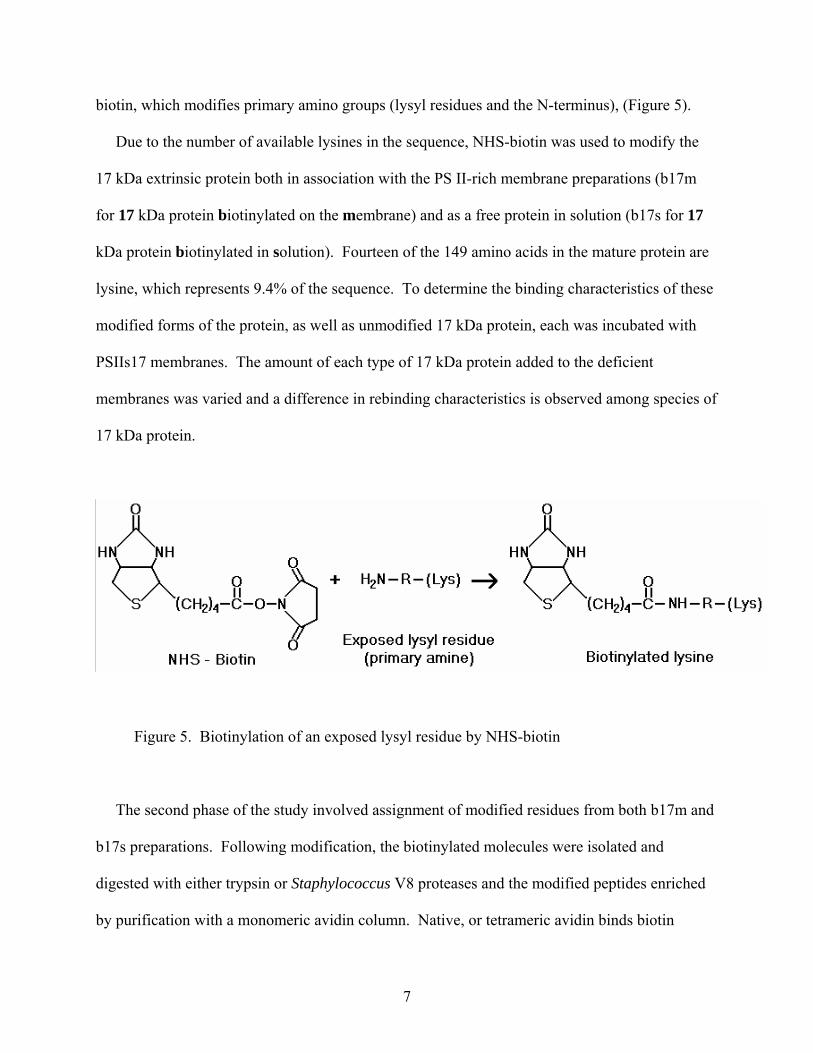
biotin, which modifies primary amino groups (lysyl residues and the N-terminus), (Figure 5).
Due to the number of available lysines in the sequence, NHS-biotin was used to modify the
17 kDa extrinsic protein both in association with the PS II-rich membrane preparations (b17m
for 17 kDa protein biotinylated on the membrane) and as a free protein in solution (b17s for 17
kDa protein biotinylated in solution). Fourteen of the 149 amino acids in the mature protein are
lysine, which represents 9.4% of the sequence. To determine the binding characteristics of these
modified forms of the protein, as well as unmodified 17 kDa protein, each was incubated with
PSIIs17 membranes. The amount of each type of 17 kDa protein added to the deficient
membranes was varied and a difference in rebinding characteristics is observed among species of
17 kDa protein.
Figure 5. Biotinylation of an exposed lysyl residue by NHS-biotin
The second phase of the study involved assignment of modified residues from both b17m and
b17s preparations. Following modification, the biotinylated molecules were isolated and
digested with either trypsin or Staphylococcus V8 proteases and the modified peptides enriched
by purification with a monomeric avidin column. Native, or tetrameric avidin binds biotin

8
molecules with a very high affinity (Kd = 10-15), however, monomeric avidin has a weaker
binding affinity (Kd = 10-7) and that binding can be reversed under milder conditions.
Figure 6. Flowchart showing the experimental protocol. The 17 kDa protein that is biotinylated either on the photosystem membranes (b17m) or in solution (b17s).
A chromatography column containing a rigid, methacrylate polymeric gel filtration matrix,
made functional with covalently bound, monomeric avidin, (SoftLink™ Soft Release Avidin
Resin, Promega) can be used to bind any biotin-containing peptides in solution, allowing

9
everything else to pass through. Elution of the bound biotinylated peptides is accomplished by
unfolding the monomeric avidin with 10% acetic acid. Such a column is used here to enrich the
sample for biotinylated peptide fragments.
These enriched samples were analyzed by nanospray LC MS/MS spectroscopy. The peptides
are separated by reverse phase (C18) chromatography and inserted into a quadrapole
spectrophotometer. Precise time-of-flight measurements give very accurate mass measurements
of the parent ions introduced into the machine.
Figure 7. Schematic of a time-of-flight mass spectrophotometer. (http://www.waters.com/WatersDivision/ContentD.asp?watersit=JDRS-5L7PBV)
Knowing the sequence of the 17 kDa protein and the sites of cleavage by the proteases, one
can sort through the parent ion mass measurements to identify biotinylated fragments. Using
information obtained from collision fragmentation (MS/MS data), some ambiguities in
assignment of modified lysines can be resolved. Fragmentation of the parent ions results in two
principal series of daughter ions, one where the peptide is fragmented at the amino side of the
peptide bond (b ion series), and one fragmented from the carboxyl side of the peptide bond (y ion
10
series). Using the program GPMAW (General Protein/Mass Analysis for Windows), a listing of
these daughter ion masses can be used to identify which lysine residues contain the additional
mass of one or more biotin moieties. Further computational analysis using MASCOT Search
(www.matrixscience.com) resolves ambiguities in biotin modification assignment.
Differential labeling of residues between the on-membrane and in-solution methods identifies
residues not accessible to the bulk solvent while in association with the membrane, but
accessible to the modifying agent once free of the membrane. This information was correlated
with those regions that inhibit rebinding to the PS II membranes lacking the 17 kDa protein.
Finally, suspect residues or regions involved in binding are visualized on the crystal structure of
the protein and a model of the 17 kDa protein’s orientation to the PSII membrane complex is
proposed.

11
METHODS AND MATERIALS
Chloroplasts were isolated from spinach (Spinacia oleracea) bought at a local market.
Oxygen-evolving PS II membranes were prepared by the method of Berthold et al. (1981) with
the modifications described by Ghanotakis and Babcock (1983). All steps in the procedure were
done at 0-4°C except where noted. Spinach leaves washed with deionized water were deveined
and ground in a cold blender with a solution of cold chloroplast isolation buffer (100 mM
sucrose, 200 mM NaCl, 5 mM MgCl2, and 50 mM Na-KPO4 buffer, pH 7.4), then filtered
through a single layer of Miracloth (CalBiochem Co.) and 2 layers of cheesecloth into a flask
kept immersed in ice. The filtrate was centrifuged for 7 minutes (min) at 4,000 x g and the
supernatant removed. The pellet (chloroplasts with intact thylakoid membranes) was
resuspended in resuspension buffer (300 mM sucrose, 10 mM MgCl2, 15 mM NaCl, and 50 mM
Mes-NaOH, pH 6.0). Chlorophyll concentration was measured by the method of Arnon (1949).
The chlorophyll a/b ratio at this point was 2.6 – 3.2, indicating that a significant amount of PS I
is present. These intact thylakoids were then subjected to detergent treatment with 20% Triton-
X-100 (w/v) to a final chlorophyll to detergent ratio of 25:1 (summer) or 22.5:1 (winter). This
treatment is 25 minutes in the dark briefly agitating every 5 min. The detergent treated
thylakoids were centrifuged at 2,000 x g for 5 min and the supernatant was immediately
centrifuged at 30,000 x g for 25 min. This pellet was resuspended in resuspension buffer and
again subjected to 30,000 x g for another 25 min. This second high speed spin served to remove
any remaining detergent. Typical preparations had a chlorophyll a/b ratio of 1.90 – 2.04,
showing enrichment for PS II over PS I in the membranes (Dunahay et al, 1984).
Overcoming the obstacles of limiting the contaminants in the purified protein solution,

12
minimizing the action of the released protease, and protecting the sample from denaturation for
further experimentation or purification was paramount in this study. In the search for an
extraction protocol that satisfied these requirements, several methods either did not prevent the
protease cleavage, or did not effectively remove only the 17 kDa protein. Solutions of varying
percentage methanol have been shown to remove the 17 kDa protein while suppressing the
release of the 24 kDa protein (Yamamoto and Kubota, 1987). High pH (>8.0) keeps the prolyl
protease in an inactive state, as does the addition of mM quantities of CuCl2 (Kuwabara et al,
1986). Each of these methods alone tackles only one of the obstacles above. Building on these
ideas, isolation of unmodified and modified 17 kDa protein from PS II membranes was
accomplished via a two-step process of high-pH, salt-methanol extraction followed by cation-
exchange chromatography. The chromatography column used (CM-Toyopearl 650M) is
ineffective at separating the 17 and 24 kDa components easily removed by 1.0 M NaCl salt-
washing, however, it is highly effective at separating the 17 kDa protein from MSP. This is
useful if a method of extraction is selective for the 17 kDa protein and MSP while leaving the 24
kDa protein on the membrane. A solution of 20% MeOH, 100 mM NaCl specifically releases
only the 17 kDa protein, but a percentage of the protein remains on the membrane. Allowing for
the concerted release of MSP, the optimum extraction buffer for this removal of the 17 kDa
protein was 40% MeOH, 1 M NaCl, and 50 mM Tricine-NaOH, pH 9.0 (extraction buffer). The
40% MeOH minimizes the removal of the 24 kDa protein, while the high pH and salinity
suppress the activity of the released prolyl protease. PS II membranes were centrifuged at
30,000 x g for 20 min and the pellet was resuspended in extraction buffer at a chlorophyll
concentration of 1 mg/ml. The extraction is 10 minutes at 0-4°C, followed by centrifugation at
30,000 x g for 20 minutes. The supernatant was then subjected to a 48 hour dialysis against

13
100:1 volume of deionized water. Subsequent concentration to 1-2 ml was achieved via a
Centricell 30 device with a molecular weight limit of 10 kDa (Polysciences, Inc.). The
concentrate was then loaded on the cation-exchange chromatography column CM-Toyopearl
650M. A NaCl gradient was used to progressively increase the concentration of eluting salt in
the column as fractions are collected. (See Figure 8) Buffer A was 20 mM NaKPO4 pH 6.5 and
buffer B was 20 mM NaKPO4 pH 6.5, 250 mM NaCl.
Figure 8. Line Trace of CM-Toyopearl 650M Column Purification. Blue trace, UV spectrum absorbance; red trace, conductivity; black dashed trace, salt concentration gradient progress bar.
The MSP does not or only very weakly binds to the column at the pH of the elution buffer. As
indicated by its low pI of approximately 5.01, this protein is not highly positively charged at pH
6.5 and so is not attracted to the negatively charged column matrix. As a result, it was eluted
immediately with no salt. The 17 kDa protein’s pI is much higher at 9.25 and thus it strongly

14
binds the column and must be eluted with salt. Interestingly, any residual 24-kDa protein
(theoretical pI of 5.94) was eluted soon after the MSP as demonstrated by a concerted
conductivity spike, unlike its normal release just before the 17-kDa protein when extracted with
a 1.0 M NaCl solution. The fractions that correspond to the 17 kDa protein were pooled and
concentrated.
PS II membranes deficient in 17 kDa protein (PSIIs17) were prepared using a different
extraction buffer, when the recovery of the 17 kDa protein free of contaminants was less of a
priority than recovering PS II membranes deficient in the 17 kDa protein but with minimal loss
of any other membrane component. This was accomplished via an extraction buffer containing
20% MeOH, 100 mM NaCl, and 50 mM Mes-NaOH, pH 6.0, (Yamamoto and Kubota, 1987).
The pH was kept low as we are unconcerned with the action of the prolyl protease here.
Analytical PAGE of the PS II proteins was performed under conditions described by
Delepelaire and Chua (1979) in 12.5-20% gradient polyacrylamide gels. The resolved proteins
were electroblotted onto PVDF membranes (Immobilon-P, Millipore Co.). Panels of the blot
were either stained with Coomassie Brilliant Blue stain or were blocked for 2 hours (h) with 5%
nonfat dry milk in development buffer (150 mM NaCl, and 10 mM Tris-HCl, pH 7.4), and then
washed extensively with the same buffer. Following this, the milk-treated PVDF panels were
incubated for 24 h with an anti-17 kDa monoclonal antibody (FCC4) derived from mice (Frankel
and Bricker, 1990). A 1:1000 dilution of an anti-mouse secondary antibody conjugated to a
peroxidase enzyme and dissolved in 1% BSA in deionized water was placed on the blot for 4 h.
Once more, the blot was washed extensively with development buffer and finally placed in a
solution of 80 mL development buffer and 20 mL of 0.3% 4-chloro-1-naphthol (dissolved in
100% ethanol). The addition of 400 µl of 3% H2O2 allowed visualization of bands
15
corresponding to the 17 kDa protein. The development process is stopped after 15-20 seconds
by extensive washing with deionized water and allowing the blot to dry on paper towels in the
dark.
Modified versions of the 17 kDa protein were prepared as follows. PS II membranes were
centrifuged and resuspended in 300 mM sucrose, 10 mM MgCl2, 15 mM NaCl, and 50 mM Tes-
NaOH, pH 7.0, at 1.0mg of chlorophyll per milliliter, and kept at 0-4°C throughout the
procedure. Exposed primary amines (lysyl residues and the N-terminus) on the 17 kDa protein
were labeled with NHS-biotin as described by Bricker et al., 1988, with a couple ok
modifications. NHS-biotin was dissolved at a concentration of 1.0 mg/ml in dimethyl sulfoxide
at 20°C. This solution was added at a concentration of 50 µM to the suspended membranes and
allowed to incubate for 1.5 hours at 0-4°C. The labeling reaction was stopped by the addition of
Tris-HCl, pH 6.8, to a concentration of 50 mM. The residual NHS-biotin was removed by
washing the membranes twice with resuspension buffer. The 17 kDa protein was then isolated as
described above. Labeling of the purified 17-kDa extrinsic protein in solution was performed in
a similar manner at a protein concentration of 137.68 µg/ml in 300 mM sucrose, 10 mM MgCl2,
15 mM NaCl, and 50 mM Tes-NaOH, pH 7.0. The labeling reaction was quenched by the
addition of Tris-HCl, pH 6.8, to a concentration of 50mM, and the residual NHS-biotin was
removed by centrifugal ultrafiltration in a Centricell 30 device (Polysciences, Inc.). The purified
proteins were quantified using an extinction coefficient of 13 mM-1 cm-1 at 277nm.
The rebinding of unmodified and modified versions of the 17 kDa protein to PSIIs17
membranes was done on an increasing molar ratio scale. Using 250 chlorophyll molecules per
PS II reaction center, 1 µmol chlorophyll = 100 µg chlorophyll, and the actual mass of the 17
kDa protein as 16,522 Da, 0.1 µmol 17 kDa protein = 6.609 µg 17 kDa protein. For a 1:1 molar

16
ratio, PS II reaction centers to 17 kDa protein, 100 µl of PSIIs17 membranes at 1.0 mg/ml
chlorophyll was incubated with 9.94 µl of 17 kDa protein at 0.6647 mg/ml. Samples with ratios
of 0.0, 0.1, 0.2, 0.5, 1.0, and 2.0 were incubated for 30 min at 0-4°C, and then brought to 500 µl
with resuspension buffer. Following centrifugation, the pellet was resuspended in 500 µl of
resuspension buffer, centrifuged once more, and the pellet resuspended in 70 µl of resuspension
buffer. Chlorophyll concentrations were measured and samples were adjusted to 1.0 mg/ml
chlorophyll. Volumes corresponding to 15 µg chlorophyll were loaded per lane on a gradient
polyacrylamide gel. The gels were electroblotted onto PVDF membranes and probed with the
FCC4 antibody as above. Density scans of the developed blots quantified the amount of 17 kDa
protein rebinding to the PSIIs17 membranes. The values were normalized to a standard curve of
known concentrations of 17 kDa protein for comparison.
For the mass spectrometry experiment, each biotinylated protein solution (b17m and b17s)
was brought to 10% TCA (trichloroacetic acid), and the protein precipitate was collected by
centrifugation, washed twice with cold 100% acetone, dried under vacuum, and dissolved in 8 M
urea and 400 mM ammonium bicarbonate (Stone et al., 1989). The solubilized protein was then
diluted to 2 M urea and 100 mM ammonium bicarbonate and digested overnight at 37°C with
either trypsin or Staphylococcus V8 protease. Trypsin cleaves at the peptide bond C-terminal to
a lysine or arginine residue, with the exception that it will not cleave at a biotinylated lysine. V8
protease can cleave at the peptide bond C-terminal to aspartate or glutamate residues, however,
in the bicarbonate buffer used here, it is specific for the glutamate residues. The final 17 kDa
protein to protease ratio was 25:1, with a 1:50 initial addition of enzyme and a second 1:50
addition 4 h later. The digestion was stopped by the introduction of a protease inhibitor cocktail
prior to purification on a monomeric avidin column (SoftLink™ Soft Release Avidin Resin,
17
Promega). This was necessary not to stop the digestion of the 17 kDa samples, but to avoid the
action of the protease on the avidin proteins in the column.
The entire avidin enrichment chromatographic procedure is done at room temperature and the
flow is due to gravity alone (~ 400 µL / min). The avidin column was preadsorbed with 5 mM
biotin in 0.1 M NaPO4, pH 7.0 to fill nonreversible binding sites. The less numerous
nonreversible biotin binding sites on the column are tetrameric avidin, which are not denatured
by the addition of 10% acetic acid. After 15 minutes to allow biotin binding, the column was
washed with 8 column volumes of 10% acetic acid to unfold the monomeric avidin, releasing the
bound biotin, followed by 8 column volumes of 100 mM NH4C2H3O2, pH 7.0. NH4C2H3O2 was
used due to its volatility, leaving minimal additional compounds in the purified solution. The pH
of the eluate was monitored until it reached 6.8, at which point the flow was arrested for 30
minutes to allow the monomeric avidin to refold. The sample was then loaded on the column
and the eluate collected. The column was washed with 5 column volumes of 100 mM
NH4C2H3O2, pH 7.0, and the wash eluate collected. The bound biotinylated molecules were
then eluted with 10% acetic acid and 1.5 mL fractions were collected. Twenty µL of each
fraction, including the load and wash eluates, was applied to a dry nitrocellulose membrane and
allowed to dry. The membrane was then incubated in the dark with 5% nonfat dry milk in
development buffer, and washed extensively with development buffer. The milk-treated PVDF
panels were then probed with a 1:1000 dilution of an avidin-peroxidase conjugate (Sigma
Chemical Co.). Color development using chloronaphthol and H2O2 as described above
visualized biotinylated molecules in the fractions. (See Figure 9) These biotin-containing
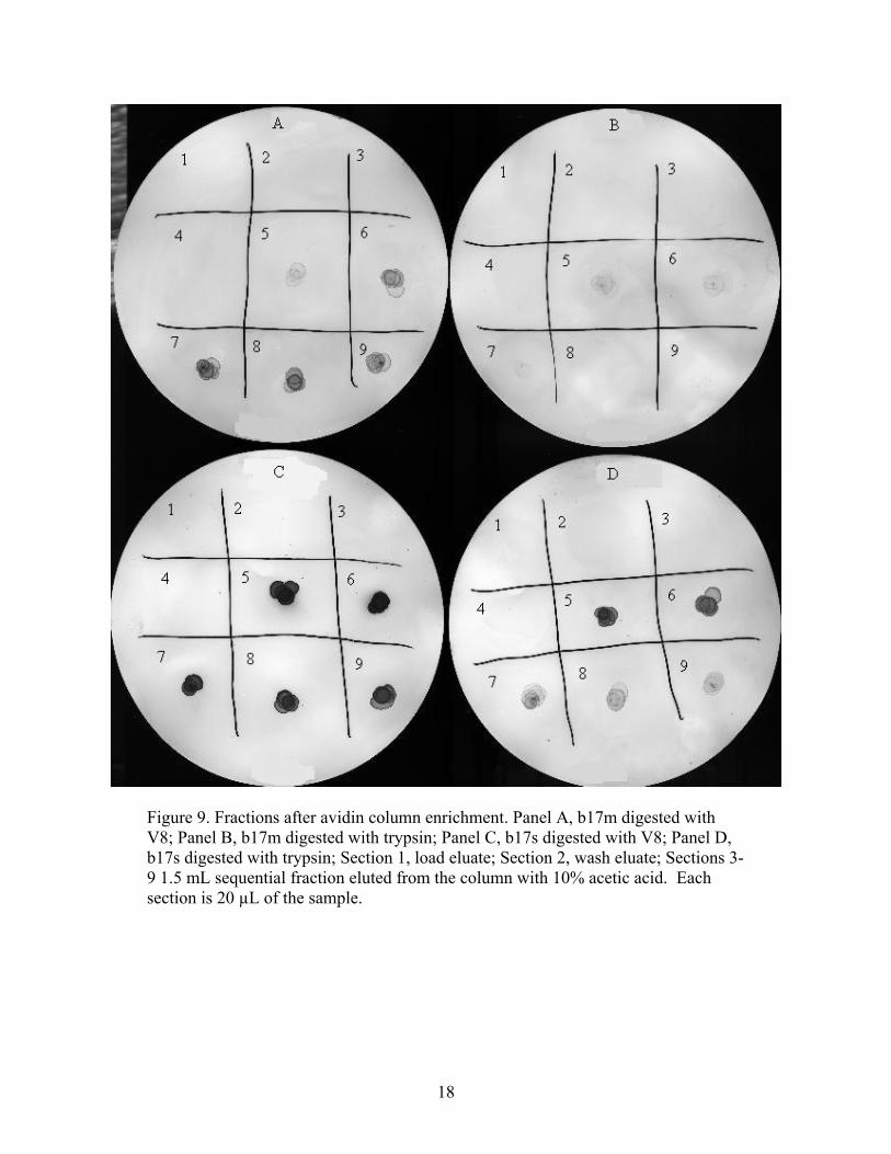
18
Figure 9. Fractions after avidin column enrichment. Panel A, b17m digested with V8; Panel B, b17m digested with trypsin; Panel C, b17s digested with V8; Panel D, b17s digested with trypsin; Section 1, load eluate; Section 2, wash eluate; Sections 3-9 1.5 mL sequential fraction eluted from the column with 10% acetic acid. Each section is 20 µL of the sample.

19
fractions were pooled and frozen for lyophilization. The lyophilized peptides were resuspended
in 100 µl of 0.1% trifluoroacetic acid and frozen at -80°C.
For liquid chromatography/mass spectrometry analysis a Waters CapLC coupled to a Q-TOF
II mass spectrometer was used. The samples were injected onto a 75µm i.d. X 10 cm spraying
capillary packed with 5 µm C18 beads. The flow rate was set to 7 µL/min split to approximately
200 nL/min before reaching the column. A long gradient of 75 minutes was used in order to
obtain good peptide separation. Buffer A consisted of 95% water, 5% acetonitrile, 0.1% formic
acid and buffer B was 95% acetonitrile, 5% water and 0.1% formic acid. Mass spectrometry
results were analyzed with the GPMAW software package (Lighthouse Data, Sweden).
20
RESULTS AND DISCUSSION
17 kDa Protein Purification
Previous experiments had shown that washing PS II membrane preparations with 1.0 M NaCl
released the 24 and 17 kDa proteins from the membrane but not the MSP. Adaptation of this
buffer to pH 9.0 in order to keep the prolyl protease specific for the 17 kDa protein N-terminus
in an inactive state additionally releases some of the MSP from the membrane (Figure 10, lane
2). While the cation exchange CM-Toyopearl column is effective at separating this additional
MSP from the supernate, it fails to separate the 24 and 17 kDa proteins independently (Figure 10,
lane 8). Favorable selective removal of the 17 and 33 kDa proteins, while minimizing the
concurrent removal of the 24 kDa protein, was achieved with a solution of 40% MeOH, 1.0 M
NaCl, and 50 mM Tricine-NaOH, pH 9.0. The ability to remove the MSP and 17 kDa proteins
without the removal of the 24 kDa protein suggests that the 24 kDa protein is not attached to the
complex solely through the MSP and may have sites of interaction with integral membrane
components. Jegerschöld, et al (1995) showed the specific release of only the 17 kDa protein
using 10 mM CuCl2. This method was not employed here as the addition of copper may
interfere with subsequent steps.

21
Figure 10. Coomassie-Stained PAGE of CM-Toyopearl 650M Column Fractions: Ineffective Separation. Lane 1, PS II membranes; lane 2, 1.0 M NaCl, 50 mM Tricine-NaOH, pH 9.0 extracted proteins; lanes 4-6, fractions containing MSP; lanes 7-9, fractions containing overlapping elution peaks for the 24 and 17 kDa proteins released from the column.
Figure 11 illustrates the extraction and purification of the 17 kDa protein from spinach PS II
oxygen-evolving membranes. Lane 2 shows the proteins remaining in the supernate after
resuspending the pelleted membranes in extraction buffer. The three extrinsic proteins were
released into solution, MSP and the 17 kDa protein, and to a much lesser extent, the 24 kDa
protein. MSP did not bind to the column and was eluted soon after loading and with no salt
present (lanes 3 through 5) with only minor traces remaining in the end. Interestingly, any
residual 24 kDa protein that was released with extraction buffer was eluted soon after the MSP

22
initial peak with an accompanying conductivity spike as opposed to its normal release along with
the 17 kDa protein within the salt gradient when no methanol was used. (See Figure 8) Lane 7
clearly shows the elution of the 17 kDa protein, devoid of any contaminating 24 kDa or MSP.
Figure 11. Coomassie- Stained PAGE of CM-Toyopearl 650M Column Fractions: Effective Separation. Lane 1, PS II membranes; lane 2, 40% MeOH, 1.0 M NaCl, 50 mM Tricine-NaOH, pH 9.0 extracted proteins; lanes 3 & 4, fractions containing MSP; lane 5, minor traces of MSP & 24 kDa protein; lane 6, minor trace of 17 kDa protein; lane 7, majority of 17 kDa protein released from the column.
Biotinylation of the 17 kDa Protein
After purifying the protein and preventing its breakdown by the prolyl endoprotease, a means
of modifying the protein was needed in order to test its ability to rebind to the PS II complex.
The 17 kDa protein has a relatively large percentage of lysine in the mature sequence (9.4%), so
NHS-biotin was chosen as the modifying agent since it modifies free amino groups. In the
23
biotinylation experiment, concentrations of substrate and labeling reagent were such that, on
average, a single biotin moiety would be added per protein molecule. This was done to minimize
the effects that could arise from the simultaneous neutralization of numerous positively charged
lysyl residues with neutral biotin molecules within a single protein molecule. Visualization by
FCC4 anti-17 kDa antibody binding and avidin peroxidase activity is shown for unmodified 17
kDa, b17m, and b17s (Figure 12) and illustrates the typical results of labeling with NHS-biotin.
Lane 1 in panel A shows that the unmodified 17 kDa protein was significantly degraded to the 16
kDa fragment as a result of N-terminal 12 residue cleavage by the protease. The biotinylation
procedure, both on membrane and in solution, appeared to further the protein from this cleavage.
The rebinding studies performed did not use this highly degraded sample of unmodified 17
kDa protein. The unmodified 17 kDa protein used there contained none of the 16 kDa
degradation fragment as a result of the high pH extraction buffer inactivating the protease. In
order to accurately compare the intensity of banding patterns from the different species of 17-
kDa proteins using the FCC4 antibody, the different versions of the protein must bind the
antibody similarly. Panel A shows that the monoclonal FCC4 antibody’s recognized surface
antigen is not altered by biotinylation. Panel B, Lane 1 serves as a negative control, showing no
bands. This indicates that there exist no natural biotin moieties in the 17 kDa protein; thus, all
biotins observed were added by reaction with NHS-biotin. The multiple bands that appear in
lanes 2 and 3 are biotinylated 17 kDa proteins that are moving through the polyacrylamide gel at
differing rates due to changes in their electrostatic properties introduced by biotinylation. As the
avidin peroxidase conjugate visualization method is much more sensitive in the detection of
biotin, more bands appear in panel B compared to FCC4 antibody sensitivity in panel A.

24
Figure 12. Typical Biotinylation of Unmodified 17 kDa Protein, b17m, & b17s. Panel A, FCC4 antibody binding; Panel B, avidin peroxidase conjugate binding. Lane 1, unmodified 17 kDa protein (with 16 kDa fragment present); lane 2, b17m; lane 3, b17s.
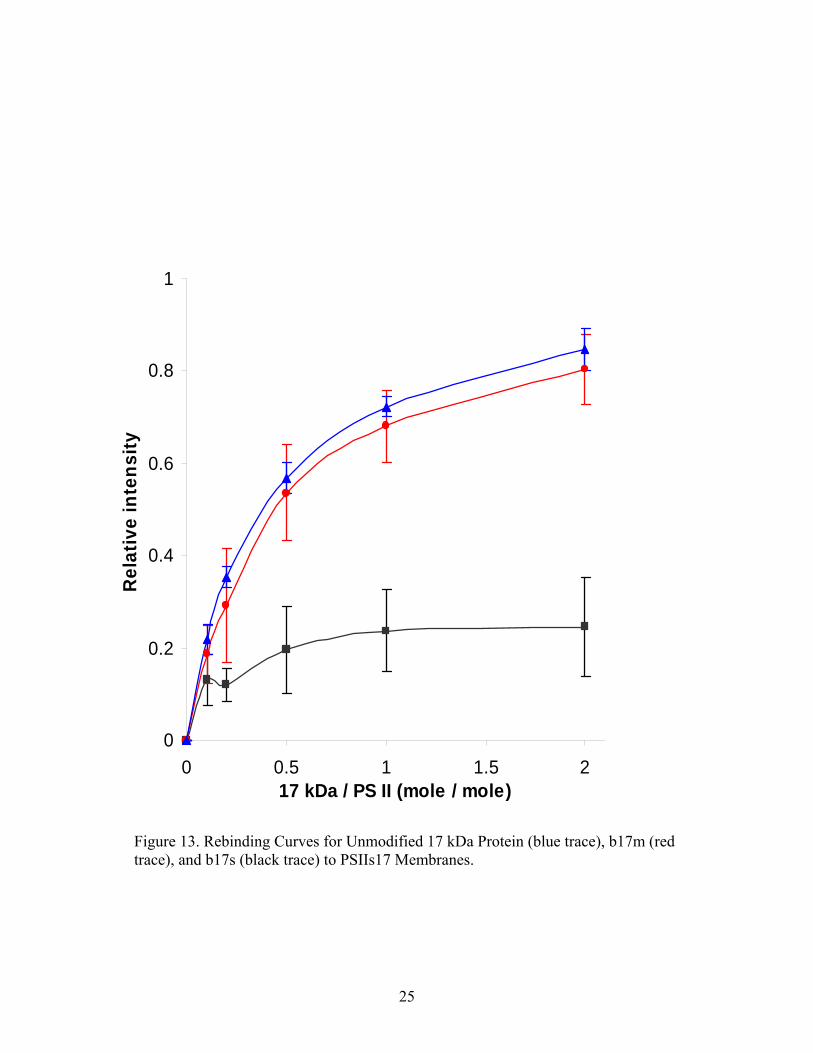
Rebinding Studies
Each of the biotinylated 17-kDa proteins as well as unmodified 17 kDa protein was incubated
with and allowed to rebind to PSIIs17 membranes (lacking the 17 kDa protein). The rebinding
curves are shown in Figure 13.
25
0
0.2
0.4
0.6
0.8
1
0 0.5 1 1.5 217 kDa / PS II (mole / mole)
Rel
ativ
e in
tens
ity
Figure 13. Rebinding Curves for Unmodified 17 kDa Protein (blue trace), b17m (red trace), and b17s (black trace) to PSIIs17 Membranes.
26
Unmodified and modified 17 kDa protein were incubated with 17 kDa-depleted PS II
membranes at molar ratios of 0.1 – 2.0. Data from 5 experiments comparing b17s to unmodified
17 kDa protein data from 3 experiments showed a marked decrease in ability to rebind to
membranes lacking the 17 kDa protein, while b17m data from 5 experiments showed an ability
to rebind with nearly the same affinity as the unmodified protein. It is clear from the data that
there exists a difference in the rebinding characteristics of 17 kDa protein modified while on the
membrane and while in solution. The inability of the b17s species to rebind to 17 kDa-deficient
membranes is similar to the incapacity of the 16 kDa fragment to rebind to the same membranes.
Mass Spectrometry Analysis
The addition of a biotin moiety to a peptide fragment results in a mass increase of 227.14
daltons. This difference can be seen in mass spectrometry fragment masses, and allows
assignment of location of biotinylated residues, although some ambiguity exists. Table 2
summarizes the results from the trypsin digestion of the 17 kDa protein that had been
biotinylated on the membrane. Ten peptides were observed ranging in mass from 1917.07 to
4604.53 Da. Of these, six contained one or more biotinylated sites as evidenced by the addition
of multiples of 227.1 Da: 52A-68R + 1 biotin, 124I-147K + 2 biotins, 52A-79R + 2 biotins, 99D-132K +
1 biotin, 70A-101K + 2 biotins, and 52A-90K + 3 biotins. By excluding the C-terminal lysine from
the list of possible biotinylated residues for each peptide fragment observed, some ambiguity can
be eliminated. This may be done, since trypsin does not cleave at a biotinylated lysine.
The same procedure was performed with Staphylococcus V8 protease digests and the results of
that digestion on the membrane are shown in Table 3. Only three peptides were observed, all
containing 1-2 biotin additions: 107L-131E + 1 biotin, 107L-131E + 2 biotins, and 101K-129E + 1

27
biotin. Using MS/MS data from the daughter ions produced from further collision fragmentation
within the mass spectrometer, one can look at the masses of those ions, with respect to the
known sequence of the peptide fragment and determine which lysine(s) in the sequence contain
the added mass of biotin. Using the data from both enzyme digestions, and making two
unambiguous assignments based on MS/MS analysis (See Table 6), the list of lysyl residues
accessible to the modifying agent NHS-biotin while the 17 kDa protein is in association with PS
II includes: 53K, 63K, 69K, 96K, 98K, 101K, 102K, 110K, 125K, and 132K.
Table 2. Assignments of Biotinylated Lysines for Peptides Produced from a Trypsin Digest of b17m. Observed mass
∆ mass from predicted
Peptide assignment Modified K
1917.07 -0.03 52A-68R + 1 biotin 53K or 63K 2776.42 -0.04 28D-51R 2975.55 -0.01 28D-53R 2681.39 -0.02 124I-147K + 2 biotins 125K and 132K 3140.70 -0.01 36D-63K 3225.76 0.02 52A-79R + 2 biotins 2 of 53K, 63K, and 69K 3774.06 -0.03 70A-101K 3814.96 -0.01 99D-132K + 1 biotin 1 of 101K, 102K, 110K, 123K, and 125K 3774.06 -0.03 70A-101K + 2 biotins 2 of 90K, 96K, and 98K 4604.53 -0.03 52A-90K + 3 biotins 53K, 63K, and 69K
Table 3. Assignment of Biotinylated Lysines for Peptides Produced from a Staphylococcus V8 Protease Digest of b17m. Observed mass
∆ mass from predicted
Peptide assignment Modified K
2729.39 -0.02 107L-131E + 1 biotin 1 of 110K, 123K, and 125K 2729.39 -0.05 107L-131E + 2 biotins 2 of 110K, 123K, and 125K 3242.71 -0.04 101K-129E + 1 biotin 1 of 101K, 102K, 110K, 123K, and 125K

28
Table 4. Assignments of Biotinylated Lysines for Peptides Produced from a Trypsin Digest of b17s. Observed mass
∆ mass from predicted
Peptide assignment Modified K
842.52 0.00 91T-98K + 1 biotin 96K 1127.60 0.00 82A-90K 1001.54 0.00 124I-132K + 1 biotin 125K 1032.58 0.01 102K-110K + 1 biotin 102K 1164.58 0.00 28D-37R + 1 biotin 35K 1214.69 0.00 91T-101K + 1 biotin 1 of 96K and 98K 1257.73 0.00 52A-63K + 1 biotin 53K 1326.70 0.00 69K-79R + 1 biotin 69K 1342.78 0.00 91T-102K + 1 biotin 1 of 96K, 98K, and 101K1361.79 0.00 87Y-98K + 1 biotin 1 of 90K and 96K 1214.69 0.00 91T-101K + 2 biotins 96K and 98K 1717.94 0.00 54V-68R + 1 biotin 63K 1733.96 0.00 87Y-101K + 1 biotin 1 of 90K, 96K, 98K 2186.21 0.00 69K-86R 1733.96 0.00 87Y-101K + 2 biotins 2 of 90K, 96K, and 98K 2324.27 0.00 82A-101K + 1 biotin 1 of 90K, 96K, and 98K 2440.21 0.00 126S-147K + 1 biotin 132K
Table 5. Assignments of Biotinylated Lysines for Peptides Produced from a Staphylococcus V8 Protease Digest of b17s. Observed mass
∆ mass from predicted
Peptide assignment Modified K
599.40 0.00 144V-149G + 1 biotin 147K 731.42 0.00 101K-106E + 2 biotins 101K and 102K 1116.59 0.00 48A-58E + 1 biotin 53K 1996.06 0.00 132K-149G + 1 biotin 147K 2729.39 -0.01 107L-131E + 1 biotin 1 of 110K, 123K, and 125K 4126.05 -0.01 107L-143E + 1 biotin 1 of 110K, 123K, 125K, and 132K

29
Table 6. Assignments of Biotinylated Lysines for Ambiguities in Modified Peptides Based on MS/MS Data Analysis. Source of Peptide Peptide assignment Ambiguously
modified K Modified K after MS/MS data analysis
V8 digest of b17m 107L-131E + 1 biotin 1 of 110K, 123K, and 125K 110K V8 digest of b17m 107L-131E + 2 biotins 2 of 110K, 123K, and 125K 110K, 1 of 123K and 125KTrypsin digest of b17s 91T-101K + 1 biotin 1 of 96K and 98K 98K Trypsin digest of b17s 87Y-101K + 2 biotins 2 of 90K, 96K, and 98K 90K and 98K V8 digest of b17s 107L-131E + 1 biotin 1 of 110K, 123K, and 125K 123K
Table 4 presents the data from the 17 kDa protein digested with trypsin in solution.
Seventeen peptides were observed ranging in mass from 842.52 to 2440.21 Da, sixteen of which
contained one or two attached biotins: 91T-98K + 1 biotin, 124I-132K + 1 biotin, 102K-110K + 1
biotin, 28D-37R + 1 biotin, 91T-101K + 1 biotin, 52A-63K + 1 biotin, 69K-79R + 1 biotin, 91T-102K + 1
biotin, 87Y-98K + 1 biotin, 91T-101K + 2 biotins, 54V-68R + 1 biotin, 87Y-101K + 1 biotin, 87Y-101K
+ 2 biotins, 82A-101K + 1 biotin, and 126S-147K + 1 biotin. The very small differences in mass
from predicted to observed should be noted, as this indicates few false positives in the
assignments of biotinylated peptides. Similarly, Table 5 lists the data from the Staphylococcus
V8 digestion in solution, having only six peptides, all with one or two biotins attached: 144V-149G
+ 1 biotin, 101K-106E + 2 biotins, 48A-58E + 1 biotin, 132K-149G + 1 biotin, 107L-131E + 1 biotin, and
107L-143E + 1 biotin. Again, using the data from both enzyme digestions, and making three
further assignments based on MS/MS analysis (See Table 6 and Figure 14), the list of lysyl
residues accessible to the modifying agent NHS-biotin while the 17 kDa protein is in solution
includes: 35K, 53K, 63K, 69K, 90K, 96K, 98K, 102K, 123K, 125K, 132K, and 147K. It should be noted,
however, that lysyl residues not listed here are not necessarily protected from the modifying
agent in solution. Those residues cannot be labeled with confidence as being modified with a

30
biotin, as ambiguities remain.
Figure 14 shows one instance where MASCOT search analysis helps to resolve an ambiguity.
The daughter ion series for the fragment 87Y-101K in the trypsin digest of b17s is shown. The
possible biotinylated lysyl residues are 90K, 96K, and 98K. 101K is excluded from this list
because trypsin will not cleave at a modified lysine.
Figure 14. MASCOT search results. Daughter ion series for 87Y-101K in trypsin digest of b17s.

31
Masses in red were the ones observed in the experiment, whereas masses in black were
only predicted to occur. In the b ion series, the mass difference from 89L to 90K is one
biotin mass greater than the addition of 90K alone, indicating that it is one of the
biotinylated residues. Similarly, in the y ion series, the mass difference from 99D to 98K
is one biotin mass greater than the addition of 98K alone, indicating that it is also one of
the biotinylated residues. Also in the y ion series, the mass difference from 97P to 96K is
comparable to the addition of 96K alone, indicating that this residue does not contain the
additional mass of an added biotin. This analysis allows the assignment of biotins to 90K
and 98K, and excludes 96K, resolving the ambiguity.
17 kDa Protein Models
Four residues which were biotinylated when the 17 kDa protein was modified in solution but
not while on the membrane (35K, 90K, 123K, and 147K) are found in regions of the protein that are
shielded from the bulk solvent while in association with the photosystem. These residues
become accessible once the 17 kDa protein is released into solution. Since a large
conformational change has never been demonstrated, the secondary structure is a stable 4-helix
bundle core, and the unmodified protein has the capacity to rebind to the PSIIs17 membranes,
the differentially modified residues are very likely shielded in this manner. Figure 15 shows the
locations of three of the four differentially modified lysyl residues.
Figures 16 and 17 are multiview renderings of the 17 kDa protein with all the atoms in van
der Waals contact. In these representations, the 4-helix nature of the protein is lost in the image,
but the pictures show the lack of empty space typical in the interior of proteins.

32
Figure 15. VMD-generated image of 17 kDa crystal structure showing the locations of three lysine residues in red van der Waals radii.

33
Figure 16. One plausible orientation of the 17 kDa protein to the photosystem II membrane complex. The VMD-rendered images illustrate the atoms in van der Waals contact. Panel A, viewed from above; panel B, viewed from below. 35K is not shown in the crystal structure since it begins at residue 38F.

34
The differentially modified lysyl residues, excluding 35K, are visible as surface moieties. Figure
16 shows one proposed orientation of the 17 kDa protein based on the shielded lysyl residues
presented here and the observation that the 16 kDa fragment lacking the N-terminal 12 residues
does not rebind to the membrane. Panel A represents looking at the protein from above,
perpendicular to the plane of the membrane and panel B represents looking up at the protein
from the perspective of the complex. The differentially modified residues are seen from below,
but are shielded from view in Panel A. From this orientation, the N-terminal tail (not shown in
the crystal structure pdb: 1NZE) can anchor the 17 kDa protein to the complex as suggested by
Kuwabara, et al (1986). This orientation would necessitate the existence of a solvent channel
between the 17 kDa protein and the complex to which the protein is bound, as 110K was shown to
be accessible by the bulk solvent while in association with the membrane.
Equally possible is the proposed orientation represented in Figure 17. This orientation allows
the modification of 110K without a solvent channel and sequesters 90K and 123K along a ridge in
contact with the complex. Residue 147K, in this model, must be shielded from NHS-biotin by
close association with another extrinsic protein.

35
Figure 17. A second plausible orientation of the 17 kDa protein to the photosystem II membrane complex. The VMD-rendered images illustrate the atoms in van der Waals contact. Panel A, viewed from above; panel B, viewed from below. 35K is not shown in the crystal structure since it begins at residue 38F.

36
SUMMARY AND CONCLUSIONS
Protein purification is a complex process in which many unforeseen problems can arise.
Purifying a membrane protein, albeit an extrinsic membrane protein, is even more difficult as the
protein may require more effort to remove it from its membranous environment. The protein
may require lipid chaperones to maintain stability in solution, a conformational change may
occur to limit the amount of hydrophobic residues exposed to aqueous solvent, or the protein my
aggregate once released into solution. Another problem in purifying a protein from a membrane-
anchored complex is the possibility of releasing other contaminating components of the complex
and/or associated proteases. A method of extraction and purification for the 17 kDa extrinsic
protein of photosystem II was established in which the additional release of contaminating 24
kDa protein was minimized and the released endoprolyl protease specific for the 17 kDa
protein’s N-terminus was kept in an inactive state. This initial extraction did not specifically
release only the 17 kDa protein, but additional proteins released could be easily removed from
the solution via ultrafiltration and chromatography. Contaminants smaller than 10 kDa were
allowed to pass out of the solution through a filter. The MSP and 17 kDa proteins have vastly
different binding characteristics on the cation exchange chromatography column used due to
differing pI’s and therefore can be easily separated. This two-step extraction and purification of
the 17 kDa protein of photosystem II overcomes the obstacles listed above.
Charged amino acid side chains localized to the surface of a protein play an important role in
the solubility characteristics of that protein. Residues composing hydrophobic patches on the
surface of an extrinsic membrane protein allow interaction with other proteins. The addition of
charged or polar groups in hydrophobic regions or the masking of a charged group may alter the

37
protein’s ability to bind in its normal location. NHS-biotinylation of the 17 kDa protein while
still present on assembled PS II membranes, and subsequent extraction with high pH NaCl-
methanol treatment, did not appear to diminish the rebinding characteristics to PS II membranes
lacking the 17 kDa protein. However, if extracted first and then biotinylated in the same manner
once in solution, there was a dramatic drop in the ability of the modified protein to rebind.
Modifying the 17 kDa protein while in association with photosystem II membranes and while
free in solution lead to differential modification of four lysyl residues. Lysyl residues 35K, 90K,
123K, and 147K were labeled with a biotin moiety by NHS-biotin if the procedure was done on 17
kDa protein free in solution; however, these four residues remain unaffected by the modifying
agent when modifying the 17 kDa protein while in association with the membrane. This
suggests the possibility that these four additional lysyl residues are those shielded while in
association with PS II membranes. These residues are part of or near regions of the 17 kDa
protein involved in binding to the other elements of the completed photosystem.
It should be kept in mind that these data do not preclude the possibility that a large
conformational change could occur in the 17 kDa protein upon its release into solution, and, if
so, the protein rebinding well in its unmodified state may imply that this conformational change
is reversible upon rebinding to the complex. However, since no evidence has been presented to
demonstrate any such conformational change, it is likely that the protein, excluding the N-
terminus, has the same rigid structure both on the membrane and in solution.
In conclusion, future models of the structural organization of PS II polypeptides must take
these data into account. Further studies whereby residues other than lysine may be modified and
analyzed in this manner could refine this orientation.

38
BIBLIOGRAPHY
Akerlund, H.-E., C. Jannson, and B. Andersson, 1982, Biochimica et Biophysica Acta 681:1-10. Reconstitution of oxygen evolution in high salt washed photosystem II particles. Arnon, D.I., 1949, Plant Physiology 24:1-15. Copper enzymes in isolated chloroplasts. Polyphenoloxidase in Beta vulgaris. Berthold, D. A., G. T. Babcock, and C. F. Yocum, 1981, FEBS Letters 134(2):231-4. A highly resolved, oxygen-evolving photosystem II preparation from spinach thylakoid membranes, EPR and electron-transport properties. Bergantino, E., A. Brunetta, E. Touloupakis, A. Segalla, I. Szabò, and G.M. Giacometti, 2003, J. Biol. Chem. 278:43, 41820-9. Role of the PSII-H Subunit in Photoprotection: Novel Aspects of D1 Turnover in Synechocystis 6803. Bricker, T. M., 1992, Biochemistry 31:4623-8. Oxygen Evolution in the Absence of the 33 kDa Manganese-Stabilizing Protein. Bricker, T. M., W. R. Odom, and C. B. Queirolo, 1988, FEBS Letters 231(1):111-7. Close association of the 33 kDa extrinsic protein with the apoprotein of CPa-1 in photosystem II. Bricker, T. M., H. B. Pakrasi, and L. A. Sherman, 1985, Archives of Biochemistry and Biophysics 237:170-6. Characterization of a spinach photosystem II core preparation isolated by a simplified method. Burnap, R. L., and L. A. Sherman, 1991, Biochemistry 30:440-6. Deletion mutagenesis in Synechocystis sp. PCC6803 indicates that the Mn-stabilizing protein of photosystem II is not essential for O2 evolution. Calderone, V., M. Trabucco, A. Vujicic, R. Battistutta, G. M. Giacometti, F. Andreucci, R. Barbato, and G. Zanotti, 2003, EMBO Reports 4:900-5. Crystal structure of the PsbQ protein of photosystem II from higher plants. Delepelaire, P., and N. Chua, 1979, PNAS 76:111-5. Lithium dodecyl sulfate/polyacrylamide gel electrophoresis of thylakoid membranes at 4°C: Characterizations of two additional chlorophyll a-protein complexes. Dunahay, T. G., L. A. Staehelin, M. Seibert, P. D. Ogilvie, and S. P. Berg, 1984, Biochimica et Biophysica Acta 764:179-93. Structural, biochemical, and biophysical characterization of four oxygen-evolving photosystem II preparations from spinach. Frankel, L. K., and T. M. Bricker, 1990, In Current Research in Photosynthesis 1:825-8. Monoclonal Antibodies Directed Against the 33, 24, and 17 kDa Extrinsic Proteins of Spinach Photosystem II.

39
Ghanotakis, D. F., and G. T. Babcock, 1983, FEBS Letters 153(1):231-4. Hydroxylamine as an inhibitor between Z and P680 in photosystem II. Ghanotakis, D. F., G. T. Babcock, and C. F. Yocum, 1984, FEBS Letters 167(1):127-30. Calcium reconstitutes high rates of oxygen evolution in polypeptide depleted Photosystem II preparations. Jegerschöld, C., J.B. Arellano, W.P. Schröder, P.J.M. van Kan, M. Barón, and S Styring, 1995, Biochemistry 34, 12747–12754, Cu (II) inhibition of the electron transfer through photosystem II studied by EPR spectroscopy. Kuwabara, T., and N. Murata, 1982, Plant Cell Physiology 23:533-9. Inactivation of photosynthetic oxygen evolution and concomitant release of three polypeptides in the photosystem II particles of spinach chloroplasts. Kuwabara, T., T. Murata, M. Miyao, and N. Murata, 1986, Biochimica et Biophysica Acta 850:146-55. Partial degradation of the 18 kDa protein of the photosynthetic oxygen-evolving complex – a study of a binding site. Miyao, M., and N. Murata, 1984, FEBS Letters 170:350-4. Role of the 33 kDa polypeptide in preserving Mn in the photosynthetic oxygen-evolving system and its replacement by chloride ions. Miyao, M., and N. Murata, 1989, Biochemica et Biophysica Acta 977 :315-21. The mode of binding of three extrinsic proteins of 33, 23, and 18 kDa in the photosystem II complex in spinach. Murata, N., M. Miyao, T. Omata, H. Matsunami, and T. Kuwabara, 1984. Biochim. Biophys. Acta 765:363-69. Stoichiometry of components in the photosynthetic oxygen evolution system of photosystem II particles prepared with Triton X-100 from spinach chloroplasts. Ono, T., and Y. Inoue, 1983, FEBS Letters 164:255-60. Mn-preserving extraction of the 33, 24, and 16 kDa proteins from O2-evolving PS II particles by divalent salt-washing. Philbrick, J. B., and B. Zilinskas, 1988, Mol. Gen. Genet 212:418-25. Cloning, nucleotide sequence, and mutational analysis of the gene encoding the photosystem II manganese-stabilizing polypeptide of Synechocystis 6803. Stone, K. L., M. B. LoPresti, J. M. Crawford, R. DeAngelis, and K. R. Williams, 1989, in A Practical Guide to Protein and Peptide Purification for Microsequencing (Matsudaira, P. T., Ed.) pp. 33-47, Academic Press, San Diego. Yamamoto, Y., and F. Kubota, 1987, Biochimica et Biophysica Acta 893:579-83. Specific release of the extrinsic 18 kDa protein from spinach Photosystem II particles by the treatment with NaCl and methanol and its application for large-scale purification of the three extrinsic proteins of Photosystem II without chromatography.

40
VITA
Glen Meades, Jr. is a native of Louisiana, born in New Orleans on January 31, 1977, to
parents Glen Meades, Sr. and Linda K. Meades. He graduated as valedictorian from French
Settlement High School in May, 1995, with a 4.00 GPA. After receiving his Bachelor of Science
degree in physics from Louisiana State University in May, 1999, he attended the LSU Health
Sciences Center School of Medicine in New Orleans, Louisiana, while concurrently working as a
research associate in the lab of Marion Freistadt, Ph.D., until 2001. Returning to LSU in Baton
Rouge in August, 2002, he began the pursuit of a Master of Science, biochemistry degree, which
he will receive in August, 2005. His future plans include returning to LSU Health Sciences
Center School of Medicine in New Orleans, Louisiana, to complete his medical education
towards the degree of Doctor of Medicine.
Glen received a teaching assistantship from the Department of Biological Sciences, LSU,
from August 2002 to May 2005. In the spring semester of 2005, he was also awarded the Simon
Chang / Ezzat Younathan Outstanding Biochemistry Teaching Assistant Award. His
publications include: Freistadt, M.S., Meades, G.D., and Cameron, C.E., Drug Resist Updat
7(1):19-24, Feb, 2004. Lethal mutagens: Broad-spectrum antivirals with limited potential for
development of resistance? He also has attended the 30th Annual Photosynthesis Conference in
October 2003, Marshall, Indiana where he presented a poster: Meades, G.D. and Bricker, T.M.
“Association of the 17 kDa Extrinsic Protein with Photosystem II: Rebinding Characteristics
After NHS-Biotinylation and Mapping of Biotinylated Sites”