tgf-beta induced erk phosphorylation of smad linker region ... · tgf-beta induced erk...
TRANSCRIPT

TGF-Beta Induced Erk Phosphorylation of Smad LinkerRegion Regulates Smad SignalingChris Hough1, Maria Radu2, Jules J. E. Dore1*
1 BioMedical Sciences, Memorial University, St. John’s, Newfoundland, Canada, 2 Cancer Biology Program, Fox Chase Cancer Center, Philadelphia, Pennsylvania, United
States of America
Abstract
The Transforming Growth Factor-Beta (TGF-b) family is involved in regulating a variety of cellular processes such asapoptosis, differentiation, and proliferation. TGF-b binding to a Serine/Threonine kinase receptor complex causes therecruitment and subsequent activation of transcription factors known as smad2 and smad3. These proteins subsequentlytranslocate into the nucleus to negatively or positively regulate gene expression. In this study, we define a second signalingpathway leading to TGF-b receptor activation of Extracellular Signal Regulated Kinase (Erk) in a cell-type dependent manner.TGF-b induced Erk activation was found in phenotypically normal mesenchymal cells, but not normal epithelial cells. Byactivating phosphotidylinositol 3-kinase (PI3K), TGF-b stimulates p21-activated kinase2 (Pak2) to phosphorylate c-Raf,ultimately resulting in Erk activation. Activation of Erk was necessary for TGF-b induced fibroblast replication. In addition, Erkphosphorylated the linker region of nuclear localized smads, resulting in increased half-life of C-terminal phospho-smad 2and 3 and increased duration of smad target gene transcription. Together, these data show that in mesenchymal cell typesthe TGF-b/PI3K/Pak2/Raf/MEK/Erk pathway regulates smad signaling, is critical for TGF-b-induced growth and is part of anintegrated signaling web containing multiple interacting pathways rather than discrete smad/non-smad pathways.
Citation: Hough C, Radu M, Dore JJE (2012) TGF-Beta Induced Erk Phosphorylation of Smad Linker Region Regulates Smad Signaling. PLoS ONE 7(8): e42513.doi:10.1371/journal.pone.0042513
Editor: Elad Katz, University of Edinburgh, United Kingdom
Received January 23, 2012; Accepted July 9, 2012; Published August 6, 2012
Copyright: � 2012 Hough et al. This is an open-access article distributed under the terms of the Creative Commons Attribution License, which permitsunrestricted use, distribution, and reproduction in any medium, provided the original author and source are credited.
Funding: This work was supported by funding through the Canadian Institutes of Health Research/Regional Partnership Program, ROP-62277 and ROP-82355.The funders had no role in study design, data collection and analysis, decision to publish, or preparation of the manuscript.
Competing Interests: The authors have declared that no competing interests exist.
* E-mail: [email protected]
Introduction
Transforming Growth Factor b (TGF-b) is the prototypic
member of a family of structurally related cytokines that control a
myriad of cellular functions. TGF-b elicits its cellular responses by
signaling through a receptor complex of serine/threonine kinase
type I (TbRI) and type II (TbRII) receptors [1,2]. Ligand binding
induced signal transduction through this receptor complex results
in receptor mediated (R-) smad2 and/or smad3 phosphorylation.
This phosphorylation at the C-terminal SSXS motif of smad2/3
allows them to complex with the common mediator (Co-) smad4
[3,4], translocate into the nucleus, and regulate target gene
expression [5,6]. Although both mesenchymal and epithelial cells
contain the canonical TGF-b/smad signaling cascade, epithelial
cell types are growth inhibited, whereas mesenchymal cells are
growth stimulated by TGF-b suggesting a fundamental mecha-
nistic difference in TGF-b signaling between cell types, suppli-
mental to the smad signaling cascade. This has lead to the
nomenclature of smad and non-smad or smad-dependant and
independent signaling cascades.
There have been a number of these ‘‘non-smad’’ signaling
pathways described including Erk, Jnk, ROCK, and more
recently, p21-activated kinase-2 (Pak2; [7–11]). In phenotypically
normal cell lines (neither virally transformed nor cancer derived),
TGF-b regulation of Pak2 activity was found to be stimulated
through cdc42/Rac1 and inhibited by Merlin/Erbin [10,11].
Pak2 is specifically activated by TGF-b only in mesenchymal cells,
as the result of phosphatidylinositol 3-kinase (PI3K) activation and
may be associated with TGF-b activation of Ras [10,12,13].
Conversely, normal epithelial cells appear to inhibit Pak2
activation through an inability to activate PI3K and/or by directly
inhibiting Pak2 through Merlin/Erbin [11]. Functionally, PAKs
regulate apoptosis, cell motility and cytoskeletal rearrangement
[14]. Relevant to this study, Paks have been implicated in mitogen
activated protein kinase/extracellular signal regulated kinase
(MAPK/Erk) signaling cascades as potential MAP kinase kinase
kinase kinases [15] by regulating the activity of both c-Raf and
MEK1 [16,17]. Classically, with tyrosine kinase receptors,
activation of Ras [18,19] results in activated Raf, which activates
MEK1/2, followed by Erk activation. However, Ras independent
mechanisms of Erk activation have been described for both
erythropoietin (Epo; [20]) and platelet derived growth factor
(PDGF; [21]), suggesting different pathways lead to Erk activation.
Although cross-talk between Erk and smad signaling was
described over a decade ago [7,18,22], the relationship and
mechanism by which this occurs is still unknown. Within the linker
region domains of smad2 and smad3 are several potential Erk
phosphorylation sites [23,24]. However, these same sites have also
been implicated in smad regulation by the cyclin dependent
kinases, CDK8 and 9 [25]. The phosphorylated linker region, has
also been shown to both inhibit smad nuclear translocation and
signaling [18,24,26–28] and enhance smad mediated transcrip-
tional activity [7,23,25], two mutually exclusive functions.
To address this controversy, in this study we further refine the
mechanism for cell type specific TGF-b activation of Erk. We
PLoS ONE | www.plosone.org 1 August 2012 | Volume 7 | Issue 8 | e42513

show that via PI3K, Pak2 activation results in Erk activation in
untransformed cells with endogenous levels of signal transduction
proteins. We also show that this activated Erk phosphorylates
smads within their linker regions, resulting in the maintenance of
smad mediated transcriptional activation, thus demonstrating
integration of the Erk and smad pathways, both under the direct
control of TGF-b.
Materials and Methods
Cell CultureAll cell lines used were maintained in high glucose Dulbecco’s
Modified Eagle Medium (DMEM; Invitrogen, Carlsbad, CA) and
purchased from American Type Culture Collection repository
(Mannassas, VA; NIH-3T3, CRL-1658; Mv1Lu, CCL-64; HEK-
293A, CRL-1573; NMuMG, CRL-1636). The murine embryonic
fibroblast cell line, AKR-2B, was grown in DMEM supplemented
with 5% Fetal Bovine Serum (FBS; PAA Labs Inc, Etobicoke,
ON)), while NIH-3T3 cells were grown in DMEM supplemented
with 10% Newborn Calf Serum (NBCS; Invitrogen, Carlsbad,
CA). Pak2 flox/flox MEF parental cell line and the Cre/Pak2
knockout derivative (kind gift of Dr. Jonathan Chernoff, Fox
Chase Cancer Centre, OH) were maintained in DMEM
supplemented with 10% FCS, as were Mv1Lu epithelial cells,
while NMuMG growth media also contained 10 mg/ml bovine
Insulin (Sigma Biochemicial, St. Louis, MO) and 5 ng/ml EGF
(Cell Signaling Technologies; Pickerington, ON). All buffer salts,
bovine serum albumin (BSA) and acrylamide were purchased from
ThermoFisher Biotechnology.
Protein AnalysisMesenchymal cell lines were plated 24 h prior to serum
depletion (0.1% NBCS/DMEM) 18 h prior to experimentation.
Epithelial cell lines were treated approximately 18 hours after
plating (Mv1Lu), or medium was replaced with 10% FCS/
DMEM, 18 hours prior to treatment to remove interference from
supplemental growth factors (NMuMG). Cells were stimulated
with 2 ng/ml TGF-b1 (US Biological, Swampscott, MA) for
indicated time periods. Inhibitors to PI3K (LY294002; Upstate
Biologicals; Billerica, MA), MEK1/2 (U0126; Cell Signal Tech-
nologies; Pickerington, ON), proteosomal inhibitor (MG132;
Tocris Bioscience; Ellisville, MO) were used at 10 mM dissolved
in DMSO, the TGF-b receptor inhibitor (LY364947, Tocris
Bioscience; Ellisville, MO) was used at 500 nM and also dissolved
in DMSO, while the Ras inhibitor (FPT II; EMD Biosciences,
Gibbstown, NJ) was used at 20 mM dissolved in water.
Total cellular protein was obtained by lysing cells with RIPA
lysis buffer [29] and quantified for total protein by BCA assay with
a standard curve generated using a BSA standard (Pierce/
ThermoFisher Scientific; Rockford, IL). Aliquots of equivalent
total protein were separated by polyacrylamide gel electrophoresis
and transferred to PVDF (Millipore; Billerica, MA) or Nitrocel-
lulose (Pall Life Sciences; Pensacola, FL) membranes prior to
antibody detection of each specific protein of interest. Primary
antibody binding was detected using a goat anti-rabbit IgG-
Horseradish peroxidase secondary antibody (Santa Cruz Biotech-
nology: Santa Cruz, CA), visualized with Supersignal West Pico
Chemiluminscent Substrate (Pierce/ThermoScientific; Rockford,
IL) by exposing Hyperfilm (GE Healthcare Bio-Sciences; Piscat-
away, NJ). All primary antibodies used were from Cell Signal
Technologies and included p44/22 MAP Kinase, Phospho-p44/
22 (Thr202/Tyr204), Phospho-Akt (Ser473), and Akt, Phospho-
smad2 (Ser245/250/255), Phospho-smad2 (Ser465/467), smad2,
Phospho-c-Raf (Ser338), and Pak2. Anti-phospho-smad3 (Ser423/
425) antiserum was the kind gift of Dr. Ed Leof (Mayo Clinic,
Rochester, Minnesota; [10]).
Cell AssaysThe thymidine incorporation assay was a modification of
previously described methods [30]. Briefly, NIH 3T3 cells were
plated at 40 000 cells/well in a 24 well plate and allowed to attach
for 24 h. Cells were then serum depleted for 24 h prior to
treatment (TGF-b, 5 ng/ml), supplemented with or without
U0126 for 18 hours. Tritiated Thymidine (1 mCi; PerkinElmer;
Shelton, CT) was added to each well and incubated for 2 h at
37uC. Macromolecular incorporated radioactivity was precipitated
with ice cold 10% trichloroacetic acid, solubilized (0.2 N NaOH,
200 mg/ml ssDNA) then quantified using a Beckman Coulter
LS6500 Liquid Scintillation Counter.
Dominant negative Pak2-EGFP (Ad-dnEGFP-Pak2) fusion
protein and control EGFP (Ad-EGFP) expressing adenoviruses
were generously provided by Dr. Ed Leof (Mayo Clinic,
Rochester, Minnesota) and Dr. Mark Natchigal (Dalhousie
University, Halifax, NS), respectively. Adenovirus constructs were
amplified by infecting HEK-293A cells. Fibroblast cells (AKR-2B
or NIH 3T3) were plated 8 h prior to infection with the indicated
virus at a multiplicity of infection (MOI) of 125:1 for 24 h, then
either serum depleted for pathway analysis (AKR-2B), or replated
for thymidine incorporation assays (NIH 3T3).
Smad signaling duration was determined using a variation of a
pulse-chase treatment of AKR-2B fibroblasts. Cells were plated
and serum depleted as previously described. Cells were pretreated
with U0126 (10 mM) 30 minutes prior to the addition of TGF-b(2 ng/ml) for 10 minutes. Medium was replaced with prewarmed
medium (37uC, 0.1%NBCS/DMEM) with or without supplimen-
tation of U0126. The indicated treatment times are relative to the
addition of TGF-b to the medium. Protein lysates were prepared
as previously described while RNA was isolated from separate
experiments using Trizol (Invitrogen; Carlsbad, CA) following the
manufacturer’s protocol. Total RNA was evaluated and quantified
using a 2100 Bioanalyzer (Agilent Technologies; Waldbronn,
Germany). Samples with a RIN value greater than 8.5 were used.
Target gene mRNA levels were evaluated using TaqMan assays
on-demand (Applied Biosystems; Foster City, CA) with 18S rRNA
as the internal standard, Mm00435860_m1 for plasminogen
activator-1 (PAI-1), or Mm00484742_m1 for Smad 7. Relative
expression levels were determined using the DDCt method [31]
where target gene expression was evaluated relative to 18S rRNA
levels. Untreated control levels were arbitrarily set at 1 to which
the treatment groups were compared. Differences between mean
levels of treatment group’s target gene expression were analyzed
with Graphpad Prism software by 1-way ANOVA using a Tukey’s
Multiple Range test for Post-hoc analysis. Differences in band
intensities of western blots were determined using Image J software
to define the amount of phospho-proteins, relative to their
respective loading control. Decay rates for C-terminal phospho-
smad2 and 3 were determined using Graphpad Prism software to
calculate exponential decay curves for each experiment (smad 2,
n = 3; smad 3, n = 4). Exposure differences between experiments
were equalized by setting the 60 min. value at 100% and
calculating the intensities relative to this value.
ImmunocytochemistryNIH 3T3 cells were plated on 4 chamber slides (Lab-Tek;
ThermoFisher Scientific; Rockford, IL), treated as previously
described. Cells were fixed with 4% Paraformaldehyde/PBS,
permeabilized with 0.2% Triton X-100, blocked [PBS, 5% BSA,
10% normal goat serum (Sigma/Aldrich Chemical Company; St.
Erk Regulates Smad Signaling
PLoS ONE | www.plosone.org 2 August 2012 | Volume 7 | Issue 8 | e42513

Louis, MO)], prior to incubation in primary antibody overnight at
4uC in a humidified chamber. Immune complexes were detected
with Rhodamine X conjugated goat anti-rabbit secondary
antibody (BioCan Scientific; Mississauga, ON), and coverslipped
with Vectashield (Vector Laboratories; Burlingame, CA). Photo-
micrographs were obtained using a Leica DMII microscope with a
TX2 filter cube and Open Lab software. Digital images were all
adjusted for size, contrast and brightness equally using Photoshop
software to preserve the original relative magnification and
intensities.
Results
TGF-b Activates Erk in a Cell Type Specific MannerSince TGF-b has been known to stimulate fibroblast replication
and had been shown to activate PI3K [10,12] we wished to further
define activation of the Erk pathway in normal (untransformed)
cell lines. Temporal changes in Erk phosphorylation as the result
of fibroblast cell lines treated with TGF-b over the course of 4 h
were determined (Figure 1). Taking into account protein loading
(total Erk) between lanes, we found Erk phosphorylation increased
significantly above background between 60–90 minutes after
TGF-b addition (Figure 1B). Previous reports have shown Erk
activation at earlier time points [7,13]. We likewise saw an
increase in phospho-Erk earlier, but only when cells were allowed
to cool when taken from the incubator during addition of TGF-b.
Cells maintained near 37uC, demonstrated no significant activa-
tion of Erk prior to the 60–90 minute window. Earlier activation
was consistent with Erk signaling being indicative of an induced
stress response [32,33]. Since TGF-b activation of PI3K has been
shown to be cell type dependent [10,12] the previous experiment
was repeated using Mv1Lu and NMuMG epithelial cells. No
increase in Erk phosphorylation was identified at any time point
(Figure 1C) following TGF-b treatment. Together these results
confirm that activation of Erk upon TGF-b treatment occurs in
phenotypically normal cells of mesenchymal origin, but not
epithelial cells.
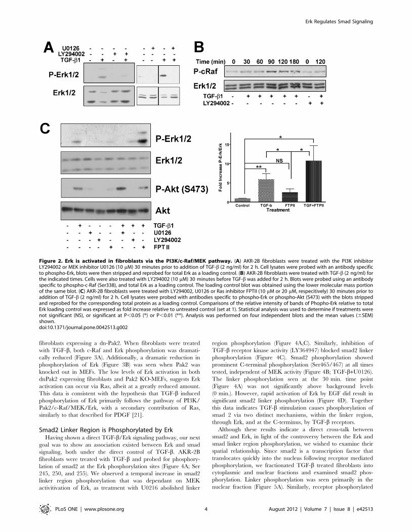
TGF-b Mechanism of Erk ActivationHaving shown Erk activation, via TGF-b, as a cell type specific
response, we next wanted to confirm the mechanism through
which the TGF-b signal was propagated. Previous studies have
shown that TGF-b induced activation of Pak2 and/or Ras
[10,12,13], we sought to identify the pathway resulting in Erk
activation. Using LY294002, a specific inhibitor of PI3K, Erk
phosphorylation was inhibited (Figure 2). Additionally, the
MEK1/2 inhibitor U0126, blocked Erk phosphorylation, demon-
strating that not only is PI3K necessary but also the MAPKK,
MEK is involved in TGF-b activation of Erk. There was also a
temporal increase in c-Raf phosphorylation (Figure 2B) dependent
on TGF-b stimulation and subsequent to PI3K activation as
shown by the phosphorylation of S338, a known Pak activation site
[34,35] and its inhibition by the PI3K inhibitor LY294002. In the
canonical Erk pathway, activated Ras initiates a cascade resulting
in Erk activation. Using small molecular weight inhibitors, we
evaluated the activation of two different PI3K activated pathways,
Erk and Akt (Figure 2C). Although both pathways were activated
by TGF-b through PI3K, only Erk phosphorylation was sensitive
to U0126 and neither pathway was inhibited by the Ras
farnysylation inhibitor, FPT II [36]. TGF-b induced Erk
phosphorylation was significantly increased, above control levels
(untreated and FTPII), with no significant difference found
between the two TGF-b treated groups (TGF-b alone and TGF-
b+FTPII).
Since inhibition of Ras farnysylation did not appear to effect
TGF-b induced Erk activation, and previous data had shown Pak
to be downstream of PI3K and phosphorylate Raf [34,35], we
wanted to determine where Pak2 acted within this signaling
pathway. Initially we assessed S338 phosphorylation of c-Raf in
Figure 1. Cell type specific activation of Erk. (A) AKR-2B and NIH 3T3 fibroblast were treated with TGF-b (2 ng/ml) for times ranging from 0 to4 h. Cell lysates were probed with antibodies specific to phospho-Erk (P-Erk). Blots were then stripped and reprobed for total Erk as a loading control.Typical results are shown representing four independent time course experiments. (B) Graph of densitometric analysis of western blots from timecourses from 0–3 h (n = 4) of phospho-Erk. Shown is the mean fold increase of Phospho-Erk relative to total Erk for each time point with the 0 time setas 1. Statistically significant change from 0 time is noted as, (*) P,0.05 and (**) P,0.01. (C) Mv1Lu and NMuMG epithelial cell lines were treated withTGF-b (2 ng/ml) for times ranging from 0 to 3 h. Cell lysates were probed with antibodies specific to phospho-Erk (P-Erk). Blots were then strippedand reprobed for total Erk as a loading control. Triplicate blots were performed on three independent time course experiments with typical resultsshown.doi:10.1371/journal.pone.0042513.g001
Erk Regulates Smad Signaling
PLoS ONE | www.plosone.org 3 August 2012 | Volume 7 | Issue 8 | e42513
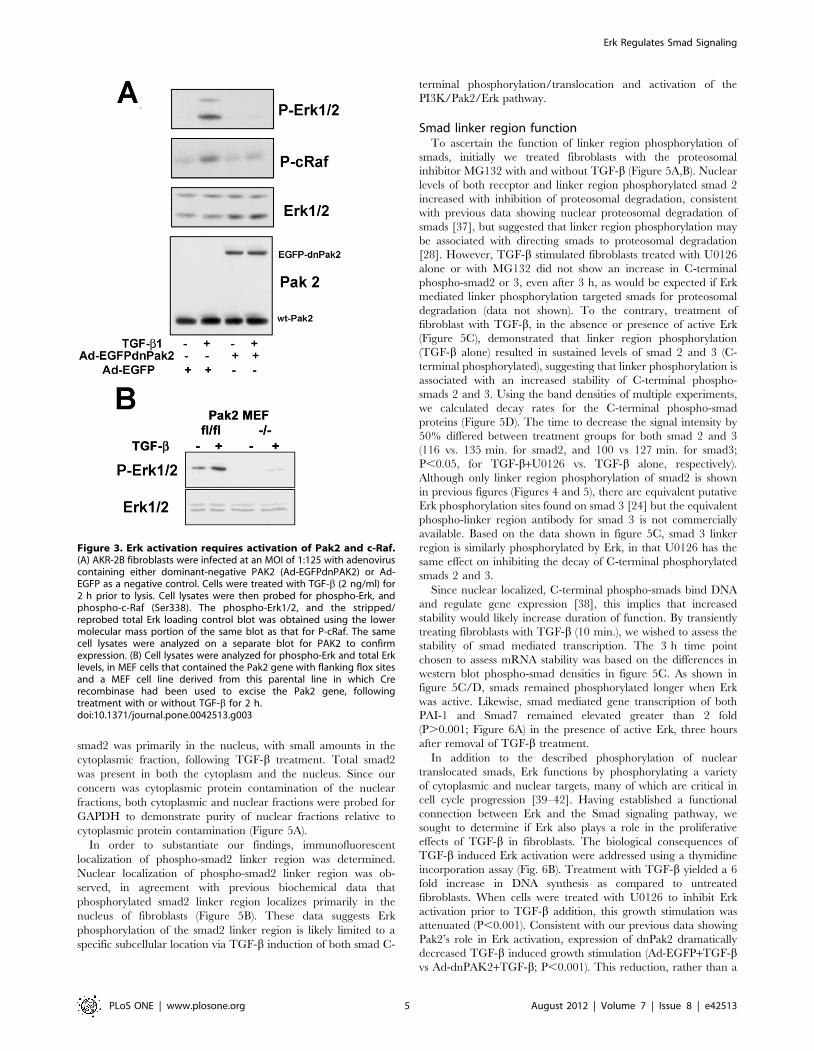
fibroblasts expressing a dn-Pak2. When fibroblasts were treated
with TGF-b, both c-Raf and Erk phosphorylation was dramati-
cally reduced (Figure 3A). Additionally, a dramatic reduction in
phosphorylation of Erk (Figure 3B) was seen when Pak2 was
knocked out in MEFs. The low levels of Erk activation in both
dnPak2 expressing fibroblasts and Pak2 KO-MEFs, suggests Erk
activation can occur via Ras, albeit at a greatly reduced amount.
This data is consistent with the hypothesis that TGF-b induced
phosphorylation of Erk primarily follows the pathway of PI3K/
Pak2/c-Raf/MEK/Erk, with a secondary contribution of Ras,
similarly to that described for PDGF [21].
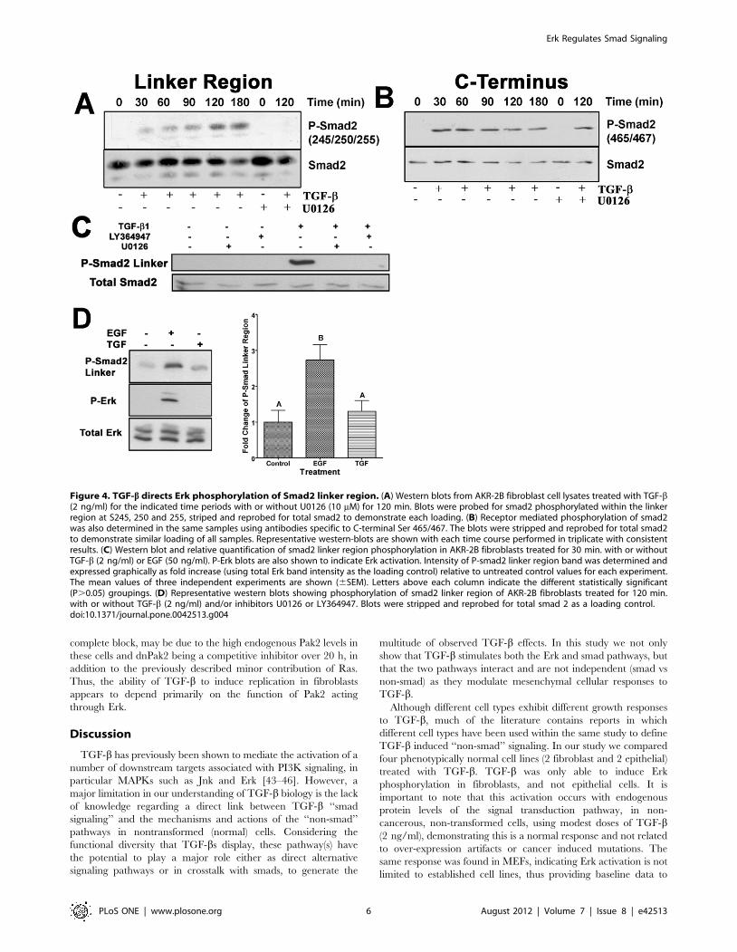
Smad2 Linker Region is Phosphorylated by ErkHaving shown a direct TGF-b/Erk signaling pathway, our next
goal was to show an association existed between Erk and smad
signaling, both under the direct control of TGF-b. AKR-2B
fibroblasts were treated with TGF-b and probed for phosphory-
lation of smad2 at the Erk phosphorylation sites (Figure 4A; Ser
245, 250, and 255). We observed a temporal increase in smad2
linker region phosphorylation that was dependant on MEK
activitivation of Erk, as treatment with U0216 abolished linker
region phosphorylation (Figure 4A,C). Similarly, inhibition of
TGF-b receptor kinase activity (LY364947) blocked smad2 linker
phosphorylation (Figure 4C). Smad2 phosphorylation showed
prominent C-terminal phosphorylation (Ser465/467) at all times
tested, independent of MEK activity (Figure 4B; TGF-b+U0126).
The linker phosphorylation seen at the 30 min. time point
(Figure 4A) was not significantly above background levels
(0 min.). However, rapid activation of Erk by EGF did result in
significant smad2 linker phosphorylation (Figure 4D). Together
this data indicates TGF-b stimulation causes phosphorylation of
smad 2 via two distinct mechanisms, within the linker region,
through Erk, and at the C-terminus, by TGF-b receptors.
Although these results indicate a direct cross-talk between
smad2 and Erk, in light of the controversy between the Erk and
smad linker region phosphorylation, we wished to examine their
spatial relationship. Since smad2 is a transcription factor that
translocates quickly into the nucleus following receptor mediated
phosphorylation, we fractionated TGF-b treated fibroblasts into
cytoplasmic and nuclear fractions and examined smad2 phos-
phorylation. Linker phosphorylation was seen primarily in the
nuclear fraction (Figure 5A). Similarly, receptor phosphorylated
Figure 2. Erk is activated in fibroblasts via the PI3K/c-Raf/MEK pathway. (A) AKR-2B fibroblasts were treated with the PI3K inhibitorLY294002 or MEK inhibitor U0126 (10 mM) 30 minutes prior to addition of TGF-b (2 ng/ml) for 2 h. Cell lysates were probed with an antibody specificto phospho-Erk, blots were then stripped and reprobed for total Erk as a loading control. (B) AKR-2B fibroblasts were treated with TGF-b (2 ng/ml) forthe indicated times. Cells were also treated with LY294002 (10 mM) 30 minutes before TGF-b was added for 2 h. Blots were probed using an antibodyspecific to phospho-c-Raf (Ser338), and total Erk as a loading control. The loading control blot was obtained using the lower molecular mass portionof the same blot. (C) AKR-2B fibroblasts were treated with LY294002, U0126 or Ras inhibitor FPTII (10 mM or 20 mM, respecitively) 30 minutes prior toaddition of TGF-b (2 ng/ml) for 2 h. Cell lysates were probed with antibodies specific to phospho-Erk or phospho-Akt (S473) with the blots strippedand reprobed for the corresponding total protein as a loading control. Comparisons of the relative intensity of bands of Phopho-Erk relative to totalErk loading control was expressed as fold increase relative to untreated control (set at 1). Statistical analysis was used to determine if treatments werenot significant (NS), or significant at P,0.05 (*) or P,0.01 (**). Analysis was performed on four independent blots and the mean values (6SEM)shown.doi:10.1371/journal.pone.0042513.g002
Erk Regulates Smad Signaling
PLoS ONE | www.plosone.org 4 August 2012 | Volume 7 | Issue 8 | e42513
smad2 was primarily in the nucleus, with small amounts in the
cytoplasmic fraction, following TGF-b treatment. Total smad2
was present in both the cytoplasm and the nucleus. Since our
concern was cytoplasmic protein contamination of the nuclear
fractions, both cytoplasmic and nuclear fractions were probed for
GAPDH to demonstrate purity of nuclear fractions relative to
cytoplasmic protein contamination (Figure 5A).
In order to substantiate our findings, immunofluorescent
localization of phospho-smad2 linker region was determined.
Nuclear localization of phospho-smad2 linker region was ob-
served, in agreement with previous biochemical data that
phosphorylated smad2 linker region localizes primarily in the
nucleus of fibroblasts (Figure 5B). These data suggests Erk
phosphorylation of the smad2 linker region is likely limited to a
specific subcellular location via TGF-b induction of both smad C-
terminal phosphorylation/translocation and activation of the
PI3K/Pak2/Erk pathway.
Smad linker region functionTo ascertain the function of linker region phosphorylation of
smads, initially we treated fibroblasts with the proteosomal
inhibitor MG132 with and without TGF-b (Figure 5A,B). Nuclear
levels of both receptor and linker region phosphorylated smad 2
increased with inhibition of proteosomal degradation, consistent
with previous data showing nuclear proteosomal degradation of
smads [37], but suggested that linker region phosphorylation may
be associated with directing smads to proteosomal degradation
[28]. However, TGF-b stimulated fibroblasts treated with U0126
alone or with MG132 did not show an increase in C-terminal
phospho-smad2 or 3, even after 3 h, as would be expected if Erk
mediated linker phosphorylation targeted smads for proteosomal
degradation (data not shown). To the contrary, treatment of
fibroblast with TGF-b, in the absence or presence of active Erk
(Figure 5C), demonstrated that linker region phosphorylation
(TGF-b alone) resulted in sustained levels of smad 2 and 3 (C-
terminal phosphorylated), suggesting that linker phosphorylation is
associated with an increased stability of C-terminal phospho-
smads 2 and 3. Using the band densities of multiple experiments,
we calculated decay rates for the C-terminal phospho-smad
proteins (Figure 5D). The time to decrease the signal intensity by
50% differed between treatment groups for both smad 2 and 3
(116 vs. 135 min. for smad2, and 100 vs 127 min. for smad3;
P,0.05, for TGF-b+U0126 vs. TGF-b alone, respectively).
Although only linker region phosphorylation of smad2 is shown
in previous figures (Figures 4 and 5), there are equivalent putative
Erk phosphorylation sites found on smad 3 [24] but the equivalent
phospho-linker region antibody for smad 3 is not commercially
available. Based on the data shown in figure 5C, smad 3 linker
region is similarly phosphorylated by Erk, in that U0126 has the
same effect on inhibiting the decay of C-terminal phosphorylated
smads 2 and 3.
Since nuclear localized, C-terminal phospho-smads bind DNA
and regulate gene expression [38], this implies that increased
stability would likely increase duration of function. By transiently
treating fibroblasts with TGF-b (10 min.), we wished to assess the
stability of smad mediated transcription. The 3 h time point
chosen to assess mRNA stability was based on the differences in
western blot phospho-smad densities in figure 5C. As shown in
figure 5C/D, smads remained phosphorylated longer when Erk
was active. Likewise, smad mediated gene transcription of both
PAI-1 and Smad7 remained elevated greater than 2 fold
(P.0.001; Figure 6A) in the presence of active Erk, three hours
after removal of TGF-b treatment.
In addition to the described phosphorylation of nuclear
translocated smads, Erk functions by phosphorylating a variety
of cytoplasmic and nuclear targets, many of which are critical in
cell cycle progression [39–42]. Having established a functional
connection between Erk and the Smad signaling pathway, we
sought to determine if Erk also plays a role in the proliferative
effects of TGF-b in fibroblasts. The biological consequences of
TGF-b induced Erk activation were addressed using a thymidine
incorporation assay (Fig. 6B). Treatment with TGF-b yielded a 6
fold increase in DNA synthesis as compared to untreated
fibroblasts. When cells were treated with U0126 to inhibit Erk
activation prior to TGF-b addition, this growth stimulation was
attenuated (P,0.001). Consistent with our previous data showing
Pak2’s role in Erk activation, expression of dnPak2 dramatically
decreased TGF-b induced growth stimulation (Ad-EGFP+TGF-bvs Ad-dnPAK2+TGF-b; P,0.001). This reduction, rather than a
Figure 3. Erk activation requires activation of Pak2 and c-Raf.(A) AKR-2B fibroblasts were infected at an MOI of 1:125 with adenoviruscontaining either dominant-negative PAK2 (Ad-EGFPdnPAK2) or Ad-EGFP as a negative control. Cells were treated with TGF-b (2 ng/ml) for2 h prior to lysis. Cell lysates were then probed for phospho-Erk, andphospho-c-Raf (Ser338). The phospho-Erk1/2, and the stripped/reprobed total Erk loading control blot was obtained using the lowermolecular mass portion of the same blot as that for P-cRaf. The samecell lysates were analyzed on a separate blot for PAK2 to confirmexpression. (B) Cell lysates were analyzed for phospho-Erk and total Erklevels, in MEF cells that contained the Pak2 gene with flanking flox sitesand a MEF cell line derived from this parental line in which Crerecombinase had been used to excise the Pak2 gene, followingtreatment with or without TGF-b for 2 h.doi:10.1371/journal.pone.0042513.g003
Erk Regulates Smad Signaling
PLoS ONE | www.plosone.org 5 August 2012 | Volume 7 | Issue 8 | e42513
complete block, may be due to the high endogenous Pak2 levels in
these cells and dnPak2 being a competitive inhibitor over 20 h, in
addition to the previously described minor contribution of Ras.
Thus, the ability of TGF-b to induce replication in fibroblasts
appears to depend primarily on the function of Pak2 acting
through Erk.
Discussion
TGF-b has previously been shown to mediate the activation of a
number of downstream targets associated with PI3K signaling, in
particular MAPKs such as Jnk and Erk [43–46]. However, a
major limitation in our understanding of TGF-b biology is the lack
of knowledge regarding a direct link between TGF-b ‘‘smad
signaling’’ and the mechanisms and actions of the ‘‘non-smad’’
pathways in nontransformed (normal) cells. Considering the
functional diversity that TGF-bs display, these pathway(s) have
the potential to play a major role either as direct alternative
signaling pathways or in crosstalk with smads, to generate the
multitude of observed TGF-b effects. In this study we not only
show that TGF-b stimulates both the Erk and smad pathways, but
that the two pathways interact and are not independent (smad vs
non-smad) as they modulate mesenchymal cellular responses to
TGF-b.
Although different cell types exhibit different growth responses
to TGF-b, much of the literature contains reports in which
different cell types have been used within the same study to define
TGF-b induced ‘‘non-smad’’ signaling. In our study we compared
four phenotypically normal cell lines (2 fibroblast and 2 epithelial)
treated with TGF-b. TGF-b was only able to induce Erk
phosphorylation in fibroblasts, and not epithelial cells. It is
important to note that this activation occurs with endogenous
protein levels of the signal transduction pathway, in non-
cancerous, non-transformed cells, using modest doses of TGF-b(2 ng/ml), demonstrating this is a normal response and not related
to over-expression artifacts or cancer induced mutations. The
same response was found in MEFs, indicating Erk activation is not
limited to established cell lines, thus providing baseline data to
Figure 4. TGF-b directs Erk phosphorylation of Smad2 linker region. (A) Western blots from AKR-2B fibroblast cell lysates treated with TGF-b(2 ng/ml) for the indicated time periods with or without U0126 (10 mM) for 120 min. Blots were probed for smad2 phosphorylated within the linkerregion at S245, 250 and 255, striped and reprobed for total smad2 to demonstrate each loading. (B) Receptor mediated phosphorylation of smad2was also determined in the same samples using antibodies specific to C-terminal Ser 465/467. The blots were stripped and reprobed for total smad2to demonstrate similar loading of all samples. Representative western-blots are shown with each time course performed in triplicate with consistentresults. (C) Western blot and relative quantification of smad2 linker region phosphorylation in AKR-2B fibroblasts treated for 30 min. with or withoutTGF-b (2 ng/ml) or EGF (50 ng/ml). P-Erk blots are also shown to indicate Erk activation. Intensity of P-smad2 linker region band was determined andexpressed graphically as fold increase (using total Erk band intensity as the loading control) relative to untreated control values for each experiment.The mean values of three independent experiments are shown (6SEM). Letters above each column indicate the different statistically significant(P.0.05) groupings. (D) Representative western blots showing phosphorylation of smad2 linker region of AKR-2B fibroblasts treated for 120 min.with or without TGF-b (2 ng/ml) and/or inhibitors U0126 or LY364947. Blots were stripped and reprobed for total smad 2 as a loading control.doi:10.1371/journal.pone.0042513.g004
Erk Regulates Smad Signaling
PLoS ONE | www.plosone.org 6 August 2012 | Volume 7 | Issue 8 | e42513

compare TGF-b responses in other cell backgrounds. Also, in our
studies fibroblasts were serum depleted, while epithelial cells were
not, since serum depletion induces senescence. This was done in
order to faithfully reflect the regulatory effects of TGF-b on each
individual cell type. As a known effector of cell replication, it is
interesting that Erk is activated in a cell type known to proliferate
in response to TGF-b, but not a growth inhibited cell type, when
using the appropriate cellular environment where these responses
would occur.
To initiate TGF-b activation of the Erk signaling pathway,
PI3K activation has been identified as necessary [12,13]. Here we
show that PI3K also acts downstream of the TGF-b receptor
complex to induce the activation of Erk, through Pak2 phosphor-
ylation of c-Raf at S338, a known site of Pak activation [34,35,47].
Unlike the canonical Ras/Raf/Erk pathway and data previously
describing Erk activation by TGF-b [13], our study showed c-Raf
activation appears to occur primarily through Pak2, similar to that
described for Epo and PDGF [20,21], and mimicing its budding
yeast homologue, Ste20, by acting as a MAP4K [15]. Since TGF-
b treatment of either Pak2-knockout or dnPak2 transfected cells
caused a dramatic reduction, but not a complete block, of c-Raf or
Erk phosphorylation or thymidine incorporation, this suggests that
Ras may still be involved, but play a minor role. Beeser et al. [21]
showed that neither EGF nor PDGF used only one pathway to
activate Erk, but had a preference for either Ras or Pak,
respectively. Our data concerrs with this, in that TGF-b appears
to have a preference for Pak2 over Ras, but the two pathways work
together to induce growth in fibroblasts through their coordinated
activation of Erk.
The linker region of smads is believed to be important in
regulating smad function [48,49]. Previous studies have addressed
the cross-talk between MAPKs and smad signaling [7,18,23],
however an unambiguous definition remains elusive, primarily due
to the lack of consistency in the cellular models used to define the
interaction between the pathways. Using both subcellular
fractionation and immunocytochemistry we demonstrated TGF-
b induced, Erk mediated linker phosphorylated smad2 is found in
the nucleus of fibroblasts. Although our data does not preclude the
possibility of cytoplasmic Erk phosphorylation of smad linker
region, followed by a rapid nuclear translocation, this seems unlike
in that previously cytoplasmic smad2 linker phosphorylation has
been shown to be associated with exclusion from the nucleus
[18,27]. Additionally, our data shows that in fibroblasts treated
with TGF-b, Erk is the primary kinase responsible for smad-linker
phosphorylation rather than CDK8 and 9 as shown in HaCat
epithelial cells [25], since U0126 was able to completely abolish
detectable smad2-linker phosphorylation. Kretzschmar et al. [18],
showed oncogenic Ras and EGF, through Erk, resulted in nuclear
Figure 5. Nuclear Smad levels are controlled by the proteasome and activated Erk. (A) AKR-2B fibroblasts were treated for 3 h with TGF-b(2 ng/ml) with or without MG132 (10 mM), 30 minutes prior to TGF-b addition. Nuclear and cytoplasmic fractions were isolated and probed for smad2linker region phosphorylation (245/250/255), or receptor phosphorylation (465/467). Linker phosphorylation blots were stripped and reprobed fortotal smad2 as a loading control, while receptor phosphorylated blots were stripped and reprobed for GAPDH to monitor the presence ofcytoplasmic protein in the nuclear fraction. (B) Photomicrographs of NIH 3T3 fibroblasts treated with TGF-b (2 ng/ml) for 3 h with or without MG132(10 mM) added 30 minutes prior to TGF-b treatment. Cells were incubated with phospho-smad2 (S245/250/255) linker antibody and specific immunecomplexes detected using Rhodamine X conjugated secondary antibody. (C) Cell lysates from AKR-2B fibroblasts pulsed for 10 minutes with TGF-b(2 ng/ml) with or without U0126 (10 mM) were probed for receptor phosphorylated smad2 and smad3. Blots were stripped and reprobed for b-actinas a loading control. (D) The density of phospho-smad2 and 3 bands for each time point relative to its b-actin control were determined. The meanvalues for each time point (n = 3 for smad 2, n = 4 for smad 3) are displayed with the solid line representing the curve for TGF-b+U0126 and the dottedline representing TGF-b treatment.doi:10.1371/journal.pone.0042513.g005
Erk Regulates Smad Signaling
PLoS ONE | www.plosone.org 7 August 2012 | Volume 7 | Issue 8 | e42513

exclusion of smad2/3, while others have shown [7,23] Erk activity
increased smad mediated transcription. Our results not only show
Erk mediated linker region phosphorylation increased the half-life
of receptor phosphorylated smad 2 and 3, but also an increase in
duration of smad transcriptional activity. The difference between
our data and Kretzchmar et al. [18] could be that our study
defined fibroblast TGF-b signaling under endogenous conditions,
while Kretzchmar et al. used epithelial cells and defined the
interactions of overexpressed Ras and EGF pre-treatment with
TGF-b signaling. Smads are rapidly phosphorylated through
receptor serine/threonine kinase activity (within 30 min as shown
in Figure 4B) and translocate into the nucleus, while TGF-bmediated Erk activation was much slower, resulting in smads being
in the nucleus prior to significant Erk activation (C-terminal
phospho-smads peak 30–60 min, while Erk is significantly above
background 60–90 min. and increases through 4 h). By activating
Erk, resulting in smad linker phosphorylation, either rapidly
through EGF (as shown in Figure 4D) or constitutively through
oncogenic Ras, Kretzchmar et al. may have changed the
subcellular location where the interaction between smads 2/3
and Erk occurs, potentially altering the function of linker region
phosphorylation to inhibiting smad2/3 from entering the nucleus
as suggested by the Agonist/Antagonist interaction concept for
Smad1 linker phosphorylation [25,50]. Additionally, this demon-
strates the complex effects of the growth factor mileu on a cell, in
Figure 6. Erk activity is integral for TGF-b signaling and inducing growth in fibroblasts. (A) Fold change in expression levels of two smadregulated genes, PAI-1 and smad7, relative to untreated controls levels are shown from AKR-2B fibroblasts treated for 10 minutes with or withoutTGF-b (2 ng/ml), with or without U0126 (10 mM) for 3 h. The mean values (6SEM) of six independent experiments are shown with statisticalevaluation indicating differences between groups as (NS) not significant, (*) P,0.05, (***) P,0.001. (B) Thymidine incorporation was determined inserum deprived NIH 3T3 cells treated with TGF-b (5 ng/ml) with or without infection with Ad-dnPAK2 or Ad-EGFP (MOI = 125:1) or U0126 (10 mM)prior to treatment. The effect of treatment is expressed as a fold change in thymidine incorporation relative to untreated cells (control = 1). The mean(6SEM) of triplicate assays is shown with statistical evaluation indicating differences between groups as previously describe. (C) Schematicrepresentation of TGF-b signaling pathways in fibroblasts, indicating the proposed interactions and their subcellular locations. Arrows represent afunctional link between the subsequent proteins, not necessarily a direct interaction.doi:10.1371/journal.pone.0042513.g006
Erk Regulates Smad Signaling
PLoS ONE | www.plosone.org 8 August 2012 | Volume 7 | Issue 8 | e42513
that a TGF-b/smad signal may be modulated by the presence of
other growth factors acting on the PI3K pathway.
In summary, as depicted in figure 6C, we propose the following
model; TGF-b induced phosphorylation of Erk begins with
TGFbR activation of the PI3K/Pak2 pathway. As described in
earlier studies [10,51,52], PI3K and smad activation differs in
their subcellular location. Smads require clathrin mediated
endocytosis prior to activation, while PI3K is activated from the
plasma membrane. Although our data does not eliminate the role
of Ras, it does indicate a subordinate role and the primary
mechanism of c-Raf activation is through Pak2. Pak2 then
activates c-Raf, followed by MEK and ultimately Erk. Nuclear
translocated, activated Erk is able to phosphorylate the linker
region of smad2/3 and increase their duration of transcriptional
activity. In addition, activated Erk is able to stimulate cellular
replication through its modulation of smad and other transcription
factor’s activity. The data presented here, defines the signaling
pathway by which TGF-b results in the activation of Erk, in
normal mesenchymal and not normal epithelial cells, thus
providing a baseline to examine these differences in transformed
cells. In addition, it clarifies and defines a role of Erk activation in
modulating smad signaling and the consequences of the cross-talk
between the two signaling pathways, suggesting that the concept of
smad-dependent and smad-independent signaling pathways may
be inappropriate. The findings that TGF-b/PI3K signaling in
fibroblasts can play a major role in direct regulation of smad
signaling indicates that TGF-b activates a complex, interacting
signaling web rather than discrete smad/non-smad pathways.
Author Contributions
Conceived and designed the experiments: CH JJD. Performed the
experiments: CH JJD. Analyzed the data: CH JJD. Contributed
reagents/materials/analysis tools: MR JJD. Wrote the paper: CH JJD.
References
1. Lin HY, Wang XF, Ng-Eaton E, Weinberg RA, Lodish HF (1992) Expression
cloning of the TGF-beta type II receptor, a functional transmembrane serine/
threonine kinase. Cell 68: 775–785.
2. Franzen P, Ten Dijke P, Ichijo H, Yamashita H, Schulz P, et al. (1993) Cloning
of a TGF beta type I receptor that forms a heteromeric complex with the TGF
beta type II receptor. Cell 75: 681–692.
3. Zhang Y, Feng X, We R, Derynck R (1996) Receptor-associated Mad
homologues synergize as effectors of the TGF-beta response. Nature 383:
168–172.
4. Zhang Y, Musci T, Derynck R (1997) The tumor suppressor Smad4/DPC 4 as a
central mediator of Smad function. Curr Biol 7: 270–276.
5. Macias-Silva M, Abdollah S, Hoodless PA, Pirone R, Attisano L, Wrana JL
(1996) MADR2 is a substrate of the TGFbeta receptor and its phosphorylation is
required for nuclear accumulation and signaling. Cell 87: 1215–1224.
6. Baker JC, Harland RM (1996) A novel mesoderm inducer, Madr2, functions in
the activin signal transduction pathway. Genes Dev 10: 1880–1889.
7. Blanchette F, Rivard N, Rudd P, Grondin F, Attisano L, Dubois CM (2001)
Cross-talk between the p42/p44 MAP kinase and smad pathways in
transforming growth factor b1-induced furin gene transactivation. J Biol Chem
276: 33986–33994.
8. Wang W, Zhou G, Hu MC, Yao Z, Tan TH (1997) Activation of the
hematopoietic progenitor kinase-1 (HPK1)-dependent, stress-activated c-Jun N-
terminal kinase (JNK) pathway by transforming growth factor beta (TGF-beta)-
activated kinase (TAK1), a kinase mediator of TGF beta signal transduction.
J Biol Chem 272: 22771–22775.
9. Kamaraju AK, Roberts AB (2005) Role of Rho/ROCK and p38 MAP kinase
pathways in transforming growth factor-beta-mediated Smad-dependent growth
inhibition of human breast carcinoma cells in vivo. J Biol Chem 280: 1024–
1036.
10. Wilkes MC, Murphy SJ, Garamszegi N, Leof EB (2003) Cell-type-specific
activation of PAK2 by transforming growth factor beta independent of Smad2
and Smad3. Mol Cell Biol 23: 8878–8889.
11. Wilkes MC, Repellin CE, Hong M, Bracamonte M, Penheiter SG, et al. (2009)
Erbin and the NF2 tumor suppressor merlin cooperatively regulate cell-type-
specific activation of PAK2 by TGF-b. Devel Cell 16: 433–444.
12. Wilkes MC, Mitchell H, Penheiter SG, Dore JJ, Suzuki K, et al. (2005)
Transforming growth factor-beta activation of phosphatidylinositol 3-kinase is
independent of Smad2 and Smad3 and regulates fibroblast responses via p21-
activated kinase-2. Cancer Res 65: 10431–10440.
13. Suzuki K, Wilkes MC, Garamszegi N, Edens M, Leof EB (2007) Transforming
growth factor beta signaling via Ras in mesenchymal cells requires p21-activated
kinase 2 for extracellular signal-regulated kinase-dependent transcriptional
responses. Cancer Res 67: 3673–3682.
14. Hofmann C, Shepelev M, Chernoff J (2004) The genetics of Pak. J Cell Sci 117:
4343–4354.
15. Dan I, Watanabe NM, Kusumi A (2001) The Ste20 group kinases as regulators
of MAP kinase cascades. Trends Cell Biol 11: 220–230.
16. Coles LC, Shaw PE (2002) PAK1 primes MEK1 for phosphorylation by Raf-1
kinase during cross-cascade activation of the ERK pathway. Oncogene 21:
2236–2244.
17. Park ER, Eblen ST, Catling AD (2007) MEK1 activation by PAK: a novel
mechanism. Cell Signal 19: 1488–1496.
18. Kretzschmar M, Doody J, Timokhina I, Massague J (1999) A mechanism of
repression of TGFbeta/Smad signaling by oncogenic Ras. Genes Dev 13: 804–
816.
19. Lo RS, Wotton D, Massague J (2001) Epidermal growth factor signaling via Ras
controls the Smad transcriptional co-repressor TGIF. EMBO J 20: 128–136.
20. Chen C, Sytkowski AJ (2004) Erythropoietin regulation of Raf-1 and MEK:
evidence for a ras-independent mechanism. Blood 104: 73–80.
21. Beeser A, Jaffer ZM, Hofmann C, Chernoff J (2005) Role of group A p21-activated kinases in activation of extracellular-regulated kinase by growth factors.
J Biol Chem 280: 36609–36615.
22. Kretzschmar M, Doody J, Massague J (1997) Opposing BMP and EGFsignalling pathways converge on the TGF-beta family mediator Smad1. Nature
389: 618–622.
23. Burch ML, Yang SNY, Ballinger ML, Getachew R, Osman N, Little PJ (2010)TGF-b stimulates biglycan synthesis via p38 and ERK phosphorylation of the
linker region of smad2. Cell Mol Life Sci 67: 2077–2090.
24. Matsurra I, Wang G, He D, Liu F (2005) Identification and characterization of
ERK MAP kinase phosphorylation sites in smad3. Biochem 44: 12546–12553.
25. Alarcon C, Zaromytidou A-I, Xi Q, Gao S, Yu J, et al. (2009) Nuclear CDKsdrive smad transcriptional activation and turnover in BMP and TGF-bpathways. Cell 139: 757–769.
26. Yue J, Frey RS, Mulder KM (1999) Cross-talk between the Smad1 and Ras/MEK signaling pathways for TGFbeta. Oncogene 18: 2033–2037.
27. Grimm OH, Gurbon JB (2002) Nuclear exclusion of smad 2 is a mechanismleading to loss of competence. Nat Cell Biol 4: 519–522.
28. Gao S, Alarcon C, Sapkota G, Rahman S, Chen P-Y, et al. (2009) Ubiquitin
ligase Nedd4L targets activated smad2/3 to limit TGF-b signaling. Mol Cell 36:457–468.
29. Liu Y, Dore JJE, Chen X (2007) Calcium influx through L-type channels
generates protein kinase M to induce burst firing of dopamine cells in the rateventral tegmental area. J Biol Chem 282: 8594–8603.
30. Wharton W, Leof E, Pledger WJ, O’Keefe EJ (1982) Modulation of the
epidermal growth factor receptor by platelet-derived growth factor andcholeragen: effects on mitogenesis. Proc Natl Acad Sci USA 79: 5567–5571.
31. Pfaffl MW (2001) A new mathematical model for relative quantification in real-time RT-PCR. Nucleic Acids Res 29:e45.
32. Guyton KZ, Liu Y, Gorospe M, Xu Q, Holbrook NJ (1996) Activation of
mitogen-activated protein kinase by H2O2. Role in cell survival followingoxidant injury. J Biol Chem 271: 4138–4142.
33. Jakobi R, Moertl E, Koeppel MA (2001) p21-activated protein kinase gamma-
PAK suppresses programmed cell death of BALB3T3 fibroblasts. J Biol Chem276: 16624–16634.
34. Diaz B, Barnard D, Filson A, MacDonald S, King A, Marshall M (1997)
Phosphorylation of Raf-1 serine 338-serine 339 is an essential regulatory eventfor Ras-dependent activation and biological signaling. Mol Cell Biol 17: 4509–
4516.
35. King AJ, Sun H, Diaz B, Barnard D, Miao W, et al. (1998) The protein kinase
Pak3 positively regulates Raf-1 activity through phosphorylation of serine 338.
Nature 396: 180–183.
36. Manne V, Ricca CS, Brown JG, Tuomari AV, Yan N, et al. (1995) Ras
farnesylation as a target for novel antitumour agents: Potent and selective
farnesyl diphosphate analog inhibitors of farnesyltransferase. Drug Devel Res 34:121–137.
37. Lo,R.S Massague,J. (1999). Ubiquitin-dependent degradation of TGF-beta-activated smad2. Nat Cell Biol 1, 472–478.
38. Massague J, Seoane J, Wotton D (2005) Smad transcription factors. Genes Dev
19: 2783–2810.
39. Gille H, Kortenjann M, Thomae O, Moomaw C, Slaughter C, et al. (1995)
ERK phosphorylation potentiates Elk-1-mediated ternary complex formation
and transactivation. EMBO J 14: 951–962.
40. Weber JD, Raben DM, Phillips PJ, Baldassare JJ (1997) Sustained activation of
extracellular-signal-regulated kinase 1 (ERK1) is required for the continued
expression of cyclin D1 in G1 phase. Biochem J 326: 61–68.
Erk Regulates Smad Signaling
PLoS ONE | www.plosone.org 9 August 2012 | Volume 7 | Issue 8 | e42513
41. Sears R, Leone G, DeGregori J, Nevins JR (1999) Ras enhances Myc protein
stability. Mol Cell 3: 169–179.42. Sears R, Nuckolls F, Haura E, Taya Y, Tamai K, Nevins JR (2000) Multiple
Ras-dependent phosphorylation pathways regulate Myc protein stability. Genes
Dev 14: 2501–2514.43. Mucsi I, Skorecki KL, Goldberg HJ (1996) Extracellular signal-regulated kinase
and the small GTP-binding protein, Rac, contribute to the effects oftransforming growth factor-beta1 on gene expression. J Biol Chem 271:
16567–16572.
44. Atfi A, Djelloul S, Chastre E, Davis R, Gespach C (1997) Evidence for a role ofRho-like GTPases and stress-activated protein kinase/c-Jun N-terminal kinase
(SAPK/JNK) in transforming growth factor beta-mediated signaling. J BiolChem 272: 1429–1432.
45. Bakin AV, Tomlinson AK, Bhowmick NA, Moses HL, Arteaga CL (2000).Phosphatidylinositol 3-kinase function is required for transforming growth factor
beta-mediated epithelial to mesenchymal transition and cell migration. J Biol
Chem 275: 36803–36810.46. Bhowmick NA, Ghiassi M, Bakin A, Aakre M, Lundquist CA, et al. (2001)
Transforming growth factor-beta1 mediates epithelial to mesenchymal transdif-ferentiation through a RhoA-dependent mechanism. Mol Biol Cell 12: 27–36.
47. Chaudhary A, King WG, Mattaliano MD, Frost JA, Diaz B, et al. (2000)
Phosphatidylinositol 3-kinase regulates Raf1 through Pak phosphorylation of
serine 338. Curr Biol 10: 551–554.
48. Inman GJ (2005) Linking smads and transcriptional activation. Biochem J
386:e1–e3.
49. Cohen-Solal KA, Merrigan KT, Chan JL-K, Goydos JS, Chen W, et al. (2011)
Constitutive smad linker phosphorylation in melanoma: a mechanism of
resistance to transforming growth factor-b-medicated growth inhibition.
Pigment Cell Melanoma Res 24: 512–524.
50. Sapkota G, Alarcon C, Spagnoli FM, Brivanlou AH, Massague J (2007)
Balancing BMP signaling through integrated inputs into smad1 linker. Mol Cell
25: 441–454.
51. Penheiter SG, Mitchell H, Garamszegi N, Edens M, Dore JJE, Leof EB (2002)
Internalization-dependent and -independent requirements for transforming
growth factor b receptor signaling via the smad pathway. Mol Cell Biol 22:
4750–4759.
52. Di Guglielmo GM, Le Roy C, Goodfellow AF, Wrana JL (2003) Distinct
endocytic pathways regulate TGF-beta receptor signalling and turnover. Nat
Cell Biol 5: 410–421.
Erk Regulates Smad Signaling
PLoS ONE | www.plosone.org 10 August 2012 | Volume 7 | Issue 8 | e42513