test directory - ok.gov - test directory 01-18... · 7/13/2018 · dengue virus (see...
TRANSCRIPT
Test Directory Last revised 07/13/2018
Table of Contents
Laboratory Information Hours of Operation Contacts Licensure and Accreditations
Specimen Collection Kits & Supplies Instructions for Specimen Collection Labeling Specimens
Test Requests Test Requisition Forms Verbal Requests Add-on Test Requests STAT Requests Medico-legal Test Requests Test Cancellations Referral Testing
Specimen Storage & Shipment Blood Tubes, CSF, Urine, Culture Tubes, Sputum, and Stool Specimens Newborn Screening, Sickle Cell and PKU Monitoring Forms Courier Service Shipping Regulations
Specimen Receipt at OSDH PHL Specimen Rejection Missing Information
Test Reports Issue of Test Reports Changes to Information on Test Reports Patient Access to Laboratory Test Results
Billing CPT Coding
Test List A
Acid-Fast Bacilli (see Mycobacteria) Arbovirus (see West Nile /St. Louis Viruses, Zika/Chikungunya/Dengue Viruses, or Zika
Virus) B
Babesia (see Parasites, Blood) Bacterial Isolate, Identification/Serotyping/Confirmation ►Go to page Biotinidase Deficiency (see Newborn Screening) Blood Parasites (see Parasites, Blood)
Bordetella - PCR ►Go to page
C Chagas’ Disease (see Parasites, Blood)
Carbapenem-resistance Testing ►Go to page
Chikungunya Virus (see Zika/Chikungunya/Dengue Viruses)
Chlamydia trachomatis/Neisseria gonorrhoeae – DNA Amplification ►Go to page
Congenital Adrenal Hyperplasia (CAH) (see Newborn Screening) Congenital Hypothyroidism (see Newborn Screening) Cystic Fibrosis (see Newborn Screening)
D Dengue Virus (see Zika/Chikungunya/Dengue Viruses)
E Enterohemorrhagic Escherichia coli (see Enteric Pathogens) Enteric Pathogens (see Gastrointestinal Pathogens or Ova and Parasites)
F Filaria (see Parasites, Blood)
Fungal Isolate Identification ►Go to page
Flu (see Influenza or Respiratory Pathogens Panel) G
Galactosemia (see Newborn Screening)
Gastrointestinal Pathogens, Isolation and Identification ►Go to page
German Measles (see Rubella) Gonorrhea (see Chlamydia trachomatis/Neisseria gonorrhoeae)
Group B Streptococcus, Isolation and Identification ►Go to page
H Hemoglobin (see Newborn Screening)
Hepatitis B Surface Antigen (HBsAg) – EIA w/ Reflex to Neutralization Test ►Go to page
Human Immunodeficiency Virus (HIV) - HIV-1/2 Antigen/Antibody EIA w/ Reflex to
HIV-1/HIV-2 Antibody Differentiation Test ►Go to page
Human Papillomavirus, High Risk – Transcription-mediated Amplification ►Go to page
I
Influenza Virus A and B - PCR (also see Respiratory Pathogens Panel) ►Go to page
M Malaria (see Parasites, Blood) Medium-chain Acyl-CoA Dehydrogenase Deficiency (MCAD) (see Newborn Screening) Microfilariae (see Parasites, Blood) Mold (see Fungal Isolate Identification) Mycobacteria (Acid-Fast Bacilli) – Smear and Culture with Reflex to Mycobacteria
Species Identification ►Go to page
Mycobacteria (Acid-Fast Bacilli), Isolate Identification ►Go to page
Mycobacterium tuberculosis Complex - PCR ►Go to page
Mycobacterium tuberculosis Complex, Antimicrobial Susceptibility - Culture ►Go to page
Mycology (see Fungal Isolate Identification) N
Neisseria gonorrhoeae/Chlamydia trachomatis – DNA Amplification ►Go to page
Newborn Screening ►Go to page Biotinidase Deficiency ►Go to page
Congenital Adrenal Hyperplasia ►Go to page
Congenital Hypothyriodism ►Go to page
Cystic Fibrosis ►Go to page
Galactosemia ►Go to page
Hemoglobinopathy (HGB) – Isoelectric Focusing ►Go to page
Fatty Acid Oxidation Disorders ►Go to page
Organic Acid Disorders ►Go to page
Amino Acid Disorders ►Go to page
Severe Combined Immunodeficiency (SCID) ►Go to page
Norovirus (see Gastrointestinal Pathogens) Norwalk Virus (see Gastrointestinal Pathogens)
O
Ova and Parasites (O&P), Fecal - Microscopic Examination ►Go to page
Organic Acid Disorders (see Newborn Screening) P
Parasites Parasites, Blood - Microscopic Examination and/or PCR ►Go to page
Gastrointestinal Pathogens, Isolation and Identification ►Go to page Ova and Parasites (O&P), Fecal - Microscopic Examination and/or PCR ►Go to page
Pertussis Syndrome (see Bordetella) Phenylketonuria (PKU) (see Newborn Screening) Plasmodium (see Parasites, Blood) Primary Immunodeficiency (see Newborn Screening, SCID)
R
Rabies – Direct Fluorescent Antibody ►Go to page
Rubella, Antibodies – Latex Particle Agglutination ►Go to page
Respiratory Pathogen Panel ►Go to page
S Severe Combined Immunodeficiency (see Newborn Screening, SCID) Shiga-like Toxin-Producing Escherichia coli (STEC) (see Gastrointestinal Pathogens) Sickle Cell Disease (see Newborn Screening, Hemoglobinopathy) Sleeping Sickness (see Parasites, Blood) Streptococcus agalactiae (see Group B Streptococcus) St. Louis Encephalitis Virus (see Arbovirus) Syphilis, Treponemal Screen with Reflex to RPR Screen with Titer and TP-PA
Confirmation, as Indicated ►Go to page
T Trypanosomes (see Parasites, Blood) Treponema (see Syphilis) Tuberculosis (see Mycobacteria)
W West Nile Virus (see Arbovirus) Whooping Cough (see Bordetella)
Y
Yeast (see Fungal Isolate Identification) Z
Zika/Chikungunya/Dengue Viruses – PCR ►Go to page
Zika Virus, Antibodies - ELISA ►Go to page
LABORATORY INFORMATION Back to Table of Contents
Hours of Operation
Monday – Friday, 8:00 am - 5:00 pm (Central Time)
The Client Services Department of the OSDH Public Health Laboratory (PHL) is staffed with experienced representatives who are dedicated to providing accurate, personal service. These individuals can help you with supply requests, courier information, as well as, providing copies of test results. If you have technical inquiries, please, inform the Client Services Department that you wish to talk to technical personnel and indicate the test for which you have questions; they will direct you to the appropriate laboratory section. Laboratory personnel are available to address your technical inquiries regarding availability of specific tests, specimen collection, storage and shipping, and patient test result availability and interpretation.
The OSDH PHL is also closed on the following holidays: New Year’s Day 1st January Martin Luther King, Jr. Day 3rd Monday of January President’s Day 3rd Monday of February Memorial Day Last Monday of May Independence Day 4th July Labor Day First Monday of September Veterans’ Day November 11th Thanksgiving Fourth Thursday & Friday of November Christmas 25th and 26th December
Generally, when a holiday falls on a non-workday — Saturday or Sunday — the OSDH will be closed on Monday (if the holiday falls on Sunday) or Friday (if the holiday falls on Saturday). Contacts
Mailing Address: Oklahoma State Department of Health Public Health Laboratory 1000 NE 10th Street Oklahoma City, Oklahoma 73117
Telephone: (405) 271-5070
Fax: (405) 271-4850
Email: [email protected]
Website: http://phl.health.ok.gov
Laboratory Director: S. Terence Dunn, PhD.
Federal Tax ID: 736017987

Licensure and Accreditations The OSDH PHL is accredited by the College of American Pathologists (CAP) Laboratory Accreditation Program and has CLIA (Clinical Laboratory Improvement Amendments) certification through CMS (Centers of Medicare and Medicaid Services).
Each laboratory is required to verify that reference laboratories utilized by the laboratory are CLIA-’88-certified for high-complexity testing in the applicable specialty/subspecialty for which testing services are sought. This requirement can be satisfied by obtaining copies of current CAP accreditation and/or CLIA certificates from each reference laboratory utilized by the referring laboratory. Since the OSDH PHL serves as a reference laboratory for Oklahoma County Health Departments and other institutions, we have made available a current copy of our CLIA certificate (#37D0656594) at the OSDH PHL website (see Public Health Laboratory/Test Directory) to allow submitting sites to comply with this regulation. If the OSDH PHL forwards submitted specimens to a reference laboratory for additional testing, there is no requirement for the submitting site to obtain verification of CLIA-’88 certification from those reference laboratories; submitting laboratories may rely on our accreditation to ensure compliance with this regulation.
SPECIMEN COLLECTION Back to Table of Contents
Kits/Supplies The OSDH PHL provides the following collection kits/supplies free-of-charge to OSDH County Health Department sites:
Ova and Parasite kit Enteric Bacteria kit TB/Sputum kit Viral kit Group B Streptococcus kit Pertussis kit CT/GC Urine Preservative Transport (UPT) Female Vaginal Swab kits
The following kits are offered to private clinics, hospitals, and physician offices for a small fee; check with the OSDH PHL Client Services Department at (405) 271-5070 during regular business hours to arrange delivery:
Ova and Parasite kit Enteric Bacteria kit TB/Sputum kit
Rabies kits for shipment of euthanized animals/animal heads for rabies virus testing at the OSDH PHL are made available free-of-charge to veterinarians and the public when there is suspicion of transmission of rabies to an animal or human. To order a rabies specimen shipping container, or to inquire about rabies testing, call the OSDH PHL Client Services Department at (405) 271-5070 during regular business hours. For consultation on animal bites and rabies risk, contact the OSDH Acute Disease Service Epidemiologist-On-Call at (405) 271-4060. If you suspect a case of human rabies, immediately contact the OSDH Disease and Prevention Services at (405) 271-4060.
Collection kits for Newborn screening and PKU monitoring (ODH #450 Newborn Screening Form) and adult/child hemoglobinopathy (sickle cell) screening (ODH #485 Child/Adult Sickle Cell Screening Kit) are available to birthing centers and other healthcare providers at no cost.
All clients can submit Supply Order Requests on-line at the OSDH PHL website (Forms, Supply Order Request). Supply Order Request Forms can also be faxed to the OSDH PHL Client Services Department at (405) 271-8755. Phone orders for supplies can also be made by calling the OSDH PHL Client Services Department at (405) 271-5070 during regular business hours. In addition, Oklahoma County Health Department sites can order supplies using the Inventory Supply System.
Supplies are shipped Monday through Thursday. Instructions for Specimen Collection Instructions for specimen collection are provided in the description of individual tests within this Test Directory. Further specifics regarding specimen collection can be obtained from technical staff of the OSDH PHL. Please, inform the OSDH PHL Client Services Department that you wish to talk to technical personnel and indicate the test for which you have questions; they will direct you to the appropriate laboratory section.
All specimens must be collected, labeled, transported, and processed according to procedures indicated in this Test Directory and/or kit instructions. Prior to collection of the specimen, review the appropriate container type, specimen volume, storage and shipping conditions, and any other special handling requirements needed for optimum analysis. If the guidelines for these processes are not met, the specimen may be rejected or the test results compromised.
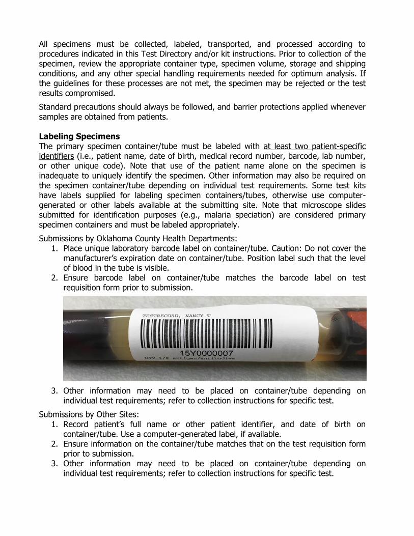
Standard precautions should always be followed, and barrier protections applied whenever samples are obtained from patients. Labeling Specimens The primary specimen container/tube must be labeled with at least two patient-specific identifiers (i.e., patient name, date of birth, medical record number, barcode, lab number, or other unique code). Note that use of the patient name alone on the specimen is inadequate to uniquely identify the specimen. Other information may also be required on the specimen container/tube depending on individual test requirements. Some test kits have labels supplied for labeling specimen containers/tubes, otherwise use computer-generated or other labels available at the submitting site. Note that microscope slides submitted for identification purposes (e.g., malaria speciation) are considered primary specimen containers and must be labeled appropriately.
Submissions by Oklahoma County Health Departments: 1. Place unique laboratory barcode label on container/tube. Caution: Do not cover the
manufacturer’s expiration date on container/tube. Position label such that the level of blood in the tube is visible.
2. Ensure barcode label on container/tube matches the barcode label on test requisition form prior to submission.
3. Other information may need to be placed on container/tube depending on individual test requirements; refer to collection instructions for specific test.
Submissions by Other Sites: 1. Record patient’s full name or other patient identifier, and date of birth on
container/tube. Use a computer-generated label, if available. 2. Ensure information on the container/tube matches that on the test requisition form
prior to submission. 3. Other information may need to be placed on container/tube depending on
individual test requirements; refer to collection instructions for specific test.

If multiple labels are used on a specimen, the last applied label will be used as the primary method of identification. It is good practice to place new specimen labels immediately beneath the name/identifier of the patient as it appears on the previous label such that the names/identifiers on both labels can be read.
For instructions on labeling collection kits for newborn screening and PKU monitoring (ODH #450 Newborn Screening Form) and adult/child hemoglobinopathy (Sickle Cell) screening (ODH #485 Child/Adult Sickle Cell Screening Kit), see the Newborn Screening section of this Test Directory.
TEST REQUESTS Back to Table of Contents
Test Requisition Forms All patient specimens submitted to the OSDH PHL for testing must be accompanied by an appropriately completed test requisition form. Submitters should use a test requisition form appropriate for the specimen being submitted:
Laboratory Requisition Form ODH #419 This form is used for submission of patient specimens/isolates other than those associated with newborn screening, PKU monitoring, or child/adult sickle cell screening. Oklahoma County Health Department sites are able to pre-populate this form electronically within the OSDH PHOCIS system; other sites can access this form at the OSDH PHL website.
Newborn Screening Form ODH #450 This form is used for submission of specimens for newborn screening, and can be ordered from the OSDH PHL Client Services Department at (405) 271-5070; also, see Kits/Supplies section of this Test Directory.
Sickle Cell Screening Form ODH #485 This form is used for screening of children/adults for sickle cell disease and can be ordered from the OSDH PHL Client Services Department at (405) 271-5070; also, see Kits/Supplies section of this Test Directory.
In addition, the OSDH PHL has forms for the submission of specimens for animal rabies testing and for submission of environmental swabs, food or water samples associated with foodborne outbreak events:
Rabies Submission Form OSD #460 This form is used for submission of specimens for rabies testing. Specimens may be submitted by a veterinarian, owner of the animal being tested, or a member of the public.
Oklahoma Foodborne Taskforce Sample Collection Sheet This form is available at the PHL website, or may be requested by phoning the OSDH PHL Client Services Department at (405) 271-5070.
Test requisition forms for patient testing must be completed in their entirety and contain the following information (CLIA Regulation 42 CFR 493.1241) prior to submission:
Patient’s name or unique patient identifier (e.g., MR#);
Patient’s sex;
Patient’s DOB or age;
Test(s) to be performed;
Source of the specimen, when appropriate;
Date and, if appropriate, time of specimen collection; Name and address or other suitable identifiers of the authorized person requesting
the test, and if appropriate the individual responsible for using the test results, OR name and address of the laboratory submitting the specimen, including, as applicable, a contact person to enable the reporting of imminently life threatening laboratory results or panic or alert values;
Any additional information relevant and necessary for a specific test to ensure accurate and timely testing and reporting of results, including interpretation, if applicable.
Note: Information provided on the test requisition form will be cross-referenced with information appearing on the last placed label on the specimen container/tube.
All patient-specific identifiers (i.e., patient name, date of birth, MR#, or other unique number or code) provided on the test requisition form must match exactly those provided on the specimen container/tube; any discordance will result in the specimen being deemed unsatisfactory for testing. Therefore, if the name on the requisition is spelled differently from that on the specimen (even by a single letter), it will be deemed unsatisfactory for testing. If a patient’s initials rather than full name are provided on the primary specimen container and the full name is provided on the test requisition, it will be deemed unsatisfactory for testing. Also, even if one patient-specific identifier is concordant between the requisition and specimen but another identifier is discordant, the specimen will be deemed unsatisfactory for testing. The PHL may contact the submitter to resolve such discrepancies in the identifiers provided on the test requisition and specimen.
Also, if other information (e.g., sex, DOC, time of collection) is discordant between the specimen container/tube and test requisition form that potentially affects the acceptability of the specimen, the submitter will be contacted for clarification.
Obvious inconsistencies between multiple labels on the same specimen container/tube will result in the specimen being deemed unsatisfactory for testing.
Information provided on the test requisition may be changed; however, the PHL will not change any information that is provided on a specimen. A corrected requisition form or other suitable documentation must be provided by the submitter before the specimen can be accepted for testing.
For instructions on completion of ODH #450 Newborn Screening Form (for newborn screening and PKU monitoring) and ODH #485 Child/Adult Sickle Cell Screening Kit (for adult/child Sickle Cell screening), see Newborn Screening section of this Test Directory. Verbal Requests The OSDH PHL does not accept verbal requests for initial testing; however, it does accept verbal requests for add-on testing to previously submitted specimens, as appropriate (see Add-on Test Requests section below). Add-on Test Requests Additional testing may be added subsequent to submission of an original test request, if volume of the original submitted specimen is adequate. Requests for add-on testing may be received verbally, but must be followed with a written request within 30 days of the verbal request. All verbal requests for add-on testing require test order ‘read-back’ to ensure accuracy. Additional testing may be delayed in the absence of a written request. A test report will not be issued for any additional requested testing in the absence of a written request. STAT Requests STAT testing is performed only on specimens to determine outbreak status as deemed necessary, and on all credible bioterrorism threat cases. STAT testing requests require notification and approval of the OSDH PHL prior to submission of the specimen.
Medico-legal Test Requests The OSDH PHL does not perform medico-legal testing. Test Cancellations Testing can only be cancelled by the original submitter of the specimen. This can be done verbally or in writing. A report will be issued indicating cancellation of the test. Referral Testing When indicated, the OSDH PHL will refer specimens to specific reference laboratories for additional testing. Test results from reference laboratories will be reported to submitting sites by the OSDH PHL.
Specimens submitted to the OSDH PHL for referral to another laboratory for testing (i.e., pass-through testing), including the Centers for Disease Control and Prevention (CDC), requires prior notification and approval by the OSDH PHL.
Specimens referred for testing to a reference laboratory should be submitted using the reference laboratory’s test requisition form, and collected, labeled, stored and shipped as instructed by the reference laboratory. A Specimen Referral Log should be used to track dates, and time as applicable, of collection, shipping and receipt of results for referred specimens. Prior to submitting specimens to a reference laboratory, the referring laboratory must obtain a copy, or have on-hand a copy, of the current CAP accreditation and/or CLIA certificate of the reference laboratory to verify that the reference laboratory is CLIA-’88 certified for testing in the applicable specialty/subspecialty.

SPECIMEN STORAGE AND SHIPMENT Back to Table of Contents
Following collection, specimens must be appropriately pre-processed and stored (as necessary) then packaged and shipped to ensure that they arrive at the OSDH PHL in a satisfactory state for testing. For detailed information on appropriate pre-processing, storage, packaging and shipping of samples for submission to the OSDH PHL for testing, refer to the individual test descriptions in this Test Directory. Some general guidance on these topics is provided below. Blood Tubes, CSF, Urine, Culture Tubes, Sputum, and Stool Specimens
1. Specimens must be packaged in a securely sealed, water-tight, primary container appropriate for the specimen being collected (e.g., blood tube, UPT tube, screw-capped plastic tube, etc.). This primary container must be appropriately labeled.
2. The primary container must be placed in a secondary container (zip-lock plastic biohazard bag) that is capable of being closed to form a water-tight seal.
The secondary container should contain an absorbent material (e.g., paper toweling or gauze) of sufficient volume to absorb the complete fluid contents of the primary container in case of leakage/breakage of the primary container.
OSDH PHL personnel will not open biohazard bags containing specimens that have leaked or broken during transit. If the specimen can be identified without opening the biohazard bag, a report will be issued to the submitter to inform them that the specimen will not be tested. If such specimens cannot be identified, a report will not be generated; therefore, submitting facilities should check their Specimen Referral Log at appropriate intervals to ensure test results have been received accordingly, and contact the OSDH PHL if results have not been received in the expected timeframe.
3. Multiple samples may be placed in a single secondary container but: The total volume of samples in one secondary container must not exceed 50
mL. Sputum samples (for tuberculosis testing), stool samples (for enteric and
parasite/ova testing), and samples for virus isolation testing should be separated from all other sample types; each specimen type should be packaged separately.
When possible, package specimens by test type requested, (e.g., all RPRs in one bag, all HIVs in one bag, etc.).
Specimens that require specific transporting conditions (ambient, refrigerated or frozen) must be packaged and shipped separately. It is strongly encouraged to mark the outside of the bag of all secondary containers with “Transport at Room Temperature”, “Transport Refrigerated” or “Transport Frozen”. If such specimens are sent via the OSDH PHL-contracted courier, the courier must be informed that the specimens are to be transported refrigerated on wet ice or frozen on dry ice, as appropriate. Alternatively, samples may be packed in insulated boxes containing cold packs at refrigerated temperatures (for samples requiring refrigerated temperatures during shipping) or with sufficient frozen ice packs or dry ice (for samples

that should remain frozen during transport) and given to the courier. Samples that should be transported frozen should be placed directly on frozen ice packs or dry ice to keep them frozen. Do not place specimens that should be transported at refrigerated temperatures directly on frozen ice packs; place cardboard or other insulator between ice packs and these specimens.
When shipping frozen specimens over long distances, it is best to use a combination of dry ice and frozen gel ice-packs; the gel ice-packs will remain frozen for a day or two after the dry ice has dissipated.
4. Requisitions should be placed in the outer pocket of the plastic biohazard bag. Do not wrap requisitions around individual specimens. Fold requisition such that the test request faces outward and can be read through the bag.
Newborn Screening, Sickle Cell and PKU Monitoring Forms For instructions on storage and shipping of collection kits for newborn screening, PKU monitoring, and adult/child hemoglobinopathy (sickle cell) screening, see Newborn Screening section of this Test Directory. Courier Service The OSDH PHL contracts with a courier service to pick-up specimens from OSDH County Health Departments and other sites for delivery to the central laboratory located in Oklahoma City. Courier pick-up occurs on a regular schedule Monday through Friday for most sites (weekend pick-up of newborn screening specimens occurs at most birthing facilities). Requests for non-scheduled specimen pick-ups must have prior approval; call the OSDH Client Services Division of the OSDH PHL at 1-405-271-5070. Pick-up occurs at approximately the same time each workday for an individual site. Specimens collected after the designated pick-up time will be picked-up from the site on the following workday. Accordingly, testing of these specimens will not begin until the day following pick-up, at the earliest. Such delays in testing should be considered especially when scheduling specimen collection from patients on Fridays; if the specimen is not picked-up on Friday, pick-up will be delayed until Monday and testing will not occur until Tuesday, at the earliest. For newborn screening specimens picked-up on weekends, testing begins on Monday or the next work day. Similar provisions should be made for holiday closings of the OSDH PHL. Specimens should be stored appropriately from the time of collection to the time of delivery to the OSDH PHL. Other Delivery Methods The OSDH PHL-contracted courier is the preferred method for delivery of specimens to the OSDH PHL; however, specimens can also be transported directly to the OSDH PHL via private courier, commercial courier (e.g., FedEx, UPS), or USPS. Note: these alternative services will not be able to deliver specimens to the OSDH on weekends. Shipping Regulations For shipping specimens, specimens should be packaged and labeled in compliance with applicable state, and federal, and international regulations covering the transport of clinical specimens and etiologic agents/infectious substances. Specific rules and
regulations set forth by the U. S. Department of Transportation (Code of Federal Regulations 49 (CFR 49) part 173.196, Category A infectious substances and part 173.199, Category B infectious substances) should be followed in order to ensure safe transport of potentially infectious substances.
A Category A infectious substance is capable of causing permanent disability, life-threatening or fatal disease in otherwise healthy humans or animals. A Category B infectious substance is an infectious substance that does not meet the criteria for inclusion in Category A.
According to The World Health Organization (WHO) Guidance on Regulations for the Transport of Infectious Substances, the proper designation for shipment of Category A substances is “UN 2814 – Infectious Substance, Affecting Humans” and that for Category B substances is “UN 3373 - Biological Substance, Category B”.
If samples are transported by air, the International Civil Aviation Organizations (ICAO) Technical Instructions for the Safe Transport of Dangerous Goods should be followed. The International Air Transportation Association (IATA) provides shipping procedures based on ICAO instructions for shipping hazardous materials by air. These can be found in packing instructions 620 for Category A infectious substances and packing instructions 650 Category B infectious substances.
Further information regarding shipping regulations and specific forms can be found at the OSDH PHL website under “Packaging and Shipping Specimens”.
SPECIMEN RECEIPT AT OSDH PHL Back to Table of Contents
Specimen Rejection Specimens will be rejected for the following reasons:
Inappropriate specimen (e.g., type; patient age; patient gender); Inappropriate specimen container or collection device/media; Insufficient volume for analysis (i.e., QNS); No or illegible patient name or other unique identifier on specimen container; No or illegible patient name or other unique identifier on requisition form; Inability to match at least one unique identifier between the test requisition form
and the specimen container due to absence or illegibility of others; No test requisition form; Inability to determine address or submitter ID for submitting laboratory/clinic (may
be able to obtain information by inquiry, if missing); Expired collection device/kit; Specimen received outside of timeframe appropriate for testing; Specimen handled improperly subsequent to collection (e.g., improper temperature
during specimen shipment; specimen container leaked, broke or otherwise compromised during shipping);
Laboratory accident (e.g., spilled sample during accessioning); Other reasons as outlined in the individual test descriptions of this Test Directory.
All specimens deemed unsatisfactory for testing will have a final report generated stating the reason. Missing Information When any of the following information is missing from the test requisition form or specimen container, or is otherwise illegible or unclear (e.g., orders are non-specific or non-standard), the submitter will be contacted by the OSDH PHL, as appropriate:
DOB or age, if appropriate; Time of birth (NBS only); DOC; TOC (NBS only; and only if less than one day difference between date of birth and
date of collection); Sex (not required for NBS unless 2nd tier CAH testing is required); Address of submitter; Test requested; Source of specimen, if appropriate.
A Request for Missing Information Form will be sent to the submitting site. Specimens with missing information will be held for 7 days or until they have exceeded the appropriate time for testing, whichever is shorter. If missing information is not received from the submitting site within 7 days, the specimen will be deemed unsatisfactory for testing and reported as “Unsatisfactory: Information requested, not received”.
Referred specimens for enteric bacteria testing with missing information and for which requested information is not received, will be tested by the OSDH PHL and test results will be communicated to the OSDH Acute Diseases Division for epidemiological purposes only.

The submitter will receive a final report that indicates “Unsatisfactory for testing” and will not receive test results, unless deemed necessary.

TEST REPORTS Back to Table of Contents
Issue of Test Reports Reports are issued via fax and/or US Mail to the submitting facility/healthcare provider using the information provided on the test requisition form. Clients must complete a Facsimile Permission Form prior to receiving reports by fax. To sign-up for this service, call the PHL Client Services Department at (405) 271-5070.
OSDH County Health Departments are able to retrieve patient test reports directly using the PHOCIS system.
Newborn Screening results are accessible to authorized healthcare providers through the Newborn Screening Results web-based portal. To sign-up for this service, call the PHL Client Services Department at (405) 271-5070.
Some test results may be reported by telephone to authorized clients (facilities/healthcare providers). HIV test results and abnormal NBS results are not provided by telephone by the OSDH PHL. All results conveyed by telephone require “read-back” confirmation by the client.
No test reports may be picked-up at the OSDH PHL location in Oklahoma City.
Requests for access to test results for patient specimens tested by the OSDH PHL from non-submitting facilities/healthcare providers will be denied. Such requests should be made directly to the submitting facility/healthcare provider. Changes to Information on Test Reports Corrections to test reports, subsequent to original issue of test results to the submitter, may be made by the OSDH PHL, as appropriate. The Corrected Report will indicate the information being changed (i.e., with explicit indication of “from” and “to”) and the need for the change, as appropriate. A request will be made to the submitting facility for the original report to be returned to the OSDH PHL or for it to be destroyed by the submitting facility.
Changes to information presented in a test report may be made at the request of an authorized individual from the submitting facility. Please, call the Client Services Department of the OSDH PHL at 405-271-5070 and ask to speak to the supervisor of the laboratory responsible for performing the test. Verbal requests for changes to information provided on test reports must be followed by a written request within 30 days. A report will not be issued on such specimens until a written request from the submitting facility is provided to the OSDH PHL. The laboratory supervisor and/or Client Services Department will provide the necessary forms for completion by the submitter in order to fulfill such requests.
Requests for changes in demographic information following issue of a test report will result in issue of a Corrected Report; the information being changed (with explicit indication of “from” and “to”), the person requesting information to be changed, and date/time of the request will be indicated on the Corrected Report. A request will be made to the submitting facility for the original report to be returned to the OSDH PHL or for it to be destroyed by the submitting facility.

The OSDH PHL will not change the name or other unique identifier of a patient on a test report unless the name/unique identifier indicated on the test requisition form or specimen container has been misinterpreted by OSD PHL staff during data entry or a typographical error occurs.
Patient Access to Laboratory Test Results On February 6th, 2014, the Centers for Medicare & Medicaid Services (CMS) published a final rule that amended both the Clinical Laboratory Improvement Amendments (CLIA) and the Health Insurance Portability and Accountability Act (HIPAA) in order to provide patients with direct access to laboratory test results. Under the final rule, laboratories that operate as covered entities under HIPAA are required to provide individual patients, or their representatives, with laboratory test results for those tests performed by the laboratory upon the patient's request.
The OSDH PHL in Oklahoma City is unable to provide laboratory test results directly to individuals presenting at this location. Patients, or their legal representatives, may obtain copies of their laboratory test reports for testing performed at the OSDH PHL by presenting at the County Health Department or other health care facility where medical care was provided. The patient, or their legal representative, will be asked to complete an Oklahoma Standard Authorization Form, provide a photo ID and/or authorization code prior to release of laboratory test results.
Alternatively, patients, or their legal representatives, can contact the OSDH PHL Client Services Department at (405) 271-5070 to obtain laboratory test reports through the mail or electronically. A patient, or their legal representative, may request laboratory test results performed by the OSDH PHL by completing a Request to Release Laboratory Test Results Form.

BILLING Back to Table of Contents
CPT Coding The OSDH PHL has provided CPT codes for testing that it performs for guidance purposes only. These codes reflect our interpretation of the coding requirements. CPT coding is the sole responsibility of the billing party. Individual facilities should contact the OSDH PHL for information regarding testing methodology and the local Medicare carrier for clarification, as appropriate.

TEST LIST Back to Table of Contents
Individual tests are listed alphabetically in the subsequent pages. Be aware that individual tests may be named differently from that expected, which may make it difficult to find information for a specific test. Please, refer to the Test List in the Table of Contents at the beginning of this Test Directory where some alternative names have been listed for you convenience.
Bacterial Isolate, Identification/Serotyping/Confirmation
Use: Identification of unusual bacterial isolates and the characterization and surveillance of organisms of public health concern.
Methodology: Isolates are identified using conventional microbiological and molecular tests.
Clinical Significance:
Referred isolates can cover a broad range of bacteria, including known common pathogens, rare opportunistic pathogens, and new and emerging pathogens. These organisms can cause a wide range of infections, ranging from abscesses to meningitis. Accurate identification of these isolates assists in the development of appropriate treatment plans and public health intervention strategies as necessary.
Further background information, fact sheets, statistics and educational resources may be found at the OSDH Acute Disease Services website.
Specimen: Type: Pure isolate
Volume: Minimum of 1 slant or plate, visible growth
Container: Petri plate; Slant
Collection: Primary specimens should be collected according the submitting institution’s policies
Interferences: None
Special
Instructions: Aerobes: Chocolate agar plates/slants or blood agar plates/slants Anaerobes: Chocolate agar plates/slants, blood agar plates/slants,
thioglycollate or chopped meat media in anaerobic atmosphere Microaerophiles: Microaerophiles, such as Campylobacter, should
be submitted on blood agar plates/slants in a microaerophilic atmosphere
Isolates that require special growth media, such as Legionella species, should be submitted on appropriate media
Incubate all isolates for 18-24 hours prior to shipping
Shipping: Ambient temperature in appropriate atmosphere
Rejection Criteria: Media expired No growth on media Specimen frozen Other criteria as outlined in Specimen Rejection section of this Test
Directory
Reported: Within 3–28 working days from receipt unless referred to CDC for further characterization, which may delay availability of final results
CPT Codes: CPT codes will vary depending on organism identified and methods used
Normal/Abnormal Organism identified (Genus or Genus/species, targeted genes,
Results: serotype, as applicable); Unable to identify isolate, referred to CDC for further testing
Interpretation: The identification of unusual bacterial species should be used in conjunction with patient symptomology to determine appropriate course of treatment. Detection of stx1 and/or stx2 in E. coli isolates indicates the presence of a shiga-like toxin-producing E. coli (STEC) strain. Non-O157 STEC strains that produce only stx2 are more often associated with HUS than strains that produce only stx1 or both stx1 and stx2.
Limitations: Some isolates may not be successfully identified to the genus/species level. Such isolates will be referred to the CDC for further characterization, which may delay availability of a final report.
Notes: These are laboratory-developed tests; performance characteristics have been validated and determined to be suitable for diagnostic purposes by the OSDH PHL. These tests have not been cleared or approved by the U.S. Food and Drug Administration.
Back to Table of Contents
Bordetella – PCR
Use: For the diagnosis of pertussis syndrome (whooping cough) in children with consistent epidemiological and clinical features of disease. Also, appropriate for adults with persistent cough in whom Bordetella pertussis infection is suspected.
Methodology: This assay targets three insertion sequences, IS481, pIS1001 and hIS1001 of Bordetella spp. using real-time, multiplexed PCR. B. pertussis or B. parapertussis-positive cases are reflexed to a second PCR that targets the ptxS1 gene that encodes the S1 subunit of pertussis toxin. Presence/absence of combinations of these sequences allows for differentiation between B. pertussis, B. parapertussis and B. holmesii. While this test may detect B. bronchiseptica, it cannot differentiate it from other Bordetella spp. CDC recommends B. pertussis culture concurrently.
Clinical Significance:
Pertussis is an upper respiratory tract infection caused by B. pertussis bacteria. It is a serious disease that can cause permanent disability in infants, and even death. In previously vaccinated children and adults in whom immunity has waned, symptoms can be mild or absent. Since adenovirus, parainfluenza viruses, CMV, Mycoplasma pneumoniae, and Chlamydia pneumoniae can also cause pertussis-like coughing, rapid and accurate diagnosis is needed to guide therapy.
Further background information, fact sheets, statistics and educational resources may be found at the OSDH Acute Disease Services website.
Specimen: Type: Nasopharyngeal swab (with flexible, fine-shaft and nylon, rayon or
Dacron tip)
Volume: Swabs: 1 or 2
Container: Regan Lowe transport media (keep refrigerated)
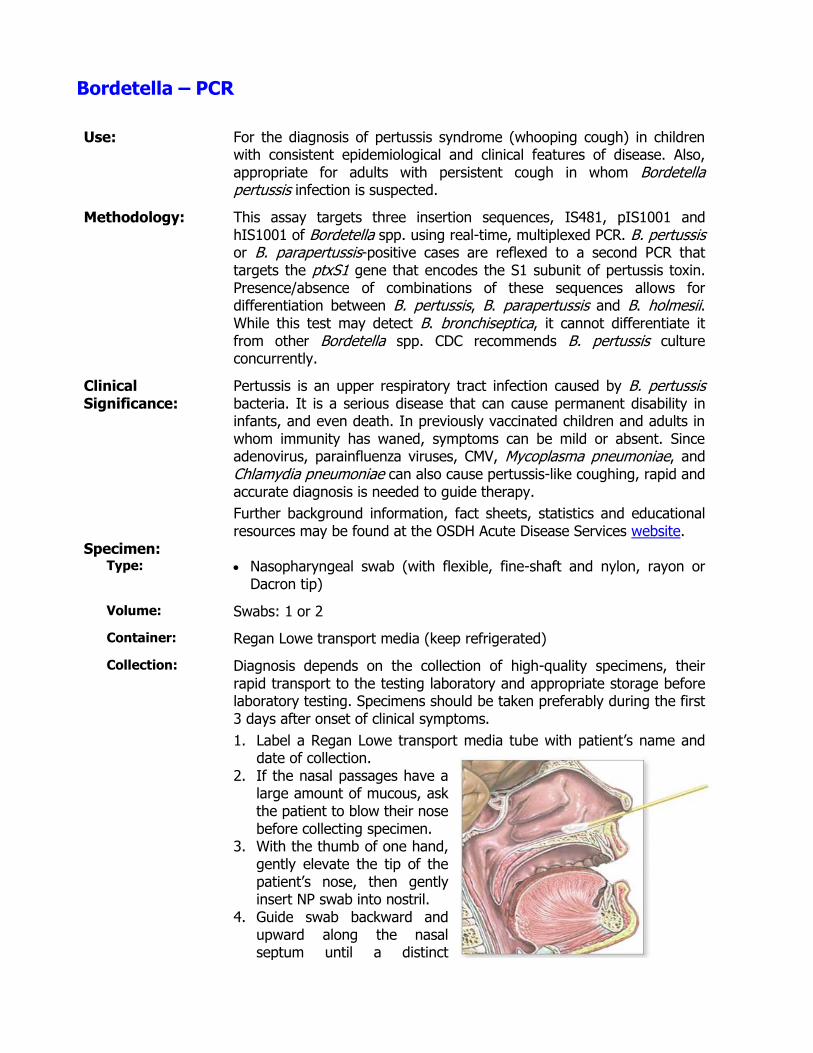
Collection: Diagnosis depends on the collection of high-quality specimens, their rapid transport to the testing laboratory and appropriate storage before laboratory testing. Specimens should be taken preferably during the first 3 days after onset of clinical symptoms.
1. Label a Regan Lowe transport media tube with patient’s name and date of collection.
2. If the nasal passages have a large amount of mucous, ask the patient to blow their nose before collecting specimen.
3. With the thumb of one hand, gently elevate the tip of the patient’s nose, then gently insert NP swab into nostril.
4. Guide swab backward and upward along the nasal septum until a distinct
resistance is met. 5. Hold it there for a few seconds then with a rotating motion gently
remove it. 6. Place swab immediately into Regan Lowe transport media,
positioning the swab about half way into media. 7. Break-off or cut excess shaft of swab so that tube can be capped. 8. Place tube in plastic biohazard bag and insert completed requisition
form in outer pouch of the bag.
Interferences: Cotton swab; Calcium alginate swab (shown to inhibit PCR)
Special Instructions:
Sampling patients as early as possible in infection is recommended. Incubate NP swabs at ambient temperature for 18-24 hours prior to
shipping at refrigerated temperature (2-8oC) or ship immediately at ambient temperature.
Contact OSDH Acute Disease Services at (405) 271-4060 regarding all suspected Bordetella pertussis cases.
It is extremely important to record date of onset, date of collection (DOC) and physician contact information on requisition form.
Shipping:
Refrigerated at 2-8oC (alternatively, ship immediately at ambient temperature); must be received within 7 days from DOC
Rejection Criteria: Incorrect media Incorrect collection device, e.g. cotton or calcium alginate swab Incorrect shipping temperature NP swab not submerged/present in transport media Specimen received > 7 days after DOC Other criteria as outlined in Specimen Rejection section of this Test
Directory
Reported: Within 5 working days from receipt
CPT Codes: 87801
Normal/Abnormal Results:
Bordetella pertussis/parapertussis/holmesii not detected Bordetella pertussis/parapertussis/holmesii detected (individually or in combinations)
Interpretation: Specimens that are positive for IS481 and ptxS1 indicate presence of B. pertussis, those positive for pIS1001 and ptxS1 indicate presence of B. parapertussis, and those with IS481 and hIS1001 products indicate B. holmesii. Dual infections of B. pertussis and B. parapertussis are indicated when IS481, pIS1001 and ptxS1 are detected.
Limitations:
A negative result does not preclude the presence of Bordetella spp. infection. The results of this test should not be used as the sole basis for diagnosis or patient management decisions. Positivity of this test may be variable following treatment.
Notes: This is a laboratory-developed test; performance characteristics have been validated and determined to be suitable for diagnostic purposes by the OSDH PHL. This test has not been cleared or approved by the U.S. Food and Drug Administration.

Back to Table of Contents
Carbapenem-resistance Testing
Use: Confirmation and characterization of carbapenem-resistant Enterobacteraceae (CRE), Pseudomonas aeruginosa (CRPA), and Acinetobacter spp. (CRA) isolates from Oklahoma healthcare facilities for epidemiological purposes. These tests are intended as an aid for infection control of carbapenem-non-susceptible organisms in healthcare settings. They are not intended to guide or monitor treatment for carbapenem-non-susceptible bacterial infections.
Methodology: Submitted isolates are initially subjected to MALDI-ToF mass spectrometry to confirm species, and then undergo Antimicrobial Sensitivity Testing (AST) using a broth microdilution method (SensititreTM System) and a Modified Carbapenemase Inactivation Method (mCIM) to confirm phenotypic carbapenem resistance. Isolates demonstrating positive or indeterminate carbapenemase activity by mCIM are reflexed to molecular PCR detection of KPC, NDM, VIM, IMP and OXA-48 antimicrobial resistance genes using the Cepheid Xpert® Carba-R IVD.
Clinical Significance:
Carbapenemase-producing Enterobacteriaceae, Pseudomonas aeruginosa and Acinetobacter spp. are a growing public health concern. They are often resistant to all beta-lactam agents and can be co-resistant to multiple classes of other antimicrobial agents. Identifying isolates that produce carbapenemase and classifying the kind of carbapenemase present is important in preventing their spread.
Further background information, fact sheets, statistics and educational resources may be found at the OSDH Acute Disease Services website.
Specimen: Type: Pure isolate of confirmed or suspected carbapenem-resistant
Enterobacteriaceae grown for 18-24 h on Trypticase Soy Agar with 5% Sheep’s Blood (BAP). o It is recommended that isolates are restricted to Escherichia
coli, Klebsiella oxytoca, Klebsiella pneumoniae, and Enterobacter spp. that are resistant to imipenem, meropenem, or doripenem (each with MICs of ≥ 4 μg/mL), or ertapenem (≥ 2 μg/mL) by standard susceptibility testing methods.
Pure isolate of confirmed or suspected carbapenem-resistant Pseudomonas aeruginosa that is resistant to imipenem, meropenem or doripenem by standard susceptibility testing methods (MIC ≥8 µg/mL). Mucoid CRPA isolates should be excluded.
Pure isolate of confirmed or suspected carbapenem-resistant Acinetobacter spp. o It is recommended that isolates are restricted to A. baumannii
or A. baumannii complex that are resistant to imipenem, meropenem, or doripenem (each with MICs of ≥ 8 μg/mL) by standard susceptibility testing methods.
Pure isolate of confirmed or suspected CRE that exhibits pan-resistance to all tested carbapenems by standard antimicrobial

susceptibility methods.
Volume: Minimum of 1 plate, visible growth
Container: Petri plate
Collection: Primary specimens should be collected according to the submitting institution’s standard procedure.
Interferences: None
Special Instructions:
Submit isolates on a blood agar plate or MacConkey agar plate Incubate all isolates in appropriate atmosphere for 18-24 hours
prior to shipping
Shipping: Ambient temperature in appropriate atmosphere
Rejection Criteria: Media expired No growth on media Specimen non-viable Specimen frozen Mucoid CRPA isolates Other criteria as outlined in Specimen Rejection section of this Test
Directory
Reported: Within 4 working days from receipt unless referred to the Antimicrobial Resistance Laboratory Network (ARLN) Regional Laboratory or CDC for further characterization, which may delay availability of final results. Results of the SensititerTM AST are for epidemiological purposes only and are not reported to the submitter.
CPT Codes: CPT codes will vary depending on organism identified and methods used
Normal/Abnormal Results:
MALDI-ToF-MS: Genus/species identified; Isolate could not be identified to species level, carbapenem resistance testing not performed mCIM: Positive; Negative; Indeterminate Xpert CarbaR PCR: [KPC, NDM, VIM, IMP or OXA-48] antimicrobial resistance genes detected; no antimicrobial resistance genes detected
Isolates demonstrating a potentially new carbapenemase variant or novel mechanism of resistance or isolates that produce discordant results may be forwarded to an ARLN Regional Laboratory or the CDC for further testing.
Interpretation: Isolates demonstrating carbapenem resistance by phenotypic AST and mCIM with a positive result for one or more resistance genes are confirmed carbapenem resistant due to the presence of one or more carbapenemases. Isolates demonstrating carbapenem resistance by AST and mCIM with a negative result for resistance genes are confirmed carbapenem resistant potentially due to the presence of a new variant carbapenemase or other novel mechanism of resistance. Isolates demonstrating carbapenem sensitivity by AST but resistance by mCIM with a positive result for one or more resistance genes could potentially harbor a carbapenemase with low activity, e.g., OXA-48.
Isolates demonstrating carbapenem sensitivity by AST and mCIM are confirmed carbapenem sensitive.
Limitations: Discordant results are expected between the different methods. Hydrolysis of carbapenem by carbapenemases is the most common mechanism of resistance for this class of antibacterial agents but other mechanisms of resistance occur and may not be detected by PCR. A carbapenemase may be weakly expressed producing a negative phenotypic test or the gene may be present in low copy numbers producing a negative PCR. Phenotypic antimicrobial susceptibility tests demonstrate variable sensitivities and specificities and use different combinations of antibiotics and inhibitors. Isolates that are not successfully identified to the genus/species level will not be tested. Isolates that are negative for both mCIM and AST will not have further testing performed.
Notes: These tests are intended as an aid for infection control of carbapenem-non-susceptible organisms in healthcare settings. These tests are not intended to guide or monitor treatment for carbapenem-non-susceptible bacterial infections. The mCIM is a laboratory-developed test; performance characteristics have been validated and determined to be suitable for diagnostic purposes by the OSDH PHL. The Bruker MALDI-ToF mass spectrometry and Xpert® Carba-R assay are approved for in vitro diagnostic use by the U.S. Food and Drug Administration.
Back to Table of Contents
Chlamydia trachomatis / Neisseria gonorrhoeae - DNA Amplification
Use: To screen symptomatic or asymptomatic males and females for the presence of Chlamydia trachomatis and/or Neisseria gonorrhoeae (CT/GC). This test is not to be used for monitoring therapeutic efficacy.
Methodology: BD ProbeTec® ET Chlamydia trachomatis and Neisseria gonorrhoeae Amplified DNA Assays; Strand Displacement Amplification (SDA). The C. trachomatis assay is able to detect the new variant strain, nvCT.
Clinical Significance:
Chlamydia trachomatis and Neisseria gonorrhoeae are the most common bacterial causes of sexually transmitted diseases in the U.S. Screening reduces the prevalence of CT/GC and potentially reduces the incidence of severe and debilitating complications associated with symptomatic infections.
Further background information, fact sheets, statistics and educational resources may be found at the OSDH Acute Disease Services website.
Specimen: Type: Urine
Vaginal Swab
Volume: 2.5-3.0 mL Dry Vaginal Swab in Transport Tube
Container: Urine Preservative Transport (UPT) tube Vaginal Specimen Transport Device
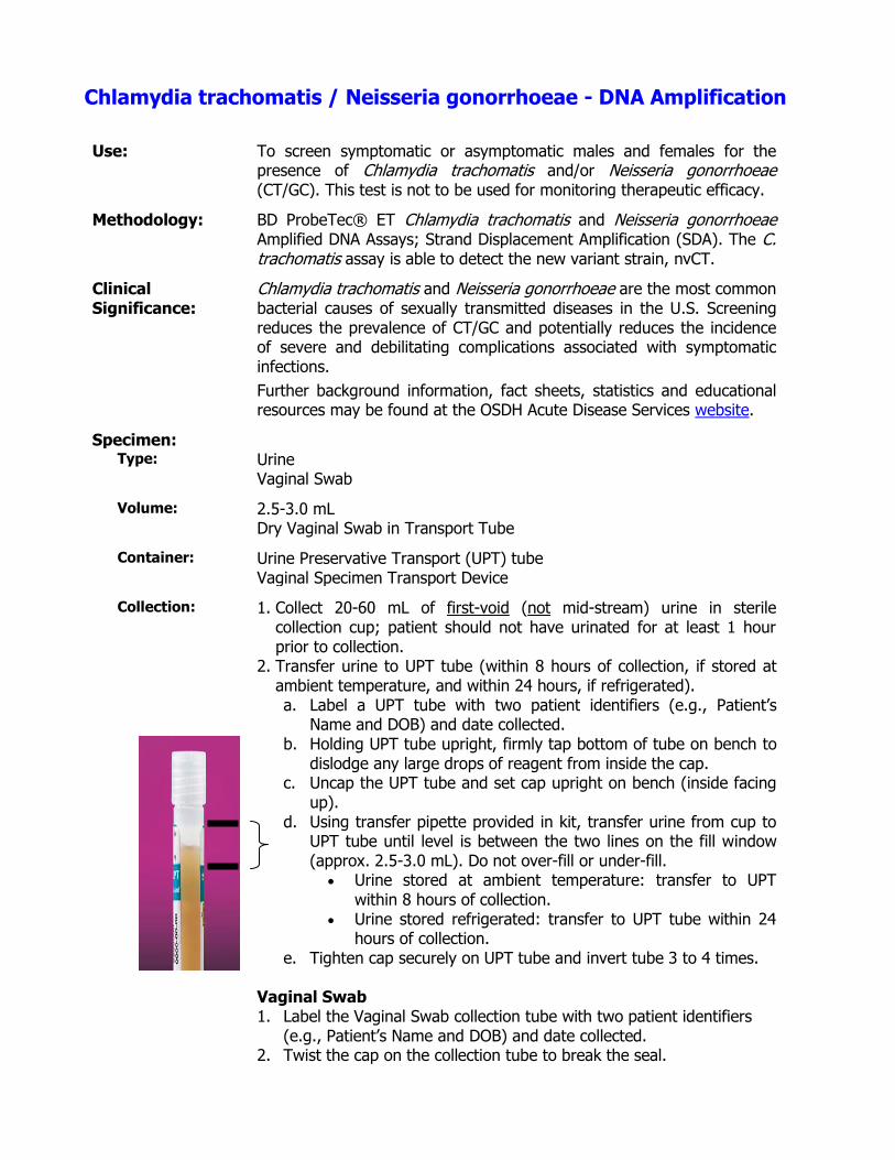
Collection: 1. Collect 20-60 mL of first-void (not mid-stream) urine in sterile collection cup; patient should not have urinated for at least 1 hour prior to collection.
2. Transfer urine to UPT tube (within 8 hours of collection, if stored at ambient temperature, and within 24 hours, if refrigerated). a. Label a UPT tube with two patient identifiers (e.g., Patient’s
Name and DOB) and date collected. b. Holding UPT tube upright, firmly tap bottom of tube on bench to
dislodge any large drops of reagent from inside the cap. c. Uncap the UPT tube and set cap upright on bench (inside facing
up). d. Using transfer pipette provided in kit, transfer urine from cup to
UPT tube until level is between the two lines on the fill window (approx. 2.5-3.0 mL). Do not over-fill or under-fill.
Urine stored at ambient temperature: transfer to UPT within 8 hours of collection.
Urine stored refrigerated: transfer to UPT tube within 24 hours of collection.
e. Tighten cap securely on UPT tube and invert tube 3 to 4 times.
Vaginal Swab 1. Label the Vaginal Swab collection tube with two patient identifiers
(e.g., Patient’s Name and DOB) and date collected. 2. Twist the cap on the collection tube to break the seal.
3. Pull the cap with attached swab off the tube. Do not touch tip or lay swab down on any surface.
4. Spread the skin around the patient’s vaginal opening, and gently insert the tip of the swab no more than 2 inches into the vaginal opening. The tip of the swab should be pointed towards the lower back of the patient. If the swab does not slide easily into the vaginal opening, rotate the swab while gently pushing.
5. While inserted, rotate the swab for 10-15 seconds. 6. Carefully, withdraw the swab, avoiding any contact with the skin or
other surfaces. 7. Immediately, place the swab in the collection tube and tightly secure
the cap.
Interferences: None
Special
Instructions: None
Shipping: Store and ship UPT or Vaginal Specimen Transport Device at refrigerated or ambient temperatures (2-30oC) for delivery within 30 days of collection for urine or within 7 days of collection for vaginal swab.
Rejection Criteria: Specimen collected in container other than UPT tube or Vaginal Specimen Transport Device
Over-filled or under-filled UPT tube Raw urine UPT specimens submitted > 30 days from date of collection Dry vaginal swabs submitted > 7 days from date of collection Other criteria as outlined in Specimen Rejection section of this Test
Directory
Reported: Within 7 working days from receipt
CPT Codes: 87491, 87591
Normal/Abnormal Results:
Chlamydia trachomatis Not Detected Chlamydia trachomatis Detected Neisseria gonorrhoeae Not Detected Neisseria gonorrhoeae Detected
Interpretation: A negative result does not preclude C. trachomatis and/or N. gonorrhoeae infection since detection is dependent on adequate specimen collection, absence of inhibitors, and sufficient levels of organisms
A positive result does not infer viability and/or infectivity for C. trachomatis and/or N. gonorrhoeae since target DNA for these organisms may persist in the patient in the absence of viable organisms (e.g., following anti-microbial therapy)
Limitations:
This assay is not appropriate for testing of cases of sexual assault/abuse or cases with other medico-legal implications. Culture is the recommended procedure for diagnosing CT/GC infections in medico-legal cases, testing of conjunctival, rectal and nasopharyngeal
specimens, and evaluating gonorrhea treatment failure; UPTs and Vaginal Specimen Transport Devices are inappropriate for culture. Test results may be affected by improper specimen collection, low levels of organisms in the sample, plasmid-free variants of C. trachomatis, technical error, specimen mix-up, or concurrent antibiotics. This test cannot be used to assess therapeutic success or failure.
Notes: This test has been cleared for in vitro diagnostic use by the U.S. Food and Drug Administration.
Back to Table of Contents
Enteric Pathogens, Isolation and Identification
Use: Isolation and identification of Salmonella spp., Shigella spp., Escherichia coli 0157, non-0157 shiga-like toxin-producing E. coli (STEC), enterotoxigenic E. coli (ETEC), Campylobacter spp., Yersinia spp., Vibrio spp., Aeromonas spp., Plesiomonas shigelloides, Bacillus cereus, Staphylococcus aureus, Adenovirus, Norovirus, Rotavirus A, Cryptosporidium, and Giardia lamblia in clinical stool samples from individuals exhibiting signs and symptoms of infectious colitis or gastroenteritis. While Clostridium difficile may be detected, results for this organism are not reported due to its high frequency in asymptomatic individuals.
Methodology: Specimens are initially screened using a PCR amplification assay (xTAG® Gastrointestinal Pathogen Panel (GPP)) that detects multiple bacterial, viral and protozoan enteric pathogens. All specimens negative by GPP are subject to limited culture to screen for potential common enteric pathogens not detected by GPP. Attempts are made to confirm GPP-positive specimens by routine microbiological procedures, including culture, biochemical, molecular, and/or serotyping analyses, as necessary; however, confirmation of certain GPP-positive results (e.g., ETEC, rotavirus, Norovirus) will be beyond the testing capabilities of the OSDH PHL and may require specimen referral by the submitter, as clinically appropriate. Isolated E. coli are checked for the presence of shiga toxin I (stx1) and/or II (stx2) genes. Isolates positive for stx1 and/or stx2 are then serotyped.
Clinical Significance:
The CDC estimates that each year 48 million Americans develop foodborne illnesses that result in 128,000 hospitalizations and 3000 deaths, and cost the U. S. economy an estimated 8.4 billion dollars in lost productivity. Considering the large impact on public health, it is critically important that these infections be identified and the isolates characterized as quickly as possible. Prompt identification of the causal agent can not only aid in diagnosis and implementation of individual patient management plans, but also ultimately reduce the number of infections in outbreak situations.
Further background information, fact sheets, statistics and educational resources may be found at the OSDH Acute Disease Services website.
Specimen: Type: Solid or liquid feces
Volume: Feces: Solid, 2 grams; Liquid, 5-10 mL GN broth: Visible growth
Container: Cary-Blair Transport Media (enteric kit); GN Broth
Collection: Stool specimens should be obtained early in the course of illness, optimally 1-3 days after onset of illness, and during early morning hours when the causative agent should be present in the greatest numbers. No more than three stool specimens, collected 24 hours apart, should be submitted for initial culture. Stool specimens should be collected

before antimicrobial therapy has been initiated. Test-of-cure stools should be collected 24 hours after completion of antibiotic treatment. 1. Collect the stool specimen in a wide mouth container, bedpan, clean
newspaper or plastic bag placed over the toilet seat opening. 2. DO NOT pass the specimen into the toilet or directly into the
collection vial. Do NOT mix urine or water with the sample. 3. Open the Carey-Blair vial then using the collection spoon attached to
the cap, add enough specimen (walnut-size portion, approximately 2 grams or 5-10 mL) until the liquid reaches the arrow on the label.
4. Important: Sample areas of the specimen which appear bloody, slimy, or watery. If the stool is hard, sample from each end and the middle of the specimen.
5. Mix the contents of the vial with the collection spoon. 6. Replace the cap tightly and shake the tube vigorously. 7. Fill-in the information on the vial label(s):
Patient name Date and time specimen was collected Specimen number if more than one specimen was collected
(e.g., No1, No2, No3) Check the appropriate box for specimen consistency (formed,
watery, bloody, loose or soft); orange-capped Meridian containers only
8. Make sure the vial(s) is closed tightly. 9. Place the vial(s) in the original package and seal. 10. Ship at ambient temperature, as soon as possible.
Interferences: Stool specimens should not be collected using laxatives, mineral oil, bismuth or barium compounds for purgation. Stools should not be collected immediately following the use of anti-diarrheal or antacid compounds.
Special Instructions:
Rectal swabs and raw stools are not acceptable
Shipping: Ambient temperature for delivery within 7 days from collection date
Rejection Criteria:
Specimen not in Cary-Blair Transport Media or GN broth Specimen received > 7 days from date of collection Other criteria as outlined in Specimen Rejection section of this Test
Directory
Reported: Within 10 working days; a preliminary report based on GPP is issued within 4 working days from receipt of specimen, followed by a final report within a further 6 working days upon completion of culture and other testing
CPT Codes: GPP: 87801; Culture: 87045, 87046 (x3), 87077; STEC genotyping 87797, included as relevant; CPT codes will vary depending on organism identified and methods used
Normal/Abnormal Results:
Pathogen Detected (Genus or Genus/species/targeted genes/serotype, as relevant); Pathogen Not Detected
Interpretation: Results from the GPP assay alone are considered presumptive and
require confirmation by other laboratory methods.
Laboratory confirmation of pathogens in fecal specimens from symptomatic individuals is evidence of fecal-oral contamination via food, water, fomites or the hands. Positive results do not rule-out co-infection with other, potentially clinically-relevant, pathogens not detected by this test. Detection of stx1 and/or stx2 in E. coli isolates indicates the presence of an STEC strain. Non-O157 STEC strains that produce only stx2 are more often associated with HUS than strains that produce only stx1 or both stx1 and stx2. Negative results do not exclude the diagnosis of enteric pathogens.
Limitations: Specimen type is not suitable for microscopic parasite examination
Notes: These are laboratory-developed tests; performance characteristics have been validated and determined to be suitable for diagnostic purposes by the OSDH PHL. These tests have not been cleared or approved by the U.S. Food and Drug Administration.
Back to Table of Contents

Fungal Isolate Identification
Use: Speciation of mold, yeast and some aerobic actinomycete isolates.
Methodology: Isolates are identified by morphological, physiological and biochemical tests. Histoplasma capsulatum, Blastomyces dermititidis, and Coccidioides immitis are identified by DNA probes. Yeast isolates are identified using substrate assimilation tests. Susceptibility testing of mycology specimens is not performed.
Clinical Significance:
Fungal infections have emerged as a significant clinical problem in recent years. Due to the increasing frequency of fungal infections, mycology identification is important for diagnosis and the possible need for treatment.
Specimen: Type: Solid cultured isolate
Volume: Visible growth
Container: Slant or sealed plate
Collection: N/A
Interferences: Impure isolate
Special Instructions:
If organism is a suspected H. capsulatum, B. dermititidis, or C. immitis, indicate on requisition form
Shipping: Store and ship at ambient temperature
Rejection Criteria: Mixed or contaminated specimen Isolates submitted on dehydrated media Frozen specimen Raw (clinical) specimen Other criteria as outlined in Specimen Rejection section of this Test
Directory
Reported: 1-30 days from receipt depending on organism
CPT Codes: 87106 (yeast), 87107 (mold), 87149 (H. capsulatum, B. dermititidis, or C. immitis), 87077 (aerobic actinomycete)
Normal/Abnormal Results:
Genus and Species Isolate Non-Viable, Unable to Culture Unidentified Non-sporulating Mold
Interpretation: Genus and species of organism identified Isolate non-viable, unable to culture: isolate fails to demonstrate
growth within 30 days Unidentified non-sporulating mold: molds that fail to produce conidia
or other characteristic features will not be identifiable
Limitations:
If isolate cannot be identified by techniques available, the isolate will be sent to the CDC for further testing.
Notes: Analyses may involve both laboratory-developed tests and tests that are

approved for in vitro diagnostic use by the U.S. Food and Drug Administration; performance characteristics have been validated and determined to be suitable for diagnostic purposes by the OSDH Public Health Laboratory.
Back to Table of Contents
Group B Streptococcus (Streptococcus agalactiae), Isolation and Identification
Use: Screening for Group B Streptococcus (GBS), also known as beta hemolytic streptococci or Streptococcus agalactiae, is routinely recommended at 35-37 weeks for pregnant women.
Methodology: GBS are isolated utilizing selective and non-selective media, and identified by catalase and latex antigen agglutination tests
Clinical Significance:
Streptococcus agalactiae is an important cause of maternal and neonatal infections and sepsis. Neonatal infections occur in about 1 in 1,000 live births, and present as two different clinical entities. Early-onset neonatal disease is characterized by sepsis and pneumonia within the first 7 days of life. Late-onset neonatal disease is characterized by meningitis and sepsis between day 7 and 3 months. Invasive GBS infections during pregnancy may also lead to preterm labor or stillbirth. The most important risk factor for the development of neonatal disease is the colonization of the maternal urogenital or gastrointestinal tracts.
This colonization occurs in about 10 to 30% of pregnant women and vertical transmission from mother to neonate occurs in about 75% of cases; therefore, it is critically important to provide rapid and accurate detection of the GBS in pregnant women. Expert groups recommend that all pregnant women have a GBS swab culture at 35 to 37 weeks of pregnancy to determine the need for intrapartum antibiotic prophylaxis (IAP). The CDC 2010 Guidelines for the Prevention of GBS also recommend IAP for:
Women who delivered a previous infant with GBS disease Women with GBS bacteriuria in the current pregnancy Women with a GBS-positive screening result in the current pregnancy Women with unknown GBS status who deliver at less than 37 weeks’
gestation, have an intrapartum temperature of 100.4°F or greater, or have rupture of membranes for 18 hours or longer.
Further background information, fact sheets, statistics and educational resources may be found at the OSDH Acute Disease Services website.
Specimen: Type: Vaginal/anal swab
Volume: One or two swabs
Container: Todd Hewitt with Colistin Nalidixic Acid (CNA) (LIM broth) – GBS Kit
Collection: Specimen Collection: GBS colonization status should be determined by collecting both vaginal and rectal specimens at 35-37 weeks' gestation. A single combined vaginal-rectal specimen can be collected. Swabbing both the lower vagina and rectum (through the anal sphincter) increases the culture yield substantially compared with sampling the cervix or the vagina without also swabbing the rectum.
1. Label the specimen transport tube with the patient’s name and date

of collection. 2. Swab the vaginal mucosa (high in the vaginal canal), followed by
the rectum (i.e., insert swab through the anal sphincter approximately 1 inch beyond the anal crypts and withdrawing) using the same swab or two different swabs. Do not use speculum with lubricant. Do not use a speculum for culture collection.
3. Place the swab(s) into LIM tube and break-off the swab shaft near the top of the tube. Leave enough of the shaft so that the swab(s) can be easily removed by the microbiologist.
4. Replace and secure the lid on the LIM tube. 5. Fill-out the Test Requisition Form ODH #419.
Specimen Transport: 1. Wrap the transport tube with the packing material provided in the
GBS kit and place tube in the plastic bag. 2. Place the completed requisition form in the outer pouch of the
plastic bag. 3. Place bag with specimen and requisition form in the mailing
container and mail using the green mailing label provided in the kit.
Interferences: Not Applicable
Special
Instructions: GBS collection kits (provided by OSDH PHL) must be refrigerated
upon arrival. Antimicrobial susceptibility testing should be performed on antenatal
GBS isolates from penicillin-allergic women at high risk for anaphylaxis because of a history of anaphylaxis, angioedema, respiratory distress, or urticaria following administration of penicillin or cephalosporin.
Test available only to County Health Department sites; other sites require pre-approval (call Microbiology Laboratory at 405-271-5070)
Shipping: Ambient temperature for delivery within 48 hours of collection (do not collect on Fridays)
Rejection Criteria: Specimen not collected using Todd Hewitt w/ CNA media (LIM broth) No swab in broth Received > 48 hours after collection Expired media Other criteria as outlined in Specimen Rejection section of this Test
Directory
Reported: Within 3 working days from receipt
CPT Codes: 87081
Normal/Abnormal Results:
GBS Detected GBS Not Detected
Interpretation: Cultures positive for GBS indicate the need for intrapartum antibiotic prophylaxis to prevent mother-to-newborn transmission. If the patient is penicillin-allergic, or is at high risk for anaphylaxis associated with GBS prophylaxis, antimicrobial susceptibility testing is recommended to ensure effectiveness of alternative therapy.

Limitations:
Culture at 35-37 weeks' gestation will fail to detect some women with intrapartum GBS colonization; therefore, this test should not be used as the sole deciding factor for administering antibiotic prophylaxis.
This test should not be used for the detection of sexually transmitted diseases.
Notes: These are laboratory-developed tests; performance characteristics have been validated and determined to be suitable for diagnostic purposes by the OSDH PHL. These tests have not been cleared or approved by the U.S. Food and Drug Administration.
Back to Table of Contents
Hepatitis B Surface Antigen (HBsAg) – EIA with Reflex to Neutralization Test
Use: This test is useful in the differential diagnosis of hepatitis. Typically, this test is only available for maternity patients from County Health Departments in Oklahoma; pre-approval is required for other patients.
Methodology: HBsAg Screen: Enzyme Immunoassay (EIA) using monoclonal antibodies to hepatitis B virus (HBV) surface antigen (HBsAg). Specimens with reactive screen results are repeated in duplicate using the same EIA. Repeatedly reactive specimens are reflexed to a neutralization confirmatory assay.
Clinical Significance:
HBV is a major public health problem world-wide, infecting approximately 350 million individuals in the world and about one million in the United States. Most adults are able to clear the virus naturally, but about 5% will develop chronic hepatitis B, which may lead to cirrhosis, acute liver failure, and hepatocellular carcinoma. However, acute HBV infections are frequently asymptomatic or are associated with mild malaise and fever, and symptoms of chronic infections progress insidiously over several decades, which often delays a diagnosis and potentially perpetuates transmission. Transmission of HBV occurs principally by exposure to contaminated blood or body secretions. In infected individuals, the virus can be found in the blood, semen, vaginal secretions, breast milk, and saliva. In the United states, sexual contact is the most common means of transmission, followed by sharing of needles used for injecting illicit drugs, tattooing, body piercing, acupuncture, and occupational exposure of medical personnel. The risk of vertical transmission of HBV from an infected mother to the fetus during pregnancy is about 90%, and is the most prevalent means of transmission in regions of the world where HBV is endemic. Also, the risk of an infected infant developing chronic hepatitis B is about 90%. Fortunately, transmission can be significantly reduced through immunopropyllaxis. Women who have been infected with HBV can receive medications during pregnancy or at delivery to reduce the likelihood of transmitting these infections to their newborn.
The presence of HBsAg in the blood indicates that the patient is currently infected with HBV. HBsAg is detected in the blood, on average, four weeks after initial exposure. Individuals who recover from acute infections clear the blood of HBsAg within approximately four months after onset of symptoms. These individuals develop antibodies to HBsAg (anti-HBs) that provide complete immunity to subsequent hepatitis B infection. Similarly, individuals who are successfully vaccinated against HBV, produce anti-HBs. Chronically infected patients are those that have HBsAg present in the blood for at least six months. In chronic hepatitis B, HBsAg can be detected for many years, and anti-HBs do not appear.
Specimen: Type: Whole blood collected in serum separator tube (SST)
Volume: 2 mL serum; draw a sufficient amount of blood to yield the necessary

serum volume
Container: SST or separated serum poured into sterile, plastic, screw-cap tube
Collection: Each facility should follow its guidelines for venipuncture collection of blood/serum. Following collection of blood, invert tube gently no more than 8 times then allow blood to clot in an upright position for at least 30 minutes and no more than 60 minutes then centrifuge at 3000 rpm for 10 minutes.
Interferences: Extensive hemolysis or lipemia; bacterial contamination
Special
Instructions: Centrifuge SST to separate serum from cells as soon as possible after clotting or within 1 hour of collection
Shipping: Store refrigerated (2-8°C) and ship using ice packs. If transit time will be > 7 days post-collection, pour serum from SST into a sterile, plastic, screw-cap tube and store/ship frozen (-20°C or colder)
Rejection Criteria: Unapproved submitting site (typically, County Health Departments only)
Non-maternity patient Blood collected in tube other than SST SST received unspun at 2-8oC, and > 2 days from DOC Specimen received at ambient temperature and > 24 hours from
collection Specimen received at 2-8oC and > 7 days from DOC SST frozen QNS Bacterial contamination Extensive hemolysis Extensive lipemia Other criteria as outlined in Specimen Rejection section of this Test
Directory
Reported: Within 10 working days from receipt
CPT Codes: 87340 (for screen), 87341 (for confirmation)
Normal/Abnormal Results:
Non-Reactive; Reactive; Indeterminate
Interpretation: Non-Reactive: suggests an absence of active HBV infection Reactive: presence of HBsAg indicates an active HBV infection Indeterminate: repeatedly reactive specimen that cannot be
confirmed by the neutralization assay
A non-reactive or indeterminate result does not exclude the possibility of HBV infection.
Limitations:
False negative results can occur if the quantity of HBsAg present in the sample is too low for the detection limits of the assay, as may happen early in infection and in the quiescent phase of chronic hepatitis B. Transient positive HBsAg results have been reported following HBV vaccination.

Notes: These tests are approved for in vitro diagnostic use by the U.S. Food and Drug Administration.
Back to Table of Contents

Human Immunodeficiency Virus (HIV) - HIV-1/2 Antigen/Antibody EIA with Reflex to HIV-1/2 Antibody Differentiation Test
Use: This test is used to screen for and confirm HIV-1/HIV-2 infection, including acute infection and to differentiate HIV-1 from HIV-2 infection.
Methodology: Sera are initially screened for HIV-1 p24 antigen and HIV-1 and HIV-2 specific antibodies using a 4th generation qualitative Enzyme Immunoassay (EIA). Specimens with an EIA-reactive screen result are repeated in duplicate using the same EIA. If either of the repeated samples is reactive, the specimen is reflexed to a supplemental HIV-1/HIV-2 antibody differentiation test.
Clinical Significance:
Two HIV serotypes, designated as HIV-1 and HIV-2, have been identified based on the results of serologic and molecular studies. Both viruses have the same morphology, lymphotropism, and modes of transmission. Following infection with HIV, an individual rapidly (within 4 weeks) develops antibodies to viral proteins, a process known as seroconversion. After seroconversion, HIV-specific antibodies can be readily detected in the blood specimen. The EIA screen detects HIV-1 p24 antigen in addition to HIV-1/HIV-2 antibodies, which allows for earlier detection of HIV in individuals who have not undergone seroconversion. The supplemental differentiation test allows for differentiation between HIV-1/HIV-2 antibodies, which is important in treatment management, since HIV-2 does not respond to some anti-retroviral agents. In acute infections, where patients have not yet seroconverted, the EIA screen may be positive while the differentiation test may be negative; patients with negative or indeterminate results for the supplemental differentiation test require follow-up HIV nucleic acid testing to resolve their infection status. This testing algorithm is consistent with the 2014 CDC Recommended Algorithm for Laboratory Diagnosis of HIV Infection.
Further background information, fact sheets, statistics and educational resources may be found at the OSDH Acute Disease Services website.
Specimen: Type: Whole blood collected in serum separator tube (SST).
Volume: 2 mL of serum; draw a sufficient amount of blood to yield the necessary serum volume
Container: SST or separated serum poured into sterile, plastic, screw-cap tube
Collection: Each facility should follow its guidelines for venipuncture collection of blood/serum. Following collection of blood, invert tube gently no more than 8 times then allow blood to clot in an upright position for at least 30 minutes and no more than 60 minutes then centrifuge at 3000 rpm for 10 minutes.
Interferences: Bacterial contamination; extensive hemolysis; extensive lipemia
Special
Instructions: This test is available for: County Health Departments in Oklahoma
Reference sites approved through the OSDH HIV/STD Division
Follow-up nucleic acid testing, as recommended by CDC guidelines, must be approved by the HIV/STD Division. See instructions for submitting specimens for HIV-1 RNA testing or HIV-2 DNA/RNA testing.
Shipping: Store refrigerated (2-8°C) and ship using ice packs. If transit time will be > 7 days post-collection, pour serum from SST into a sterile, plastic, screw-cap tube and store/ship frozen (-20°C or colder)
Rejection Criteria: Facility not approved for testing Patient younger than 2 years of age Blood collected in tube other than SST SST received unspun at 2-8oC, and > 2 days from DOC Specimen received at ambient temperature and > 24 hours from
collection Specimen received at 2-8oC and > 7 days from DOC SST frozen QNS Bacterial contamination Extensive hemolysis Extensive lipemia Other criteria as outlined in Specimen Rejection section of this Test
Directory
Reported: Within 10 working days from receipt
CPT Codes: 86701/86689
Normal/Abnormal Results:
Non-reactive: HIV-1 antigen and HIV-1/HIV-2 antibodies not detected; if recent HIV exposure is suspected, consider re-testing HIV-1 Reactive: Positive for HIV-1 antibodies; consistent with established HIV-1 infection HIV-2 Reactive: Positive for HIV-2 antibodies; consistent with established HIV-2 infection HIV Reactive, Undifferentiated: Evidence of HIV infection is present but unable to differentiate antibodies as HIV-1 or HIV-2; recommend referral testing for HIV-1 RNA and HIV-2 RNA or DNA to verify or rule-out dual infection. Inconclusive: Reactive HIV-1/HIV-2 screening test result could not be confirmed by the differentiation test; this may be due to an acute/early infection or false positive reaction. Recommend repeat HIV antigen/antibody testing and HIV-1 RNA testing.
Interpretation: The OSDH PHL follows the CDC-recommended laboratory HIV testing algorithm for serum specimens, which is indicated below:
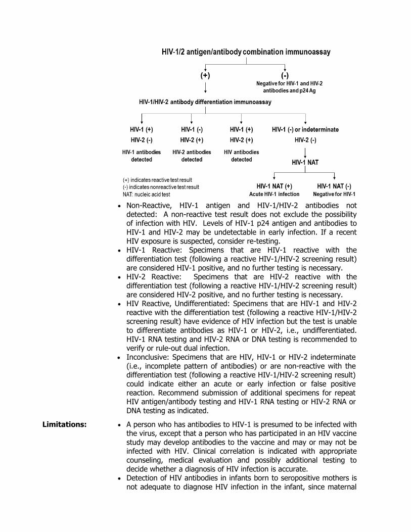
Non-Reactive, HIV-1 antigen and HIV-1/HIV-2 antibodies not
detected: A non-reactive test result does not exclude the possibility of infection with HIV. Levels of HIV-1 p24 antigen and antibodies to HIV-1 and HIV-2 may be undetectable in early infection. If a recent HIV exposure is suspected, consider re-testing.
HIV-1 Reactive: Specimens that are HIV-1 reactive with the differentiation test (following a reactive HIV-1/HIV-2 screening result) are considered HIV-1 positive, and no further testing is necessary.
HIV-2 Reactive: Specimens that are HIV-2 reactive with the differentiation test (following a reactive HIV-1/HIV-2 screening result) are considered HIV-2 positive, and no further testing is necessary.
HIV Reactive, Undifferentiated: Specimens that are HIV-1 and HIV-2 reactive with the differentiation test (following a reactive HIV-1/HIV-2 screening result) have evidence of HIV infection but the test is unable to differentiate antibodies as HIV-1 or HIV-2, i.e., undifferentiated. HIV-1 RNA testing and HIV-2 RNA or DNA testing is recommended to verify or rule-out dual infection.
Inconclusive: Specimens that are HIV, HIV-1 or HIV-2 indeterminate (i.e., incomplete pattern of antibodies) or are non-reactive with the differentiation test (following a reactive HIV-1/HIV-2 screening result) could indicate either an acute or early infection or false positive reaction. Recommend submission of additional specimens for repeat HIV antigen/antibody testing and HIV-1 RNA testing or HIV-2 RNA or DNA testing as indicated.
Limitations:
A person who has antibodies to HIV-1 is presumed to be infected with the virus, except that a person who has participated in an HIV vaccine study may develop antibodies to the vaccine and may or may not be infected with HIV. Clinical correlation is indicated with appropriate counseling, medical evaluation and possibly additional testing to decide whether a diagnosis of HIV infection is accurate.
Detection of HIV antibodies in infants born to seropositive mothers is not adequate to diagnose HIV infection in the infant, since maternal
IgG frequently persists in the infant’s blood for as long as 18 months after birth. Supplemental assays designed specifically for neonatal specimens may be helpful in resolving such cases.
Non-reactive, inconclusive and undifferentiated HIV-1/HIV-2 Differentiation Test results should be referred for HIV-1 and/or HIV-2 nucleic acid testing as per CDC guidelines.
Notes: These tests are approved for in vitro diagnostic use by the U.S. Food and Drug Administration.
Back to Table of Contents
Human Papillomavirus, High-risk - Transcription-Mediated Amplification
Use: To screen women 25 years and older with atypical squamous cells of undetermined significance (ASC-US) cervical cytology results to determine the need for referral to colposcopy.
Note: High-risk human papillomavirus (hrHPV) testing of standard-risk women aged 21-24 years with ASC-US cervical cytology results is no longer recommended. However, it is acceptable if done. See the updated American Society for Colposcopy and Cervical Pathology (ASCCP) Consensus Screening Guidelines.
To screen women 30-65 years old adjunctively with cervical cytology to assess for the presence/absence of hrHPVs.
Other uses of this test may include those indicated by consensus guidelines for cervical cancer screening and the management of women with abnormal cervical cytology and/or hrHPV results, such as those from the ASCCP; clinical indications must be explicitly documented on the requisition form.
Methodology: Transcription-mediated amplification of E6/E7 viral mRNA from 14 hrHPVs: 16, 18, 31, 33, 35, 39, 45, 51, 52, 56, 58, 59, 66, and 68.
Clinical Significance:
hrHPVs detected by the assay are associated with cervical pre-cancerous and cancerous lesions.
Specimen: Type: ThinPrep Pap specimens
Volume: 1.0 mL
Container: ThinPrep Pap Test vials containing PreservCyt Solution
Collection: Collect cervical specimens in ThinPrep Pap Test vials containing
PreservCyt Solution with broom-type or cytobrush/spatula collection devices according to the manufacturer’s instructions
Interferences: Personal lubricants containing Polyquaternium 15 at concentrations > 0.025% of test sample
Anti-fungals containing tioconazole at concentrations > 0.075% of test sample
Effects of other potential variables such as vaginal discharge, use of tampons, douching, etc. and specimen collection variables have not been evaluated.
Shipping: Store and ship ThinPrep liquid cytology specimens at 2°C to 30°C for delivery within 30 days from collection
Rejection Criteria: Specimen collection > 30 days prior to receipt Patient under 21 years of age Patient 21 to 29 years of age without a cytology diagnosis of ASC-
US or other prior diagnosis/treatment that would warrant hrHPV testing per consensus guidelines
Patient 21 to 29 years of age with no cytology diagnosis provided or other prior diagnosis/treatment that would warrant HPV testing per
consensus guidelines Patient of any age with “HPV Reflex Testing Only” marked on
requisition form and without a cytology diagnosis of ASC-US
QNS: < 1 mL ThinPrep liquid cytology specimen Other criteria as outlined in Specimen Rejection section of this Test
Directory
Note: requisitions marked “Co-testing” will be primarily associated with patients 30 years and older; however, if a patient on such a requisition is 25-29 years old and has a prior diagnosis/treatment that would warrant hrHPV testing per consensus guidelines, then the specimen is still acceptable for testing.
Reported: Within 7 working days of receipt
CPT Codes: 87621
Normal/Abnormal Results:
High-risk HPV Detected High-risk HPV Not Detected
Interpretation: A positive result may be due to the presence of one or more hrHPV types. Infection with HPV is not an indicator of cytologic HSIL or underlying high-grade CIN, nor does it imply that CIN2/3 or cancer will develop; most women infected with hrHPV do not develop CIN2/3 or cancer. False positive results may occur; some cross-reactivity is exhibited with low-risk HPV types 26, 67, 70, and 82. A negative result does not exclude the possibility of, underlying or future cytologic abnormalities, CIN2, CIN3, or cancer, infection with low levels of hrHPV, or other HPV types not detected by this assay. Detection of HPV is dependent on adequate specimen collection and handling, absence of inhibitors, and sufficient levels of virus. Results should be interpreted in conjunction with other laboratory and clinical data. Consensus guidelines for cervical cancer screening and the management of women with abnormal cervical cytology and/or hrHPV results should be followed, such as those from the ASCCP.
Limitations:
This test is not intended for use in cases of suspected sexual abuse or for screening for anal and penile cancers; currently, availability of this test is restricted to County Health Departments in Oklahoma
Notes: This test is approved for in vitro diagnostic use by the U.S. Food and Drug Administration.
Back to Table of Contents

Influenza Virus A and B – PCR
Use: Detection and characterization of influenza virus types A (subtypes H1pdm09, H3, H5, H7), or B (Victoria and Yamagata lineages) in patients with influenza-like symptoms.
Methodology: Specimens are tested using the Human Influenza Virus Real-time Reverse Transcriptase-PCR Diagnostic Panel, which was developed by the CDC to diagnose human infections with seasonal influenza viruses and novel influenza A viruses. Specimens may be initially screened using the Respiratory Pathogen Panel (see this Test Directory), which is able to detect a spectrum of viruses and bacteria associated with respiratory infections, including influenza viruses A and B.
Clinical Significance:
Influenza virus types A, B, and C are RNA viruses that cause acute respiratory disease with associated fever, shivering, chills, headache, malaise, dry cough, loss of appetite, body aches and nausea. Severe cases are associated with prostration, hemorrhagic bronchitis, pneumonia and occasionally death, especially in the young and elderly. Most influenza-like infections are not caused by influenza but by other viruses (e.g., rhinoviruses and respiratory syncytial virus, adenoviruses, and parainfluenza viruses).
Further background information, fact sheets, statistics and educational resources may be found at the OSDH Acute Disease Services website.
Specimen: Type: Nasopharyngeal swab (recommended); Nasal swab; Throat swab
Volume: 1 or 2 swabs
Container: Tube containing viral transport medium (VTM; provided by OSDH PHL) or other appropriate commercial medium (UTM, M4, and M4RT). VTM contains antibiotics and must be stored in the refrigerator prior to and subsequent to specimen collection. Do not use media if cloudy or when past expiration date.
Collection: Respiratory virus diagnosis depends on the collection of high-quality specimens, their rapid transport to the testing laboratory and appropriate storage before laboratory testing. Specimens for the direct detection of viral nucleic acids should be taken preferably during the first 3 days after onset of clinical symptoms.
Nasopharyngeal Swab1. Label a sterile tube
containing 2-3 mL of VTM or other suitable collection media with patient’s name and date of collection.
2. If the nasal passages have a large amount of mucus, ask the patient to blow their nose before collecting specimen.
3. With the thumb of one hand, elevate the tip of the patient’s nose then gently insert the NP swab into the nostril.
4. Guide the swab backward and upward along the nasal septum until a distinct resistance is met, hold it there for a few seconds, then with a rotating motion gently remove it.
5. Immediately, place swab into tube containing VTM or other suitable collection media.
6. Break-off or cut excess shaft of swab so that tube can be capped. 7. Place tube in plastic biohazard bag and insert completed requisition
form in outer pouch of the bag. 8. Refrigerate (2-8oC) immediately.
Nasal Swab 1. Label a sterile tube containing 2-3 mL of VTM or other suitable
collection media with patient’s name and date of collection. 2. If the nasal passages have a large amount of mucus, ask the patient
to blow their nose before collecting specimen. 3. With the thumb of one hand, elevate the tip of the patient’s nose
then gently insert a dry polyester swab into one nostril, keeping swab shaft parallel to the palate.
4. Hold it there for a few seconds then with a rotating motion gently remove it.
5. Using the same swab, repeat the process, inserting the swab in the other nostril.
6. Place swab immediately into VTM or other suitable collection media. 7. Break-off or cut excess shaft of swab so that tube can be capped. 8. Place tube in plastic biohazard bag and insert completed requisition
form in outer pouch of the bag. 9. Refrigerate (2-8oC) immediately.
Interferences: Cotton swab; Calcium alginate swab (shown to inhibit PCR)
Special Instructions:
Only synthetic (nylon, rayon or Dacron)-tipped swabs with plastic or aluminum shafts are acceptable.
Note: Submission of specimens from patients with suspected influenza A/H5 or Eurasian H7 requires OSDH Acute Disease Service pre-approval to verify the patient meets clinical case criteria, including travel history to an area of novel influenza circulation.
Shipping: Store and ship specimens refrigerated (2-8oC) for delivery within 3 days (72 hours) of collection. If delivery will be delayed for more than 72 hours, specimens should be frozen at -70°C or colder and shipped with dry ice. 1. Place specimen in round cardboard container and place this in
Styrofoam mailer box (both containers are supplied by OSDH PHL for influenza surveillance).
2. Remove frozen cold pack from freezer and place in the mailer with specimen then fill the mailer with packing material.
3. Mailers are returned to submitting sites with fresh tubes of VTM corresponding to the number of specimens submitted. If additional VTM is required, fill-out the order form and return with the specimens in the mailer.

4. Send via OSDH PHL courier service.
Rejection Criteria: Incorrect collection device (cotton, wooden, or calcium alginate swab) or media
Received > 72 hours from time of collection and not frozen at -70°C or colder
Swab without transport medium Specimen at ambient temperature Other criteria as outlined in Specimen Rejection section of this Test
Directory
Reported: Within 5 working days of receipt. Specimens initially screened using the Respiratory Pathogens Panel, and in which influenza virus A and/or B is detected, will be issued a preliminary report pending testing by the Human Influenza Virus rRT-PCR Diagnostic Panel. Specimens in which variant or potential novel influenza viruses are detected by the Human Influenza Virus rRT-PCR Diagnostic Panel will be issued a preliminary report, pending further characterization by the CDC.
CPT Codes: 87502, 87503 (x3)
Normal/Abnormal Results:
Influenza Not Detected Influenza Virus A Detected, Subtype: H1 2009 pandemic strain Influenza Virus A Detected, Subtype: H3 Influenza Virus A Detected, Subtype: Eurasian H7 Influenza Virus A Detected, Subtype: Undetermined Influenza Virus A Detected, Subtype: Undetermined, referred to CDC
for subtyping Presumptive Positive for Influenza A/H3N2 variant; referred to CDC
for confirmation Presumptive Positive for Influenza A/H5; referred to CDC for
confirmation Influenza B Detected Influenza B Victoria Lineage Detected Influenza B Yamagata Lineage Detected Indeterminate, potential PCR inhibitor/poor sample quality
Interpretation: When a clinical sample fails to amplify, the sample is reported as “Indeterminate, potential PCR inhibitor/poor sample quality”. It is recommended that a new sample is submitted for testing.
Limitations:
Negative results do not preclude influenza virus infection and should not be used as the sole basis for treatment or other patient management decisions. Positive results do not rule-out other viral or bacterial co-infections. Individuals immunized with live attenuated influenza nasal spray vaccine may be positive for one or more influenza virus targets for several days post-vaccination; vaccination history should be considered when interpreting positive test results, especially early in the flu season. Lineage typing of influenza B is used for epidemiological purposes, and has no bearing on patient treatment. Performance characteristics of this assay may vary in the detection of

novel or emerging influenza viruses.
Notes: This test is approved for in vitro diagnostic use by the U.S. Food and Drug Administration.
Back to Table of Contents
Mycobacteria (Acid-Fast Bacilli) – Smear and Culture With Reflex to Mycobacteria Identification
Use: For isolation and detection of Mycobacterium species.
Methodology: Concentrated smears are prepared, stained with Auramine O and read by fluorescence microscopy. Broth-based BACTEC MGIT media and conventional Lowenstein-Jensen media are used for culture and isolation. Blood and urine are grown on two different solid media. AFB smears are only performed in conjunction with culture. Identification testing will be reflexively performed on all culture-positive specimens (see Mycobacteria (Acid-Fast Bacilli) – Species Identification in this Test Directory).
Clinical Significance:
Many species within the genus Mycobacterium are prominent pathogens, especially the members of Mycobacterium tuberculosis Complex as well as M. leprae and M. ulcerans. In addition, numerous species of environmental mycobacteria, called non-tuberculosis mycobacteria (NTM), are responsible for various mycobacterioses. The route of transmission of pulmonary tuberculosis (TB) is through the air, which makes this a highly transmissible disease. Given the infectious nature of pulmonary TB, fast and accurate diagnosis is an important element of TB treatment and control.
Further background information, fact sheets, statistics and educational resources may be found at the OSDH Acute Disease Services website.
Specimen: Type: Sputum, expectorated or induced (early-morning recommended);
bronchoalveolar lavage (BAL); bronchial washings; bronchial brush; tracheal aspirate; urine; blood; sterile body fluids; gastric aspirates (must be neutralized with 100 grams of NaCO3 at time of collection); tissue; wound/lesion; other.
Submission of swabs is not recommended, and requires prior authorization from the Mycobacteriology/Mycology Laboratory (call 405-271-5070).
Volume: Fluids: 5-10 mL Sterile fluids: > 2 mL Tissue: 1 gram
Container: Sterile, leak-proof, screw-cap container; TB/sputum kit
Collection: As appropriate for specimen type. Instructions for collection of the most common specimen types are provided below:
Sputum Patient Preparation: 1. For initial diagnosis, patients should submit a series of 3 sputum
samples over a period of 3 days (one/day). 2. Specimens should be collected early in the morning. 3. Patient should brush teeth/dentures and/or rinse with water, not an
antiseptic solution, immediately before obtaining specimen.

4. Steam from a hot shower or boiling kettle and/or drinking several glasses of water or non-alcoholic liquids may help raise sputum from lungs.
5. Nurses/other individuals remaining with patient during specimen collection coughing when patient is known or suspected of being infectious (smear or culture positive), should wear a particulate respirator.
Collection: 1. Sputum specimens should be collected using the TB/Sputum kit
(available from OSDH PHL and contains a 50 mL sterile plastic tube, collection instructions, dual-pocket specimen bag and cardboard mailing container).
2. Remove cap from sterile 50 mL container without touching inside of container.
3. Patient should take a deep breath, hold it momentarily then cough deeply from the deepest part of the chest and expectorate sputum into container. This should be repeated until 5-10 mL or 1-2 teaspoons are obtained. Saliva (spit) and nasal secretions contain few acid-fast bacteria and should not be collected. Sputum specimens should be free of food particles and other extraneous material.
4. Replace cap tightly on container. 5. Avoid soiling outside of container; if outside of container becomes
soiled with sputum, wipe with a clean cloth wetted with alcohol, soap and water, or 1:10 bleach solution.
Shipping: 1. Label container with appropriate patient ID. 2. Place container inside plastic specimen bag (provided with kit). 3. Place completed Test Requisition Form in outside pocket of bag. 4. Place bag, containing specimen and requisition form, in mailing
container and ship to OSDH PHL.
Urine For initial diagnosis, patients should submit a series of 3 urine samples over a period of 3 days (one/day). Collection: Females: 1. Thoroughly clean urethral area with soap and water. 2. Sit on toilet and manually separate labia minora with one hand. 3. Keep labia separated and begin urinating into toilet. 4. After several mLs have passed, use other hand to catch midstream
portion of urine in specimen cup; test requires approx. 10 mL or 2 teaspoons. Avoid touching the lip or inside of the container.
5. Finish urinating into toilet. Males: 1. Thoroughly clean glans (head) of penis with soap and water. 2. Hold foreskin retracted, if appropriate, and begin urinating in toilet. 3. After several mLs have passed, use other hand to catch midstream
portion of urine in specimen cup; test requires approx. 10 mL or 2 teaspoons. Avoid touching the lip or inside of the container.

4. Finish urinating into toilet. Shipping: 1. Pour urine from specimen cup into sterile screw-capped tube (or
container from TB/Sputum kit) and tightly secure cap. Do not send urine in specimen cup.
2. Label tube as “Urine” with appropriate patient ID. 3. Place container inside plastic specimen bag (provided with
TB/Sputum kit). 4. Place completed Test Requisition Form in outside pocket of bag. 5. Place bag, containing specimen and requisition form, in mailing
container and ship to OSDH PHL.
Interferences: Fixatives
Special Instructions:
M. marinum and M. haemophilum require special conditions for culture; notify the laboratory if either of these organisms is suspected.
Shipping: Store and ship samples refrigerated, except blood, which should be shipped at ambient temperatures. It is recommended that specimens be delivered to the Public Health Laboratory within 24 hours of collection. Specimens must be received within 7 days from date of collection.
Rejection Criteria: Stool Specimen frozen Specimen containing fixative, foreign object/food particles Specimen received > 7 days post-collection Refrigerated blood specimen Pooled specimens Other criteria as outlined in Specimen Rejection section of this Test
Directory
Reported: Smear: within 24 hours of receipt Positive culture: 2-42 days from receipt Negative culture: at 42 days
CPT Codes: 87015 (except for sterile body fluids), 87206, 87116
Normal/Abnormal Results:
AFB Smear: AFB Stain-Negative; AFB Stain-Positive (with quantification per high dry field) AFB Culture: Negative for AFB; Positive for AFB; Indeterminate, Over-growth by Non-acid Fast Microorganisms
Interpretation: AFB Smear: AFB Stain-Negative: no AFB observed in 30 fields of view at 200x
magnification. Negative results do not preclude the presence of other clinically relevant microorganisms.
AFB Stain-Positive: o < 1 AFB observed per high dry field of view (600x magnification) o 1-10 AFB observed per high dry field of view (600x
magnification) o > 10 AFB observed per high dry field of view (600x
magnification) AFB Culture: Cultures are negative if no growth is observed at 42 days. Negative
results do not preclude the presence of other clinically relevant microorganisms.
Limitations:
Because detection of mycobacteria is dependent on the number of organisms present in the sample, proper sample collection, handling, and storage are important components of testing.
Notes: Analyses involve both laboratory-developed tests and tests that are approved for in vitro diagnostic use by the U.S. Food and Drug Administration; performance characteristics have been validated and determined to be suitable for diagnostic purposes by the OSDH Public Health Laboratory.
Back to Table of Contents
Mycobacteria (Acid-Fast Bacilli) – Isolate Identification
Use: Identification of mycobacteria to the species or species group/complex level from cultured isolates
Methodology: Primary identification involves mycolic acid analysis by high performance liquid chromatography (HPLC). DNA probes and biochemical testing supplement HPLC, as needed.
Clinical Significance:
Laboratory diagnosis of M. tuberculosis Complex has vital epidemiological and public health consequences. Differentiation of M. tuberculosis Complex from non-tuberculosis mycobacteria (e.g., M. avium Complex), which can cause pulmonary disease resembling tuberculosis, lymphadenitis, skin and disseminated disease, is important for diagnosis and treatment. Identification of the species of non-tuberculosis mycobacteria can be important in the treatment of patients.
Further background information, fact sheets, statistics and educational resources may be found at the OSDH Acute Disease Services website.
Specimen: Type: Liquid or solid cultured isolate
Volume: Liquid: > 3 mL Solid: must have visible growth
Container: Sealed container; slant or sealed plate
Collection: N/A
Interferences: Impure isolates
Special
Instructions: None
Shipping: Store and ship at ambient temperature
Rejection Criteria: Mixed or contaminated specimen Frozen specimen Dehydrated media Other criteria as outlined in Specimen Rejection section of this Test
Directory
Reported: Within 7 working days from receipt (longer if biochemical testing is necessary). M. tuberculosis Complex identifications are telephoned to submitter on previously undiagnosed patients.
CPT Codes: 87143 (HPLC, all mycobacteria cultures), 87149 (DNA probe, if required), 87118 (biochemical testing, if required)
Normal/Abnormal Results:
Genus/Species; Genus/Species Group; Genus/Species Complex; Unidentified Non-tuberculosis Mycobacteria Species; Isolate Non-viable, Unable to Sub-culture; Mixed Culture, Could Not Isolate AFB; Over-growth by Non-acid Fast Microorganisms; Could Not Isolate AFB
Interpretation: Genus/species or Genus/species group or complex: M. tuberculosis Complex: includes M. tuberculosis, M. bovis, M. bovis
BCG, M. africanum, M. caprae, M. microti, M. canettii, and M. pinnipedii
M. avium Complex: includes M. avium and M. intracellulare M. terrae Complex: includes M. terrae, M. nonchromagenicum, and M.
triviale M. fortuitum Group: includes M. fortuitum, M. peregrinum, M.
senegalense, M. setense, M. septicum, M. porcinum, M. houstonense, M. boenickei, M. brisbanense, and M. neworleansense
M. chelonae Group: includes M. chelonae, M. immunogenum, M. abscessus subsp. abscessus, M. abscessus, subsp. bolleetii, and M. salmoniphilum
M. asiaticum M. celatum M. flavescens M. gordonae M. haemophilum M. kansasii M. marinum M. mucogenicum M. phlei M. scrofulaceum M. smegmatis M. szulgai M. ulcerans M. vaccae M. xenopi
Unidentified Non-tuberculosis Mycobacteria Species: cannot be identified by techniques used at the OSDH PHL; isolates that cannot be identified will be sent to the CDC for further characterization should they meet specified CDC submission criteria
Isolate Non-viable, Unable to Sub-culture: isolate could not be grown on solid media
Limitations: Differentiation of species within complexes or groups is not performed
Notes: Analyses involve both laboratory-developed tests and tests that are approved for in vitro diagnostic use by the U.S. Food and Drug Administration; performance characteristics have been validated and determined to be suitable for diagnostic purposes by the OSDH Public Health Laboratory.
Back to Table of Contents
Mycobacterium tuberculosis Complex - PCR
Use: For detection of Mycobacterium tuberculosis Complex DNA in respiratory specimens from patients showing signs and symptoms consistent with active pulmonary tuberculosis (TB).
Methodology: The Cepheid Xpert® MTB/RIF assay is a nested, real-time PCR assay that detects the presence of M. tuberculosis Complex DNA.
Clinical Significance:
Many species within the genus Mycobacterium are prominent human pathogens, especially members of the M. tuberculosis Complex and M. leprae and M. ulcerans. In addition, numerous species of environmental mycobacteria, called non-tuberculosis mycobacteria (NTM), are responsible for various mycobacterioses. The route of transmission of pulmonary TB is through the air, which makes this a highly transmissible disease. Given the infectious nature of pulmonary TB, fast and accurate diagnosis is an important element of treatment and control.
Further background information, fact sheets, statistics and educational resources may be found at the OSDH Acute Disease Services website.
Specimen: Type: Sputum (early-morning recommended; expectorated or nubulizer-
induced); bronchoalveolar lavage (BAL); bronchial washings, bronchial brush or tracheal aspirate
Volume: 5-10 mL
Container: Sterile, leak-proof, screw-cap container
Collection: As appropriate for specimen; instructions for collection of sputum are provided in test description for Mycobacteria (Acid-Fast Bacilli) – Smear and Culture of this Test Directory
Interferences: Includes, but not limited to, blood, pus, mammalian cells and hemoglobin
Special
Instructions: Typically, performed as a reflex test on previously undiagnosed, AFB
smear-positive patients Otherwise, only performed by physician order
o County Health Departments must seek prior approval from the OSDH Tuberculosis Program (405-271-4060)
Shipping: Store and ship samples refrigerated for delivery within 7 days from collection
Rejection Criteria: Received > 7 days post-collection Specimen decontaminated/processed prior to submission Patient previously identified with mycobacterium species within prior 6
months (excluding M. gordonae) Extensively bloody Specimen containing fixative, foreign object/food particles Non-pulmonary specimen Sample from patient being treated with anti-tuberculosis drugs
(unless < 3 days treatment)
Specimen from pediatric patient (< 18 years old) Other criteria as outlined in Specimen Rejection section of this Test
Directory
Reported: Within 24 hours post-concentration. Results are telephoned to the submitter.
CPT Codes: 87556
Normal/Abnormal Results:
M. tuberculosis Complex Detected M. tuberculosis Complex Not Detected Indeterminate: Inhibitor Present, Unable to Amplify
Interpretation: A positive result infers the presence of M. tuberculosis Complex DNA, not viable organisms. The assay does not differentiate between species within the M. tuberculosis Complex.
A negative result does not exclude the possibility of M. tuberculosis Complex infection; detection is dependent on appropriate specimen collection and handling, absence of inhibitors, and sufficient levels of organisms. Since the assay cannot detect non-tuberculosis mycobacterial species, culture should be used to determine if non-tuberculosis mycobacteria are present.
An indeterminate result may be due to improper processing of sample, or presence of a PCR inhibitor; collection of a fresh specimen for testing is recommended.
Limitations: This assay is not suitable for monitoring therapeutic efficacy.
Notes: Sputum samples are processed using the unmodified Cepheid Xpert® MTB/RIF IVD as approved by the U.S. Food and Drug Administration. Bronchial and tracheal aspirates and lavages are processed using a modified version of the assay, the performance characteristics of which have been validated by the OSDH Public Health Laboratory. This assay has not been verified by the OSDH Public Health Laboratory for use in evaluating rifampin resistance.
Back to Table of Contents
Mycobacterium tuberculosis Complex, Antimicrobial Susceptibility - Culture
Use: A qualitative procedure for susceptibility testing of Mycobacterium tuberculosis Complex culture to anti-tuberculosis agents: streptomycin, isoniazid, rifampin, ethambutol and pyrazinamide.
Methodology: Anti-tuberculosis susceptibility testing is performed using the BD BACTEC Mycobacterial Growth Indicator Tube (MGIT) 960 system. Susceptibility results are expressed as concentrations of specific agents (see list below).
Clinical Significance:
Anti-mycobacterial susceptibility testing is valuable in defining optimum drug treatment for patients with tuberculosis. TB patients commonly receive a multiple-drug regimen that includes streptomycin, isoniazid, rifampin, ethambutol and pyrazinamide. It is important that the anti-mycobacterial drugs prescribed show appropriate activity against infecting agent.
Further background information, fact sheets, statistics and educational resources may be found at the OSDH Acute Disease Services website.
Specimen: Type: Liquid or solid culture
Volume: Liquid: > 3 mL Solid: must have visible growth
Container: Sealed container; Slant or sealed plate
Collection: N/A
Interferences: Impure isolates
Special Instructions:
None
Shipping: Store and ship at ambient temperature
Rejection Criteria: Mixed or contaminated specimen Dehydrated media Frozen specimen Other criteria as outlined in Specimen Rejection section of this Test
Directory
Reported: Approximately 10-28 working days after receipt; a preliminary report is issued on all potential drug-resistant results pending confirmatory re-testing
CPT Codes: 87188 (x5)
Normal/Abnormal Results:
Results are provided for the five individual anti-tuberculosis drugs: Susceptible to <Anti-tuberculosis Agent> at <Concentration>; Resistant to <Anti-tuberculosis Agent> at <Concentration>; Isolate Non-viable, Unable to Sub-culture; Mixed Culture, Could Not Isolate AFB; Over-growth by Non-acid Fast Microorganisms, Could Not Isolate AFB
Interpretation: Susceptible to Streptomycin at 1.0 µg/mL Resistant to Streptomycin at 1.0 µg/mL Susceptible to Isoniazid at 0.1 µg/mL Resistant to Isoniazid at 0.1 µg/mL Susceptible to Isoniazid at 0.4 µg/mL Resistant to Isoniazid at 0.4 µg/mL Susceptible to Rifampin at 1.0 µg/mL Resistant to Rifampin at 1.0 µg/mL Susceptible to Ethambutol at 5.0 µg/mL Resistant to Ethambutol at 5.0 µg/mL Susceptible to Pyrazinamide at 100 µg/mL Resistant to Pyrazinamide at 100 µg/mL
Limitations:
The BACTEC MGIT system susceptibility test does not provide the degree of susceptibility of the isolate to the drugs (i.e., the percentage of organisms that are resistant).
Notes: Resistance to any of the drugs is confirmed by re-testing. Isoniazid will be tested at a higher concentration (0.4 µg/mL), if resistance is observed at 0.1 µg/mL. If resistance to any of the drugs is confirmed, the isolate will be sent to CDC for additional second-line drug susceptibility testing (with the exception of mono-resistant streptomycin). The OSDH PHL does not perform susceptibility testing on non-tuberculosis mycobacteria.
This test is approved for in vitro diagnostic use by the U.S. Food and Drug Administration.
Back to Table of Contents
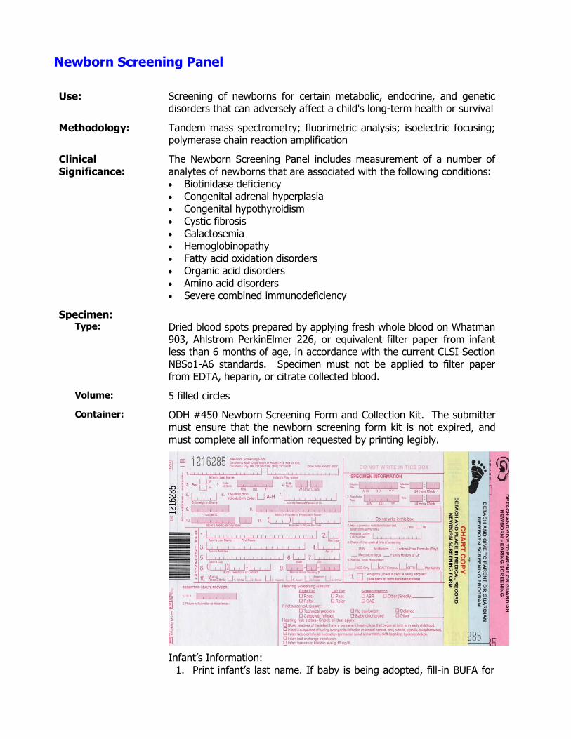
Newborn Screening Panel
Use: Screening of newborns for certain metabolic, endocrine, and genetic disorders that can adversely affect a child's long-term health or survival
Methodology: Tandem mass spectrometry; fluorimetric analysis; isoelectric focusing; polymerase chain reaction amplification
Clinical Significance:
The Newborn Screening Panel includes measurement of a number of analytes of newborns that are associated with the following conditions: Biotinidase deficiency Congenital adrenal hyperplasia Congenital hypothyroidism Cystic fibrosis Galactosemia Hemoglobinopathy Fatty acid oxidation disorders Organic acid disorders Amino acid disorders Severe combined immunodeficiency
Specimen: Type: Dried blood spots prepared by applying fresh whole blood on Whatman
903, Ahlstrom PerkinElmer 226, or equivalent filter paper from infant less than 6 months of age, in accordance with the current CLSI Section NBSo1-A6 standards. Specimen must not be applied to filter paper from EDTA, heparin, or citrate collected blood.
Volume: 5 filled circles
Container: ODH #450 Newborn Screening Form and Collection Kit. The submitter must ensure that the newborn screening form kit is not expired, and must complete all information requested by printing legibly.
Infant’s Information: 1. Print infant’s last name. If baby is being adopted, fill-in BUFA for
last name and mark #11. Print infant’s first name or fill-in with Male/Female.
2. Mark appropriate gender. 3. Print date of birth. 4. Print time of birth using military time. 5. Print birth weight in grams. 6. If multiple birth, indicate birth order (i.e., A,B,C). If not a multiple
birth, leave blank. 7. Print infant’s medical record number. 8. Print the provider’s first and last name that the infant will follow-
up with after discharge. If this is left blank, the follow-up responsibility for abnormal results falls back on the submitter of the specimen.
9. Print mom’s medicaid number, if available. 10. Print provider’s phone number.
Mom’s Information:
If infant is not in mom’s care, put information for person responsible for getting infant to doctor’s appointments (e.g., other family member, case worker, social worker) in the Mom’s Information section.
1. Print last and first name. 2. Print age, if available. 3. Print address. 4. Print apartment number, as applicable. If not applicable, leave
blank. 5. Print name of town where mom resides. 6. Print name of state where mom resides. 7. Print zip code of town where mom resides. 8. Print mom’s telephone or contact number. 9. Print mom’s social security number, if available. 10. Mark appropriate box to indicate mom’s race/ethnicity.
Submitting Health Provider’ Information: 1. Print facility ID. 2. Print facility address. A sticker or stamp may be used for this
section.
Specimen Information: 1. Print collection date and time of specimen collection. 2. Print transfusion date and time if infant was transfused. If infant
was not transfused, leave blank. 3. Mark appropriate box if infant has had a previous newborn
screen. If infant has had a previous screen, print previous OSDH lab number.
4. Mark appropriate fields to indicate treatments/health history of infant (i.e., TPN, antibiotics, lactose-free formula, meconium illeus, family history of CF). GALT enzyme testing will be performed if lactose-free formula is marked. CFTR mutation analysis will be performed if meconium ileus or family history of CF are marked. If none of the fields apply to the infant, then leave blank.
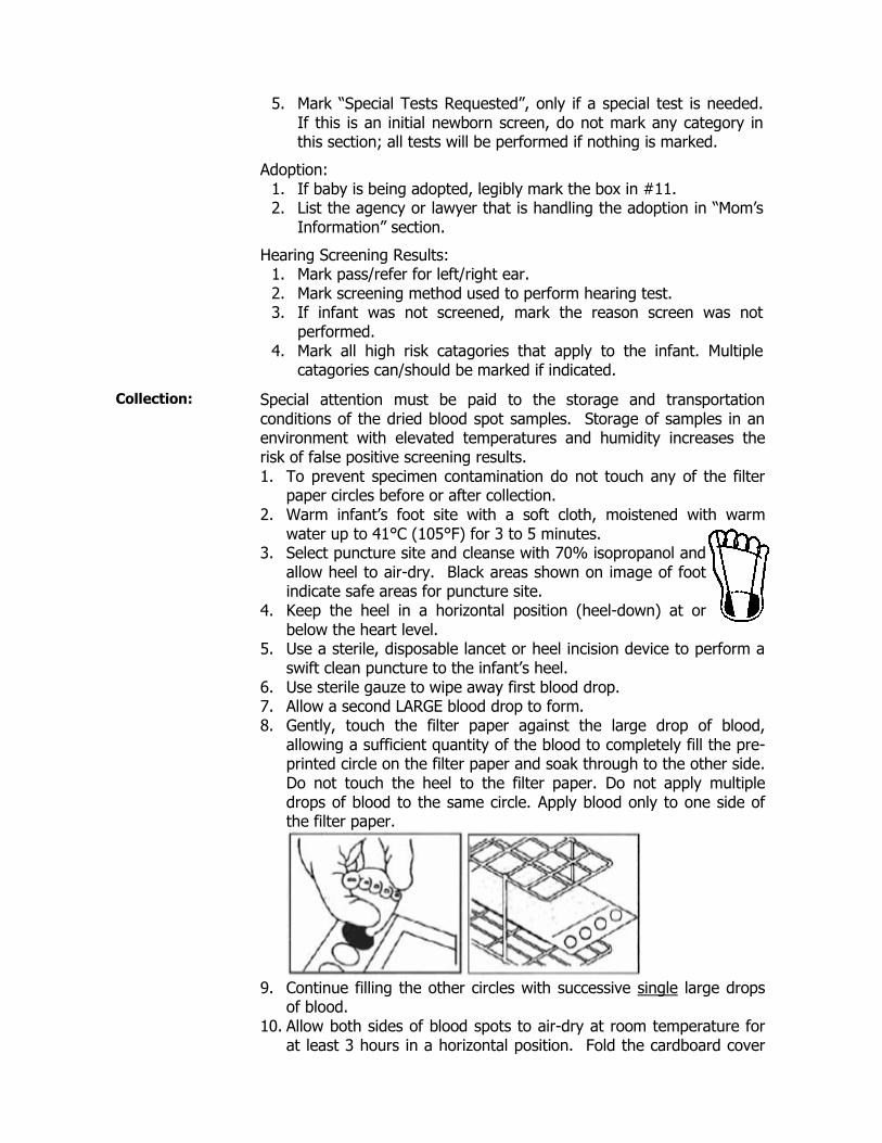
5. Mark “Special Tests Requested”, only if a special test is needed. If this is an initial newborn screen, do not mark any category in this section; all tests will be performed if nothing is marked.
Adoption: 1. If baby is being adopted, legibly mark the box in #11. 2. List the agency or lawyer that is handling the adoption in “Mom’s
Information” section.
Hearing Screening Results: 1. Mark pass/refer for left/right ear. 2. Mark screening method used to perform hearing test. 3. If infant was not screened, mark the reason screen was not
performed. 4. Mark all high risk catagories that apply to the infant. Multiple
catagories can/should be marked if indicated.
Collection: Special attention must be paid to the storage and transportation conditions of the dried blood spot samples. Storage of samples in an environment with elevated temperatures and humidity increases the risk of false positive screening results. 1. To prevent specimen contamination do not touch any of the filter
paper circles before or after collection. 2. Warm infant’s foot site with a soft cloth, moistened with warm
water up to 41°C (105°F) for 3 to 5 minutes. 3. Select puncture site and cleanse with 70% isopropanol and
allow heel to air-dry. Black areas shown on image of foot indicate safe areas for puncture site.
4. Keep the heel in a horizontal position (heel-down) at or below the heart level.
5. Use a sterile, disposable lancet or heel incision device to perform a swift clean puncture to the infant’s heel.
6. Use sterile gauze to wipe away first blood drop. 7. Allow a second LARGE blood drop to form. 8. Gently, touch the filter paper against the large drop of blood,
allowing a sufficient quantity of the blood to completely fill the pre-printed circle on the filter paper and soak through to the other side. Do not touch the heel to the filter paper. Do not apply multiple drops of blood to the same circle. Apply blood only to one side of the filter paper.
9. Continue filling the other circles with successive single large drops
of blood. 10. Allow both sides of blood spots to air-dry at room temperature for
at least 3 hours in a horizontal position. Fold the cardboard cover
to hold the blood spots off of any surface or place on a rack to dry. Be sure the attached coverslip does not come into contact with the blood until completely dry. NOTE: Do not use artificial heat (lamps, incubators, etc) to dry the samples. Keep away from direct sunlight. Do not stack wet specimens. Insufficient drying will adversely affect the test results.
11. Replace the coverslip over the blood spots when completely dry. 12. Check the quality of the specimens (see Simple Spot Check below).
All specimens are judged for acceptability at the OSDH PHL, and those not acceptable are reported as “Unsatisfactory”.
Special
Instructions: Screening requirements for all newborns: Collect specimen as early as possible after 24 hours of age or
immediately prior to discharge or prior to blood transfusion, whichever comes first.
If infant is screened at ≤ 24 hours of age, repeat screen at 3-5 days of age (if premature or sick infant, repeat screen at 7-14 days of age).
All premature and sick infants should have a repeat screen at 14 days of age.
Shipping: Specimens must be transported within 24 hours after collection but send specimens as soon as possible after drying.
Transport specimens at ambient temperature of 2–30oC (35–86oF) in a sealed paper envelope or container that will provide protection from moisture, light, heat, and contact with other materials. o Never place filter paper specimens in plastic bags. o Do not batch specimens from multiple days except for
days when there is no courier service. Send specimens via OSDH PHL courier service.
o Courier pick-up occurs daily at most sites. o For sites without weekend courier service, specimens collected
on the weekend will be picked-up on the next workday. Specimens older than 14 days from collection are unsatisfactory for
testing and repeat collection will be required.
Rejection Criteria: Circles not completely filled or not thoroughly saturated Uneven saturation of circles or multiple sample applications Specimen appears diluted, discolored, or contaminated Clotted or caked blood on filter paper Peripheral serum rings present on filter paper Specimen appears scratched or abraded or filter paper damaged No blood applied to filter paper Specimen received on incorrect or expired form Specimen received more than 14 days from date of collection Serial number on form and (detached) filter paper do not match Other criteria as outlined in Specimen Rejection section of this Test
Directory
Reported: Within 5 working days of receipt (7 working days if reflex testing is required)
Initial normal/abnormal results:

o Written report sent to baby’s physician and sample collection site
Repeat of initial abnormal results: o Written report to physician and collection site
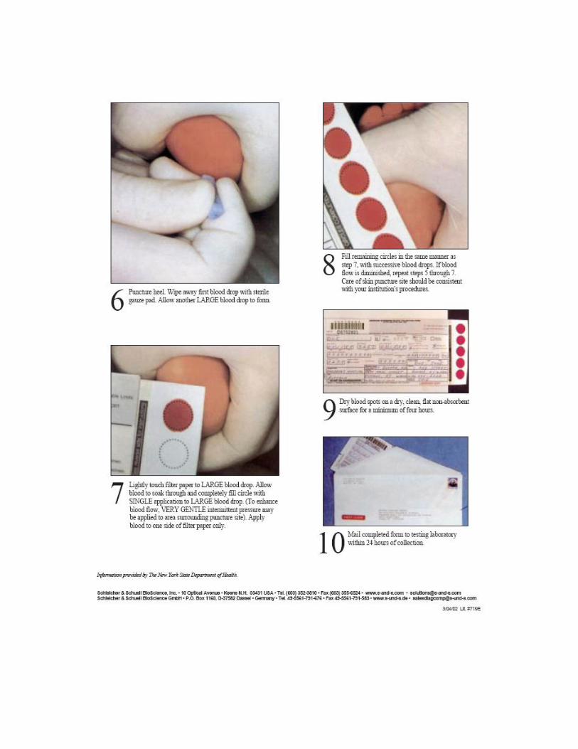
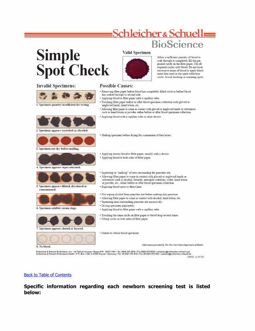
Back to Table of Contents
Specific information regarding each newborn screening test is listed below:
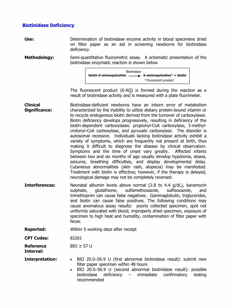
Biotinidase Deficiency
Use: Determination of biotinidase enzyme activity in blood specimens dried on filter paper as an aid in screening newborns for biotinidase deficiency.
Methodology: Semi-quantitative fluorometric assay. A schematic presentation of the biotinidase enzymatic reaction is shown below.
The fluorescent product (6-AQ) is formed during the reaction as a result of biotinidase activity and is measured with a plate fluorimeter.
Clinical Significance:
Biotinidase-deficient newborns have an inborn error of metabolism characterized by the inability to utilize dietary protein-bound vitamin or to recycle endogenous biotin derived from the turnover of carboxylases. Biotin deficiency develops progressively, resulting in deficiency of the biotin-dependent carboxylases: propionyl-CoA carboxylase, 3-methyl-crotonyl-CoA carboxylase, and pyruvate carboxylase. The disorder is autosomal recessive. Individuals lacking biotinidase activity exhibit a variety of symptoms, which are frequently not present at birth, thus making it difficult to diagnose the disease by clinical observation. Symptoms and the time of onset vary greatly. Affected infants between two and six months of age usually develop hypotonia, ataxia, seizures, breathing difficulties, and display developmental delay. Cutaneous abnormalities (skin rash, alopecia) may be manifested. Treatment with biotin is effective; however, if the therapy is delayed, neurological damage may not be completely reversed.
Interferences: Neonatal albumin levels above normal (2.8 to 4.4 g/dL), kanamycin sulphate, glutathione, sulfamethoxazole, sulfisoxazole, and trimethoprim can cause false negatives. Gammaglobulin, triglycerides, and biotin can cause false positives. The following conditions may cause anomalous assay results: poorly collected specimen, spot not uniformly saturated with blood, improperly dried specimen, exposure of specimen to high heat and humidity, contamination of filter paper with feces.
Reported: Within 5 working days after receipt
CPT Codes: 82261
Reference Interval:
BIO ≥ 57 U
Interpretation: BIO 20.0–56.9 U (first abnormal biotinidase result): submit new filter paper specimen within 48 hours
BIO 20.0–56.9 U (second abnormal biotinidase result): possible biotinidase deficiency – immediate confirmatory testing recommended
Biotinidase biotin 6-aminoquinoline 6-aminoquinoline* + biotin
*Fluorescent product
BIO < 20.0 U: Consistent with biotinidase deficiency – immediate confirmatory testing recommended
Limitations:
This is a screening test only. It should not be used to distinguish partial from profound biotinidase deficiency. A diagnostic procedure should be used to confirm a diagnosis of biotinidase deficiency.
Notes: This test is approved for in vitro diagnostic use by the U.S. Food and Drug Administration.
Back to Table of Contents
Congenital Adrenal Hyperplasia
Use: -OH-progesterone (17-OHP) in blood specimens dried on filter paper as an aid in screening newborns for congenital adrenal hyperplasia (CAH). If the 17-OHP value is elevated, the specimen is referred for 2nd tier MS/MS steroid profiling.
Methodology: The Neonatal 17-OHP assay is a solid phase, time-resolved fluoroimmunoassay based on the competitive reaction between europium-labeled 17-OHP specific polyclonal antibodies (derived from rabbit). Danazol facilitates the release of 17-OHP from the binding proteins. A second antibody, directed against rabbit IgG, is coated to the solid phase, giving convenient separation of the antibody-bound and free antigen. Enhancement Solution dissociates europium ions from the labeled antigen into solution where they form highly fluorescent chelates with components of the Enhancement Solution. The fluorescence in each well is then measured; fluorescence is inversely proportional to the concentration of 17-OHP in the sample.
Clinical Significance:
There are various congenital enzyme defects of the steroid biosynthesis, which cause CAH. They are genetically different, but are all transmitted in an autosomal recessive manner. The most frequent types are 21 β-hydroxylase deficiency (about 80% of all cases) and 11 β-hydroxylase deficiency (about 15% of all cases). CAH due to 21 β-hydroxylase deficiency is characterized by a deficiency in the hormones cortisol and aldosterone and an over-production of androgen. Serious loss of body salt and water can result in death. In girls, the genitalia may appear like that of a male, and can result in incorrect sex assignment. Symptoms of adrenal insufficiency include: emesis, excessive weight loss relative to birth weight, diaphoresis, hyperventilation, pallor, dry mucosa, and lethargy. 17-OHP, a precursor of cortisol, is increased in both 21 and 11 β-hydroxylase deficiency. Its determination is thus useful as a screening method for the two most frequent types of CAH or about 95% of all cases.
Interferences: Do not use EDTA or citrate tubes or capillaries to collect blood, as these anticoagulation reagents will affect the assay by chelating the europium label. Treatment of the mother or the child with dexamethasone, hydrocortisone or prednisone may result in false-negative results. The following conditions may cause anomalous assay results: poorly collected specimen, spot not uniformly saturated with blood, improperly dried specimen, exposure of specimen to high heat and humidity, contamination of filter paper with feces.
Reported: Within 5 working days after receipt Within 7 working days after receipt if 2nd tier steroid profile is required
CPT Codes: 83498
Reference Interval:
17-OHP < 30 ng/mL if birth weight is ≥ 2500 grams 17-OHP < 50 ng/mL if birth weight is < 2500 grams
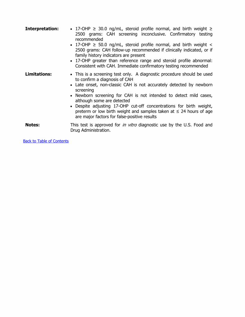
Interpretation: 17-OHP ≥ 30.0 ng/mL, steroid profile normal, and birth weight ≥ 2500 grams: CAH screening inconclusive. Confirmatory testing recommended
17-OHP ≥ 50.0 ng/mL, steroid profile normal, and birth weight < 2500 grams: CAH follow-up recommended if clinically indicated, or if family history indicators are present
17-OHP greater than reference range and steroid profile abnormal: Consistent with CAH. Immediate confirmatory testing recommended
Limitations:
This is a screening test only. A diagnostic procedure should be used to confirm a diagnosis of CAH
Late onset, non-classic CAH is not accurately detected by newborn screening
Newborn screening for CAH is not intended to detect mild cases, although some are detected
Despite adjusting 17-OHP cut-off concentrations for birth weight, preterm or low birth weight and samples taken at ≤ 24 hours of age are major factors for false-positive results
Notes: This test is approved for in vitro diagnostic use by the U.S. Food and Drug Administration.
Back to Table of Contents
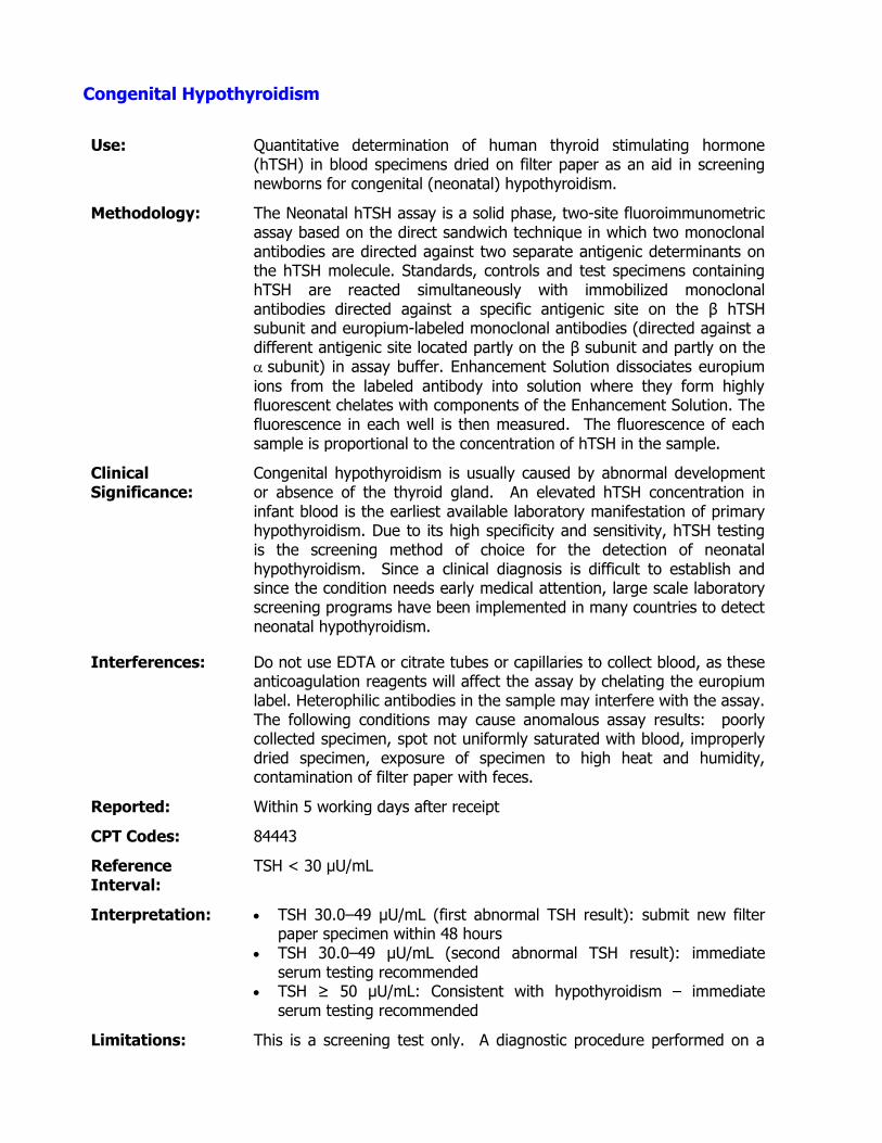
Congenital Hypothyroidism
Use: Quantitative determination of human thyroid stimulating hormone (hTSH) in blood specimens dried on filter paper as an aid in screening newborns for congenital (neonatal) hypothyroidism.
Methodology: The Neonatal hTSH assay is a solid phase, two-site fluoroimmunometric assay based on the direct sandwich technique in which two monoclonal antibodies are directed against two separate antigenic determinants on the hTSH molecule. Standards, controls and test specimens containing hTSH are reacted simultaneously with immobilized monoclonal antibodies directed against a specific antigenic site on the β hTSH subunit and europium-labeled monoclonal antibodies (directed against a different antigenic site located partly on the β subunit and partly on the subunit) in assay buffer. Enhancement Solution dissociates europium
ions from the labeled antibody into solution where they form highly fluorescent chelates with components of the Enhancement Solution. The fluorescence in each well is then measured. The fluorescence of each sample is proportional to the concentration of hTSH in the sample.
Clinical Significance:
Congenital hypothyroidism is usually caused by abnormal development or absence of the thyroid gland. An elevated hTSH concentration in infant blood is the earliest available laboratory manifestation of primary hypothyroidism. Due to its high specificity and sensitivity, hTSH testing is the screening method of choice for the detection of neonatal hypothyroidism. Since a clinical diagnosis is difficult to establish and since the condition needs early medical attention, large scale laboratory screening programs have been implemented in many countries to detect neonatal hypothyroidism.
Interferences: Do not use EDTA or citrate tubes or capillaries to collect blood, as these anticoagulation reagents will affect the assay by chelating the europium label. Heterophilic antibodies in the sample may interfere with the assay. The following conditions may cause anomalous assay results: poorly collected specimen, spot not uniformly saturated with blood, improperly dried specimen, exposure of specimen to high heat and humidity, contamination of filter paper with feces.
Reported: Within 5 working days after receipt
CPT Codes: 84443
Reference Interval:
TSH < 30 µU/mL
Interpretation: TSH 30.0–49 µU/mL (first abnormal TSH result): submit new filter paper specimen within 48 hours
TSH 30.0–49 µU/mL (second abnormal TSH result): immediate serum testing recommended
TSH ≥ 50 µU/mL: Consistent with hypothyroidism – immediate serum testing recommended
Limitations: This is a screening test only. A diagnostic procedure performed on a

serum sample should be used to confirm a diagnosis of congenital hypothyroidism.
Notes: This test is approved for in vitro diagnostic use by the U.S. Food and Drug Administration.
Back to Table of Contents
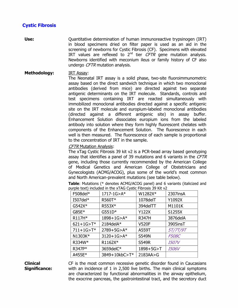
Cystic Fibrosis
Use: Quantitative determination of human immunoreactive trypsinogen (IRT) in blood specimens dried on filter paper is used as an aid in the screening of newborns for Cystic Fibrosis (CF). Specimens with elevated IRT values are reflexed to 2nd tier CFTR gene mutation analysis. Newborns identified with meconium ileus or family history of CF also undergo CFTR mutation analysis.
Methodology: IRT Assay: The Neonatal IRT assay is a solid phase, two-site fluoroimmunometric assay based on the direct sandwich technique in which two monoclonal antibodies (derived from mice) are directed against two separate antigenic determinants on the IRT molecule. Standards, controls and test specimens containing IRT are reacted simultaneously with immobilized monoclonal antibodies directed against a specific antigenic site on the IRT molecule and europium-labeled monoclonal antibodies (directed against a different antigenic site) in assay buffer. Enhancement Solution dissociates europium ions from the labeled antibody into solution where they form highly fluorescent chelates with components of the Enhancement Solution. The fluorescence in each well is then measured. The fluorescence of each sample is proportional to the concentration of IRT in the sample.
CFTR Mutation Analysis: The xTag Cystic Fibrosis 39 kit v2 is a PCR-bead array based genotyping assay that identifies a panel of 39 mutations and 6 variants in the CFTR gene, including those currently recommended by the American College of Medical Genetics and American College of Obstetricians and Gynecologists (ACMG/ACOG), plus some of the world’s most common and North American-prevalent mutations (see table below).
Table: Mutations (* denotes ACMG/ACOG panel) and 6 variants (italicized and purple text) included in the xTAG Cystic Fibrosis 39 Kit v2
F508del* 1717-1G>A* W1282X* 2307insA
I507del* R560T* 1078delT Y1092X
G542X* R553X* 394delTT M1101K
G85E* G551D* Y122X S1255X
R117H* 1898+1G>A* R347H 3876delA
621+1G>T* 2184delA* V520F 3905insT
711+1G>T* 2789+5G>A* A559T 5T/7T/9T
N1303K* 3120+1G>A* S549N F508C
R334W* R1162X* S549R I507V
R347P* 3659delC* 1898+5G>T I506V
A455E* 3849+10kbC>T* 2183AA>G
Clinical Significance:
CF is the most common recessive genetic disorder found in Caucasians with an incidence of 1 in 2,500 live births. The main clinical symptoms are characterized by functional abnormalities in the airway epithelium, the exocrine pancreas, the gastrointestinal tract, and the secretory duct
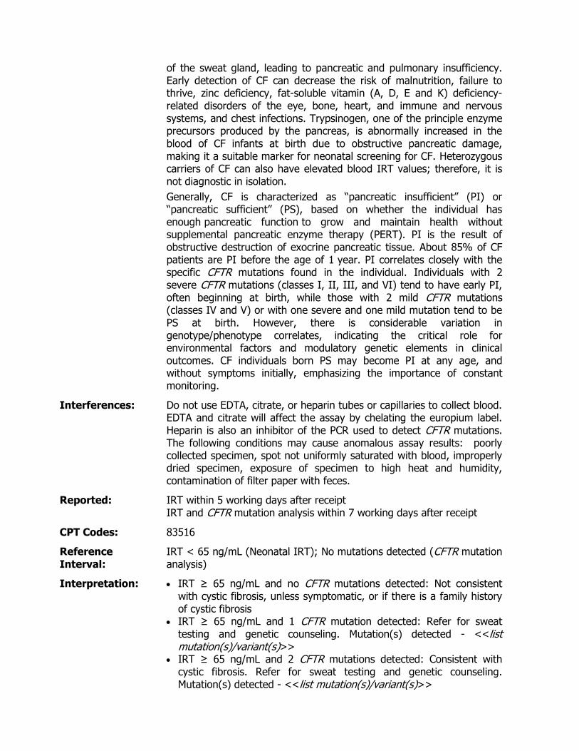
of the sweat gland, leading to pancreatic and pulmonary insufficiency. Early detection of CF can decrease the risk of malnutrition, failure to thrive, zinc deficiency, fat-soluble vitamin (A, D, E and K) deficiency-related disorders of the eye, bone, heart, and immune and nervous systems, and chest infections. Trypsinogen, one of the principle enzyme precursors produced by the pancreas, is abnormally increased in the blood of CF infants at birth due to obstructive pancreatic damage, making it a suitable marker for neonatal screening for CF. Heterozygous carriers of CF can also have elevated blood IRT values; therefore, it is not diagnostic in isolation.
Generally, CF is characterized as “pancreatic insufficient” (PI) or “pancreatic sufficient” (PS), based on whether the individual has enough pancreatic function to grow and maintain health without supplemental pancreatic enzyme therapy (PERT). PI is the result of obstructive destruction of exocrine pancreatic tissue. About 85% of CF patients are PI before the age of 1 year. PI correlates closely with the specific CFTR mutations found in the individual. Individuals with 2 severe CFTR mutations (classes I, II, III, and VI) tend to have early PI, often beginning at birth, while those with 2 mild CFTR mutations (classes IV and V) or with one severe and one mild mutation tend to be PS at birth. However, there is considerable variation in genotype/phenotype correlates, indicating the critical role for environmental factors and modulatory genetic elements in clinical outcomes. CF individuals born PS may become PI at any age, and without symptoms initially, emphasizing the importance of constant monitoring.
Interferences: Do not use EDTA, citrate, or heparin tubes or capillaries to collect blood. EDTA and citrate will affect the assay by chelating the europium label. Heparin is also an inhibitor of the PCR used to detect CFTR mutations. The following conditions may cause anomalous assay results: poorly collected specimen, spot not uniformly saturated with blood, improperly dried specimen, exposure of specimen to high heat and humidity, contamination of filter paper with feces.
Reported: IRT within 5 working days after receipt IRT and CFTR mutation analysis within 7 working days after receipt
CPT Codes: 83516
Reference Interval:
IRT < 65 ng/mL (Neonatal IRT); No mutations detected (CFTR mutation analysis)
Interpretation: IRT ≥ 65 ng/mL and no CFTR mutations detected: Not consistent with cystic fibrosis, unless symptomatic, or if there is a family history of cystic fibrosis
IRT ≥ 65 ng/mL and 1 CFTR mutation detected: Refer for sweat testing and genetic counseling. Mutation(s) detected - <<list mutation(s)/variant(s)>>
IRT ≥ 65 ng/mL and 2 CFTR mutations detected: Consistent with cystic fibrosis. Refer for sweat testing and genetic counseling. Mutation(s) detected - <<list mutation(s)/variant(s)>>
Refer to the Physicians Guide for Cystic Fibrosis for further details on interpretation of CFTR mutation analysis
Limitations:
The Neonatal IRT assay is a screening test only. Other diseases may mimic PI CF, including other causes of PI, intestinal malabsorption, and some behavioral problems. Therefore, a diagnostic procedure should be used to confirm a diagnosis of CF. The Cystic Fibrosis 39 kit v2 assay can be used to confirm abnormal IRT values when two mutations are present in either homozygous or compound heterozygous states. However, because this kit only detects a subset of CFTR mutations, albeit common mutations for the US population, individuals with CFTR mutations may be missed. Testing of individuals with an expanded CFTR mutation panel that includes other less common mutations may be indicated if the initial panel of mutations demonstrates a single mutation, or is suspected of having CF. A sweat chloride test should be an early step in the differential diagnosis of PI or malabsorption in newborns, and remains the “gold standard” for diagnosis of CF.
Notes: False negative IRT values are known to occur in some CF newborns that present with meconium ileus. If an infant has meconium ileus or there is a family history of CF, it is important to mark the appropriate area of the NBS collection form, so CFTR mutation analysis will be performed even if the IRT level is < 65 ng/mL.
Both the Neonatal IRT assay and the xTag® Cystic Fibrosis 39 kit v2 assay are approved for in vitro diagnostic use by the U.S. Food and Drug Administration.
Back to Table of Contents
Galactosemia
Use: Quantitative determination of total galactose (galactose and galactose-1-phosphate) in blood specimens dried on filter paper as an aid in screening newborns for classic galactosemia (GALT), galactokinase deficiency (GALK), and galactoepimerase deficiency (GALE). Specimens with elevated total galactose are reflexed to analysis of galactose-1-phosphate uridyl transferase (GALT) enzyme activity. Newborns identified as receiving lactose-free formula also undergo GALT enzyme testing.
Methodology: The GSP Neonatal Total Galactose kit quantitatively measures total galactose, i.e. both galactose and galactose-1-phosphate, in dried blood spot specimens based on a fluorescent galactose oxidase method.
Clinical Significance:
Galactosemia is an autosomal recessive disorder that is characterized by elevated concentrations of galactose in the blood resulting from the absence or dysfunction of any of the three enzymes responsible for the transformation of galactose to glucose, i.e., D-galactose-1-
phosphotransferase, -D-galactose-1-phosphate-uridyltransferase, or UDP-glucose-4-epimerase. If not diagnosed and treated within the newborn period, galactosemia can lead to diarrhea, dehydration, jaundice, hepatic failure, hypoglycemia, cataracts, developmental retardation and death within a few weeks. Sepsis due to Escherichia coli seems to be particularly frequent among galactosemic neonates and is usually the cause of death. Treatment of the disease consists of withdrawal of all foods containing lactose and galactose from the diet.
Interferences: Infants that have not ingested breast milk or formula containing lactose prior to the sample collection may have lower total galactose values. Conjugated bilirubin concentrations greater than 16.6 mg/dL and acetaminophen concentrations greater than 2.75 mg/dL in the blood of infants may decrease measured TG concentrations, which may cause false negative results. Exchange transfusions may also lead to a false negative screening test. The following conditions may cause anomalous assay results: poorly collected specimen, spot not uniformly saturated with blood, improperly dried specimen, exposure of specimen to high heat and humidity, contamination of filter paper with feces.
Reported: Within 5 working days after receipt
CPT Codes: 82760
Reference Interval:
TG < 7 mg/dL or TG < 10 mg/dL with GALT Enzyme Activity Within Normal Limits (WNL)
Interpretation: TG < 10 mg/dL and GALT enzyme WNL: Not consistent with classic galactosemia
TG 10 - < 17 mg/dL and GALT enzyme WNL: Not consistent with classic galactosemia; repeat testing at provider’s discretion
TG ≥ 17 mg/dL and GALT enzyme WNL: Possible variant form of galactosemia, carrier or normal genotype; consult with metabolic specialist
TG ≥ 7 mg/dL and GALT enzyme Low [2.5 to 3.5 U GALT/g Hb]: Low GALT enzyme; submit repeat specimen as soon as possible
TG ≥ 7 mg/dL and GALT enzyme Absent [< 2.5 U GALT /g Hb]: Consistent with classic galactosemia; recommend immediate confirmatory testing
Note: Interpretations and recommendations may differ when screening involves a repeat specimen.
Limitations:
This is a screening test only. This test is not designed to screen for GALT heterozygosity. A diagnostic procedure should be used to confirm a diagnosis of galactosemia.
Notes: Samples subjected to heat, light, or moisture may have decreased total galactose levels. Samples stored in warm and/or humid conditions lose GALT activity. Therefore, improper handling and storage of specimens may cause both false positive and false negative galactosemia screening results.
This test is approved for in vitro diagnostic use by the U.S. Food and Drug Administration.
Back to Table of Contents

Hemoglobinopathy - Isoelectric Focusing
Use: Qualitative detection of normal and variant hemoglobins in blood specimens dried on filter paper used as an aid in the detection of sickle cell anemia, sickle C disease, S/beta-thalassemia, homozygous C disease, and variant hemoglobinopathies in newborns and children, and in adults.
Methodology: Hemoglobin variants are separated by isoelectric focusing in pH gradient agarose gels. Hemoglobin bands are visualized by staining with o-dianisidine, which is readily oxidized by the heme portion of the hemoglobin molecule in the presence of hydrogen peroxide. The reaction forms an insoluble precipitate that intensifies each band.
Clinical Significance:
Detection of hemoglobin variants is important as a diagnostic tool and for genetic counseling. Detecting hemoglobins S, C and E (Hb S, Hb C, and Hb E) is particularly important because each of these variants in their homozygous state produces clinically significant effects. Additionally, these variants are found as double heterozygotes (Hb SC, Hb SE) that also produce disease states.
Screening of blood for hemoglobin variants has become important mainly for detection of Hb S. Early treatment of sickle cell anemia (Hb SS) with antibiotics has lessened the infections associated with the disease.
Specimen: Type: Dried blood spot collected from infant in accordance with the current
CLSI Section NBSo1-A6 standards; or dried blood spot collected from adult of childbearing age (females < 46 years of age, maternity or family planning patients of any age, males of any age). Newborn screening specimen must not be applied to filter paper from EDTA, heparin, or citrate collected blood.
Volume: 5 filled circles (Newborn) 3 filled circles (Adult/Child)
Container: ODH #450 Newborn Screening Form (Newborn) ODH #485 Child/Adult Sickle Cell Screening Form (Child/Adult) The submitter must ensure that the expiration date of the screening form has not passed. Legibly print and complete all information requested. See sample collection section for detailed instructions on completion of screening form.
Interferences: Older samples may yield “aging bands” due to decomposition and/or oxidation of hemoglobin. The following conditions may cause anomalous assay results: poorly collected specimen, spot not uniformly saturated with blood, improperly dried specimen, exposure of specimen to high heat and humidity, contamination of filter paper with feces.
Reported: Within 5 working days after receipt (Newborn) Within 10 working days after receipt (Child/Adult)
CPT Codes: 83020-52
Reference Interval:
Hb FA (Newborn) Hb AA (Child/Adult)
Interpretation: F Only: Submit new filter paper specimen at 2 months of age. Consistent with transfusion: Submit new filter paper specimen at 4
months of age FA, Bart’s: Recommend CBC with indices at 1 year of age; or at 3
months if of Asian descent FAS: Consistent with S trait. Submit new filter paper specimen at 4
months of age FAC: Consistent with C trait. Submit new filter paper specimen at 4
months of age FA, Other: Consistent with trait. Submit new filter paper specimen at
4 months of age FS: Consistent with SS disease. Refer to pediatric hematologist for
confirmatory testing FC: Consistent with CC disease. Refer to pediatric hematologist for
confirmatory testing FSC: Consistent with SC disease. Refer to pediatric hematologist for
confirmatory testing FSA: Consistent with S-thalassemia. Refer to pediatric hematologist
for confirmatory testing F, Other: Consistent with disease. Refer to pediatric hematologist to
confirm
Note: Any variant reported as “F, Other” or “FA, Other” will include an additional comment to convey location of the unidentified variant, e.g., “Other Band in D/G Region” or “Other Band in E/O Region”, etc.
Limitations:
Isoelectric focusing is able to separate hemoglobin variants with isoelectric points (pI) that differ by 0.01 pH units or greater. Variants with the same pI migrate together.
This is a screening test only. A diagnostic procedure should be used to confirm the identity of variant hemoglobins.
Notes: This test is approved for in vitro diagnostic use by the U.S. Food and Drug Administration.
Back to Table of Contents

Fatty Acid Oxidation Disorders
Use: Quantitative measurement of acylcarnitines C0, C4, C6, C8, C10, C10:1, C14, C16, C16OH, C16:1OH, C18, C18OH, C18:1, C18:1OH, and C18:2 in blood specimens dried on filter paper as an aid in screening newborns for medium-chain acyl-CoA dehydrogenase deficiency and other fatty acid oxidation (FAO) and organic acid disorders, including carnitine uptake defect, short-chain acyl-CoA dehydrogenase deficiency, glutaric acidemia type II, very long-chain acyl-CoA dehydrogenase deficiency, carnitine acylcarnitine translocase deficiency, carnitine palmitoyltransferase I deficiency, carnitine palmitoyltransferase II deficiency, long-chain L-3-hydroxyacyl-CoA dehydrogenase deficiency, and trifunctional protein deficiency.
Methodology: The measurement of free carnitine and acylcarnitines uses an Amino Acids and Acylcarnitines tandem mass spectrometry (MS/MS) kit to extract dried blood spots with a solution containing stable-isotope labeled internal standards with subsequent analysis by MS/MS. The response of each analyte relative to its corresponding stable-isotope labeled internal standard is proportional to analyte concentration.
Clinical Significance:
Elevated or decreased free carnitine and elevated acylcarnitine levels in newborn blood can be indicative of one or more of several metabolic disorders. Free carnitine and acylcarnitines are markers for disorders that are generally classified as FAO disorders and organic aciduria (OA) disorders. These disorders are inborn errors of metabolism (or genetic metabolic deficiencies).
In FAO disorders, enzymes necessary for fatty acid breakdown are unavailable or have reduced activity. Breakdown, or oxidation, of fatty acids is necessary for energy production when glucose levels are low. Without this energy supply, some individuals may have recurring incidences of low blood sugar levels. In cases of fasting, often caused by illnesses such as ear infections or flu, there may be metabolic crisis. Affected individuals may show vomiting, diarrhea, lethargy, seizures or coma. Failure to diagnose FAO disorders may result in excessive fat build-up in the liver, heart and kidneys. This build-up can cause a variety of symptoms, ranging from hepatic failure, encephalopathy, heart and eye complications to general problems with muscle development. Many of these clinical symptoms can lead to death. Many deaths due to FAO disorders have been misdiagnosed as Sudden Infant Death Syndrome (SIDS) or Reye’s Syndrome.
Interferences: High nutritional intake of carnitine or medium-chain triglyceride (MCT) oil may interfere with the measurement of acylcarnitines. Variables such as hematocrit, prematurity, and age of infant may affect the interpretation of values produced by the assay. The following conditions may cause anomalous assay results: poorly collected specimen, spot not uniformly saturated with blood, improperly dried specimen, exposure of specimen to high heat and humidity, contamination of filter paper with feces.
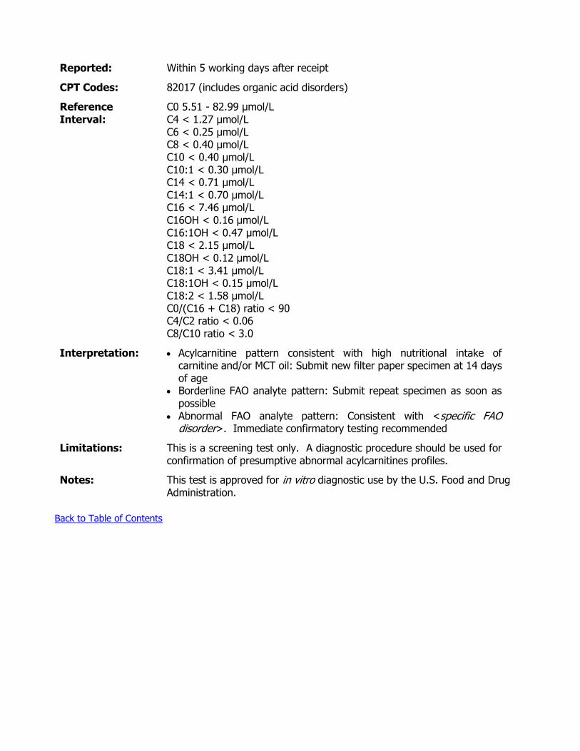
Reported: Within 5 working days after receipt
CPT Codes: 82017 (includes organic acid disorders)
Reference Interval:
C0 5.51 - 82.99 µmol/L C4 < 1.27 µmol/L C6 < 0.25 µmol/L C8 < 0.40 µmol/L C10 < 0.40 µmol/L C10:1 < 0.30 µmol/L C14 < 0.71 µmol/L C14:1 < 0.70 µmol/L C16 < 7.46 µmol/L C16OH < 0.16 µmol/L C16:1OH < 0.47 µmol/L C18 < 2.15 µmol/L C18OH < 0.12 µmol/L C18:1 < 3.41 µmol/L C18:1OH < 0.15 µmol/L C18:2 < 1.58 µmol/L C0/(C16 + C18) ratio < 90 C4/C2 ratio < 0.06 C8/C10 ratio < 3.0
Interpretation: Acylcarnitine pattern consistent with high nutritional intake of carnitine and/or MCT oil: Submit new filter paper specimen at 14 days of age
Borderline FAO analyte pattern: Submit repeat specimen as soon as possible
Abnormal FAO analyte pattern: Consistent with <specific FAO disorder>. Immediate confirmatory testing recommended
Limitations:
This is a screening test only. A diagnostic procedure should be used for confirmation of presumptive abnormal acylcarnitines profiles.
Notes: This test is approved for in vitro diagnostic use by the U.S. Food and Drug Administration.
Back to Table of Contents
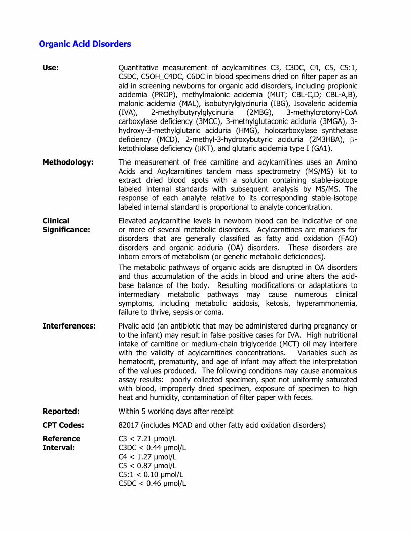
Organic Acid Disorders
Use: Quantitative measurement of acylcarnitines C3, C3DC, C4, C5, C5:1, C5DC, C5OH_C4DC, C6DC in blood specimens dried on filter paper as an aid in screening newborns for organic acid disorders, including propionic acidemia (PROP), methylmalonic acidemia (MUT; CBL-C,D; CBL-A,B), malonic acidemia (MAL), isobutyrylglycinuria (IBG), Isovaleric acidemia (IVA), 2-methylbutyrylglycinuria (2MBG), 3-methylcrotonyl-CoA carboxylase deficiency (3MCC), 3-methylglutaconic aciduria (3MGA), 3-hydroxy-3-methylglutaric aciduria (HMG), holocarboxylase synthetase
deficiency (MCD), 2-methyl-3-hydroxybutyric aciduria (2M3HBA), -
ketothiolase deficiency (KT), and glutaric acidemia type I (GA1).
Methodology: The measurement of free carnitine and acylcarnitines uses an Amino Acids and Acylcarnitines tandem mass spectrometry (MS/MS) kit to extract dried blood spots with a solution containing stable-isotope labeled internal standards with subsequent analysis by MS/MS. The response of each analyte relative to its corresponding stable-isotope labeled internal standard is proportional to analyte concentration.
Clinical Significance:
Elevated acylcarnitine levels in newborn blood can be indicative of one or more of several metabolic disorders. Acylcarnitines are markers for disorders that are generally classified as fatty acid oxidation (FAO) disorders and organic aciduria (OA) disorders. These disorders are inborn errors of metabolism (or genetic metabolic deficiencies).
The metabolic pathways of organic acids are disrupted in OA disorders and thus accumulation of the acids in blood and urine alters the acid-base balance of the body. Resulting modifications or adaptations to intermediary metabolic pathways may cause numerous clinical symptoms, including metabolic acidosis, ketosis, hyperammonemia, failure to thrive, sepsis or coma.
Interferences: Pivalic acid (an antibiotic that may be administered during pregnancy or to the infant) may result in false positive cases for IVA. High nutritional intake of carnitine or medium-chain triglyceride (MCT) oil may interfere with the validity of acylcarnitines concentrations. Variables such as hematocrit, prematurity, and age of infant may affect the interpretation of the values produced. The following conditions may cause anomalous assay results: poorly collected specimen, spot not uniformly saturated with blood, improperly dried specimen, exposure of specimen to high heat and humidity, contamination of filter paper with feces.
Reported: Within 5 working days after receipt
CPT Codes: 82017 (includes MCAD and other fatty acid oxidation disorders)
Reference Interval:
C3 < 7.21 µmol/L C3DC < 0.44 µmol/L C4 < 1.27 µmol/L C5 < 0.87 µmol/L C5:1 < 0.10 µmol/L C5DC < 0.46 µmol/L
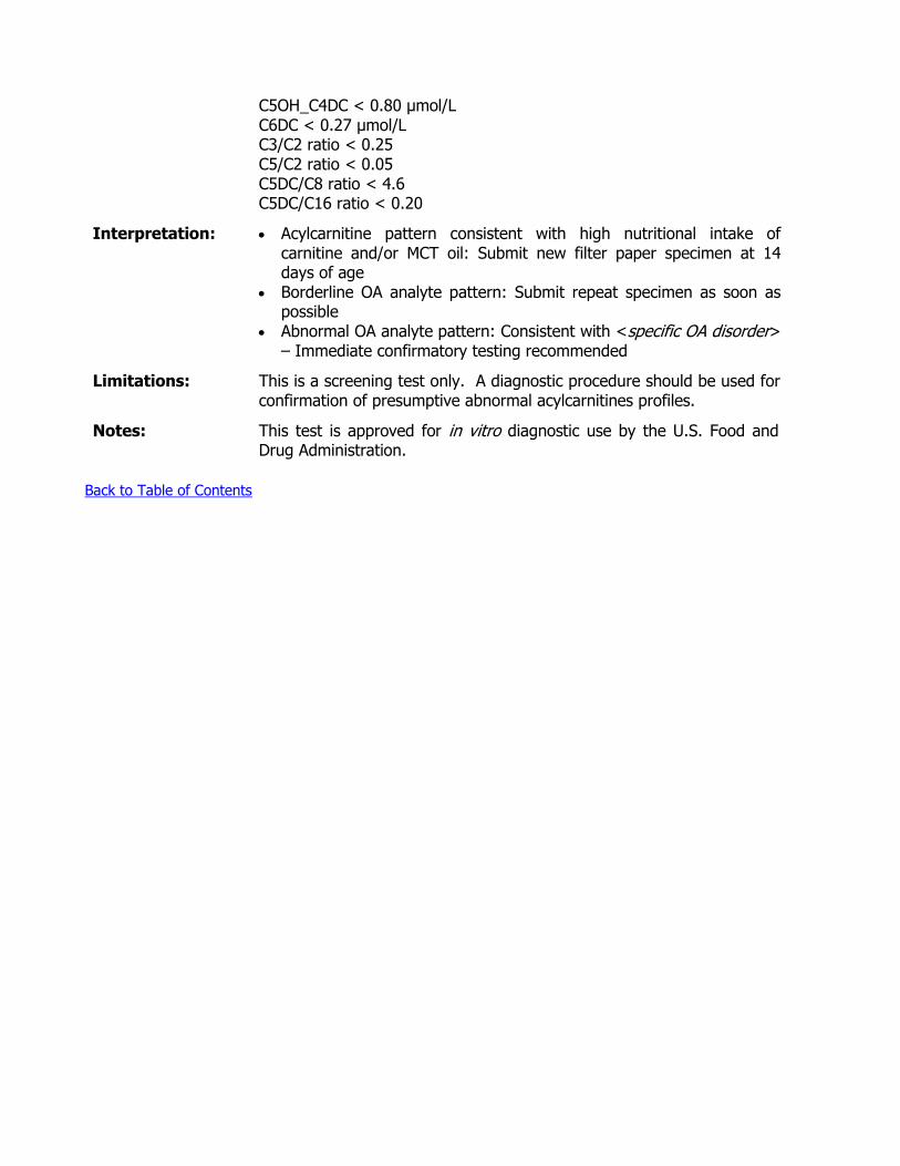
C5OH_C4DC < 0.80 µmol/L C6DC < 0.27 µmol/L C3/C2 ratio < 0.25 C5/C2 ratio < 0.05 C5DC/C8 ratio < 4.6 C5DC/C16 ratio < 0.20
Interpretation: Acylcarnitine pattern consistent with high nutritional intake of carnitine and/or MCT oil: Submit new filter paper specimen at 14 days of age
Borderline OA analyte pattern: Submit repeat specimen as soon as possible
Abnormal OA analyte pattern: Consistent with <specific OA disorder> – Immediate confirmatory testing recommended
Limitations:
This is a screening test only. A diagnostic procedure should be used for confirmation of presumptive abnormal acylcarnitines profiles.
Notes: This test is approved for in vitro diagnostic use by the U.S. Food and Drug Administration.
Back to Table of Contents
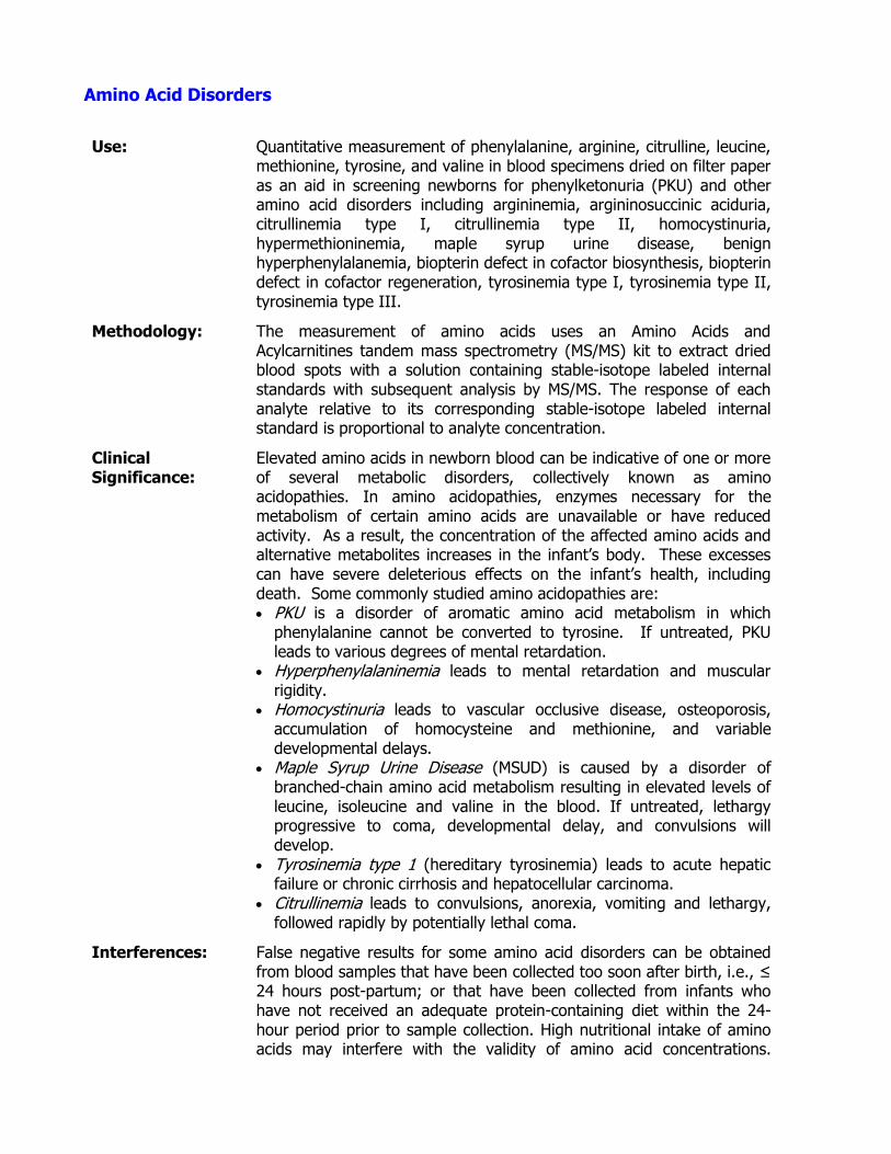
Amino Acid Disorders
Use: Quantitative measurement of phenylalanine, arginine, citrulline, leucine, methionine, tyrosine, and valine in blood specimens dried on filter paper as an aid in screening newborns for phenylketonuria (PKU) and other amino acid disorders including argininemia, argininosuccinic aciduria, citrullinemia type I, citrullinemia type II, homocystinuria, hypermethioninemia, maple syrup urine disease, benign hyperphenylalanemia, biopterin defect in cofactor biosynthesis, biopterin defect in cofactor regeneration, tyrosinemia type I, tyrosinemia type II, tyrosinemia type III.
Methodology: The measurement of amino acids uses an Amino Acids and Acylcarnitines tandem mass spectrometry (MS/MS) kit to extract dried blood spots with a solution containing stable-isotope labeled internal standards with subsequent analysis by MS/MS. The response of each analyte relative to its corresponding stable-isotope labeled internal standard is proportional to analyte concentration.
Clinical Significance:
Elevated amino acids in newborn blood can be indicative of one or more of several metabolic disorders, collectively known as amino acidopathies. In amino acidopathies, enzymes necessary for the metabolism of certain amino acids are unavailable or have reduced activity. As a result, the concentration of the affected amino acids and alternative metabolites increases in the infant’s body. These excesses can have severe deleterious effects on the infant’s health, including death. Some commonly studied amino acidopathies are: PKU is a disorder of aromatic amino acid metabolism in which
phenylalanine cannot be converted to tyrosine. If untreated, PKU leads to various degrees of mental retardation.
Hyperphenylalaninemia leads to mental retardation and muscular rigidity.
Homocystinuria leads to vascular occlusive disease, osteoporosis, accumulation of homocysteine and methionine, and variable developmental delays.
Maple Syrup Urine Disease (MSUD) is caused by a disorder of branched-chain amino acid metabolism resulting in elevated levels of leucine, isoleucine and valine in the blood. If untreated, lethargy progressive to coma, developmental delay, and convulsions will develop.
Tyrosinemia type 1 (hereditary tyrosinemia) leads to acute hepatic failure or chronic cirrhosis and hepatocellular carcinoma.
Citrullinemia leads to convulsions, anorexia, vomiting and lethargy, followed rapidly by potentially lethal coma.
Interferences: False negative results for some amino acid disorders can be obtained from blood samples that have been collected too soon after birth, i.e., ≤ 24 hours post-partum; or that have been collected from infants who have not received an adequate protein-containing diet within the 24-hour period prior to sample collection. High nutritional intake of amino acids may interfere with the validity of amino acid concentrations.

Variables such as hematocrit, prematurity, and age of infant may affect the interpretation of the values produced. The following conditions may cause anomalous assay results: poorly collected specimen, spot not uniformly saturated with blood, improperly dried specimen, exposure of specimen to high heat and humidity, contamination of filter paper with feces.
Reported: Within 5 working days after receipt
CPT Codes: 82139
Reference Interval:
Phenylalanine < 150 µmol/L Arginine < 100 µmol/L Citrulline < 55 µmol/L Leucine < 355 µmol/L Methionine < 75 µmol/L Tyrosine < 400 µmol/L Valine < 330 µmol/L Phe/Tyr ratio < 2.0 Cit/Arg ratio < 6.5 Leu/Phe ratio < 4.8 Met/Phe ratio < 1.2
Interpretation: Pattern of multiple amino acid elevations consistent with TPN: Submit new filter paper specimen at 14 days of age
Borderline amino acid pattern: Submit repeat specimen as soon as possible
Abnormal amino acid pattern: Consistent with <specific AA disorder>; Immediate confirmatory testing recommended
Limitations:
This is a screening test only. A diagnostic procedure should be used for confirmation of presumptive abnormal amino acid profiles.
Notes: This test is approved for in vitro diagnostic use by the U.S. Food and Drug Administration.
Back to Table of Contents
Severe Combined Immunodeficiency (SCID)
Use: Quantitative determination of T-Cell Receptor Excision Circles (TRECs) in dried blood spots (DBSs) as an aid in screening newborns for severe combined immunodeficiency (SCID).
Methodology: Real-time, quantitative polymerase chain reaction (rt Q-PCR) amplification is used to assess levels of TRECs in DBSs collected from newborn infants, as a marker of T-cell development.
Clinical Significance:
The initial protective effects afforded by maternal antibodies transferred during pregnancy and nursing can initially mask the inherent immunodeficiency of the SCID infant during the first weeks of life. Subsequently, SCID infants experience recurrent infections, including pneumonia, bronchitis, meningitis, ear infections, thrush, chronic diarrhea, fail to thrive, and often develop a scaly erythematous rash over the entire body. Left undiagnosed, SCID infants usually die from severe infections within the first year of life. Identification of the asymptomatic infant with a primary immunodeficiency during the first few weeks of life is essential for successful treatment, which generally involves immune reconstitution by hematopoietic cell transplantation (HCT), gene therapy or enzyme replacement (for ADA deficiency); thereafter, morbidity from infections makes treatment much less effective.
Interferences: Do not use heparinized tubes or capillaries to collect blood; heparin is a known inhibitor of PCR. The following conditions may cause anomalous assay results: poorly collected specimen, spot not uniformly saturated with blood, improperly dried specimen, exposure of specimen to high heat and humidity, contamination of filter paper with feces.
Reported: Within 5 working days after receipt
CPT Codes: 81479
Reference Interval:
Term (gestational age ≥ 37 weeks) TREC threshold cycle (Ct) < 33.5 (i.e., > 26 TREC copies) Preterm (gestational age < 37 weeks) TREC Ct < 33.8 (i.e., > 21.5 TREC copies)
Interpretation: Term TREC Ct < 33.5: In-range TREC Ct 33.5 – 35.4: Submit repeat specimen as soon as possible TREC Ct 33.5 – 35.4 (for a previously abnormal term specimen):
Consistent with primary immunodeficiency due to two abnormal screen results; immediate confirmatory testing recommended
TREC Ct ≥ 35.5: Consistent with primary immunodeficiency; immediate confirmatory testing recommended
RNAseP ≥ 28.0 Ct: Poor DNA amplification; submit repeat specimen as soon as possible
Preterm TREC Ct < 33.8: In-range
TREC Ct ≥ 33.8 and ≤ 45: Submit new filter paper specimen at 37 weeks gestational age
TREC Ct = > 45: Consistent with primary immunodeficiency; immediate confirmatory testing recommended
RNAseP ≥ 28.0 Ct: Poor DNA amplification; submit repeat specimen as soon as possible
Limitations:
This is a screening test only. A diagnostic procedure should be used to confirm a diagnosis of SCID. Other primary or secondary immunodeficiencies may be detected by the assay; while potentially beneficial, this is not the intended use of the assay.
Notes: This is a laboratory-developed test; performance characteristics have been validated and determined to be suitable for diagnostic purposes by the OSDH PHL. This test has not been cleared or approved by the U.S. Food and Drug Administration.
Back to Table of Contents
Ova and Parasites (O&P), Fecal - Microscopic Examination
Use: Detection of intestinal protozoa (amoeba, flagellates, ciliates, coccidia), microsporidia, nematodes, trematodes, cestodes and helminths.
Methodology: Wet mounts and modified acid-fast staining of formalin-fixed specimens following ethyl acetate concentration, and trichrome staining of PVA-fixed specimens. Undiagnosed specimens may be referred to the CDC for further testing.
Clinical Significance:
It is a common conception that the burden of parasitic diseases lies in the tropics and subtropics; however, parasitic disease is also a significant issue for the United States, where it is estimated that alone 7.4 million cases of trichomoniasis, 2 million cases Giardia, and 300,000 cases of Cryptosporidium occur each year. With the advent of managed care, many laboratories have ceased traditional parasite testing and perform only enzyme immunoassays for Giardia, Cryptosporidium and Entamoeba histolytica/dispar. While these tests are very sensitive and specific, a negative test result does not rule-out the possibility of parasitic infection. In such cases, further testing using traditional parasite methodologies may be indicated.
Further background information, fact sheets, statistics and educational resources may be found at the OSDH Acute Disease Services website.
Specimen: Type: Solid or liquid feces
Volume: Solid feces: 2 grams Liquid feces: 5-10 mL
Container: O & P collection kit (contains one vial of PVA and one vial of 10% formalin; available from OSDH PHL)
Collection: Stool specimens should be obtained early in the course of illness; optimum time is 1-3 days after onset of illness, during the early morning hours when the parasite(s) should be present in the greatest numbers.
Many parasites are shed intermittently during the course of an infection; therefore, for initial detection, 3 separate specimens should be collected on separate days, at a minimum of 24 hours apart – one every other day (over 5 days) is optimal but collection should not exceed 10 days. Note: specimens may need to be submitted individually to avoid delayed submission and rejection (> 7 days from date of collection).
Stool specimens should be collected before antibiotic therapy has been initiated. Post-therapy specimens should be collected approximately 3 to 4 weeks after completion of therapy using the same collection procedure/timing as that used for pre-therapy detection.
1. Stool specimens are ideally passed into a clean, dry bedpan or container without being contaminated with urine. Alternatively, a clean newspaper, or large plastic bag or plastic wrap can be placed over the toilet seat opening. DO NOT pass specimen into the toilet or directly into the collection vials. Do NOT mix specimen with urine

or water. Infant specimens may be collected in a disposable diaper by turning the diaper inside-out with the plastic side facing the skin. Specimens collected on the absorbent side are not acceptable.
2. Open the O&P kit and label both vials (10% formalin and PVA) with the following information: Patient name Date and time specimen was collected Check appropriate box for specimen description
3. Carefully, open the 10% formalin vial. 4. Using the collection spoon attached to the cap, add enough stool
specimen (about a walnut-size portion, ~2 grams or 5-10 mL) until the liquid in the vial reaches the arrow on the label. Do not over-fill the vial. Take care to sample areas of the specimen that appear bloody, slimy, or watery. If the stool is hard, sample from each end and the middle of the stool.
5. Thoroughly mix the contents of the vial using the collection spoon. Solid stool needs to be well broken-up to fix the specimen adequately.
6. Replace the cap on the vial and secure tightly. 7. Shake the vial vigorously to mix. 8. Repeat this process using the PVA vial. 9. Place both vials in the biohazard bag, and place the completed
requisition form in the outer pouch. 10. Place bag containing specimens and requisition form in metal can
and seal. 11. Transport specimens to the laboratory (or return specimens to clinic)
as soon as possible. Store specimens at room temperature.
Interferences: Certain drugs/compounds may render stool specimens unsatisfactory for examination; therefore, specimens should be collected before these drugs/compounds are administered, or collection should be delayed until effects of these drugs/compounds have passed. Such substances include: antacids, kaolin, mineral oil and other oily materials, non-absorbable antidiarrheal preparations, barium or bismuth (7-10 days needed for clearance of effects), antibiotics (2-3 weeks), and gallbladder dyes (3 weeks).
Special
Instructions: Not applicable
Shipping: Ambient temperature for delivery within 7 days of collection
Rejection Criteria: Specimen collected using device other than O&P collection kit Specimens collected on the same day (one will be tested) Specimen received > 7 days from date of collection Other criteria as outlined in Specimen Rejection section of this Test
Directory
Reported: Within 3 days working days after receipt
CPT Codes: 87206, 87207, 87209, 87210
Normal/Abnormal Results:
No Parasite Detected; <Genus or Genus/species> Detected; <Genus or Group> Detected, Unable to Speciate
Interpretation: No Parasite Detected: a negative result does not exclude the possibility of parasitic infection since many parasites are shed intermittently Parasite Detected: diagnostic Parasite Detected, Unable to Speciate: speciation may not be possible in all cases due to poor morphology, low parasitemia, or lack of diagnostic features. Undiagnosed specimens may be referred to the CDC for testing; further specimen collection may be necessary
Limitations: Fixed specimens cannot be used to culture bacterial pathogens.
Notes: This is a laboratory-developed test; performance characteristics have been validated and determined to be suitable for diagnostic purposes by the OSDH PHL. This test has not been cleared or approved by the U.S. Food and Drug Administration.
Back to Table of Contents
Parasites, Blood – Microscopic Examination and/or PCR
Use: Screen for and identification of blood parasites, including species of Plasmodium, Babesia, filariae and Trypanosoma.
Methodology: Light microscopy examination of Giemsa- or Wright-Giemsa-stained blood smears. Malaria specimens are PCR-amplified to aid in species identification. Undiagnosed specimens may be referred to the CDC for further testing.
Clinical Significance:
Plasmodium Malaria occurs mostly in poor, tropical and subtropical areas of the world, and is transmitted by mosquitoes. In many countries where malaria is endemic, it is a leading cause of illness and death. In 2012, an estimated 219 million cases of malaria occurred worldwide and 627,000 people died, mostly comprising children in sub-Saharan Africa. The vast majority of cases in the United States occur in travelers and immigrants returning from countries where malaria is endemic. People with malaria often experience fever, chills, and flu-like illness. Left untreated, they may develop severe complications and die.
Babesia Babesiosis is an infection caused by a malaria-like parasite that also infects the red blood cells, but is transmitted by ticks. Babesia microti is the most common piroplasm infecting humans across the U. S., Europe and Asia. Symptoms of babesiosis often start with high fever and chills. As the infection progresses, patients may develop fatigue, headache, drenching sweats, muscle aches, nausea, and vomiting. Babesiosis is often very mild but can be life-threatening to immunocompromised individuals and the elderly.
Filariae Filariasis is caused by thread-like nematodes belonging to the superfamily Filarioidea. Adult worms reside in the lymphatic system, subcutaneous tissues, eye, or abdominal cavity, depending of the species, and release first-stage larvae (microfilariae) into the bloodstream to be taken up with a blood meal by the arthropod vector, either black flies or mosquitoes. In the vector, they develop into infective larvae that can be transmitted to a new host. Filariasis is usually diagnosed through clinical features and direct observation of microfilariae in Geimsa-stained peripheral blood smears.
Lymphatic filariasis is a painful and profoundly disfiguring disease that occurs in over 120 million people worldwide, and is caused by Wuchereria bancroftii, Brugia malayi, and Brugia timori. While the infection is usually acquired in childhood, its visible manifestations occur later in life, including swollen limbs and breasts (lymphedema), damage to the genitals (hydrocele) and swollen limbs with thickened, hardened skin (elephantiasis). Those with severe symptoms of the disease are often unable to work and may suffer significant social stigma as a result of their disfigurement. In endemic countries, lymphatic filariasis has a major social and economic impact.
Subcutaneous filariasis or loaisis is caused by the eye worm, L. loa, which is associated with migratory angioedemas, known as Calabar
swellings, and damaging migration through the conjunctiva.
Infections of the serous cavity of the abdomen by Mansonella perstans or Mansonella ozzardi, while often asymptomatic, can be associated with angioedema, pruritus, fever, headaches, arthralgias, pulmonary symptoms, adenopathy, hepatomegaly, and neurologic manifestations.
Given the potential for severe damage to major organ systems and the potential psychological damage associated with lymphatic filariasis, obtaining a proper diagnosis is critical to patient care.
Microfilariae of some filarial nematodes (e.g., Onchocerca volvulus, and Mansonella streptocerca) are found in the skin rather than the blood, and skin snips are the specimen of choice.
Trypanosomes Human African trypanosomiasis is caused by infection with Trypanosoma; T. brucei gambiense and T. brucei rhodesiense account for > 98% and 2% of reported cases, respectively, most of which occur in 36 sub-Saharan African countries where tsetse flies (Glossina genus) transmit the disease. In 2009, the number of cases reported dropped below 10,000 for first time in 50 years and in 2012 there were 7216 cases recorded. People most exposed to the tsetse fly live in rural areas. The first stage, or hemolymphatic phase, of infection entails bouts of fever, headaches, joint pains and itching. During the second stage, or neurological or meningoencephalic phase, the parasites infect the central nervous system and symptoms include changes of behavior, disturbance of the sleep cycle, which gives the disease its name, confusion, sensory disturbances and poor coordination. Without treatment, sleeping sickness is considered fatal although cases of healthy carriers have been reported.
Another form of trypanosomiasis, American trypanosomiasis or Chagas disease, occurs mainly in Latin America and is caused by Trypanosoma cruzii. T. cruzi is found mostly in blood-sucking triatome insects (kissing bugs) and small mammals in a sylvatic cycle that is enzootic from the southern and southwestern U. S. to central Argentina and Chile. Inflammatory skin lesions occur at the site of the bite but the infection spreads systemically to include muscles, including the myocardium, and various other tissues. Pathologic changes are often associated with the heart and gut. Approximately 12,000 deaths are attributable to Chagas’ disease each year. It is estimated that 23 million immigrants living in the U. S. are from endemic countries, and about 300,000 of these have chronic T cruzi infection. Approximately two thirds of these immigrants are from Mexico, where the overall prevalence of T cruzi infection is 0.5%-1%. Microscopic detection of circulating trypomastigotes of T. cruzii in blood is usually only possible in acute infections since the level of parasites in the blood of chronic infections is very low.
Further background information, fact sheets, statistics and educational resources may be found at the OSDH Acute Disease Services website.
Specimen: Type: Blood and stained blood smears. Travel history is required.
Volume: Blood: 2-6 mL (malaria only)
Geimsa or Wright-Giemsa stained smears: 1 thin and 1 thick, minimum
Container: Blood: EDTA tube (malaria only) Stained slides: in plastic slide holder
Collection: When malaria, trypanosomiasis or babesiosis is suspected, blood smears should be obtained and examined without delay; rapid diagnosis is critical for appropriate patient management. Extended exposure to EDTA anticoagulants can result in altered parasite morphology. Blood should be collected by venipuncture according to established policies and procedures of the submitting site. Whenever possible, specimens should be collected before treatment is initiated. Since parasitemias may fluctuate, multiple smears might be needed. These can be taken at 8 to 12 hour intervals for 2 to 3 days.
Prior consultation with the OSDH PHL is required before submitting specimens for suspected filarial infection. These parasites have very specific time ranges for proper specimen collection. 90% of the specimens collected outside of these time frames will be negative; therefore, strict adherence to the collection times in the following table is important:
Organism Location Periodicity Collection Time
W. bancroftii Tropics/Subtropics Nocturnal 10:00pm-4:00am
W. bancroftii Pacific Diurnal/sub-periodic 2:00pm-6:00pm
B. malayi SE Asia/SW India Nocturnal 10:00pm-2:00am
B. malayi Indonesia Diurnal/sub-periodic 6:00pm-12:00am
B. timori Indonesia Nocturnal 10:00pm-2:00am
L.loa West/Central Africa Nocturnal 10:00pm-2:00am
M. perstans Africa/S. America Non-periodic Anytime
M. ozzardi Central/S. America Non-periodic Anytime
In most cases, collecting venipuncture blood at the mid-point of the time range will yield optimal results.
Stained, thick and thin blood smears should be prepared by the submitter immediately after collection of venipuncture blood, or at most, within 1 hour of collection; delay can result in changes in parasite morphology and staining characteristics. Giemsa stain is the preferred stain; if another stain is used, such as hematoxylin or Wright stain, note this on the test requisition form.
Preparation of Thick Smears 1. Label slide using at least two patient-specific identifiers (e.g., name
and DOB). 2. Place a small drop of blood (10 to 20 µL) in the center of the pre-
cleaned, labeled slide. 3. Using the corner of another slide or glass, wooden or plastic
applicator, spread the drop in a circular pattern until it is about the size of a dime (1.5 cm2). A thick smear of proper density is: if smear is placed (wet) over newsprint, the words can be barely read.
4. Lay the slides flat and allow smear to dry thoroughly at room temperature (30 minutes to several hours). Protect from dust.
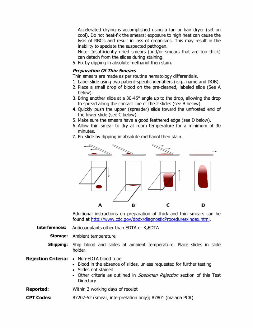
Accelerated drying is accomplished using a fan or hair dryer (set on cool). Do not heat-fix the smears; exposure to high heat can cause the lysis of RBC’s and result in loss of organisms. This may result in the inability to speciate the suspected pathogen. Note: Insufficiently dried smears (and/or smears that are too thick) can detach from the slides during staining.
5. Fix by dipping in absolute methanol then stain.
Preparation Of Thin Smears Thin smears are made as per routine hematology differentials. 1. Label slide using two patient-specific identifiers (e.g., name and DOB). 2. Place a small drop of blood on the pre-cleaned, labeled slide (See A
below). 3. Bring another slide at a 30-45° angle up to the drop, allowing the drop
to spread along the contact line of the 2 slides (see B below). 4. Quickly push the upper (spreader) slide toward the unfrosted end of
the lower slide (see C below). 5. Make sure the smears have a good feathered edge (see D below). 6. Allow thin smear to dry at room temperature for a minimum of 30
minutes. 7. Fix slide by dipping in absolute methanol then stain.
Additional instructions on preparation of thick and thin smears can be found at http://www.cdc.gov/dpdx/diagnosticProcedures/index.html.
Interferences: Anticoagulants other than EDTA or K2EDTA
Storage: Ambient temperature
Shipping: Ship blood and slides at ambient temperature. Place slides in slide holder.
Rejection Criteria: Non-EDTA blood tube Blood in the absence of slides, unless requested for further testing Slides not stained Other criteria as outlined in Specimen Rejection section of this Test
Directory
Reported: Within 3 working days of receipt
CPT Codes: 87207-52 (smear, interpretation only); 87801 (malaria PCR)
Normal/Abnormal Results:
No Parasite Detected; <Genus or Genus/species> Detected; <Genus or Group> Detected, Unable to Speciate
Interpretation: No Parasite Detected: a negative result does not exclude the diagnosis of malaria, babesiosis, trypanosomiasis, or filariasis; parasitemias can be periodic and/or vary widely Parasite Detected: diagnostic Parasite Detected, Unable to Speciate: speciation may not be possible in all cases due to poor morphology, low parasitemia, or lack of diagnostic features. Undiagnosed specimens may be referred to the CDC for testing; further specimen collection may be necessary
Limitations:
Multiple, timed collections may be necessary for diagnosis, and microfilariae may be absent in early (pre-patent) or single-sex infections
Notes: This is a laboratory-developed test; performance characteristics have been validated and determined to be suitable for diagnostic purposes by the OSDH PHL. This test has not been cleared or approved by the U.S. Food and Drug Administration.
Back to Table of Contents
Rabies – Direct Fluorescent Antibody
Use: To diagnose rabies virus in animal brain tissue
Methodology: Direct fluorescent antibody (DFA) detection of rabies virus in brain stem and cerebellum (or hippocampus) tissue smears using fluorescence microscopy
Clinical Significance:
Rapid and accurate laboratory diagnosis of rabies infection in animals is imperative for appropriate management/treatment of exposed persons and instigation of control measures in the community.
Further background information, fact sheets, statistics and educational resources may be found at the OSDH Acute Disease Services website.
Specimen: Type: Whole animal: if under 12 inches (excluding tail)
Animal head: if animal is over 12 inches (excluding tail) Brain only: on large animals such as cattle and horses
Volume: Whole, intact brain
Container: Rabies shipping container (available from OSDH PHL Client Services at (405) 271-5070 during regular business hours) or leak-proof container.
Collection: For animals more than 12 inches from nose to base of tail, the head must be removed from the body. For small animals (12 inches or less), the whole animal can be submitted. For very large animals (e.g., cattle and horses), a veterinarian should remove and submit only the brain. Specific instructions are available at the OSDH PHL website, Rabies
Interferences: Chemical fixatives; decomposed brain
Special Instructions:
Do not submit live animals Do not club or shoot animal in head; intact brain is needed for
examination Do not fix brain tissue in formalin or other preservatives Do not freeze Submit specimen with completed Rabies Submission Form OSD Form
460
Shipping: Ship specimens refrigerated on ice packs; do not freeze
Rejection Criteria: Live animal Wild rodent that has exposed only other animals (not humans) Birds, turtles or reptiles Cage-raised animals Whole animal > 12 inches in length (excluding tail) Large animal heads, such as cattle and horses Brain not intact Brain tissue fixed in preservative Brain grossly decomposed Human specimen
Reported: Within 3 days working days after receipt
CPT Codes: N/A
Normal/Abnormal Results:
Rabies Detected; Rabies Not Detected; Indeterminate: sparse, weakly staining pattern; cannot rule-out rabies; Unsatisfactory specimen
Interpretation: Detected: at least +3 to +4 intensity and +2 to +4 distribution of rabies virus antigen in brain stem and cerebellum (or hippocampus)
Not detected: no specific staining Indeterminate: sparse, weakly staining pattern Unsatisfactory:
o Brain stem unavailable for testing while other brain tissues are negative
o Non-specific staining indistinguishable from rabies virus inclusions or that may mask rabies-specific staining
o Decomposed, fixed or morphology compromised
Persons exposed to an animal with unsatisfactory rabies results may be treated on the basis of clinical suspicion of rabies in the animal. Alternative tests (virus isolation, PCR) may be indicated to rule-out or confirm rabies in cases with indeterminate results, or in cases with unsatisfactory or negative results where rabies infection is suspected. Samples requiring confirmation, variant typing, or formalin-fixed tissues may be sent to the CDC for testing.
Limitations:
Decomposed or fixed (formalin or other fixative) brain tissue or brains with compromised morphology (e.g., shooting, clubbing) may produce false negative results. Non-specific staining that is indistinguishable from rabies virus inclusions may mask specific staining.
Notes: The OSDH PHL does not perform rabies testing on human specimens. For consultation on animal bites and rabies risk, contact the OSDH Acute Disease Service Epidemiologist-On-Call at (405) 271-4060. If you suspect a case of human rabies immediately, contact the OSDH Disease and Prevention Services at 405-271-4060. The disease is fatal for humans once signs of the disease appear; it is only prevented when persons are treated soon after exposure.
To inquire about rabies testing, call the OSDH PHL Client Services at (405) 271-5070 during regular business hours.
This is a laboratory-developed test; performance characteristics have been validated and determined to be suitable for diagnostic purposes by the OSDH PHL. This test has not been cleared or approved by the U.S. Food and Drug Administration.
Back to Table of Contents
Respiratory Pathogen Panel (RPP)
Use: Detection and identification of nucleic acids from multiple respiratory viral and bacterial pathogens extracted from nasopharyngeal swabs collected from individuals with clinical signs and symptoms of respiratory tract infection.
Methodology: Specimens are tested using the NxTAG® Respiratory Pathogen Panel (RPP), a qualitative PCR amplification assay.
Clinical Significance:
Respiratory pathogens are responsible for an estimated 80% of respiratory tract infections annually. A respiratory tract infection can be the result of one of dozens of viral or bacterial pathogens. The symptoms caused by these different pathogens are nearly indistinguishable, but how a healthcare provider chooses to treat a respiratory infection may depend greatly on a rapid and accurate diagnosis of the responsible pathogen. These infections can range from a mild, self-limiting illness to severe disease that can cause death. More severe disease is seen in the young, the immunocompromised, and the elderly. The frequency of respiratory viral infections is highest in children under 4 years of age. School children become infected, on average, with 5 to 8 respiratory viruses per year, and adults average 2 to 4 respiratory viruses annually. Bacteria that cause respiratory infections represent approximately 10% of all upper respiratory tract infections. In children, respiratory syncytial virus (RSV) is the most common cause of severe lower respiratory tract infection worldwide. Timely detection of these viruses can lead to initiation of proper antiviral treatment, decreased use of unnecessary antibiotics, reduced transmission of disease from person to person and better clinical outcomes.
Specimen: Type: Nasopharyngeal swab
Volume: 1 or 2 swabs
Container: Tube containing viral transport medium (VTM; provided by OSDH PHL) or other appropriate commercial medium (UTM, M4, and M4RT). VTM contains antibiotics and must be stored in the refrigerator prior to and subsequent to specimen collection. Do not use media if cloudy or when past expiration date.
Collection: Respiratory virus diagnosis depends on the collection of high-quality specimens, their rapid transport to the testing laboratory and appropriate storage before laboratory testing. Specimens for RPP analysis should be taken preferably during the first 3 days after onset of clinical symptoms.
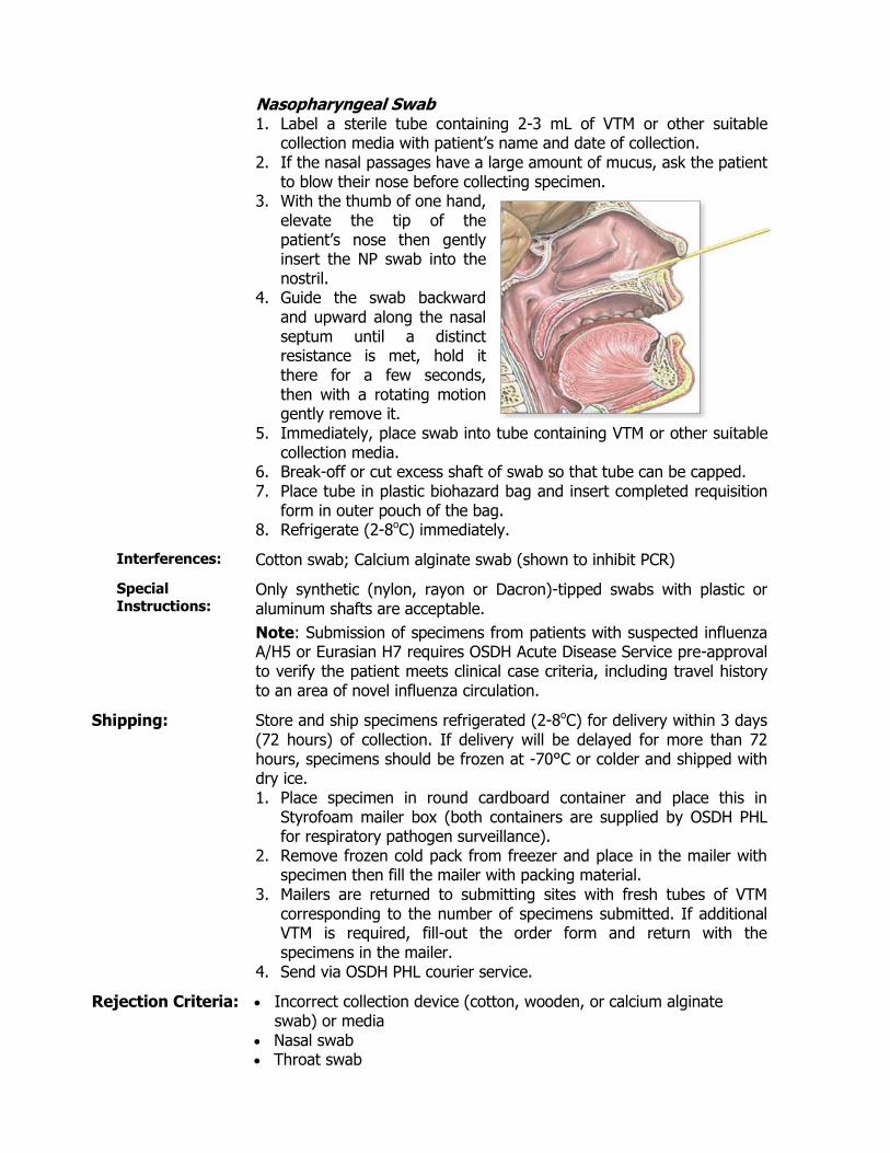
Nasopharyngeal Swab 1. Label a sterile tube containing 2-3 mL of VTM or other suitable
collection media with patient’s name and date of collection. 2. If the nasal passages have a large amount of mucus, ask the patient
to blow their nose before collecting specimen. 3. With the thumb of one hand,
elevate the tip of the patient’s nose then gently insert the NP swab into the nostril.
4. Guide the swab backward and upward along the nasal septum until a distinct resistance is met, hold it there for a few seconds, then with a rotating motion gently remove it.
5. Immediately, place swab into tube containing VTM or other suitable collection media.
6. Break-off or cut excess shaft of swab so that tube can be capped. 7. Place tube in plastic biohazard bag and insert completed requisition
form in outer pouch of the bag. 8. Refrigerate (2-8oC) immediately.
Interferences: Cotton swab; Calcium alginate swab (shown to inhibit PCR)
Special
Instructions: Only synthetic (nylon, rayon or Dacron)-tipped swabs with plastic or aluminum shafts are acceptable.
Note: Submission of specimens from patients with suspected influenza A/H5 or Eurasian H7 requires OSDH Acute Disease Service pre-approval to verify the patient meets clinical case criteria, including travel history to an area of novel influenza circulation.
Shipping: Store and ship specimens refrigerated (2-8oC) for delivery within 3 days (72 hours) of collection. If delivery will be delayed for more than 72 hours, specimens should be frozen at -70°C or colder and shipped with dry ice. 1. Place specimen in round cardboard container and place this in
Styrofoam mailer box (both containers are supplied by OSDH PHL for respiratory pathogen surveillance).
2. Remove frozen cold pack from freezer and place in the mailer with specimen then fill the mailer with packing material.
3. Mailers are returned to submitting sites with fresh tubes of VTM corresponding to the number of specimens submitted. If additional VTM is required, fill-out the order form and return with the specimens in the mailer.
4. Send via OSDH PHL courier service.
Rejection Criteria: Incorrect collection device (cotton, wooden, or calcium alginate swab) or media
Nasal swab Throat swab
Swab without transport medium Received > 72 hours from time of collection and not frozen at -70°C
or colder Specimen at ambient temperature Other criteria as outlined in Specimen Rejection section of this Test
Directory
Reported: Within 5 working days of receipt
CPT Codes: 87633, 87581, 87486
Normal/Abnormal Results:
Pathogen Not Detected Adenovirus Detected Chlamydophilia pneumoniae Detected Coronavirus 0C43 Detected Coronavirus 229E Detected Coronavirus HKU1 Detected Coronavirus NL63 Detected Human Bocavirus Detected Human Metapneumovirus Detected Influenza Detected; presumptive positive pending additional testing
For specimens in which influenza virus is detected by RPP, a preliminary report will be issued, pending testing by the OSDH PHL using the CDC Human Influenza Virus rRT-PCR Diagnostic Panel. For specimens in which variant or potential novel influenza viruses are detected by the Human Influenza Virus rRT-PCR Diagnostic Panel, a preliminary report will be issued, pending further characterization by the CDC.
Mycoplasma pneumoniae Detected Parainfluenza Virus 1 Detected Parainfluenza Virus 2 Detected Parainfluenza Virus 3 Detected Parainfluenza Virus 4 Detected Respiratory Syncytial Virus A Detected Respiratory Syncytial Virus B Detected Rhinovirus/Enterovirus Detected Indeterminate, potential PCR inhibitors/poor sample quality; suggest
additional sample submission
Interpretation: The RPP test is indicated to aid in the detection and identification of viral and bacterial agents causing respiratory tract infections in symptomatic adult and pediatric patients, who are either hospitalized, admitted to emergency departments or who are outpatients with suspected respiratory tract infection. The results of this test should not be used as the sole basis for diagnosis, treatment, or other patient management decisions. The use of additional laboratory testing and clinical findings must be taken into consideration in order to obtain the final diagnosis of respiratory tract infection.
Limitations:
Negative results in the setting of a respiratory illness may be due to infection with pathogens not detected by this test or lower respiratory tract infection that is not detected by a nasopharyngeal swab specimen.

Positive results do not rule out co-infection with other pathogens. The agent(s) detected may not be the definite cause of disease.
Notes: This test is approved for in vitro diagnostic use by the U.S. Food and Drug Administration.
Back to Table of Contents
Rubella, Antibodies – Latex Particle Agglutination
Use: This test is used for the qualitative detection of Rubella virus antibodies in sera of women prior to or during pregnancy in order to assess previous infection and immunity to Rubella or vaccination against Rubella. Typically, this test is only available for maternity patients from County Health Departments in Oklahoma; pre-approval is required for other patients.
Methodology: Latex Particle Agglutination
Clinical Significance:
Rubella virus, the etiological agent of German measles, generally causes a mild disease, which sometimes resembles common measles, but with none of the serious consequences often seen in young measles patients. However, German measles can be dangerous for the fetus when women become infected during pregnancy, especially during the first trimester. The virus can infect the fetus through the placenta causing deafness, cataracts, microcephaly and/or cardiac abnormalities in addition to hepatosplenomegaly, icterus, thrombocytopenic purpura, anemia and low birth weight. These multiple abnormalities are commonly referred to as congenital Rubella syndrome (CRS). Clinical manifestations of CRS may also be delayed by up to 4 years, and include diabetes mellitus, progressive encephalopathy, impaired immunity, and autism in children. Other consequences of Rubella infection during pregnancy may include fetal demise, spontaneous abortion, or preterm delivery. Non-immune women should be vaccinated to prevent contracting Rubella during pregnancy.
Further background information, fact sheets, statistics and educational resources may be found at the OSDH Acute Disease Services website.
Specimen: Type: Whole blood collected in serum separator tube (SST)
Volume: 1 mL serum; draw a sufficient amount of blood to yield the necessary serum volume
Container: SST or separated serum in sterile, plastic, screw-cap tube
Collection: Each facility should follow its guidelines for venipuncture collection of blood/serum. Following collection of blood, invert tube gently no more than 8 times then allow blood to clot in an upright position for at least 30 minutes and no more than 60 minutes then centrifuge at 3000 rpm for 10 minutes.
Interferences: Bacterial contamination, hemolysis, plasma
Special Instructions:
This test is available for maternity patients only at OSDH-approved submitting sites
Shipping: Store and ship SST refrigerated (2-8C) for delivery within 7 days Pour serum into sterile, plastic, screw-cap tube and store/ship frozen
at -20oC or colder if delivery will be > 7 days after collection
Rejection Unapproved submitting site (typically, County Health Departments
Criteria: only) Non-maternity patient Blood collected in tube other than SST SST received unspun at 2-8oC, and > 2 days from DOC Specimen received at ambient temperature and > 24 hours from
collection Specimen received at 2-8oC and > 7 days from DOC SST frozen Bacterial contamination Extensive hemolysis Extensive lipemia Other criteria as outlined in Specimen Rejection section of this Test
Directory
Reported: Within 7 days working days after receipt
CPT Codes: 86762
Normal/Abnormal Results:
Antibodies Detected; Antibodies Not Detected
Interpretation: Antibodies Detected: antibodies at levels ≥ 10 IU/mL (equivalent to hemagglutination inhibition test at 1:8), indicating previous infection or vaccination to Rubella. The CDC considers any detectable antibody levels presumptive evidence of immunity.
Antibodies Not Detected: suggests no prior infection or vaccination to Rubella
Notes: This test has been approved for in vitro diagnostic use by the U.S. Food and Drug Administration.
Back to Table of Contents
Syphilis – Treponemal Screen with Reflex to RPR, with Titer and TP-PA Confirmation, as Indicated
Note: This reverse sequence algorithm test for syphilis replaced the Syphilis - RPR Screen with Reflex to RPR Titer and TP-PA Confirmation test on March 1st 2018. Specimens received on March 1st onwards will be tested using the reverse sequence algorithm.
Use: This test comprises a set of serologic treponemal and non-treponemal assays that are used in a “reverse sequence algorithm” as an aid in the diagnosis of syphilis. The Lumipulse G TP-N chemiluminescent enzyme immunoassay (CLEIA) qualitatively detects antibodies (IgG and IgM) to Treponema pallidum in human serum.
Methodology: The reverse sequence algorithm for syphilis testing initially uses a qualitative treponemal screening assay, the Lumipulse G TP-N chemiluminescent immunoassay, to detect antibodies (IgG and IgM) to T. pallidum in human serum. Screen-reactive specimens are reflexed to a semi-quantitative Rapid Plasma Reagin (RPR) flocculation test for the detection of reagin antibodies and endpoint titer determination. RPR-negative specimens are reflexed to the qualitative T. pallidum Particle Agglutination (TP-PA) treponemal test to verify the initial screen-reactive result.
Clinical Significance:
T. pallidum, the etiological agent of syphilis, induces the production of at least two types of antibodies in human infection: anti-treponemal antibodies that can be detected by treponemal antigen, and anti-non-treponemal antibodies (reagin) produced as the result of reaction to cellular breakdown due to infection that can be detected by RPR antigen. Diagnosis of syphilis relies of the use of two types of serologic tests: non-treponemal and treponemal. The use of only one type of serologic test is generally insufficient for diagnosis because each test has limitations: false-positive RPR results may occur in persons without syphilis and treponemal tests are unable to distinguish between recent and past infections.
Non-treponemal test titers usually correlate with current disease activity, and the results are reported quantitatively. Sequential non-treponemal test endpoint titers can be used to monitor effectiveness of treatment. A 4-fold change in titers (e.g., from 1:16 to 1:4 or from 1:8 to 1:32) is considered necessary to demonstrate a clinically significant difference. Non-treponemal tests usually become non-reactive with time after treatment. In some patients, however, non-treponemal antibodies can persist at a low titer for a long period of time (i.e., "serofast reaction"), sometimes for the life of the patient. By contrast, reactive treponemal tests usually remain reactive for the life of the patient. While rare, non-treponemal tests can also produce false negative results, particularly during tertiary syphilis, due to the “prozone effect” whereby the reaction is overwhelmed by excess antibody.
Traditional serologic screening for syphilis initially uses non-treponemal testing, with confirmation of reactive results using a treponemal test; however, reverse sequence algorithms, which initially use treponemal testing, with confirmation of reactive results using a non-treponemal

test, are gaining popularity, and offer certain advantages analytically and clinically. Detects early primary and treated infection that might be missed with
traditional algorithm Automated and lower cost in high-volume settings No false negatives as a result of prozone reaction
Because the treponemal screen cannot differentiate active versus previously treated infections, all screen-reactive specimens are reflexed to a non-treponemal test with titer to define those with active infections. False-positive screen results (i.e., initial reactive treponemal screen with negative reflexed non-treponemal test) are resolved by further testing using a second treponemal test.
Further background information, fact sheets, statistics and educational resources may be found at the OSDH Acute Disease Services website.
Specimen: Type: Whole blood collected in serum separator tube (SST)
Volume: 2 mL serum; draw a sufficient amount of blood to yield the necessary serum volume
Minimal acceptable volume for testing is 1 mL serum (this volume is sufficient for initial screen but does not allow for reflex testing if the specimen is screen-reactive)
Container: SST or separated serum poured into sterile, plastic, screw-cap tube
Collection: Each facility should follow its guidelines for venipuncture collection of blood/serum. Following collection of blood, invert tube gently no more than 8 times then allow blood to clot in an upright position for at least 30 minutes and no more than 60 minutes then centrifuge at 3000 rpm for 10 minutes.
Interferences: Bacterial contamination, hemolysis, lipemia
Special
Instructions: The RPR has utility in monitoring the effectiveness of treatment by comparing sequential RPR titer results. Because treponemal antibodies are generally detectable for the lifetime of the patient after syphilis infection, sera from previously-treated patients will remain reactive by the Lumipulse TP-N screen, and will be automatically reflexed to RPR with titer. Therefore, when monitoring the effectiveness of treatment, specific requests for an RPR titer should not be needed. RPR titer requests on screen-negative specimens require advance notice of the OSDH Public Health Laboratory. Also, such requests must be clearly indicated on the Laboratory Test Requisition Form.
Shipping: Separated serum (in spun SSTs or poured-off serum) to be received within 5 days of date of collection (DOC) when stored/transported at 2-8°C
If specimen will be > 5 days from DOC when received at the testing laboratory, pour serum into sterile, plastic, screw-cap tube and freeze at -20°C or colder, then ship on dry ice; specimens may be stored at -20°C or colder for a maximum of 14 days from DOC
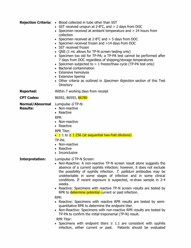
Rejection Criteria:
Blood collected in tube other than SST SST received unspun at 2-8oC, and > 2 days from DOC Specimen received at ambient temperature and > 24 hours from
collection Specimen received at 2-8oC and > 5 days from DOC Specimen received frozen and >14 days from DOC SST received frozen QNS (1 mL allows for TP-N screen testing only) Specimen too old for TP-PA; a TP-PA test cannot be performed after
7 days from DOC regardless of shipping/storage temperatures Specimen subjected to > 1 freeze/thaw cycle (TP-PA test only) Bacterial contamination Extensive hemolysis Extensive lipemia Other criteria as outlined in Specimen Rejection section of this Test
Directory
Reported: Within 7 working days from receipt
CPT Codes: 86592, 86593, 86780
Normal/Abnormal Results:
Lumipulse G TP-N: Non-reactive Reactive
RPR: Non-reactive Reactive
RPR Titer: 1:1 to ≥ 1:256 (at sequential two-fold dilutions)
TP-PA: Non-reactive Reactive Inconclusive
Interpretation: Lumipulse G TP-N Screen: Non-Reactive: A non-reactive TP-N screen result alone suggests the
absence of a current syphilis infection; however, it does not exclude the possibility of syphilis infection. T. pallidum antibodies may be undetectable in some stages of infection and in some clinical conditions. If recent exposure is suspected, re-draw sample in 2-4 weeks.
Reactive: Specimens with reactive TP-N screen results are tested by RPR to determine potential current or past infection.
RPR: Reactive: Specimens with reactive RPR results are tested by semi-
quantitative RPR to determine the endpoint titer. Non-Reactive: Specimens with non-reactive RPR results are tested by
TP-PA to confirm the initial treponemal (TP-N) result.
RPR Titer: Specimens with endpoint titers ≥ 1.1 are consistent with syphilis
infection, either current or past. Patients should be evaluated
clinically to identify signs, symptoms, or past history of infection. RPR titers can be used to monitor a patient’s response to treatment.
RPR titers usually become non-reactive with time, following successful treatment. o A ≥ 4-fold decline in titers at 12 months signifies successful
treatment. o A < 4-fold decline in titers at 12 months indicates treatment
failure or re-infection, or “serofast” condition.
TP-PA: Reactive: A reactive TP-PA test result, together with a reactive TP-N
and non-reactive RPR, is consistent with past or potential early syphilis infection.
Non-Reactive: A non-reactive TP-PA result, together with a reactive TP-N and non-reactive RPR, is considered inconclusive for syphilis infection, likely signifying a false positive TP-N or potentially an early infection.
Inconclusive: Patients with reactive TP-N, non-reactive RPR, and inconclusive TP-PA results should have blood drawn in 2-4 weeks for re-testing and/or syphilis testing performed by an alternative method.
Limitations:
Lumipulse TP-N: All treponemal tests tend to remain reactive for the life of a
treponeme-infected individual, even after treatment. Treponemal antibody titers correlate poorly with disease activity; therefore, they should not be used to evaluate response to therapy. Because of the persistence of reactivity, treponemal tests are of no value in determining relapse or re-infection in a patient who has had a reactive TP-PA result.
False positives, especially in low prevalence populations, and false negatives may occur.
Interference may be encountered with certain sera containing non-specific and/or unidentified reactive substances.
Test results from specimens obtained from immunosuppressed patients should be interpreted with caution.
Assay interference due to possible circulating antibodies against pinta, yaws, and bejel has not been evaluated.
RPR: False negative reactions may occur, particularly in tertiary syphilis but
also in early primary and late latent stages. Rarely, false negative results arise from a phenomenon called
“prozone effect”, whereby the reaction is overwhelmed by excess antibody.
False positive reactions occur occasionally in samples from individuals with a history of drug abuse, with febrile illness, women who are pregnant or with diseases such as lupus erythrematosus, malaria, vaccinia, mononucleosis, leprosy, viral pneumonia, and after smallpox vaccinations.
Pinta, yaws, bejel and other treponemal diseases produce positive reactions in this test.
TP-PA:
TP-PA may be reactive in a small percentage (< 1%) of normal or healthy persons; these false positive results are often transient, and their cause is unknown.
TP-PA may be reactive in persons from areas where yaws or pinta, bejel, and other treponemal diseases were, or are, endemic.
Samples from patients with HIV, leprosy, toxoplasmosis, H. pylori, cardiovascular disease, and drug addiction may react, on occasion, causing false positive or indeterminate results.
TP-PA is less sensitive than the fluorescent treponemal antibody absorption (FTA-ABS) test in untreated primary syphilis but compares favorably in all other stages of syphilis.
In accord with all diagnostic methods, a final diagnosis should not be made on the result of a single test, but should be based on a correlation of all laboratory test results with other clinical findings.
Notes: These tests are approved for in vitro diagnostics use by the U.S. Food and Drug Administration.
Back to Table of Contents
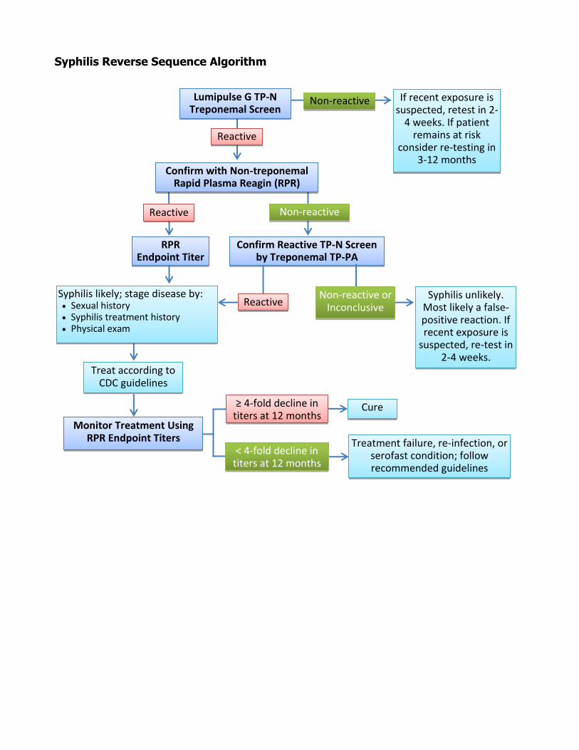
Syphilis Reverse Sequence Algorithm
Lumipulse G TP-N Treponemal Screen
Non-reactive
Reactive
If recent exposure is suspected, retest in 2-
4 weeks. If patient remains at risk
consider re-testing in 3-12 months
Confirm with Non-treponemal Rapid Plasma Reagin (RPR)
Non-reactive Reactive
Syphilis unlikely. Most likely a false-positive reaction. If recent exposure is
suspected, re-test in 2-4 weeks.
Confirm Reactive TP-N Screen by Treponemal TP-PA
Non-reactive or Inconclusive Reactive
Syphilis likely; stage disease by: Sexual history Syphilis treatment history Physical exam
Treat according to CDC guidelines
Monitor Treatment Using RPR Endpoint Titers
RPR Endpoint Titer
Treatment failure, re-infection, or serofast condition; follow recommended guidelines
≥ 4-fold decline in titers at 12 months
Cure
< 4-fold decline in titers at 12 months
West Nile Virus and St. Louis Encephalitis Virus, IgM Antibodies - ELISA
Use: To support a diagnosis for West Nile Virus (WNV) and St. Louis Encephalitis virus (SLE) infection in symptomatic individuals through detection of IgM antibodies.
Methodology: Specimens are tested using a semi-quantitative IgM antibody capture enzyme-linked immunosorbent assay (MAC-ELISA). Serum specimens that are reactive on initial screen are re-tested by titration using six 2-fold dilutions. CSF specimens receive initial screening only, and are not reflexed to titration.
Clinical Significance:
WNV and SLE are members of the Flavivirus group of Arboviruses, and are transmitted to humans by the bite of an infected mosquito. WNV can also be transmitted through contact with infected animals, blood and other tissues. Most persons infected with WNV or SLE have no apparent illness. Some people, especially the young, elderly and immunocompromised, will develop severe disease with symptoms including high fever, headache, nausea, vomiting, body and joint pains. Less than 1% of infected individuals develop neuroinvasive disease. Some neurologic effects may be permanent.
Further background information, fact sheets, statistics and educational resources may be found at the OSDH Acute Disease Services website.
Specimen: Type: Whole blood collected in serum separator tube (SST)
CSF (a serum specimen must accompany all CSF specimens); CSF is preferred specimen for detection of neuroinvasive disease
Volume: Serum: 1 mL, draw a sufficient amount of blood to yield the necessary serum volume; CSF: 1 mL
Container: Serum: SST or separated serum poured into sterile, plastic, screw-cap tube; CSF: sterile, plastic, screw-cap tube
Collection: Each facility should follow its guidelines for venipuncture collection of blood/serum and for collection of CSF. Following collection of blood, invert SST gently no more than 8 times then allow blood to clot in an upright position for at least 30 minutes and no more than 60 minutes then centrifuge at 3000 rpm for 10 minutes.
Interferences: Bacterial contamination; extensive hemolysis; extensive lipemia
Shipping: Serum: 2-8oC for delivery within 7 days from date of collection (DOC) o If transit time will be > 7 days post-collection, pour serum into
sterile, plastic, screw-cap tube and store/ship frozen at -20⁰C or colder
CSF: 2-8oC for delivery within 7 days from DOC o If transit time will be > 7 days post-collection, freeze and
store/ship frozen at -20⁰C or colder

Rejection Criteria: Blood collected in tube other than SST SST received unspun at 2-8oC, and > 2 days from DOC Specimen received at ambient temperature and > 24 hours from
collection Specimen received at 2-8oC and > 7 days from DOC SST frozen CSF received unfrozen and > 7 days from DOC CSF received at ambient temperature and > 24 hours from collection CSF received without a patient-matched serum specimen QNS Bacterial contamination Extensive hemolysis of serum Extensive lipemia of serum Other criteria as outlined in Specimen Rejection section of this Test
Directory
Reported: Within 10 working days from receipt
CPT Codes: 86788, 86653
Normal/Abnormal Results:
Separate results are provided for WNV and SLE virus and each type of specimen CSF: Non-reactive; Reactive Serum: Non-reactive, < 1:400; Weakly reactive, 1:400, recommend repeat testing in 10-14 days or testing by alternative method; Reactive, 1:800, recommend confirmatory testing by alternative method; Reactive, 1:1600, recommend confirmatory testing by alternative method; Reactive, 1:3200, recommend confirmatory testing by alternative method; Reactive, 1:6400, recommend confirmatory testing by alternative method; Reactive, 1:12800, recommend confirmatory testing by alternative method; Reactive > 1:12800, recommend confirmatory testing by alternative method
Interpretation: Non-reactive – no significant level of WNV or SLE IgM antibody detected
Reactive – presence of WNV or SLE IgM antibody detected, suggestive of current or recent infection
A titration cannot be performed for CSF specimens. CSF specimens will be screened only, and no titer will be reported. Positive results are only suggestive of current or recent infection with WNV and/or SLE since IgM cross-reactivity between the different flaviviruses is common; secondary testing, such as nucleic acid amplification or a neutralization assay, is required to confirm positive results and differentiate the flavivirus(es) involved. Also, positive results may occur in persons previously infected with flaviviruses or vaccinated for flaviviruses. Detection of WNV or SLE IgM in CSF suggests CNS involvement by WNV or SLE. A negative result in serum or CSF does not rule-out infection with WNV or SLE; IgM may be undetected in early infections, or in severely immunocompromised individuals. IgM titers should not be used to predict the severity of an individual’s symptoms or their prognosis. Results of this test should not be used as the sole
basis for diagnosis, treatment, or other patient management decisions.
Limitations:
In the event that an early acute CSF or serum is negative, a convalescent serum specimen should be drawn and tested two weeks after the initial specimen. Without testing of a convalescent specimen, a negative result may reflect testing of an acute-phase specimen. Antibody levels may be undetectable in early infection. In most patients, IgM is detectable 8 days post-onset of symptoms from a flavivirus infection.
Notes: This is a laboratory-developed test; performance characteristics have been validated and determined to be suitable for diagnostic purposes by the OSDH PHL. This test has not been cleared or approved by the U.S. Food and Drug Administration.
Back to Table of Contents
Zika Virus, IgM Antibodies - ELISA
Use: To support a diagnosis for Zika virus infection through detection of IgM antibodies in serum or CSF collected from individuals meeting the Centers for Disease Control and Prevention (CDC) Zika virus clinical and/or epidemiological criteria. Multiple assays and specimen types are often needed to establish a definitive laboratory diagnosis of Zika virus infection due to the temporal nature of biologic analytes in the infected person. Viral ribonucleic acid (RNA) is the first analyte that can be detected in an infected person in multiple specimen types. As the immune response develops, immunoglobulin M (IgM) titers rise in peripheral blood and the level of viral RNA generally declines. However, viral RNA may be detectable in some infected people for longer periods in certain specimen types. Nucleic acid testing (NAT) is most informative in the first 6 weeks after symptom onset. IgM antibodies are most likely detectable in the first 12 weeks after symptom onset, but may persist longer.
An Updated Interim Guidance for Healthcare Providers Caring for Pregnant Women with Possible Zika Exposure was released by the CDC in July 2017 in response to declining numbers of reported cases of Zika virus infection in the Americas, emerging evidence on prolonged detection of Zika IgM, and new limitations for interpreting serologic tests during pregnancy. Although IgM is most likely to be detected in the first 12 weeks after infection, emerging data indicate that Zika virus IgM may persist beyond 12 weeks in a subset of infected individuals, limiting the ability of testing to determine whether an infection occurred during or prior to pregnancy. IgM tests are also susceptible to false positives and cross-reactivity with other flaviviruses, especially when an individual has been vaccinated against or previously infected with a related flavivirus. In the United States, as the decline in reported Zika virus cases (including travel-associated cases) continues, the proportion of positive tests that are false positives for Zika virus are expected to increase due to a low positive predictive value. Key changes in the updated guidance for testing pregnant women have been made taking these testing limitations into consideration.
The CDC now recommends that symptomatic, pregnant women with possible exposure, and pregnant women with possible Zika virus exposure who have a fetus with prenatal ultrasound findings consistent with congenital Zika virus infection, be tested by for viral RNA and IgM concurrently. Specimens should be collected as soon as possible, up to 12 weeks following symptom onset or exposure. Routine IgM testing for asymptomatic pregnant women, with or without ongoing possible exposure to Zika virus, is no longer recommended.
For symptomatic, non-pregnant individuals that meet the CDC Zika virus clinical and/or epidemiological criteria, laboratory testing will be based on the date of symptom onset.
This test is only available following consultation with the OSDH Acute
Disease Service (ADS); healthcare providers seeking testing should call the OSDH ADS epidemiologist-on-call at (405) 271-4060. Prior to specimen collection/submission, an epidemiologist from the OSDH ADS must gather pertinent clinical signs and symptoms, travel, and other epidemiologic information from the clinician to determine if a patient meets required criteria for Zika virus testing.
The OSDH PHL is a CDC-designated laboratory that performs Zika virus testing by Emergency Use Authorization using the CDC Zika IgM Antibody Capture (MAC) ELISA and the CDC Trioplex Real-time RT-PCR Assay. Specimens may be referred by the OSDH PHL to the CDC or other laboratories for additional testing, as indicated.
Methodology: Specimens are tested using an immunoglobulin M (IgM) antibody capture enzyme-linked immunosorbent assay (MAC-ELISA), designed to detect IgMs to Zika virus. Per CDC testing guidelines, specimens that are ‘presumptive positive’ or ‘equivocal’ on initial screen may be reflexed to an arbovirus panel plaque reduction neutralization test (PRNT). Additional serologic testing for dengue and/or chikungunya viruses may also be performed.
Clinical Significance:
Zika virus is a member of the Flavivirus group of Arboviruses, and is transmitted to humans primarily by the bite of an infected mosquito. Perinatal, in utero, sexual (from men to his sexual partners), and blood transfusion transmissions have also been documented. Most (~80%) persons infected with Zika virus have no apparent illness. When symptoms do occur, they are usually mild and may include fever, maculopapular rash, arthralgia, myalgia, headache and non-purulent conjunctivitis. Symptoms typically last from 2 to 7 days. The virus has been associated with Guillain-Barré syndrome, microcephaly, intracranial calcifications, and other CNS abnormalities.
Further background information, fact sheets, statistics and educational resources may be found at the OSDH Acute Disease Service website and the CDC website.
Specimen: Type: Whole blood collected in serum separator tube (SST)
CSF (must be accompanied by a serum specimen); preferred specimen for detection of neuroinvasive disease
Volume: Serum: 2 mL, collect additional tubes to meet volume requirements, as needed; CSF: 1 mL
Container: Serum: SST or separated serum poured into sterile, plastic, screw-cap tube; CSF: sterile, plastic, screw-cap tube
Collection: Specimen collection must be coordinated with OSDH ADS.
Each facility should follow its guidelines for venipuncture collection of blood/serum and for collection of CSF. Following collection of blood, invert SST gently no more than 8 times then allow blood to clot in an upright position for at least 30 minutes and no more than 60 minutes then centrifuge at 3000 rpm for 10 minutes.

Interferences: Bacterial contamination; extensive hemolysis; extensive lipemia
Shipping: Serum: Store refrigerated (2-8°C) and ship using ice packs. If transit time will be > 7 days post-collection, pour serum into a sterile, plastic, screw-cap tube and store/ship frozen (-20°C or colder)
CSF: preferably, freeze at -20⁰C or colder, then ship on dry ice; however, refrigerated (2-8°C) shipment using ice-packs is acceptable
Rejection Criteria: Specimens received without documented OSDH Acute Disease Service consultation
Blood collected in tube other than SST SST received unspun at 2-8oC, and > 2 days from DOC Specimen received at ambient temperature and > 24 hours from
collection Specimen received at 2-8oC and > 7 days from DOC SST frozen CSF received without a patient-matched serum specimen QNS, depending on testing algorithm Bacterial contamination Extensive hemolysis of serum Extensive lipemia of serum Other criteria as outlined in Specimen Rejection section of this Test
Directory
Reported: Within 10 working days from receipt
CPT Codes: 86790
Normal/Abnormal Results:
Negative; Presumptive Positive; Equivocal; Inconclusive
Interpretation: Negative o If specimen collected ≥ 8 days and ≤ 12 weeks post-onset of
symptoms, then report will read, “No serological evidence of recent Zika virus infection”
o If specimen collected ≤ 7 days post-onset of symptoms, then report will read, “Not suggestive of a recent Zika virus infection but anti-Zika virus IgM levels may be undetectable in early acute infections; if signs and symptoms suggest Zika infection, submit a convalescent specimen”
o If specimen collected > 12 weeks post-onset of symptoms, then report will read, “Not suggestive of a recent Zika virus infection but anti-Zika virus IgM antibodies may decrease to undetectable levels in specimens collected after 12 weeks post-onset of symptoms”
o If specimen collected from an asymptomatic individual, then the report will read, “Not suggestive of a recent Zika virus infection but specimen could have been collected outside the window when anti-Zika virus IgM antibodies would normally be detected in a Zika virus infected individual (i.e., 4 days to 12 weeks post-infection)”
o If fetus/infant born to symptomatic woman or asymptomatic woman with travel history to or residence in a country with Zika
virus transmission or fetus/infant with CNS abnormalities suggestive of Zika virus infection, then report will read “Not suggestive of a recent Zika virus infection but anti-Zika virus IgM antibody levels in fetus or infant serum or CSF are not well understood”
“Presumptive Positive – serological evidence of possible recent Zika virus infection”
“Equivocal – repeatedly above negative cut-off and below presumptive positive cut-off”
“Inconclusive – Unable to determine presence of anti-Zika virus IgM antibodies due to high background signal; suggest submit alternative serum specimen”
Since it is common for IgM cross-reactivity to occur between different flaviviruses, a positive MAC-ELISA result only indicates a probable infection with Zika virus; other testing may be performed to define the infecting flavivirus. Positive results may also occur in persons previously infected with flaviviruses or vaccinated against flaviviruses. Negative results do not preclude infection with Zika virus; IgMs to Zika virus may be low and undetectable by MAC-ELISA in early stages of infection, > 12 weeks post-onset of symptoms, or in severely immunocompromised individuals.
Guidance documents to help patients and healthcare providers interpret results are available at https://www.cdc.gov/zika/hc-providers/testresults.html. For further guidance, contact OSDH ADS at 405-271-4060.
Limitations:
In the event that an early acute CSF or serum is negative by MAC-ELISA and clinical signs and symptoms suggest Zika infection, a convalescent serum specimen should be drawn and tested two to three weeks after the initial specimen. Without testing of a convalescent specimen, a negative result may reflect testing of an acute-phase specimen. Results of this test should not be used as the sole basis of patient treatment/management. As with other laboratory tests, false negative and false positive results may occur. Clinical decisions surrounding patient management should not be made until all testing is complete and should be considered within the context of all test results, and clinical and epidemiologic criteria as specified for the testing algorithm appropriate for the person being tested. Current CDC testing guidance does not recommend Zika virus testing for pre-conception screening or non-pregnant asymptomatic individuals.
Notes: Specimens must be submitted with a completed OSDH Test Requisition Form.
This test is performed in compliance with a US Food and Drug Administration (FDA) Emergency Use Authorization (EUA).
Back to Table of Contents
Zika Virus, Chikungunya Virus, and Dengue Virus – PCR
Use: To support a diagnosis for arbovirus infection through detection and differentiation of Zika virus, dengue virus or chikungunya virus RNA in sera or cerebrospinal fluid (CSF), or Zika virus RNA in urine or amniotic fluid collected from individuals meeting the Centers for Disease Control and Prevention (CDC) Zika virus clinical and/or epidemiological criteria. Updated Interim Guidance for Healthcare Providers Caring for Pregnant Women with Possible Zika Exposure was released by the CDC MMWR July 2017. The interim guidance has been updated based on declining trends in the number of reported cases of Zika virus infection in Region of the Americas, emerging evidence on prolonged detection of Zika immunoglobulin M (IgM) antibodies, and new limitations for interpreting serologic tests during pregnancy. Although IgM is most likely to be detected in the first 12 weeks after infection, emerging data indicate that Zika virus IgM may persist beyond 12 weeks in a subset of infected individuals, limiting the ability of testing to determine whether an infection occurred during or prior to pregnancy. IgM tests are also susceptible to false positives and cross-reactivity with other flaviviruses, especially when an individual has been vaccinated against or previously infected with a related flavivirus. In the United States, as the decline in reported Zika virus cases (including travel-associated cases) continues, the proportion of positive tests that are false positives for Zika virus are expected to increase due to a low positive predictive value. Key changes in the updated guidance for testing pregnant women have been made taking these testing limitations into consideration.
The CDC now recommends that symptomatic pregnant women with possible exposure, pregnant women with possible Zika virus exposure who have a fetus with prenatal ultrasound findings consistent with congenital Zika virus infection, and asymptomatic pregnant women with ongoing possible exposure to Zika virus be tested.
This test is only available following consultation with the OSDH Acute Disease Service; healthcare providers seeking testing should call the OSDH ADS epidemiologist-on-call at (405) 271-4060. Prior to specimen collection/submission, an epidemiologist from the OSDH Acute Disease Service (ADS) must gather pertinent clinical signs and symptoms, travel, and other epidemiologic information from the clinician to determine if a patient meets the required criteria for Zika virus testing as indicated by the CDC. All specimens must be approved by the ADS prior to shipping to the PHL for testing.
The OSDH PHL is a CDC-designated laboratory that performs Zika virus testing by Emergency Use Authorization using the CDC Zika IgM Antibody Capture (MAC) ELISA and the CDC Trioplex Real-time RT-PCR Assay. Specimens may be referred by the OSDH PHL to the CDC or other laboratories for additional testing, as indicated.
This test is only available following consultation with the OSDH Acute Disease Service (ADS); healthcare providers seeking testing should call the OSDH ADS epidemiologist-on-call at (405) 271-4060. Prior to specimen

collection/submission, an epidemiologist from the OSDH ADS must gather pertinent clinical signs and symptoms, travel, and other epidemiologic information from the clinician to determine if a patient meets required criteria for Zika virus testing.
Methodology: Specimens are tested using a qualitative real-time, reverse transcription-polymerase chain reaction (rRT-PCR) assay developed by the CDC (Trioplex Real-time RT-PCR Assay).
Clinical Significance:
Zika virus, dengue virus (both flaviviruses) and chikungunya virus (an alphavirus) are transmitted to humans primarily by the bite of infected Aedes mosquitoes, although in utero and blood transfusion transmissions have also been documented. Sexual transmission (from men to his sexual partners) has been documented only for Zika virus. Approximately, 1 in 5 Zika virus infections or 1 in 4 dengue virus infections cause symptoms, whereas most chikungunya infections (60-95%) are symptomatic. Each virus can cause self-limiting, acute febrile illness, with myalgia, arthralgia, headache, conjunctivitis, and maculopapular rash, typically lasting from 2 to 7 days; however, each has unique clinical features: acute dengue infection is complicated by hemorrhage, vascular leak, and shock syndrome in rare cases; chikungunya infections can cause persistent disabling joint symptoms, especially in older individuals; Zika virus has been associated with microcephaly in infants born to mothers infected during pregnancy, as well as Guillain-Barré and other neurologic complications. Nevertheless, because of similarities in patient symptoms for the three viruses, laboratory testing is important in establishing an accurate diagnosis.
Further background information, fact sheets, statistics and educational resources may be found at the OSDH Acute Disease Service website and the CDC website.
Specimen: Type: Whole blood collected in serum separator tube (SST)
Urine (must be accompanied by a serum specimen); preferred specimen for early acute phase infection
CSF (must be accompanied by a serum specimen); preferred specimen for detection of neuroinvasive disease
Amniotic fluid (must be accompanied by a serum specimen)
Volume: Serum: 2 mL; collect additional tubes to meet volume requirements, as needed; Urine: 1 mL; CSF: 1 mL; Amniotic fluid: 1 mL
Container: Serum: SST or separated serum poured into sterile, plastic screw-cap tube; Urine: sterile, plastic, screw-cap tube (do not send urine collection cups); CSF: sterile, plastic, screw-cap tube; Amniotic fluid: sterile, plastic, screw-cap tube
Collection: Specimen collection must be coordinated with OSDH ADS.
Each facility should follow its guidelines for venipuncture collection of blood/serum and for collection of CSF. Following collection of blood, gently invert SST no more than 8 times, allow blood to clot in an upright position for at least 30 minutes and no more than 60 minutes, then

centrifuge at 3000 rpm for 10 minutes.
Interferences: None
Shipping: SST: Store refrigerated (2-8°C) and ship using ice packs. If transit time will be > 7 days post-collection, pour serum into a sterile, plastic, screw-cap tube and store/ship frozen (-20°C or colder)
Urine, CSF, and amniotic fluid: preferably, freeze at -20⁰C or colder, then ship on dry ice; however, refrigerated shipment using ice-packs is acceptable
Rejection Criteria: Specimens received without documented OSDH Acute Disease Service consultation
Blood collected in tube other than SST SST received unspun at 2-8oC, and > 2 days from DOC Specimen received at ambient temperature and > 24 hours from
collection Specimen received at 2-8oC and > 7 days from DOC SST frozen CSF, urine or amniotic fluid received without a patient-matched serum
specimen QNS, depending on testing algorithm Bacterial contamination Extensive hemolysis of serum Extensive lipemia of serum Other criteria as outlined in Specimen Rejection section of this Test
Directory
Reported: Within 10 working days from receipt
CPT Codes: 87798
Normal/Abnormal Results:
Sera and CSF: Negative, Positive, or Inconclusive for Zika virus, dengue virus and/or chikungunya virus
Urine and amniotic fluid: Negative, Positive, or Inconclusive for Zika virus
Interpretation: Negative rRT-PCR results indicate that Zika, chikungunya, and dengue virus RNA was not detected in the submitted serum and/or CSF specimen, or Zika virus RNA was not detected in the submitted urine or amniotic fluid specimen. Viral RNA is typically detectable in serum during the acute phase of infection (generally up to 7 days post-symptom onset). Zika virus RNA has been detected in serum up to 13 days post-symptom onset in non-pregnant patients, and up to 62 days post-symptom onset in pregnant patients. In addition, Zika virus RNA has been detected up to 53 days after the last known possible exposure in an asymptomatic pregnant woman. Because viremia may decrease rapidly shortly after onset of symptoms, a negative rRT-PCR result for specimens collected during the latter part of the acute phase of infection does not exclude flavivirus/alphavirus infection.
Positive rRT-PCR results indicate that Zika, chikungunya, and/or dengue virus RNA was detected in the submitted serum
and/or CSF specimen, or Zika virus RNA was detected in the submitted urine or amniotic fluid specimen.
Inconclusive rRT-PCR results indicate the presence of a potential PCR inhibitor in the submitted sample or poor sample quality; submission of an alternative specimen is suggested.
Guidance documents to help patients and healthcare providers interpret results are available at https://www.cdc.gov/zika/hc-providers/testresults.html. For further guidance, contact OSDH ADS at 405-271-4060.
Limitations:
Results of this test should not be used as the sole basis of patient treatment/management. As with other laboratory tests, false negative and false positive results may occur. Clinical decisions surrounding patient management should not be made until all testing is complete and should be considered within the context of all test results, and clinical and epidemiologic criteria as specified for the testing algorithm appropriate for the person being tested. Current CDC testing guidance does not recommend Zika virus testing for pre-conception screening or non-pregnant asymptomatic individuals.
Notes: Specimens must be submitted with completed OSDH Test Requisition Form.
This test is performed in compliance with a US Food and Drug Administration (FDA) Emergency Use Authorization (EUA).
Back to Table of Contents