tesi dottorato corr prof detmar - research collection1694/eth... · and in the production of...
TRANSCRIPT

Research Collection
Doctoral Thesis
Antibody engineeringadvances in phage display technology and in the production oftherapeutic immunocytokines
Author(s): Sommavilla, Roberto
Publication Date: 2010
Permanent Link: https://doi.org/10.3929/ethz-a-006155036
Rights / License: In Copyright - Non-Commercial Use Permitted
This page was generated automatically upon download from the ETH Zurich Research Collection. For moreinformation please consult the Terms of use.
ETH Library

DISS. ETH NO. 18980
ANTIBODY ENGINEERING: ADVANCES IN PHAGE DISPLAY TECHNOLOGY AND IN THE PRODUCTION OF THERAPEUTIC IMMUNOCYTOKINES
A dissertation submitted to the
ETH Zürich
for the degree of
Doctor of Sciences
presented by
Roberto Sommavilla
Laurea in Biotecnologie Farmaceutiche
Università degli Studi di Padova
born on 6th May 1982
citizen of Italy
accepted on the recommendation of
Prof. Dr. Dario Neri, examiner
Prof. Dr. Michael Detmar, co-examiner
2010

2

3
To my family

4

5
Table of contents
1 Summary 7
2 Introduction 9
2.1 Antibody-based targeting of disease 11
2.1.1 General concepts on antibodies 11
2.1.2 Mechanisms of action 12
2.1.3 Therapeutic applications 14
2.1.4 Immunogenicity of approved antibodies 17
2.2 Antibody phage display technology 19
2.2.1 The phage display technology 19
2.2.2 Antibody phage display libraries 24
2.2.3 Murine antibodies and antibody libraries 29
2.2.4 Other selections methodologies 30
2.3 Vascular tumor targeting of interleukin 12 40
2.3.1 Antibody-based tumor vascular targeting 40
2.3.2 Immunocytokines 44
2.3.3 Interleukin 12 46
2.4 The aim of this thesis 52
3 Design and construction of a naïve mouse antibody phage display library 55
3.1 Results 55
3.1.1 A novel approach for the generation of murine antibodies 55
3.1.2 Design and construction of PHILOtop, a synthetic naïve mouse antibody phage display library 56
3.1.3 Isolation, characterization and affinity maturation of murine monoclonal antibodies specific to the extra domain B of fibronectin 65
3.2 Discussion 70
3.3 Material and methods 73
4 Expression, engineering and characterization of the tumor-targeting heterodimeric immunocytokine F8-IL12 81
4.1 Results 81
4.1.1 Optimization and stable transfection of cells expressing F8-IL12 81
4.1.2 Production and characterization of F8-IL12 84

6
4.2 Discussion 91
4.3 Material and methods 93
5 References 100
6 Curriculum Vitae 112

7
1 Summary
Antibody phage technology greatly facilitates the isolation of good-quality
monoclonal antibodies to virtually any target antigen. Large combinatorial phage
display libraries of human antibodies are routinely being used for the
identification of antibody candidates for clinical applications. However, preclinical
studies in rodents would benefit from the availability of good-quality single-pot
mouse synthetic naïve antibody libraries, which at present are not available.
Such libraries would be particularly useful for the generation of murine antibodies
against self or highly conserved antigens or in case of highly toxic or deadly
pathogenic antigens which do not allow animal immunization.
This thesis reports on the construction of a mouse antibody phage display library,
containing over 1 billion antibody clones, based on the combinatorial
mutagenesis of residues in the CDR3 loops of a given antibody scaffold. The
library was shown to reliably yield good-quality antibodies towards all protein
antigens used so far in selection experiments, including three tumor-associated
antigens. The modular structure of the library facilitates a simple affinity-
maturation procedure based on the combinatorial mutagenesis of CDR1 and
CDR2 loops of the VH domain, which has led to the isolation of a high-affinity
antibody [scFv(H7), Kd = 6 nM] specific to the EDB domain of fibronectin, a
marker of angiogenesis. This single-pot antibody library may thus represent a
useful source of binding specificities, facilitating preclinical studies in
immunocompetent syngeneic mouse models of pathology.
Interleukin 12 (IL12) is a key mediator of innate and adaptive immunity. The
immunomodulating and antiangiogenic functions of IL12 have provided the
rationale for exploiting this cytokine as an anticancer agent in patients with
advanced cancer, but its administration is typically associated with severe
toxicity, hampering dose escalation to therapeutically-active regimens and the
development as anti-cancer drug.

8
To overcome the clinical drawbacks associated to the administration of cytokines
(and IL12 in particular) to patients, the use of “immunocytokines” (i.e., cytokines
fused to antibodies or antibody fragments) has been proposed, with the aim to
concentrate the immune stimulating activity at the site of disease while sparing
normal tissues.
In this thesis I describe the design and construction of a heterodimeric
immunocytokine F8-IL12, consisting of scFv(F8) (an antibody fragment, specific
to the alternatively-spliced EDA domain of fibronectin) fused to both p35 and p40
subunits of human IL12. This immunocytokine could be stably expressed in
mammalian cells and purified to homogeneity with full retention of cytokine
activity. The resulting product exhibited an impressive tumor targeting
performance in a mouse model of cancer.

9
1 Riassunto
La tecnologia del phage display di frammenti anticorpali ha reso possibile la
generazione di anticorpi monoclonali contro potenzialmente qualsiasi antigene di
interesse. Librerie combinatoriali di anticorpi umani, esposti sulla superficie di
fagi, sono utlizzate di routine per l’identificazione di anticorpi a fini terapeutici.
Gli studi preclinici in roditori beneficerebbero di librerie sintetiche, naïve e
funzionali di anticorpi murini che al giorno d’oggi non sono disponibili.
Tali librerie sarebbero particolarmente utili al fine di ottenere anticorpi murini
contro antigeni self o conservati o in caso di antigeni tossici, gravementi
patogenici o letali, caratteristiche che impediscono l’immunizzazione degli
animali.
In questa tesi si descrive la costruzione di una libreria di phage display di
anticorpi murini contenente oltre 1 miliardo di diversi cloni, basata sulla
mutagenesi combinatoriale dei residui nei loop dei CDR3 di una data struttura
anticorpale. La libreria si e’ dimostrata un’ottima fonte di anticorpi contro tutti gli
antigeni presi in esame, tra cui tre antigeni presenti esclusivamente a livello
tumorale. La struttura modulare della libreria rende possibile una semplice
procedura per l’aumento dell’affinita’ mediante la mutagenesi combinatoriale dei
loop del CDR1 e del CDR2 del dominio VH. Tale strategia ha cosentito
l’isolamento di un anticorpo ad alta affinita’ [scFv(H7), Kd = 6 nM] specifico per
l’extra-dominio B della fibronectina, un antigene espresso nel processo di
angiogenesi tumorale. La libreria potrebbe quindi divenire un’utile fonte di
anticorpi in grado di permettere di effettuare studi preclinici in modelli murini
singenici ed immunocompetenti di una data patologia.
L’interleuchina 12 (IL12) e’ un mediatore chiave della risposta immunitaria innata
e adattativa. Le funzioni immunomodulanti e antiangiogeniche della IL12 hanno
motivato lo studio di tale citochina come agente per la terapia del cancro in
pazienti con forme neoplastiche avanzate. Tuttavia la somministrazione di IL12
induce grave tossicita’ nei pazienti, impedendo il raggiungimento della dose

10
terapeuticamente efficace e conseguentemente lo sviluppo come farmaco
anticancro.
Al fine di superare i problemi derivanti dall’uso nei pazienti delle citochine (e
dell’IL12 in particolare), e’ stato proposto l’impiego di immunocitochine (citochine
fuse ad anticorpi o frammenti anticorpali) con lo scopo di concentrare l’attivita’
del sistema immunitario esclusivamente nella regione interessata dalla malattia.
In questa tesi descrivo il disegno e la costruzione di F8-IL12, una
immunocitochina eterodimerica, composta da scFv(F8) (un frammento
anticorpale, specifico per l’extra-dominio A della fibronectina, regione soggetta a
splicing alternativo) fuso alle due subunita’ dell’interleuchina 12 umana. Tale
immunocitochina e’ stata stabilmente espressa in cellule di mammifero e
purificata all’omogeneita’ mantenendo l’attivita’ biologica. In experimenti preclinici
in topi, questo nuovo prodotto farmaceutico ha mostrato di possedere
straordinarie capacita’ di accumulo selettivo a livello tumorale.

11
2 Introduction
2.1 Antibody-based targeting of disease
2.1.1 General concepts on antibodies
Antibodies are a class of glycoproteins belonging to the immunoglobulin super-
family found in blood or other bodily fluids of vertebrates.
Five classes of immunoglobulins are known - IgM, IgD, IgG IgA and IgE - that
differ for their constant region. IgGs are the most relevant class of antibodies for
pharmaceutical applications (Table 2.1). The classic structure of an IgG consists
of two identical light chains (25 kDa) and two identical heavy chains (50 kDa)
covalently linked by a disulphide bridge between the two heavy chains and
between heavy and light chains (Figure 2.1). Each heavy and light chain
contains a variable domain (VH and VL respectively) of around 110 amino acids
where the variability on the antibody structure is located. Within each variable
domain, three hypervariable loops confer the binding properties and are defined
as complementarity determining regions (CDRs).
Figure 2.1. This figure shows the modular structure of an IgG. molecule. All immunoglobulin
monomers are composed of two identical light (L) chains and two identical heavy (H) chains.
Light chains are composed of one constant domain (CL) and one variable domain (V
L), whereas
heavy chains are composed of three constant domains (CH1, C
H2 and C
H3) and one variable
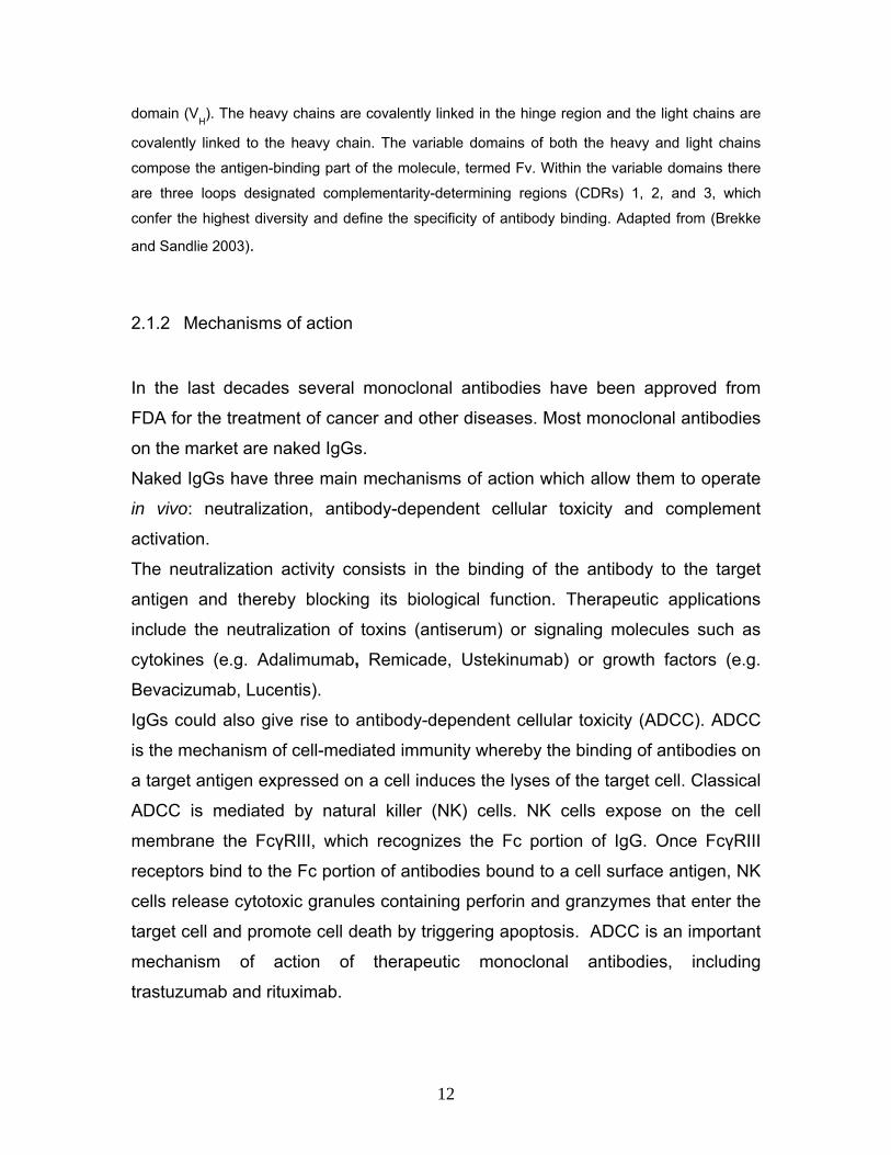
12
domain (VH). The heavy chains are covalently linked in the hinge region and the light chains are
covalently linked to the heavy chain. The variable domains of both the heavy and light chains
compose the antigen-binding part of the molecule, termed Fv. Within the variable domains there
are three loops designated complementarity-determining regions (CDRs) 1, 2, and 3, which
confer the highest diversity and define the specificity of antibody binding. Adapted from (Brekke
and Sandlie 2003).
2.1.2 Mechanisms of action
In the last decades several monoclonal antibodies have been approved from
FDA for the treatment of cancer and other diseases. Most monoclonal antibodies
on the market are naked IgGs.
Naked IgGs have three main mechanisms of action which allow them to operate
in vivo: neutralization, antibody-dependent cellular toxicity and complement
activation.
The neutralization activity consists in the binding of the antibody to the target
antigen and thereby blocking its biological function. Therapeutic applications
include the neutralization of toxins (antiserum) or signaling molecules such as
cytokines (e.g. Adalimumab, Remicade, Ustekinumab) or growth factors (e.g.
Bevacizumab, Lucentis).
IgGs could also give rise to antibody-dependent cellular toxicity (ADCC). ADCC
is the mechanism of cell-mediated immunity whereby the binding of antibodies on
a target antigen expressed on a cell induces the lyses of the target cell. Classical
ADCC is mediated by natural killer (NK) cells. NK cells expose on the cell
membrane the FcγRIII, which recognizes the Fc portion of IgG. Once FcγRIII
receptors bind to the Fc portion of antibodies bound to a cell surface antigen, NK
cells release cytotoxic granules containing perforin and granzymes that enter the
target cell and promote cell death by triggering apoptosis. ADCC is an important
mechanism of action of therapeutic monoclonal antibodies, including
trastuzumab and rituximab.

13
Antibodies (mainly IgM, IgG1 and IgG3) are able to trigger the classical pathway
of complement activation: the starting event is the binding of C1q to the Fc region
of antibodies bound to multiple sites of a cell surface (typically a pathogen).
Subsequent activation of the complement cascade ultimately leads to three
possible events: (i) direct killing of the cell via lysis (by creating pores on the
membrane), (ii) opsonization and engulfment of the cell by phagocytes and (iii)
uptake of the complement-coated-antigen by antigen presenting cells and its
presentation to the adaptive immune system.
Antibodies and their derivatives can also be used as vehicle to concentrate a
therapeutic agent specifically at the site of disease sparing healthy tissues. A
targeted therapy is the ideal approach for the therapy of cancer where often
potent drugs cannot be used at therapeutic relevant regimens because of their
severe side effects. The targeted delivery of drugs by means of antibodies
promises to enhance their therapeutic index and limit their side effects.
Figure 2.2. IgG and antibody fragments. IgG is a 150 kDa molecule comprising two variable
fragments (Fv) which contain the antigen binding sites, and an Fc portion which mediated the
effector function. The single-chain Fv (scFv) fragment, in which the variable domain of the heavy
and light chains are joined by a polypeptide linker, is the smallest fragment which still contains the
intact binding site. Whereas the Fab fragment, which naturally occur within the IgG molecule, is a
monovalent fragment. The engineered mini-antibody format is a bivalent binding molecule
obtained by fusing a scFv to a constant εCH4 domain of a human IgE

14
For tumor targeting applications the use of antibody fragments (i.e. scFv,
diabody, or mini-antibody format; Figure 2.2), is preferable to full IgGs as they
allow a better extravasation from the blood circulation and a superior tissue
penetration. In comparison to naked IgG, antibody fragments per se do not
mediate any therapeutic activity and need to be linked to bioactive agents such
as cytokines, toxins, procoagulant factors, drugs with a cleavable linker,
radionuclides and photosensitizers (Figure 2.11).
2.1.3 Therapeutic applications
Since their introduction, monoclonal antibodies have had a progressively
increasing impact on different areas of medical diagnostics and therapeutics.
Monoclonal antibodies have been approved for the treatment of cancer,
inflammatory diseases, cardiovascular diseases, macular degeneration,
transplant rejection, multiple sclerosis, and viral infection (Table 2.1). Most of
these antibodies have the neutralization of their target or the induction of ADCC
as mechanism of action. In the future, we may also see approval of antibody
derivatives linked to a bioactive agent. These “immuno-derivatives” may offer the
possibility to deliver a potent therapeutic agent that acts selectively at the site of
the disease, sparing healthy tissues, and may considerably enhance the
therapeutic index of the bioactive agent.
For the therapy of cancer the U.S. FDA approved a number of monoclonal
antibodies (Table 2.1), some of which have become blockbusters in spite of their
generally modest clinical benefit.
Bevacizumab is a humanized monoclonal antibody that targets and blocks the
VEGF-A and was approved for the first-line treatment of metastatic colorectal
cancer and as second line for other solid cancers. The neutralization of VEGF-A
seemed to be a promising strategy as this molecule is supposed to be a key
factor for the neoangiogenesis process required for tumor growth. Clinical studies
showed that bevacizumab combined with chemotherapy can prolong life of

15
patients of 4-5 months compared to patients treated with standard chemotherapy
(Hurwitz, Fehrenbacher et al. 2004)
Trastuzumab is a humanized monoclonal antibody approved for the therapy of
metastatic breast cancer. The antibody targets the extracellular domain of the
human epidermal growth factor receptor 2 (HER2), a protein over-expressed in
~20% of women with breast cancer. ADCC and arrest of cell proliferation by
disrupture of receptor dimerization or downstream signaling pathways may be
possible mechanisms of action in vivo, though they have been only demonstrated
in vitro (Sarup, Johnson et al. 1991; Cooley, Burns et al. 1999).
Trastuzumab combined with chemotherapy increases response rates, time to
progression, and survival (Cobleigh, Vogel et al. 1999). However, the majority of
cancers that initially respond to trastuzumab begin to progress again within one
year. The discovery that signaling by the epidermal growth factor receptor (EGFR)
plays a key role in tumorigenesis prompted efforts to target this receptor in
anticancer therapy. Two different types of EGFR-targeted monoclonal antibodies
were developed: first cetuximab, a chimeric monoclonal IgG1, and subsequently
panitumumab a fully human IgG2. Both antibodies target the extracellular domain
of the receptor, thereby inhibiting ligand-dependent EGFR signal transduction
(Okamoto). Both therapeutics were approved for the treatment of patients with
EGFR expressing KRAS wild-type metastatic colorectal cancer and head and
neck cancer.
Clinical studies of cetuximab in combination with irinotecan showed tumor
shrinking in 22.9% of patients and delayed tumor growth by 4 months. For
patients who received cetuximab alone, the tumor response rate was 10.8% and
tumor growth was delayed by 4 months (Alberts, Sinicrope et al. 2005).
Rituximab (Rituxan) is a chimeric monoclonal antibody against CD20, which is
primarily found on the surface of B cells. Rituximab was approved for the
treatment of certain lymphomas and some autoimmune disorders. In the
treatment of lymphomas, Rituximab has significantly improved the response
duration and time to disease progression (Van Oers, Hagenbeek et al. 2002).

16
Table 2.1. List of antibodies approved for therapy or diagnosis.

17
2.1.4 Immunogenicity of approved antibodies
The first murine monoclonal antibody approved from FDA, Muromonab-CD3
(orthoclone OKT3) for the prevention and treatment of solid organ transplant
rejection (Smith 1996) was a scientific and commercial success. However it
turned out that murine monoclonal antibodies lead to the development of human
anti-mouse antibodies (HAMA) that cause rapid clearance and loss of efficacy of
the therapeutic antibody and can induce severe allergic reactions. Moreover
murine IgGs do not mediate ADCC and complement activation, thus limiting
therapeutic applications.
In order to reduce the HAMA response, chimeric monoclonal antibodies were
produced combining the murine variable regions of the immunoglobulin genes
with constant domains of the human IgG. The use of chimeric antibodies
substantially reduced the HAMA response but could not completely eliminate it.
Although several chimeric antibodies achieved regulatory approval (Table 2.1), it
became clear the need of a further reduction of murine sequences in the
antibody scaffold in order to minimize the HAMA response. With the hope to
solve this problem, humanized antibodies were generated. Humanized
antibodies were constructed by grafting the CDRs of the heavy and light chains
and a limited number of structural amino acids of the murine monoclonal
antibody to the CDR-depleted human IgG scaffold. Although this process further
reduced the HAMA response, in many cases, substantial additional antibody
design procedures were needed to reestablish the required specificity and affinity
of the original murine antibody (Presta, Lahr et al. 1993). In order to minimize the
development of HAMA responses, the generation of fully human antibodies
would be desirable. By means of phage display technology, it has been possible
to obtain fully human antibodies that reached regulatory approval (Adalimumab).
Fully human antibodies (e.g., Panitumumab) can be also generated by
transgenic animals whose murine immunoglobulin loci have been replaced by the

18
entire human immunoglobulin loci determining a human antibody response in
mice.
Immunogenicity problems, however, have not been totally solved. Adalimumab, a
fully human antibody for the treatment of rheumatoid arthritis, was supposed to
be less immunogenic than murine, chimeric and humanized monoclonal
antibodies. Surprisingly it was observed to induce the formation of human anti-
human antibodies (HAHA) in 12% of patients (Weinblatt, Keystone et al. 2003;
van de Putte, Atkins et al. 2004). It is believed that the HAHA reactivity is either
directed against non-human glycosylated sites on the surface of the antibody
expressed in CHO cells or against the antigenic determinants in VL and VH
regions of adalimumab that include the CDRs complementary determining
regions (anti-idiotype antibodies).
The development of auto-antibodies in some cases can be related to degradation
or aggregation of the antibody or to presence of impurities, such as toll-like
receptor (TLR) ligands.

19
2.2 Antibody phage display technology
2.2.1 The phage display technology
Phage display is a powerful methodology that allows the selection of a particular
phenotype (e.g., a ligand specific to a desired antigen) from repertoires of
proteins displayed on bacteriophage. Phage display was first described in 1985
by Smith (Smith 1985), who presented the use of the non-lytic filamentous
bacteriophage fd for the display of specific binding peptides on the phage coat.
The power and applications of the methodology were further improved by the
groups of Winter (McCafferty, Griffiths et al. 1990) and Wells (Lowman, Bass et
al. 1991) who demonstrated the display of functional folded proteins on the
phage surface (an antibody fragment and a hormone, respectively). Phage
technology is based on the fact that a polypeptide (capable of performing a
function, classically the specific binding to an antigen) can be displayed on the
phage surface by inserting the gene coding for the polypeptide into the phage
genome. Thus the phage particle physically links genotype to phenotype (Figure 2.3).
Figure 2.3. Phage displaying a binding protein: in this case a scFv antibody fragment as fusion
protein of a minor coat protein pIII

20
It is possible to create large repertoires of phage (phage display libraries) in
which the proteins displayed on each phage are slightly different from each other.
If one is able to purify from this large phage repertoire a phage particle by virtue
of the phenotype (e.g., the binding specificity) displayed on its surface, one also
isolates the genetic information coding for the binding protein, and can amplify
the corresponding phage by means of bacterial infection.
As an example, one can consider the selection of a binding specificity from
repertoires of binders (Figure 2.4).
Figure 2.4. Selection of binding specificity from a phage display library. A library of proteins
displayed on the phage surface is used as input for the selection. Phage displaying binding
proteins are captured on immobilized target molecules, and after washing, bound phage can be
eluted. This phage population is then propagated in E.coli cells and can be used for further
rounds of selection.
The library on phage is panned against an antigen of interest. Unbound phage

21
are discarded whereas specifically binding phage are collected and amplified in
bacteria. Several rounds of selection can be performed (in general 2-4 rounds
using antibody phage libraries). As a result, even very rare phenotypes present in
large repertoires can be selected and amplified from a background of phage
carrying undesired phenotypes.
The possibility to amplify the selected phage in bacteria during biopanning
experiments allows the enrichment of the pool of phage with the desired
phenotype. Filamentous phage infect strains of E.coli that harbour the F
conjugative episome by attaching to the tip of the F pilus and translocating the
phage genome (a circular single-strand DNA molecule) into the bacterial
cytoplasm. The genome is replicated involving both phage- and host-derived
proteins, and packaged into elongated “filamentous” viral particles of roughly 6
nm in diameter and 900 nm in length (Figure 2.5).
Figure 2.5. Life cycle of filamentous phage f1 (M13/fd). Sequential binding of pIII to the tip of the
F-pilus and then the host Tol protein complex results in depolymerization of the phage coat
proteins, their deposition in the cytoplasmic membrane (where they are available for reutilization),
and entry of the ssDNA into the cytoplasm. The ssDNA is converted by host enzymes to a
double-stranded reading frame (RF), the template for phage gene expression. Progeny ssDNA,
coated by pV dimers (except for the packaging sequence hairpin (PS) that protrudes from one
end), is the precursor of the virion. A multimeric complex that spans both membranes mediates
conversion of the pV-ssDNA complex to virions and secretion of virions from the cell. This
process involves removal of pV dimers and their replacement by the five coat proteins that

22
transiently reside in the cytoplasmic membrane. Adapted from “Phage display: a practical
approach” Edited by Clackson T. and Lowman HB. Oxford University Press.
Filamentous phage particles are covered by several thousand copies of a small
major coat protein (pVIII). Few copies of the minor coat proteins pIII and pVI are
displayed at one extremity of the phage particle, while pVII and pIX are present
at the opposite extremity. The minor coat protein pIII (the product of gene III), is
displayed in 3-5 copies and mediates the adsorption of the phage to the bacterial
pilus. Peptides and proteins have been displayed on phage as fusions with the
coat proteins pIII (Smith 1985; Parmley and Smith 1988) or pVIII (Greenwood,
Willis et al. 1991). Display of proteins encoded by a cDNA library as carboxy-
terminal fusion with the minor coat protein pVI has also been described
(Greenwood, Willis et al. 1991).
The first peptides and proteins were displayed on phage using phage vectors
(essentially the phage genome with suitable cloning sites for pVIII or pIII fusions
and an antibiotic resistance gene). Phage vectors carry all the genetic
information necessary for the phage life cycle.
With pIII fusions in phage vectors, each pIII coat protein displayed on phage is
fused with the heterologous polypeptide. Using phage vectors, most peptides
and folded proteins can be displayed as pIII fusions, while only short peptides of
6-7 residues containing no cysteine give rise to functional phage when displayed
as pVIII fusions (Iannolo, Minenkova et al. 1995).
Phagemids, a more popular type of vector for phage display, are plasmids that
carry the gene III with appropriate cloning sites and a packaging signal (Figure 2.6; (Hoogenboom, Griffiths et al. 1991)).
For the production of functional phage particles, phagemid containing bacteria
have to be superinfected with helper phage particles, which contain a complete
phage genome. Phagemid vectors encoding the polypeptide-pIII fusion are
preferentially packaged into the phage particles, because the typically used
helper phage (M13K07 or VCS-M13) have a slightly defective origin of
replication, which also serves as packaging signal.

23
Figure 2.6. General scheme for phage display using phage or phagemid vectors. The difference
between phage and phagemid vectors is illustrated for pIII display. Sequences for display are
inserted between a secretion signal sequence (Sig.) and gene III. Both phage and phagemid
vectors carry an Ff origin of replication to permit production of ssDNA and hence virions.
Phagemid vectors also have a plasmid origin (here pBR322) and an antibiotic resistance marker
to allow propagation as plasmids in E .coli. Phage vectors are also often modified with antibiotic
resistance markers for convenience, as illustrated here. In many phagemid vectors, an amber
stop codon (TAG) is interposed between the displayed sequence and gene III to allow soluble
protein expression by transferring the vector into a non-supE suppressor strain. Adapted from
“Phage display: a practical approach” Edited by Clackson T. and Lowman HB. Oxford University
Press.
The resulting phage particles may incorporate either pIII derived from the helper
phage or the polypeptide-pIII fusion, encoded by the phagemid. Depending on
the type of phagemid, growth conditions used, and the nature of the polypeptide
fused to pIII, ratios of polypeptide-pIII : pIII ranging between 1:9 and 1:10000
have been reported (Kristensen and Winter 1998). Furthermore, the proteolytic
cleavage of protein-pIII fusions has been described, contributing to further
elevated levels of wild type pIII (McCafferty, Griffiths et al. 1990). The number of
displayed protein-pIII fusion per phage particle has important implications for

24
selection experiments. Whereas phage particles obtained by using a phage
vector are polyvalent (i.e. 3 to 5 identical polypeptides displayed on one phage
particle), the use of phagemids often delivers monovalent protein-pIII display,
which is instrumental for the isolation of high-affinity binders (e.g., for affinity
maturation procedures). In reverse, polyvalent phage leads to the selection of
lower affinity binders due to avidity effects. By performing superinfections with
hyperphage, a phage which lacks gene III in the genome, polyvalent phage can
still be generated with phagemids (Rondot, Koch et al. 2001).
2.2.2 Antibody phage display libraries
Antibody phage display technology is the display and use of repertoires of
antibody fragments on the surface of bacteriophage. The filamentous phage
surface constitutes a physical link between genotype and phenotype of the
antibody, in the same way that surface immunoglobulins are linked to the B cells
in vivo. The antibody fragments can be displayed as single chain Fv fragments,
in which VH and VL domains of a full immunoglobulin are connected on the same
polypeptide chain by a flexible polypeptide spacer or as Fab fragments where the
association of the variable domains is stabilized by the first constant domain of
the heavy chain and the first constant domain of the light chain (Better, Chang et
al. 1988; Cabilly 1989).
ScFv fragments have a molecular weight of about 25 kDa and are not
glycosylated. In a scFv fragment the order of the V domains may vary, with the
VH domain at the N-terminus or at the C-terminus (Bird, Hardman et al. 1988;
Huston, Levinson et al. 1988), whereby the linker length has to be adjusted for
optimal spatial arrangement of the two V domains (Huston and Haber 1996). The
most common format VH-(Gly4Ser)3-VL has been also used for the construction
of various phage libraries (Clackson, Hoogenboom et al. 1991; Marks,
Hoogenboom et al. 1991; Hoogenboom and Winter 1992)
By definition the term “antibody phage display library” refers to a collection of

25
recombinant phage which display an antibody fragment on their surface. The
total number of different phage particles displaying each a unique antibody
fragment in the repertoire defines the size of the library, which is a critical
parameter for the success of antibody phage technology. The larger the library,
the greater the chance of finding antibodies that bind to any given epitope, and
the higher the affinity (Perelson and Oster 1979). The second key parameter
which defines library performance is the diversity carried by the amino acid
sequences of the antibodies in the library. As in the immune system, the
antibodies of a phage display library may have a common scaffold, while
diversity is inserted in the amino acid positions which determine the specificity of
binding. CDR3 loops represent the antibody region in which diversity is mainly
concentrated in nature. Therefore, also for synthetic libraries, the amino acid
diversity is generally localized in CDR3 residues.
There are different ways to create diversity when building an antibody phage
display library, which rely on the possibility to harvest VH and VL genes by PCR.
Thanks to the extensive characterization of the V-genes and their flanking
regions, several sets of “universal” PCR primers have been described for the
cloning of human (Marks, Hoogenboom et al. 1991; Tomlinson, Walter et al.
1992), murine (also usable for rat) (Orlandi, Gussow et al. 1989; Clackson,
Hoogenboom et al. 1991; Kettleborough, Saldanha et al. 1993; Orum, Andersen
et al. 1993; Ridder, Schmitz et al. 1995), rabbit (Ridder, Schmitz et al. 1995), and
chicken V-genes repertoires (Davies, Smith et al. 1995).
On the basis of the strategy followed to obtain diversity, antibody phage display
libraries can be classified in “Immune repertoires” (antigen-biased), and “Single-
pot” libraries (antigen-unbiased).
- Immune antibody phage display libraries
Immune antibody phage display libraries (Burton, Barbas et al. 1991; Clackson,
Hoogenboom et al. 1991) take advantage of the diversity created in vivo by the

26
immune system: in this case the source of variable immunoglobulin genes are B-
cells from an animal immunized with the antigen of interest or an immune patient.
The resulting libraries are enriched in antigen-specific immunoglobulin domains,
some of which have already been matured by the immune system, and may
therefore yield high-affinity antibodies even when the library size is not
spectacular (e.g., 107
clones). For example, Chester et al. (Chester, Robson et
al. 1994) isolated from a murine immune library a well-behaved scFv fragment
specific for the carcinoembryonic antigen (CEA) with a dissociation constant in
the low nanomolar range. This scFv (MFE-23) has been shown to selectively
target human tumors xenografted in nude mice (Verhaar, Chester et al. 1995).
There are some disadvantages in isolating antibodies from immune repertoires.
When the source of V genes is an immunized animal, the resulting antibodies are
not human and therefore potentially immunogenic. Animal immunization and
library construction are necessary for each individual antigen, making the whole
procedure long and labour intensive.
However, the isolation of human anti-tumor antibodies from phage repertoires of
antibodies derived from cancer patients immunized with autologous tumor cells
(Cai and Garen 1995), or from their tumor-draining lymph nodes (Kettleborough,
Ansell et al. 1994) is a powerful strategy for the isolation of novel tumor-
associated binding specificities. We foresee that immune libraries, obtained by
immunization against complex antigen mixtures and analyzed using efficient
selection schemes (Hoogenboom 1997) and screening methodologies (e.g.,
high-throughput immunohistochemistry), will continue to be useful tools for the
discovery of novel tumor markers.
- Single-pot libraries
Single-pot libraries contain virtually all possible binding specificities and are not
biased for a particular antigen. They are cloned once, with the aim to reach a
complexity > 108
clones and, if possible, > 109-10
10 clones. The corresponding
phage are stored frozen in aliquots (Neri, Pini et al. 1998) and can directly be
used in panning experiments against a variety of different antigens. Typically,

27
when using pure antigen preparations, specific monoclonal antibodies are almost
always isolated in 2-4 rounds of panning (5-10 days of work). In general, both
library design and library size contribute to the performance of the library, and to
the quality of the isolated antibodies. Larger libraries have a higher probability of
containing high affinity antibodies (Griffiths, Williams et al. 1994). It is technically
possible to make phage display libraries of complexity >109
using brute force
electroporation, and >1011
using combinatorial infection and cre-lox mediated
recombination (Waterhouse, Griffiths et al. 1993; Griffiths, Williams et al. 1994).
However, the combinatorial diversity that can in practice be explored in panning
experiments is limited by several factors, including the solubility of phage
particles (typically ≤1013
transforming units/ml), the efficiency of antibody display
on phage, and the phage recovery yields in biopanning experiments (de Haard,
Kazemier et al. 1998).
Single-pot libraries can be classified as naïve or synthetic.
- Naïve repertoires
In this case V-genes are isolated from unimmunized animals or human donors,
and are combinatorially assembled to create large arrays of antibodies.
The murine naïve repertoire has been estimated to contain <5x10⁸
different B-
lymphocytes, while the human repertoire may be a hundred to a thousand times
bigger (Winter, Griffiths et al. 1994). This array of antibodies may be cloned as a
“naïve” repertoire of rearranged genes, by harvesting the V genes from the IgM
mRNA of B-cells isolated from peripheral blood lymphocytes (PBLs), bone
marrow, or spleen cells.
Several naïve human antibody phage libraries have been cloned so far. The first
library of Marks et al. (Marks, Hoogenboom et al. 1991) was made from the PBLs
of two healthy human volunteers and has yielded several antibodies with different
specificities.
While it is by now clear that high-affinity antibodies can easily be isolated from
large naïve libraries if the corresponding pure antigen is available, potential
disadvantages are (1) the lower affinity rescued when smaller repertoires are

28
used; (2) the time and effort needed to construct these libraries; (3) the largely
unknown and uncontrolled content of the library; (4) the need to sequence the
isolated antibodies and to design custom primers for affinity maturation strategies
based on combinatorial mutagenesis of CDRs. Furthermore (5), it remains to be
seen how well naïve libraries perform against self-antigens for which the immune
system is tolerant.
- Synthetic repertoires
In synthetic repertoires, variability is entirely created outside the natural host. To
construct a synthetic antibody library, V-genes are typically assembled by
introducing randomized CDRs into germline V-gene segments (Hoogenboom
and Winter 1992). The antibody residues in which synthetic diversity is
concentrated are chosen to correspond to regions of natural sequence diversity
of the primary antibody repertoire. Since the VH CDR3 is the most diverse loop,
in composition, length and structure, it is usually chosen for partial or complete
randomization.
The choice of the germline V-genes into which one can insert combinatorial
diversity can greatly vary. The variable regions of human antibodies are
assembled from 51 different VH germline genes (Chothia, Lesk et al. 1992) and
70 different functional VL segments (40 Vκ and 30 Vλ; (Tomlinson, Cox et al.
1995; Tomlinson, Walter et al. 1996; Ignatovich, Tomlinson et al. 1997). One can
choose to use only one type of scaffold, based on qualities of the scaffold (Pini,
Viti et al. 1998), or keep one of the heavy or light chains constant and use
different scaffolds of the other one (Nissim, Hoogenboom et al. 1994), or take full
advantage of the diversity of the scaffolds and combine the different heavy and
light chains as much as possible (Griffiths, Williams et al. 1994).
Since not all of the different chain variants are equally well represented in the
functional repertoire, there might be a disadvantage using such a great variation
of scaffolds. Indeed, there is evidence that only a few germline V-genes
dominate the functional repertoire (Kirkham, Mortari et al. 1992; Tomlinson, Cox
et al. 1995). By using scaffolds that are not often represented among the binders,

29
library diversity would be wasted. To avoid this, one could consider constructing
libraries with only one light chain and concentrate combinatorial diversity solely in
the heavy chain (Nissim, Hoogenboom et al. 1994). This approach has proven to
work well in practice and offers the possibility to affinity mature the binders by
randomizing the light chain in a second step (Neri, Carnemolla et al. 1997).
One of the main advantages of synthetic antibody phage display libraries is that
the content of the library (antibody structure, codon usage, knowledge of the
antibody portions that are randomized and of those that are kept constant) is
defined a priori. Moreover, since antibody genes have not undergone any
immunological selection, the library is not biased against self antigens. Synthetic
libraries have already allowed to isolation of good-quality antibodies against
conserved antigens such as calmodulin (Griffiths, Williams et al. 1994), the EDA
(Borsi, Castellani et al. 1998; Villa, Trachsel et al. 2008) and EDB domains of
fibronectin (Carnemolla, Neri et al. 1996; Neri, Carnemolla et al. 1997; Pini, Viti et
al. 1998) or against “difficult” antigens such as BiP (a molecular chaperone)
(Nissim, Hoogenboom et al. 1994).
2.2.3 Murine antibodies and antibody libraries
Mouse monoclonal antibodies are routinely generated by means of hybridoma
technology (Kohler and Milstein 1975). B lymphocytes of immunized animals are
fused with immortalized myeloma cells producing hybrid cell lines (hybridomas)
that able to secrete monoclonal antibodies and can be screened for the desired
binding specificity.
The development of antibody-based therapeutics often requires the development
and in vivo testing of antibody-based products in syngeneic preclinical settings
(e.g., murine antibodies in mouse models of pathology). Certain antigens are not
immunogenic in rodents (Carnemolla, Leprini et al. 1992; Melkko and Neri 2003)
(i.e. self proteins and conserved antigens) or cannot be used for immunization
(lethal toxins or highly toxic proteins) that would allow the development of a B

30
lymphocytes population specific for the desired antigen necessary to generate
hybridomas.
Current preclinical therapy studies involving human antibodies and antibody
derivative therapeutics in mouse models are limited by the mouse anti-human
antibody (MAHA) response. To minimize the MAHA response preclinical studies
are limited to use of immunodeficient mouse strains or to short-time therapies.
To overcome this problem it would be useful to rely on good-quality mouse
antibodies, as mice are often the standard animal model for the in vivo testing of
novel antibody-based therapeutics.
A mouse synthetic antibody phage display library would thus fulfill the increasing
need of mouse monoclonal antibodies that cannot be generated by hybridoma
technology.
While several antibody libraries from immunized mice have been described so far
(Clackson, Hoogenboom et al. 1991), there are only few reports of naïve single
pot mouse antibody libraries (Gao, Huang et al. 1999; Okamoto, Mukai et al.
2004; Imai, Mukai et al. 2006), which have so far been used only for the isolation
of few monoclonal antibodies. Such naïve libraries, based on the combinatorial
assembly of VH - VL genes extracted from non immunized mice, have still an
unsatisfactory performance towards self antigens and conserved antigens due to
immunological tolerance. Therefore a naïve synthetic mouse antibody phage
display library may overcome this limitation and allow the isolation of mouse
antibodies against self and conserved antigens of pharmaceutical relevance (i.e.
EDB of fibronectin). At present no library with these prerequisites is available.
2.2.4 Other selections methodologies
2.2.4.1 Yeast display Yeast display of antibody fragments has demonstrated to be an efficient and
productive methodology for directed evolution of scFv fragments for increased

31
affinity and thermal stability and for the display of naïve scFv and immune Fab
libraries. A major advantage of yeast display is the possibility to characterize the
binding properties, such as the affinity and epitope binding characteristics, of a
clone without the need of subcloning, expression and purification of the antibody
fragment. A further strength of yeast display is the compatibility with Fluorescent-
Activated-Cell-Sorting (FACS). By means of FACS, one can isolate yeast clones
of interest, based on their ability to bind fluorescently labelled antigen.
In yeast display the a-agglutinin yeast adhesion receptor is used for the display
of recombinant proteins on the surface of Saccharomyces cerevisiae. In S.
cerevisiae, the a-agglutinin receptor acts as an adhesion molecule to stabilize
cell-cell interactions and facilitate fusion between mating a and α haploid yeast
cells. The receptor is composed of two proteins, Aga1 and Aga2: once Aga1 is
secreted from the cell it becomes covalently attached to β-glucan in the
extracellular matrix of the yeast cell wall. Aga2 binds to Aga1 through two
disulfide bonds and after secretion remains attached to the cell through Aga1
(Figure 2.7). The display on the yeast surface of a recombinant protein profit by
the association of Aga1 and Aga2 . The gene of interest is cloned into a vector as
an in frame fusion with the AGA2 gene. Expression of both the Aga2 fusion
protein from the vector and the Aga1 protein in the host strain is regulated by a
tightly regulated promoter, GAL1. The use of this promoter allows the expansion
of a scFv library 1010
-fold without any recognizable changes in either the
percentage of antibody expression or the frequency of specific clones within the
library . Upon induction with galactose, the Aga1 protein and Aga2-scFv fusion
protein associate within the secretory pathway, and the epitope tagged scFv
antibody is displayed on the cell surface at 10’000-100’000 copies per cell
(Figure 2.7). ScFv antibody expression on the yeast cell surface can be
monitored by flow cytometry with fluorescently labelled antibodies recognizing
either the C-terminal c-myc or the N-terminal hemagglutinin (HA) epitope tags
encoded by the display vector. The extracellular surface display of scFv by S.
cerevisiae allows the detection of appropriately labelled antigen-antibody
interactions by flow cytometry. The binding interactions between antigen and

32
scFv antibody are easily visualized by either direct or indirect fluorescent
labelling of the antigen of interest. In a appropriate concentration range the
fluorescent signal for antigen binding correlates to the affinity of the clone for this
antigen. The use of yeast display of non-immune human scFv libraries is still
limited as it has only been available since 2002. However, yeast display has
some unique strengths as a platform for affinity reagent discovery and
optimization. When screening a non-immune library for specific binders,
enrichments of 109
can be achieved through multiple rounds of enrichment on a
cell sorter, magnetic and/or flow cytometry based.
Figure 2.7. The scFv-Aga2 fusion protein surface expression system. Aga1 is bound to a cell wall
glycan and connected by a disulfide bond to Aga2. The protein to be displayed is cloned in frame
with the Aga2 gene. Using suitable antibodies, N-terminal hemagglutinin (HA) tag and C-terminal
c-myc tag allow the monitoring of fusion protein expression. By addition of labelled antigen, yeast
cells displaying antibody fragments binding the antigen can be isolated by affinity purification
(e.g., biotinylated antigen) or FACS (e.g., fluorescently labelled antigen).
An additional advantage of yeast display is the ease of discriminating between
clones with different affinities for the antigen on a flow cytometer during the

33
selection facilitating the isolation of higher affinity clones from lower affinity
clones . Moreover yeast display selections are performed in solution, allowing the
investigator to precisely control the concentration of antigen and establish a
lower affinity threshold preventing the accumulation of low affinity clones, thereby
facilitating clone characterization at the end of the selection. However, in cases
where the antigen is not monovalent, strong avidity effects may come into play
due to dense display of scFv on the cell wall of the yeast cell.
The characterization of binding clones is a time consuming and labor-intensive
step in any antibody discovery process. Characterizations usually include:
dissociation constant (KD) determination, determination of off-rate (koff) and of on-
rate (kon) constants and stability analysis. Yeast display is well suited for these
analytical tasks, as the binding properties of multiple individually isolated scFv
fragments can be rapidly and quantitatively determined directly on the yeast
surface using flow cytometry.
Yeast surface display of scFv antibodies has also been successfully utilized to
isolate higher affinity clones from small mutagenic libraries (1x10⁶
clones)
created from a single antigen-binding scFv clone. These libraries are constructed
by amplifying the parental scFv gene for affinity maturation using error-prone
PCR incorporating three to seven point mutations per scFv. One type of selection
of a mutagenized library is based on equilibrium antigen binding at defined
concentration, usually at a concentration equal to the KD of the parental clone.
Selecting the brightest antigen-binding fraction of the population will often identify
clones with increased affinity. Screening for slower off rates can also be
performed. This requires saturating the antigen-binding sites and then allowing
dissociation to occur in a large volume of buffer that does not contain antigen
(“infinite dilution”). Yeast clones still binding biotinylated antigen are visualized on
a flow cytometer and the clones retaining the highest degree of binding are
sorted. Selections can also be focused on increasing the kon rate constant of an
antigen-scFv interaction by using shorter incubation times with a specific

34
concentration of antigen.
The directed evolution of an anti-CEA scFv antibody fragment with a 4-day
monovalent dissociation half-time at 37°C was reported. The previously
described scFv antibody fragment MFE-23 was first humanized by replacing 28
amino acid residues (hMFE-23) and then affinity matured by two rounds of
mutagenesis and screening of yeast surface-displayed libraries. Several variants
of hMFE-23 were isolated which showed 10-, 100-, and 1000-fold improvement
in the off-rate over the original scFv. The biggest improvement corresponded to a
half-life for binding to CEA of 4-7 days at 37° (versus 10 min for the parental
antibody hMFE-23). This is the slowest reported dissociation rate constant
engineered for an antibody against a protein antigen.
2.2.4.2 Ribosome display The screening methodologies mentioned so far have some restrictions: library
size is a limiting factor, due to cell transformation efficiency and the cloning of
large libraries (≥109) can require a considerable amount of time and work. In
order to circumvent these problems, fully in vitro selection techniques have been
proposed.
In vitro display technologies combine two important benefits for identifying and
optimizing ligands by evolutionary strategies. First, by obviating the need to
transform cells in order to generate and select libraries, they allow higher library
diversity. Second, by including PCR as an internal step in the procedure, they
make PCR-based mutagenesis strategies convenient.
The concept underlying ribosome display was first described by Mattheakis et. al
(Mattheakis, Bhatt et al. 1994). The key idea is to translate a library of mRNA
molecules with a stoichiometric amount of ribosomes. The functional library size
is limited by the quantity of in vitro transcription translation mixture used. There
are two primary requirements for an efficient ribosome display. Firstly, it is
necessary that the ribosome stalls on reaching the 3’ end of the mRNA without
dissociating. This was achieved by removing translation termination codons from

35
the mRNA (Hanes and Pluckthun 1997). This strategy also allows virtually all the
full length translated protein to remain attached to the ribosome (Payvar and
Schimke 1979). The second indispensable requirement is the correct folding of
the protein while still attached to the ribosome in order to obtain satisfactory
results in selections experiments. This can be achieved by introducing an
unstructured tether or spacer region to the C-terminal end of a library of proteins,
which is genetically encoded as a 3’ end fusion to the DNA library. Another issue
is the stability of the ternary complex (mRNA-ribosome-protein). Jermutus and
co-workers could show that under appropriate experimental conditions, the
complexes are stable for more than 10 days, allowing very extensive off-rate
selections One unexpected advantage of these ternary complexes is that
proteins displayed on the ribosome seem to be less aggregation-prone,
expanding the range of proteins for which this technology can be applied.
After in vitro translation the ribosomal complexes are directly used for selection
either on a ligand immobilized on a surface or in solution, with the bound
ribosomal complexes subsequently being captured with, e.g. magnetic beads.
The mRNA incorporated in the bound ribosomal complexes is eluted by addition
of EDTA, purified, reverse-transcribed, and amplified by PCR. During the PCR
step, the T7 promoter and the Shine-Dalgarno sequence are reintroduced by
appropriate primers. Therefore the PCR product can be directly used for further
selection cycles. Ribosome display is schematically depicted in Figure 2.8.

36
Figure 2.8. Schematic representation of the selection cycle of ribosome display. Linear DNA
fragments coding for a protein library (here scFv antibody fragments) are transcribed in vitro and
then purified before subsequent translation in vitro. After having reached the end of the mRNA
translation, the ribosome is unable to dissociate from the mRNA because the stop codon is
missing. The resulting ternary complex comprising the ribosome, mRNA and the nascent
polypeptide can be stabilized by high concentrations of magnesium ions and at low temperature,
therefore creating a stable linkage between the mRNA (genotype) and the encoded protein
(phenotype). Ribosomes displaying a binding protein can be isolated by affinity selection on
immobilized antigen, and the genetic information is amplified by reverse transcription and PCR
after elution of selected mRNA molecules by addition of EDTA.
A large synthetic antibody library, HUCAL-1, of 2x10⁹
independent members
(Knappik, Ge et al. 2000), was used directly as the starting material for ribosome
display selections (Hanes, Schaffitzel et al. 2000). This naïve library was applied
for six rounds of selection using insulin as antigen. In three independent
experiments, different scFv families with different framework combinations were
isolated. Since the library was completely synthetic (Knappik, Ge et al. 2000), the
starting scFv sequences were known and any mutation could be directly
identified as being generated during the ribosome display procedure by non-

37
proofreading polymerases in the PCR steps. In summary, this procedure mimics
to a certain degree the process of somatic hypermutation of antibodies during
secondary immunization.
Ribosome display has been shown to work especially well for affinity maturation
of scFv fragments. Two studies, here described, have been reported in which a
given antibody was evolved to higher affinity. In both cases, off-rate selection
combined with error-prone PCR was used.
An antibody fragment specific to fluorescein was evolved (Jermutus, Honegger et
al. 2001) using selections in which the antibody-antigen complex needed to last
up to 10 days, resulting in final dissociation constants of about 100 pM. The
evolved scFv fragments all contained between 4 and 11 mutations, with the
majority unlikely to be in contact with the antigen.
In another study the dissociation constant of a scFv fragment specific to a
peptide from the transcription factor GCN4 was improved from 40 to 5 pM
(Zahnd, Spinelli et al. 2004). In both cases libraries were generated with error-
prone PCR and DNA shuffling, and selected for decreased off-rates.
ScFv antibody fragments have also been evolved for stability by ribosome
display (Jermutus, Honegger et al. 2001) .
In a recent work, ribosome display was used in order to generate the tightest
peptide-binding antibody reported to date. A single-chain Fv antibody fragment,
showing a binding affinity of 1 pM to a peptide derived from the unstructured
region of bovine PrP, was obtained by applying several rounds of directed
evolution and off-rate selection with ribosome display using an antibody library
generated from a single PrP binder with error-prone PCR and DNA-shuffling
(Luginbuhl, Kanyo et al. 2006).
2.2.4.3 Iterative colony filter screening Iterative colony filter screening is rapid methodology that allows the isolation of
binding specificities from a large synthetic repertoire of human antibody
fragments (Giovannoni, Viti et al. 2001). In this procedure a positional linkage

38
between the binding phenotype and the bacterial colony (therefore the genotype)
is kept by two overlaid filters. Bacterial cells, expressing the library of antibody
fragment, are spread and grown on a first master porous filter (membrane A,
Figure 2.9). A second nitrocellulose filter (membrane B, Figure 2.9) is coated
with the antigen of interest and transferred on a solid medium able to induce
soluble antibody fragments expression. Placing the bacterial colony filter onto the
antigen-coated filter allows the diffusion and binding of antibody fragments to the
antigen immobilized on the nitrocellulose membrane. Detection of antibody
fragment on the antigen filters and by overlaying it with the bacterial colony filter
leads to the identification of clones displaying the desired binding phenotype.
This procedure can be iteratively reproduced until single clones are isolated.
Typically two or three rounds of colony filter screening are applied in order to
isolate monoclonal antibodies from big library repertoires. Iterative colony filter
screening has been successfully used for the isolation of antibodies against EDB
(Giovannoni, Viti et al. 2001) and EDA (Villa, Trachsel et al. 2008) domains of
fibronectin.
Figure 2.9. Schematic representation of the iterative colony filter screening method. Bacterial
expressing a library of antibody fragments (i.e., scFv) are spread on a Durapore filter membrane
A. On the filter, placed on a solid medium that allows the bacterial growth, colonies become
visible after about 8h incubation at 37°C. Membrane B is a nitrocellulose filter coated with the
antigen of interest. This second filter is placed onto a solid medium capable of inducing soluble
antibody fragment expression. Once bacterial colonies are grown, membrane A is overlaid onto

39
membrane B, where after a few hours of incubation induced soluble antibody fragments are able
to diffuse through membrane A and reach antigen coated membrane B. Membrane B is then
developed for instance with colorimetric or electrochemoluminescence techniques in order to
identify the position of the colonies expressing binding antibodies.

40
2.3 Vascular tumor targeting of interleukin 12
2.3.1 Antibody-based tumor vascular targeting
Most conventional pharmaceuticals in use for the treatment of cancer do not
selectively accumulate in the tumor tissue, leading to poor efficacy and severe
side effects. In general intravenously administered drugs equally distribute within
the different organs and tissues of the body. The extravasation and accumulation
of cytotoxic drugs in neoplastic lesions is especially poor as a consequence of
the high interstitial pressure in the tumor environment, and the sluggish blood
flow in the tumor neovasculature (Tozer, Ameer-Beg et al. 2005).
One promising approach to circumvent the disadvantages of conventional cancer
therapy is based on the preferential delivery of a therapeutic agent to the tumor
site by means of a binding molecule (for example human antibodies) specific for
a tumor-associated marker (Figure 2.10). The selective targeting of a drug to the
tumor environment will ultimately result in an increased local concentration at its
site of action, while the host’s healthy organs can be effectively spared. In most
cases, this will lead to an improvement of the therapeutic index of the delivered
pharmaceutical, that is, a higher efficacy with minimized side effects.
Figure 2.10. The concept of antibody-based tumor vascular targeting. The targeted compound,
consisting of an antibody as a carrier molecule and an effector moiety, is applied intravenously

41
and homes to the tumor-specific vascular antigen, resulting in the accumulation of the
pharmaceutical at the tumor site. Vascular antigen can be either expressed on the luminal
surface of endothelial cells (EC) or in the perivascular extracellular matrix (ECM).
The favorable toxicity profile of site-specific therapeutics may open new avenues
in cancer therapy, allowing the systemic administration of highly potent agents,
which are currently either given at suboptimal doses or whose clinical application
has been to date impeded by unacceptable toxicities (for example cytokines)
when applied in an unmodified form.
Tumor vascular targeting is thus defined as the targeted delivery of a bioactive
effector molecule (such as a drug, cytokine, toxin, procoagulant factor,
radionuclide, or photosensitizer) to the tumor site by means of a binding molecule
(typically a human antibody) specific to a tumor-associated vascular marker.
Due to their accessibility from the bloodstream and to the therapeutic options that
they offer, vascular markers selectively expressed on tumor blood vessels seem
to be ideally suited for antibody-based tumor-targeting strategies, opening a new
avenue for the therapy of cancer.
Markers expressed on tumor neovasculature have the theoretical potential to
meet all desired criteria for an efficient drug targeting approach. Vascular
markers are accessible from the bloodstream to systemically administered
agents. Endothelial cells and perivascular cells, that produce and secrete the
vascular matrix proteins where tumor markers are generally found, are
genetically more stable than tumor cells, therefore they bear a lower risk of drug
resistance development. Moreover, a single vascular targeting agent could in
principle be applicable to a wide range of different tumor types, because
angiogenesis is a common feature of virtually all malignancies and tumor vessels
in histologically distinct tumors express common markers.
Among all vascular tumor markers known so far, splice isoforms of fibronectin
and tenascin are some of the most promising markers of tumor angiogenesis as
they have been found in the stroma of the majority of human solid tumors (Brack,
Silacci et al. 2006; Rybak, Roesli et al. 2007; Pedretti, Soltermann et al. 2009;

42
Schliemann, Wiedmer et al. 2009). Because of their abundant and selective
expression at tumor site and limited presence in healthy organs they are
excellent candidates for the antibody-based vascular tumor targeting.
Tenascins are extracellular matrix components primarily synthesized by cells in
the connective tissue. Tenascin C is highly expressed during embryogenesis and
is transiently expressed during organogenesis, while absent or much reduced in
developed organs. Tenascin C reappears under pathological conditions caused
by infections, inflammation or during tumorigenesis (Chiquet-Ehrismann and
Chiquet 2003), where it is expressed by activated fibroblasts, endothelial cells or
by cancer cells. Several isoforms of tenascin C can be generated, as a result of
alternative splicing which may lead to the inclusion of (multiple) fibronectin-type
III homology repeats in the protein, ranging from domain A1 to domain D (Borsi,
Carnemolla et al. 1992; Carnemolla, Borsi et al. 1992). These large isoforms of
tenascin C have been associated with tissue remodeling and with invasiveness
of carcinomas (Jones and Jones 2000) and angiogenesis (Zagzag, Friedlander
et al. 1995) as well as with wound healing. The large isoform of tenascin C is
undetectable in most normal tissue making it an extremely interesting candidate
for antibody-based tumor targeting.
Currently an antibody fragment scFv(F16) specific to the oncofetal domain A1 of
tenascin C is investigated in the clinic as fusion protein with the human
interleukin 2 for the therapy of breast cancer (Marlind, Kaspar et al. 2008).
Fibronectins are high molecular weight adhesive glycoproteins found in soluble
form in plasma and other body fluids and in insoluble form in the extracellular
matrix. Fibronectins play an essential role in several processes as in
maintenance of normal cell morphology, cell adhesion, migration, hemostasys,
thrombosis, wound healing, differentiation and proliferation.
By alternative splicing of the pre-mRNA and post-translational modifications, up
to twenty isoforms of fibronectin are produced. Splicing occurs in three regions of
the fibronectin gene. Two type-III exons known as EDA (extra-domain A) or EDB

43
(extra-domain B), can be either totally included or totally excluded. The other
type-III repeats of fibronectin are coded by two exons. A third region, the variable
V or IIICS, at the C-terminal of the molecule, may be spliced in several positions,
giving rise to five potential variants (Schwarzbauer, Tamkun et al. 1983).
The alternatively-spliced EDB domain of fibronectin represents one of the best
characterized markers of angiogenesis (Zardi, Carnemolla et al. 1987; Castellani,
Viale et al. 1994; Kaczmarek, Castellani et al. 1994; Carnemolla, Neri et al. 1996;
Castellani, Borsi et al. 2002; Kaspar, Zardi et al. 2006) .
EDB is virtually absent in normal adult tissues (exception made for the
endometrium in the proliferative phase and some vessels of the ovaries), but is
strongly expressed in tissue remodelling as in most aggressive solid cancer
types, with a prominent vascular and/or stromal pattern of expression. The high-
affinity human antibody L19 (Pini, Viti et al. 1998; Borsi, Balza et al. 2002)
specific to EDB, has been shown to selectively localize around tumor neo-
vascular structures in animal models of cancer (Borsi, Balza et al. 2002;
Berndorff, Borkowski et al. 2005; Spaeth, Wyss et al. 2006; Tijink, Neri et al.
2006; Wyss, Spaeth et al. 2007) and in patients with cancer (Santimaria,
Moscatelli et al. 2003; Sauer, Erba et al. 2009).
A mentioned above fibronectin contains a second alternatively spliced domain
(EDA) (Muro, Caputi et al. 1999) which has been shown to display restricted
pattern of expression in plasma and normal human tissues, while being over-
expressed in the neo-vasculature and/or stroma structures of several aggressive
solid tumors (Borsi, Castellani et al. 1998; Rybak, Roesli et al. 2007)
A high-affinity human antibody fragment, termed scFv(F8), specific to EDA was
generated. Quantitative biodistribution studies of F8 exhibited an impressive
tumor targeting performance (Villa, Trachsel et al. 2008).

44
2.3.2 Immunocytokines
Based on the evidence that impaired immune responses to tumors are due to
tumor’s inability to appropriately activate the immune system (Van Pel and Boon
1982), immunotherapy evolved rapidly as an alternative to the conventional
therapies and immunoregulatory cytokines have been demonstrated to improve
antitumor immune responses (Soiffer, Robertson et al. 1993; Rosenberg, Yang et
al. 1998)
Cytokines are key mediators of innate and adaptive immunity. Physiologically
cytokines exert their function as auto- or paracrine factors that reach high
concentrations only in the close vicinity of the producing cell. Many cytokines has
been used for therapeutic purposes in patients with advanced cancer, but their
systemic administration is typically associated with severe toxicity, hampering
dose escalation to therapeutically-active regimens and their development as anti-
cancer drugs (Janeway`s Immunobiology);
To overcome these problems, the use of “immunocytokines” (i.e., cytokines fused
to antibodies or antibody fragments) has been proposed, with the aim to
concentrate the immune system stimulating activity at the site of disease while
sparing normal tissues.
The group of Reisfeld has pioneered the fusion of interleukin-2 (IL2) at the C-
terminal end of the heavy chain of intact immunoglobulins and has brought these
products to clinical trials in patients with cancer (Osenga, Hank et al. 2006). Our
group has focused on the engineering of polypeptides consisting of individual
cytokines fused to antibody fragments in scFv format, in order to reduce the
circulatory half-life of these biopharmaceuticals to a minimum, while retaining
their tumor homing properties (Neri and Bicknell 2005; Schliemann and Neri
2007). For pharmacodelivery applications, we have mainly focused on antibodies
specific to splice isoforms of fibronectin and of tenascin-C, as these tumor targets
are strongly expressed in the stroma and neo-vasculature of virtually all cancer
types, while being undetectable in most normal adult tissues (Neri and Bicknell
2005; Brack, Silacci et al. 2006; Pedretti, Soltermann et al. 2009; Schliemann,

45
Wiedmer et al. 2009; Schwager, Kaspar et al. 2009). The antibody fragments
scFv(L19) (specific to the alternatively-spliced EDB domain of fibronectin (Pini,
Viti et al. 1998) ), scFv(F8) (specific to the EDA domain of fibronectin (Villa,
Trachsel et al. 2008)) and scFv(F16) (specific to the A1 domain of tenascin-C
(Brack, Silacci et al. 2006)) have been genetically fused to several cytokines.
Currently the immunocytokines L19-IL2 (Carnemolla, Borsi et al. 2002), L19-TNF
(Borsi, Balza et al. 2003), F16-IL2 (Marlind, Kaspar et al. 2008) and F8-IL10
(Schwager, Kaspar et al. 2009) are being tested in Phase I and Phase II clinical
trials. In addition, a number of other immunocytokines (IL12, IFNγ, GM-CSF, IL15
fused to antibody fragments) have been tested in preclinical models of cancer
(Halin, Rondini et al. 2002; Halin, Gafner et al. 2003; Ebbinghaus, Ronca et al.
2005; Gafner, Trachsel et al. 2006; Rybak, Roesli et al. 2007; Wyss, Spaeth et
al. 2007). In our experience the fusion of cytokines to a tumor targeting antibody
fragment could significantly improve the therapeutic potency of the cytokine
compared to the untargeted form in animal experiments (Halin, Rondini et al.
2002; Kaspar, Trachsel et al. 2007; Rybak, Roesli et al. 2007)

46
Figure 2.11: Scheme of scFv(L19) derivatives evaluated in pre-clinical model of cancer and in
clinical studies. On the right upper part of the figure scFv(L19)-cytokines fusion proteins are
grouped.
2.3.3 Interleukin 12
2.3.3.1 Structure and function Interleukin 12 (IL12) is a heterodimeric cytokine, consisting of a p35 and a p40
subunits covalently linked by a disulfide bridge. The crystal structure of IL12
shows that the heterodimer p35-p40, named also p70, is similar to a class 1
cytokine-receptor complex in its architecture. The similarities of IL6 and its
receptor to IL12 might suggest a possible evolution of this cytokine from a
primordial cytokine of the IL-6 family and one of its receptors. IL23 and IL27, two
other heterodimeric cytokines that are related to IL12, have been identified
(Oppmann, Lesley et al. 2000; Pflanz, Timans et al. 2002) which indicates that

47
IL12 is the prototype member of a small family of heterodimeric cytokines. IL12
activity is mediated by the binding to a high affinity heterodimeric receptor,
termed IL12R, that is composed of two subunits IL12Rβ1 and IL12Rβ2.
The binding of IL12 to its receptor leads to the activation of the Janus kinase
(JAK)–STAT (signal transducer and activator of transcription) pathway of signal
transduction.
IL12 is a key regulator of cell-mediated immune responses as it regulates the
balance between Th1 and Th2 responses.
The more important functions of IL12 are the priming of the T helper 1 (Th1)
immune responses and IFN-γ secretion by NK cells. In vitro studies in both
human (Manetti, Parronchi et al. 1993) and murine (Schmitt, Hoehn et al. 1994)
systems and in vivo studies in mice (Sypek, Chung et al. 1993; Magram,
Connaughton et al. 1996) have confirmed that IL12 plays a critical role in the
promotion of Th1 responses. IL12 generates the Th1 response in three
modalities: (i) it promotes the differentiation of naïve T cells, during initial
encounter with an antigen, into a population of Th1-cells capable of producing
large amounts of IFN-γ following activation (Magram, Connaughton et al. 1996),
(ii) it serves as a costimulus required for maximum secretion of IFN-γ by
differentiated Th1 cells responding to a specific antigen (Murphy, Terres et al.
1994), and (iii) it stimulates the development of IFN-γ -producing Th1 cells from
populations of resting memory T cells interacting with an antigen to which they
have been previously exposed (Manetti, Parronchi et al. 1993). More recently
IL12 was shown to play a role also in the differentiation of cytotoxic T
lymphocytes (Kalinski, Hilkens et al. 1999; Curtsinger, Lins et al. 2003) and in the
reactivation and survival of memory CD4+ T cells (Yoo, Cho et al. 2002).
Moreover it potentiates the activity of cytotoxic T lymphocytes and lymphokine-
activated killer cells.
IL12 directly stimulates early hematopoietic progenitor cells and synergizes with
other hematopoietic growth factors in promoting proliferation and differentiation of
bone marrow progenitors.

48
2.3.3.2 Anticancer properties and clinical studies The immunomodulating and antiangiogenic functions of IL12 have provided the
rationale for exploiting this cytokine as an anticancer agent in preclinical studies
(Brunda, Luistro et al. 1993; Trinchieri 1997; Tsung, Meko et al. 1997; Rodolfo
and Colombo 1999; Colombo and Trinchieri 2002). Brunda et al. have
demonstrated in a number of murine tumor models the inhibition of established
experimental pulmonary or hepatic metastases and a reduction in spontaneous
metastases by treatment with murine IL12 (Brunda, Luistro et al. 1993). Mice
bearing different subcutaneous tumor types treated with IL12 resulted in tumor
growth inhibition, prolongation of survival, and, in some models, tumor
regression. Interestingly, after depletion of TCD8+ cells the antitumor efficacy of
IL12 was dramatically decreased while remained intact in TCD4+ depleted mice;
these results may suggest that CD8+ cells are the critical cell type for mediating
the antitumor activity of IL12 (Brunda, Luistro et al. 1993).
Some groups have shown that the anti-tumor activity of IL12 relies also on the
inhibition of tumor neoangiogenesis (Voest, Kenyon et al. 1995).
Possible mechanisms are the cross-talk between the induced IFN-γ activated
lymphocytes and endothelial cells, the reduction of vascular endothelial growth
factor production by tumor cells (Dias, Boyd et al. 1998) observed also in several
patients exposed to IL12 (Younes, Pro et al. 2004) and the reduction of
metalloproteases production that play a role in matrix remodeling processes
(Mitola, Strasly et al. 2003)
The encouraging preclinical data of IL12 as agent for cancer therapy suggested
the possibility to extend the IL12-based treatment also in humans.
In spite of promising antitumor activity in preclinical studies, clinical trials
revealed limited efficacy and extreme toxicity of systemic administration of IL12
at doses lower than 1 µg per kg per day, hampering dose escalation to
therapeutically-active regimens (Atkins, Robertson et al. 1997; Bajetta, Del
Vecchio et al. 1998; Motzer, Rakhit et al. 1998; Robertson, Cameron et al. 1999;
Rook, Wood et al. 1999; Gollob, Mier et al. 2000; Mortarini, Borri et al. 2000;
Motzer, Rakhit et al. 2001; Lenzi, Rosenblum et al. 2002; Portielje, Lamers et al.

49
2003; Weiss, O'Donnell et al. 2003; Wadler, Levy et al. 2004; van Herpen, van
der Laak et al. 2005).
Not even the combination of IL12 with different kinds of vaccine (Lee, Wang et al.
2001; Cebon, Jager et al. 2003), IL2 (Gollob, Veenstra et al. 2003), interferon
alfa (Alatrash, Hutson et al. 2004), trastuzumab (Parihar, Nadella et al. 2004)
showed any relevant clinical benefit.
With the exception of cutaneous T-cell lymphoma variants (Rook, Wood et al.
1999), AIDS-related Kaposi sarcoma (Little, Pluda et al. 2006) and non–
Hodgkin’s lymphoma (Younes, Pro et al. 2004), where more than 20% of patients
had a partial or complete response, efficacy was generally minimal, with an
objective response rate ranging between 0% and 11%.
2.3.3.3 Antibody – interleukin 12 fusion proteins Cytokines, as IL12, may be potent anticancer agents as they can enhance the
capacity of the immune system to react against tumors. However in clinical
studies exposure to IL12 was associated to unacceptable toxicity already at very
low doses, preventing dose escalation to therapeutically active concentrations.
Generally cytokines act locally in a paracrine or autocrine way and do not have
any therapeutic action at distant sites. Therefore the systemic administration of
an untargeted cytokine leads to an insufficient therapeutic concentration at the
site of disease.
As IL12 and generally all cytokines do not preferentially accumulate at the tumor
site following i.v. administration, the antibody-based targeted delivery of IL12 to
the tumor environment seems to be a promising strategy for enhancing the
therapeutic index and reducing side effects as lower doses are required.
Several antibody-IL12 fusion proteins for the therapy of cancer were reported.
Lo et al fused the IL12 to a fully humanized IgG (BC1) (Lo, Lan et al. 2007)
targeting a cryptic epitope on the domain 7 of B fibronectin (Carnemolla, Leprini
et al. 1992). By coupling IL12 to a full IgG a multifunctional cytokine fusion
protein is obtained with a mass of about 300 kDa. This high molecular weight

50
impairs extravasation of the protein from the blood vessel into the extracellular
matrix where the target protein is expressed and leads to a long half life with a
slow clearance from the circulation. Phase I clinical trial of AS1409 (BC1-IL12)
showed some clinical benefit at a MTD of 15 µg/kg. The clinical use of a such
high dose, was not correlated to usual extreme side effects (the MTD of IL12 is
0.5 µg/kg) suggesting a partial loss of bioactivity of the cytokine by the fusion to
the IgG. Further clinical trials will be needed to asses the real potential of this
fusion protein.
The generation of a fully bioactive and efficient tumor targeting version of IL12 is
not a trivial accomplishment. First a tumor targeting antibody must be available.
Second, the fusion strategy of the IL12 to an antibody moiety must not alter the
bioactive properties. Last, the fusion protein must selectively accumulate at the
tumor site.
Preclinical potential of the antibody targeted delivery of IL12 at the tumor site has
been reported and it was shown that fusions at N-terminus of IL12 can abrogate
the cytokine bioactivity (Lieschke, Rao et al. 1997). Large molecular weight
antibody-IL12 fusion proteins (>200 kDa) ((Gafner, Trachsel et al. 2006);
Sommavilla R, Kaspar M, Neri D, unpublished data) generally abolish tumor
extravasation, while the simple fusion of a single antibody moiety to the IL12
results in a poor tumor targeting performance (Halin, Rondini et al. 2002). Gafner
et al could show that the fusion of the murine IL12 to an antibody fragment into
the heterodimeric format scFv-p35/p40-scFV with a size of ~120 kDa combines
perfect balance between extravasation and elimination from the blood, resulting
in excellent tumor targeting performance (Gafner, Trachsel et al. 2006) (Figure 2.12). This complex heterodimeric protein was shown to have impressive
therapeutic potentials and could serve as model for the creation of a fully human
antibody-IL12 fusion protein.

51
Figure 2.12. The schematic structure of the heterodimeric format scFv-p35/p40-scFv is depicted
in the picture. The two subunits are covalently linked by a disulphide bridge between p35 and
p40.
52
2.4 The aim of this thesis
Thanks to the pioneering work of Milsteins and Köhler, mouse monoclonal
antibodies have been generated since 1975 via hybridoma technology (Kohler
and Milstein 1975). No other technology was shown to yield good quality mouse
antibodies so far.
Hybridoma technology has clear limitations when the antigen is not immunogenic
(self and conserved proteins) or in case of highly toxic or deadly pathogenic
antigens. Moreover the entire process, from immunization to the isolation of the
desired hybridoma cell line, is very complex, laborious and expensive.
The mouse is the most frequent animal model for the in vivo testing of antibody-
based therapeutics. Human antibodies and antibody derivatives studies in mouse
models are limited by the mouse anti-human antibody (MAHA) response that
force to use immuno-deficient mouse strains or to short-time therapies. Fully
murine antibody-based products would allow long-term preclinical studies in
syngeneic settings resulting in more significant preclinical results and in a better
understanding of the immunological mechanisms of the therapeutic.
The aim of my thesis was the design and construction of a novel synthetic naïve
mouse antibody phage display library from no prior art which could allow the
isolation of fully murine monoclonal antibodies against virtually any desired
antigen. The library could be especially useful for the generation of mouse
monoclonal antibodies against conserved antigens, like the extra domain B of
fibronectin where all immunization attempts failed (Carnemolla, Leprini et al.
1992; Peters, Trevithick et al. 1995).
IL12 is a highly potent and multifunctional cytokine acting as a key regulator of
cell-mediated immune responses. The immunomodulating and antiangiogenic
functions of IL12 have provided the rationale for exploiting this cytokine as
anticancer agent. In spite of promising antitumor activity in preclinical studies,
clinical trials with IL12 have shown limited efficacy and severe side effects in

53
most instances. The selective delivery of cytokines to the tumor site by means of
suitable tumor targeting antibodies promises to enhance the potency of cytokines
and reduce their side effects. Among all promising antibody-cytokine fusion
proteins that have been produced in our laboratory, the IL12 fusion proteins
showed impressive preclinical results (Halin, Rondini et al. 2002; Halin, Gafner et
al. 2003; Gafner, Trachsel et al. 2006) without worsening of inflammatory
processes (Trachsel, Kaspar et al. 2007).
The second aim of my thesis was the cloning, expression and characterization of
a fully human heterodimeric immunocytokine for clinical use between the human
IL12 and a tumor specific antibody scFv(F8). This project posed significant
challenges since gene dosage studies were necessary for the optimal expression
of the chosen heterodimeric format and since clinical-grade material should not
contain the peptidic tags which were previously used to facilitate purification of
heterodimeric proteins.

54

55
3 Design and construction of a naïve mouse antibody phage display library
3.1 Results
3.1.1 A novel approach for the generation of murine antibodies
There is a growing interest in the development of human monoclonal antibodies
as therapeutics. Fully human monoclonal antibodies can be rapidly isolated from
antibody phage display libraries to virtually any target antigen. However,
preclinical studies in rodents are limited by the immunogenicity of human
antibodies that induce mouse anti human antibody responses (HAMA). For this
reason, immunodeficient mouse strains or short-time therapies are frequently
used to evaluate human antibodies in rodent models of pathology. In order to
perform more significant and reliable preclinical studies, one should execute
therapy experiments in a syngeneic setting, such as the testing of murine
antibodies in mouse models of pathology.
Murine monoclonal antibodies are routinely generated from mice using
hybridoma technology. This approach has limitations when the antigen is highly
conserved among species or is a self protein (resulting in low immunogenicity
due to immunological tolerance), or in case of highly toxic or deadly pathogenic
antigens. Antibody phage technology represents a valid alternative to overcome
such problems. Phage display technology, not requiring immunization of the
donor with antigen, avoids laborious laboratory work. Furthermore, production in
E.coli, facilitates the genetic manipulation of antibodies. At present there is no
functional synthetic naïve mouse antibody phage display library available.
For this reason, we aimed at designing and constructing a large mouse antibody
phage display library, which would capitalize on previous experience of our lab in
the construction of antibody libraries based on a single antibody scaffold with

56
combinatorial mutations at residues of the CDR3 loops of heavy and light chains
(Pini, Viti et al. 1998; Silacci, Brack et al. 2005; Villa, Trachsel et al. 2008).
3.1.2 Design and construction of PHILOtop, a synthetic naïve mouse antibody
phage display library
In order to generate a large, stable and highly diverse library of functional
antibody fragments with similar biophysical properties, we chose to clone our
synthetic antibody libraries in a phagemid vector of proven quality (Hoogenboom,
Griffiths et al. 1991; Silacci, Brack et al. 2005) restricting sequence diversity to
the CDR3 of VH and Vk domains, in which all other positions were kept constant
(Pini, Viti et al. 1998). We chose to express antibody fragments in scFv format
(Huston, Levinson et al. 1988). As this single polypeptide recombinant antibody
format exhibits better expression yields and phage display properties compared
to Fab fragments. The complete sequence of the library can be found in Table 3.1.
The PHILOtop library design was based on the germline gene 186.2 for the VH
(Bothwell, Paskind et al. 1981; Williams, Martinez et al. 2001; Chang and Mohan
2005) and on the Vk germline gene kv4-72 (Kirschbaum, Roschenthaler et al.
1999). The germline gene 186.2 was chosen as VH scaffold for library
construction due to the predominant frequency of occurrence of this germline in
Variable heavy chain (VH)
Linker Variable light chain (Vk)
QVQLQQPGAELVKPGASVKMSCKASGYTFTSYNMHWVKQTPGRGLEWIGAIYPGNGYTSYNQKFKGKATLTADKSSSTAYMQLSSLTSEDSAVYYCARXXXX(X)(X)FDYWGQGTTVTVSS
GGGGSGGGGSGGGG
QIVLSQSPAILSASPGEKVTMTCRASSSVSYMHWFQQKPGSSPKPWIYATSNLASGVPVRFSGSGSGTSYSLTISRVEAEDAATYYCQQXXXXPXTFGGGTKLEIKRTVAA
Table 3.1. Aminoacid sequence of PHILOtop library The amino acid sequence of the murine
scFv antibody phage display library PHILOtop is represented in the table. The scFv is composed
of a heavy chain, a linker and a variable chain; the randomized CDR3 regions are underlined.

57
antibody responses in mice (Chang and Mohan 2005) suggesting that VH186.2
is a versatile scaffold able to bind a broad spectrum of antigens. As variable light
chain, the Vk germline gene kv4-72 was chosen due to the fact that it is often
found to pair with the VH186.
Figure 3.1. Representation of the scFv scaffold of the PHILOtop library. Heavy variable chain in
blue: blue spheres CRD3 randomized residues of PHILOtop library, grey spheres CDR1 and
CDR2 randomized residues in the affinity maturation library. In red the variable light chain: red
spheres CDR3 randomized residues.
Figure 3.2. Design of the PHILOtop, scFv antibody phage display library, is depicted in the figure.
Variable heavy and light domains are connected by a 14 aminoacid linker that allows the pairing
of the two domains into a scFv format. Randomized positions in CDR3 of variable heavy and light
chains are indicated in red.

58
The two germline genes, with only four amino acids mutated within the VH and
the conserved germline gene sequence of the kv4-72, are contained in rituximab,
a chimeric monoclonal antibody approved for the therapy of lymphoma and
certain inflammatory diseases (Molina 2008).
A completely randomized sequence of four, five or six amino acids residues
(followed by the conserved Phe-Asp-Tyr sequence) was appended to the VH
germline segment giving rise to the three sub-libraries H4K, H5K and H6K,
respectively. Five aminoacid positions were randomized in the CDR3 loop of the
Vk domain (Figure 3.2).
The partially degenerate primers used for library construction can be found in
Table 3.6. Library cloning yielded three sub-libraries (H4K, H5K and H6K)
corresponding to the different length of the CDR3 loops of the VH domain, which
contained a total of 1.5 x 10⁹ individual clones (Table 3.2).
PHILOtop library
sub-library
H4K
H5K
H6K
titer
5.6 X 10⁸
3.2 X 10⁸
6.2 X 10⁸
total titer
1.5 x 10⁹
Table 3.2. Titers of the three sub-libraries composing the PHILOtop library
PCR screening showed that 36/36 randomly picked clones from the library
contained an insert of the correct size (Figure 3.3 A). A dot blot experiment
revealed that approximately 90% of the analyzed library clones expressed
soluble scFv fragments (Figure 3.3 B). Importantly, the majority of bacterial
supernatants that scored positive with an anti-myc reagent could also be
detected with Protein L (Figure 3.4).

59
The library was also tested for Protein A dot blot analysis but results were all
negative.
In line with the good antibody expression data observed in the bacterial
supernatants, a Western blot analysis of PHILOtop clones indicated that the
band corresponding to pIII was approximately four times more intense than the
scFv-pIII band (Figure 3.3 C), compatible with an average expression of one
antibody fragment per phage particle.
Fifteen randomly picked clones were sequenced, revealing that all amino acid
sequences in the CDR3 regions of both heavy and light variable chains were
diverse (Table 3.3). However sequence analysis showed that 2/15 clones
contained frameshift mutations. These results were in agreement with the soluble
antibody expression data presented in Figure 3.3 B.

60
Figure 3.3. Characterization of the PHILOtop library. (A) PCR colony screening of 12 clones for
each sub-library. As negative (-) control the PCR reaction mix was prepare omitting the template;
as positive control (+) a scFv from the human antibody phage display library - ETH-2 Gold
(Silacci, Brack et al. 2005)) was amplified; as internal control a BirA insert (1200bp) of a pHEN1

61
vector was amplified. All the tested clones showed an insert with the correct size of approximately
1000 bp. (B) Dot blot analysis of 92 induced supernatants of individual library clones for each of
the three sub-libraries. The soluble scFv fragments were detected with the anti-myc-tag mAb
9E10. As positive control (+) a clone from the ETH-2 Gold library was expressed; as negative
control (-) only 2xYT was blotted on the membrane. Approximately 90% of the clones express a
detectable amount of soluble sc-Fv fragment. (C) Western blot analysis to evaluate the efficiency
of the display of the scFv-pIII fusion protein on the surface if the phage particle. Different amounts
of purified phage of each sub-library were analyzed. The protein pIII and the fusion protein scFv-
pIII were detected with a monoclonal anti-pIII antibody.
Figure 3.4. Dot blot analysis of supernatants of individual clones from PHILOtop library. The
soluble scFv fragments were detected with Protein L-HRP conjugate. As positive control (+)
clones from the ETH-2 Gold library were used; as negative control (-) only 2xYT was blotted on
the membrane. The majority of antibody clones from PHILOtop library were able to bind to
Protein L.

62
Table 3.3. Sequence diversity of VH and VL CDR of 15 randomly picked clones from PHILOtop
sub-libraries.
The functionality of the PHILOtop library was tested performing selections on
immunotubes against a panel of antigens. It was possible to isolate specific
antibody clones against all the tested antigens (Table 3.4), including splice
isoforms of fibronectin and of tenascin-C as biomedically-relevant tumor-
associated antigens (Neri and Bicknell 2005).
PHILOtop Clone VH CDR3 Vk CDR3 PHILOtop H4K-1 T H S G P M Q D P S PHILOtop H4K-2 A V V L Q R F N P R PHILOtop H4K-3 M K E L G R R Q P W PHILOtop H4K-4 P M P S S I T I P D PHILOtop H4K-5 Y D V G N S N T P P PHILOtop H5K-1 N T L R H H Y P P P A PHILOtop H5K-2 S T L L C H T G H P R PHILOtop H5K-3 Y L H F E S A V S P R PHILOtop H5K-4 S L L L Y E A E P P A PHILOtop H5K-5 M A I * N S H H P S PHILOtop H6K-1 T D V L M T W W L A P Q PHILOtop H6K-2 C G P V R M S N P E P S PHILOtop H6K-3 W R I K S L V L L T P N PHILOtop H6K-4 P H M Y C T T T S S P K PHILOtop H6K-5 Q R K D G I V A N E P *
* Nucleotide missing in the CDR3 (frame shift)

63
Table 3.4. The PHILOtop library was biopanned against several different antigens. The ratio
between “positive antibody clones/screened antibody clones” and the number of rounds
performed for each antigen are shown in this table.
For the selections towards fibronectin splice isoforms, larger recombinant
proteins containing the EDA or EDB domains were used in analogy to what
previously described by our group (Carnemolla, Neri et al. 1996; Borsi, Castellani
et al. 1998), yielding at least 2 and 10 different sequences which were also
capable of specific recognition of the recombinant EDA and EDB domains,
respectively (Table 3.5). Sequence analysis of individual selected clones
revealed substantial aminoacid diversity in the CDR3 regions of selected clones,
with certain consensus preferences for antibody clones specific to the EDB
domain of fibronectin and to transferrin (Table 3.5).

64
Table 3.5. VH and Vk CDR3 randomized positions of PHILOtop selected antibody clones. The
conserved proline residue of the Vk CDR3 is underlined. Single amino acid codes are used
according to standard IUPAC nomenclature.

65
3.1.3 Isolation, characterization and affinity maturation of murine monoclonal
antibodies specific to the extra domain B of fibronectin
We successfully isolated murine monoclonal antibodies against the EDB domain
of fibronectin, a conserved tumor associated antigen for which there has been a
series of documented failures i.e. the attempt to generate murine antibodies by
hybridoma technology (Peters, Trevithick et al. 1995)
One anti-EDB antibody named scFv(A9) was produced in E.coli, purified to
homogeneity and analyzed both by BIAcore and by immunohistochemistry.
BIAcore analysis revealed a dissociation constant (KD) of 100 nM (koff = 1.1 x 10⁻¹
s-1; kon = 1.1 x 10⁶ M-1 s-1) (Figure 3.7 A, B). Immunohistochemistry experiments
performed on frozen tumor sections confirmed the ability of the scFv(A9) clone to
recognize the cognate antigen with good affinity and in tumor neo-vascular
structures (Figure 3.7 E). Finally, we tested the possibility of affinity-maturing the
A9 antibody by combinatorial mutagenesis of residues in the CDR1 (residue 33)
and CDR2 loops (residues 50, 52, 54, 56, 58) of the VH domain. The six amino
acid residues randomized in the affinity maturation library were chosen for their
high degree of variability in naturally-occurring antibodies, as shown in Figure 3.5, suggesting a probable relevance in predicting a contact with the antigen
(Chang and Mohan 2005).
An affinity-maturation library containing 2.2 x 10⁷ clones was constructed starting
from the anti-EDB antibody A9. One round of selection on immobilized antigen
and a BIAcore analysis (Figure 3.6 C) of the clones exhibiting the strongest
ELISA signals (Figure 3.6 B) led to the identification of scFv(H7), an antibody
fragment with a KD = 6 nM (koff = 1.0 x 10⁻³ s-1; kon = 1.6 x 10⁵ M-1 s-1; Figure 3.7 C, D). All clones in analysis showed a CDR1 and CDR2 sequence diversity
(Figure 3.6 A) from the parental clone A9 and exhibited certain consensus
preferences both in CDR1 and CDR2 randomized residues (VH33 L; VH50 S/F;
VH52 N; VH56 aromatic residue)

66
Figure 3.5. The amino acid usage in the CDR1 and CDR2 regions of the VH1/J558 germlines
family, in which the VH186.2 gene is one of the predominants, is depicted in the figure.
Aminoacid residues VH33, VH 50, VH52, VH54, VH56, VH58 show the highest variability index
and have been chosen for randomization in the affinity maturation library of scFv(A9). Adapted
from Chang and Mohan 2005.

67
Figure 3.6. Analysis of eight affinity matured clones against EDB. A9 is the parental clone of the
affinity maturation library. H7 is a 6nM affinity matured clone. Table A shows the sequence of
randomized aminoacid residues in CDR1 and CDR2 of VH of the clones. Panel B shows the
ELISA signal of the eight affinity matured clones. Negative control: 2xYT medium. Panel C:
Biacore analysis on supernatants of the eight affinity matured clones.

68
Figure 3.7. Anti-7B89 domain of fibronectin antibody. (A) Size-exclusion chromatography profile
of the purified scFv(A9). The retention volume (mL) of the major peak corresponds to the

69
monomeric form (m) of the scFv fragment: this fraction was collected and used for BIAcore
measurements. (B) BIAcore analysis of the binding of the scFv(A9) to the 7B89 domain of
fibronectin. Different concentration of purified scFv(A9) were injected and the kinetic constant
were calculated with the BIAevaluation 3.1 software. (C) Size-exclusion chromatography profile of
the purified scFv(H7). The retention volume (mL) of the major peak corresponds to the
monomeric form (m) of the scFv fragment: this fraction was collected and used for BIAcore
measurements. (D) BIAcore analysis of the binding of the scFv(H7) to the 7B89 domain of
fibronectin. Different concentration of purified scFv(H7) were injected and the kinetic constant
were calculated with the BIAevaluation 3.1 software. (E) Immunohistochemical analysis on F9
murine teratocarcinoma and SL4 murine colon carcinoma sections. The scFv(A9) stains the
vascular structure in red. Negative control: ScFv(A9) was omitted. Scale bars: 100 µm.

70
3.2 Discussion
Based on our previous experience with synthetic antibody libraries (Pini, Viti et al.
1998; Viti, Nilsson et al. 2000; Silacci, Brack et al. 2005) we here described the
design, construction and characterization of a large synthetic naïve mouse
antibody phage display library containing 1.5 billions antibody clones.
The library, termed PHILOtop, largely fulfils the quality requisites as percentage
of clones carrying the full-length insert, bacterial expression of the soluble scFv
fragments and display of the scFv-pIII fusion protein on the phage particle. The
library was shown to reliably yield good-quality antibodies towards all protein
antigens used so far in selection experiments. As an illustrative example of the
practical performance of the PHILOtop library, we presented details on the
isolation of a specific monoclonal antibody to the EDB domain of fibronectin, a
marker of angiogenesis (Carnemolla, Neri et al. 1996), showing the ability of the
antibody to recognize the native antigen in ELISA, real-time interaction analysis
in a BIAcore 3000 instrument, as well as in sections of F9 teratocarcinoma. One
of the anti-EDB clones isolated from the PHILOtop library (scFv(A9)) was affinity-
matured by combinatorial mutagenesis of CDR1 and CDR2 loops of VH domain,
leading to clone H7, which displays a 6 nM dissociation constant to the antigen
and which is now being used in our laboratory for the production of fully murine
therapeutic immunocytokines.
To our knowledge, only constructions of non-immune scFv phage display
libraries from spleen and bone marrow of unimmunized mice were described so
far. However cloning of antibody gene from non-immune donors is inefficient: this
due to the fact that their RT-PCR primers sets, PCR conditions and efficiency of
subcloning for construction of a antibody gene library cannot encompass all the
antibody diversity (Okamoto, Mukai et al. 2004).
Monoclonal antibodies are routinely generated from mice or rats using hybridoma
technology: this approach has clear limitations when the antigen is highly
conserved among species (resulting in low immunogenicity), or in case of highly

71
toxic or deadly pathogenic antigens. Antibody phage technology represents a
valid alternative to overcome such problems. Moreover phage display
technology, not requiring immunization of the donor with antigen, avoids much
laborious laboratory work. Furthermore production in E.coli facilitates further
genetic manipulation of antibodies.
Current preclinical therapy studies involving human antibodies and antibody
derivative therapeutics in mouse models are limited by the mouse anti-human
antibody (MAHA) response. To overcome this problem, such studies are limited
to use of immuno-deficient mouse strains or to short-time therapies: a mouse
antibody phage display library would fulfil the increasing need of mouse
monoclonal antibodies. For instance, antibody clones selected from the
PHILOtop could be affinity matured and fused to murine cytokines allowing the
use of syngeneic mouse models of disease: the use of fully murine
immunocytokines will enable more significant therapy studies.
The PHILOtop library design is compatible with affinity maturation strategies,
based on library construction by combinatorial mutagenesis of residues in CDR1
and/or CDR2 loops (Figure 3.6). An affinity maturation strategy consisting in the
randomization of VH CDR1 and VH CDR2 residues with the greatest degree of
variability in J558 family mouse antibodies (Figure 3.5)(Chang and Mohan 2005)
led to the isolation of an antibody clone with a 16 fold increased affinity (Figure 3.6 and 3.7). CDR1 and or CDR2 combinatorial mutagenesis is an affinity
maturation strategy which have extensively been used by our group in the past
(Pini, Viti et al. 1998; Brack, Silacci et al. 2006; Villa, Trachsel et al. 2008)
yielding the L19, F16 and F8 antibodies, whose derivatives are currently being
investigated in multiple clinical trials (Santimaria, Moscatelli et al. 2003; Sauer,
Erba et al. 2009).
We anticipate that the PHILOtop library may provide a useful complement to the
many human antibody phage display libraries described so far. We mainly
foresee applications for preclinical research activities, when fully murine
antibodies are needed for the in vivo implementation of biomedical strategies in
mouse models of pathology. In particular, the PHILOtop library has solved an old

72
problem of our lab, namely the isolation of mouse monoclonal antibodies to
extra-domains of fibronectin and of tenascin-C which can be used for the cloning
and in vivo testing of fully murine immunocytokines for therapeutic applications in
rodents (Carnemolla, Borsi et al. 2002; Halin, Rondini et al. 2002; Gafner,
Trachsel et al. 2006; Schliemann, Palumbo et al. 2009)

73
3.3 Material and methods
Growth media, helper phage, and general procedures used for library selections
and screening procedures were essentially as described by (Viti, Nilsson et al.
2000).
Unless stated otherwise, chemicals were purchased from Sigma-Aldrich (Buchs,
Switzerland).
Library construction and cloning
Synthetic genes (GenScript Corporation; NJ, USA) were used as templates for
the PCR amplification of the variable heavy chain (VH) gene 186.2 (Bothwell,
Paskind et al. 1981; Williams, Martinez et al. 2001; Chang and Mohan 2005) and
for the amplification of the light variable chains (Vk) gene kv4-72 (Kirschbaum,
Roschenthaler et al. 1999). The amplified fragments were used for the
construction in the scFv format of the PHILOtop library. the linker
Gly4SerGly4SerGly4 was introduced between the heavy and the light variable
chain amplified genes (Table 3.1 and Figure 3.2). The resulting scFv segments
were used for library construction as follows.
Antibody residues are numbered according to (Chothia and Lesk 1987;
Tomlinson, Cox et al. 1995) and are indicated in Figure 3.2. Sequence variability
in the variable heavy chains component of the libraries was introduced by PCR
using partially degenerated primers (Table 3.6 and Figure 3.2), in a process that
generate random mutations at position 95-99 of the VH CDR3. The variable light
chain components of the libraries were generated in a similar fashion, introducing
random mutations at position 91, 92, 93, 94 and 96 in the Vk CDR3 (Table 3.6
and Figure 3.2). VH/Vk combinations were assembled in scFv format by PCR
assembly, using gel purified VH and Vk segments as templates. The assembled
VH/Vk fragments were doubly-digested with NcoI/NotI (New England Biolabs;
MA, USA) and cloned (T4 DNA ligase, New England Biolabs; MA, USA) into
NcoI/NotI-digested pHEN1 phagemid vector (Hoogenboom, Griffiths et al. 1991).

74
The resulting ligation product was electroporated into electrocompetent
Escherichia coli TG1 cells according to Viti et al. (Viti, Nilsson et al. 2000). The
library was electroporated on three different days, thereby obtaining three
different sub-libraries, named PHILOtop H4K, H5K and H6K. The sub-libraries
were stored as glycerol stocks, rescued and used for phage production according
to standard protocols (Viti, Nilsson et al. 2000).
Table 3.6. Primers for the construction of the PHILOtop library and the affinity maturation library
Library characterization
A total of 36 clones, 12 for each sub-library, were tested by PCR screening using
the primers LMB3long and fdseqlong (Table 3.6) and the REDTaq ReadyMix
(Sigma) to verify the correct size of the insert. For all the libraries fifteen clones
(five for each sub-library) were selected at random and sequenced (Big Dye
Terminator v1.1 Cycle Sequencing kit; ABI PRISM 310 Genetic Analyzer; Applied
Biosystems, Foster City, CA, USA) to check for the absence of frameshifts and
pervasive contamination. The percentage of clones expressing soluble antibody
fragments was determined by dot blot analysis of bacterial supernatants (ELIFA
system; Perbio, Lausanne, Switzerland) using anti-myc mouse mAb 9E10
(Marks, Hoogenboom et al. 1991). and anti-mouse horseradish peroxidase
PHILOtop library construction primers a LMB3long 5'-CAGGAAACAGCTATGACCATGATTAC-3'
b1 186_CDR304ba 5'-GTCCCTTGGCCCCAGTAGTCAAAMNNMNNMNNMNNTCTTGCACAATAATAGACCGCA-3'
b2 186_CDR305ba 5'-GTCCCTTGGCCCCAGTAGTCAAAMNNMNNMNNMNNMNNTCTTGCACAATAATAGACCGCA-3'
b3 186_CDR306ba 5'-GTCCCTTGGCCCCAGTAGTCAAAMNNMNNMNNMNNMNNMNNTCTTGCACAATAATAGACCGCA-3'
c 186_CDR3fo 5'-TTTGACTACTGGGGCCAAGGGAC-3'
d kv4-72_CDR3ba 5'-CGAGCTTGGTCCCCCCTCCGAACGTMNNCGGMNNMNNMNNMNNCTGCTGGCAGTAATAAGTGGC-3'
e kv4-72_FR4NotIba 5'-TTTTCCTTTTGCGGCCGCTGCAGCCACAGTCCGTTTGATCTCGAGCTTGGTCCCCCCTCC-3'
fdseqlong 5'-GACGTTAGTAAATGAATTTTCTGTATGAGG-3'
PHILOtop affinity maturation library primers
a LMB3long 5'-CAGGAAACAGCTATGACCATGATTAC-3'
f CDR1VH 1aa ba 5'-GTGTCTGCTTTACCCAGTGCATMNNGTAGCTGGTGAAGGTGTAGC-3'
g CDR2 VH 5aa fo 5'-CAGGGGTCTGGAATGGATCGGGNNMATTNNMCCTGGANNMGGTNNMACTNNMTACAATCAGAAGTTCAA GGGCAAG -3'
h Ext FR1/2 ba 5'-CCGATCCATTCCAGACCCCTGCCAGGTGTCTGCTTTACCCAGTG -3'
fdseqlong 5'-GACGTTAGTAAATGAATTTTCTGTATGAGG-3'
M and N are defined according to the IUPAC nomenclature (M=A/C, N=A/C/G/T)

75
immunoglobulins (Sigma-Aldrich) as detecting reagents or Protein L-horseradish
peroxidase (Pierce; Rockford, IL, USA). Peroxidase activity was detected using
the ECL plus Western blotting detection system (Amersham Biosciences). The
display of the scFv-pIII fusion protein on the phage surface was evaluated by
Western blot. Different amounts of purified phage were loaded on a SDS gel and
then transferred to nitrocellulose membrane (Protran BA 85; Schleicher &
Schuell, Dassel Germany). As detecting reagents anti-pIII mouse mAb (MoBiTec;
Göttingen, Germany) and anti-mouse horseradish peroxidase immunoglobulins
(Sigma-Aldrich) were used. Peroxidase activity was detected using the ECL plus
Western blotting detection system (Amersham Biosciences).
Construction of the affinity maturation library
Antibody fragment scFv(A9) specific to the extra-domain B (EDB) of fibronectin
was isolated from the PHILOtop library. The affinity maturation library was cloned
by introducing sequence variability in the CDR1 and CDR2 of heavy chain.
Mutations at position 33 of the VH CDR1 and 50, 52, 54, 56, 58 of the VH CDR2
were introduced by PCR using partially degenerated primers (Table 3.6).
First, two fragments were obtained by PCR on the parental clone plasmid as
template, using primer pairs a/f and g/fdseqlong (Table 3.6). After gel-purification
of both segments, a/f amplified segment was PCR amplified using primers pair
a/h and gel-purified. The fragments obtained by PCR using a/h and g/fdseqlong
primers were assembled by PCR and further amplified using primers
a/fdseqlong. The VH-VL combinations were doubly digested with NcoI/NotI and
cloned into the NcoI/NotI-digested expression vector pHEN1. The resulting
ligation mixture was purified and electroporated into fresh electrocompetent E.
coli TG-1 cells. Electrocompetent E. coli TG-1 cells were prepared by washing
the cells twice with 1 mM HEPES / 5% glycerol and twice with 10% glycerol in
water. Finally, cells were resuspended in 10% glycerol. Electroporated cells were
spread on 2xYT-agar plates (16 g/l bacto-tryptone, 10 g/l bacto-yeast extract, 5
g/l NaCl pH 7.4 – 15 g/l agar – 100 µg/ml ampicillin, 1% glucose) and incubated

76
at 30 ºC overnight. On the next day, cells were rescued with 2xYT-10% glycerol
from the plates, snap-frozen in liquid nitrogen and stored at -80 ºC.
Antigens
Human α-2-Macroglobulin was purchased by BIODESIGN International (MILAN
ANALYTICA AG, La Roche, Switzerland). Glutathione-s-transferase (GST) was
expressed and purified from the GST Gene Fusion Vector pGEX-4-T2
(Amersham Biosciences) according to the manufacturer's instructions. Human
apo-transferrin as well as bovine Hemoglobin were purchased by Sigma-Aldrich.
The recombinant extra-domain A (EDA) containing human fibronectin fragment
(termed 11A12) (Borsi, Castellani et al. 1998); the recombinant EDB containing
human fibronectin fragment (termed 7B89) (Carnemolla, Neri et al. 1996); and
the recombinant domain C of human tenascin-C (termed BCD) (Silacci, Brack et
al. 2006) were expressed and purified as previously described.
Library selection on immunotubes
All selections were performed using recombinant or commercially available
antigens with purity >90%.
Immunotubes (Nunc; Wiesbaden, Germany) were coated with antigen at a
concentration of 50 µg/ml in PBS, over-night at room temperature. Immunotubes
were then rinsed with PBS and blocked for 2 hours at room temperature with 2%
MPBS. After rinsing with PBS, >1012 phage particles in 2% MPBS were added to
the immunotubes. The immunotubes were first incubated on a shaker for 30
minutes and then for 90 minutes standing upright at room temperature. Unbound
phage were washed away by rinsing the immunotubes ten times with PBS 0.1%
Tween 20 and ten times with PBS. The bound phages were eluted by means of
incubation of 1 ml 100 mM triethylamine for 5 minutes. Triethylamine was
neutralized by adding 0.5 ml 1 M Tris-HCl pH 7.4. The eluted phage were used
for the infection of exponentially growing E.coli TG1, for 40 min at 37°C. Dilution
series of bacteria were then plated on small 2xYT agar plates, 100 μg/ml
ampicillin (Applichem; Darmstadt, Germany), 0.1% glucose (2xYT-Amp-Glu), and

77
incubated at 30°C overnight to determine the titer of the eluted phage. The
remaining phage-infected bacteria were centrifuged for 10 min at 3300 g and
4°C. The pellet was resuspended in 0.5 ml 2xYT, spread on a large 2xYT-Amp-
Glu agar plate and incubated at 30°C overnight. The following day the bacteria
were rescued from the large plate using 5 ml 10% glycerol 2xYT by means of a
sterile glass loop. The rescued bacteria were stored at -80°C.
The rescued bacteria were used to inoculate 50 ml 2xYT-Amp-Glu (initial OD600
nm between 0.05 and 0.1) and the culture was grown at 37°C and 200 rpm until
OD600 nm 0.4 – 0.5. Of this culture, 10 ml (4 – 5 x 10⁸ bacteria/ml) were infected
with 100 μl helper phage VCS-M13 (1x10¹² phage) at 37°C for 40 min. The
bacteria were then centrifuged for 10 min at 3300 g and 4°C, the pellet
resuspended in 100 ml 2xYT-Amp-Glu including kanamycin 33 μg/ml (2xYT-
Amp-Kan-Glu) and the culture incubated at 30°C overnight. The overnight culture
was centrifuged for 30 min at 3300 g and 4°C. The supernatant was transferred
to new bottles and 10 ml 20% PEG/2.5 M NaCl was added for each 40 ml of
supernatant. The mixture was incubated on ice for 40 min and then centrifuged
for 30 min at 3300 g and 4°C. The pellet was resuspended in 40 ml sterile H2O
and 10 ml PEG/NaCl was added. The mixture was again incubated on ice for 40
min and then centrifuged for 30 min at 3300 g and 4°C. The pellet containing the
precipitated phage was resuspended in 2 ml sterile PBS containing 15% glycerol.
In order to remove cell debris, the solution was then centrifuged for 3 min at
15000 g and the pellet was discarded. Phage used on the same day for a further
round of panning were kept on ice; otherwise they were stored at -20°C. More
details about selection protocols can be found in (Viti, Nilsson et al. 2000).
Elisa screening
Bacterial supernatants containing scFv fragments were screened for binding to
antigen by ELISA essentially as described in (Viti, Nilsson et al. 2000) Individual
colonies were inoculated in 180 μl 2xYT-Amp-Glu in 96-well plates (Nunclon
Surface, Nunc). The plates were incubated for 3 hours at 37°C in a shaker
incubator. The cells were then induced with isopropyl-thiogalactopyranoside

78
(IPTG; Applichem) at a final concentration of 1 mM, and grown overnight at 30°C.
The bacterial supernatants assayed were tested in ELISA experiment as
described in (Marks, Hoogenboom et al. 1991) using the anti-myc tag 9E10 mAb
and anti-mouse horseradish peroxidase immunoglobulins.
7B89 affinity resin preparation
Recombinant 7B89 of human fibronectin was conjugated to sepharose beads as
follows: 1.5 mg of CNBr-sepharose (GE-Healthcare) beads were resuspended in
cold 1 mM HCl resulting in approximately 5 ml of resin after swelling. The resin
was washed several times with cold 1 mM HCl (200 ml / g resin) and 100 mM
NaHCO3, 500 mM NaCl, pH 8.2 (coupling buffer, 20 ml / g resin) followed by
addition of 7B89 (5 mg / ml resin) dissolved in coupling buffer. After incubation
overnight at 4°C, the resin was washed again several times with coupling buffer
(minimal 5 column volumes) and free reactive groups on the beads were
quenched by addition of 100 mM Tris / HCl, pH 8.9. After blocking for 2 hours at
room temperature, the beads were washed repeatedly with alternating buffers
(100 mM sodium aceteate, 500 mM NaCl, pH4, and 100 mM Tris / HCl, 500 mM
NaCl, pH 8). The resin was washed with PBS, resuspended in PBS, 0.1%
sodium azide and stored at 4°C.
Expression and purification of scFv antibody fragments ScFv(A9) and scFv(H7) antibody fragments from a selected bacterial clone were
produced by inoculating a single fresh colony in 10 ml 2xYT medium, 100 μg/ml
ampicillin, and 1% glucose. This preculture was grown overnight at 37°C to an
OD600nm > 0.9 and then diluted 1:100 in 1 L of 2xYT medium, 100 μg/ml
ampicillin, and 0.1% glucose and grown at 37°C to an OD600nm = 0.4. The protein
expression was then induced by the addition of IPTG (final concentration 1mM)
and grown at 30°C overnight. The scFv fragment was purified by affinity
chromatography using a 7B89 coupled resin (see Antigens and 7B89 affinity
resin preparation)

79
Size-exclusion chromatography
Size-exclusion chromatography of purified scFv(A9) and ScFv(H7), at a
concentration of 2.3 x 10⁻⁶ M and 1.6 x 10⁻⁵ M respectively, was performed on a
ÄKTA FPLC system using the Superdex 75 column (Amersham Biosciences). Surface Plasmon resonance experiments
For affinity measurements and kinetic constant determination, monomeric form of
scFv(A9) and scFv(H7) were isolated by size exclusion chromatography. Serial
two-fold dilutions of the antibody fractions were then analyzed by surface
plasmon resonance (BIAcore 3000 system) using a low-density coated chip.
CM5 chip (BIAcore) was prepared by covalent coupling the recombinant 7B89, at
a concentration of 2.5 x 10⁻⁷ M. 20 μl of monomeric antibody were injected using
the kinject command at a flow of 20 μl/min. The binding curves were analysed
with the BIAevaluation 3.2 software. As negative control, an ETH-2 GOLD library
(Silacci, Brack et al. 2005) derived antibody fragment (scFv(9A) specific to the
fluorescent dye Cy5), which did not display any detectable affinity to 7B89, was
used. Immunohistochemistry mmunohistochemistry with scFv(A9) fragment was performed essentially as
described by (Brack, Silacci et al. 2006). Tissues of a thickness of 10 μm were
treated with ice-cold acetone, rehydrated in TBS (50mM Tris, 100mM NaCl, pH
7.4), blocked with fetal bovine serum (Invitrogen) and then incubated with 10
μg/ml of purified scFv together with the biotinylated 9E10 anti-myc antibody (5
μg/ml). Bound antibody was detected using streptavidin:biotinylated alkaline
phosphatase complex (Biospa, Milano, Italy) and subsequent staining reaction
with Fast-Red TR (Sigma). Hematoxylin solution (Sigma) was used for
counterstaining.

80

81
4 Expression, engineering and characterization of the tumor-targeting heterodimeric immunocytokine F8-IL12
4.1 Results
4.1.1 Optimization and stable transfection of cells expressing F8-IL12
The heterodimeric protein scFv(F8)-p35/p40-scFv(F8) consists of two
polypeptides joined together by a disulfide bond between C74 of p35 and C177
of p40 (Yoon, Johnston et al. 2000) (Figure 4.1 B). The C-terminal fusion of
scFv(F8) with the p40 subunit is recommended, since the N-terminus of this
subunit should not be chemically modified in order to retain maximal biological
activity (Lieschke, Rao et al. 1997)
Figure 4.1. Structure of F8-IL12. (A) Schematic representation of the cloning strategy of the F8-
IL12 fusion protein. (B) Protein structure and binding sites of antigen, Protein A, anti-p35
antibody, anti-p70 antibody and anti-p40 antibody.
For immunochemical detection, the scFv moiety can be detected with a
recombinant fibronectin fragment containing the EDA domain (Villa, Trachsel et
al. 2008) and with Protein A (Hoogenboom and Winter 1992; Silacci, Brack et al.

82
2005). The p35 subunit is recognized by monoclonal antibody “affinity purified
anti-human IL-12, p35” (irrespective of whether it is free or paired with p40 or
p35) and the p40 subunit by monoclonal antibody “affinity purified anti-human IL-
12, p40” (only when it is not associated with p35) (Figure 4.1 B). Furthermore,
the heterodimeric moiety p70, consisting of the disulfide-linked p35 and p40, can
be detected using monoclonal antibody “anti-human IL-12 p70 antibody”.
The genes coding for the two chains, featuring a 14-aminoacid linker between
VH and VL domain, and a 6-aminoacid linker between scFv and IL12 subunit,
were assembled by PCR and cloned into the pcDNA3.1 expression vectors
carrying different antibiotic resistance genes, yielding plasmids pRS1a and
pRS1b (Figure 4.1 A and Table 4.1). In order to investigate the effect of gene
dosage on the correct assembly and production of F8-IL12, Chinese hamster
ovary (CHO) cells were stably co-transfected with pRS1a and pRS1b used at
different ratios (Figure 4.2). The highest protein expression (as assessed by
ELISA with Protein-A-based detection) and the highest titer of heterodimeric
protein (as assessed with the anti-human IL-12 p70 antibody) were observed at a
pRS1a/pRS1b ratio of 3/7 (Figure 4.2). These transfectants were then used for
the isolation and characterization of monoclonal cell lines.
Figure 4.2. ELISA on supernatants of co-transfected CHO-S cells with pRS1a and pRS1b at
different ratios. The x axis shows the percentage of pRS1a (F8-p35 plasmid) used. The arrow
indicates the highest titer of heterodimeric protein (anti-human IL-12 p70 antibody) that was
observed at a pRS1a/pRS1b ratio of 3/7.

83
Figure 4.3 A shows the results of an ELISA characterization of culture cell
supernatants derived from different monoclonal cell lines expressing F8-IL12,
which retained the high-titer formation of heterodimer observed at the polyclonal
level. Clone G11 was used for further subcloning steps and for stability studies.
Figure 4.3 B shows that 26/26 daughter clones derived from G11 stably
produced F8-IL12, as assessed by anti-p70 ELISA. Subclone 26 was used for
cell bank production according to GMP guidelines (ICH guidelines Q5B, Q5C,
Q5D).
Figure 4.3. Monoclonal cell lines. (A) ELISA on 11 monoclonal cell lines supernatants using
four different detection systems. The arrow indicates clone G11 chosen for subcloning (B) ELISA
on 26 monoclonal cell lines supernatants with an anti-human IL-12 p70 antibody that recognizes
the heterodimeric protein. The arrow indicates clone 26 that was used for large scale production
of F8-IL12.

84
4.1.2 Production and characterization of F8-IL12
The stable clone 26, driving F8-IL12 expression was used for large scale culture
in bioreactor (see also Materials and Methods). A 7 days fed batch fermentation
process was found to be compatible with the production of F8-IL12. The cell
culture was interrupted after 7 days as the cell viability was drastically decreased
(<65%) and no increase in total protein yield could be observed from the previous
24 h measurement (Figure 4.4)
Figure 4.4. Large scale production in bioreactor. Upper panel shows cell growth, middle panel
viability and lower panel productivity over time.

85
At the end of the fermentation, the culture supernatant was recovered by cell
filtration on vacuum filtration flasks (Stericups, Millipore) and purified by Protein A
chromatography. The resulting product was further purified by ion exchange
chromatography and size exclusion chromatography, yielding the final product
after a sterile filtration step with an overall yield of 7 mg/liter. This yield is
satisfactory, in consideration of the complex expression of heterodimeric
immunocytokines and of the fact that the maximal tolerated dose of IL12 in
patients is 0.5 µg/kg of body weight which would correspond to approximately 70
µg of immunocytokine per patient and administration (yielding 100 doses of
fusion protein per liter production).
The resulting purified F8-IL12 was characterized by SDS-PAGE analysis,
confirming the presence of a single band of apparent molecular weight equal to
120 kDa in non-reducing conditions and of two bands of approx. 55 and 65 kDa
in reducing conditions, as expected (Figure 4.5 A). The product eluted as a
single peak in size-exclusion chromatography on a Superdex-200 column, with
no evidence of protein aggregation (Figure 4.5 B). The heterodimeric F8-IL12
exhibited a high functional affinity towards the cognate antigen, with flat
dissociation profile (apparent koff = 3,3 x 10⁻⁴ s-1) as assessed by real time
interaction analysis on a BIAcore 3000 instrument (Figure 4.5 C).

86
Fig. 4.5. Characterization of F8-IL12. (A) SDS-PAGE analysis of the protein under non reducing
and reducing conditions. (B) Gel filtration analysis reveals one single peak when analyzed under
native conditions for the heterodimer F8-IL12. (C) Surface Plasmon Resonance analysis shows
flat dissociation phase of F8-IL12 (Biacore 3000, flow 20ul/min, F8-IL12 5x10⁻⁷M).

87
Fig. 4.6. Mass spectrometric analysis of F8-IL12. Following reduction of the heterodimer F8-IL12,
the two subunits F8-p35 and p40-F8 were identified by MALDI-TOF mass spectrometry. The
determined mass of both subunits (Mass MS) was considerably higher than the mass calculated
according to the amino acid composition (Mass AA). This difference is due to glycosylation of the
two subunits.
A mass spectrometric characterization on a 4800 MALDI-TOF/TOF analyzer
revealed that the two subunits F8-p35 and p40-F8 of the heterodimer F8-IL12
have molecular weights of 53969 ± 88 Da and 64246 ± 46 Da, respectively
(Figure 4.6). We experimentally detected an increased molecular weight for both
subunits [Figure 4.6] compared to the calculated molecular mass of the amino
acid sequence [Figure 4.6 and Table 4.1], in agreement with the previously
reported complex glycosylation patterns of unmodified IL12 (Carra, Gerosa et al.
2000)
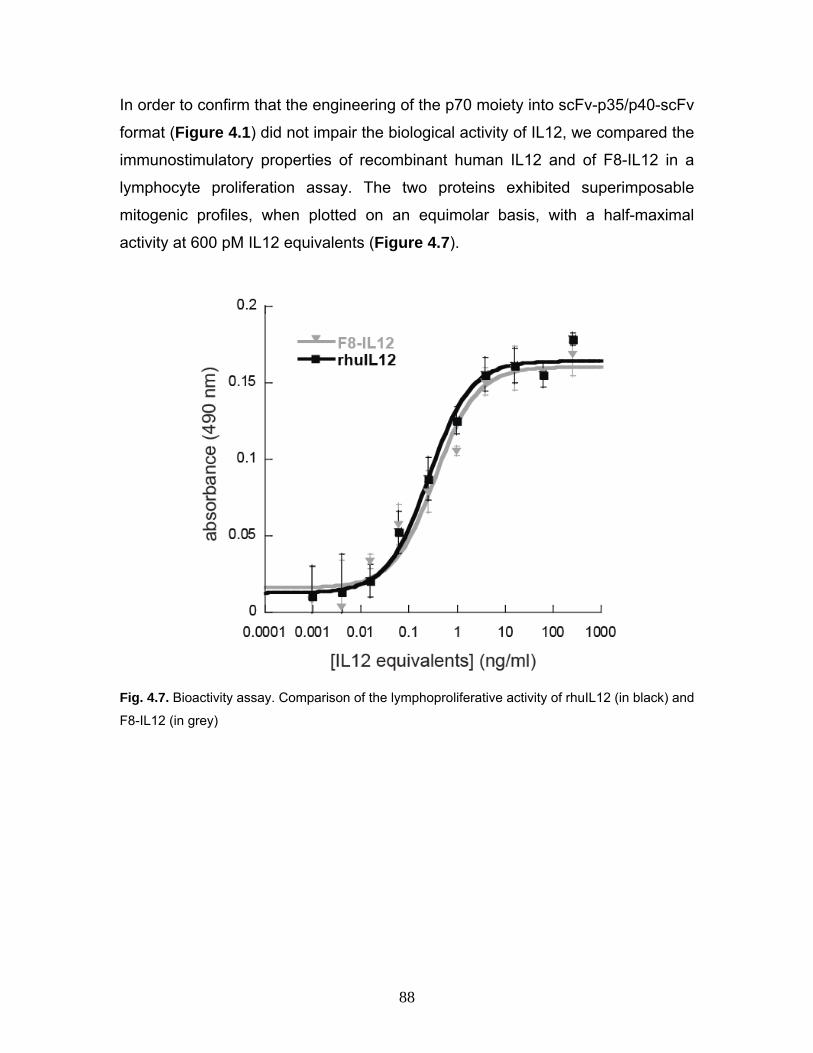
88
In order to confirm that the engineering of the p70 moiety into scFv-p35/p40-scFv
format (Figure 4.1) did not impair the biological activity of IL12, we compared the
immunostimulatory properties of recombinant human IL12 and of F8-IL12 in a
lymphocyte proliferation assay. The two proteins exhibited superimposable
mitogenic profiles, when plotted on an equimolar basis, with a half-maximal
activity at 600 pM IL12 equivalents (Figure 4.7).
Fig. 4.7. Bioactivity assay. Comparison of the lymphoproliferative activity of rhuIL12 (in black) and
F8-IL12 (in grey)

89
Finally, we investigated the tumor targeting properties of F8-IL12 by performing a
quantitative biodistribution analysis in mice bearing F9 tumors (Villa, Trachsel et
al. 2008) with a radioiodinated protein preparation. Cohorts of 4 mice were
sacrificed 24 and 48 hours after intravenous injection and the resulting organs
and body fluids were weighed and counted. F8-IL12 selectively accumulated in
the tumor, with percent injected dose per gram (%ID/g) values of 22 and 15 at
the two time points, respectively ([Figure 4.8 A). When correcting for tumor
growth during the experiment (F9 tumors have a doubling time of 40 hours; (Tarli,
Balza et al. 1999)). one observes a stable accumulation of immunocytokine in the
tumor at high levels (approx. 35 % ID/g) which is consistent with the inert nature
of EDA-containing fibronectin fibers (Figure 4.8 B). Furthermore an ex vivo
microautoradiography of tumors from biodistribution revealed selective
accumulation of the radiolabeled F8-IL12 protein around tumor vessels (Figure 4.8 C).

90
Fig. 4.8. Biodistribution studies. (A) F9 tumor bearing mice were injected intravenously with 125I-
labeled F8-IL12 and sacrificed 24 or 48 hours after injection. (B) Tumor values are corrected for
tumor growth during the experiment (F9 tumors have a doubling time of 40 hours). (C) An ex vivo
microautoradiography shows selective accumulation of 125I-labeled F8-IL12 around tumor
vessels.

91
4.2 Discussion
In this thesis, we have described the cloning, expression and characterization of
a novel fully human immunocytokine, suitable for clinical development programs
in patients with cancer. Compared to monomeric immunocytokines, the fusion of
heterodimeric cytokines to a full antibody or antibody fragment presents several
biotechnological challenges. First, multiple formats can be considered for the
construction of fusion proteins, which do not exhibit identical in vivo
biodistribution properties (Gafner, Trachsel et al. 2006). Moreover, vector design
and gene dosage effects influence the stability of the resulting stably-transfected
cell lines and product quality.
We used the strategy of Gafner et al (2006) for the cloning of the heterodimeric
cytokine IL12 to the tumor-targeting antibody fragment scFv(F8). The resulting
immunocytokine consists of the disulfide-linked IL12 subunits p35 and p40, each
joined to a scFv fragment. In contrast to the cloning strategy of Gafner et al, we
omitted all C-terminal peptide tags, an essential requirement for compatibility with
current GMP guidelines and subsequent clinical development of this
biopharmaceutical. In spite of the complex cloning strategy with two independent
vectors, the absence of peptidic tags for affinity purification and the need for cell
line stability over multiple passages, we were able to generate a clone
expressing the F8-IL12 heterodimer at 7 mg/l. These yields are compatible with
clinical development programs, in consideration of the fact that the Maximal
Tolerated Dose of IL12 in cancer patients is as low as 0.5 µg per kilogram of
body weight. The fusion protein retained its full bioactivity as shown by in vitro
proliferation assay and kept its high binding affinity to the cognate antigen, the
alternatively spliced extradomain A of fibronectin. Biodistribution studies revealed
an impressive tumor uptake of F8-IL12, with excellent tumor to organ ratio.
Microutoradiography confirmed the selective accumulation of F8-IL12 at vascular
structures of the tumor.
Our group has generated and investigated in clinical trials the human monoclonal
antibodies F8, L19 and F16 (Pini, Viti et al. 1998; Brack, Silacci et al. 2006; Villa,

92
Trachsel et al. 2008) specific to the alternatively spliced EDA and EDB domains
of fibronectin, and to the alternatively spliced A1 domain of tenascin-C,
respectively. The tumor targeting properties of these antibodies have been
extensively studied in animal models of pathology (Carnemolla, Borsi et al. 2002;
Brack, Silacci et al. 2006; Kaspar, Trachsel et al. 2007; Marlind, Kaspar et al.
2008; Villa, Trachsel et al. 2008). In addition, a radioiodinated version of the L19
antibody in scFv format (Santimaria, Moscatelli et al. 2003) and in SIP format
(Sauer, Erba et al. 2009) have been studied in imaging and radioimmunotherapy
clinical trials in patients with cancer. Similar studies are currently being
conducted also for SIP(F16) and SIP(F8).
The clinical-stage L19, F8, and F16 antibodies have been used as modular
building blocks for the construction of immunocytokines based on IL2
(Carnemolla, Borsi et al. 2002; Marlind, Kaspar et al. 2008), IIL10 (Trachsel,
Bootz et al. 2007; Schwager, Kaspar et al. 2009), IL12 (Halin, Rondini et al.
2002; Gafner, Trachsel et al. 2006), TNF (Borsi, Balza et al. 2003), IFN-γ
(Ebbinghaus, Ronca et al. 2005), IL15, GM-CSF (Kaspar, Trachsel et al. 2007)
and other cytokines.
Interleukin-12-based immunocytokines appear to display potent anti-tumor
effects, without worsening inflammation in animal models of chronic autoimmune
diseases (Trachsel, Kaspar et al. 2007).
At present, the immunocytokines L19-IL2, F16-IL2, L19-TNF and F8-IL10 are
being investigated in multiple clinical trials at 18 European Centers. The
development activities for F8-IL12 described in this article represent the basis for
the future clinical studies of this product. Investigations in patients with cancer
are particularly encouraged by the excellent biodistribution results observed with
F8-IL12 (Figure 4.8) and by the fact that the EDA domain of fibronectin is
virtually undetectable in normal adult organs, while being strongly expressed in
the majority of aggressive solid tumors and lymphomas (Pedretti, Rancic et al.;
Rybak, Roesli et al. 2007; Villa, Trachsel et al. 2008; Schliemann, Wiedmer et al.
2009)

93
4.3 Material and methods
Cloning of F8-p35
For the cloning of F8-p35, the scFv(F8), containing a NheI restriction site and a
leader sequence (Li, Pedraza et al. 1997) at the 5`, was amplified by PCR with
primer RS9fo (5’-ctagctagcgtcgaccatgggctggagcctgat-3`) and NP5ba (5`-
atcagcgcttcctttgatttccaccttggtcccttg-3`) which appends a part of the human p35
sequence. Human p35 gene was isolated from a human cDNA library (Human
immune system MTC panel; Clontech, US) and was amplified by PCR with
primer NP6fo (5’-gtggaaatcaaaggaagcgctgatggaggtagaaac-3’) that appends an
overlapping sequence to the VL of scFv(F8) and RS12ba (5`-
atagtttagcggccgctcattaggaagcattcagatagctcatcac-3’) which contains two stop
codons and a NotI restriction site at the 3`. The scFv(F8) antibody sequence and
the human p35 sequence were PCR assembled. The double digested NheI-NotI
assembly product was cloned into the mammalian cell expression vector
pcDNA3.1(+) (Invitrogen, Switzerland) containing the resistence for neomycin.
Cloning of p40-F8 For the cloning of p40-F8, the human p40 gene and its leader sequence was
isolated from a human cDNA library (Human immune system MTC panel;
Clontech, US) and was PCR amplified with primer KS5fo (5’-
cccaagcttatgtgtcaccagcagttggtcatc-3`) that anneals on the leader sequence and
introduces a HindIII restriction site at the 5` and RS8ba (5’-
cccagactccaacagctgcacctcacctccatcagcgcttccactgca-3’) that contains an
overlapping sequence with the scFv(F8). The scFv(F8) was amplified with
primers RS3 (5’-gaggtgcagctgttggagtctggg-3’) which partially overlaps with the
sequence of human p40 and NP7 (5’cctttgcggccgctcactatttgatttccaccttggtcccttg-
3’) that introduces two stop codons and a NotI restriction site. Human p40 and
the scFv(F8) sequences were PCR assembled and double digested with NheI
and NotI restriction enzymes. The digestion product was cloned into the
mammalian cell expression vector pcDNA3.1(+) (Invitrogen, Switzerland)

94
containing the resistance for hygromycin. All PCR reactions were performed
using the High Fidelity Taq Polymerase (Roche Diagnostics, Germany).
Nucleotide sequence of F8-p35
GCTAGCGTCGACCATGGGCTGGAGCCTGATCCTCCTGTTCCTCGTCGCTGTGGCTACAGGTAAGGGGCTCACAGTAGCAGGCTTGAGGTCTGGACATATATATGGGTGACAATGACATCCACTTTGCCTTTCTCTCCACAGGTGTGCACTCGGAGGTGCAGCTGTTGGAGTCTGGGGGAGGCTTGGTACAGCCTGGGGGGTCCCTGAGACTCTCCTGTGCAGCCTCTGGATTCACCTTTAGCCTGTTTACGATGAGCTGGGTCCGCCAGGCTCCAGGGAAGGGGCTGGAGTGGGTCTCAGCTATTAGTGGTAGTGGTGGTAGCACATACTACGCAGACTCCGTGAAGGGCCGGTTCACCATCTCCAGAGACAATTCCAAGAACACGCTGTATCTGCAAATGAACAGCCTGAGAGCCGAGGACACGGCCGTATATTACTGTGCGAAAAGTACTCATTTGTATCTTTTTGACTACTGGGGCCAGGGAACCCTGGTCACCGTCTCGAGTGGTGGAGGCGGTTCAGGCGGAGGTGGCTCTGGCGGTGGCGGAGAAATTGTGTTGACGCAGTCTCCAGGCACCCTGTCTTTGTCTCCAGGGGAAAGAGCCACCCTCTCCTGCAGGGCCAGTCAGAGTGTTAGCATGCCGTTTTTAGCCTGGTACCAGCAGAAACCTGGCCAGGCTCCCAGGCTCCTCATCTATGGTGCATCCAGCAGGGCCACTGGCATCCCAGACAGGTTCAGTGGCAGTGGGTCTGGGACAGACTTCACTCTCACCATCAGCAGACTGGAGCCTGAAGATTTTGCAGTGTATTACTGTCAGCAGATGCGTGGTCGGCCGCCGACGTTCGGCCAAGGGACCAAGGTGGAAATCAAAGGAAGCGCTGATGGAGGTAGAAACCTCCCCGTGGCCACTCCAGACCCAGGAATGTTCCCATGCCTTCACCACTCCCAAAACCTGCTGAGGGCCGTCAGCAACATGCTCCAGAAGGCCAGACAAACTCTAGAATTTTACCCTTGCACTTCTGAAGAGATTGATCATGAAGATATCACAAAAGATAAAACCAGCACAGTGGAGGCCTGTTTACCATTGGAATTAACCAAGAATGAGAGTTGCCTAAATTCCAGAGAGACCTCTTTCATAACTAATGGGAGTTGCCTGGCCTCCAGAAAGACCTCTTTTATGATGGCCCTGTGCCTTAGTAGTATTTATGAAGACTTGAAGATGTACCAGGTGGAGTTCAAGACCATGAATGCAAAGCTTCTGATGGATCCTAAGAGGCAGATCTTTCTAGATCAAAACATGCTGGCAGTTATTGATGAGCTGATGCAGGCCCTGAATTTCAACAGTGAGACTGTGCCACAAAAATCCTCCCTTGAAGAACCGGATTTTTATAAAACTAAAATCAAGCTCTGCATACTTCTTCATGCTTTCAGAATTCGGGCAGTGACTATTGATAGAGTGATGAGCTATCTGAATGCTTCCTAATGAGCGGCCGC
Aminoacid sequence of F8-p35
EVQLLESGGGLVQPGGSLRLSCAASGFTFSLFTMSWVRQAPGKGLEWVSAISGSGGSTYYADSVKGRFTISRDNSKNTLYLQMNSLRAEDTAVYYCAKSTHLYLFDYWGQGTLVTVSSGGGGSGGGGSGGGGEIVLTQSPGTLSLSPGERATLSCRASQSVSMPFLAWYQQKPGQAPRLLIYGASSRATGIPDRFSGSGSGTDFTLTISRLEPEDFAVYYCQQMRGRPPTFGQGTKVEIKGSADGGRNLPVATPDPGMFPCLHHSQNLLRAVSNMLQKARQTLEFYPCTSEEIDHEDITKDKTSTVEACLPLELTKNESCLNSRETSFITNGSCLASRKTSFMMALCLSSIYEDLKMYQVEFKTMNAKLLMDPKRQIFLDQNMLAVIDELMQALNFNSETVPQKSSLEEPDFYKTKIKLCILLHAFRIRAVTIDRVMSYLNAS
Nucleotide sequence of p40-F8
AAGCTTATGTGTCACCAGCAGTTGGTCATCTCTTGGTTTTCCCTGGTTTTTCTGGCATCTCCCCTCGTGGCCATATGGGAACTGAAGAAAGATGTTTATGTCGTAGAATTGGATTGGTATCCGGATGCCCCTGGAGAAATGGTGGTCCTCACCTGTGACACCCCTGAAGAAGATGGTATCACCTGGACCTTGGACCAGAGCAGTGAGGTCTTAGGCTCTGGCAAAACCCTGACCATCCAAGTCAAAGAGTTTGGAGATGCTGGCCAGTACACCTGTCACAAAGGAGGCGAGGTTCTAAGCCATTCGCTCCTGCTGCTTCACAAAAAGGAAGATGGAATTTGGTCCACTGATATTTTAAAGGACCAGAAAGAACCCAAAAATAAGACCTTTCTAAGATGCGAGGCCAAGAATTATTCTGGACGTTTCACCTGCTGGTGGCTGACGACAATCAGTACTGATTTGACATTCAGTGTCAAAAGCAGCAGAGGCTCTTCTGACCCCCAAGGGGTGACGTGCGGAGCTGCTACACTCTCTGCAGAGAGAGTCAGAGGGGACAACAAGGAGTATGAGTACTCAGTGGAGTGCCAGGAGGACAGTGCCTGCCCAGCTGCTGAGGAGAGTCTGCCCATTGAGGTCATGGTGGATGCCGTTCACAAGCTCAAGTATGAAAACTACACCAGCAGCTTCTTCATCAGGGACATCATCAAACCTGACCCACCCAAGAACTTGCAGCTGAAGCCATTAAAGAATTCTCGGCAGGTGGAGGTCAGCTGGGAGTACCCTGACACCTGGAGTACTCCACATTCCTACTTCTCCCTGACATTCTGCGTTCAGGTCCAGGGCAAGAGCAAGAGAGAAAAGAAAGATAGAGTCTTCACGGACAAGACCTCAGCCACGGTCATCTGCCGCAAAAATGCCAGCATTAGCGTGCGGGCCCAGGACCGCTACTATAGCTCATCTTGGAGCGAATGGGCATCTGTGCCCTGCAGTGGAAGCGCTGATGGAGGTGAGGTGCAGCTGTTGGAGTCTGGGGGAGGCTTGGTACAGCCTGGGGGGTCCCTGAGACTCTCCTGTGCAGCCTCTGGATTCACCTTTAGCCTGTTTACGATGAGCTGGGTCCGCCAGGCTCCAGGGAAGGGGCTGGAGTGGGTCTCAGCTATTAGTGGTAGTGGTGGTAGCACATACTACGCAGACTCCGTGAAGGGCCGGTTCACCATCTCCAGAGACAATTCCAAGAACACGCTGTATCTGCAAATGAACAGCCTGAGAGCCGAGGACACGGCCGTATATTACTGTGCGAAAAGTACTCATTTGTATCTTTTTGACTACTGGGGCCAGGGAACCCTGGTCACCGTCTCGAGTGGTGGAGGCGGTTCAGGCGGAGGTGGCTCTGGCGGTGGCGGAGAAATTGTGTTGACGCAGTCTCCAGGCACCCTGTCTTTGTCTCCAGGGGAAAGAGCCACCCTCTCCTGCAGGGCCAGTCAGAGTGTTAGCATGCCGTTTTTAGCCTGGTACCAGCAGAAACCTGGCCAGGCTCCCAGGCTCCTCATCTATGGTGCATCCAGCAGGGCCACTGGCATCCCAGACAGGTTCAGTGGCAGTGGGTCTGGGACAGACTTCACTCTCACCATCAGCAGACTGGAGCCTGAAGATTTTGCAGTGTATTACTGTCAGCAGATGCGTGGTCGGCCGCCGACGTTCGGCCAAGGGACCAAGGTGGAAATCAAATAGTGAGCGGCCGC

95
Aminoacid sequence of p40-F8
IWELKKDVYVVELDWYPDAPGEMVVLTCDTPEEDGITWTLDQSSEVLGSGKTLTIQVKEFGDAGQYTCHKGGEVLSHSLLLLHKKEDGIWSTDILKDQKEPKNKTFLRCEAKNYSGRFTCWWLTTISTDLTFSVKSSRGSSDPQGVTCGAATLSAERVRGDNKEYEYSVECQEDSACPAAEESLPIEVMVDAVHKLKYENYTSSFFIRDIIKPDPPKNLQLKPLKNSRQVEVSWEYPDTWSTPHSYFSLTFCVQVQGKSKREKKDRVFTDKTSATVICRKNASISVRAQDRYYSSSWSEWASVPCSGSADGGEVQLLESGGGLVQPGGSLRLSCAASGFTFSLFTMSWVRQAPGKGLEWVSAISGSGGSTYYADSVKGRFTISRDNSKNTLYLQMNSLRAEDTAVYYCAKSTHLYLFDYWGQGTLVTVSSGGGGSGGGGSGGGGEIVLTQSPGTLSLSPGERATLSCRASQSVSMPFLAWYQQKPGQAPRLLIYGASSRATGIPDRFSGSGSGTDFTLTISRLEPEDFAVYYCQQMRGRPPTFGQGTKVEIK
Table 4.1. Nucleotidic and aminoacid sequence of scFv(F8)-p35 and p40-scFv(F8) are
represented in the table.
Cell culture
Adherent CHO-S cells were cultured in RPMI 1640 (Gibco, Switzerland)
containing 10% FCS (Invitrogen, Switzerland) and incubated at 37ºC and 5%
CO2. Suspension CHO-S cells were cultivated in PowerCHO-2 CD (Lonza,
Switzerland) containing 8mM ultraglutamine (Lonza, Belgium) incubated at 37 °C
and 5% CO2. When necessary G418 (Calbiochem, Germany) and Hygromicin
(Invivogen, Switzerland) were added to the medium at a concentration of 0.5
mg/ml.
Generation of a cell line expressing F8-IL12
Adherent CHO-S cells were electroporated using an Amaxa nucleofector
according to the manufacturer’s protocol. Different ratios of plasmids F8-p35 /
p40-F8 were used to find the best conditions to maximize the expression of the
heretodimer. Three days after electroporation, stably-transfected cells were
selected in presence of G418 0.5 mg/ml (Calbiochem, Germany) and 0.5 mg/ml
hygromycin (Invivogen, Switzerland). Monoclonal cell lines were obtained by
serial dilutions and screened for expression of the fusion protein by ELISA.
Large scale culture of F8-IL12 in bioreactor
Stably-transfected suspension CHO-S cells driving F8-IL12 expression were
grown in PowerCHO-2 CD containing 8mM ultraglutamine, 0.5 mg/ml G418 and
0.5 mg/ml Hygromicin. The cell culture inoculum for the 3 liters Labfors bioreactor

96
(Infors AG, Switzerland) was obtained starting from a static culture of the cells of
the master cell bank. The cell culture was expanded in roller bottles untill cell
density reached 2-3x10⁶ cells/ml. The fed batch fermentation process was used
for the production of F8-IL12. Cells were seeded at 5x10⁵ cells/ml in the
bioreactor. Fixed cell culture parameters of F8-IL12 in the bioreactor were
defined as 37 °C temperature, 50% pO2, 130 rpm helical stirring, pH 7.25.
Glucose concentration was monitored and manual feed were applied to bring its
concentration up to 2 g/l. In the exponential and plateau growth phase (72h -
144h) feeds of fresh medium supplemented with ultraglutamine were introduced
to maintain cell vitality. In order to monitor the total protein productivity, 100 ml
supernatant samples were daily collected and affinity purified on HiTrap Protein
A resin (GE Healthcare, Sweden).
Purification of F8-IL12
The fusion protein could be purified from cell culture medium by protein A affinity
chromatography, as there is a staphylococcal protein A binding site present on
most VH3 domains (Sasso, Silverman et al. 1991; Hoogenboom and Winter
1992; Silacci, Brack et al. 2005) Ion exchange chromatography (Ceramic hyper
D; PALL, USA) and preparative size exclusion chromatography on a HiLoad
26/60 Superdex 200 size exclusion column (GE Healthcare, Sweden) of the
eluted protein A preparation allowed the isolation of the F8-IL12 heterodimer
from a mix containing also aggregates and polymers.
Elisa of supernatants ELISA experiments on the conditioned culture media were performed according
to Carnemolla et al. (Carnemolla, Neri et al. 1996) To detect different clones
expressing F8-IL12, the biotinylated 11-EDA-12 domain of fibronectin, which
includes the epitope recognized by scFv(F8), was immobilized on a streptavidin
coated plate (StreptaWell, Roche Applied Bioscience, Germany). Four different
detection systems were used and allowed the evaluation of the expression of the
different components of the fusion protein. Horseradish peroxidase conjugated

97
Protein A (GE Healthcare, Sweden), diluted according to the manufacturer’s
recommendations, was used as agent to detect the VH domain of the fusion
protein. In order to detect the heterodimer p70, the p35 subunit and the free p40
subunit, three different mouse monoclonal antibodies were used (“affinity purified
anti-human IL-12, p35” (eBioscience, Germany); “affinity purified anti-human IL-
12, p40” (eBioscience, Germany); “anti-human IL-12 p70 antibody” (R&D
Systems, Germany)) and detected with a goat anti-muose IgG peroxidase
conjugate (Sigma-Aldrich, USA). In all cases, the immunoreactivity with the
immobilized antigen was detected using the substrate BluePOD (Roche
Diagnostics, Germany) for peroxidase, and photometric absorbance at 405 nm
was measured.
Bioactivity assay
The biological activity of F8-IL12 was determined by a T-cell proliferation assay
(Gately, Chizzonite et al. 2001) Freshly isolated human peripheral blood
mononuclear cells (PBMC) were cultured immediately after isolation with 25
µg/ml mitogen Phytohemagglutinin-M (Roche Diagnostics, Germany) for 3 days.
Cells were diluted 1:2 by adding equal volume of supplement medium and further
cultivated with 50 I.U./ml of human Interleukin-2 (Roche Diagnostics, Germany).
After 24 hr cells were seeded in 96-well plates at a density of 2 x 10⁴ cells/well in
200 µl of medium containing serial dilutions of either F8-IL12 or commercially
available, recombinant, human IL12 as standard (R&D Systems, Germany) or
culture medium as a negative control. After 48 h, 20 µl Cell Titer 96 Aqueous
One Solution (Promega, USA) was added to each well. The plate was incubated
for 4 h and absorbance was read at 490 nm. The experiment was performed in
triplicate.
Biodistribution studies
The in vivo targeting performance of F8-IL12 was evaluated by biodistribution
analysis as described before (Carnemolla, Borsi et al. 2002) Briefly, purified F8-
IL12 was radioiodinated and injected into the tail vein of immunocompetent

98
129SvEv mice bearing s.c. implanted F9 murine teratocarcinoma. Mice were
sacrificed 24 h or 48 h after injection of the fusion protein (8 µg, 7 µCi/mouse).
Organs were weighed and radioactivity was counted with a Packard Cobra
gamma counter. Radioactivity content of representative organs was expressed
as the percentage of the injected dose per gram of tissue (%ID/g).
Microautoradiography
Twenty-four hours after the tail injection of radiolabeled F8-IL12, mice were
sacrificed and tumors were embedded in paraffin. Ten μm sections were cut and
fixed with paraformaldehyde. Sections were then coated with NBT KODAK
autoradiography emulsion (KODAK, US), dried and stored at 4°C in the dark for
approximately three weeks. The autoradiography emulsions were developed
(Developer D-19, KODAK, France) and fixed (EASTMAN Fixer, KODAK France).
Finally, slides were rinsed with deionized water and counterstained with
hematoxilin (SIGMA, Switzerland).
Mass spectrometry
Following reduction of F8-IL12 with 10 mM TCEP for 30 min at room
temperature, the solution was desalted and concentrated using C4 microcolumns
(OMIX™ tips, Varian Inc., Paolo Alto, CA, USA) according to the manufacturer’s
guidelines. The reduced and desalted protein was mixed with sinapinic acid (20
mg/ml in 70% ACN, 0.1% TFA) and spotted on a MALDI (matrix assisted laser
desorption/ionization) target plate. The mass spectrometric analysis was carried
out using a 4800 MALDI-TOF/TOF Analyzer (Applied Biosystems, Foster City,
CA, USA). All spectra were acquired in the linear mode in a mass range of
10’000 to 100’000 m/z with a solid-state laser (355 nm) at a laser repetition rate
of 200 Hz. A total of 1500 laser shots were summed for each spectra. Spectra
were further processed using the Data Explorer software (Applied Biosystems).

99

100
5 References
Alatrash, G., T. E. Hutson, et al. (2004). "Clinical and immunologic effects of
subcutaneously administered interleukin-12 and interferon alfa-2b: phase I trial of patients with metastatic renal cell carcinoma or malignant melanoma." J Clin Oncol 22(14): 2891-900.
Alberts, S. R., F. A. Sinicrope, et al. (2005). "N0147: a randomized phase III trial of oxaliplatin plus 5-fluorouracil/leucovorin with or without cetuximab after curative resection of stage III colon cancer." Clin Colorectal Cancer 5(3): 211-3.
Atkins, M. B., M. J. Robertson, et al. (1997). "Phase I evaluation of intravenous recombinant human interleukin 12 in patients with advanced malignancies." Clin Cancer Res 3(3): 409-17.
Bajetta, E., M. Del Vecchio, et al. (1998). "Pilot study of subcutaneous recombinant human interleukin 12 in metastatic melanoma." Clin Cancer Res 4(1): 75-85.
Berndorff, D., S. Borkowski, et al. (2005). "Radioimmunotherapy of solid tumors by targeting extra domain B fibronectin: identification of the best-suited radioimmunoconjugate." Clin Cancer Res 11(19 Pt 2): 7053s-7063s.
Better, M., C. P. Chang, et al. (1988). "Escherichia coli secretion of an active chimeric antibody fragment." Science 240(4855): 1041-3.
Bird, R. E., K. D. Hardman, et al. (1988). "Single-chain antigen-binding proteins." Science 242(4877): 423-6.
Borsi, L., E. Balza, et al. (2002). "Selective targeting of tumoral vasculature: comparison of different formats of an antibody (L19) to the ED-B domain of fibronectin." Int J Cancer 102(1): 75-85.
Borsi, L., E. Balza, et al. (2003). "Selective targeted delivery of TNFalpha to tumor blood vessels." Blood 102(13): 4384-92.
Borsi, L., B. Carnemolla, et al. (1992). "Expression of different tenascin isoforms in normal, hyperplastic and neoplastic human breast tissues." Int J Cancer 52(5): 688-92.
Borsi, L., P. Castellani, et al. (1998). "Preparation of phage antibodies to the ED-A domain of human fibronectin." Exp Cell Res 240(2): 244-51.
Bothwell, A. L., M. Paskind, et al. (1981). "Heavy chain variable region contribution to the NPb family of antibodies: somatic mutation evident in a gamma 2a variable region." Cell 24(3): 625-37.
Brack, S. S., M. Silacci, et al. (2006). "Tumor-targeting properties of novel antibodies specific to the large isoform of tenascin-C." Clin Cancer Res 12(10): 3200-8.
Brekke, O. H. and I. Sandlie (2003). "Therapeutic antibodies for human diseases at the dawn of the twenty-first century." Nat Rev Drug Discov 2(1): 52-62.
Brunda, M. J., L. Luistro, et al. (1993). "Antitumor and antimetastatic activity of interleukin 12 against murine tumors." J Exp Med 178(4): 1223-30.

101
Burton, D. R., C. F. Barbas, 3rd, et al. (1991). "A large array of human monoclonal antibodies to type 1 human immunodeficiency virus from combinatorial libraries of asymptomatic seropositive individuals." Proc Natl Acad Sci U S A 88(22): 10134-7.
Cabilly, S. (1989). "Growth at sub-optimal temperatures allows the production of functional, antigen-binding Fab fragments in Escherichia coli." Gene 85(2): 553-7.
Cai, X. and A. Garen (1995). "Anti-melanoma antibodies from melanoma patients immunized with genetically modified autologous tumor cells: selection of specific antibodies from single-chain Fv fusion phage libraries." Proc Natl Acad Sci U S A 92(14): 6537-41.
Carnemolla, B., L. Borsi, et al. (2002). "Enhancement of the antitumor properties of interleukin-2 by its targeted delivery to the tumor blood vessel extracellular matrix." Blood 99(5): 1659-65.
Carnemolla, B., L. Borsi, et al. (1992). "Comparison of human tenascin expression in normal, simian-virus-40-transformed and tumor-derived cell lines." Eur J Biochem 205(2): 561-7.
Carnemolla, B., A. Leprini, et al. (1992). "The inclusion of the type III repeat ED-B in the fibronectin molecule generates conformational modifications that unmask a cryptic sequence." J Biol Chem 267(34): 24689-92.
Carnemolla, B., D. Neri, et al. (1996). "Phage antibodies with pan-species recognition of the oncofoetal angiogenesis marker fibronectin ED-B domain." Int J Cancer 68(3): 397-405.
Carra, G., F. Gerosa, et al. (2000). "Biosynthesis and posttranslational regulation of human IL-12." J Immunol 164(9): 4752-61.
Castellani, P., L. Borsi, et al. (2002). "Differentiation between high- and low-grade astrocytoma using a human recombinant antibody to the extra domain-B of fibronectin." Am J Pathol 161(5): 1695-700.
Castellani, P., G. Viale, et al. (1994). "The fibronectin isoform containing the ED-B oncofetal domain: a marker of angiogenesis." Int J Cancer 59(5): 612-8.
Cebon, J., E. Jager, et al. (2003). "Two phase I studies of low dose recombinant human IL-12 with Melan-A and influenza peptides in subjects with advanced malignant melanoma." Cancer Immun 3: 7.
Chang, S. and C. Mohan (2005). "Identification of novel VH1/J558 immunoglobulin germline genes of C57BL/6 (Igh b) allotype." Mol Immunol 42(11): 1293-301.
Chester, K. A., L. Robson, et al. (1994). "Production and tumour-binding characterization of a chimeric anti-CEA Fab expressed in Escherichia coli." Int J Cancer 57(1): 67-72.
Chiquet-Ehrismann, R. and M. Chiquet (2003). "Tenascins: regulation and putative functions during pathological stress." J Pathol 200(4): 488-99.
Chothia, C. and A. M. Lesk (1987). "Canonical structures for the hypervariable regions of immunoglobulins." J Mol Biol 196(4): 901-17.
Chothia, C., A. M. Lesk, et al. (1992). "Structural repertoire of the human VH segments." J Mol Biol 227(3): 799-817.

102
Clackson, T., H. R. Hoogenboom, et al. (1991). "Making antibody fragments using phage display libraries." Nature 352(6336): 624-8.
Cobleigh, M. A., C. L. Vogel, et al. (1999). "Multinational study of the efficacy and safety of humanized anti-HER2 monoclonal antibody in women who have HER2-overexpressing metastatic breast cancer that has progressed after chemotherapy for metastatic disease." J Clin Oncol 17(9): 2639-48.
Colombo, M. P. and G. Trinchieri (2002). "Interleukin-12 in anti-tumor immunity and immunotherapy." Cytokine Growth Factor Rev 13(2): 155-68.
Cooley, S., L. J. Burns, et al. (1999). "Natural killer cell cytotoxicity of breast cancer targets is enhanced by two distinct mechanisms of antibody-dependent cellular cytotoxicity against LFA-3 and HER2/neu." Exp Hematol 27(10): 1533-41.
Curtsinger, J. M., D. C. Lins, et al. (2003). "Signal 3 determines tolerance versus full activation of naive CD8 T cells: dissociating proliferation and development of effector function." J Exp Med 197(9): 1141-51.
Davies, E. L., J. S. Smith, et al. (1995). "Selection of specific phage-display antibodies using libraries derived from chicken immunoglobulin genes." J Immunol Methods 186(1): 125-35.
de Haard, H. J., B. Kazemier, et al. (1998). "Selection of recombinant, library-derived antibody fragments against p24 for human immunodeficiency virus type 1 diagnostics." Clin Diagn Lab Immunol 5(5): 636-44.
Dias, S., R. Boyd, et al. (1998). "IL-12 regulates VEGF and MMPs in a murine breast cancer model." Int J Cancer 78(3): 361-5.
Ebbinghaus, C., R. Ronca, et al. (2005). "Engineered vascular-targeting antibody-interferon-gamma fusion protein for cancer therapy." Int J Cancer 116(2): 304-13.
Gafner, V., E. Trachsel, et al. (2006). "An engineered antibody-interleukin-12 fusion protein with enhanced tumor vascular targeting properties." Int J Cancer 119(9): 2205-12.
Gao, X., Y. Huang, et al. (1999). "Construction of murine phage antibody library and selection of ricin-specific single-chain antibodies." IUBMB Life 48(5): 513-7.
Gately, M. K., R. Chizzonite, et al. (2001). "Measurement of human and mouse interleukin-12." Curr Protoc Immunol Chapter 6: Unit 6 16.
Giovannoni, L., F. Viti, et al. (2001). "Isolation of anti-angiogenesis antibodies from a large combinatorial repertoire by colony filter screening." Nucleic Acids Res 29(5): E27.
Gollob, J. A., J. W. Mier, et al. (2000). "Phase I trial of twice-weekly intravenous interleukin 12 in patients with metastatic renal cell cancer or malignant melanoma: ability to maintain IFN-gamma induction is associated with clinical response." Clin Cancer Res 6(5): 1678-92.
Gollob, J. A., K. G. Veenstra, et al. (2003). "Phase I trial of concurrent twice-weekly recombinant human interleukin-12 plus low-dose IL-2 in patients with melanoma or renal cell carcinoma." J Clin Oncol 21(13): 2564-73.

103
Greenwood, J., A. E. Willis, et al. (1991). "Multiple display of foreign peptides on a filamentous bacteriophage. Peptides from Plasmodium falciparum circumsporozoite protein as antigens." J Mol Biol 220(4): 821-7.
Griffiths, A. D., S. C. Williams, et al. (1994). "Isolation of high affinity human antibodies directly from large synthetic repertoires." Embo J 13(14): 3245-60.
Halin, C., V. Gafner, et al. (2003). "Synergistic therapeutic effects of a tumor targeting antibody fragment, fused to interleukin 12 and to tumor necrosis factor alpha." Cancer Res 63(12): 3202-10.
Halin, C., S. Rondini, et al. (2002). "Enhancement of the antitumor activity of interleukin-12 by targeted delivery to neovasculature." Nat Biotechnol 20(3): 264-9.
Hanes, J. and A. Pluckthun (1997). "In vitro selection and evolution of functional proteins by using ribosome display." Proc Natl Acad Sci U S A 94(10): 4937-42.
Hanes, J., C. Schaffitzel, et al. (2000). "Picomolar affinity antibodies from a fully synthetic naive library selected and evolved by ribosome display." Nat Biotechnol 18(12): 1287-92.
Hoogenboom, H. R. (1997). "Designing and optimizing library selection strategies for generating high-affinity antibodies." Trends Biotechnol 15(2): 62-70.
Hoogenboom, H. R., A. D. Griffiths, et al. (1991). "Multi-subunit proteins on the surface of filamentous phage: methodologies for displaying antibody (Fab) heavy and light chains." Nucleic Acids Res 19(15): 4133-7.
Hoogenboom, H. R. and G. Winter (1992). "By-passing immunisation. Human antibodies from synthetic repertoires of germline VH gene segments rearranged in vitro." J Mol Biol 227(2): 381-8.
Hurwitz, H., L. Fehrenbacher, et al. (2004). "Bevacizumab plus irinotecan, fluorouracil, and leucovorin for metastatic colorectal cancer." N Engl J Med 350(23): 2335-42.
Huston, J. S. and E. Haber (1996). "An overview of the 1996 Keystone meeting. Exploring and exploiting antibody and Ig superfamily combining sites." Immunotechnology 2(4): 253-60.
Huston, J. S., D. Levinson, et al. (1988). "Protein engineering of antibody binding sites: recovery of specific activity in an anti-digoxin single-chain Fv analogue produced in Escherichia coli." Proc Natl Acad Sci U S A 85(16): 5879-83.
Iannolo, G., O. Minenkova, et al. (1995). "Modifying filamentous phage capsid: limits in the size of the major capsid protein." J Mol Biol 248(4): 835-44.
Ignatovich, O., I. M. Tomlinson, et al. (1997). "The creation of diversity in the human immunoglobulin V(lambda) repertoire." J Mol Biol 268(1): 69-77.
Imai, S., Y. Mukai, et al. (2006). "Quality enhancement of the non-immune phage scFv library to isolate effective antibodies." Biol Pharm Bull 29(7): 1325-30.
Jermutus, L., A. Honegger, et al. (2001). "Tailoring in vitro evolution for protein affinity or stability." Proc Natl Acad Sci U S A 98(1): 75-80.

104
Jones, P. L. and F. S. Jones (2000). "Tenascin-C in development and disease: gene regulation and cell function." Matrix Biol 19(7): 581-96.
Kaczmarek, J., P. Castellani, et al. (1994). "Distribution of oncofetal fibronectin isoforms in normal, hyperplastic and neoplastic human breast tissues." Int J Cancer 59(1): 11-6.
Kalinski, P., C. M. Hilkens, et al. (1999). "T-cell priming by type-1 and type-2 polarized dendritic cells: the concept of a third signal." Immunol Today 20(12): 561-7.
Kaspar, M., E. Trachsel, et al. (2007). "The antibody-mediated targeted delivery of interleukin-15 and GM-CSF to the tumor neovasculature inhibits tumor growth and metastasis." Cancer Res 67(10): 4940-8.
Kaspar, M., L. Zardi, et al. (2006). "Fibronectin as target for tumor therapy." Int J Cancer 118(6): 1331-9.
Kettleborough, C. A., K. H. Ansell, et al. (1994). "Isolation of tumor cell-specific single-chain Fv from immunized mice using phage-antibody libraries and the re-construction of whole antibodies from these antibody fragments." Eur J Immunol 24(4): 952-8.
Kettleborough, C. A., J. Saldanha, et al. (1993). "Optimization of primers for cloning libraries of mouse immunoglobulin genes using the polymerase chain reaction." Eur J Immunol 23(1): 206-11.
Kirkham, P. M., F. Mortari, et al. (1992). "Immunoglobulin VH clan and family identity predicts variable domain structure and may influence antigen binding." Embo J 11(2): 603-9.
Kirschbaum, T., F. Roschenthaler, et al. (1999). "The central part of the mouse immunoglobulin kappa locus." Eur J Immunol 29(7): 2057-64.
Knappik, A., L. Ge, et al. (2000). "Fully synthetic human combinatorial antibody libraries (HuCAL) based on modular consensus frameworks and CDRs randomized with trinucleotides." J Mol Biol 296(1): 57-86.
Kohler, G. and C. Milstein (1975). "Continuous cultures of fused cells secreting antibody of predefined specificity." Nature 256(5517): 495-7.
Kristensen, P. and G. Winter (1998). "Proteolytic selection for protein folding using filamentous bacteriophages." Fold Des 3(5): 321-8.
Lee, P., F. Wang, et al. (2001). "Effects of interleukin-12 on the immune response to a multipeptide vaccine for resected metastatic melanoma." J Clin Oncol 19(18): 3836-47.
Lenzi, R., M. Rosenblum, et al. (2002). "Phase I study of intraperitoneal recombinant human interleukin 12 in patients with Mullerian carcinoma, gastrointestinal primary malignancies, and mesothelioma." Clin Cancer Res 8(12): 3686-95.
Li, E., A. Pedraza, et al. (1997). "Mammalian cell expression of dimeric small immune proteins (SIP)." Protein Eng 10(6): 731-6.
Lieschke, G. J., P. K. Rao, et al. (1997). "Bioactive murine and human interleukin-12 fusion proteins which retain antitumor activity in vivo." Nat Biotechnol 15(1): 35-40.
Little, R. F., J. M. Pluda, et al. (2006). "Activity of subcutaneous interleukin-12 in AIDS-related Kaposi sarcoma." Blood 107(12): 4650-7.

105
Lo, K. M., Y. Lan, et al. (2007). "huBC1-IL12, an immunocytokine which targets EDB-containing oncofetal fibronectin in tumors and tumor vasculature, shows potent anti-tumor activity in human tumor models." Cancer Immunol Immunother 56(4): 447-57.
Lowman, H. B., S. H. Bass, et al. (1991). "Selecting high-affinity binding proteins by monovalent phage display." Biochemistry 30(45): 10832-8.
Luginbuhl, B., Z. Kanyo, et al. (2006). "Directed evolution of an anti-prion protein scFv fragment to an affinity of 1 pM and its structural interpretation." J Mol Biol 363(1): 75-97.
Magram, J., S. E. Connaughton, et al. (1996). "IL-12-deficient mice are defective in IFN gamma production and type 1 cytokine responses." Immunity 4(5): 471-81.
Manetti, R., P. Parronchi, et al. (1993). "Natural killer cell stimulatory factor (interleukin 12 [IL-12]) induces T helper type 1 (Th1)-specific immune responses and inhibits the development of IL-4-producing Th cells." J Exp Med 177(4): 1199-204.
Marks, J. D., H. R. Hoogenboom, et al. (1991). "By-passing immunization. Human antibodies from V-gene libraries displayed on phage." J Mol Biol 222(3): 581-97.
Marlind, J., M. Kaspar, et al. (2008). "Antibody-mediated delivery of interleukin-2 to the stroma of breast cancer strongly enhances the potency of chemotherapy." Clin Cancer Res 14(20): 6515-24.
Mattheakis, L. C., R. R. Bhatt, et al. (1994). "An in vitro polysome display system for identifying ligands from very large peptide libraries." Proc Natl Acad Sci U S A 91(19): 9022-6.
McCafferty, J., A. D. Griffiths, et al. (1990). "Phage antibodies: filamentous phage displaying antibody variable domains." Nature 348(6301): 552-4.
Melkko, S. and D. Neri (2003). "Calmodulin as an affinity purification tag." Methods Mol Biol 205: 69-77.
Mitola, S., M. Strasly, et al. (2003). "IL-12 regulates an endothelial cell-lymphocyte network: effect on metalloproteinase-9 production." J Immunol 171(7): 3725-33.
Molina, A. (2008). "A decade of rituximab: improving survival outcomes in non-Hodgkin's lymphoma." Annu Rev Med 59: 237-50.
Mortarini, R., A. Borri, et al. (2000). "Peripheral burst of tumor-specific cytotoxic T lymphocytes and infiltration of metastatic lesions by memory CD8+ T cells in melanoma patients receiving interleukin 12." Cancer Res 60(13): 3559-68.
Motzer, R. J., A. Rakhit, et al. (1998). "Phase I trial of subcutaneous recombinant human interleukin-12 in patients with advanced renal cell carcinoma." Clin Cancer Res 4(5): 1183-91.
Motzer, R. J., A. Rakhit, et al. (2001). "Randomized multicenter phase II trial of subcutaneous recombinant human interleukin-12 versus interferon-alpha 2a for patients with advanced renal cell carcinoma." J Interferon Cytokine Res 21(4): 257-63.

106
Muro, A. F., M. Caputi, et al. (1999). "Regulation of fibronectin EDA exon alternative splicing: possible role of RNA secondary structure for enhancer display." Mol Cell Biol 19(4): 2657-71.
Murphy, E. E., G. Terres, et al. (1994). "B7 and interleukin 12 cooperate for proliferation and interferon gamma production by mouse T helper clones that are unresponsive to B7 costimulation." J Exp Med 180(1): 223-31.
Neri, D. and R. Bicknell (2005). "Tumour vascular targeting." Nat Rev Cancer 5(6): 436-46.
Neri, D., B. Carnemolla, et al. (1997). "Targeting by affinity-matured recombinant antibody fragments of an angiogenesis associated fibronectin isoform." Nat Biotechnol 15(12): 1271-5.
Neri, D., A. Pini, et al. (1998). "Antibodies from phage display libraries as immunochemical reagents." Methods Mol Biol 80: 475-500.
Nissim, A., H. R. Hoogenboom, et al. (1994). "Antibody fragments from a 'single pot' phage display library as immunochemical reagents." Embo J 13(3): 692-8.
Okamoto, I. "Epidermal growth factor receptor in relation to tumor development: EGFR-targeted anticancer therapy." Febs J 277(2): 309-15.
Okamoto, T., Y. Mukai, et al. (2004). "Optimal construction of non-immune scFv phage display libraries from mouse bone marrow and spleen established to select specific scFvs efficiently binding to antigen." Biochem Biophys Res Commun 323(2): 583-91.
Oppmann, B., R. Lesley, et al. (2000). "Novel p19 protein engages IL-12p40 to form a cytokine, IL-23, with biological activities similar as well as distinct from IL-12." Immunity 13(5): 715-25.
Orlandi, R., D. H. Gussow, et al. (1989). "Cloning immunoglobulin variable domains for expression by the polymerase chain reaction." Proc Natl Acad Sci U S A 86(10): 3833-7.
Orum, H., P. S. Andersen, et al. (1993). "Efficient method for constructing comprehensive murine Fab antibody libraries displayed on phage." Nucleic Acids Res 21(19): 4491-8.
Osenga, K. L., J. A. Hank, et al. (2006). "A phase I clinical trial of the hu14.18-IL2 (EMD 273063) as a treatment for children with refractory or recurrent neuroblastoma and melanoma: a study of the Children's Oncology Group." Clin Cancer Res 12(6): 1750-9.
Parihar, R., P. Nadella, et al. (2004). "A phase I study of interleukin 12 with trastuzumab in patients with human epidermal growth factor receptor-2-overexpressing malignancies: analysis of sustained interferon gamma production in a subset of patients." Clin Cancer Res 10(15): 5027-37.
Parmley, S. F. and G. P. Smith (1988). "Antibody-selectable filamentous fd phage vectors: affinity purification of target genes." Gene 73(2): 305-18.
Payvar, F. and R. T. Schimke (1979). "Improvements in immunoprecipitation of specific messenger RNA. Isolation of highly purified conalbumin mRNA in high yield." Eur J Biochem 101(1): 271-82.

107
Pedretti, M., Z. Rancic, et al. "Comparative immunohistochemical staining of atherosclerotic plaques using F16, F8 and L19: Three clinical-grade fully human antibodies." Atherosclerosis 208(2): 382-9.
Pedretti, M., A. Soltermann, et al. (2009). "Comparative immunohistochemistry of L19 and F16 in non-small cell lung cancer and mesothelioma: two human antibodies investigated in clinical trials in patients with cancer." Lung Cancer 64(1): 28-33.
Perelson, A. S. and G. F. Oster (1979). "Theoretical studies of clonal selection: minimal antibody repertoire size and reliability of self-non-self discrimination." J Theor Biol 81(4): 645-70.
Peters, J. H., J. E. Trevithick, et al. (1995). "Expression of the alternatively spliced EIIIB segment of fibronectin." Cell Adhes Commun 3(1): 67-89.
Pflanz, S., J. C. Timans, et al. (2002). "IL-27, a heterodimeric cytokine composed of EBI3 and p28 protein, induces proliferation of naive CD4(+) T cells." Immunity 16(6): 779-90.
Pini, A., F. Viti, et al. (1998). "Design and use of a phage display library. Human antibodies with subnanomolar affinity against a marker of angiogenesis eluted from a two-dimensional gel." J Biol Chem 273(34): 21769-76.
Portielje, J. E., C. H. Lamers, et al. (2003). "Repeated administrations of interleukin (IL)-12 are associated with persistently elevated plasma levels of IL-10 and declining IFN-gamma, tumor necrosis factor-alpha, IL-6, and IL-8 responses." Clin Cancer Res 9(1): 76-83.
Presta, L. G., S. J. Lahr, et al. (1993). "Humanization of an antibody directed against IgE." J Immunol 151(5): 2623-32.
Ridder, R., R. Schmitz, et al. (1995). "Generation of rabbit monoclonal antibody fragments from a combinatorial phage display library and their production in the yeast Pichia pastoris." Biotechnology (N Y) 13(3): 255-60.
Robertson, M. J., C. Cameron, et al. (1999). "Immunological effects of interleukin 12 administered by bolus intravenous injection to patients with cancer." Clin Cancer Res 5(1): 9-16.
Rodolfo, M. and M. P. Colombo (1999). "Interleukin-12 as an adjuvant for cancer immunotherapy." Methods 19(1): 114-20.
Rondot, S., J. Koch, et al. (2001). "A helper phage to improve single-chain antibody presentation in phage display." Nat Biotechnol 19(1): 75-8.
Rook, A. H., G. S. Wood, et al. (1999). "Interleukin-12 therapy of cutaneous T-cell lymphoma induces lesion regression and cytotoxic T-cell responses." Blood 94(3): 902-8.
Rosenberg, S. A., J. C. Yang, et al. (1998). "Durability of complete responses in patients with metastatic cancer treated with high-dose interleukin-2: identification of the antigens mediating response." Ann Surg 228(3): 307-19.
Rybak, J. N., C. Roesli, et al. (2007). "The extra-domain A of fibronectin is a vascular marker of solid tumors and metastases." Cancer Res 67(22): 10948-57.

108
Santimaria, M., G. Moscatelli, et al. (2003). "Immunoscintigraphic detection of the ED-B domain of fibronectin, a marker of angiogenesis, in patients with cancer." Clin Cancer Res 9(2): 571-9.
Sarup, J. C., R. M. Johnson, et al. (1991). "Characterization of an anti-p185HER2 monoclonal antibody that stimulates receptor function and inhibits tumor cell growth." Growth Regul 1(2): 72-82.
Sasso, E. H., G. J. Silverman, et al. (1991). "Human IgA and IgG F(ab')2 that bind to staphylococcal protein A belong to the VHIII subgroup." J Immunol 147(6): 1877-83.
Sauer, S., P. A. Erba, et al. (2009). "Expression of the oncofetal ED-B-containing fibronectin isoform in hematologic tumors enables ED-B-targeted 131I-L19SIP radioimmunotherapy in Hodgkin lymphoma patients." Blood 113(10): 2265-74.
Schliemann, C. and D. Neri (2007). "Antibody-based targeting of the tumor vasculature." Biochim Biophys Acta 1776(2): 175-92.
Schliemann, C., A. Palumbo, et al. (2009). "Complete eradication of human B-cell lymphoma xenografts using rituximab in combination with the immunocytokine L19-IL2." Blood 113(10): 2275-83.
Schliemann, C., A. Wiedmer, et al. (2009). "Three clinical-stage tumor targeting antibodies reveal differential expression of oncofetal fibronectin and tenascin-C isoforms in human lymphoma." Leuk Res 33(12): 1718-22.
Schmitt, E., P. Hoehn, et al. (1994). "T helper type 1 development of naive CD4+ T cells requires the coordinate action of interleukin-12 and interferon-gamma and is inhibited by transforming growth factor-beta." Eur J Immunol 24(4): 793-8.
Schwager, K., M. Kaspar, et al. (2009). "Preclinical characterization of DEKAVIL (F8-IL10), a novel clinical-stage immunocytokine which inhibits the progression of collagen-induced arthritis." Arthritis Res Ther 11(5): R142.
Schwarzbauer, J. E., J. W. Tamkun, et al. (1983). "Three different fibronectin mRNAs arise by alternative splicing within the coding region." Cell 35(2 Pt 1): 421-31.
Silacci, M., S. Brack, et al. (2005). "Design, construction, and characterization of a large synthetic human antibody phage display library." Proteomics 5(9): 2340-50.
Silacci, M., S. S. Brack, et al. (2006). "Human monoclonal antibodies to domain C of tenascin-C selectively target solid tumors in vivo." Protein Eng Des Sel 19(10): 471-8.
Smith, G. P. (1985). "Filamentous fusion phage: novel expression vectors that display cloned antigens on the virion surface." Science 228(4705): 1315-7.
Smith, S. L. (1996). "Ten years of Orthoclone OKT3 (muromonab-CD3): a review." J Transpl Coord 6(3): 109-19; quiz 120-1.
Soiffer, R. J., M. J. Robertson, et al. (1993). "Interleukin-12 augments cytolytic activity of peripheral blood lymphocytes from patients with hematologic and solid malignancies." Blood 82(9): 2790-6.
Spaeth, N., M. T. Wyss, et al. (2006). "Radioimmunotherapy targeting the extra domain B of fibronectin in C6 rat gliomas: a preliminary study about the

109
therapeutic efficacy of iodine-131-labeled SIP(L19)." Nucl Med Biol 33(5): 661-6.
Sypek, J. P., C. L. Chung, et al. (1993). "Resolution of cutaneous leishmaniasis: interleukin 12 initiates a protective T helper type 1 immune response." J Exp Med 177(6): 1797-802.
Tarli, L., E. Balza, et al. (1999). "A high-affinity human antibody that targets tumoral blood vessels." Blood 94(1): 192-8.
Tijink, B. M., D. Neri, et al. (2006). "Radioimmunotherapy of head and neck cancer xenografts using 131I-labeled antibody L19-SIP for selective targeting of tumor vasculature." J Nucl Med 47(7): 1127-35.
Tomlinson, I. M., J. P. Cox, et al. (1995). "The structural repertoire of the human V kappa domain." Embo J 14(18): 4628-38.
Tomlinson, I. M., G. Walter, et al. (1996). "The imprint of somatic hypermutation on the repertoire of human germline V genes." J Mol Biol 256(5): 813-17.
Tomlinson, I. M., G. Walter, et al. (1992). "The repertoire of human germline VH sequences reveals about fifty groups of VH segments with different hypervariable loops." J Mol Biol 227(3): 776-98.
Tozer, G. M., S. M. Ameer-Beg, et al. (2005). "Intravital imaging of tumour vascular networks using multi-photon fluorescence microscopy." Adv Drug Deliv Rev 57(1): 135-52.
Trachsel, E., F. Bootz, et al. (2007). "Antibody-mediated delivery of IL-10 inhibits the progression of established collagen-induced arthritis." Arthritis Res Ther 9(1): R9.
Trachsel, E., M. Kaspar, et al. (2007). "A human mAb specific to oncofetal fibronectin selectively targets chronic skin inflammation in vivo." J Invest Dermatol 127(4): 881-6.
Trinchieri, G. (1997). "Function and clinical use of interleukin-12." Curr Opin Hematol 4(1): 59-66.
Tsung, K., J. B. Meko, et al. (1997). "IL-12 induces T helper 1-directed antitumor response." J Immunol 158(7): 3359-65.
van de Putte, L. B., C. Atkins, et al. (2004). "Efficacy and safety of adalimumab as monotherapy in patients with rheumatoid arthritis for whom previous disease modifying antirheumatic drug treatment has failed." Ann Rheum Dis 63(5): 508-16.
van Herpen, C. M., J. A. van der Laak, et al. (2005). "Intratumoral recombinant human interleukin-12 administration in head and neck squamous cell carcinoma patients modifies locoregional lymph node architecture and induces natural killer cell infiltration in the primary tumor." Clin Cancer Res 11(5): 1899-909.
Van Oers, M. H., A. Hagenbeek, et al. (2002). "Chimeric anti-CD20 monoclonal antibody (Mabthera) in remission induction and maintenance treatment of relapsed follicular non-Hodgkin's lymphoma: a phase III randomized clinical trial--Intergroup Collaborative Study." Ann Hematol 81(10): 553-7.
Van Pel, A. and T. Boon (1982). "Protection against a nonimmunogenic mouse leukemia by an immunogenic variant obtained by mutagenesis." Proc Natl Acad Sci U S A 79(15): 4718-22.

110
Verhaar, M. J., K. A. Chester, et al. (1995). "A single chain Fv derived from a filamentous phage library has distinct tumor targeting advantages over one derived from a hybridoma." Int J Cancer 61(4): 497-501.
Villa, A., E. Trachsel, et al. (2008). "A high-affinity human monoclonal antibody specific to the alternatively spliced EDA domain of fibronectin efficiently targets tumor neo-vasculature in vivo." Int J Cancer 122(11): 2405-13.
Viti, F., F. Nilsson, et al. (2000). "Design and use of phage display libraries for the selection of antibodies and enzymes." Methods Enzymol 326: 480-505.
Voest, E. E., B. M. Kenyon, et al. (1995). "Inhibition of angiogenesis in vivo by interleukin 12." J Natl Cancer Inst 87(8): 581-6.
Wadler, S., D. Levy, et al. (2004). "A phase II trial of interleukin-12 in patients with advanced cervical cancer: clinical and immunologic correlates. Eastern Cooperative Oncology Group study E1E96." Gynecol Oncol 92(3): 957-64.
Waterhouse, P., A. D. Griffiths, et al. (1993). "Combinatorial infection and in vivo recombination: a strategy for making large phage antibody repertoires." Nucleic Acids Res 21(9): 2265-6.
Weinblatt, M. E., E. C. Keystone, et al. (2003). "Adalimumab, a fully human anti-tumor necrosis factor alpha monoclonal antibody, for the treatment of rheumatoid arthritis in patients taking concomitant methotrexate: the ARMADA trial." Arthritis Rheum 48(1): 35-45.
Weiss, G. R., M. A. O'Donnell, et al. (2003). "Phase 1 study of the intravesical administration of recombinant human interleukin-12 in patients with recurrent superficial transitional cell carcinoma of the bladder." J Immunother 26(4): 343-8.
Williams, G. S., A. Martinez, et al. (2001). "Unequal VH gene rearrangement frequency within the large VH7183 gene family is not due to recombination signal sequence variation, and mapping of the genes shows a bias of rearrangement based on chromosomal location." J Immunol 167(1): 257-63.
Winter, G., A. D. Griffiths, et al. (1994). "Making antibodies by phage display technology." Annu Rev Immunol 12: 433-55.
Wyss, M. T., N. Spaeth, et al. (2007). "Uptake of 18F-Fluorocholine, 18F-FET, and 18F-FDG in C6 gliomas and correlation with 131I-SIP(L19), a marker of angiogenesis." J Nucl Med 48(4): 608-14.
Yoo, J. K., J. H. Cho, et al. (2002). "IL-12 provides proliferation and survival signals to murine CD4+ T cells through phosphatidylinositol 3-kinase/Akt signaling pathway." J Immunol 169(7): 3637-43.
Yoon, C., S. C. Johnston, et al. (2000). "Charged residues dominate a unique interlocking topography in the heterodimeric cytokine interleukin-12." Embo J 19(14): 3530-41.
Younes, A., B. Pro, et al. (2004). "Phase II clinical trial of interleukin-12 in patients with relapsed and refractory non-Hodgkin's lymphoma and Hodgkin's disease." Clin Cancer Res 10(16): 5432-8.

111
Zagzag, D., D. R. Friedlander, et al. (1995). "Tenascin expression in astrocytomas correlates with angiogenesis." Cancer Res 55(4): 907-14.
Zahnd, C., S. Spinelli, et al. (2004). "Directed in vitro evolution and crystallographic analysis of a peptide-binding single chain antibody fragment (scFv) with low picomolar affinity." J Biol Chem 279(18): 18870-7.
Zardi, L., B. Carnemolla, et al. (1987). "Transformed human cells produce a new fibronectin isoform by preferential alternative splicing of a previously unobserved exon." Embo J 6(8): 2337-42.

112

113
6 Curriculum Vitae
Roberto Sommavilla
Personal details
Date of birth 6th May 1982
Place of birth Belluno / Italy
Nationality Italian
Sex Male
Legal status Unmarried
Address Farmanstrasse 49 8152 Opfikon- Switzerland Contact [email protected] mobile +41 77 41 20 327
Education
Nov. 2006 to present Ph.D thesis in the group of Prof. Dr. Dario Neri,
Swiss Federal Institute of Technology - Zurich (ETHZ) and Philochem AG, Switzerland Title of the thesis: “Antibody engineering: advances in phage display technology and in the production of therapeutic immunocytokines”.
Oct. 2004 to Oct. 2006 M.Sc. in Pharmaceutical Biotechnologies
Summa cum Laude Title of the thesis: “Novel immunocytokines for vascular tumour targeting”. Faculty of Pharmac. Sc., University of Padua, Italy.

114
Academic advisor: Prof. Dr. Dario Neri and Dr. Patrizia de Laureto Polverino.
Mar. 2006 to Oct. 2006 Erasmus fellowship at Swiss Federal Institute of Technology – Zurich (ETHZ) Master Thesis in the research group of Prof. Dr. Dario Neri
Sep. 2001 to Sep. 2004 B.Sc. in Health Biotechnologies
Title of the thesis: “Use of calcium-sensitive photoprotein aequorin for the study of calcium homeostasis in starfish oocytes”. Faculties of Medicine and Pharmac. Sc., University of Padua, Italy. Academic advisor: Prof. Ernesto Carafoli and Dr. Marisa Brini.
Sep. 1996 to June 2001 High school “Liceo Scientifico G. Galilei” -Belluno, Italy
Teaching and relevant work experience
Two months internship at Philogen S.p.A. (Siena, Italy) - a biotech company specialized in GMP grade antibodies production (2009).
Speaker at the “Third Experimental Course on Antibody Phage Technology”, Swiss Federal Institute of Technology – Zurich (ETH) (2008)
Supervision of one master thesis student (2007)
Languages
Italian Native speaker English Fluent German Intermediate level French Basic

115
Patents and publications
Sommavilla R, Villa A, Lovato V (2009). US patent application 61/221,913 “Murine Antibody Display Library” Sommavilla R, Valeria L, Villa A, Sgier D, Neri D (2010). “Design and construction of a naïve mouse antibody phage display library”. Journal of Immunological Methods; 352 (1-2):31-43. Sommavilla R, Pasche N, Trachsel E, Giovannoni L, Roesli C, Villa A, Neri D, Kaspar M. “Expression, engineering and characterization of the tumor-targeting heterodimeric immunocytokine F8-IL12”. Manuscript submitted to Protein Engineering Design & Selections.
Melkko S, Mannocci L, Dumelin C, Villa A, Sommavilla R, Scheuermann J, Zhang Y, Grutter M, Keller N, Jermutus L, Jackson R, Neri D (2010). “Isolation of a small molecule inhibitor of the antiapoptotic protein BCL-xL from a DNA-encoded chemical library”. ChemMedChem, article in press. Zuberbühler K, Palumbo A, Bacci C, Giovannoni L, Sommavilla R, Kaspar M, Trachsel E, Neri D (2009). “A general method for the selection of high-level scFv and IgG antibody expression by stably transfected mammalian cells” Protein Engineering Design & Selections; 22 (3): 169-174.
Mårlind J, Kaspar M, Trachsel E, Sommavilla R, Hindle S, Bacci C, Giovannoni L, Neri D (2008). “Antibody-mediated delivery of interleukin-2 to the stroma of breast cancer strongly enhances the potency of chemotherapy”. Clinical Cancer Research; 14 (20) 6515-24. Villa A, Trachsel E, Kaspar M, Schliemann C, Sommavilla R, Rybak JN, Roesli C, Borsi L, Neri D (2008). “A high-affinity human monoclonal antibody specific to the alternatively spliced EDA domain of fibronectin efficiently targets tumor neo-vasculature in vivo”. International Journal of Cancer; 122 (11): 2405-13.

116
Voluntary activities
2005-present: Member ofDoctors with Africa (CUAMM), Padua, Italy. CUAMM is the largest Italian non-governmental organization to improve and safeguard the health of African populations. 2005: Speaker at Radio Sherwood – Northeastern Italy broadcast 2001-1998 Voluntary fireman – Protezione Civile del Veneto, Italy Hobbies
Volleyball, swimming, diving, fencing, hiking, climbing, skiing.

117
Acknowledgements First of all I would like to thank my supervisor Prof. Dr. Dario Neri for the great
opportunity to work in his laboratory in the field of cancer research and for
supporting and encouraging me during my PhD.
I would like also to thank Prof. Dr. Michael Detmar for having kindly accepted to
be my co-examiner.
I am grateful to current and former members of the Neri group and Philochem for
the nice atmosphere and the pleasant moments spent together.
In particular, for their scientific contribution and support during my PhD, I would
like to thank Nadine Pasche, Alessandra Villa, Sarah Wulhfard, Samu Melkko,
Luca Mannocci and Jörg Scheuermann.
I am also grateful to Manuela Kaspar and Eveline Trachsel for their constant help
and advices during my PhD, in particular in the immunocytokine projects.
I thank also Christoph Rösli for the help in the MS experiments.
I acknowledge Valeria Lovato for her contribution in the construction of the
murine library and Stefanie Pfaffen for answering all my phage display related
questions.
I am thankful to the Philogen group and in particular to Leonardo Giovannoni,
Serenza Lorenzini, Paola Battestin, Alessandro Redi, Giulio Nanni, for their
generous help and for the fantastic working atmosphere.
For their sincere friendship I will always be grateful to all my friends met during
these years in Switzerland: Francesca Bacchion, Michele Celebrano, Anna
Cibinel Thomas Bschorr, Andrea Rizzi, Alex Howard, Daniel Egli, Stefan
Schneider, Sevrin Heynen, Domenick Grasso, Cesare Borgia, Francesca Pretto,
Marta Pedretti David Sgier and Giulio Casi.
Finally, I owe my deepest gratitude to my family, for their love and for having
always encouraged and believed in me.