terry kotrla, ms, mt(ascp)bb unit 1 part 6 hypersensitivity reactions
TRANSCRIPT

Terry Kotrla, MS, MT(ASCP)BB
Unit 1 Part 6Hypersensitivity Reactions

Hypersensitivity ReactionsWhen the immune system "goes wrong" .
Immune response should be protective.In this process damage to host occurs.
Hypersensitivity denotes a state of increased reactivity of the host to an antigen and implies that the reaction is damaging to the host.The individual must first have become sensitized by
previous exposure to the antigen.On second and subsequent exposures, symptoms and
signs of a hypersensitivity state occur.

Hypersensitivity ReactionsImmediate hypersensitivity refers to
antibody mediated reactions – symptoms develop within minutes to hours
Delayed hypersensitivity refers to cell mediated immunity, symptoms not observed for 24 to 48 hours.

Four ClassificationsType I (Immediate) HypersensitivityType II (cytotoxic) hypersensitivityType III (immune complex mediated)
hypersensitivityType IV (delayed) hypersensitivity

Type I (Immediate) HypersensitivityDistinguishing feature short lag time.Key reactant is IgEAntigens which trigger response called atopic
antigens or allergens.Atopy – inherited tendency to immunologically
respond to inhaled or ingested allergens with increased IgE production.
Type I (Immediate) HypersensitivityIgE primarily synthesized in lymphoid tissue
of respiratory and GI tract.Regulated by T helper cells.Specific interleukins are involved in
development of eosinophils and promote development of mast cells.
All act to stimulate overproduction of mucus.
Basophils and mast cells have highest number of receptors for Fc portion of IgE on surface.
Type I (Immediate) HypersensitivityReactions range from mild manifestations
associated with food allergies to life-threatening anaphylactic shock.Atopic allergies include hay fever, asthma,
food allergies and eczema.Exposure to allergens can be through inhalation,
absorption from the digestive tract or direct skin contact.
Extent of allergic response related to port of entry, i.e., bee sting introduces allergen directly into the circulation.
Caused by inappropriate IgE productionThis antibody has an affinity for mast cells or
basophils.

Type I (Immediate) Hypersensitivity

Type I (Immediate) HypersensitivityWhen IgE meets its specific allergen it
causes the mast cell to discharge its contents of vasoactive substances into the circulation.
This release leads to symptoms of: sneezing, runny noses, red watery eyes and wheezing.
Symptoms subside when allergen is gone.The most common immunological
abnormality seen in medical practice, estimated that 30% of US population has allergies.

Type I (Immediate) HypersensitivityAnaphylactic shock is the most serious and
fortunately the rarest form of this Type I hypersensitivity.
Symptoms are directly related to the massive release of vasoactive substances leading to fall in blood pressure, shock, difficulty in breathing and even death.
It can be due to the following:Horse gamma globulin given to patients who are
sensitized to horse protein.Injection of a drug that is capable of acting as a
hapten into a patient who is sensitive, ie, penicillin.Following a wasp or bee sting in highly sensitive
individuals.Foods – peanuts, shellfish, etc.

Type I (Immediate) HypersensitivityAnaphylaxis

Type I (Immediate) HypersensitivityAnaphylaxis

Type I (Immediate) HypersensitivityAnaphylaxis
Type I (Immediate) HypersensitivityTreatment
Avoidance of known allergensLocalized reactions use OTC antihistamines and
decongestants.Asthma uses combination – antihistamines,
bronchodilators and corticosteroids.Systemic use epinephrineHyposensitization – inject antigen to cause
production of IgG which binds to antigen (allergen) before it reaches IgE coated cells.
Monocolonal anti-IgE – inject, binds to receptors on mast cells blocking them from the IgE.

Type I (Immediate) HypersensitivityEpipen

Type I (Immediate) HypersensitivityTesting
In-Vivo Tests - Skin testsSmall amount of allergen injected into skinLook for wheal formation of 3mm or greater
in diameterSimple, inexpensive, can screen for
multiple allergens.Stop anti-histamines 24-72 hours before
test.Danger of systemic reactionNot for children under 3
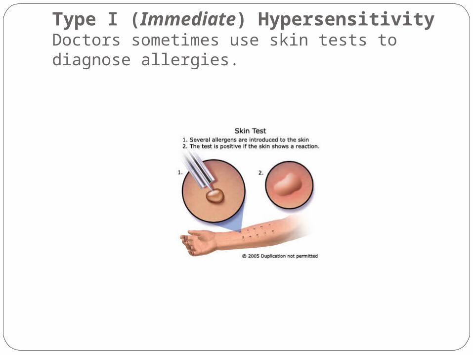
Type I (Immediate) HypersensitivityDoctors sometimes use skin tests to diagnose allergies.

Type I (Immediate) HypersensitivityThe reactions shown here demonstrate allergic
response.

Type I (Immediate) Hypersensitivity
In-Vitro TestsMeasure total IgE or antigen-specific IgELess sensitive than skin tests.RIST, RAST, Allergen specific and
Microarray will be covered later.

Type II (Cytotoxic) HypersensitivityTriggered by antigens found on cell surfaces
Altered self antigensHeteroantigens
Manifested by the production of IgG or IgM antibodies which coat the antigens.
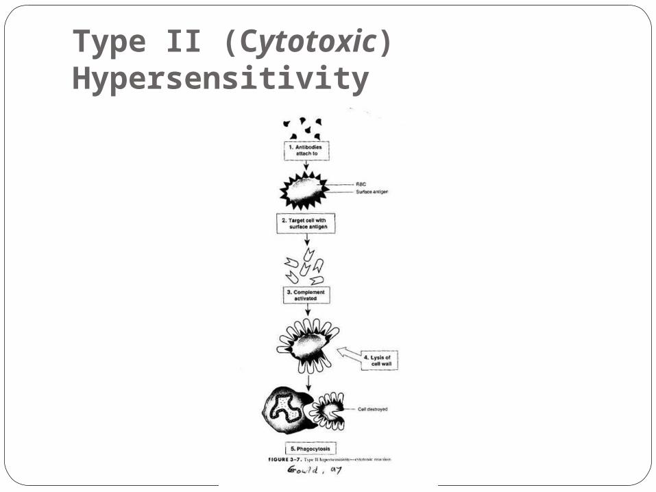
MechanismsAntibody coats cell surface promotes phagocytosis
– macrophages, neutrophils and eosinophils have Fc receptors to bind to antibody on target cell.
Natural Kill cells have Fc receptors, bind, results in cytotoxicity
Complement Coats cells which enhances phagocytosisComplement cascade goes to completion results in cell
lysis.

Type II (Cytotoxic) HypersensitivityTransfusion reactions
Hundreds of different antigens expressed on RBCsAntibodies can be produced naturally or through
exposure, transfusion or pregnancy most commonMost well known example due to ABO
incompatibility.Individuals form potent antibodies against ABO
antigens not present on their red blood cells.Group O individuals have anti-A and if transfused
with group A blood will have an immediate, and possibly fatal, reaction
Other blood groups may cause delayed reaction or acute reactions.

Type II (Cytotoxic) HypersensitivityHemolytic disease of the fetus and newborn
Mother exposed to blood group antigens due to previous pregnancy with antigen positive child or transfusion.
Antibody must be IgGCrosses placenta and coats fetal RBCs, destruction
of RBCs causes increased bilirubin and anemia.If first pregnancy is first exposure infant usually not
affected.Subsequent pregnancies have increased risk and
the disease ranges from mild to fatal.All pregnant women are screened for blood group
antibodies.
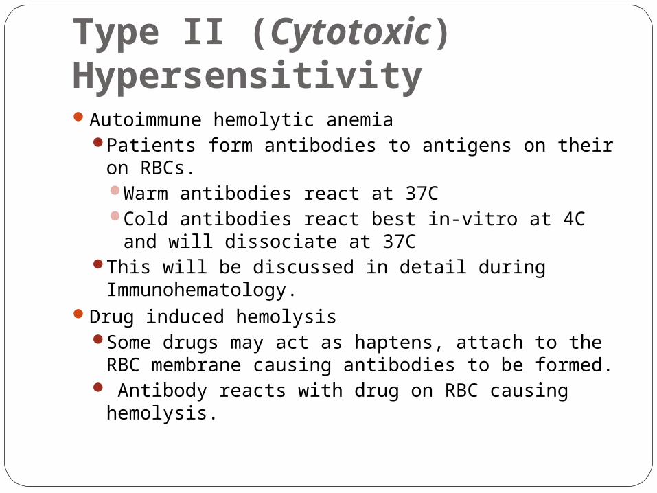
Type II (Cytotoxic) HypersensitivityAutoimmune hemolytic anemia
Patients form antibodies to antigens on their on RBCs.Warm antibodies react at 37CCold antibodies react best in-vitro at 4C and will
dissociate at 37CThis will be discussed in detail during
Immunohematology.Drug induced hemolysis
Some drugs may act as haptens, attach to the RBC membrane causing antibodies to be formed.
Antibody reacts with drug on RBC causing hemolysis.
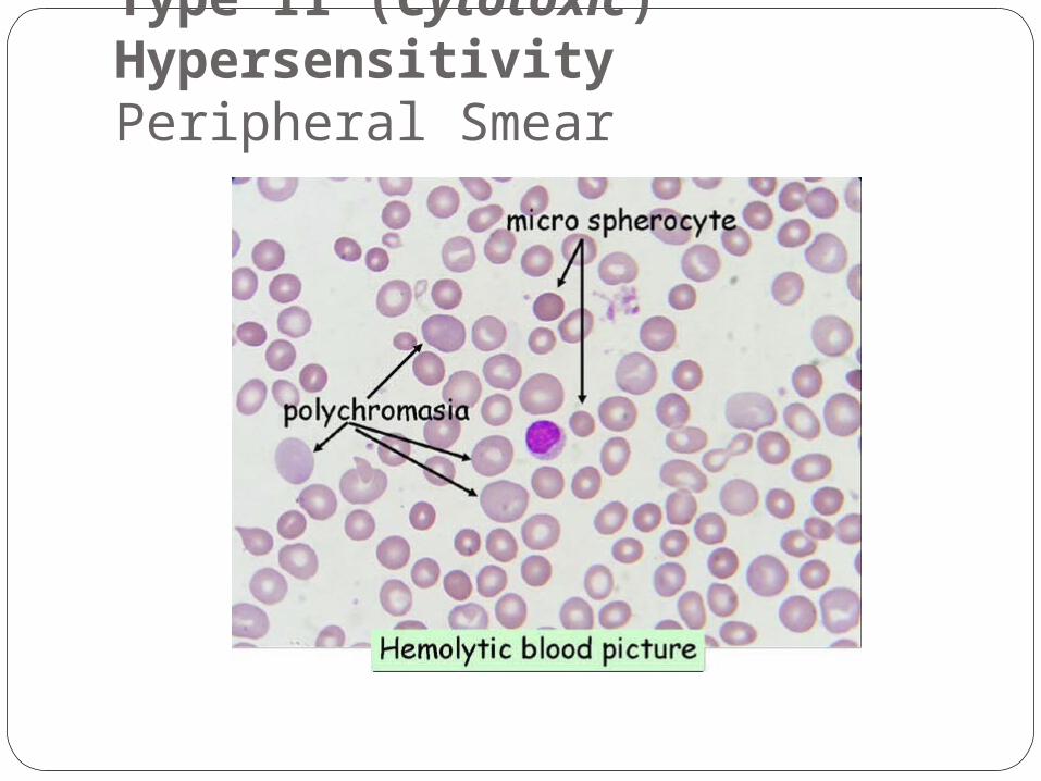
Type II (Cytotoxic) HypersensitivityPeripheral Smear

Type II (Cytotoxic) HypersensitivityTests
Coomb’s or anti-human globulin test.Direct Coomb’s
Add anti-IgG to washed drop of RBCsIf cells are coated with IgG then
agglutination will occur.Indirect Coomb’s
Incubate patient serum with RBCs of known antigenic make up.
Wash and add anti-IgGIf patient has antibody against antigen on
RBC agglutination will occur.

Type II (Cytotoxic) HypersensitivitySome individuals make antibody which cross
reacts with self antigens found in both the lung and kidney.
Goodpasture syndrome most well known exampleAntibody produced against basement membrane
protein.This protein present in lungs and kidneys.Antibody binding results in inflammationSymptoms are hemoptysis and hematuria.
Others will be discussed laterHashimoto’s diseaseMyasthenia GravisDiabetes mellitus
Type II (Cytotoxic) Hypersensitivity
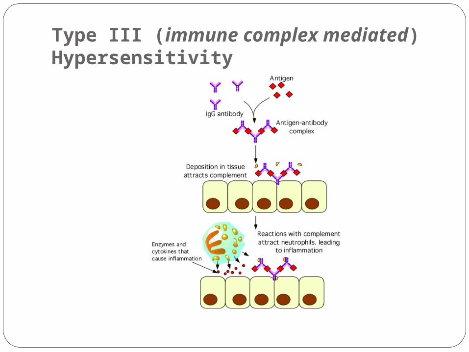
Type III (immune complex mediated) HypersensitivitySimilar to Type II, IgG or IgM involved and
destruction is complement mediated.Difference is that antigen is SOLUBLE.Soluble antigen and antibody combine to form
complexes.Usually complexes cause no symptoms, quickly
disappear from the circulation.Size of complexes produced seems important
in determining whether they will be eliminated quickly from the body or retained long enough to cause damage.
In some individuals the immune complexes persist in circulation causing clinical symptoms, some of them serious.

Type III (immune complex mediated) HypersensitivityMechanism
Soluble immune complexes which contain a greater proportion of antigen than antibody penetrate blood vessels and lodge on the basement membrane
At the basement membrane site, these complexes activate the complement cascade.
During complement activation, certain products of the cascade are produced,`attract neutrophils to the area. Such substances are known as chemotactic substances.
Once the polymorphs reach the basement membrane they release their granules, which contain lysosomal enzymes which are damaging to the blood vessel.
This total process leads to the condition recognized histologically as vasculitis.
Type III (immune complex mediated) Hypersensitivity

Type III (immune complex mediated) HypersensitivityTissues most frequently affected are:
Glomerular basementVascular endotheliumJoint liningsPulmonary alveolar membranes
Classical clinical symptoms of immune complex disease are due to blood vessel involvement, i.e., vasculitis.
Blood vessels of joints and the kidney are most frequently affected, giving rise to symptoms of arthritis and glomerulonephritis.
Type III (immune complex mediated) Hypersensitivity Arthus Reaction
Immunized rabbits to antigen Rabbits then injected intradermally with antigen Localized inflammatory reaction occurred followed by hemorrhagic
necrotic lesion. Occurred due to immune complexes depositing in dermal blood
vessels. Complement, neutrophils and platelets caused toxic affects. Rare in humans.
Serum Sickness Due to passive immunization with animal serum, bovine or horse. Vaccines and bee stings may also trigger. Symptoms appear 7 – 21 days after exposure to animal serum. Headache, fever, nausea, vomiting, joint pain, rashes and
lymphadenopathy. Symptoms due to antibody being formed at same time antigen is
present = immune complexes form. Benign, self limiting, 7-30 days for recovery.
Type III (immune complex mediated) HypersensitivityChronic immune complex diseases are
naturally occurring diseases caused by deposits of immune complex and complement in the tissues.Systemic Lupus Erythematosus (SLE)Acute glomerulonephritisRheumatic feverRheumatoid arthritis

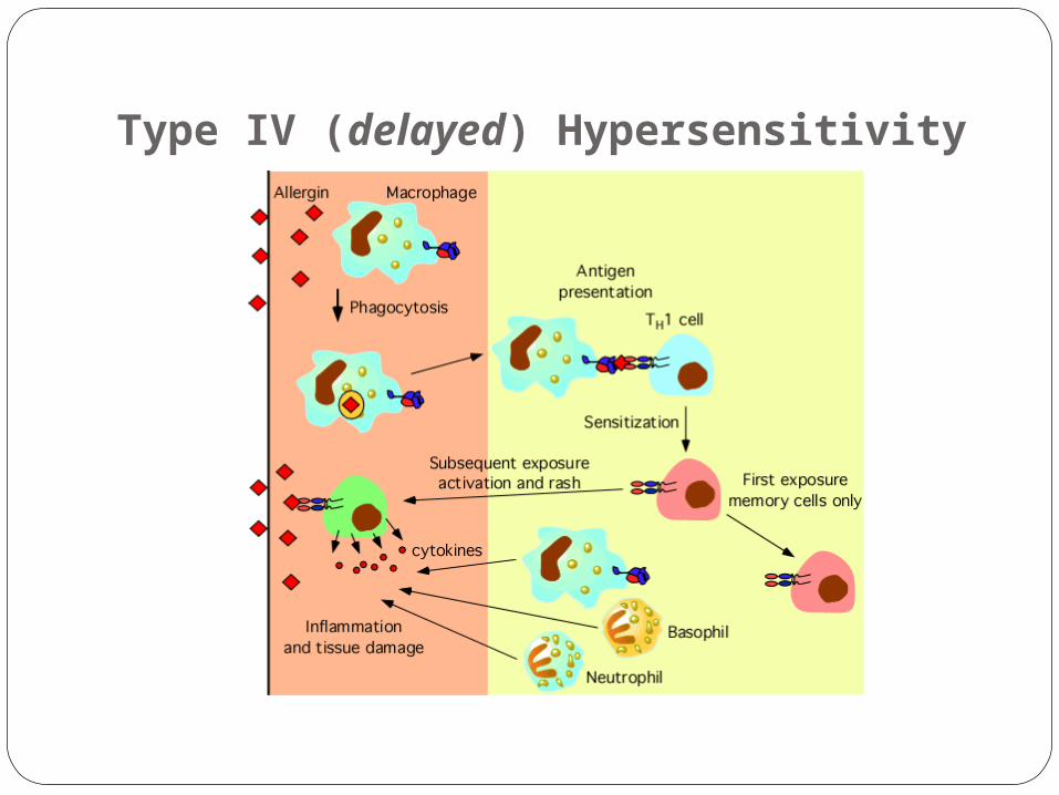
Type IV (delayed) HypersensitivityUsed to describe the signs and symptoms
associated with a cell mediated immune response.
Results from reactions involving T lymphocytes.
Characteristics of this phenomenon are:Delayed, taking 12 hours to develop.Causes accumulation of lymphs and
macrophages.Reaction is not mediated by histamine.Antibodies are not involved in the
reaction.

Type IV (delayed) HypersensitivityMost well known is the Koch
PhenomenonInject tuberculoprotein (PPD test)
intradermally Reaction results in an area of induration of 5
mm or more in diameter and surrounded by erythema
Reaction which occurs within 48 hours is a positive.
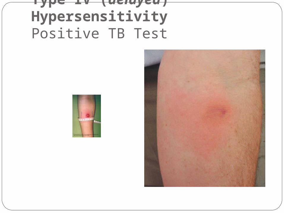
Type IV (delayed) HypersensitivityPositive TB Test

Type IV (delayed) HypersensitivityContact dermatitis due to contact with chemicals
Poison ivy, oak and sumac give off urushiol.Nickel, rubber, formaldehyde, hair dyes,
comseticsLatex allergies Function as haptensCauses erythema, swelling and formation of
papulesHypersensitivity Pneumonitis
Response of sensitized T cells to inhaled allergens.
Caused by chronic inhalation of microorganisms.Occupationally related – pigeons, farmers

Type IV (delayed) Hypersensitivity
Type IV (delayed) Hypersensitivity

Summary
Referenceshttp://www.thebody.com/nih/immune_system.h
tmlhttp://pathmicro.med.sc.edu/ghaffar/hyper00.h
tmhttp://home.kku.ac.th/acamed/kanchana/bsi.ht
ml