terramycin in trachoma of belgrade - bjo.bmj.com · terramycin, which was discovered by finlay...
TRANSCRIPT
Brit. J. Ophthal. (1954) 38, 692.
TERRAMYCIN IN TRACHOMABY
GJORGJE PAVKOVIC-BUGARSKI AND MILICA CVETOJEWICFrom the Regional Eye Hospital and Anti-Trachonm Centre of Vojvodina, PanJewp,mu4he Institute
ofPathology of the Faculty of Medicine, BelgradeTERRAmycIN, which was discovered by Finlay and his collaborators (1950),was isolated from Streptomyces rumozus. The first experiments with it weremade at the Mayo Clinic, Rochester, Minn., U.S.A., and it was found toproduce an effectuponGram-positive andGram-negative bacteria, Rickettsiae,and viruses.
Reports in the LiteratureComparatively few papers have been published on the results achieved by treating
trachoma with terramycin, but Bietti (1953), Bengisu Naci (1952), and Mitsui (1951)have published the results of their researches. The Japanese have been the mostfrequent users of terramycin in trachoma. Mitsui and others (1951) administeredthis antibiotic in seven hundred cases, 80 per cent. of which were reported to havebeen cured. These clinical findings were corroborated by their laboratory work.Inclusion bodies disappeared after a few days; histopathological analyses showedimprovement; pannus disappeared after 6-10 days. Bengisu Naci (1952) treatedten patients with terramycin capsules, drops, and ointment, and later treated ahundred children, administering terramycin ointment five times a day. Laboratoryand clinical control was introduced before and after the treatment. Bietti (1953)treated 206 eyes with terramycin, and thought that it influenced the virus. Goodresults with terramycin were obtained by Izzet (1951) in Turkey, and by Geraci(1951) in Italy. Rals (1950) investigated cases of pure trachoma and also cases inwhich trachoma was combined with conjunctivitis. Naccache (1953) treated38 patients by oral administration and locally, without any toxic or allergic symp-toms. Siniscal (1952) asserted that terramycin produced an effect only in cases ofsuperinfection. Very good results were achieved by Town (1951) and Thygeson(1951), and moderate results by Focosi (1953), Lepri (1951), and Caterini andDe Vincentiis (1951).
Present ResearchHaving investigated the results of treating trachoma with the sulphon-
amides, penicillin, and aureomycin, we decided to apply terramycin andobserve its effect upon trachoma in the province of Vojvodina, Jugoslavia.
692
copyright. on 19 M
arch 2019 by guest. Protected by
http://bjo.bmj.com
/B
r J Ophthalm
ol: first published as 10.1136/bjo.38.11.692 on 1 Novem
ber 1954. Dow
nloaded from

TERRAMYCIN IN TRACHOMA
Material.-We treated eighty patients with terramycin. Our research began onOctober 1, 1952. The cases were classified in four stages according to MacCallan(1936). We had seven cases in Stage I, forty in Stage II, and 33 in Stage III.59 of the patients were under 20, eight between 20 and 30, and thirteen over 30years of age. We chose patients who had not been treated previously, and thosein whom the trachoma process was not old-established, so that most of them werein a progressive stage of the disease. Clinical observation was carried out bymeans of a biomicroscope or a loupe. Microbiological and histopathologicalexaminations enabled us to see the effect produced on the trachomatous processitself. Some of the patients stayed in hospital for 2-3 months, and others weresegregated in a home or boarding-school for trachomatous children, but most ofthe latter group were also treated in hospital for some time.The patients were divided into two groups: one was treated locally with terra-
mycin, and the other received sulphonamides, besides local therapy. All the cases,with few exceptions, were under our supervision even after the termination oftreatment. In all the hospitalized cases, before the actual treatment was begun, ageneral examination was made. The conjunctival flora was examined in each case,and preparations of conjunctival scrapings were taken. These examinations weremade frequently in the course of the treatment, and once a month or once in 2months thereafter. In order to examine the influence of the drug on the patho-logical process, we made histopathological examinations, trying to find out whether,even a long time after treatment had been stopped, the pathological process stillexisted. Sections were taken from the upper palpebral conjunctiva, and, having beenfixed in Zenker, were stained with haematoxylin and eosin or by Van Gieson'smethod. These were taken before and after treatment, and in seven cases alsoafter an interval of 2-4 months. 140 sections were made from 54 treated eyes.
Clinical Features.-The peculiarity of trachoma in Vojvodina is the rare inci-dence of acute cases. A chronic, follicular, form of the disease, without dis-charge, is prevalent, and during the last 20 years it has been occurring in a verymild form. A developed pannus and damage to the eyelids are extremely rare inyounger persons. The number of cases of pure trachoma is large. A fine diffusedinfiltration, a few follicles, opaque and differing in size, and still fewer papillae,are generally found in Stage I cases. The process begins in the upper tarsalconjunctiva, near to the free border of the tarsus or on the semilunar fold, wheretwo or three follicles are usually aggregated. There is no discharge. In Stage II adiffused infiltration develops, there are often many opaque follicles, different in sizeand sometimes aggregated. There is some papillary hypertrophy, but no rough-ness. The process gradually penetrates deeper into the tarsus, covering the wholetarsal conjunctiva and the upper conjunctival fornix. There are follicles in thebulbar conjunctiva, especially in the upper part. Sometimes there are signsof an initial pannus in the limbus in the upper part with superficial infiltration1 mm. wide and initial superficial vascularization. Pannus is rarely developed,but if it occurs, the infiltration seldom penetrates deeply. In most of theStage III cases some fine scarring is to be found. In a small number of cases scarringis prevalent, opaque follicles being found here and there with the infiltration andpapillary hypertrophies, and the tarsus being infiltrated and thickened. In suchcases pannus is developed and often involves the whole cornea, with deep penetra-tion, and vision is very poor.
693
copyright. on 19 M
arch 2019 by guest. Protected by
http://bjo.bmj.com
/B
r J Ophthalm
ol: first published as 10.1136/bjo.38.11.692 on 1 Novem
ber 1954. Dow
nloaded from

694 GJORGJE PA VKOVIC-BUGARSKI AND MILICA CVETOJEVIC
Treatment.-In 41 cases the treatment was exclusively local. Ointment wasapplied six times a day. In 39 cases the patients also received sulphathiazole tabletsby mouth. There was little change in the clinical appearances in the first month oftreatment, except that in the Stage I cases one had an impression that the diffusedinfiltration had become smaller, especially under combined therapy. In the secondmonth, besides the regular oral treatment, the antibiotic was administered locallyby massage once a day. During the second, and, in more serious cases, during thethird month, the diffuse infiltration gradually disappeared, and the follicles andpapillary hypertrophies became smaller and then vanished. In a number ofcases fine scarring remained, while in others the process was completely curedwithout scarring. In Stage II regression was slower, and in Stage III it was veryslight, but it frequently occurred without any new scarring.
After the termination of the treatment the conjunctivae were pinkish-grey, thin,and smooth, sometimes with fine scarring on the surface, and sometimes without.In Stage II cases some follicles and papillary hypertrophies remained.The effect on the pannus was favourable. We treated only eleven patients with
pannus; the subjective irritation rapidly ceased, hyperaemia disappeared after2-5 days, the conjunctival infiitration was resorbed within 5-10 days, and aftera month the pannus was resorbed completely. After the termination of the treat-ment vascularization persisted on the corneal surface; this was very rare, but therewas scarring in the long-standing cases.
Vision was considerably improved in all cases, and terramycin was seen to in-fluence the true trachomatous pannus.Of eighty patients, 37 (46-25 per cent.) were clinically cured: of seven Stage I
cases, four (57 per cent.); of forty Stage II cases, 24(60 per cent.); and of 33 Stage IIIcases, nine (27 per cent.). However, in some cases the pathological changes didnot quite disappear even after 3 months' treatment. Follow-up examinationsshowed that there was a further regression of pathological changes, and so far nocase has relapsed. Terramycin combined with sulphathiazole produces quickerand better effects than when it is administered locally only. In general, themore superficial the process the sooner it is cured.
Biomicroscopical InvestigationBefore treatment the conjunctival surface in Stage I cases is uneven,
diffused, opaque, and opalescent. Longitudinal blood vessels and the tarsusare transparent. The papillae, with a characteristic perpendicular vascular-ization, in the form of a bouquet, are a little increased and infiltrated, and lesstransparent, and form groups, causing the uneveness of the conjunctival surf-ace. The papillary hypertrophy is greater near the conjunctival fornix.Here and there small follicles can be seen; they are whitish-grey, transparent,and not vascularized at first, but later fine capillaries can be seen on the sur-face. This state is directly followed by cicatrization, and fine scarring isformed, but in some cases follicles- disappear without any residual scarring.
In Stage II the opalescence is greater. Some longitudinal blood vesselscan be seen dimly; there are many papillary hypertrophies, the bouquetof blood vessels is very developed, and infiltration is greater. Among thepapillae are seen whitish-grey follicles, hemi-ampullar in form and non-
copyright. on 19 M
arch 2019 by guest. Protected by
http://bjo.bmj.com
/B
r J Ophthalm
ol: first published as 10.1136/bjo.38.11.692 on 1 Novem
ber 1954. Dow
nloaded from

TERRAMYCIN IN TRACHOMA
transparent, their surface covered with a loose net of blood vessels. Most ofthem are near the canthi and in the conjunctival fornix. Sometimes there isan enormous number of follicles, so that the conjunctiva has a gelatinouslook. During treatment the follicles diminish gradually, become clear andwhite, lose their hemi-ampullar appearance, become transparent and vanish.Fine scarring is formed, and whitish lines can be seen. The papillae becomeflat and disappear gradually. In the cases considered as clinically cured,the conjunctiva is light, with a smooth surface, transparent, vascularizationis parallel to the surface, and there are newly-formed vessels in front of andbehind the fine surface scarring.
In the majority of our Stage III cases cicatrization was initial, so thatbiomicroscopically there was no significant difference between them and theStage II cases. In a smaller number of cases the conjunctiva was covered witha net of fine linear scarring, sometimes forming islets with a smooth surface.Generally, blood vessels are no longer perpendicular, but parallel to the surf-ace, behind the scars. The papillae are flat and smaller in size. The innerblood vessels can be seen. There is a fine greyish, diffused infiltration. A smallnumber of regressing follicles can be found on the tarsal conjunctiva, especiallylaterally and in the conjunctival fornix. New blood vessels develop fromthe inner or conjunctival fornix vessels, forming a dense, superficialnet. Regression and cicatrization progress during treatment. In thebulbar conjunctiva, hyperaemia and infiltration are continued into a moreor less developed pannus. As for the pannus, the limbus area is infiltrated,the papillae are small, and blood vessels form a bouquet. The direct branchesof the foremost conjunctival vessels penetrate into the infiltration, by whichthey are covered. In the distal part of the limbus can be seen round follicles,surrounded by fine blood vessels. Some of their branches cover the surfaceof the follicles, and there are many maculae and papillae. During treatmentthe infiltration is resorbed, and instead of the follicles, fine pits are formed.Vascularization is persistent.
Microbiological InvestigationIn all the cases before treatment, often during the treatment, and at inter-
vals after discontinuing treatment, the conjunctival flora was examined andpreparations stained by the method of Lindner (1927) and Poleff (1951) weremade from the conjunctival scrapings. Before treatment we found inclusionbodies in five of the seven Stage I cases, in thirty of the forty Stage II cases,and in eight of the 33 Stage III cases. We thus found them in 43 out ofeighty cases (54 per cent.). After treatment we found them only in twoStage I cases, four Stage II cases, and one Stage III case. During the follow-up period, 2-4 months after the termination of the treatment, they were notfound at all. The conjunctival flora became sterile within 2-3 days afterthe treatment was started. There had been many epithelial cells, which in thecourse of the treatment and after its discontinuance became gelatinously
695
copyright. on 19 M
arch 2019 by guest. Protected by
http://bjo.bmj.com
/B
r J Ophthalm
ol: first published as 10.1136/bjo.38.11.692 on 1 Novem
ber 1954. Dow
nloaded from
696 GJORGJE PA VKOVIC-BUGARSKI AND MILICA CVETOJEVIC
degenerate; their nucleus could not be stained and they disappeared gradu-ally. We found Prowazek-Halberstiidter bodies, initial and elementary, inthe epithelial cells. Pastille and rickettsoid bodies, Leber's cells, smalllymphocytes, and erythrocytes were also found.
Since the trachoma bodies disappeared in almost all cases, this couldbe taken as a proof that terramycin destroyed the virus. However, thefact that the inclusion bodies were found in 54 per cent. of the cases suggeststhat the virus may exist in an invisible form.
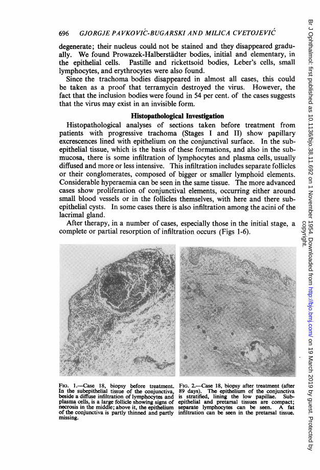
Histopathological InvestigationHistopathological analyses of sections taken before treatment from
patients with progressive trachoma (Stages I and II) show papillaryexcrescences lined with epithelium on the conjunctival surface. In the sub-epithelial tissue, which is the basis of these formations, and also in the sub-mucosa, there is some infiltration of lymphocytes and plasma cells, usuallydiffused and more or less intensive. This infiltration includes separate folliclesor their conglomerates, composed of bigger or smaller lymphoid elements.Considerable hyperaemia can be seen in the same tissue. The more advancedcases show proliferation of conjunctival elements, occurring either aroundsmall blood vessels or in the follicles themselves, with here and there sub-epithelial cysts. In some cases there is also infiltration among the acini of thelacrimal gland.
After therapy, in a number of cases, especially those in the initial stage, acomplete or partial resorption of infiltration occurs (Figs 1-6).
41~~4
FIG. l.-Case 18, biopsy before treatment. FIG. 2.-Case 18, biopsy after treatment (afterIn the subepithelial tissue of the conjunctiva, 89 days). The epithelium of the conjunctivabeside a diffuse infiltration of lymphocytes and is stratified, lining the low papillae. Sub-plasma cells, is a large follicle showing signs of epithelial and pretarsal tissues are compact;necrosis in the middle; above it, the epithelium separate lymphocytes can be seen. A fatof the conjunctiva is partly thinned and partly infiltration can be seen in the pretarsal tissue.missing.
copyright. on 19 M
arch 2019 by guest. Protected by
http://bjo.bmj.com
/B
r J Ophthalm
ol: first published as 10.1136/bjo.38.11.692 on 1 Novem
ber 1954. Dow
nloaded from
TERRAMYCIN IN TRACHOMA
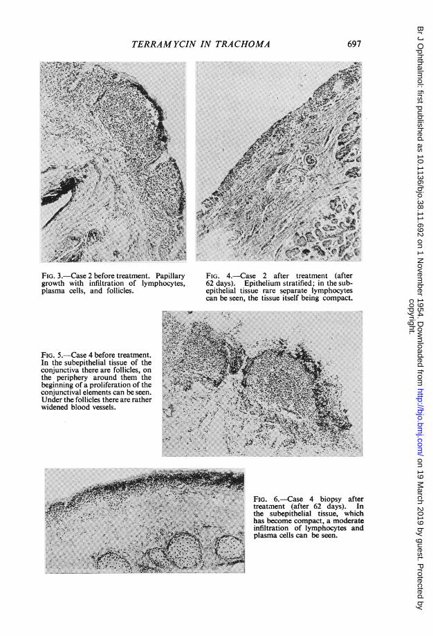
FIG. 3.-Case 2 before treatment. Papillary FIG. 4.-Case 2 after treatment (aftergrowth with infiltration of lymphocytes, 62 days). Epithelium stratified; in the sub-plasma cells, and follicles. epithelial tissue rare separate lymphocytes
can be seen, the tissue itself being compact.
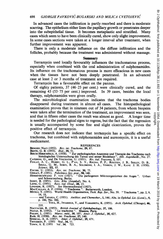
FIG. 5.-Case 4 before treatment.~In the subepithelial tissue of theconjunctiva there are follicles, on ,the periphery around them thebeginning of a proliferation of the ,conjunctival elements can besenUnder the follicles there are ratherwidened blood vessels. ~~~~~~~
_ ~ FIG. 6.-Case 4 biopsy afterX > .*- ^ pltreatment (after 62 days). In
the subepithelial tissue, whichinfiltration of lymphocytes and
V~~ ~ ~ ~~~ ~~plasma cells can be seen.
697
copyright. on 19 M
arch 2019 by guest. Protected by
http://bjo.bmj.com
/B
r J Ophthalm
ol: first published as 10.1136/bjo.38.11.692 on 1 Novem
ber 1954. Dow
nloaded from
698 GJORGJE PA VKOVIC-BUGARSKI AND MILICA CVETOJEVIC
In advanced cases the infiltration is partly resorbed and there is moderatescarring. The epithelium either lines the papillary growth or penetrates deeperinto the subepithelial tissue. It becomes metaplastic and stratified. Manycases which seem to have been clinically cured, show only slight improvement.In some cases sections were taken at a longer interval after treatment, whenfurther improvement was apparent.
There is only a moderate influence on the diffuse infiltration and thefollicles, probably because the treatment was administered without massage.
SummaryTerramycin used locally favourably influences the trachomatous process,
especially when combined with the oral administration of sulphonamides.Its influence on the trachomatous process is more efficacious in new caseswhen the tissues have not been deeply penetrated. In an advancedcase at least 2 or 3 months of treatment are required.
Terramycin has a favourable effect on the pannus.Of eighty patients, 37 (46.25 per cent.) were clinically cured, and the
remaining 43 (53.75 per cent.) improved. In 39 cases, besides the localtherapy, sulphonamides were given orally.The microbiological examination indicates that the trachoma bodies
disappeared during treatment in almost all cases. The histopathologicalexamination proves that in nineteen out of 34 patients, from whom biopsieswere taken after the termination of the treatment, an improvement was seen,and that in fifteen other cases the result was almost as good. A longer timeis needed for the pathological signs to regress, but the fact that the regressionis usually accompanied by some fine and slight cicatrization, proves thepositive effect of terramycin.Our research does not indicate that terrmycin has a specific effect on
trachoma, but combined with sulphonamides and aureomycin, it is a usefulmedicament.
REFERENCESBENGISU NACI (1952). Rev. int. Trachome, 29, 87.BIETrI, G. B. (1953). Ibid., 30, 51.BIRCH-HiRSCHFELD, A. (1938). " Zur pathologischen Anatomie und Therapie des Trachoma nach
histologischer Untersuchung des Tarsus und seiner Bindehaut", Abh. Augenheilk., No. 27.CATERINI, G., and DE VINCENTIIS, U. (1951). Riv. ital. Tracoma, 3, 107.FINLAY, A. C., HOBBY, G. L., P'AN, S. Y., REGNA, P. P., ROUTIEN, J. B., SEELEY, D. B.,
SHULL, G. M., SOBIN, B. A., SOLOMONS, I. A., VINSON, J. W., KANE J. H. (1950).Science, mH, 85.
Focosi, U. (1953). Rev. int. Trachome, 30, 75.GERACI, P. (1951). Policlinico Sez. prat., 58, 140.HERRENSCHWAND F. VON (1927). Die pathogenen Mikroorganismen des Auges ". Urban
and Schwarzenberg, Berlin.IZZET, B. (1951). GOz. Klin. (Istanbul), No. 6.LEPRI, G. (1951). Rass. ital. Ottal., 20, 15.LINDNER, K. (1927). See Herrenschwand (1927).MACCALLAN, A. F. (1936). " Trachoma ". Butterworth, London.MiTsui, Y. (1951). World Health Organisation, Tech. Rep. Ser., No. 59. "Trachoma ", pp. 2, 9,
14, 16.and TANAKA, C. (1951). Antibiot. and Chemother., 1, 146; Abs. in Ophihal. Lit. (Lond.)., 5,p. 186, No. 586.
- ~~TOYA, H., IWASHIGE, Y., and YAMASHITA, K. (1951). Arch. Ophthal. (Chicago), 46,235.
NACCACHE, R. (1953). British Journal of Ophthalmology, 37, 106.NATAF, R. (1952). "Le trachome ". Masson, Paris.POLEFF, L. (1951). Maroc. med., 30, 107; Amer. J. Ophthal., 35, 627.RAIS, H. (1950). Rev. int. Trachome, 27, 140.SIMNSCAL, A. A. (1952). Amer. J. Ophthal., 35, 671.THYGESON, P. (1951 ibid., 34, 11.TOWN, A. E. (1951 5id, 34. 723.
copyright. on 19 M
arch 2019 by guest. Protected by
http://bjo.bmj.com
/B
r J Ophthalm
ol: first published as 10.1136/bjo.38.11.692 on 1 Novem
ber 1954. Dow
nloaded from