temporal patterns in innate immunity parameters in reef ... de water et al... · ecosphere 7 (11):...
TRANSCRIPT

November 2016 v Volume 7(11) v Article e015051 v www.esajournals.org
Temporal patterns in innate immunity parameters in reef- building corals and linkages with local climatic conditions
Jeroen A. J. M. van de Water,1,2,3,4,5,† Joleah B. Lamb,1,2,3,6 Scott F. Heron,7,8 Madeleine J. H. van Oppen,1,3,4,9 and Bette L. Willis1,2,3
1ARC Centre of Excellence for Coral Reef Studies, James Cook University, Townsville, Queensland 4811 Australia2College of Marine and Environmental Sciences, James Cook University, Townsville, Queensland 4811 Australia
3AIMS@JCU, James Cook University, Townsville, Queensland 4811 Australia4Australian Institute of Marine Science, PMB 3, Townsville MC, Townsville, Queensland 4810 Australia
5Centre Scientifique de Monaco, MC 98000 Monaco6Department of Ecology and Evolutionary Biology, Cornell University, Ithaca, New York 14850 USA
7National Oceanic and Atmospheric Administration–Coral Reef Watch, James Cook University, Townsville, Queensland 4811 Australia8Marine Geophysical Laboratory, Physics Department, College of Science, Technology and Engineering, James Cook University,
Townsville, Queensland 4811 Australia9School of BioSciences, The University of Melbourne, Parkville, Victoria 3010 Australia
Citation: van de Water, J. A. J. M., J. B. Lamb, S. F. Heron, M. J. H. van Oppen, and B. L. Willis. 2016. Temporal patterns in innate immunity parameters in reef- building corals and linkages with local climatic conditions. Ecosphere 7(11): e01505. 10.1002/ecs2.1505
Abstract. Extremes in seasonal environmental conditions can significantly impact the health and phys-iological functioning of reef corals, underscoring the need for knowledge of seasonally specific baselines from which to monitor and forecast impending stress. Increases above summertime means in seawater tem-perature, sunlight intensity, turbidity, or sedimentation may reduce coral immunocompetency and increase disease and bleaching susceptibility. We analyzed temporal patterns in innate immunity parameters over nine time points throughout one year to establish baseline levels from which anomalies might be detected for representative species from three major reef- building coral families (Acroporidae, Faviidae, and Porit-idae). Temporal patterns in both phenoloxidase activity and expression of green fluorescent protein- like proteins varied among the three families, as did overall constitutive levels. For example, Porites cylindrica had 2.8- fold higher yearly average levels of phenoloxidase activity than Acropora millepora, which had the lowest levels. In contrast, mean fluorescence was lowest in Acropora millepora and highest in Echinopora mammiformis. Relationships between the potential physical drivers (seasonal variation in seawater tempera-ture, rainfall, salinity) and temporal patterns in these parameters also differed among the three species. For example, phenoloxidase activity was positively correlated with seawater temperature in A. millepora, but negatively correlated in both E. mammiformis and P. cylin drica. Distinctions in constitutive levels and tempo-ral patterns in these parameters among species suggest that corals from these three families have evolved different strategies for investing resources into innate immune parameters. Such differences highlight the need for species- specific baselines and long- term assessments to accurately predict coral reef trajectories in rapidly changing environments.
Key words: chromoprotein; coral; fluorescence; green fluorescent protein-like proteins; immunity; indicator; phenoloxidase; prophenoloxidase; salinity; temperature.
Received 23 January 2016; revised 18 May 2016; accepted 22 July 2016. Corresponding Editor: Andrew W. Park. Copyright: © 2016 van de Water et al. This is an open access article under the terms of the Creative Commons Attribution License, which permits use, distribution and reproduction in any medium, provided the original work is properly cited.† E-mail: [email protected]

November 2016 v Volume 7(11) v Article e015052 v www.esajournals.org
van de WATER ET AL.
IntroductIon
Global declines in coral reef ecosystem func-tion is a critical concern, both for vertebrates and invertebrates dependent on reefs for food and shelter, and for the millions of people who draw extensively on them for food, coastal protection, tourism income, and cultural values (Burke et al. 2011). Increasing levels of environmental and anthropogenic pressures facing corals world-wide (Hughes et al. 2003) highlight the urgent need to establish reference baselines for coral physiological parameters that could be used as tools for evaluating, monitoring, and forecasting coral health to preempt and manage activities that might cause further declines. As a compe-tent immune system is integral to maintaining an organism’s health, parameters associated with the coral innate immune system, such as activity of the prophenoloxidase (proPO) sys-tem and expression of green fluorescent protein- like (GFP- like) proteins, have been suggested as potential indicators of stress based on their detectable responses to the variations in local environmental conditions (D’Angelo et al. 2008, Palmer et al. 2010, Roth and Deheyn 2013).
Both parameters vary in response to warm sea-water temperature anomalies, which are known to significantly affect the health of the coral holo-biont. In particular, warm temperature anoma-lies cause disruptions to the coral–Symbiodinium endosymbiosis leading to bleaching (Douglas 2003), as well as shifts in coral- associated micro-bial communities toward potential pathogenic microbes (Littman et al. 2010) and increases in the virulence of coral pathogens (Ben- Haim et al. 2003) that may be linked to increased coral dis-ease prevalence in summer. Heavy monsoonal rainfall, which results in reduced salinity and increased terrestrial runoff leading to increased levels of turbidity and agricultural pollutants, is also a major stressor for corals. Warm seawater temperatures and changes in salinity and agri-cultural pollutants have all been shown to com-promise the functioning of the innate immune systems of a range of marine invertebrates, thereby increasing disease susceptibility and mortality (Tseng and Chen 2004, Ellis et al. 2011, Lin et al. 2012). In addition, both low salinity (Kerswell and Jones 2003, Jones and Berkelmans 2014) and cold sea surface temperatures (Saxby
et al. 2003) have been implicated in coral bleaching. Extremes in seasonal environmen-tal conditions can therefore significantly affect coral health, fitness, and disease susceptibility. Although targeted, short- term studies are begin-ning to explore how environmental stressors affect the proPO- activating system and GFP- like protein expression, an understanding of what constitutes seasonal baselines in healthy levels is needed to explore these parameters as potential indicators of immune- compromising environmental conditions.
The prophenoloxidase (proPO)- activating sys-tem is part of the melanin synthesis pathway and forms an important, highly conserved com-ponent of the innate immune system of inver-tebrates (Cerenius et al. 2010), including both gorgonian and scleractinian corals (Mydlarz et al. 2008, Palmer et al. 2008). In corals, higher levels of immune parameters associated with this system, particularly phenoloxidase activity and melanin content, have been linked to higher resistance to both bleaching and disease (Palmer et al. 2010). The proPO system is activated when components of microbial cell walls are detected, leading to the activation of a protease cascade that cleaves proPO into PO (Cerenius et al. 2010). In turn, PO oxidizes phenolic compounds into cytotoxic quinone intermediates, which non- enzymatically form melanin. Whereas mel-anin forms a barrier and immobilizes microbes, cytotoxic quinones and reactive oxygen species (ROS) formed eliminate invading organisms. In corals, melanin and PO have been found to play a significant role in wound healing (Palmer et al. 2011b, D’Angelo et al. 2012, van de Water et al. 2015a) and in anti-microbial (Mydlarz et al. 2008, Palmer et al. 2011a, van de Water et al. 2015b) and anti-parasite (Palmer et al. 2008, Burge et al. 2013) immune responses. Additionally, mela-nin deposits have been implicated in bleaching mitigation via photoprotection (Palmer et al. 2010). However, knowledge of baseline levels of PO activity characteristic of healthy corals and of the additional capacity for activation represented by the stored inactive proenzyme proPO is currently lacking. Studies of baseline levels of these parameters and how they vary seasonally are essential first steps in evaluating the natural variation in immune parameters of reef corals.
November 2016 v Volume 7(11) v Article e015053 v www.esajournals.org
van de WATER ET AL.
Expression of GFP- like proteins is thought to contribute significantly to the cellular- based stress responses of corals, ameliorating bleach-ing by absorbing and dissipating solar radiation that could otherwise trigger photoinhibition (Baird et al. 2009). In contrast to their photopro-tective role at high light intensities (Salih et al. 2000, Dove et al. 2001), fluorescent proteins (FPs) may also augment light for photosynthesis at low light intensities (Kawaguti 1969, Schlichter et al. 1986), although this role appears minimal (Gilmore et al. 2003). Similarly, there is strong evidence that non- fluorescent chromoprotein (CP) has a photoprotective role (Salih et al. 2000, Dove et al. 2001) for Symbiodinium photosys-tems (Smith et al. 2013). Increased expression of GFP- like proteins has been found in areas of high tissue proliferation, such as at branch tips and in regenerating tissues during wound heal-ing (D’Angelo et al. 2012, van de Water et al. 2015a), potentially protecting both coral and Symbiodinium cells from light stress in areas with low levels of Symbiodinium pigmentation. GFP- like proteins may also exhibit antioxidant prop-erties (Bou- Abdallah et al. 2006, Palmer et al. 2009a) by mitigating the effects of ROS produced by defective Symbiodinium photosystems (Lesser 1996), and via the proPO- activating system and the oxidative burst as part of an innate immune response (Palmer et al. 2008, 2009b, D’Angelo et al. 2012, van de Water et al. 2015a).
Coral families differ in their susceptibilities to disease (Willis et al. 2004, Raymundo et al. 2005, Lamb and Willis 2011) and bleaching (Marshall and Baird 2000, Loya et al. 2001); thus, evidence that taxonomic (family- level) trends in disease susceptibility are inversely correlated with con-stitutive levels of PO activity (Palmer et al. 2010) underscores the need to examine baseline levels that are at least family- or pre ferably species- specific. On the Great Barrier Reef (GBR), coral species in the family Acroporidae are among the most vulnerable to coral diseases, whereas species in the family Poritidae are among the most resistant; species in the family Faviidae tend to be intermediate in their susceptibility to disease (Willis et al. 2004, Palmer et al. 2010). Similarly, there is a hierarchy in bleaching sus-ceptibility among coral taxa, with massive taxa (e.g., Poritidae) being more resistant than fast- growing branching taxa (e.g., Acroporidae)
(Marshall and Baird 2000, Loya et al. 2001). However, within- species susceptibility can be significantly influenced by various other phys-iological and environmental factors, such as thermal history (Guest et al. 2012, Howells et al. 2013), Symbiodinium endosymbiont composi-tion (Howells et al. 2012) and density (Cunning and Baker 2013). Such variations in bleaching and disease susceptibility highlight the need to investigate temporal patterns in baseline levels of parameters that may influence the stress suscep-tibility among coral families.
In this study, we investigated the seasonal patterns in activity of the proPO system and the expression levels of GFP- like proteins to estab-lish the baseline levels for a common species in each of three major reef- building coral families. Species selected span the range from resistant to highly susceptible to bleaching and disease. Seasonal patterns in sea temperature, cloud cover, rainfall, and salinity were documented over the corresponding year- long period to iden-tify the potential links between the variation in environmental factors and the immune param-eters investigated. For example, we evaluated whether corals upregulate GFP- like proteins and the proPO system during summer months, potentially to cope with the increased environ-mental stressors that can lead to bleaching and disease.
MaterIals and Methods
Field sampling design and sample collectionThis study was conducted in Pioneer Bay (coor-
dinates: 18°36′24.9114″ S, 146°29′20.205″ E), situ-ated on the sheltered, western side of Orpheus Island within the Great Barrier Reef Marine Park. Samples of corals in the families Acroporidae (Acropora millepora—red color morph), Faviidae (Echinopora mammiformis), and Poritidae (Porites cylindrica) were collected from similar- sized tag-ged colonies over the course of one year, from October 2009 to September 2010 (Fig. 1). Numbe-red plastic cattle tags and cable ties were used to tag 10 colonies of E. mammiformis located at 2–4 m depth and eight colonies of each of A. millepora (2–3 m depth) and P. cyclindrica (4–5 m depth). Colonies were sampled and photographed at nine time points: October 2009 (late austral spring); December 2009 and January, February, and March

November 2016 v Volume 7(11) v Article e015054 v www.esajournals.org
van de WATER ET AL.
of 2010 (austral summer); May, July, and August of 2010 (austral winter); and September 2010 (early austral spring). At each time point, one branch (approximately 5 cm in length) was sampled from the middle of each tagged colony using surgical bone cutters, placed in a plastic bag underwater, transferred to a cryogenic tube and snap- frozen in liquid nitrogen, and stored at −80°C until processed.
Although sampling generates lesions and elic-its an immune response in the coral through the increased activity of the proPO system and GFP- like protein expression, this response is localized to the lesion area (Palmer et al. 2011b, D’Angelo et al. 2012, van de Water et al. 2015a). Sealing of lesions and recovery of tissue structures is accomplished within ~48 h (Palmer et al. 2011b). Given the localized response and the sufficient recovery time between sampling time points, our sampling regime is not expected to have had an effect on the immune parameters analyzed here in other locations within the coral colonies, and to have influenced our results.
Sample preparationTo prepare the tissue lysates, frozen fragments
were thawed on ice and approximately 4 cm2 of coral tissue was removed from the fragment using an airbrush into 10 mL of ice- cold extraction buffer (50 mM Tris–HCl, pH 7.8, with 50 mM dithiothreitol). Each tissue sample was homoge-nized for 45 s (IKA T10 Basic homogenizer, IKA-Werke GmbH & Co. KG, Staufen, Germany). The resulting slurry was centrifuged at 3500 rpm for 5 min to remove the cell debris and Symbiodinium, and the supernatant was collected and stored at −30°C. Total protein content of each
sample was determined using the DC Protein Assay kit (Bio- Rad, Hercules, California, USA) following the manufacturer’s instructions, and the endpoint absorbance was read using a SpectraMax M2 (Molecular Devices, Sunnyvale, California, USA).
Phenoloxidase activityPhenoloxidase activity was assayed according
to the procedures outlined in Palmer et al. (2011a), with some modifications. Both total potential (trypsin- activated) phenoloxidase (tpPO) activity (as defined in van de Water et al. 2015a) and active phenoloxidase (PO) activity were measured to analyze the total capacity and the active fraction of the proPO system, respectively, in each sam-ple. To analyze tpPO activity, 20 μL of coral tissue lysate was loaded in triplicate into wells of a 96- well plate, to which Tris- buffered saline (50 mM, pH 7.8; 40 μL) and trypsin (25 μL 0.1 mg/mL) were added. Reaction mixtures were incubated for 20 min to allow for activation of prophenoloxidase by trypsin, and then, 30 μL of 10 mM dopamine hydrochloride (Sigma- Aldrich, St. Louis, Missouri, USA) was added to each mix-ture. As a blank, 20 μL of extraction buffer was used. The same procedure was followed to ana-lyze PO activity, except that 25 μL double- distilled water was substituted for the trypsin solution. Absorbance was measured at 490 nm every 5 min for 45 min using the SpectraMax M2 (Molecular Devices). Data for each sample were inde-pendently obtained in triplicate. Phenoloxi dase activity was calculated as the change in absor-bance using the linear portion of the reaction curve over time and standardized to the total pro-tein content of each sample.
Fig. 1. Study species from three important reef- building coral families. (A) Acropora millepora (Acroporidae), (B) Echinopora mammiformis (Faviidae), and (C) Porites cylindrica (Poritidae).
November 2016 v Volume 7(11) v Article e015055 v www.esajournals.org
van de WATER ET AL.
Chromoprotein and fluorescent protein expressionTwenty microliters of tissue lysate was added
to each well of a black, clear bottom 384- well plate in triplicate for each sample. Expression of chromoprotein was analyzed by measuring the absorbance at 588 nm using a SpectraMax M2. The fluorescence spectrum was analyzed by measuring the emission wavelengths between 400 and 700 nm, with a 5- nm resolution, emitted upon the excitation of FPs at 280 nm. The 280- nm excitation wavelength was used, as it is optimal for the excitation of FPs across the spectrum and is sufficiently distant from the cyan fluorescent protein (CFP) emission peak to prevent interfer-ence with its excitation and emission (Shagin et al. 2004). All data were normalized to the total protein content. Fluorescence spectra and FP expression levels were calculated as described in van de Water et al. (2015a). In summary, the exponentially decaying background scatter was subtracted from each spectrum between 445 and 645 nm and multiple regression models based on the purified FP spectra were fitted to the data to calculate the proportions of the individual FPs (cyan, green, and red fluorescent protein) present using the formula B = F450/exp(k × (w − 450)), where F450 is the fluorescence reading at 450 nm (no FP emission), w is wavelength in nanometers, and k is an empirically determined coefficient that provides a good fit of the model. Parameter k was set at 0.1, 0.12, and 0.25 for A. millepora, E. mammiformis, and P. cylindrica, respectively. The relative proportions of FPs were multiplied by the coefficient reflecting the relative excitabil-ity of purified A. millepora FPs at 280 nm (for A. millepora: CFP: 0.85, GFP: 0.92, RFP: 1; for E. mammiformis and P. cylindrica: CFP: 1, GFP: 1, RFP: 1). Samples where the R2 of the model fit was less than 0.9 were excluded from the analysis.
Environmental parametersDaily environmental data (seawater tempera-
ture, rainfall, and salinity) were obtained at a depth of 3 m from the Australian Institute of Marine Science Orpheus Island Platform and Sensor Float 1 (Integrated Marine Observing System [IMOS]) located in Pioneer Bay (see http://www.aims.gov.au). Daily cloud fraction data (Level 2, MOD06) were acqui-red from the Moderate Resolution Imaging
Spectroradiometer (MODIS) on board the Terra satellite (downloaded via ladsweb.nascom.nasa.gov; algorithm description in King et al. 1997). As cloud cover is variable on local scales and MODIS data result from a single snapshot (or, at most, two snapshots) around 10:30 local time, cloud fraction conditions across each day-time period were inferred by considering the data from pixels in the vicinity of the study site. The radial extent of cloud influence was esti-mated using local wind information. Near- surface wind speeds in the IMOS data during the study period had a mean value of 19.8 km/h, which for an 8-h daytime period corresponds to a distance of 158.4 km. However, upper- level wind speeds are typically greater than those near the surface, plus the geometry of sun angle at the location and wind direction through each day confer the additional variability affecting spatial values to be included in the daily cloud fraction estimate. Accordingly, the distance was reduced (by half) to 79.2 km, which is approxi-mately 0.7 arc degrees, and cloud fraction val-ues within a radius of 0.35 arc degrees of Pioneer Bay were averaged for each day. All environ-mental parameter data are displayed in Appendix S1: Fig. S1.
Statistical analysisDifferences in immune parameters between
the yearly average and sampling times were analyzed using a linear mixed- effects (LME) model. Pairwise comparisons on the temporal patterns between the consecutive months were made using LME models, and the Bonferroni correction was applied to obtain the critical P- value based on α = 0.05. Immune parameters were added to the model as dependent fixed fac-tors and sampling time and/or species as inde-pendent fixed factors. In addition, colony was inclu ded as a random factor to account for the repeated sampling of the same individual col-ony. A t- test was used to test for the differences in the fluorescence levels between summer and winter for each species. Correlations between immune and environmental parameters (aver-age of 10 d prior to sampling) were analyzed using Pearson’s r. All analyses were conducted using the statistical software package S- PLUS 8.0. Differences were considered significant when P < 0.05, or P < 0.05/n, in the case of

November 2016 v Volume 7(11) v Article e015056 v www.esajournals.org
van de WATER ET AL.
Bonferroni- adjusted P- values, with n being the number of pairwise comparisons.
results
Phenoloxidase activityOn average, mean constitutive levels of the
two parameters associated with the proPO- activating system differed among the three coral species over the one- year duration of the study. The yearly average of active phenoloxidase (PO)
activity (measured as ΔOD 490 nm per mg pro-tein per min) was more than twofold greater in Porites cylindrica (10.70 ± 0.54) than in Acropora millepora (3.73 ± 0.48), while Echinopora mammi-formis (5.62 ± 0.31) had intermediate levels of PO activity (dashed lines in Fig. 2A–C). A similar pattern was found for yearly averages of the total potential phenoloxidase (tpPO) activity, which was more than twofold greater in P. cylindrica (10.89 ± 0.55) than in A. millepora (4.65 ± 0.53), and again intermediate in E. mammiformis (6.88 ± 0.41)
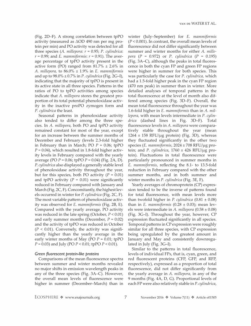
Fig. 2. Seasonal patterns in the proPO system. Temporal patterns in (A–C) active phenoloxidase (PO) activity; (D–F) the total potential phenoloxidase (tpPO) activity; and (G–I) ratio of PO activity and tpPO activity. Asterisk (*) indicates the statistically significant difference (P < 0.05/n) with previous time point. Letter “a” indicates the statistically significant difference (P < 0.05) from the yearly average.
PO
act
ivity
(∆O
D49
0 nm
∙mg–1
pro
tein
∙min
–1)
tpP
O a
ctiv
ity (∆
OD
490
nm∙m
g–1 p
rote
in∙m
in–1
) P
O/tp
PO
ratio
Porites cylindricaEchinopora mammiformisAcropora millepora
02468
1012141618
*
*
02468
1012141618
0.0
0.2
0.4
0.6
0.8
1.0
1.2
* **
*
**
**
*
*
*
Time2009 2010
Oct Dec FebJan
Mar AugMay SepJul
Time2009 2010
Oct Dec FebJan
Mar AugMay SepJul
Time2009 2010
Oct Dec FebJan
Mar AugMay SepJul
a
a
a
a
aa
aa
*
*
a
a
a
A B C
D E F
G H I
November 2016 v Volume 7(11) v Article e015057 v www.esajournals.org
van de WATER ET AL.
(Fig. 2D–F). A strong correlation between tpPO activity (measured as ΔOD 490 nm per mg pro-tein per min) and PO activity was detected for all three species (A. millepora: r = 0.95; P. cylindrica: r = 0.99; and E. mammiformis: r = 0.91). The aver-age percentage of tpPO activity present in the active form (PO) ranged from 81.7% ± 2.6% in A. millepora, to 86.0% ± 1.9% in E. mammiformis and up to 98.0% ± 0.7% in P. cylindrica (Fig. 2G–I), indicating that the majority of tpPO is present in its active state in all three species. Patterns in the ratios of PO to tpPO activities among species indicate that A. millepora stores the greatest pro-portion of its total potential phenoloxidase activ-ity in the inactive proPO zymogen form and P. cylindrica the least.
Seasonal patterns in phenoloxidase activity also tended to differ among the three spe-cies. In A. millepora, both PO and tpPO activity remained constant for most of the year, except for an increase between the summer months of December and February (levels 2.3- fold higher in February than in March; PO P = 0.06; tpPO P = 0.04), which resulted in 1.8- fold higher activ-ity levels in February compared with the yearly average (PO P = 0.06; tpPO P = 0.04) (Fig. 2A, D). P. cylindrica also displayed a generally stable level of phenoloxidase activity throughout the year, but for this species, both PO activity (P = 0.01) and tpPO activity (P = 0.01) were significantly reduced in February compared with January and March (Fig. 2C, F). Concomitantly, the highest lev-els occurred in winter for P. cylindrica (Fig. 2C, F). The most variable pattern of phenoloxidase activ-ity was observed for E. mammiformis (Fig. 2B, E). Compared with the yearly average, PO activity was reduced in the late spring (October, P < 0.01) and early summer months (December, P = 0.02) and the activity of tpPO was reduced in October (P < 0.01). Conversely, the activity was signifi-cantly higher than the yearly average in the early winter months of May (PO P = 0.01; tpPO P = 0.03) and July (PO P < 0.01; tpPO P < 0.01).
Green fluorescent protein- like proteinsComparisons of the mean fluorescence spectra
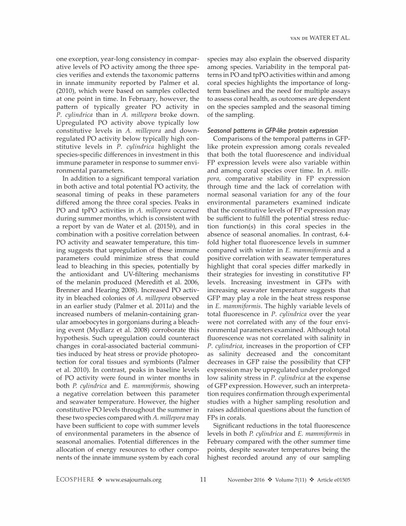
between summer and winter months revealed no major shifts in emission wavelength peaks in any of the three species (Fig. 3A–C). However, the overall mean levels of fluorescence were higher in summer (December–March) than in
winter (July–September) for E. mammiformis (P < 0.001). In contrast, the overall mean levels of fluorescence did not differ significantly between summer and winter months for either A. mille-pora (P = 0.972) or P. cylindrica (P = 0.195) (Fig. 3A–C), although the peaks in total fluores-cence in both the cyan FP and green FP regions were higher in summer for both species. This was particularly the case for P. cylindrica, which had a 1.5- fold higher peak in the cyan FP region (470 nm peak) in summer than in winter. More detailed analyses of temporal patterns in the total fluorescence at the level of month also dif-fered among species (Fig. 3D–F). Overall, the mean total fluorescence throughout the year was 1.6- fold higher in E. mammiformis than in A. mil-lepora, with mean levels intermediate in P. cylin-drica (dashed lines in Fig. 3D–F). Total fluorescence levels in A. millepora were compara-tively stable throughout the year (mean 1264 ± 158 RFU/μg protein) (Fig. 3D), whereas they fluctuated significantly in the other two species (E. mammiformis, 2024 ± 708 RFU/μg pro-tein; and P. cylindrica, 1760 ± 426 RFU/μg pro-tein). Fluctuations in total fluorescence were particularly pronounced in summer months in E. mammiformis, reflecting the 8.1- to 13.5- fold reduction in February compared with the other summer months, and in both summer and winter months in P. cylindrica (Fig. 3E, F).
Yearly averages of chromoprotein (CP) expres-sion tended to be the inverse of patterns found for total fluorescence, with mean levels more than twofold higher in P. cylindrica (0.81 ± 0.08) than in E. mammiformis (0.28 ± 0.03); mean lev-els were intermediate in A. millepora (0.58 ± 0.08) (Fig. 3G–I). Throughout the year, however, CP expression fluctuated significantly in all species. Temporal patterns in CP expression were roughly similar for all three species, with CP expression being upregulated by the greatest amount in January and May and consistently downregu-lated in July (Fig. 3G–I).
Similar to the patterns in total fluorescence, levels of individual FPs, that is, cyan, green, and red fluorescent proteins (CFP, GFP, and RFP, respectively), expressed as a proportion of total fluorescence, did not differ significantly from the yearly average in A. millepora, in any of the 9 months (Fig. 4A, D, G). Proportional levels of each FP were also relatively stable in P. cylindrica,

November 2016 v Volume 7(11) v Article e015058 v www.esajournals.org
van de WATER ET AL.
with the exception of levels recorded in March, when the contribution of CFP to the total flu-orescence was significantly increased and, con-comitantly, the relative contribution of GFP and RFP was decreased (Fig. 4C, F, I). In contrast, individual FP proportions in E. mammiformis fluctuated significantly over time and around the yearly average, with no evident association with season (Fig. 4B, E, H).
Correlations between biological and environmental parameters
Based on the monthly average rainfall and sea-water temperatures from the five years prior to our study (Appendix S1: Fig. S2) as a reference, no anomalies in these two environmental param-eters were observed during our sampling period (Appendix S1: Fig. S1). Between 2004 and 2009, the hottest monthly average temperature was
Fig. 3. Seasonal patterns in coral fluorescence. (A–C) Fluorescence spectra in summer and winter; shaded area indicates the SE of the mean (SEM); (D–F) total fluorescence levels; and (G–I) chromoprotein levels. Asterisk (*) indicates the statistically significant difference (P < 0.05/n) with previous time point. Letter “a” indicates the statistically significant difference (P < 0.05) from the yearly average. RFU = relative fluorescence units; OD588nm = optical density at 588- nm wavelength.
012345678
Rel
ativ
e to
tal f
luor
esce
nce
(RFU
∙ μg
–1 p
rote
in; ×
1000
) *
*
*
**
*
*
*
*
*
0
50
100
450 500 550 600Emission wavelength (nm)
Fluo
resc
ence
(RFU
∙ μg
–1 p
rote
in)
Summer ± SEWinter ± SE
Acropora millepora
450 500 550 600Emission wavelength (nm)
Echinopora mammiformis
450 500 550 600Emission wavelength (nm)
Porites cylindrica
a
a
a a
a
a
a
a
a
A B C
D F
Chr
omop
rote
in e
xpre
ssio
n(O
D58
8 nm
∙μg–1
pro
tein
)
0.0
0.5
1.0
1.5
2.0
2.5
*
*
*
*
Time2009 2010
Oct Dec FebJan
Mar AugMay SepJul
*
*a
a
a
a
Time2009 2010
Oct Dec FebJan
Mar AugMay SepJul
* ** *
*
*a
aa
a
a
Time2009 2010
Oct Dec FebJan
Mar AugMay SepJul
*
*
**
*a
aa
a
a
G H I
0
25
50
75
0
50
100
150
200
9 E

November 2016 v Volume 7(11) v Article e015059 v www.esajournals.org
van de WATER ET AL.
29.2° ± 0.2°C (mean ± SEM), while the highest temperature recorded in our study was 29.4°C. Similarly, no anomalies in rainfall were detected, with a peak of 948 mm in January 2010, compared with peaks of 1068 and 1007 mm in October 2005 and January 2009, respectively (744 ± 134 mm (mean ± SEM) rainfall in the wettest month in previous years).
Levels of PO activity were significantly cor-related with temperature in all three coral spe-cies (Table 1). However, whereas PO activity was
positively correlated with temperature in A. mille-pora, it was negatively correlated with temperature in E. mammiformis and P. cyclindrica. In A. millepora, PO and tpPO activities were also positively cor-related with rainfall, but in contrast, in P. cylindrica, both parameters showed a negative correlation with rainfall (Table 1). No significant correlations between rainfall and any of the two biochemical parameters were observed in E. mammiformis.
Chromoprotein expression was correlated with one environmental parameter in one species;
Fig. 4. Seasonal patterns in the proportion of individual fluorescent proteins (FP) to the total fluorescence. (A–C) cyan fluorescent protein; (D–F) green fluorescent protein; and (G–I) red fluorescent protein. An asterisk (*) indicates the statistically significant difference (P < 0.05/n) with previous time point. Letter “a” indicates the statistically significant difference (P < 0.05) from the yearly average.
0.00
0.20
0.40
0.60
0.80
1.00
0.0
0.1
0.2
0.3
0.4
Porites cylindricaAcropora milleporaPr
opor
�on
of c
yan
FP e
xpre
ssio
nto
tota
l fluo
resc
ence
0.00
0.10
0.20
0.30
Echinopora mammiformis
**
*
**
*
* *
*
*
*
*
*
** *
Time2009 2010
Oct DecFe
bJan Mar AugMay Se
pJul
Time2009 2010
Oct DecFe
bJan Mar AugMay Se
pJul
Time2009 2010
Oct DecFe
bJan Mar AugMay Se
pJul
*
a a
a
*
*
a
a
a
a
a
a
Prop
or�o
n of
gre
en F
P ex
pres
sion
to to
tal fl
uore
scen
cePr
opor
�on
of re
d FP
exp
ress
ion
to to
tal fl
uore
scen
ceA B C
D E F
G H I
a
a
a
*
**

November 2016 v Volume 7(11) v Article e0150510 v www.esajournals.org
van de WATER ET AL.
that is, CP was positively correlated with tem-perature in A. millepora (Table 1). Interestingly, a strong correlation was observed between CP and both PO (r = 0.72, P < 0.01) and tpPO (r = 0.67, P < 0.01) in A. millepora, but not in the other two species tested. With the exception of the correlation with temperature in E. mammi-formis, total fluorescence did not correlate with any of the environmental parameters in any of the species examined (Table 1). In addition, we did not observe any correlation between rainfall and GFP- like protein expression (Table 1). The proportion of GFP showed a positive correlation with temperature in E. mammiformis (Table 1). In contrast, GFP was negatively correlated with cloud cover in P. cylindrica, whereas CFP was pos-itively correlated with this parameter in this spe-cies (Table 1). Our findings also show that there is a strong positive correlation between salinity and the proportion of GFP in P. cylindrica and a strong
negative correlation with the proportion of CFP in this species (Table 1). An overview of the envi-ronmental parameters assessed can be found in Appendix S1: Fig. S1.
dIscussIon
Seasonal patterns in the prophenoloxidase systemIn this study, we show that there is high vari-
ability in mean levels of both activated and total potential PO activity over a year, and this occurs both within and among corals from three com-mon reef- building families. The typically more than twofold higher levels of PO activity found in Porites cylindrica, a disease- and bleaching- resistant species, compared with the more susceptible Acropora millepora, suggest a signifi-cantly higher level of resource investment in innate immunity by P. cylindrica, which likely contributes to its greater stress resistance. With
Table 1. Correlations (Pearson’s r) between immune parameters (PO activity and GFP- like proteins) and envi-ronmental factors (temperature, rainfall, salinity, and cloud cover).
Species Temperature Rainfall Salinity Cloud coverPO activity
Acropora millepora 0.26 (P = 0.049)* 0.34 (P = 0.012)* −0.18 (P = 0.240) 0.24 (P = 0.068)Echinopora mammiformis −0.25 (P = 0.022)* −0.01 (P = 0.949) −0.01 (P = 0.947) 0.15 (P = 0.160)Porites cylindrica −0.34 (P = 0.005)* −0.34 (P = 0.009)* 0.25 (P = 0.106) −0.13 (P = 0.298)
Total potential PO activityAcropora millepora 0.21 (P = 0.1065) 0.35 (P = 0.009)* −0.19 (P = 0.249) 0.24 (P = 0.067)Echinopora mammiformis −0.20 (P = 0.068) −0.06 (P = 0.639) 0.07 (P = 0.614) 0.11 (P = 0.315)Porites cylindrica −0.36 (P = 0.003)* −0.35 (P = 0.006)* 0.29 (P = 0.055) −0.13 (P = 0.314)
Chromoprotein expressionAcropora millepora 0.26 (P = 0.046)* 0.22 (P = 0.113) −0.11 (P = 0.130) 0.13 (P = 0.308)Echinopora mammiformis 0.10 (P = 0.343) −0.001 (P = 0.989) −0.12 (P = 0.391) 0.08 (P = 0.461)Porites cylindrica 0.03 (P = 0.815) −0.12 (P = 0.375) 0.22 (P = 0.161) −0.19 (P = 0.112)
Total fluorescenceAcropora millepora −0.09 (P = 0.532) 0.01 (P = 0.975) −0.10 (P = 0.591) 0.16 (P = 0.255)Echinopora mammiformis 0.30 (P = 0.009)* 0.05 (P = 0.707) 0.06 (P = 0.685) −0.04 (P = 0.737)Porites cylindrica −0.03 (P = 0.781) −0.09 (P = 0.513) −0.08 (P = 0.615) −0.10 (P = 0.427)
CFP proportionAcropora millepora 0.07 (P = 0.625) 0.05 (P = 0.755) −0.09 (P = 0.573) 0.10 (P = 0.471)Echinopora mammiformis −0.16 (P = 0.181) −0.04 (P = 0.745) −0.06 (P = 0.684) −0.12 (P = 0.295)Porites cylindrica 0.05 (P = 0.714) 0.18 (P = 0.177) −0.57 (P = 0.0001)* 0.39 (P = 0.001)*
GFP proportionAcropora millepora −0.06 (P = 0.653) 0.09 (P = 0.539) −0.08 (P = 0.670) −0.03 (P = 0.827)Echinopora mammiformis 0.29 (P = 0.012)* 0.06 (P = 0.616) 0.10 (P = 0.519) −0.01 (P = 0.911)Porites cylindrica 0.03 (P = 0.808) −0.20 (P = 0.122) 0.52 (P = 0.0003)* −0.36 (P = 0.003)*
RFP proportionAcropora millepora −0.06 (P = 0.652) −0.08 (P = 0.582) 0.14 (P = 0.444) −0.11 (P = 0.428)Echinopora mammiformis −0.15 (P = 0.203) −0.02 (P = 0.861) −0.07 (P = 0.645 −0.15 (P = 0.209)Porites cylindrica −0.13 (P = 0.283) 0.09 (P = 0.504) 0.01 (P = 0.959) 0.03 (P = 0.834)
Note: Significant correlations (P < 0.05) are indicated with an asterisk (*) and appear in boldface.
November 2016 v Volume 7(11) v Article e0150511 v www.esajournals.org
van de WATER ET AL.
one exception, year- long consistency in compar-ative levels of PO activity among the three spe-cies verifies and extends the taxonomic patterns in innate immunity reported by Palmer et al. (2010), which were based on samples collected at one point in time. In February, however, the pattern of typically greater PO activity in P. cylindrica than in A. millepora broke down. Upregulated PO activity above typically low constitutive levels in A. millepora and down-regulated PO activity below typically high con-stitutive levels in P. cylindrica highlight the species- specific differences in investment in this immune parameter in response to summer envi-ronmental parameters.
In addition to a significant temporal variation in both active and total potential PO activity, the seasonal timing of peaks in these parameters differed among the three coral species. Peaks in PO and tpPO activities in A. millepora occurred during summer months, which is consistent with a report by van de Water et al. (2015b), and in combination with a positive correlation between PO activity and seawater temperature, this tim-ing suggests that upregulation of these immune parameters could minimize stress that could lead to bleaching in this species, potentially by the antioxidant and UV- filtering mechanisms of the melanin produced (Meredith et al. 2006, Brenner and Hearing 2008). Increased PO activ-ity in bleached colonies of A. millepora observed in an earlier study (Palmer et al. 2011a) and the increased numbers of melanin- containing gran-ular amoebocytes in gorgonians during a bleach-ing event (Mydlarz et al. 2008) corroborate this hypothesis. Such upregulation could counteract changes in coral- associated bacterial communi-ties induced by heat stress or provide photopro-tection for coral tissues and symbionts (Palmer et al. 2010). In contrast, peaks in baseline levels of PO activity were found in winter months in both P. cylindrica and E. mammiformis, showing a negative correlation between this parameter and seawater temperature. However, the higher constitutive PO levels throughout the summer in these two species compared with A. millepora may have been sufficient to cope with summer levels of environmental parameters in the absence of seasonal anomalies. Potential differences in the allocation of energy resources to other compo-nents of the innate immune system by each coral
species may also explain the observed disparity among species. Variability in the temporal pat-terns in PO and tpPO activities within and among coral species highlights the importance of long- term baselines and the need for multiple assays to assess coral health, as outcomes are dependent on the species sampled and the seasonal timing of the sampling.
Seasonal patterns in GFP- like protein expressionComparisons of the temporal patterns in GFP-
like protein expression among corals revealed that both the total fluorescence and individual FP expression levels were also variable within and among coral species over time. In A. mille-pora, comparative stability in FP expression through time and the lack of correlation with normal seasonal variation for any of the four environmental parameters examined indicate that the constitutive levels of FP expression may be sufficient to fulfill the potential stress reduc-tion function(s) in this coral species in the absence of seasonal anomalies. In contrast, 6.4- fold higher total fluorescence levels in summer compared with winter in E. mammiformis and a positive correlation with seawater temperatures highlight that coral species differ markedly in their strategies for investing in constitutive FP levels. Increasing investment in GFPs with increasing seawater temperature suggests that GFP may play a role in the heat stress response in E. mammiformis. The highly variable levels of total fluorescence in P. cylindrica over the year were not correlated with any of the four envi-ronmental parameters examined. Although total fluorescence was not correlated with salinity in P. cylindrica, increases in the proportion of CFP as salinity decreased and the concomitant decreases in GFP raise the possibility that CFP expression may be upregulated under prolonged low salinity stress in P. cylindrica at the expense of GFP expression. However, such an interpreta-tion req uires confirmation through experimental studies with a higher sampling resolution and raises additional questions about the function of FPs in corals.
Significant reductions in the total fluorescence levels in both P. cylindrica and E. mammiformis in February compared with the other summer time points, despite seawater temperatures being the highest recorded around any of our sampling
November 2016 v Volume 7(11) v Article e0150512 v www.esajournals.org
van de WATER ET AL.
times, raise the possibility that fluorescence plays a role in mitigating stress induced by an envi-ronmental parameter other than temperature. Interestingly, the cloud cover signature at this time was distinctly different from all other time points, showing 13 consecutive days with more than 90% cloud cover in the study region, while cloud cover was below 90% at all other sampling times. Although solar radiation is increased in summer and heats up the seawater, clouds can reduce the amount of solar radiation that reaches corals. In accordance with a photoprotective function for FPs (Salih et al. 2000, Dove et al. 2001), it may be that these two species increased their fluorescence levels in summer in response to higher levels of solar radiation and then down-regulated levels when clouds reduced solar radi-ation to non-stress levels. These results would be consistent with the results of experimental studies that found strong positive correlations between GFP expression and light intensities (D’Angelo et al. 2008, Roth et al. 2010). Taken together, the total fluorescence levels appear to be upregulated in response to increased levels of solar radiation, providing corroborative evidence for a photopro-tective function for FPs in corals.
Non-fluorescent chromoprotein levels fluc-tuated throughout the year in all coral species, although a positive correlation with seawater tem-perature was found in A. millepora. This suggests a role for chromoprotein in the thermal stress response, which is consistent with the observa-tion of elevated chromoprotein expression lev-els in naturally bleached colonies of this species (Seneca et al. 2010), where it could play a photo-protective role by reducing the levels of solar radi-ation reaching Symbiodinium (Smith et al. 2013). Chromoprotein may also exhibit antioxidant properties (Palmer et al. 2009a) and could there-fore be upregulated in response to the increased PO activity at higher temperatures to mitigate the adverse effects of reactive oxygen produced by the proPO system. The strong correlations found between CP expression and PO activities in this species support this interpretation. Surprisingly, in E. mammiformis and P. cylindrica, there was no correlation between temperature and CP, or between CP and PO activities. Corals do, how-ever, possess other more effective antioxidants, such as superoxide dismutase, catalase, and per-oxidase (Mydlarz and Palmer 2011). Potentially,
these enzymes are the primary antioxidants in E. mammiformis and P. cylindrica, while in A. mille-pora, CP may play a more significant role in neu-tralizing ROS.
Comparative ecological immunologyComparative differences in immune and stress
response parameters among coral species repre-senting three coral families strongly suggest that corals differ in the allocation of resources to con-stitutive levels of immune and stress response systems. Higher levels of PO activity in the resis-tant coral P. cylindrica compared with the inter-mediately resistant E. mammiformis and the susceptible A. millepora are consistent with the positive correlations between immune status and both disease and bleaching resistance, as found by Palmer et al. (2010). In addition, we also found that a significantly higher proportion of PO is in its active form in P. cylindrica than in A. millepora, which would further contribute to the differences in disease and bleaching susceptibilities between these species. P. cylindrica appears to have consti-tutively high levels of PO activity and is thereby capable of preventing microbial infection and responding immediately to disturbances. Due to the short half- life of activated PO, maintaining such high PO activity requires significant energy resources. A. millepora, on the other hand, has lower constitutive levels of PO activity, but does have the capability of inducing additional activa-tion of PO in response to infections when and where necessary, by storing more PO in its inac-tive proPO form, likely in immune cells that migrate toward lesions (Palmer et al. 2011b). While this strategy may require fewer resources and thereby allows the allocation of more resources for other life history traits, such as col-ony growth (Palmer et al. 2010), these relatively low constitutive PO activity levels may provide a minimal protection against invading microbes, thus increasing the chance of infection and dis-ease before a significant immune response can be orchestrated by the host. It should, however, be noted that none of the coral colonies followed in this study developed signs of disease or bleach-ing. Evaluating both PO and tpPO activity levels in corals may significantly increase our under-standing of the ecological immunology of corals.
Differences in constitutive levels of fluorescence among the coral species studied also correlate
November 2016 v Volume 7(11) v Article e0150513 v www.esajournals.org
van de WATER ET AL.
with life history differences. Fast- growing A. mil-lepora generally maintained relatively stable high fluorescence levels across seasons compared with the other two species, which exhibited typ-ically lower and more variable total fluorescence levels and generally have slower growth rates. Fluorescent proteins are known to be more highly expressed in growing tissues (D’Angelo et al. 2012) and positively correlate with coral growth rates (Roth et al. 2010, Roth and Deheyn 2013), possibly because of their photoprotective func-tion, which may explain the relatively higher con-stitutive levels of fluorescence in the fast- growing species. Maintaining fluorescence is considered costly due to the relatively high FP expression levels, although studies of FP turnover sug-gest that due to their long half- life, maintaining high levels of FPs may be comparatively cheap (Leutenegger et al. 2007a). However, the apparent lack of induced FP expression in summer when solar irradiance and temperature, two important factors in coral bleaching, are highest was surpris-ing. This could indicate that FP levels in A. mille-pora are not sufficiently adequate to mitigate the effects when these environmental factors reach stressful levels, or that summer temperatures and solar irradiance were not stressful. In contrast, E. mammiformis significantly upregulates FP lev-els, in particular GFP, in summer, which likely protects this coral from high irradiance and tem-perature and ameliorates bleaching (Baird et al. 2009). P. cylindrica may use its high constitutive levels of PO activity to produce photoprotective melanin, a function recently suggested for this compound (Palmer et al. 2010). Overall, these differences show the variability in strategies for investing resources among life history traits by different coral species.
Biochemical tools for coral reef managementAn urgent need for tools to evaluate and moni-
tor coral health to manage and preempt activities that might cause further declines has arisen over recent years due to the increasing environmental and anthropogenic pressures facing corals world-wide (Burke et al. 2011). In this study, we provide initial baseline references of various innate immu-nity parameters for three coral species belonging to three major reef- building coral families, which can be used to assess the suitability of PO activity and FP expression as potential indicators for
monitoring environmental stressors. During the 2009–2010 sampling period, no anomalies in either seawater temperature or rainfall occurred, making this a suitable period for the establish-ment of baseline immune parameter levels. A number of recent studies have suggested that these immune parameters may be suitable tools for reef managers (Palmer et al. 2010), such as using FP expression for monitoring coral health (Roth et al. 2010, D’Angelo et al. 2012). Generally, biomarkers should (1) be specific to a stressor and not other factors, (2) have a magnitude in change that reflects the magnitude of the stressor, and (3) have low background variability (Cooper et al. 2009). Our results show, however, that there is a significant variability over time in most parame-ters tested, although the degree of variability depended on the coral species. Temporal vari-ability may be acceptable in cases where exten-sive baseline data are available. While the changes in GFP- like protein expression do not appear to be specific to particular stressors (e.g., responses to high solar irradiation [D’Angelo et al. 2008], heat stress [Dove et al. 2006], bleaching [Leu-tenegger et al. 2007b], and coral translocation [Bay et al. 2009]), the range of stressors that induce the changes in FP expression or in the ratio of indi-vidual FPs suggests that FP expression levels may function as a general stress biomarker for some corals. Although correlations are good indicators of relationships between immune parameters and environmental factors, experimental studies exploring how these potential biomarkers res-pond to stressors, as well as the magnitude of these responses, need to be addressed under con-trolled conditions. In addition, several key com-mon indicator species should be identified and, for each of these, extensive baseline data for each biomarker should be obtained on both temporal and spatial scales. Overall, PO activity and FP expression can potentially be used as biomarkers for coral health, but additional research on base-line data and stressors is required before we can address their suitability and applicability.
concludIng stateMent
Given the increasing environmental and ant hropogenic pressures and stresses corals face worldwide, there is an urgent need to under-stand the coral stress responses to facilitate the

November 2016 v Volume 7(11) v Article e0150514 v www.esajournals.org
van de WATER ET AL.
development of coral health monitoring tools and techniques. In this study, initial baselines of multiple ecological immune parameters were compared between three species from three major reef- building coral families. The signifi-cant differences observed in constitutive levels and the temporal patterns in PO activity and GFP- like protein expression among these species suggest that they have evolved different ecologi-cal immune strategies. Such variations under-score the need to obtain species- specific baseline levels and further long- term data before consid-ering the suitability of these ecological immune parameters for routine coral health monitoring.
acknowledgMents
Orpheus Island Research Station is thanked for logistical support. Rhondda Jones is thanked for advice on statistical analyses. Ian McLeod, Stuart Beveridge, Raechel Littman, Crystal Neligh, Tom Heintz, and Lisa Kelly are thanked for field assistance and Caroline Palmer and Lisa Kelly also for laboratory support. The authors are also grateful to the Australian Research Council for funds allocated by the ARC Centre of Excellence for Coral Reef Studies to Bette Willis, which funded this project. The manuscript con-tents are solely the opinions of the authors and do not constitute a statement of policy, decision, or position on behalf of NOAA or the U.S. Government.
lIterature cIted
Baird, A. H., R. Bhagooli, P. J. Ralph, and S. Takahashi. 2009. Coral bleaching: the role of the host. Trends in Ecology & Evolution 24:16–20.
Bay, L. K., K. E. Ulstrup, H. B. Nielsen, H. Jarmer, N. Goffard, B. L. Willis, D. J. Miller, and M. J. H. van Oppen. 2009. Microarray analysis reveals transcriptional plasticity in the reef building coral Acropora millepora. Molecular Ecology 18:3062–3075.
Ben-Haim, Y., M. Zicherman-Keren, and E. Rosenberg. 2003. Temperature- regulated bleaching and lysis of the coral Pocillopora damicornis by the novel patho-gen Vibrio coralliilyticus. Applied and Environmen-tal Microbiology 69:4236–4242.
Bou-Abdallah, F., N. D. Chasteen, and M. P. Lesser. 2006. Quenching of superoxide radicals by green fluorescent protein. Biochimica et Biophysica Acta 1760:1690–1695.
Brenner, M., and V. J. Hearing. 2008. The protective role of melanin against UV damage in human skin. Photochemistry and Photobiology 84:539–549.
Burge, C. A., M. E. Mouchka, C. D. Harvell, and S. Roberts. 2013. Immune response of the Carib-bean sea fan, Gorgonia ventalina, exposed to an Aplanochytrium parasite as revealed by transcrip-tome sequencing. Frontiers in Physiology 4:180.
Burke, L., K. Reytar, M. Spalding, and A. Perry. 2011. Reefs at risk revisited. World Resources Institute, Washington, D.C., USA.
Cerenius, L., S. Kawabata, B. L. Lee, M. Nonaka, and K. Soderhall. 2010. Proteolytic cascades and their involvement in invertebrate immunity. Trends in Biochemical Sciences 35:575–583.
Cooper, T. F., J. P. Gilmour, and K. E. Fabricius. 2009. Bioindicators of changes in water quality on coral reefs: review and recommendations for monitoring programmes. Coral Reefs 28:589–606.
Cunning, R., and A. C. Baker. 2013. Excess algal sym-bionts increase the susceptibility of reef corals to bleaching. Nature Climate Change 3:259–262.
D’Angelo, C., A. Denzel, A. Vogt, M. V. Matz, F. Oswald, A. Salih, G. U. Nienhaus, and J. Wieden-mann. 2008. Blue light regulation of host pigment in reef- building corals. Marine Ecology Progress Series 364:97–106.
D’Angelo, C., E. G. Smith, F. Oswald, J. Burt, D. Tchernov, and J. Wiedenmann. 2012. Locally accelerated growth is part of the innate immune response and repair mechanisms in reef- building corals as detected by green fluorescent protein (GFP)- like pigments. Coral Reefs 31:1045–1056.
Douglas, A. E. 2003. Coral bleaching – How and why? Marine Pollution Bulletin 46:385–392.
Dove, S. G., O. Hoegh-Guldberg, and S. Ranganathan. 2001. Major colour patterns of reef- building cor-als are due to a family of GFP- like proteins. Coral Reefs 19:197–204.
Dove, S., J. C. Ortiz, S. Enríquez, M. Fine, P. Fisher, R. Iglesias-Prieto, D. Thornhill, and O. Hoegh-Guld-berg. 2006. Response of holosymbiont pigments from the scleractinian coral Montipora monasteriata to short- term heat stress. Limnology and Oceanog-raphy 51:1149–1158.
Ellis, R. P., H. Parry, J. I. Spicer, T. H. Hutchinson, R. K. Pipe, and S. Widdicombe. 2011. Immunolog-ical function in marine invertebrates: responses to environmental perturbation. Fish & Shellfish Immunology 30:1209–1222.
Gilmore, A. M., A. W. D. Larkum, A. Salih, S. Itoh, Y. Shibata, C. Bena, H. Yamasaki, M. Papina, and R. Van Woesik. 2003. Simultaneous time resolution of the emission spectra of fluorescent proteins and zooxanthellar chlorophyll in reef- building corals. Photochemistry and Photobiology 77:515–523.
Guest, J. R., A. H. Baird, J. A. Maynard, E. Muttaqin, A. J. Edwards, S. J. Campbell, K. Yewdall, Y. A.

November 2016 v Volume 7(11) v Article e0150515 v www.esajournals.org
van de WATER ET AL.
Affendi, and L. M. Chou. 2012. Contrasting patterns of coral bleaching susceptibility in 2010 suggest an adaptive response to thermal stress. PLoS ONE 7:e33353.
Howells, E. J., V. H. Beltran, N. W. Larsen, L. K. Bay, B. L. Willis, and M. J. H. van Oppen. 2012. Coral thermal tolerance shaped by local adaptation of photosymbionts. Nature Climate Change 2: 116–120.
Howells, E. J., R. Berkelmans, M. J. H. van Oppen, B. L. Willis, and L. K. Bay. 2013. Historical thermal regimes define limits to coral acclimatization. Ecol-ogy 94:1078–1088.
Hughes, T. P., et al. 2003. Climate change, human impacts, and the resilience of coral reefs. Science 301:929–933.
Jones, A. M., and R. Berkelmans. 2014. Flood impacts in Keppel Bay, southern Great Barrier Reef in the aftermath of cyclonic rainfall. PLoS ONE 9:e84739.
Kawaguti, S. 1969. Effect of the green fluorescent pig-ment on the productivity of the reef corals. Micro-nesica 5:313.
Kerswell, A. P., and R. J. Jones. 2003. Effects of hypo- osmosis on the coral Stylophora pistillata: nature and cause of low- salinity bleaching. Marine Ecol-ogy Progress Series 253:145–154.
King, M. D., S. C. Tsay, S. E. Platnick, M. Wang, and K. N. Liou. 1997. Cloud retrieval algorithms for MODIS: optical thickness, effective particle radi-us, and thermodynamic phase. MODIS Algorithm Theoretical Basis. Document No. ATBD-MOD-05, MOD06 – Cloud product. NASA Goddard Space Flight Center, Greenbelt, Maryland, USA.
Lamb, J. B., and B. L. Willis. 2011. Using coral dis-ease prevalence to assess the effects of concentrat-ing tourism activities on offshore reefs in a tropical marine park. Conservation Biology 25:1044–1052.
Lesser, M. P. 1996. Elevated temperatures and ultra-violet radiation cause oxidative stress and inhibit photosynthesis in symbiotic dinoflagellates. Lim-nology and Oceanography 41:271–283.
Leutenegger, A., C. D’Angelo, M. V. Matz, A. Denzel, F. Oswald, A. Salih, G. U. Nienhaus, and J. Wieden-mann. 2007a. It’s cheap to be colorful. FEBS Journal 274:2496–2505.
Leutenegger, A., S. Kredel, S. Gundel, C. D’Angelo, A. Salih, and J. Wiedenmann. 2007b. Analysis of fluorescent and non- fluorescent sea anemones from the Mediterranean Sea during a bleaching event. Journal of Experimental Marine Biology and Ecology 353:221–234.
Lin, Y. C., et al. 2012. Modulation of the innate immune system in white shrimp Litopenaeus vannamei fol-lowing long- term low salinity exposure. Fish & Shellfish Immunology 33:324–331.
Littman, R. A., D. G. Bourne, and B. L. Willis. 2010. Responses of coral- associated bacterial commu-nities to heat stress differ with Symbiodinium type on the same coral host. Molecular Ecology 19: 1978–1990.
Loya, Y., K. Sakai, K. Yamazato, Y. Nakano, H. Sambali, and R. Van Woesik. 2001. Coral bleach-ing: the winners and the losers. Ecology Letters 4:122–131.
Marshall, P. A., and A. H. Baird. 2000. Bleaching of corals on the Great Barrier Reef: differen-tial susceptibilities among taxa. Coral Reefs 19: 155–163.
Meredith, P., B. J. Powell, J. Riesz, S. P. Nighswander- Rempel, M. R. Pederson, and E. G. Moore. 2006. Towards structure- property- function relationships for eumelanin. Soft Matter 2:37–44.
Mydlarz, L. D., S. F. Holthouse, E. C. Peters, and C. D. Harvell. 2008. Cellular responses in sea fan corals: Granular amoebocytes react to pathogen and cli-mate stressors. PLoS ONE 3:e1811.
Mydlarz, L. D., and C. V. Palmer. 2011. The presence of multiple phenoloxidases in Caribbean reef- building corals. Comparative Biochemistry and Physiology. Part A: Molecular & Integrative Physi-ology 159:372–378.
Palmer, C. V., J. C. Bythell, and B. L. Willis. 2010. Levels of immunity parameters underpin bleaching and disease susceptibility of reef corals. FASEB Journal 24:1935–1946.
Palmer, C. V., J. C. Bythell, and B. L. Willis. 2011a. A comparative study of phenoloxidase activity in diseased and bleached colonies of the coral Acro-pora millepora. Developmental and Comparative Immunology 35:1098–1101.
Palmer, C. V., C. K. Modi, and L. D. Mydlarz. 2009a. Coral fluorescent proteins as antioxidants. PLoS ONE 4:e7298.
Palmer, C. V., L. D. Mydlarz, and B. L. Willis. 2008. Evidence of an inflammatory- like response in non- normally pigmented tissues of two scleractinian corals. Proceedings. Biological Sciences/The Royal Society 275:2687–2693.
Palmer, C. V., M. S. Roth, and R. D. Gates. 2009b. Red fluorescent protein responsible for pigmentation in trematode- infected Porites compressa tissues. Bio-logical Bulletin 216:68–74.
Palmer, C. V., N. G. Traylor-Knowles, B. L. Willis, and J. C. Bythell. 2011b. Corals use similar immune cells and wound- healing processes as those of higher organisms. PLoS ONE 6:e23992.
Raymundo, L. J., K. B. Rosell, C. T. Reboton, and L. Kaczmarsky. 2005. Coral diseases on Philippine reefs: Genus Porites is a dominant host. Diseases of Aquatic Organisms 64:181–191.
November 2016 v Volume 7(11) v Article e0150516 v www.esajournals.org
van de WATER ET AL.
Roth, M. S., and D. D. Deheyn. 2013. Effects of cold stress and heat stress on coral fluorescence in reef- building corals. Scientific Reports 3:1421.
Roth, M. S., M. I. Latz, R. Goericke, and D. D. Deheyn. 2010. Green fluorescent protein regulation in the coral Acropora yongei during photoacclimation. Journal of Experimental Biology 213:3644–3655.
Salih, A., A. Larkum, G. Cox, M. Kuhl, and O. Hoegh-Guldberg. 2000. Fluorescent pigments in corals are photoprotective. Nature 408:850–853.
Saxby, T., W. C. Dennison, and O. Hoegh-Guldberg. 2003. Photosynthetic responses of the coral Mon-tipora digitata to cold temperature stress. Marine Ecology Progress Series 248:85–97.
Schlichter, D., H. W. Fricke, and W. Weber. 1986. Light harvesting by wavelength transformation in a sym-biotic coral of the Red Sea twilight zone. Marine Biology 91:403–407.
Seneca, F. O., S. Foret, E. E. Ball, C. Smith-Keune, D. J. Miller, and M. J. H. van Oppen. 2010. Patterns of gene expression in a scleractinian coral undergo-ing natural bleaching. Marine Biotechnology 12: 594–604.
Shagin, D. A., et al. 2004. GFP- like proteins as ubiqui-tous metazoan superfamily: evolution of functional
features and structural complexity. Molecular Biol-ogy and Evolution 21:841–850.
Smith, E. G., C. D’Angelo, A. Salih, and J. Wieden-mann. 2013. Screening by coral green fluorescent protein (GFP)- like chromoproteins supports a role in photoprotection of zooxanthellae. Coral Reefs 32:463–474.
Tseng, I. T., and J. C. Chen. 2004. The immune response of white shrimp Litopenaeus vannamei and its sus-ceptibility to Vibrio alginolyticus under nitrite stress. Fish & Shellfish Immunology 17:325–333.
van de Water, J. A. J. M., T. D. Ainsworth, W. Leggat, D. G. Bourne, B. L. Willis, and M. J. H. van Oppen. 2015a. The coral immune response facilitates pro-tection against microbes during tissue regenera-tion. Molecular Ecology 24:3390–3404.
van de Water, J. A. J. M., J. B. Lamb, M. J. H. van Oppen, B. L. Willis, and D. G. Bourne. 2015b. Comparative immune responses of corals to stressors associated with offshore reef- based tourist platforms. Conser-vation Physiology 3:cov32.
Willis, B. L., C. Page, and E. Dinsdale. 2004. Coral disease on the Great Barrier Reef. Pages 69–104 in E. Rosenberg and Y. Loya, editors. Coral health and disease. Springer, Berlin, Germany.
supportIng InforMatIon
Additional Supporting Information may be found online at: http://onlinelibrary.wiley.com/doi/10.1002/ecs2.1505/full