temporal and spatial regulation of targeting aurora b to
TRANSCRIPT

For the proper partition of chromosomes to daughtercells, sister chromatids must be held together before mi-totic chromosome segregation. This cohesion of sisterchromatids is established during DNA replication depend-ent on the cohesin complex and is maintained untilmetaphase when sister kinetochores attach to micro-tubules (MTs) emanating from the opposite spindle poles(bipolar attachment) (Nasmyth et al. 2000). In animal mi-totic cells, however, most cohesin dissociates from thechromosome arms during prophase (called “prophasepathway”), in a manner dependent on the phosphorylationof cohesin by mitotic kinases such as Polo-like kinase(Losada et al. 2002; Hauf et al. 2005). However, cen-tromeric cohesin is retained until metaphase through thefunction of shugoshin (Sgo/MEI-S332) (Lee et al. 2005;Watanabe 2005). This shugoshin-dependent centromericprotection is essential for mitotic chromosome segregationin mammalian cells. In most organisms, shugoshin is re-quired for meiotic chromosome segregation, where thestepwise dissociation of cohesin is more crucial (Miyazakiand Orr-Weaver 1994; Watanabe 2005). Shugoshin col-laborates with protein phosphatase 2A (PP2A) to preventphosphorylation of cohesin and, thereby, its dissociation(Kitajima et al. 2006; Riedel et al. 2006; Tang et al. 2006).Although shugoshin was first characterized as a protectorof centromeric cohesin (Kitajima et al. 2004; Marston etal. 2004; Rabitsch et al. 2004), a study in budding yeastsuggested that it was also involved in sensing the lack oftension on mitotic chromosomes (Indjeian et al. 2005).Accurate segregation of chromosomes during cell divi-
sion requires sister chromatids to be captured by MTsfrom the opposite spindle poles (bipolar attachment or
biorientation). This is accomplished by a trial-and-errorprocess of kinetochore-microtubule (KT-MT) attachmentthat is essential and relies largely on the CPC, which in-cludes catalytic kinase Aurora B and regulatory subunitsBorealin, Survivin, and INCENP and locates at cen-tromeres (Hauf et al. 2003; Cheeseman et al. 2006;DeLuca et al. 2006; Ciferri et al. 2008; Tanaka 2008). Inthe absence of tension across centromeres, Aurora B phos-phorylates the MT-anchoring kinetochore proteins,thereby destabilizing the attachment, whereas the bipolarattachment of kinetochores produces a tension that bringsabout the spatial separation of kinetochores from AuroraB, thereby stabilizing the attachment (Fig. 1) (Pinsky andBiggins 2005; Tanaka 2008; Liu et al. 2009). Thus, AuroraB must be targeted and anchored precisely at the center ofpaired kinetochores, a site called the inner centromere(Ruchaud et al. 2007; Kelly and Funabiki 2009).
SHUGOSHIN REQUIRED FOR CPCTARGETING TO CENTROMERES
Fission yeast possess two shugoshin-like proteins, Sgo1and Sgo2, both of which localize at the pericentromericheterochromatin region, the site enriched with cohesin andcrucial for centromeric cohesion. Sgo1 is meiosis I spe-cific and has a crucial role in protecting centromeric co-hesin during meiosis I, whereas Sgo2 is ubiquitouslyexpressed and required for faithful chromosome segrega-tion in mitosis and meiosis (Kitajima et al. 2004; Rabitschet al. 2004). Sgo2 associates closely with Bir1/Survivinand is required for the full localization of the CPC at cen-tromeres (Kawashima et al. 2007; Vanoosthuyse et al.
Temporal and Spatial Regulation of TargetingAurora B to the Inner Centromere
Y. WATANABELaboratory of Chromosome Dynamics, Institute of Molecular and Cellular Biosciences,
University of Tokyo, Yayoi, Tokyo 113-0032, JapanCorrespondence: [email protected]
Successful partitioning of chromosomes in mitosis relies on the bipolar attachment of sister chromatids at metaphase. Forthis biorientation, the chromosomal passenger complex (CPC), composed of catalytic kinase Aurora B and regulatory com-ponents (INCENP, Survivin, and Borealin), must be localized at the center of paired kinetochores, the site called the innercentromere. It is largely unknown what defines the inner centromere and how the CPC is targeted to this site. Recent studiespoint out that the shugoshin protein (SGO), originally identified as a cohesin protector, also acts as a conserved centromericadapter of the CPC. Phosphorylation of the CPC by Cdk1 promotes direct binding with shugoshin, thus explaining how theCPC is targeted to the centromere in a timely manner at prometaphase during the cell cycle. Moreover, the phosphorylationof histone H3 threonine 3 (H3-pT3) mediated by Haspin cooperates with Bub1-mediated H2A-S121 phosphorylation in tar-geting the CPC to the inner centromere. H3-pT3 promotes nucleosome binding of Survivin, whereas H2A-pS121 facilitatesthe binding of shugoshin. Haspin colocalizes with cohesin by associating with Pds5, a cohesin-binding protein, and Bub1 lo-calizes at kinetochores. Thus, the inner centromere is defined by the spatial intersection of two histone marks mediated bycohesin- and kinetochore-associated kinases.
Cold Spring Harbor Symposia on Quantitative Biology,Volume LXXV. ©2010 Cold Spring Harbor Laboratory Press 978-1-936113-07-1 419

2007). The overall phenotypes of sgo2∆, not only duringmitosis but also during meiosis, are explainable by the re-duced activity of Aurora B. These results imply that in ad-dition to its protective function, shugoshin has a crucialrole in promoting the centromeric localization of the CPC,thereby ensuring bipolar attachment of kinetochores.The requirement of shugoshin for CPC targeting to cen-
tromeres has also been shown in Xenopus (Rivera andLosada 2009). Although depletion of hSgo1, a canonicalshugoshin in human mitotic cells, causes little defect inCPC localization, depletion of both shugoshin-like pro-teins hSgo1 and hSgo2 abolishes CPC localization to cen-tromeres, validating that the second role of shugoshin isalso conserved in mammals (Tsukahara et al. 2010). Cen-tromeric localization of shugoshin is also influenced bythe CPC, implying that centromere targeting of shugoshinand the CPC is interdependent (Resnick et al. 2006;Kawashima et al. 2007; Vanoosthuyse et al. 2007). Thus,whereas shugoshin was initially identified as a cohesinprotector, the same or related protein acts as the cen-tromeric CPC adapter in eukaryotes.
CDK1-DEPENDENT CPC PHOSPHORYLATIONREQUIRED FOR CENTROMERE TARGETING
The cyclin-dependent kinase 1 (Cdk1)-cyclin B com-plex functions as a cell cycle engine to promote mitoticprogression. The newly isolated fission yeast cyclin B mu-tant cdc13-M7 shows a marked defect in chromosomebiorientation but not in mitotic entry, suggesting that theCdk1-cyclin B complex specifically targets the regulationof chromosome biorientation in addition to cell cycle pro-
gression (Tsukahara et al. 2010). Accordingly, fissionyeast Survivin/Bir1 is phosphorylated by Cdc2 in mitosis,and the nonphosphorylatable bir1-8Amutant impairs CPClocalization and its function at centromeres. In contrast,the phosphomimetic bir1-8D mutant shows intact CPCtargeting to centromeres and, crucially, suppresses thebiorientation defect of the cdc13-M7 mutant. Thus, phos-phorylation of Bir1 is important for targeting the CPC tocentromeres in prometaphase (Tsukahara et al. 2010).The amino-terminal half of Bir1 (Bir1-N), which con-
tains a CDK phosphorylation cluster, binds Sgo2, a cen-tromeric CPC adapter, and phosphorylation of Bir1 isrequired for this interaction (Fig. 2). As in fission yeast,the inhibition of human cyclin B causes defects in chro-mosome biorientation (Chen et al. 2008). Consistently, theCdk1-dependent phosphorylation of hBorealin (ratherthan Survivin in this case) promotes centromere targetingthrough association with both hSgo1 and hSgo2 (Tsuka-hara et al. 2010). Although the CPC subunit that interactswith shugoshin thus moves between yeast and metazoa,the overall regulation of CPC targeting to the centromereby Cdk1 is conserved, explaining how the cell cycle islinked to CPC targeting to centromeres and chromosomebiorientation in eukaryotes (Fig. 2).
BUB1-DEPENDENT H2A PHOSPHORYLATIONREQUIRED FOR SHUGOSHIN LOCALIZATION
Shugoshin localization at centromeres depends on theconserved SAC protein kinase Bub1, which is also re-quired for proper chromosome segregation (Kitajima etal. 2004, 2005; Boyarchuk et al. 2007). Although severalBub1 substrate candidates have been suggested, the canon-ical substrate remained unknown more than 15 years afterits discovery, even in genetically tractable organisms suchas yeast. Recently, a biochemical approach in fission yeastidentified serine 121 of histone 2A (H2A) as a Bub1 sub-strate (Kawashima et al. 2010). Crucially, the h2a-S121A
420 WATANABE
Figure 1. Establishment of bipolar attachment. Kinetochore-as-sembling core centromere (pink globule) protrudes from innercentromere, the site where Aurora B kinase accumulates anddestabilizes kinetochore-microtubule attachment. Therefore, mi-crotubule (MT) attachment to the kinetochore within the Aurora-B-active region (dark blue and light blue) is unstable (dashedpink line), whereas MT attachment far from Aurora B activity isstable (black line). Spindle assembly checkpoint is activatedwhen unstable attachment is disconnected but is inactivated whenall attachment is bipolar and thus stabilized (bottom).
c
Figure 2. Interaction between the CPC and shugoshin. Phospho-rylation of fission yeast Survivin or human Borealin by Cdk1promotes CPC binding to coiled-coil region of shugoshin. Inter-twined configuration of Survivin, Borealin, and INCENP isbased on the crystallographic study of the complex (Jeyaprakashet al. 2007). (Modified, with permission, from Tsukahara et al.2010 [Nature Publishing Group].)
mutant, in which all cellular H2A-S121 is replaced by ala-nine, phenocopies the bub1 kinase-dead (bub1-KD) mu-tant in losing the centromeric localization of shugoshinproteins, indicating that H2A-S121 is the canonical cellu-lar substrate of Bub1. Artificial tethering of shugoshin tocentromeres largely restores the h2a-S121A– or bub1-KD–related defects in chromosome segregation. The Bub1-H2A-shugoshin pathway is also conserved in human cells(Kawashima et al. 2010).Shugoshin family proteins, although conserved among
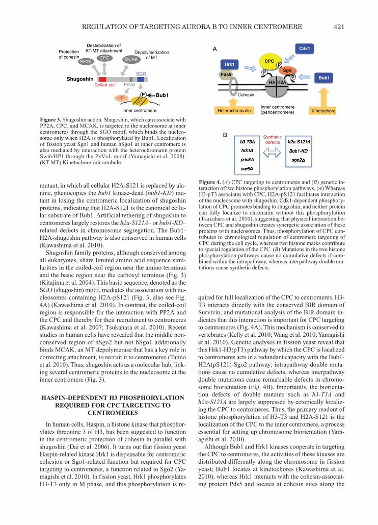
all eukaryotes, share limited amino acid sequence simi-larities in the coiled-coil region near the amino terminusand the basic region near the carboxyl terminus (Fig. 3)(Kitajima et al. 2004). This basic sequence, denoted as theSGO (shugoshin) motif, mediates the association with nu-cleosomes containing H2A-pS121 (Fig. 3, also see Fig.4A) (Kawashima et al. 2010). In contrast, the coiled-coilregion is responsible for the interaction with PP2A andthe CPC and thereby for their recruitment to centromeres(Kawashima et al. 2007; Tsukahara et al. 2010). Recentstudies in human cells have revealed that the middle non-conserved region of hSgo2 but not hSgo1 additionallybinds MCAK, an MT depolymerase that has a key role incorrecting attachment, to recruit it to centromeres (Tannoet al. 2010). Thus, shugoshin acts as a molecular hub, link-ing several centromeric proteins to the nucleosome at theinner centromere (Fig. 3).
HASPIN-DEPENDENT H3 PHOSPHORYLATIONREQUIRED FOR CPC TARGETING TO
CENTROMERES
In human cells, Haspin, a histone kinase that phosphor-ylates threonine 3 of H3, has been suggested to functionin the centromeric protection of cohesin in parallel withshugoshin (Dai et al. 2006). It turns out that fission yeastHaspin-related kinase Hrk1 is dispensable for centromericcohesion or Sgo1-related function but required for CPCtargeting to centromeres, a function related to Sgo2 (Ya-magishi et al. 2010). In fission yeast, Hrk1 phosphorylatesH3-T3 only in M phase, and this phosphorylation is re-
quired for full localization of the CPC to centromeres. H3-T3 interacts directly with the conserved BIR domain ofSurvivin, and mutational analysis of the BIR domain in-dicates that this interaction is important for CPC targetingto centromeres (Fig. 4A). This mechanism is conserved invertebrates (Kelly et al. 2010; Wang et al. 2010; Yamagishiet al. 2010). Genetic analyses in fission yeast reveal thatthis Hrk1-H3(pT3) pathway by which the CPC is localizedto centromeres acts in a redundant capacity with the Bub1-H2A(pS121)-Sgo2 pathway; intrapathway double muta-tions cause no cumulative defects, whereas interpathwaydouble mutations cause remarkable defects in chromo-some biorientation (Fig. 4B). Importantly, the biorienta-tion defects of double mutants such as h3-T3A andh2a-S121A are largely suppressed by ectopically localiz-ing the CPC to centromeres. Thus, the primary readout ofhistone phosphorylation of H3-T3 and H2A-S121 is thelocalization of the CPC to the inner centromere, a processessential for setting up chromosome biorientation (Yam-agishi et al. 2010).Although Bub1 and Hrk1 kinases cooperate in targeting
the CPC to centromeres, the activities of these kinases aredistributed differently along the chromosome in fissionyeast; Bub1 locates at kinetochores (Kawashima et al.2010), whereas Hrk1 interacts with the cohesin-associat-ing protein Pds5 and locates at cohesin sites along the
REGULATION OF TARGETING AURORA B TO INNER CENTROMERE 421
Figure 3. Shugoshin action. Shugoshin, which can associate withPP2A, CPC, and MCAK, is targeted to the nucleosome at innercentromeres through the SGO motif, which binds the nucleo-some only when H2A is phosphorylated by Bub1. Localizationof fission yeast Sgo1 and human hSgo1 at inner centromere isalso mediated by interaction with the heterochromatin proteinSwi6/HP1 through the PxVxL motif (Yamagishi et al. 2008).(KT-MT) Kinetochore-microtubule.
p
A
B
Figure 4. (A) CPC targeting to centromeres and (B) genetic in-teraction of two histone phosphorylation pathways. (A) WhereasH3-pT3 associates with CPC, H2A-pS121 facilitates interactionof the nucleosome with shugoshin. Cdk1-dependent phosphory-lation of CPC promotes binding to shugoshin, and neither proteincan fully localize to chromatin without this phosphorylation(Tsukahara et al. 2010), suggesting that physical interaction be-tween CPC and shugoshin creates synergetic association of theseproteins with nucleosomes. Thus, phosphorylation of CPC con-tributes to chronological regulation of centromere targeting ofCPC during the cell cycle, whereas two histone marks contributeto special regulation of the CPC. (B) Mutations in the two histonephosphorylation pathways cause no cumulative defects if com-bined within the intrapathway, whereas interpathway double mu-tations cause synthetic defects.
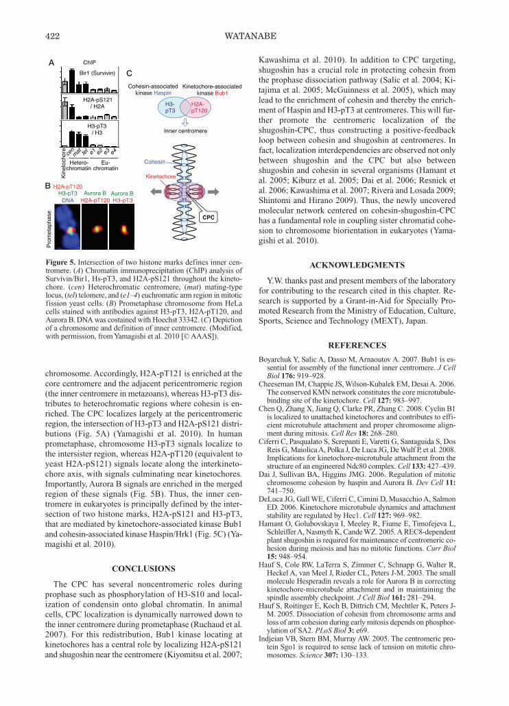
chromosome. Accordingly, H2A-pT121 is enriched at thecore centromere and the adjacent pericentromeric region(the inner centromere in metazoans), whereas H3-pT3 dis-tributes to heterochromatic regions where cohesin is en-riched. The CPC localizes largely at the pericentromericregion, the intersection of H3-pT3 and H2A-pS121 distri-butions (Fig. 5A) (Yamagishi et al. 2010). In humanprometaphase, chromosome H3-pT3 signals localize tothe intersister region, whereas H2A-pT120 (equivalent toyeast H2A-pS121) signals locate along the interkineto-chore axis, with signals culminating near kinetochores.Importantly, Aurora B signals are enriched in the mergedregion of these signals (Fig. 5B). Thus, the inner cen-tromere in eukaryotes is principally defined by the inter-section of two histone marks, H2A-pS121 and H3-pT3,that are mediated by kinetochore-associated kinase Bub1and cohesin-associated kinase Haspin/Hrk1 (Fig. 5C) (Ya-magishi et al. 2010).
CONCLUSIONS
The CPC has several noncentromeric roles duringprophase such as phosphorylation of H3-S10 and local-ization of condensin onto global chromatin. In animalcells, CPC localization is dynamically narrowed down tothe inner centromere during prometaphase (Ruchaud et al.2007). For this redistribution, Bub1 kinase locating atkinetochores has a central role by localizing H2A-pS121and shugoshin near the centromere (Kiyomitsu et al. 2007;
Kawashima et al. 2010). In addition to CPC targeting,shugoshin has a crucial role in protecting cohesin fromthe prophase dissociation pathway (Salic et al. 2004; Ki-tajima et al. 2005; McGuinness et al. 2005), which maylead to the enrichment of cohesin and thereby the enrich-ment of Haspin and H3-pT3 at centromeres. This will fur-ther promote the centromeric localization of theshugoshin-CPC, thus constructing a positive-feedbackloop between cohesin and shugoshin at centromeres. Infact, localization interdependencies are observed not onlybetween shugoshin and the CPC but also betweenshugoshin and cohesin in several organisms (Hamant etal. 2005; Kiburz et al. 2005; Dai et al. 2006; Resnick etal. 2006; Kawashima et al. 2007; Rivera and Losada 2009;Shintomi and Hirano 2009). Thus, the newly uncoveredmolecular network centered on cohesin-shugoshin-CPChas a fundamental role in coupling sister chromatid cohe-sion to chromosome biorientation in eukaryotes (Yama-gishi et al. 2010).
ACKNOWLEDGMENTS
Y.W. thanks past and present members of the laboratoryfor contributing to the research cited in this chapter. Re-search is supported by a Grant-in-Aid for Specially Pro-moted Research from the Ministry of Education, Culture,Sports, Science and Technology (MEXT), Japan.
REFERENCES
BoyarchukY, Salic A, Dasso M, Arnaoutov A. 2007. Bub1 is es-sential for assembly of the functional inner centromere. J CellBiol 176: 919–928.
Cheeseman IM, Chappie JS,Wilson-Kubalek EM, DesaiA. 2006.The conserved KMN network constitutes the core microtubule-binding site of the kinetochore. Cell 127: 983–997.
Chen Q, Zhang X, Jiang Q, Clarke PR, Zhang C. 2008. Cyclin B1is localized to unattached kinetochores and contributes to effi-cient microtubule attachment and proper chromosome align-ment during mitosis. Cell Res 18: 268–280.
Ciferri C, Pasqualato S, Screpanti E, Varetti G, Santaguida S, DosReis G, Maiolica A, Polka J, De Luca JG, DeWulf P, et al. 2008.Implications for kinetochore-microtubule attachment from thestructure of an engineered Ndc80 complex. Cell 133: 427–439.
Dai J, Sullivan BA, Higgins JMG. 2006. Regulation of mitoticchromosome cohesion by haspin and Aurora B. Dev Cell 11:741–750.
DeLuca JG, Gall WE, Ciferri C, Cimini D, Musacchio A, SalmonED. 2006. Kinetochore microtubule dynamics and attachmentstability are regulated by Hec1. Cell 127: 969–982.
Hamant O, Golubovskaya I, Meeley R, Fiume E, Timofejeva L,Schleiffer A, Nasmyth K, Cande WZ. 2005. A REC8-dependentplant shugoshin is required for maintenance of centromeric co-hesion during meiosis and has no mitotic functions. Curr Biol15: 948–954.
Hauf S, Cole RW, LaTerra S, Zimmer C, Schnapp G, Walter R,Heckel A, van Meel J, Rieder CL, Peters J-M. 2003. The smallmolecule Hesperadin reveals a role for Aurora B in correctingkinetochore-microtubule attachment and in maintaining thespindle assembly checkpoint. J Cell Biol 161: 281–294.
Hauf S, Roitinger E, Koch B, Dittrich CM, Mechtler K, Peters J-M. 2005. Dissociation of cohesin from chromosome arms andloss of arm cohesion during early mitosis depends on phosphor-ylation of SA2. PLoS Biol 3: e69.
Indjeian VB, Stern BM, Murray AW. 2005. The centromeric pro-tein Sgo1 is required to sense lack of tension on mitotic chro-mosomes. Science 307: 130–133.
422 WATANABE
k k
AC
B
Figure 5. Intersection of two histone marks defines inner cen-tromere. (A) Chromatin immunoprecipitation (ChIP) analysis ofSurvivin/Bir1, Hs-pT3, and H2A-pS121 throughout the kineto-chore. (cen) Heterochromatic centromere, (mat) mating-typelocus, (tel) telomere, and (e1–4) euchromatic arm region in mitoticfission yeast cells. (B) Prometaphase chromosome from HeLacells stained with antibodies against H3-pT3, H2A-pT120, andAurora B. DNA was costained with Hoechst 33342. (C) Depictionof a chromosome and definition of inner centromere. (Modified,with permission, from Yamagishi et al. 2010 [© AAAS]).

Jeyaprakash AA, Klein UR, Lindner D, Ebert J, Nigg EA, ContiE. 2007. Structure of a Survivin-Borealin-INCENP core com-plex reveals how chromosomal passengers travel together. Cell131: 271–285.
Kawashima SA, Tsukahara T, Langegger M, Hauf S, Kitajima TS,Watanabe Y. 2007. Shugoshin enables tension-generating at-tachment of kinetochores by loading Aurora to centromeres.Genes Dev 21: 420–435.
Kawashima SA, Yamagishi Y, Honda T, Ishiguro K, Watanabe Y.2010. Phosphorylation of H2A by Bub1 prevents chromosomalinstability through localizing shugoshin. Science 327: 172–177.
Kelly AE, Funabiki H. 2009. Correcting aberrant kinetochore mi-crotubule attachments: An Aurora B-centric view. Curr OpinCell Biol 21: 51–58.
Kelly AE, Ghenoiu C, Xue JZ, Zierhut C, Kimura H, Funabiki H.2010. Survivin reads phosphorylated histone H3 threonine 3 toactivate the mitotic kinase Aurora B. Science 330: 235–239.
Kiburz BM, Reynolds DB, Megee PC, Marston AL, Lee BH, LeeTI, Levine SS, Young RA, Amon A. 2005. The core centromereand Sgo1 establish a 50-kb cohesin-protected domain aroundcentromeres during meiosis I. Genes Dev 19: 3017–3030.
Kitajima TS, Kawashima SA, Watanabe Y. 2004. The conservedkinetochore protein shugoshin protects centromeric cohesionduring meiosis. Nature 427: 510–517.
Kitajima TS, Hauf S, Ohsugi M, Yamamoto T, Watanabe Y. 2005.Human Bub1 defines the persistent cohesion site along the mi-totic chromosome by affecting shugoshin localization. Curr Biol15: 353–359.
Kitajima TS, Sakuno T, Ishiguro K, Iemura S, Natsume T,Kawashima SA, Watanabe Y. 2006. Shugoshin collaborates withprotein phosphatase 2A to protect cohesin. Nature 441: 46–52.
Kiyomitsu T, Obuse C, Yanagida M. 2007. HumanBlinkin/AF15q14 is required for chromosome alignment andthe mitotic checkpoint through direct interaction with Bub1 andBubR1. Dev Cell 13: 663–676.
Lee JY, Hayashi-Hagihara A, Orr-Weaver TL. 2005. Roles and reg-ulation of the Drosophila centromere cohesion protein MEI-S332 family. Philos Trans R Soc Lond B Biol Sci 360: 543–552.
Liu D, Vader G, Vromans MJ, Lampson MA, Lens SM. 2009.Sensing chromosome bi-orientation by spatial separation of au-rora B kinase from kinetochore substrates. Science 323: 1350–1353.
Losada A, Hirano M, Hirano T. 2002. Cohesin release is requiredfor sister chromatid resolution, but not for condensin-mediatedcompaction, at the onset of mitosis. Genes Dev 16: 3004–3016.
Marston AL, Tham WH, Shah H, Amon A. 2004. A genome-widescreen identifies genes required for centromeric cohesion. Sci-ence 303: 1367–1370.
McGuinness BE, Hirota T, Kudo NR, Peters J-M, Nasmyth K.2005. Shugoshin prevents dissociation of cohesin from cen-tromeres during mitosis in vertebrate cells. PLoS Biol 3: e86.
Miyazaki WY, Orr-Weaver TL. 1994. Sister-chromatid cohesionin mitosis and meiosis. Annu Rev Genet 28: 167–168.
Nasmyth K, Peters J-M, Uhlmann F. 2000. Splitting the chromo-some: Cutting the ties that bind sister chromatids. Science 288:
1379–1385.Pinsky BA, Biggins S. 2005. The spindle checkpoint: Tension ver-sus attachment. Trends Cell Biol 15: 486–493.
Rabitsch KP, Gregan J, Schleiffer A, Javerzat JP, Eisenhaber F, Nas-myth K. 2004. Two fission yeast homologs of DrosophilaMei-S332 are required for chromosome segregation during meiosisI and II. Curr Biol 14: 287–301.
Resnick TD, Satinover DL, MacIsaac F, Stukenberg PT, EarnshawWC, Orr-Weaver TL, Carmena M. 2006. INCENP and AuroraB promote meiotic sister chromatid cohesion through localiza-tion of the Shugoshin MEI-S332 in Drosophila. Dev Cell 11:57–68.
Riedel CG, Katis VL, Katou Y, Mori S, Itoh T, Helmhart W, GalovaM, Petronczki M, Gregan J, Cetin B, et al. 2006. Protein phos-phatase 2A protects centromeric sister chromatid cohesion dur-ing meiosis I. Nature 441: 53–61.
Rivera T, Losada A. 2009. Shugoshin regulates cohesion by drivingrelocalization of PP2A in Xenopus extracts. Chromosoma 118:223–233.
Ruchaud S, Carmena M, Earnshaw WC. 2007. Chromosomal pas-sengers: Conducting cell division. Nat Rev Mol Cell Biol 8:798–812.
Salic A, Waters JC, Mitchison TJ. 2004. Vertebrate shugoshin linkssister centromere cohesion and kinetochore microtubule stabil-ity in mitosis. Cell 118: 567–578.
Shintomi K, Hirano T. 2009. Releasing cohesin from chromosomearms in early mitosis: Opposing actions of Wapl-Pds5 and Sgo1.Genes Dev 23: 2224–2236.
Tanaka TU. 2008. Bi-orienting chromosomes: Acrobatics on themitotic spindle. Chromosoma 117: 521–533.
Tang Z, Shu H, Qi W, Mahmood NA, Mumby MC, Yu H. 2006.PP2A is required for centromeric localization of Sgo1 andproper chromosome segregation. Dev Cell 10: 575–585.
Tanno Y, Kitajima TS, Honda T, Ando Y, Ishiguro K, Watanabe Y.2010. Phosphorylation of mammalian Sgo2 by Aurora B re-cruits PP2A and MCAK to centromeres. Genes Dev 24: 2169–2179.
Tsukahara T, Tanno Y, Watanabe Y. 2010. Phosphorylation of theCPC by Cdk1 promotes chromosome bi-orientation. Nature467: 719–723.
Vanoosthuyse V, Prykhozhij S, Hardwick KG. 2007. Shugoshin 2regulates localization of the chromosomal passenger proteinsin fission yeast mitosis. Mol Biol Cell 18: 1657–1669.
Wang F, Dai J, Daum JR, Niedzialkowska E, Banerjee B, Stuken-berg PT, Gorbsky GJ, Higgins JM. 2010. Histone H3 Thr-3phosphorylation by Haspin positions Aurora B at centromeresin mitosis. Science 330: 231–235.
Watanabe Y. 2005. Shugoshin: Guardian spirit at the centromere.Curr Opin Cell Biol 17: 590–595.
Yamagishi Y, Sakuno T, Shimura M, Watanabe Y. 2008. Hete-rochromatin links to centromeric protection by recruitingshugoshin. Nature 455: 251–255.
Yamagishi Y, Honda T, Tanno Y, Watanabe Y. 2010. Two histonemarks establish the inner centromere and chromosome bi-ori-entation. Science 330: 239–243.
REGULATION OF TARGETING AURORA B TO INNER CENTROMERE 423