temperature characterization of scintillation detectors … for radiation monitoring applications...
TRANSCRIPT
ARTICLE IN PRESS
Nuclear Instruments and Methods in Physics Research A 620 (2010) 351–358
Contents lists available at ScienceDirect
Nuclear Instruments and Methods inPhysics Research A
0168-90
doi:10.1
� Corr
E-m
journal homepage: www.elsevier.com/locate/nima
Temperature characterization of scintillation detectors using solid-statephotomultipliers for radiation monitoring applications
Clarisse Tur a,�, Vladimir Solovyev a, Jeremy Flamanc b
a Saint-Gobain Crystals, 17900 Great Lakes Parkway, Hiram, OH 44234, USAb Saint-Gobain Cristaux et Detecteurs, B. P. 521, 77794 Nemours Cedex, France
a r t i c l e i n f o
Article history:
Received 16 November 2009
Received in revised form
8 March 2010
Accepted 25 March 2010Available online 3 April 2010
Keywords:
Scintillation detector
Temperature characterization
Silicon photomultiplier
SPM
SiPM
Multi-pixel photon counter
MPPC
Solid-state photomultiplier
SSPM
Geiger-mode avalanche photodiode array
G-APD array
02/$ - see front matter & 2010 Elsevier B.V. A
016/j.nima.2010.03.141
esponding author. Tel.: +1 440 834 5706; fax
ail address: [email protected] (C.
a b s t r a c t
We have characterized two state-of-the-art solid-state photomultipliers, one by SensL, the other by
Hamamatsu, coupled to scintillators by Saint-Gobain Crystals in the �25 to +50 1C temperature range.
At room temperature, the energy resolution at 661.6 keV measured with both detectors is worse than
the resolution obtained when the crystals are coupled to a regular photomultiplier tube. Both the pulse
height and pulse height resolution of the 661.6 keV gamma rays in the 137Cs spectrum vary strongly
with temperature. The noise threshold determined from the 22Na spectrum increases quadratically as
the temperature is increased to well above 100 keV at +50 1C for both detectors.
& 2010 Elsevier B.V. All rights reserved.
1. Introduction
In recent years, solid-state photomultipliers (SSPMs) haveemerged as a promising light sensor for compact, low-bias scintilla-tion detectors. They have a gain comparable to that of the typicalphotomultiplier tube (PMT) while offering a number of keyadvantages compared to the latter such as compactness, operationat low bias voltage, robustness, insensitivity to magnetic fields, and insome cases, fast timing properties. In addition, operation underambient light, while not recommended for optimum noise levels, willnot cause permanent damage to the SSPM. Sizes are increasing assmaller individual sensors are tiled together.
The SSPM is an array of a large number of small pixels (typically afew tens of mm), each consisting of a Geiger-mode avalanchephotodiode coupled to a quenching resistor. When used in ascintillation detector, each pixel of the SSPM will emit the samesaturated signal if hit by an optical photon when operated at a reversebias above the breakdown voltage (Vbr) signaling the presence of thephoton. The Geiger discharge responsible for the signal is stopped
ll rights reserved.
: +1 440 834 7683.
Tur).
when the voltage is brought back to below Vbr by the quenchingresistor. Thus each pixel is operated in a binary, on/off, mode toindicate the presence or absence of a photon. The SSPM is thereforefundamentally a photon counting device where the number of ‘‘on’’pixels is proportional to the energy deposited in the scintillator, solong as the number of pixels excited is small compared to the total. Inother words, the number of pixels available is the limiting factor forthe dynamic linear range of the SSPM.
Much progress has been made on the SSPM technology frontsince the first publications on the topic [1–5] and a number ofpapers have followed since on its various possible applicationsranging from the general subject of radiation detection [6–13] tomore specific applications such as medical imaging [14–22], basicresearch in physics [23–25], low-intensity light biosensors [26],and laser radar systems [27,28].
In the present paper, we characterize two silicon-based SSPMs,one by SensL [29], the other by Hamamatsu [30], coupled toscintillators by Saint-Gobain Crystals [31], CsI(Tl) for the former andLaBr3:Ce for the latter. We will use Hamamatsu’s trademarkdesignation of MPPC (Multi-Pixel Photon Counter) hereafter when-ever we refer to the Hamamatsu sensor. At the time of the tests, theSensL SSPM offered the largest area of a single unit detector, while theHamamatsu MPPC offered the highest density of Geiger cells among
ARTICLE IN PRESS
C. Tur et al. / Nuclear Instruments and Methods in Physics Research A 620 (2010) 351–358352
the commercially available sensors of this type. Beyond the basicperformance, our main interest is temperature dependence of the twoscintillation detectors within the �25 to +50 1C range. To ourknowledge, such a complete temperature analysis of an SSPM-basedscintillation system has not been performed prior to this study. Moreprecisely, we quantified the variations within the afore-mentionedtemperature range of the noise threshold using a 22Na source, and ofthe pulse height (PH) and pulse height resolution (PHR) of the661.6 keV peak in 137Cs.
In Section 2, we describe our scintillation detectors andexperimental setups. We report the results of our characterizationin Section 3, and discuss those results in Section 4.
2. Experimental setup
2.1. The SensL SSPM setup
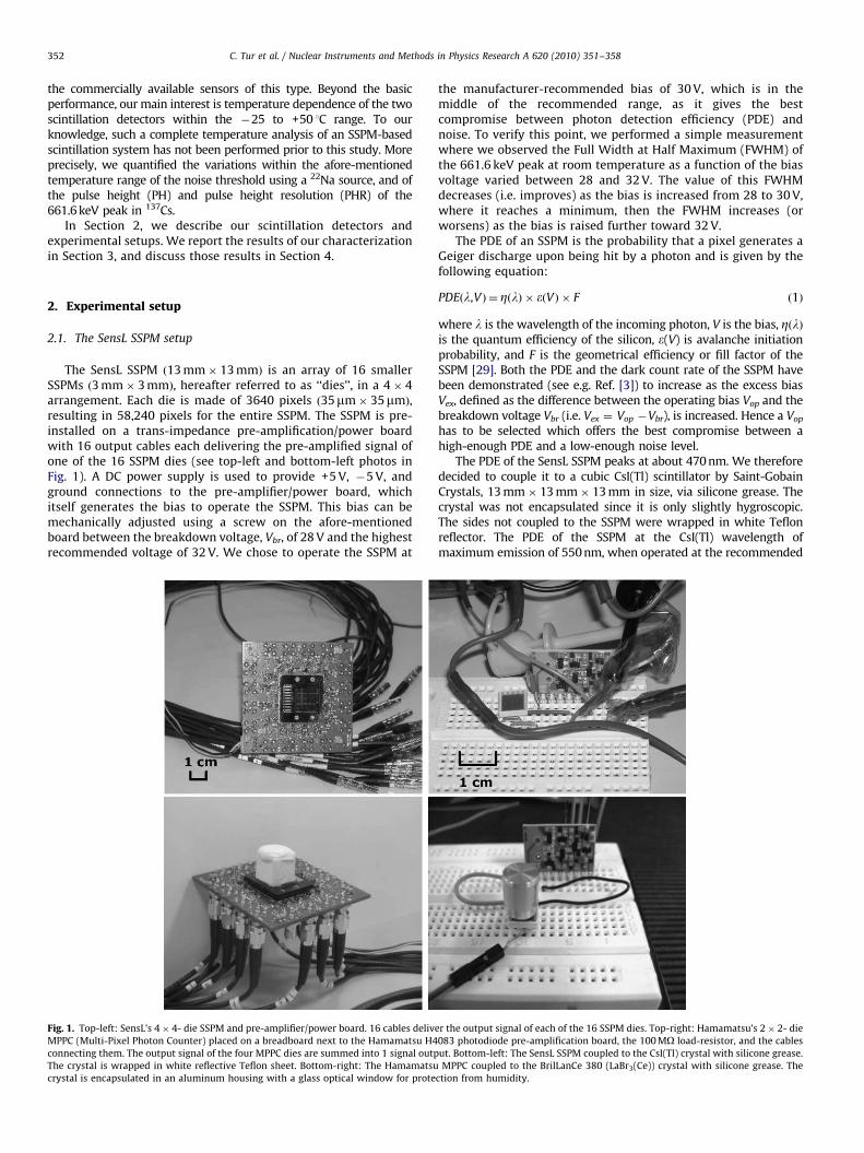
The SensL SSPM ð13 mm� 13 mmÞ is an array of 16 smallerSSPMs ð3 mm� 3 mmÞ, hereafter referred to as ‘‘dies’’, in a 4� 4arrangement. Each die is made of 3640 pixels ð35mm� 35mmÞ,resulting in 58,240 pixels for the entire SSPM. The SSPM is pre-installed on a trans-impedance pre-amplification/power boardwith 16 output cables each delivering the pre-amplified signal ofone of the 16 SSPM dies (see top-left and bottom-left photos inFig. 1). A DC power supply is used to provide +5 V, �5 V, andground connections to the pre-amplifier/power board, whichitself generates the bias to operate the SSPM. This bias can bemechanically adjusted using a screw on the afore-mentionedboard between the breakdown voltage, Vbr, of 28 V and the highestrecommended voltage of 32 V. We chose to operate the SSPM at
Fig. 1. Top-left: SensL’s 4� 4- die SSPM and pre-amplifier/power board. 16 cables deliv
MPPC (Multi-Pixel Photon Counter) placed on a breadboard next to the Hamamatsu H4
connecting them. The output signal of the four MPPC dies are summed into 1 signal outp
The crystal is wrapped in white reflective Teflon sheet. Bottom-right: The Hamamatsu
crystal is encapsulated in an aluminum housing with a glass optical window for prote
the manufacturer-recommended bias of 30 V, which is in themiddle of the recommended range, as it gives the bestcompromise between photon detection efficiency (PDE) andnoise. To verify this point, we performed a simple measurementwhere we observed the Full Width at Half Maximum (FWHM) ofthe 661.6 keV peak at room temperature as a function of the biasvoltage varied between 28 and 32 V. The value of this FWHMdecreases (i.e. improves) as the bias is increased from 28 to 30 V,where it reaches a minimum, then the FWHM increases (orworsens) as the bias is raised further toward 32 V.
The PDE of an SSPM is the probability that a pixel generates aGeiger discharge upon being hit by a photon and is given by thefollowing equation:
PDEðl,VÞ ¼ ZðlÞ � eðVÞ � F ð1Þ
where l is the wavelength of the incoming photon, V is the bias, ZðlÞis the quantum efficiency of the silicon, e(V) is avalanche initiationprobability, and F is the geometrical efficiency or fill factor of theSSPM [29]. Both the PDE and the dark count rate of the SSPM havebeen demonstrated (see e.g. Ref. [3]) to increase as the excess biasVex, defined as the difference between the operating bias Vop and thebreakdown voltage Vbr (i.e. Vex ¼ Vop �Vbr), is increased. Hence a Vop
has to be selected which offers the best compromise between ahigh-enough PDE and a low-enough noise level.
The PDE of the SensL SSPM peaks at about 470 nm. We thereforedecided to couple it to a cubic CsI(Tl) scintillator by Saint-GobainCrystals, 13 mm� 13 mm� 13 mm in size, via silicone grease. Thecrystal was not encapsulated since it is only slightly hygroscopic.The sides not coupled to the SSPM were wrapped in white Teflonreflector. The PDE of the SSPM at the CsI(Tl) wavelength ofmaximum emission of 550 nm, when operated at the recommended
er the output signal of each of the 16 SSPM dies. Top-right: Hamamatsu’s 2� 2- die
083 photodiode pre-amplification board, the 100 MO load-resistor, and the cables
ut. Bottom-left: The SensL SSPM coupled to the CsI(Tl) crystal with silicone grease.
MPPC coupled to the BrilLanCe 380 (LaBr3(Ce)) crystal with silicone grease. The
ction from humidity.
ARTICLE IN PRESS
C. Tur et al. / Nuclear Instruments and Methods in Physics Research A 620 (2010) 351–358 353
30 V bias at room temperature, is about 8% according to SensL. Aradioactive source was positioned on top of the pre-amplifier/powerboard, which was placed in a black plastic box to ensure light-tightness. The black box itself was placed in a temperature testchamber (by Thermotron Industries) in which the temperature canbe varied within the �70 to +180 1C range. The moisture level in thetemperature test chamber was not monitored or controlled. The DCpower supplies and the readout electronics were located outside thetemperature test chamber.
The 16 output signals from the SensL pre-amplifier board wereadded using 2 LeCroy 428F linear fan-in-fan-out modules. Thissummed output was amplified by a Canberra 2020 spectroscopyamplifier (with a 3ms shaping time, since this setting gave thebest PHR at 661.6 keV at room temperature) before beingdigitized by a Canberra 8075 analog-to-digital converter (ADC)module. The resulting output was analyzed and saved by a multi-channel analyzer (MCA).
2.2. The Hamamatsu MPPC setup
The Hamamatsu MPPC ð6 mm� 6 mmÞ is an array of 4 diesð3 mm� 3 mmÞ in a 2� 2 arrangement. Each die consists of14,400 pixels ð25mm� 25mmÞ without any dead space betweenthe 4 dies, resulting in 57,600 pixels for the entire MPPC. Therecommended Vop is about 70 V; we chose to bias the MPPC at65 V (for reasons which will become clear in the next section)through a load resistor of 100 MO.
The MPPC and its load resistor were both set on a breadboard,on which the four signals from the MPPC dies were summed intoone output which was pre-amplified using a Hamamatsu H4083photodiode charge-sensitive pre-amplifier board, also installed onthe breadboard. This arrangement is shown on the top-right andbottom-right photos of Fig.1.
The MPPC is blue-enhanced with a PDE which peaks at about420 nm. We therefore chose to couple it to an encapsulatedcylindrical (6 mm diameter � 6 mm long) Saint-Gobain Crystals’BrilLanCe 380 (LaBr3:Ce; abbreviated B380 in the figures)scintillator using silicone grease. The PDE of the MPPC at theBrilLanCe 380 wavelength of maximum emission of 380 nm, whenoperated at the recommended 70 V at 25 1C, is about 23%according to Hamamatsu. The hygroscopic crystal was hermeti-cally sealed in an aluminum enclosure with a 5 mm thick opticalexit made of glass. We placed a radioactive source (e.g. 137Cs or22Na) directly on top of the breadboard next to our scintillationdetector. The entire setup on the breadboard was placed in analuminum can to ensure isolation from external noise and light-tightness. All external voltages were supplied through thisaluminum can: we used one DC power supply to provide +12 V,�12 V, and ground connections to the pre-amplifier board andanother DC power supply (Tennelec TC 954) to provide the 65 Vbias to the MPPC. The aluminum can itself was placed in thetemperature test chamber specified in the previous subsection.
The pre-amplified signal of the summed MPPC output wasfurther amplified by a Canberra 2020 spectroscopy amplifier(with a 250 ns shaping time, the smallest setting available on ouramplifier module) and then digitized by a Canberra 8075 ADCmodule. The resulting output was analyzed and saved by an MCA.
3. Results
3.1. Description of the measurements
We studied the variations with temperature of the PH and PHRof the 661.6 keV peak in the 137Cs spectrum within the �25 to
+50 1C range. Since the variations in PH were quite significantwithin this range, we broke it up into three sub-ranges: �25 to0 1C, 0 to +25 1C, and +25 to +50 1C. The amplifier gain setting wasdifferent for each of those three ranges.
For each range, we began by soaking the detector at thehighest temperature in that range for about 1 h and then adjustingthe amplifier gain so that the 661.6 keV peak was at the lowestend of the MCA display, since the PH is expected to go up as thetemperature is decreased. We then programmed the Thermotrontemperature test chamber for a 28 h duration cycle: the cyclebegins with a 2 h soak at the highest temperature in the range;then the temperature is decreased by 25 1C in 5 h (at the rate of5 1C per hour); this is followed by another 2 h soak at the newlyreached temperature, the lowest in the cycle; then the tempera-ture is ramped up again by 25 1C in 5 h, at which point the highesttemperature in the range is reached again. This 14 h cycle isrepeated one more time to complete the 28 h cycle for thatparticular range.
As the Thermotron chamber was cycling, data for a 137Csspectrum were automatically accumulated for 60 s by the MCAand saved. As soon as one spectrum was saved, the next wasstarted, with virtually no time gap between them. The tempera-ture change within the 60 s acquisition time is negligibly small:about 0.08 1C. A record of the temperature was also made at thestart of each acquisition period. More than 1600 137Cs spectrawere collected per temperature range and detector and thenanalyzed offline for PH and PHR.
An energy linearity study was then made for each scintillationdetector using a 137Cs, a 22Na, and a 60Co source. Once thelinearity was confirmed up to 1332 keV, a noise-threshold studywas performed using the 22Na source, utilizing its 511 and1274.5 keV lines for calibration. For these measurements, theThermotron chamber was set to soak at the highest temperature(50 1C) for 1 h; 50 min into the soak, a spectrum was acquired fortypically 300 s which clearly showed the noise threshold and thetwo 22Na lines. Then the temperature was decreased (by typically10 1C) in 15 min. The same procedure was repeated for a spectrumat this new temperature.
3.2. Results for the CsI(Tl)/SensL SSPM scintillation detector
3.2.1. Temperature dependence of the pulse height and pulse height
resolution of the 661.6 keV peak in 137Cs
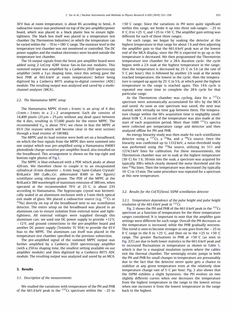
Fig. 2 shows the PH and PHR of the 661.6 keV peak in the 137Csspectrum as a function of temperature for the three temperatureranges considered. It is important to note that the amplifier gainsettings were different for each range. Overall the PH decreases asthe temperature is increased, while the PHR gradually worsens.This trend is seen to become stronger as one goes from the �25 to0 1C range to the 0 to +25 1C, and then on to the +25 to +50 1Crange. The greater fluctuations in PHR at +50 1C (as seen inFig. 2(f)) are due to both lower statistics in the 661.6 keV peak andto increased fluctuations in temperature as shown in Table 1,which is due to a marginal insulation system where the cablesexit the thermal chamber. The seemingly erratic jumps in boththe PH and PHR for small changes in temperature are presumablydue to the fact that the detector never quite gets a chance tostabilize at any given temperature even at the relatively slowtemperature change rate of 5 1C per hour. Fig. 2 also shows thatthe SSPM exhibits a slight hysteresis: the PH evolves on twoslightly different curves when one decreases the temperaturefrom the highest temperature in the range to the lowest versuswhen one increases it from the lowest temperature in the rangeback to the highest.
ARTICLE IN PRESS
Fig. 2. Temperature dependence, for the SensL SSPM + CsI(Tl) scintillation detector, of (a) the PH from �25 to 0 1C, (b) the PHR from �25 to 0 1C, (c) the PH from 0 to 25 1C,
(d) the PHR from 0 to 25 1C, (e) the PH from 25 to 50 1C, and (f) the PHR from 25 to 50 1C of the 661.6 keV peak in the 137Cs spectrum. The amplifier gain settings are different
for each of the three temperature ranges.
Table 1Standard deviation (denoted by s) and mean (denoted by m) of the measured
temperature during soaks at constant temperature for four temperature settings
and the same statistics for the 661.6 keV PHR as measured by the SensL SSPM +
CsI(Tl) scintillation detector at those four temperatures.
Temperature m (temperature) s (temperature) m (PHR) s (PHR)
�25 �24.67 0.40 11.03 0.23
0 0.18 0.08 12.36 0.29
25 24.99 0.09 16.84 1.19
50 49.31 0.33 32.15 2.57
The statistics on the temperature are in 3C and those on the PHR in %.
C. Tur et al. / Nuclear Instruments and Methods in Physics Research A 620 (2010) 351–358354
In Fig. 3, the PH of the 661.6 keV peak has been plotted againstthe temperature in arbitrary units in the entire �25 to +50 1Crange, when the contribution of CsI(Tl) has been subtracted outand the change in the amplifier gain between the three
temperature ranges has been compensated for. A normalizationhas also been performed so that the PH at 25 1C is 1. The PHattributable to the SSPM is seen to vary by a factor of 16.1between �25 and +50 1C.
An explanation about how we subtracted out the crystalcontribution is in order here: the CsI(Tl) contribution is a numberbetween 0 and 1 and was obtained from data available in theSaint-Gobain Crystals’ CsI(Tl) data sheet (see Ref. [31]). The dataconsist of a curve showing the crystal’s light output for 661.6 keVgamma rays as a function of temperature, normalized to thecrystal’s light output at 25 1C. We derived a functional form forthis curve by a least squares fit. We used this functional form tocalculate the crystal contribution for each temperature. We thensimply divided the full detector response at a given temperatureby the crystal contribution at that temperature.
The best PHR at 661.6 keV which we observed at roomtemperature was about 12% FWHM. When we coupled the same

ARTICLE IN PRESS
C. Tur et al. / Nuclear Instruments and Methods in Physics Research A 620 (2010) 351–358 355
CsI(Tl) crystal to a Hamamatsu R1306-01 PMT (using siliconegrease), the PHR at 661.6 keV was 7.9%.
3.2.2. Linearity and temperature dependence of the noise threshold
We checked the linearity of the detector using the 661.6 keVline in 137Cs, the 511 and 1274.5 keV lines in 22Na, and the 1173and 1332 keV lines in 60Co. The results are illustrated in Fig. 4(a)and show that the detector exhibits good linearity up to 1332 keV.
Once the linearity was checked, we performed a study of thenoise threshold in the 22Na spectrum as a function of tempera-ture, using its 511 and 1274.5 keV lines for calibration. Thethreshold is determined by the intersection of a linear fit to thesteep fall-off of the noise edge and a linear fit to the relatively flatCompton continuum in the low-energy region. Fig. 4(b) showsthat this threshold quadratically increases from essentially 0 keVat �25 1C to about 362 keV at +50 1C. It is worth noting thesignificant deterioration in performance in terms of noise levelsnear +50 1C.
Fig. 3. PH (in arbitrary units) of the 661.6 keV 137Cs peak acquired with the SensL
SSPM + CsI(Tl) scintillation detector as a function of the temperature when the
contribution of CsI(Tl) has been subtracted out and the change in the amplifier
gain between the three temperature ranges has been compensated for. A
normalization has also been performed so that the PH at 25 1C is 1.
Fig. 4. For the SensL SSPM + CsI(Tl) scintillation detector: (a) linearity check using lines
points, and (b) noise threshold versus temperature obtained using a 22Na source, wher
3.3. Results for the BrilLanCe 380/Hamamatsu MPPC scintillation
detector
3.3.1. Temperature dependence of the pulse height and pulse height
resolution of the 661.6 keV peak in 137Cs
Fig. 5 shows the variations in the PH and PHR of the 661.6 keVpeak in the 137Cs spectrum as a function of temperature for thethree temperature ranges studied. The PH decreases as thetemperature is increased, as expected. The PHR, however, hasthe expected behavior only in the 0 to +50 1C range, as it worsenswhen the temperature is increased, while in the �25 to 0 1Crange, the PHR improves as the temperature is increased, theopposite of what is expected and observed for the SensL SSPM. Itis useful to note here a difference in the room temperaturebehavior of the SensL and Hamamatsu sensors which weobserved: while the PHR of 661.6 keV peak as a function of biasvoltage at room temperature as measured by the SensL SSPM-based scintillation detector is mostly constant over much of therecommended bias range (i.e. within 29 and 32 V), the behavior ofHamamatsu MPPC-based detector appears to be morecomplicated as illustrated in Fig. 6. As the temperature ischanged, Vbr changes. Since Vop is held constant, this results inchanges in the excess voltage Vex, which explains much of thechanges in both the PH and PHR observed. This situation is similarto one where Vbr is held constant while Vop is changed in theopposite direction. Otherwise stated, changing the temperature issomewhat equivalent to going up or down the curve in Fig. 6.Presumably, the more complicated shape of this curve isresponsible for the difference in the PHR behavior observed forthe Hamamatsu MPPC-based detector compared the one using theSensL SSPM. The hysteresis, which we observed for the SensLSSPM, is manifest even more clearly in the PH and PHR results forthe Hamamatsu SSPM as seen in Fig. 5. This phenomenon seemsto be an intrinsic feature of these sensors.
We also chose to operate the MPPC at a bias of 65 V based onFig. 6. A bias of about 66.5 V gives the best PHR at roomtemperature. However, since moving the temperature is equiva-lent to moving along the PHR versus bias curve, we chose to avoidgetting too close to any of the extremities of this curve as wechange the temperature, at the expense of a somewhat worseresolution at room temperature.
The large fluctuations in temperature at both �25 and +50 1Care clearly visible in Fig. 5 and Table 2. They are bigger in
in the 137Cs, 22Na, and 60Co spectra, the thick black line being a linear fit to the data
e the thick black curve is the result of a quadratic fit to the data.

ARTICLE IN PRESS
Fig. 5. Temperature dependence, for the Hamamatsu MPPC + BrilLanCe 380 scintillation detector, of (a) the PH from �25 to 0 1C, (b) the PHR from �25 to 0 1C, (c) the PH
from 0 to 25 1C, (d) the PHR from 0 to 25 1C, (e) the PH from 25 to 50 1C, and (f) the PHR from 25 to 50 1C of the 661.6 keV peak in the 137Cs spectrum. The amplifier gain
settings are different for each of the three temperature ranges.
C. Tur et al. / Nuclear Instruments and Methods in Physics Research A 620 (2010) 351–358356
magnitude than for the SensL setup since we used one particularlythick cable for the Hamamatsu setup (the one used to supply+12 V, �12 V, and the ground connections to the pre-amplifierboard through the aluminum can). The use of this thick cablemeant a larger hole through the foam-stuffed exit window of thethermal chamber to reach the voltage supply located outside ofthe chamber. Hence it was more difficult to achieve good thermalinsulation, especially at the extreme temperatures of our range.
Fig. 7 shows the PH of the 661.6 keV peak versus the temperaturein arbitrary units in the entire �25 to +50 1C range, when thecontribution of BrilLanCe 380 has been subtracted out and thechange in the amplifier gain between the three temperature rangeshas been compensated for. A normalization has also been performedso that the PH at 25 1C is 1. The BrilLanCe 380 contribution wasobtained from data available in the Saint-Gobain Crystals’ BrilLanCe380 data sheet (see Ref. [31]). The PH varies by a factor of 11.2between �25 and +50 1C.
The best PHR at 661.6 keV which we observed at roomtemperature was 6.9% (at a bias setting of 66.5 V). When wecoupled the same BrilLanCe 380 crystal to a Hamamatsu R1306-01 PMT (with silicone grease), however, the PHR at 661.6 keV was3.5%.
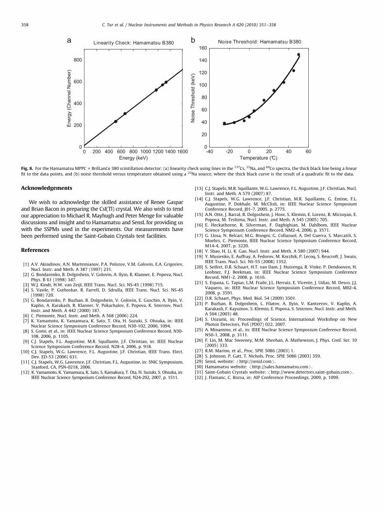
3.3.2. Linearity and temperature dependence of the noise threshold
We used the 661.6 keV line in 137Cs, the 511 and 1274.5 keVlines in 22Na, and the 1173 and 1332 keV lines in 60Co to check thelinearity of the scintillation detector. The results are illustrated inFig. 8(a) and show that the detector exhibits good linearity up to1332 keV.
Once the linearity checked, we performed a study of the noisethreshold in the 22Na spectrum as a function of temperature,using its 511 and 1274.5 keV lines for calibration. Fig. 8(b) showsthat this threshold quadratically increases from 39 keV at �25 1C

ARTICLE IN PRESS
Fig. 6. PHR, for the Hamamatsu MPPC + BrilLanCe 380 scintillation detector, of the
661.6 keV peak in the 137Cs spectrum as a function of bias at room temperature.
The thick black curve is the result of a 4th-order polynomial fit to the data points.
Table 2Standard deviation (denoted by s) and mean (denoted by m) of the measured
temperature during soaks at constant temperature for four temperature settings
and the same statistics for the 661.6 keV PHR as measured by the Hamamatsu
MPPC + BrilLanCe 380 scintillation detector at those four temperatures.
Temperature m (temperature) s (temperature) m (PHR) s (PHR)
�25 �24.42 0.46 13.64 0.68
0 1.13 0.25 7.73 0.25
25 26.14 0.24 9.34 0.31
50 50.75 0.28 13.18 0.56
The statistics on the temperature are in 3C and those on the PHR in %.
Fig. 7. PH (in arbitrary units) of the 661.6 keV 137Cs peak acquired with the
Hamamatsu MPPC + BrilLanCe 380 scintillation detector as a function of the
temperature when the contribution of the BrilLanCe 380 crystal has been
subtracted out and the change in the amplifier gain between the three
temperature ranges has been compensated for. A normalization has also been
performed so that the PH at 25 1C is 1.
C. Tur et al. / Nuclear Instruments and Methods in Physics Research A 620 (2010) 351–358 357
to about 150 keV at +50 1C. The noise level is worse than for SensLat �25 1C, but much better at +50 1C. The fact that the noisethreshold at �25 1C is not zero as it was for the SensL SSPM maybe due to the fact that our aluminum can provides worse isolationfrom external noise than the SensL pre-amplifier/power board.
4. Discussion
Our measurements show a very strong dependence of the PH of661.6 keV gamma rays on the temperature due to drifts in Vbr, andhence in the SSPM gain via Vex. When the contribution of the crystal issubtracted out, the PH varies by a factor of 16.1 and 11.2 for the SensLand Hamamatsu SSPM-based detectors, respectively, when thetemperature is raised from �25 to +50 1C. The slope, at +25 1C, ofthe curves showing the PH at 661.6 keV as a function of temperatureis typically a few percent per degree Celcius, which is about the sameas what has been reported for an avalanche photodiode [32]. PMTsalso have a gain that varies with temperature, but to a lesser degreefor typical bi-alkali PMTs (o0:1% per degree Celcius; see Ref. [30]).As with PMT systems, but to a greater degree, an SSPM used in anapplication where the temperature will vary, would require a circuitto monitor temperature or gain, and feedback to readjust Vop to holdVex and the overall gain constant.
Gain stabilization will not affect noise edge: the noise thresh-old for the SensL SSPM-based detector is 362 keV at +50 1C, whilefor the Hamamatsu MPPC-based detector, it is 150 keV at thesame temperature. A cooling system, for example based on aPeltier module, could be used to maintain temperature andperformance roughly constant with some increase in overall sizeand power consumption.
Finally, both SSPMs tested underperformed in terms of PHR atroom temperature compared to a regular PMT; the best PHR weobserved for our CsI(Tl) crystal using the SensL SSPM at roomtemperature was about 12%, while we were able to achieve 7.9%with a Hamamatsu R1306-01 PMT coupled to the same crystalusing silicone grease. The best room temperature PHR which weobserved for the BrilLanCe 380 crystal using the HamamatsuMPPC was 6.9%, while the same crystal, when coupled withsilicone grease to a Hamamatsu R1306-01 PMT, yielded 3.5%.
5. Conclusion
We have studied the variations in the PH and PHR of the661.6 keV gamma rays in 137Cs as a function of temperaturewithin the �25 to +50 1C range using two state-of-the-art SSPM-based scintillation detectors: one where a 13 mm� 13 mm, 16-die SSPM by SensL was coupled to a 13 mm� 13 mm� 13 mmCsI(Tl) crystal by Saint-Gobain Crystals; one where a6 mm� 6 mm, 4-die MPPC by Hamamatsu was coupled to a6 mm (diameter) � 6mm (length) BrilLanCe 380 (LaBr3:Ce) crystalalso by Saint-Gobain Crystals. We also measured the noisethreshold in the 22Na spectrum for both detectors as a functionof the temperature within the same range and checked theirlinearity at room temperature.
Both detectors exhibited good linearity up to 1332 keV. Thereis a strong dependence of the PH and PHR on the temperature:while the PH for 661.6 keV gamma rays varied by a factor of 16.1in the temperature range considered using the SensL SSPM-baseddetector, it varied by a factor of 11.2 for the Hamamatsu MPPC-based detector in the same range. PHR also increased withtemperature for both devices, but a direct comparison is notpossible because of the difference in their design as well as thefact they were tested with different crystals. At +50 1C, the SensLdevice with CsI(Tl) gave 32.1% FWHM while the best roomtemperature value observed was about 12%. At +50 1C, theHamamatsu sensor registered 13.2% with LaBr3:Ce and at best6.9% at room temperature. The same two crystals measured witha Hamamatsu R1306-01 PMT showed 7.9% (CsI(Tl)) and 3.5%(LaBr3:Ce) at room temperature. Noise edges rise with tempera-ture reaching 362 and 150 keV at +50 1C for the SensL andHamamatsu SSPM-based detectors, respectively.
ARTICLE IN PRESS
Fig. 8. For the Hamamatsu MPPC + BrilLanCe 380 scintillation detector: (a) linearity check using lines in the 137Cs, 22Na, and 60Co spectra, the thick black line being a linear
fit to the data points, and (b) noise threshold versus temperature obtained using a 22Na source, where the thick black curve is the result of a quadratic fit to the data.
C. Tur et al. / Nuclear Instruments and Methods in Physics Research A 620 (2010) 351–358358
Acknowledgements
We wish to acknowledge the skilled assistance of Renee Gasparand Brian Bacon in preparing the CsI(Tl) crystal. We also wish to tendour appreciation to Michael R. Mayhugh and Peter Menge for valuablediscussions and insight and to Hamamatsu and SensL for providing uswith the SSPMs used in the experiments. Our measurements havebeen performed using the Saint-Gobain Crystals test facilities.
References
[1] A.V. Akindinov, A.N. Martemianov, P.A. Polozov, V.M. Golovin, E.A. Grigoriev,Nucl. Instr. and Meth. A 387 (1997) 231.
[2] G. Bondarenko, B. Dolgoshein, V. Golovin, A. Ilyin, R. Klanner, E. Popova, Nucl.Phys. B 61 (1998) 347.
[3] W.J. Kindt, H.W. van Zeijl, IEEE Trans. Nucl. Sci. NS-45 (1998) 715.[4] S. Vasile, P. Gothoskar, R. Farrell, D. Sdrulla, IEEE Trans. Nucl. Sci. NS-45
(1998) 720.[5] G. Bondarenko, P. Buzhan, B. Dolgoshein, V. Golovin, E. Guschin, A. Ilyin, V.
Kaplin, A. Karakash, R. Klanner, V. Pokachalov, E. Popova, K. Smirnov, Nucl.Instr. and Meth. A 442 (2000) 187.
[6] C. Piemonte, Nucl. Instr. and Meth. A 568 (2006) 224.[7] K. Yamamoto, K. Yamamura, K. Sato, T. Ota, H. Suzuki, S. Ohsuka, in: IEEE
Nuclear Science Symposium Conference Record, N30-102, 2006, 1094.[8] S. Gomi, et al., in: IEEE Nuclear Science Symposium Conference Record, N30-
108, 2006, p. 1105.[9] C.J. Stapels, F.L. Augustine, M.R. Squillante, J.F. Christian, in: IEEE Nuclear
Science Symposium Conference Record, N28-4, 2006, p. 918.[10] C.J. Stapels, W.G. Lawrence, F.L. Augustine, J.F. Christian, IEEE Trans. Elect.
Dev. ED-53 (2006) 631.[11] C.J. Stapels, W.G. Lawrence, J.F. Christian, F.L. Augustine, in: SNIC Symposium,
Stanford, CA, PSN-0218, 2006.[12] K. Yamamoto, K. Yamamura, K. Sato, S. Kamakura, T. Ota, H. Suzuki, S. Ohsuka, in:
IEEE Nuclear Science Symposium Conference Record, N24-292, 2007, p. 1511.
[13] C.J. Stapels, M.R. Squillante, W.G. Lawrence, F.L. Augustine, J.F. Christian, Nucl.Instr. and Meth. A 579 (2007) 87.
[14] C.J. Stapels, W.G. Lawrence, J.F. Christian, M.R. Squillante, G. Entine, F.L.Augustine, P. Dokhale, M. McClish, in: IEEE Nuclear Science SymposiumConference Record, J01-7, 2005, p. 2775.
[15] A.N. Otte, J. Barral, B. Dolgoshein, J. Hose, S. Klemin, E. Lorenz, R. Mirzoyan, E.Popova, M. Teshima, Nucl. Instr. and Meth. A 545 (2005) 705.
[16] E. Heckathorne, R. Silverman, F. Daghighian, M. Dahlbom, IEEE NuclearScience Symposium Conference Record, NM2-4, 2006, p. 3571.
[17] G. Llosa, N. Belcari, M.G. Bisogni, G. Collazuol, A. Del Guerra, S. Marcatili, S.Moehrs, C. Piemonte, IEEE Nuclear Science Symposium Conference Record,M14-4, 2007, p. 3220.
[18] Y. Shao, H. Li, K. Gao, Nucl. Instr. and Meth. A 580 (2007) 944.[19] Y. Musienko, E. Auffray, A. Fedorov, M. Korzhik, P. Lecoq, S. Reucroft, J. Swain,
IEEE Trans. Nucl. Sci. NS-55 (2008) 1352.[20] S. Seifert, D.R. Schaart, H.T. van Dam, J. Huizenga, R. Vinke, P. Dendooven, H.
Leohner, F.J. Beekman, in: IEEE Nuclear Science Symposium ConferenceRecord, NM1-2, 2008, p. 1616.
[21] S. Espana, G. Tapias, L.M. Fraile, J.L. Herraiz, E. Vicente, J. Udias, M. Desco, J.J.Vaquero, in: IEEE Nuclear Science Symposium Conference Record, M02-4,2008, p. 3591.
[22] D.R. Schaart, Phys. Med. Biol. 54 (2009) 3501.[23] P. Buzhan, B. Dolgoshein, L. Filatov, A. Ilyin, V. Kantzerov, V. Kaplin, A.
Karakash, F. Kayumov, S. Klemin, E. Popova, S. Smirnov, Nucl. Instr. and Meth.A 504 (2003) 48.
[24] S. Uozumi, in: Proceedings of Science, International Workshop on NewPhoton Detectors, PoS (PD07) 022, 2007.
[25] A. Minamino, et al., in: IEEE Nuclear Science Symposium Conference Record,N50-1, 2008, p. 3111.
[26] F. Lin, M. Mac Sweeney, M.M. Sheehan, A. Mathewson, J. Phys. Conf. Ser. 10(2005) 333.
[27] R.M. Marino, et al., Proc. SPIE 5086 (2003) 1.[28] S. Johnson, P. Gatt, T. Nichols, Proc. SPIE 5086 (2003) 359.[29] SensL website: /http://sensl.comS.[30] Hamamatsu website: /http://sales.hamamatsu.comS.[31] Saint-Gobain Crystals website: /http://www.detectors.saint-gobain.comS.[32] J. Flamanc, C. Rozsa, in: AIP Conference Proceedings, 2009, p. 1099.