telaviv university
TRANSCRIPT

TELAVIV UNIVERSITY
th4 SSNN South East European
5>10/July/Eforie Nord/Romania

Dafin F. Mureşanu
Natan M. Bornstein
Director of Stroke Unit, Department of Neurology, Tel Aviv Medical Center, Tel Aviv University, IsraelVice President of the World Stroke Organization (WSO)
Chairman Department of Neurology, University of Medicine and Pharmacy “Iuliu Hatieganu”, Cluj-Napoca, RomaniaSecretary General of the Society for the Study of Neuroprotection and Neuroplasticity (SSNN)
PROGRAM COORDINATORS
5>10/July/Eforie Nord/Romaniath4 SSNN South East European

ORGANIZERS
CO-ORGANIZERS
02
5>10/July/Eforie Nord/Romaniath4 SSNN South East European
World Federation of Neurology

FACULTY/in alphabetical order
Raul Arizaga /
Ovidiu Băjenaru
Natan Bornstein /
László Csiba /
Anna Członkowska /
Antonio Federico /
Amos Korczyn /
Maximilian Mehdorn /
Dafin F. Mureşanu /
Bogdan O. Popescu /
C. D. Popescu /
Luiza Spiru /
Jean-Luc Truelle /
Arne Voss /
Klaus von Wild /
Argentina
/ Romania
Israel
Hungary
Poland
Italy
Israel
Germany
Romania
Romania
Romania
Romania
France
Germany
Germany
03
5>10/July/Eforie Nord/Romaniath4 SSNN South East European

5>10/July/Eforie Nord/Romaniath4 SSNN South East European
04
GENERAL INFORMATION
ANA Hotels – Eforie Nord - Europa and Astoria Hotels
Phone: 0040241 / 741.710, fax: 0040241 / 741.720Republicii Street no 13, Eforie Nord, Constanta – Romania
Scientific Secretariat
Society for the Study of Neuroprotection and Neuroplasticity Cluj-Napoca, Romania33A Teleorman StreetOffice phone: +40264431924E-mail:[email protected]
Registration Desk
All materials and documentation will be available at the registration desk located at SSNN booth.The staff will be pleased to help you with all enquiries regarding registration, materials and program. Please do not hesitate to contact the staff members if there is anything they can do to make your stay more enjoyable.
Opening hours:th th
From Monday 6 to Thursday 9 of July 2009; 08:45 - 13:00 h

5>10/July/Eforie Nord/Romaniath4 SSNN South East European
05
The organizers cannot assume liability for any changes in the program due to external or unforeseen circumstances.
Changes in Program
The official language is English. Simultaneous translation will not be provided.
Language
On-site registration will be processed on a first-come, first-served basis. Priority will be given to pre-registered delegates.
On-Site Registration
Participants are kindly requested to wear their name badge at all times.The badge constiutes admission to the scientific sessions and gala dinner.
Name Badges

Certificate of Attendance
For a certificate of attendance please come to the registration desk.
Final Program & Abstract Book
The participants documents include the final program and abstract book which will be handed out together with the congress bag at the registration desk.
Coffee Breaks
Coffee, tea and mineral water are served during the morning and afternoon coffee breaks free of charge to all registered participants.
Mobile Phones
Participants are kindly requested to keep their mobile phones turned off while attending the scientific sessions in the meeting rooms.
06
5>10/July/Eforie Nord/Romaniath4 SSNN South East European

Currency
The official Romanian currency is RON.
Electricity
Electrical current is 220 volts, 50Hz. Two-prong plugs are standard.
Time
The time in Romania is Eastern European Time (GMT+2).
CONTACT:
If you need further information technical details, please contact:
regarding
Ovidiu Selejan/e-mail/[email protected] For updates and details please visit our websitewww.ssnn.ro
07
5>10/July/Eforie Nord/Romaniath4 SSNN South East European

Scientific Program

Monday
/
06.07.09
Module coordinators: Ovidiu Băjenaru, Romania; Dafin F. Mureşanu, Romania
Welcome Address: Laurenţiu M. Popescu, Dafin F. Mureşanu,
Ovidiu Băjenaru
Diagnosis of neurological emergencies in the emergency department
/ Ovidiu Băjenaru, Romania
CADASIL: An emerging form of late onset vascular leukoencephalopathy
and dementia / Antonio Federico, Italy
11:00 – 11:15
Rare neurological diseases: Siena experience in diagnosis, treatment, research
and teaching / Antonio Federico, Italy
Social reintegration following traumatic brain injury: the French experience / Jean-Luc Truelle, France
12:45 – 13:00
What quality of life after traumatic brain injury? QOLIBRI,
a disease-specific quality of life tool / Jean-Luc Truelle, France
14:00
Case presentations and discussions
09:00 – 09:30
09:30 – 10:15
10:15 – 11:00
Coffee Break
11:15 – 12:00
12:00 – 12:45
Coffee Break
13:00 – 13:45
Lunch
18:00 - 20:00
09
5>10/July/Eforie Nord/Romaniath4 SSNN South East European

10
Tuesday Dementia - Vascular cognitive impairment 07.07.09/ Module coordinator: Amos Korczyn, Israel
Is mild cognitive impairment a single nosologic entity?
Difficulties with the definition / Amos Korczyn, Israel
Biomarkers for cognitive impairment and dementia
/ Bogdan Popescu, Romania
10:30 – 10:45
Normal versus pathological brain aging. Critical matters for the medical attempt
/ Luiza Spiru, Romania
Frontal lobe disorders / Raul Arizaga, Argentina
14:00
Case presentations and discussions
09:00 – 09:45
09:45 – 10:30
Coffee Break
10:45 – 11:30
11:30 – 12:15
Lunch
18:00 - 20:00
5>10/July/Eforie Nord/Romaniath4 SSNN South East European

11
Wednesday Neurorecovery /
08.07.09Module coordinator: Klaus von Wild, Germany
Basic principles of neurorecovery after stroke
and pharmacological influences / Dafin F. Mureşanu, Romania
Frontal cortical excitability modulation - evaluation
/ C.D. Popescu, Romania
10:30 – 10:45
Rehabilitation after TBI- a multidisciplinary challenge. How to humanize
human skills following acute brain lesions/ Klaus von Wild, Germany
Neurological Rehabilitation - Presentation of the German Model
/ Arne Voss, Germany
12:15 – 12:30
Deep brain stimulation for central movement disorders / Maximilian Mehdorn, Germany
Neuromodulation in neurorestoratology/ Maximilian Mehdorn, Germany
14:00
Case presentations and discussions
09:00 – 09:45
09:45 – 10:30
Coffee Break
10:45 – 11:30
11:30 – 12:15
Coffee Break
12:30 – 13:15
13:15 – 14:00
Lunch
18:00 - 20:00
5>10/July/Eforie Nord/Romaniath4 SSNN South East European

12
Thursday Stroke /
09.07.09Module coordinator: Natan Bornstein, Israel
09:00 – 09:45
09:45 – 10:30
Coffee Break
10:45 – 11:30
11:30 – 12:15
Coffee Break
12:30 – 13:15
Lunch
18:00 - 20:00
20:30
Endogenous defense activity – a valid model to be pharmacologically
replicated in acute stroke treatment / Dafin F. Mureşanu, Romania
Secondary stroke prevention / Natan Bornstein, Israel
10:30 – 10:45
The heart’s effect on the brain / Natan Bornstein, Israel
Why is it so difficult to implement current recommendations
for stroke management?/ Anna Czlonkowska, Poland
12:15 – 12:30
Acute stroke management / Laszlo Csiba, Hungary
14:00
Case presentations and discussions
Farewell party
5>10/July/Eforie Nord/Romaniath4 SSNN South East European

13
Friday 10.07.09
Final Examination
5>10/July/Eforie Nord/Romaniath4 SSNN South East European

AbstractsIn alphabetical order

15
FRONTAL LOBE DISORDERS
RAUL ARIZAGA/, ,
Chief Cognitive Neurology Unit Neuraxis Institute Neurological Foundation Buenos AiresArgentina
The frontal lobes are one the newest brain structures. They are crucial for all purposeful behavior from the generation of the ideas to the final judging of the results. This implies a continuum that includes identifying the objective, conceiving the goal, developing of plans to reach it, organizing tools and processes to carry out them and continuous simultaneous monitoring. Responsible for executive functions, consciousness, judgment and social behavior, frontal lobes are what make us humans. Disorders that impair the frontal lobes have an impact on patients’ behavior and cognition in a wide spectrum both in symptoms as in severity.In the first part, this presentation will deal with normal frontal lobes filo and ontogenetic evolution, structure, connections and functions. The focus will center, then, on frontal major deficits: frontotemporal dementia complex, stroke, traumatic brain injury and tumors will be reviewed. The discussion will include disorders where frontal lobes play a primary role without evidence of neuropathological lesions but with both biochemical and structural abnormalities discovered on patients’ brains like schizophrenia, depression, attention deficit disorder or Tourette’s syndrome.The practical evaluation of patients in the clinical practice and the usefulness of neuroimages and neurocognitive tools also will be discussed.
5>10/July/Eforie Nord/Romaniath4 SSNN South East European

Neurological emergencies are one of the most difficult problems to solve in particular in primary medical examination at the emergency department, because the clinical picture could be misleading if the physician does not make a rapid, accurate, systematic neurological and general medical examination in order to be able to proceed imediately to a differential and positive diagnosis. The aim of this teaching course is to make an overview of the main clinical problems which could be the first manifestations of neurological emergencies, both peripheral and central and to discuss the practical modalities allowing the correct positive diagnosis in the first minutes after the presentation of the patient in the emergency department of a medical institution.
16
DIAGNOSIS OF NEUROLOGICAL EMERGENCIES IN THE EMERGENCY DEPARTMENT
OVIDIU BĂJENARU
/University Hospital of Emergency Bucharest, Department of Neurology „Carol Davila” University of Medicine and Pharmacy, Bucharest, Romania
5>10/July/Eforie Nord/Romaniath4 SSNN South East European
17
SECONDARY STROKE PREVENTION
NATAN M. BORNSTEIN/Director of Stroke Unit, Department of Neurology, Tel Aviv Medical Center, Tel Aviv University, IsraelVice President of the World Stroke Organization (WSO)
Patients with TIA or ischemic stroke carry a risk of recurrent stroke between 5 and 20% per year. In patients with TIA or ischemic stroke of noncardiac origin antiplatelet drugs are able to decrease the risk of stroke by 11-15% and the risk of stroke, MI and vascular death by 15-22%. Aspirin is the most widely used drug. It is affordable and effective. Low doses of 50-325 mg aspirin are as effective as high doses and cause less gastrointestinal side effects. Severe bleeding complications are dose-dependent. The combination of aspirin with slow release dipyridamole is superior to aspirin alone for stroke prevention (ESPS-2 and ESPRIT1). Both studies have shown approximately 20%-24% relative risk reduction (RRR) of stroke and death. Clopidgrel is superior to aspirin in patients at high risk of recurrence by about 8.7% RRR (CAPRIE2). The combination of aspirin plus clopidogrel is not more effective than clopidogrel alone but carries a higher bleeding risk (MATCH3 and CHARISMA4). None of the antiplatelet agents is able to significantly reduce mortality. The recent results of the PRoFESS5,6 will be presented..
References1.Lancet 2006;367:1665-732.Lancet 1996;348:1392-13393.Lancet 2004;364:331-3374.N Eng J Med 2006;354(16):1744-65.Cerebrovasc Dis 2007;23:368-3806.N Engl J Med 2008;359:1238-51
5>10/July/Eforie Nord/Romaniath4 SSNN South East European

18
THE HEART’S EFFECT ON THE BRAIN
NATAN M. BORNSTEIN/Director of Stroke Unit, Department of Neurology, Tel Aviv Medical Center, Tel Aviv University, IsraelVice President of the World Stroke Organization (WSO)
Cardiogenic embolism is recognized as an important cause of stroke. Approximately 20%-25% of all ischemic strokes are attributed to emboli from the heart cavities namely, cardioembolic strokes. Several pathological conditions of the heart are known to be associated with embolic strokes. No single mechanism is responsible for the development of cardiac emboli, the underlying cardiac disease determines the pathophysiology and the mechanism of the cardioembolic stroke. This lecture will discuss the major cardiac pathologies which are associated with cardioembolic stroke.
Atrial Fibrillation Atrial fibrillation is the most frequently found arrhythmia with a prevalence of 0.4 – 0.7% in the general population (1), and has been described as one of the “epidemics” in the cardiovascular diseases with the number of hospital discharges with this diagnosis more than doubling over a decade (2). The prevalence of AF is 0.5% in the group aged 50 to 59 years, it rises to approximately 6% in population older than 65 years, and up to 10% in people older than 75 years (3, 4, 5).About 20% of all ischemic strokes are attributed to cardioembolism (6), and AF related stroke comprises approximately 45% of all cardioembolic strokes (7, 8). Ninety-one percent of clinically evident embolic events in AF patients are affecting the brain, according to results of several clinical trials (9, 10, 11, 12).
Risk for stroke. Atrial fibrillation is a well-established independent risk factor for stroke, leading to 5.6-fold increase of risk according to data from Framingham study (3). About 16% to 25% of ischemic strokes are associated with AF, the percentage being higher in patients with large supratentorial infarcts (59%). It is the most common condition predisposing to thromboembolism in patients with and without valvular heart disease (7, 13). The randomized clinical trials of AF confirmed an overall annual stroke incidence of about 5% in the general population of patients with AF not treated with anticoagulation (7,14). Risk for recurrent stroke in AF patients without antithrombotic treatment is 12% per year (15), which is strikingly high comparing with rate of 5% annually after the first year for AF-free patients after first stroke or TIA (17). An ischemic stroke will occur
5>10/July/Eforie Nord/Romaniath4 SSNN South East European

19
during lifetime of about 35% non-anticoagulated AF patients (13, 14).
The attributable risk of stroke from AF is rising from 1.5% in 50-59-year-old age group to 15% in the 70s-old age group (3). Atrial fibrillation is present in over one third of individuals aged 80-89 years with acute ischemic stroke and is considered to be a leading cause of stroke in the elderly (3, 7).Increased stroke severity, disability and mortality in AF patients have been documented (16, 18, 19). Atrial fibrillation heralds a mortality rate double that of control subjects, mostly due to a predisposition to serious ventricular arrhythmias or fatal pulmonary embolism, leading to sudden death, but also due to stroke and its consequences (19, 20).Recommendations. According to Class I evidence from previous trials, adjusted-dose warfarin reduces risk of stroke in AF patients by about 70%. Though aspirin has some efficacy in reducing stroke risk in AF patients for about 20%, it is clearly less efficacious than warfarin. For patients with AF aged 75 or younger who are at high risk for stroke and are considered to be safe candidates for anticoagulation, treatment with warfarin is recommended with target INR of 2.5 (range 2.0-3.0). Warfarin dose in elderly AF patients (more than 75 years) is optional, since warfarin may be used with lower INR target of 2.0 (target range 1.6 to 2.5) in order to decrease risk of hemorrhage; however, there are experts who disregard age and accept higher INR target of 2.5 (target range 2.0-3.0), considering it appropriate and safe. For patients with AF considered to be unable to receive anticoagulation therapy or to be at low risk of stroke, aspirin 325 mg/day is recommended. In a group of AF patients with moderate risk of stroke, decision between warfarin and aspirin should be made considering individual patient's bleeding risk and preferences (1, 21). Decision-analysis models have been developed and can be used in making clinical decision on anticoagulation treatment (22).
Implementation of treatment in clinical practice. Although benefit from warfarin treatment in AF patients is clear, not all appropriate candidates for anticoagulation actually receive this treatment (23, 24, 25). Anticoagulation is underused in high-risk elderly patients, especially women, often is used in low risk patients, and frequently used in an inadequate dosage, with a peak of INR in a zone lower than recommended
5>10/July/Eforie Nord/Romaniath4 SSNN South East European

20
(2.0-2.4), indicating that "the doctors were playing safe" (23, 24). In the SAFE II trial has been designed to enroll 500 known-AF-patients from 40 centers in six European Union countries, Deplanque and co-workers suggested several reasons for the gap between guidelines and practice: contraindications, differences in patients' profiles between trials and real life setting, the different atmosphere and conditions of trials and poor translation of trials' results in clinical practice. Both physicians and patients influence this problem, risk-versus-benefit evaluation of anticoagulation use being the essential physician-related factor (23). Anticoagulation seems to be underused and misdirected in managing AF in different communities. Efforts to promote and support wider and more appropriate use of anticoagulation are needed.
Conclusion. Atrial fibrillation is the most common clinically significant cardiac arrhythmia and a potent risk factor for ischemic stroke. The number of patients with AF is likely to increase 2.5-fold during the next 50 years. It has been established that anticoagulation treatment is capable of preventing more than two third of cardioembolic events. The optimal intensity of anticoagulation for the prevention of both first and recurrent stroke seems to be an INR of between 2.0 and 3.0. Carefully selected and monitored NVAF patients at high risk for stroke have benefit form anticoagulation with very low risk. A target INR between 1.6 and 2.6 may be an alternative for stroke prevention in elderly NVAF patients with a potential risk of hemorrhage. Antiplatelet treatment may be administered in patients who are at risk of bleeding or in whom anticoagulation is contraindicated, although the efficacy of antiplatelets for secondary prevention of stroke in NVAF has not yet been established. Use of anticoagulants in clinical practice still lags considerably behind proved benefit-versus-risk data. Management of NVAF complications and the use of anticoagulants are still challenging issues and further efforts are needed in resolving this matter.
Patent Foramen Oval: Persistent connection between the right and left atrium has a prevalence of 25% in the general population. It is present by chance association in patients with stroke at least one half of the time. Several uncontrolled studies suggested a relationship between patent
5>10/July/Eforie Nord/Romaniath4 SSNN South East European
21
foramen ovale (PFO) and stroke. However, recent data indicate that previous studies may overestimate the association between PFO and stroke. It is assumed that the rate of stroke recurrence in this condition is 1-2% per year. Larger size, spontaneous right-to-left shunting, and associated atrial septal aneurysm are postulated to identify subgroups at high risk for recurrence. However, the main data from the French PFO/atrial septal aneurysm (ASA) (26) and PICSS (Patent Foramen Ovale in Cryptogenic Stroke Study) (27), analyzed separately and in combination, indicate that PFO alone does not carry a significantly increased risk of recurrent stroke or death. The data concerning the association between PFO and ASA are inconsistent: the French PFO/ASA study found a significantly increased risk of recurrent stroke in patients with cryptogenic stroke when treated medically. In contrast, PICSS found no association between the combined PFO-ASA with stroke or death, but the two populations had meaningful differences. Antiplatelet therapy is advocated initially, and warfarin is recommended empirically if stroke recurs during antiplatelet therapy. Elucidation of the role of other therapeutic approaches, especially in young patients under the age of 55 with PFO and ASA, such as surgical closure (transthoracic, percutaneous) awaits better characterization of the natural history and their efficacy and safety are not yet assessed by large randomized trials. At present, patent foramen ovale should not be considered the cause of stroke until other etiologies have been excluded thoroughly (28).
References
1.1. Hart RG, Sherman DG, Easton JD, Cairns JA. Prevention of stroke in patients with nonvalvular atrial fibrillation: Views and reviews. Neurology 1998;51:674-681.2.Braunwald E. Shattuck lecture - cardiovascular medicine at the turn of the millennium: triumphs, concerns, and opportunities. N Engl J Med 1997;337:1360-13693.Wolf PA, Abbott RD, Kannel WB. Atrial fibrillation as an independent risk factor for stroke: the Framingham study. Stroke 1991;22:983-988.4.Furberg CD, Psaty BM, Manolio TA, et al. Prevalence of atrial fibrillation in elderly subjects (the Cardiovascular Health Study). Am J Cardiol 1994;74:236-241.5.Feinberg W, Blackshear J, Laupacis A, et al. Prevalence, age distribution, and gender of patients with atrial fibrillation. Arch Inter Med 1995;155:469-473.
5>10/July/Eforie Nord/Romaniath4 SSNN South East European

22
6.Wein TH, Bornstein NM. Stroke Prevention. Cardiac and Carotid-Related Stroke. In: Morgenstern LB, ed. Neurologic Clinics. Stroke. Philadelphia: W.B. Saunders Company, 2000:321-341.7.Sherman DG. Atrial fibrillation. In: Fieschi C, Fisher M, eds. Prevention of ischemic stroke. London: Matrin Dunitz, 2000:137-145.8.Rosenthal L. "Atrial Fibrillation." EMedicine Journal 2001, vol 2, No9. www. eMedicine.com (11 Sep 2001).9.Petersen P, Boysen G, Godtfredsen J, et al. Placebo-controlled, randomized trial of warfarin and aspirin for prevention of thromboembolic complications in chronic atrial fibrillation: The Copenhagen AFASAK Study. Lancet 1989;I:175-179.10.Boston Area Anticoagulation Trial for Atrial Fibrillation Investigators. The effect of low-dose warfarin on the risk of stroke in nonrheumatic atrial fibrillation. N Engl J Med 1990;323:1505-1511.11.Connolly SJ, Paupacis A, Gent M, et al. Canadian Atrial Fibrillation Anticoagulation (CAFA) study. J Am Coll Cardiol 1991;18:349-355.12.Ezekowitz MD, Bridgers SL, James KE, Carliner NH, Colling CL, Gornick CG, et al., and SPINAF Investigators. Warfarin in the prevention of stroke associated with nonrheumatic atrial fibrillation. N Engl J Med 1992;327:406-412.13.Ezekowitz MD, Cohen IS, Gornick CG, Tunick PA, Kronzon I. Atrial Fibrillation. In: Daniel WG, Kronzon I, Mugge A, eds. Cardiogenic Embolism. Baltimore: Williams & Wilkins, 1996;27-44.14.Atrial Fibrillation Investigators. Risk factors for stroke and efficacy of antithrombotic therapy in atrial fibrillation: analysis of pooled data from five randomized controlled trials. Arch Intern Med 1994;154:1449-1457.15.European Atrial Fibrillation Trial Study Group. Secondary prevention of vascular events in patients with nonrheumatic atrial fibrillation and recent transient ischemic attack or minor ischemic stroke. Lancet 1993;342:1255-1262.16.Prystowsky EN, Benson DW Jr, Fuster V, Hart RG, Kay GN, Myerburg RJ, Naccarelli GV, Wyse DG. A Statement for Healthcare Professionals From the Subcommittee on Electrocardiography and Electrophysiology, American Heart Association, 1996, 1998). Circulation 1996;93:1262-1277.17.Whisnant JP. Natural history of transient ischemic attack and ischemic stroke. In: JP Whisnant, ed. Stroke: Populations, Cohorts, and Clinical Trials. Oxford: Butterworth-Heinemann, 1993:135-153.18.Lip G, Lowe G. Antithrombotic therapy for atrial fibrillation. BMJ 1996;312:45-49.19.Jorgensen J, Nakayama H, Reith J, et al. Acute stroke with atrial fibrillation. The Copenhagen Stroke Study. Stroke 1996;27:218-224.20.Lin H, Kelly-Hayes M, Beiser A, et al. Stroke severity in atrial fibrillation: The Framingham Study. Stroke 1996;27:1760-1764.21.European Atrial Fibrillation Trial Study Group. Secondary prevention of vascular events in patients with nonrheumatic atrial fibrillation and recent transient ischemic attack or minor ischemic stroke. Lancet 1993;342:1255-1262.
5>10/July/Eforie Nord/Romaniath4 SSNN South East European

23
22.Thomson R, Parkin D, Eccles M, Sudlow M, Robinson A. Decision analysis and guidelines for anticoagulant therapy to prevent stroke in patients with atrial fibrillation. Lancet 2000;355:956-962.23.Cohen N, Almoznino-Sarafian D, Alon I, Gorelik O, et al. Warfarin for stroke prevention still underused in atrial fibrillation. Patterns of omission. Stroke 2000;31:1217-1222.24.Scott ME, Wallace WFM. Patients and doctors play it safe. BMJ 1996;312:51-52 (Letter).25.Deplanque D, Corea F, Arquizan C, Parnetti L, Mas JL, Gallai V, Leys D. Stroke and atrial fibrillation: is stroke prevention treatment appropriate beforehand? SAFE I Study Investigators. Heart 1999 Nov;82(5):563-569.26.MASS JL, Zuber M. Recurrent cerebrovascular events in patients with patent foramen ovale atrial septal aneurysm, or both and creptogenic stroke or TIA. French study group on Patient Foramen Ovale and Atrial Septal Aneurysm. Ann Heart J. 1995; 5: 1083-1088.27.Homma S., Sacco RL., Di-Tuillo MR. et al. Effect of medical treatment in stroke patients with patent foramen ovale: patent foramen ovale in Cryptogenic Stroke Study. Circulation. 2002; 22: 2625-2631.28.Devuyst G, Bogousslavsky J. Patent Foramen Ovale: The never-ending story. Curr Treat Options Cardiovasc Med. 2005; 3: 227-239..
5>10/July/Eforie Nord/Romaniath4 SSNN South East European

24
ACUTE STROKE MANAGEMENT
LÁSZLÓ CSIBA/Debrecen University, Department of Neurology, Debrecen, Hungary
All acute stroke and TIA patients should have a 12-lead electrocardiography (ECG). In addition,continuous ECG recording is recommended for ischaemic stroke and TIA patients. 24-hour Holter ECG monitoring should be performed when arrhythmias are suspected. Echocardiography is recommended in selected patients. Seventy two hours monitoring is also recommended (glucose, oxygen saturation>95%, electrolites, swallowing). Cautious blood pressure lowering (if >220/120 mmHg), treatment of serum glucose levels (if >10 mmol/l) with insulin titration. Treatment of pyrexia (temperature >37.5°C) with paracetamol and fanning is recommended. Intravenous rtPA (0.9 mg/kg body weight, maximum 90 mg), with 10% of the dose given as a bolus followed by a 60-minute infusion, is recommended within 4.5 (BP 185/110 mmHg or higher should be lowered before thrombolysis). Intravenous rtPA may also be administeredin selected patients under 18 years and over 80 years of age, although this is outside the current European labelling. Intra-arterial treatment of acute MCA occlusion within a 6-hour time window is an option. Intravenous thrombolysis for basilar occlusion is an acceptable alternative even after 3 h. If thrombolytic therapy is planned or given, aspirin or other antithrombotic therapy should not be initiated within 24 hours. Early administration of unfractionated heparin, low molecular weight heparin or heparinoids is not recommended for the treatment of patients with acute ischaemic stroke, currently. Low-dose subcutaneous heparin or low molecular weight heparins should be considered for patients at high risk of DVT or PE. There is no recommendation for neuroprotective substances. Surgical decompressive therapy within 48 hours in patients <60 yrs. No recommendation can be given regarding hypothermic therapy in patients with space-occupying infarctions. Ventriculostomy or surgical decompression shoul be be considered for treatment of large cerebellar infarctions that compress the brainstem. (PEG) feeding should not be considered in stroke patients in the first 2 weeks
5>10/July/Eforie Nord/Romaniath4 SSNN South East European
25
WHY IS IT SO DIFFICULT TO IMPLEMENT CURRENT RECOMMENDATIONS FOR STROKE MANAGEMENT?
ANNA CZLONKOWSKA/2nd Department of Neurology, Institute of Psychiatry and Neurology and Department of Experimental and Clinical Pharmacology, Medical University, Warsaw, Poland
Early comprehensive treatment of acute stroke in a specialized stroke unit and effective primary and secondary prevention significantly reduces the burden of the disease associated with death, disability and recurrence. The currently used many guidelines are consistent in indicating that immediate therapeutic intervention is critical in stroke. Nevertheless compliance in many countries and places is still not satisfactory. The barriers for implementation of recommended services involve not only poor social awareness of symptoms and the burden of stroke, but also insufficient medical care.Generally in many societies stroke risk factors are poorly recognized. Stroke symptoms are known by about 1/3 of population, and only 20% of them know that stroke and TIA needs the urgent medical care. There are significant differences in the access to thrombolysis between and even within countries. Proper organization of stroke service is crucial. Secondary stroke prevention is an other matter of concern. In some studies have been shown that only 50% of patients admitted with the recurrent stroke had antiplatelet or oral anticoagulant drugs. Especially oral anticoagulation is underused. One important barrier is need to monitor INR. Number of early interventions on carotid arteries is limited in some countries by low access to carotid ultrasound or other investigations. It is crucial to reduce barriers in implementation of proper stroke care an improvement in stroke services.
5>10/July/Eforie Nord/Romaniath4 SSNN South East European

26
CADASIL: AN EMERGING FORM OF LATE ONSET VASCULAR LEUKOENCEPHALOPATHY AND DEMENTIA
ANTONIO FEDERICO/Department of Neurological, Neurosurgical and Behavioural Sciences, Medical School, University of Siena, Siena, Italy
Cerebral autosomal dominant arteriopathy with subcortical infarcts and leukoencephalopathy (CADASIL) is a familial small vessels disease which appears to become the most common form of hereditary stroke and vascular dementia. The disease is caused by highly stereotyped mutations in the Notch3 gene encoding for a transmembrane receptor involved in cell fate specification during development. In the adults, expression of Notch3 is highly restricted to vascular smooth muscle cells (VSMCs). Progressive degeneration of VSMCs, and accumulation of granular osmiophilic material (GOM) are the pathological hallmark of CADASIL microangiopathy. The exact mechanism leading to subcortical brain infarcts and leukoencephalopathy is still debated. The onset of symptoms occurs generally midlife, but it can be highly variable. Clinical features are characterized by recurrent stroke or TIA, migraine with aura (usually the presenting symptom), progressive cognitive decline, and depression. However, the phenotype is extremely variable also in the same family and atypical cases are increasingly reported. MRI is the most relevant diagnostic tool. White matter changes, especially involving anterior temporal lobes, periventricular regions and external capsule are the characteristic findings, eventually present also in presymptomatic stage of the disease. Variable numbers of small lacunar infarcts can be identified in the white matter and deep grey matter, whereas the cerebral cortex is substantially spared. CADASIL seems still to be underestimated. Clinical and MRI overlap with other disorders including multiple sclerosis and late-onset leukodystrophies should be taken into account in the diagnostic screening of targeted groups.We will report on our clinical and research experience in this disease. In particular atypical phenotypes will be described and their genetic heterogeneity. Possible involvement of other system different fron CNS will be discussed as eye, heart, peripheral nerve and muscle.A guideline for diagnostic suspicion and diagnostic confirmation will be proposed.Finally, we will report some basic results on Nothc 3 involvement in apoptosis process in vivo and in vitro, and some preliminary results in therapy.
5>10/July/Eforie Nord/Romaniath4 SSNN South East European

27
Rare neurological diseases are an heterogeneous group of disorders mainly affecting central, peripheral nervous system and muscle, representing almost 50% of all the rare diseases, and indicating that neurologists are one of the main specialists involving in diagnosis and research. But the classical interest of neurologists is dedicated to the main common diseases as dementia, multiple sclerosis, headache, epilepsy, stroke, avoiding to follow these diseases, that have, taken togheter, an high impact on the health system in Europe as well as in all countries. Rare diseases are also considered orphan diseases, since for few of them a treatment exists. In Europe, like in USA, in the recent years, a great interest has been dedicated to such disorders and the organization of dedicated care systems have been stimulated. In fact, the difficulty of the diagnosis and the need of superspecialization on this topic, lead to the organization of few Centres in the different Countries, that can collect patients and organize a network for diagnosis, treatment and research.We will report our experience in Siena, as a reference Centre for these disorders, for diagnosis and treatment. We will discuss also our experience in teaching in medical school and in PhD programme, for qualification researchers in these topics. Since rare neurological diseases are a nice model for understanding the pathogenesis of more common nervous system dysfunctions, we will report some our research data on the pathogenesis of mitochondrial, lysosomal and peroxisomal diseases and also in some neurogenetic conditions, some of them leading to innovative treatments. Finally, data on the activity of an Information Centre that is able to give informations to patients and doctors will be reported.
RARE NEUROLOGICAL DISEASES: SIENA EXPERIENCE IN DIAGNOSIS, TREATMENT, RESEARCH AND TEACHING
ANTONIO FEDERICO/Department of Neurological, Neurosurgical and Behavioural Sciences, Medical School, University of Siena, Siena, Italy
5>10/July/Eforie Nord/Romaniath4 SSNN South East European
28
IS MILD COGNITIVE IMPAIRMENT A SINGLE NOSOLOGIC ENTITY? DIFFICULTIES WITH THE DEFINITION
AMOS D. KORCZYN/Sackler School of Medicine, Tel-Aviv University, Israel
Mild cognitive impairment (MCI) is defined as a condition characterized by newly acquired cognitive loss without dementia or significant impairment of other cognitive functions to an extent that is beyond that expected for age or educational background. MCI has received considerable attention in the literature over the past few years, and aspects related to its definition, prevalence, and its transformation to dementia, have been extensively studied and reviewed. Increase in the knowledge about MCI clinical characteristics, its diagnosis and treatment are accompanied by a myriad of ethical and social implications.The natural history of MCI demonstrates deterioration in some cases but recovery (or stability) in many others. Thus, it is incorrect to regard MCI as incipient AD.
5>10/July/Eforie Nord/Romaniath4 SSNN South East European

5>10/July/Eforie Nord/Romaniath4 SSNN South East European
29
Since its introduction into the armamentarium of modern neurosciences in 1987 by Benabid and Siegfried, Deep Brain Stimulation (DBS) has gained widespread use for a variety of neurological disorders. Initially a mitigating effect of electrical stimulation of basal ganglia upon tremor was realized when pain was the relevant clinical symptom and the target of distruction needed to be defined during surgery. This observation was the clue for the refinement of DBS. It has been used for Movement Disorders such as Parkinson’s Disease, Essential Tremor, tremor in the setting of Multiple Sclerosis, Dystonia. Positive side effects on other clinical conditions were observed during treatment for the initial indications, furthering the indications such as mood and memory, bladder dysfunction and obesity.Details of DBS techniques, established indications and the future of DBS will be critically discussed. A close cooperation between neurosurgeons and neurologists is of primordeal importance in treating patients as well as promoting knowledge about basal ganglia and their surrounding structures in order to further improve therapeutic options. Ethical issues also need to be discussed with patients, relatives and the community.
DEEP BRAIN STIMULATION FOR MOVEMENTS DISORDERS, OTHER MEDICAL CONDITIONS AND REHABILITATION
MAXIMILIAN MEHDORN Depts of Neurosurgery and Neurology Kiel University, Gemany
/

Current presentation will introduce the concept of neurorecovery, restitution, substitution, compensation and will highlight the therapeutical factors capable to support and enhance endogenous defense activity in order to improve patient condition after stroke.
After the presentation of neurobiological background of neurorecovery we will analyze the influence of pharmacological interventions from both perspectives: positives (supporting) and negative (deleterious). This issue is a challenging one, especially in the context of complex co-morbidity of stroke patients. The last part of the presentation will focus on important issues like the need for early neurorehabilitation in stroke and basic principles and guidelines of rehabilitation in acute and early phase. In this respect we will briefly touch the following aspects:
• prevention of recurrence• skin protection• dysphagia • bladder control• sensorimotor rehabilitation• perceptual deficits• communication• neurophysiological alterations• discharge planning
BASIC PRINCIPLES OF NEURORECOVERY AFTER STROKE AND PHARMACOLOGICAL INFLUENCES
DAFIN FIOR MURESANU
,
/Chairman Department of Neurology, University of Medicine and Pharmacy “Iuliu Hatieganu” Cluj-Napoca, Romania Secretary General of the Romanian Society for the Study of Neuroprotection and Neuroplasticity (SSNN)
5>10/July/Eforie Nord/Romaniath4 SSNN South East European
30

ENDOGENOUS DEFENSE ACTIVITY – A VALID MODEL TO BE PHARMACOLOGICALLY REPLICATED IN ACUTE STROKE TREATMENT
DAFIN FIOR MUREŞANU
,
/Chairman Department of Neurology, University of Medicine and Pharmacy “Iuliu Hatieganu” Cluj-Napoca, Romania Secretary General of the Romanian Society for the Study of Neuroprotection and Neuroplasticity (SSNN)
Neurotrophicity, neuroprotection, neuroplasticity and neurogenesis are basic biological processes of paramount importance, overlapping and acting under genetic control to generate the endogenous defense activity (EDA) which continually counteracts pathophysiological processes.Pathological cascades contain a limited number of pathophysiological processes. Stroke is characterized mainly by excitotoxicity, oxidative stress, inflammation and apoptotic-like processes. Pathophysiological processes share some common mechanisms with endogenous basic biological processes (e.g. excitotoxicity and neurotrophicity together with neuroplasticity have, as a common important driver, the NMDAR activity; inflammation has an important contribution for neuroregeneration, stimulating neuroplasticity, via trophic factors).Every lesion in the nervous system triggers in the first minute an endogenous neuroprotective reaction. An endogenous repair process, combining neuroplasticity and neurogenesis follow this as a second answer. All these processes are initiated and regulated by neurotrophic factors.Neurotrophic factors are produced by different players in the brain tissue and are acting in a pleotropic way against pathological cascades.The same molecules, due to a complex genetically regulated process, are able to induce, immediately after achieving the endogenous neuroprotective effect, neuroplasticity and neurogenesis as well. Therefore, they have also not only pleotropic activity but also multimodal way.Neuroprotection, neuroplasticity and neurogenesis, processes that are apparently independent, with different control, represent in fact sequences of the same process (EDA), regulated by neurotrophic factors.Considering this, neurotrophic factors are attractive candidates as therapeutic agents in most important neurological disorders, including stroke and there are already positive clinical data proving this.This presentation will give an overview on particular aspects in stroke and therapeutic intervention with trophic factors.
5>10/July/Eforie Nord/Romaniath4 SSNN South East European
31

32
5>10/July/Eforie Nord/Romaniath4 SSNN South East European
BIOMARKERS FOR COGNITIVE IMPAIRMENT AND DEMENTIA
BOGDAN O. POPESCU/Department of Neurology, University Hospital, School of Medicine, ‘Carol Davila’ University of Medicine and Pharmacy Bucharest, Laboratory of Molecular Medicine, ‘Victor Babes’ National Institute of PathologyBucharest, Romania
In a large sense, biomarkers are molecules that enable the evaluation of an organ function or the detection of a disease state. By extension, the term imaging biomarkers refers to new available investigations, such as functional MRI or PET, which allow a quantitative evaluation of different molecules and compounds in human organs, including the brain. Dementia is one important health burden in the elderly, the most frequent dementia type being Alzheimer’s disease (AD). The current diagnostic criteria for dementia imply an advanced stage of cognitive deterioration, which corresponds with widespread brain pathology and a substantial loss of synapses and neurons. Therefore, there is an important search for biomarkers in people with mild cognitive impairment, which precedes dementia, in order to enable an earlier diagnosis and a treatment that could delay the development of characteristic brain pathology. In the field of AD, quantification of CSF concentration of beta-amyloid and tau/phosphorylated tau, hippocampal volumetry on MRI and brain PET able to detect amyloid in the brain were already proposed as biomarkers which could detect the disease at an earlier stage.
References
1.Dubois B, Feldman HH, Jacova C, Dekosky ST, Barberger-Gateau P, Cummings J, Delacourte A, Galasko D, Gauthier S, Jicha G, Meguro K, O'brien J, Pasquier F, Robert P, Rossor M, Salloway S, Stern Y, Visser PJ, Scheltens P. Research criteria for the diagnosis of Alzheimer's disease: revising the NINCDS-ADRDA criteria. Lancet Neurol. 2007; 6:734-46.2.Popescu BO. Still debating a cause and diagnostic criteria for Alzheimer's disease. J Cell Mol Med. 2007 Nov-Dec;11(6):1225-6.3.Jellinger KA, Janetzky B, Attems J, Kienzl E. Biomarkers for early diagnosis of Alzheimer disease: 'ALZheimer ASsociated gene'--a new blood biomarker? J Cell Mol Med. 2008;12:1094-117.

33
FRONTAL CORTICAL EXCITABILITY MODULATION - EVALUATION
C. D. POPESCU/Department of Neurology, University of Medicine and Pharmacy “Gr. T. Popa” Iasi, Romania
Positive influencing of the cortical neuroplasticity remains an important topic for neurorehabilitation. Physical and pharmacological proceedings are usually used for this purpose. Besides immediate effects, long lasting modulation of cortical function is also targeted, as it may be the basis for clinical and functional recovery. Using TMS we have measured the motor treshold and the motor evoked potentials’ amplitude. There is a great interindividual variability of these parameters. They can be used to create maps of the cortical areas. These maps are influenced by stroke itself and then by the recovery processes. We have investigated the projection area of the hand, with an initial mapping of this area both in stroke patients and in healthy individuals. Both the stroke patients and the healthy individuals have performed standardised movements with the upper limb for 30 minutes each day, 5 consecutive days. The first group practiced skilled hand movements usind a special tracking system (computer – interfaced biofeedback); the second group has used functional electrical stimulation for extensor muscles of the wrist and forearm; the third group had passive movements of the wrist and forearm. Active movement using the tracking system has induced important and long lasting modifications of the motor area both in healthy volunteers and in stroke patients. FES had the same effects, but less impotant if compared to the first group. No modifications were found in subjects that have used the passive movements. Influencing excitability and broadening of the cortical motor area seem to involve sensitive information, including the muscle spindle system.
5>10/July/Eforie Nord/Romaniath4 SSNN South East European

34
5>10/July/Eforie Nord/Romaniath4 SSNN South East European
NORMAL VERSUS PATHOLOGICAL BRAIN AGING. CRITICAL MATTERS FOR THE MEDICAL ATTEMPT.
LUIZA SPIRU/Ana Aslan International Academy of Aging, Bucharest, Romania
Background. The boundary between the normal biological and behavioural changes during aging and those induced by the age-related pathological events may be difficult to mark. Most of the causes of age-related neurodegeneration – an umbrella term for the progressive loss of structure or function of nervous cells, including their death, involve a combination of senescent brain changes and physiological alterations outside the central nervous system, especially under the environmental impact. The presentation overviews the latest insights issued in the field of normal versus pathological events during aging and their impact on medical attempt of brain aging.
Content. The main part of the presentation reviews the cut-edge insights related to natural changes that occur during aging in the absence of disease, their cultural, gender and bio-medical particularities, and points out the debates around the true impact of neuronal loss and huge individual variation. The overview of the causal aspects of aging focus on programmed versus wear & tear aging, free radical and glycation theory, theory, glucocorticoid cascade hypothesis, stress and depression, the crucial role of inflammation-related events and the impact of C1q protein activation, telomeres as the aging pacemaker and the hope of gene therapy, sirtuins in the context of apoptosis and possible methods for their activation, presenilin mutations, notch protein in the context of neurogenesis etc. A synopsis related to the dynamics of pathological brain aging, the “nature and nurture” as well as the “brain fitness” concepts are also commented.
Conclusions. The central conclusion should be that the actual conceptual and technological developments are called and could fulfill the request of accurate, high resolution early diagnosis methods able to distinguish the naturally occurring changes from the impairments associated with disease, as well as to conduce to refined preventive and curative algorithms.

35
TBI accounts for more years of life lost than Breast Cancer, Stroke and HIV combined.It is a specific handicap, often hidden, mainly due to cognitive and, moreover, behavioural sequelae. Mental recovery is a long-term, needing several years, fluctuant and precarious. The French experience will be illustrated by 4 initiatives answering to 4 challenges to do with TBI mental recovery and specificities: 1. bridging the gap ,between initial rehabilitation and community re-entry, by the creation of transitional units dealing with assessment, retraining, social/vocational orientation and follow-. up. Today, there are 30 such units based on multidisciplinary teams the goal of which are mainly implemented by mental recovery and readaptation. The programme lasts for a maximum of 6 months.2. assessing mental recovery by TBI-specific evaluation tools, built throughout an international network: EBIS holistic document and, recently, QOLIBRI, a TBI-specific quality of life tool, validated in 6 languages and on the way in Russia and Asian countries. This subjective outcome measure is mainly based on mental components reflecting that specific handicap.It is noteworthy that the prognostic factors of a good community re-entry are almost based on mental recovery: motivation, awareness, emotional control, coping, ego-identity.3. facing medico-legal consequences not enough implemented by mental recovery assessment: In that perspective, we developed guidelines for TBI-specific expert appraisal, including mandatory neuro-psychological assessment, family interview and an annual forum gathering lawyers and health professionals.4. promoting specific rehabilitation programmes founded on: - limited medication - ecological neuro-psychological rehabilitation - exchange groups and workshops - violence prevention - continuity of care - and, mainly, environmental structuration, family involvement and “resocialisation”
SOCIAL REINTEGRATION FOLLOWING TRAUMATIC BRAIN INJURY: THE FRENCH EXPERIENCE
JEAN-LUC TRUELLE/
Department of Neurorehabilitation University Hospital, Garches, France
5>10/July/Eforie Nord/Romaniath4 SSNN South East European
36
Objective: There is no disease-specific Health-related Quality of Life (HRQOL) tool dedicated to assess people after Traumatic Brain Injury (TBI) yet. QOLIBRI was developed by an international research group.
Material and method: 1568 TBI patients from 10 countries and 8 languages filled-out a preliminary version of the QOLIBRI taking into account specificities, sequelae and well-being of persons after TBI. Therefore, the QOLIBRI was developed through 3 successive versions and consecutive statistical analyses in order to obtain a psychometrically valid and self-reported questionnaire.
Results: The QOLIBRI final version, filled-out in 15 minutes, consists of two parts. The first part assesses satisfaction level with HRQOL and is composed of 6 overall items and 29 items assigned to 4 subscales: thinking, self and emotion, autonomy in daily life and social aspects. The second part is devoted to "bothered" questions and composed of 12 items in 2 subscales: negative feelings and restrictions. The 6 subscales meet standard psychometric criteria. In addition, 2 items assess more medical aspects. The questionnaire was validated in English, Finnish, French, German and Italian.
Conclusion: TBI patients may now be assessed, beyond objective measures including handicap and recovery, with a new subjective measure assessing the TBI patient's own opinion on his/her HRQOL, applicable across different populations and cultures.Validations in China Mainland, Hong-Kong, Taïwan, Egypt, Indonesia, Japan, Poland, Norway, Malaya, Spain, Portugal and Brazil are on the way.
WHAT QUALITY OF LIFE AFTER TRAUMATIC BRAIN INJURY? QOLIBRI, A DISEASE-SPECIFIC QUALITY OF LIFE TOOL.
JEAN-LUC TRUELLE/
Department of Neurorehabilitation University Hospital, Garches, France
5>10/July/Eforie Nord/Romaniath4 SSNN South East European
37
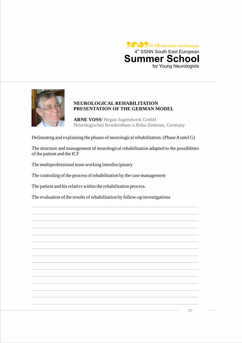
NEUROLOGICAL REHABILITATIONPRESENTATION OF THE GERMAN MODEL
ARNE VOSS/ Hegau-Jugendwerk GmbH Neurologisches Krankenhaus u.Reha-Zentrum, Germany
Delineating and explaining the phases of neurological rehabilitation. (Phase A until G)
The structure and management of neurological rehabilitation adapted to the possiblities of the patient and the ICF
The multiprofessional team working ínterdiscipinary
The controling of the process of rehabilitation by the case management
The patient and his relativs within the rehabilitation process.
The evaluation of the results of rehabilitation by follow-up investigations
5>10/July/Eforie Nord/Romaniath4 SSNN South East European

38
REHABILITATION AFTER TBI- A MULTIDISCIPLINARY CHALLENGE. HOW TO HUMANIZE HUMAN SKILLS FOLLOWING ACUTE BRAIN LESIONS
KLAUS VON WILD/ Professor of Neurosurgery, Medical Faculty University Münster, Prof. of Neurorehabilitation and Reconstructive Neurosurgery of Brain and Spinal Cord Lesions, INI Hannover, Germany
Objective: To review quality management of functional neurorehabilitation in patients after craniocerebral trauma with an emphasis on factors that may explain variability of early and outcomes after one year and how this might be influenced. to improve health related quality of life after traumatic brain injury (TBI).
Methods: First ever prospective controlled, population based, multicentre study on epidemiology and quality management of acute craniocerebral injuries (CCI) in Germany . Analysis of acute medical care and functional rehabilitation concerning the early outcome with one year follow up (telephone interview). Catchments areas were Hannover and Münster (sum of inhabitants 2,114 million). The definition of acute CCI was according to the ICD 10 S-02, S-04, S-06, S-07, S-09 in combination with dizziness or vomiting; retrograde or anterograde amnesia, impaired consciousness, skull fracture, and/or focal neurological impairment
Results: 6.783 patients CCI (58% male) were admitted for emergent hospital treatment. 28% patients were < 1 to 15 years, 18% > 65 years of age. Completed questionnaires of 63.5% were analysed. One year follow up of two third of acute CCI.. Incidence was 321/100.000 Initial CCI severity (GCS) of 55% patients showed 90,9% mild, 3,9% moderate, and 5,2% severe TBI. 5.221patients (= 77%) were hospitalised, 1,4% of them died. Follow-up of 63.5%. Only 258 patients (=3,8%) received neurological- neurosurgical rehabilitation (73% male), 68% within one month after injury, 5% were <16 years , 25% >65 years. Early rehabilitation of 100 patients (= 39%), one fifth referred within first week. Outcome end of “B”: GOS 1 = 4%; GOS 2 = 2,7%, GOS 3 = 37,3%, GOS 4 = 26,7%, GOS 5 = 29,3% and end of rehab” B – E” GOS 1 = 1,2%, 2 = 1,7%, 3 = 21,8%, 4 = 36,2%, and 5 = 39,1
Discussion: Data on epidemiology and quality management of early functional rehabilitation met the criterion set in 1993 (1). Key issue is a multidisciplinary approach for early posttraumatic neurorehabilitation in neurosurgery and neurology. Management of frequent multiple organ lesions and complications (= 57%) without referring the patient to another hospital and early functional outcome confirm the
5>10/July/Eforie Nord/Romaniath4 SSNN South East European

39
authors concept of neurosurgical early rehabilitation (2).
1.Ortega-Suhrkamp, E and von Wild, K.R.H Standards of neurologic-neurtosurgical early rehabilitation Acta Neurochir (2001) Suppl 79, 11-19
2. von Wild Klaus RH in cooperation with the TBI study council Neurorehabilitation following craniocerebral trauma European Journal of Trauma 2005, 4: 344-358
Figure 1: The interdisciplinary team for neurorehabilitation
5>10/July/Eforie Nord/Romaniath4 SSNN South East European
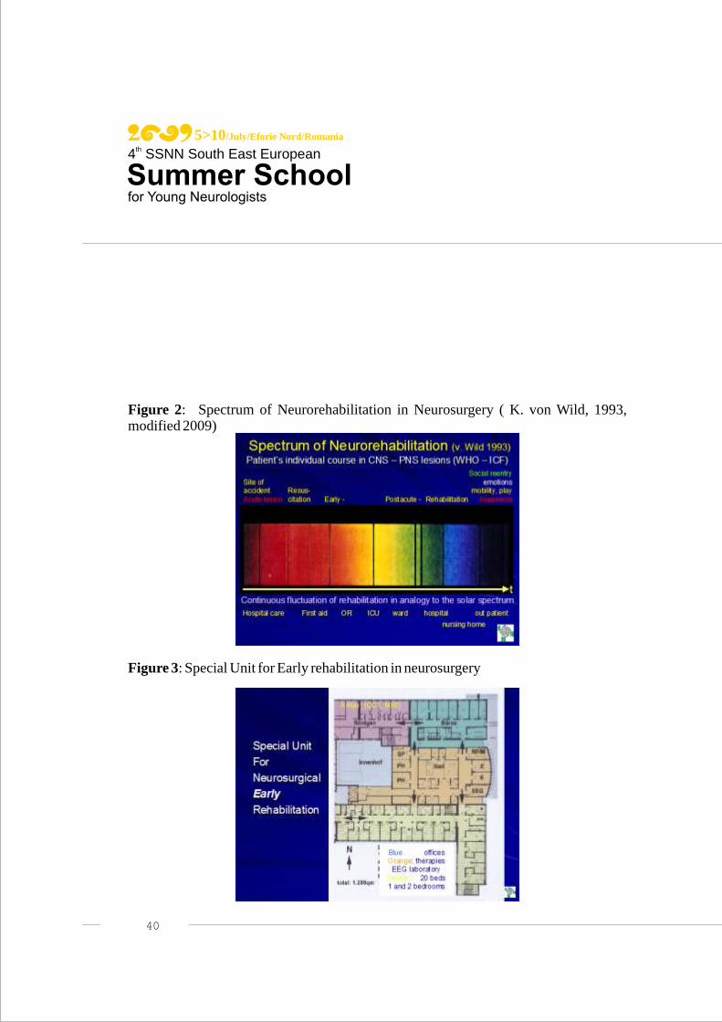
Figure 2: Spectrum of Neurorehabilitation in Neurosurgery ( K. von Wild, 1993, modified 2009)
Figure 3: Special Unit for Early rehabilitation in neurosurgery
40
5>10/July/Eforie Nord/Romaniath4 SSNN South East European

41
Purpose: Outcome following traumatic brain injury (TBI) is not only dependent on the nature and severity of injury and subsequent acute treatment, but also on quality management early and long-term rehabilitation and constituent characteristics of injured individuals. The extent of recovery and quality of life following TBI can be estimated early on by prognostic factors reflecting injury severity in the acute phase. Computerized tomography scan characteristics and Glasgow Coma Scale (GCS) score on hospital admission, length of coma (LOC), and duration of post-traumatic amnesia (PTA) are well known factors in the literature, but practically only 55% out of 6000 acute TBI were assessed accordingly in our study(1). To restore the impaired higher cortical brain functioning and for preservation and ongoing support of brain plasticity TBI patients first and foremost require Cerbroprotection. This demands great skill and a multidisciplinary approach, starting at the site of the impact, all the way long until social reintegration of the victim.
Methods: According to our daily practice we produced a short film (15 min CD-R) on Cerbprotection in TBI for teaching to improve personnel’s awareness of prognostic (pathological) early signs and symptoms in order to enhance knowledge of basics of quality management and rehabilitation after acute TBI to improve the final functional outcome. CCT and MRI scan characteristics are the main diagnostic adjuncts today that should be known.
Results: The film is based on and will demonstrate exemplarily the author’s neurosurgical experience in Cerbroprotection by multidisciplinary approach of the neurotrauma team, including intensive care, early, long-term, and special paediatric neurehabilitation.
Discussion: Up to now prospective clinical studies on functional outcome following TBI with special emphasis on the role of clinical Cerebroprotection , neuropsychology, and neurorehabilitation are rare (1,4,5 ). Relatively little is known about the longer-term impact of TBI on children's daily functioning (6-10). TBI is a significant problem in older adults (11). Predicting long-term clinical outcome for patients with traumatic
CERBROPROTECTION IN NEUROSURGERY- A MULTIDISCIPLINARY CHALLENGE
KLAUS VON WILD
Professor of Neurosurgery, Medical Faculty University Münster, Prof. of Neurorehabilitation and Reconstructive Neurosurgery of Brain and Spinal Cord Lesions, INI Hannover, Germany
/
5>10/July/Eforie Nord/Romaniath4 SSNN South East European

42
brain injury (TBI) at the beginning of rehabilitation provides essential information for counselling of the family and priority-setting for the limited resources in intensive rehabilitation. Out of a certain number of prognostic data at admission , e.g. age, GCS and ISS scores, pupil response, during intensive care management, e.g. duration of coma, intracranial pressure, secondary insults, and at early rehabilitation the German Coma Remission Scale (CRS) score, Early Barthel Index, and FIM that reflect restoration of impaired cortical functioning over time, have recently been found to be independent predictors for one-year outcome in prospective series (1-3). The accuracy of prediction for a good Glasgow Outcome Score is only 68% while for an outcome for disability (either moderate or severe) it is 83% (2,3). Children sustaining complicated mild TBI may be more vulnerable to development of chronic mild neuropsychological dysfunction than adults sustaining similar head injuries. Older age and long-term persisting cognitive sequelae are known to negatively influence functional outcome. TBI outcome is not only dependent on the nature and severity of injury but also on both constituent characteristics of injured individuals and quality management of Cerbroprotection by the neurotrauma team.
References:1) von Wild, KRH in cooperation with the TBI study group (2005): Neurorehabilitation following craniocerebral trauma European J of Trauma,4,344-3582) Marmarou A, Lu J. Butcher I et al ( 2007): J. Neurotrauma 4(2):270-803) Maas AI, Marmarou A, Murray GD, et al (2007): J. Neurotrauma 24(2):232-84) Poon WS, Zhu XL, Ng SC et al (2005): Acta Neurochir Suppl.93:207-85) Asikainen I, Kaste M, Sarna S. (1998): Brain Inj. Feb;12(2):95-1076) Keenan HT, Hooper SR, Wetherington CE et al.( 2007):Neurodevelopmental consequences of early traumatic brain injury in 3-year-old children Pediatrics Mar;119(3):e616-23.7) Klein M, Houx PJ, Jolles J(1996): J Nerv Ment Dis Aug;184(8):459-678) Stancin T, Drotar D, Taylor HG et al (2002): Pediatrics Feb;109(2):E349) Yeates KO, Swift E, Taylor HG et al (2004): J Int Neuropsychol Soc 10(3):412-26 2004 May;10(3):412-26.10) Ewing-Cobbs L, Fletcher JM, Levin HS et al (1997) J Int Neuropsychol Soc Nov;3(6):581-91. 11)Thompson HJ, McCormick WC, Kagan SH (2006): J.Am Geriat Soc Oct; 54 (10):1590-5
5>10/July/Eforie Nord/Romaniath4 SSNN South East European
