techniques used for morphological and ultrastructural ... used for morphological and ultrastructural...
TRANSCRIPT

Techniques Used for Morphological and Ultrastructural Description from Teeth White-Tufted-Ear-Marmosets (Callithrix jacchus)
B. Machado Bertassoli*1, T. Borges Lessa1, F. Delys de Oliveira1, D. Kelly de Abreu. L. C. Stunitz da Silva1, A. Cesar dos Santos1, R. Eli Grassi Rici, A. C. Assis Neto1 1 Department of Anatomy of Domestic and Wild Animals. Faculty of Veterinary Medicine, University of São Paulo –
USP, Av. Dr. Orlando Marques de Paiva 87, CEP 05508-270, São Paulo, SP, Brazil.
The scanning electron microscope (SEM) is a device capable of producing images of high magnification allowing a more detailed morphological analysis. Mineralized structures (hard), like teeth, can benefit from a differentiated protocol when submitted to the SEM technique, thus aiming at a considerable decrease of time, however when processed in routine histology techniques suffer from an increase in processing time due to decalcification. The family Callitrichidae is divides into six genera: Callithrix, Callimico, Cebuella, Saguinus, Leontopithecus and Mico. They have 32 teeth in the formula dental 2x: incisors 2/2; Canines 1/1, pre-molars 3/3 molar and 2/2. We used teeth of tufted marmosets white (Callithrix jacchus) for studies macroscopic, microscopic and ultrastructural. Considering the above and taking into account the importance of processing time and acceleration data capture, was adapted a protocol of the SEM to reduce processing time, and for histology was used descaling technique using formic acid, hydrochloric acid and distilled water. The results show the exact structures of the teeth and ensuring the method and analysis results.
Keywords decalcification; histology; scaning electron microscopy; teeth
1. Introduction
The family Callitrichidae is represented by the smallest primates in the world, encompassing six different genera, all of them arboreal and found in the rainforests of Central and South Americas [1]. Specifically regarding the genus Callithrix, composed of six different species, it may have about 70% of its diet in wildlife based on the intake of exudates, thus it is the group whose anatomical characteristics are the most adapted ones to the withdrawal and processing of such a material [1-2]. In this context, the teeth, considered as part of a very important and complex organ, the dental organ, which is formed by the dental element (tooth) and the periodontium, that fixates and supports the teeth. The tooth itself is formed by enamel, dentin, and pulp, and the periodontium is formed by cementum, periodontal ligament, alveolar bone, and gingiva [3]. Enamel is the hardest and densest mineral substance in the animal body, and it covers from the outside of the dental crown into the neck of the tooth. Dentin has a white-yellowish color and it is related to the pulp, internally, and to the enamel and cementum, externally. Pulp is the loose connective tissue located within the inner chamber of the tooth. Cementum is an osteoid layer covering the dentin root portion, from the center neck to the root apex, with a chemical composition similar to bone. The periodontal ligament is composed of collagen fibers spanning the space between the alveolar bone and cementum. And the alveolar bone is that underlying the tooth which holds its roots and develops inter-relations with the dental element. In the case of gingiva, it is a term applied to the epithelial and connective tissues surrounding and adhering to the teeth and the alveolar process [4]. Often, the diagnosis and treatment of certain pathologies only become possible after the morphological study of the dental tissue. However, the mineralization of this tissue may hinder the processing of the specimen, in this case, there’s a need to change its consistency to allow microtomy through conventional methods [5]. The consistency of mineralized tissues can be modified through the decalcifying process, which involves the removal of calcium and phosphate salts deposited on the organic matrix [6]. These procedures may be performed with the aid of acids or chelants. The choice of decalcifying agent depends on factors such as: tissue mineralization degree, aim of the study, and staining technique to be used [7]. The main difficulties of the decalcifying process are the preservation of cellular structures and the time required to accomplish it. Although some substances promote decalcification in a short period of time, they almost always cause tissue disruption, a factor which limits their use [5]. Partial decalcification of teeth for the visualization of calcium crystals, which make up enamel and enameloid, for analysis through scanning electron microscope (SEM) is widely used in studies of dental tissues of recent and fossil vertebrates, from fishes to mammals [8]. This decalcification or demineralization technique aims to soften hard tissues, through the removal of the hydroxyapatite crystals, to allow inclusion and posterior section of the paraffin. These cuts may then be microscopically analyzed [9]. These solutions may be composed of acids, such as a solution of nitric acid, and chelating compounds, such as tetraacetic diamine ethylene acid (EDTA) [9- 10 – 11].
Current Microscopy Contributions to Advances in Science and Technology (A. Méndez-Vilas, Ed.)
© 2012 FORMATEX 245
However, decalcification is a time-consuming step that requires systematic replacement of solutions employed [12], for instance, EDTA, which demands a very long time for acting (about 135 days). Given the above-mentioned facts, and taking into account the need for obtaining data in a short period of time, this study aimed to morphologically analyze the teeth of the common marmoset (Callithrix jacchus), using SEM and decalcification.
2. Material and Method
Teeth from three adult male common marmosets (Callithrix jacchus), that died due to natural causes, from a wild animals breeding center located in Atibaia, Sao Paulo, Brazil (Registration Ibama 1/35/93/0849-8, CTF 2029), were used. These animals were sent to the Domestic and Wild Animals Laboratory of Universidade de São Paulo (USP) for anatomical studies, where they were fixed in a 10% formaldehyde solution for at least 72 hours for subsequent dissection. The animals had their oral cavity exposed with the aid of anatomical tweezers, scissors, and scalpel for the extraction, identification, and measurement of the teeth with a digital caliper (Starrett®). The incisors, canines, premolars, and molars of the three specimens analyzed were taken from the mandible and maxilla, and one molar, one premolar, one canine, and one incisor underwent the modified decalcification technique, with a formulation using 800 mL of distilled water, 120 mL of formic acid, and 80 mL of hydrochloric acid. The teeth were immersed in the decalcifying solution, which was changed every three days over a 24-day period. After this processing, it was possible to continue the routine histological procedures through embedding in paraffin (Histosec® - Merck), inclusion and cutting on a Leica RM2165® microtome, and then cut to a 5 μm thickness and stained with hematoxylin-eosin (HE) and Masson trichrome to identify the structures [13]. They were photographed using a camera Sony Mavica 3.2 Mp, as well as photomicrographed through a photomicroscope Leica DM 2000. For scanning electron microscopy, the teeth were not fixed, they underwent a comprehensive analysis and a cryofracture technique; they were placed in liquid nitrogen to fracture, dried, and then they were placed on stubs with carbon glue for posterior gold "sputtering" coverage (2 min) in the sputter Emitech 550 K and analyzed (photomicrographed) in a scanning electron microscope (Leo 435 VP). All terms adopted in this study were based on the Veterinary Anatomical Nomenclature (2005) [14].
3. Results
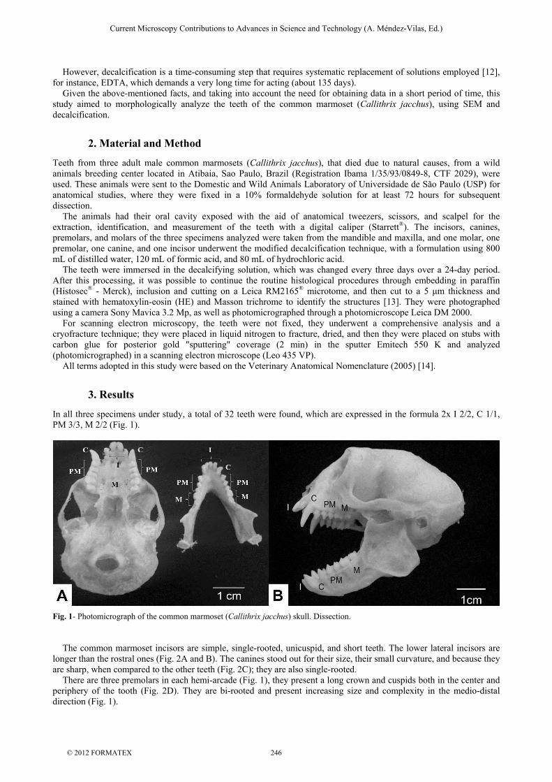
In all three specimens under study, a total of 32 teeth were found, which are expressed in the formula 2x I 2/2, C 1/1, PM 3/3, M 2/2 (Fig. 1).
Fig. 1- Photomicrograph of the common marmoset (Callithrix jacchus) skull. Dissection. The common marmoset incisors are simple, single-rooted, unicuspid, and short teeth. The lower lateral incisors are longer than the rostral ones (Fig. 2A and B). The canines stood out for their size, their small curvature, and because they are sharp, when compared to the other teeth (Fig. 2C); they are also single-rooted. There are three premolars in each hemi-arcade (Fig. 1), they present a long crown and cuspids both in the center and periphery of the tooth (Fig. 2D). They are bi-rooted and present increasing size and complexity in the medio-distal direction (Fig. 1).
Current Microscopy Contributions to Advances in Science and Technology (A. Méndez-Vilas, Ed.)
© 2012 FORMATEX 246

The molars of specimens were smaller than the premolars, and they also had several cusps, where 3 out of 4 cusps are sharp and 1 is more rounded (Fig. 2E), as well as 3 roots, that is, they are tri-rooted. On the ultrastructural analysis (cryofracture) it was possible to observe the division of the layers of the tooth (Fig. 2F, G, H, and I).
Fig. 2- Electromicrograph of the common marmoset (Callithrix jacchus) teeth. A) Upper central incisor; B) lateral incisor; C) upper canine; D) upper premolar; E) upper molar. Crown (C), root (R), and cusps (*). F, G, H, and I) Cryofracture of upper canine, where enamel (E), dentin (D), predentin (arrow), and pulp (P) can be seen. SEM technique.
The measurement of the animals’ teeth is represented in Table 1.
TABLE 1 – Average measurements of the Callithrix jacchus teeth..
TEETH
AVERAGE (cm) TEETH AVERAGE (cm)
UM 0,5 ± 0,07 LM 0,4 ± 0,07 UPM 0,6 ± 0,07 LPM 0,6 ± 0,07 UC 1,1 ± 0,07 LC 0,9 ± 0,07 ULI 0,5 ± 0,07 LLI 0,9 ± 0,07 UCI 0,6 ± 0,07 LCI 0,8 ± 0,07
* UM = Upper molar; UPM = upper premolar; UC = upper canine; ULI = upper lateral incisor; UCI = upper central incisor; LM = lower molar; LPM = lower premolar; LC = lower canine; LLI = lower lateral incisor; and LCI = lower central incisor.
The teeth were classified as heterodonts, brachydonts, since they presented a crown covered by enamel (Fig. 3) and the root, crown, and neck regions were well-defined (Fig. 2).
Current Microscopy Contributions to Advances in Science and Technology (A. Méndez-Vilas, Ed.)
© 2012 FORMATEX 247

When submitted to the modified decalcification technique, the material took on average 24 days to be ready for histological sections; in these cuts, all structures could be analyzed and photomicrographed with no changes. For the scanning electron microscope (SEM) technique, the time for preparation of the material took less than 30 minutes from the wash (start) to the bath with gold for obtaining the electromicrographs (end).
Fig.3- Photomicrographs of the common marmoset (Callithrix jacchus) teeth. A) Upper canine. LP = periodontal ligament; D = dentin; E = enamel; Pd = predentin; P = pulp. Masson trichrome staining. B) Lower premolar. LP = periodontal ligament; D = dentin; Pd = predentin; E = enamel. Hematoxylin and eosin staining.
4. Discussion
4.1 Procedures and Techniques
The decalcification time with the modified protocol was about 24 days, thus it is more efficient than EDTA, since this kind of decalcificator needs a very long time for acting (about 135 days), something which, in certain cases, hampers its use [15-16]. The 5% nitric acid, although widely used due to the reduced time required for decalcification, does not properly preserve cell morphology [16]. In our studies, we have shown the use of hydrochloric acid and formic acid, since they, contrary to nitric acid, preserve the cell structure and morphology, as seen in Fig. 3. The mixture of two or more decalcifying agents, such as formic acid and picric acid (Lillie’s method), formic acid, and nitric acid (Cajal’s method), and formic acid and hydrochloric acid (Biodec-R) have been used for many professional [5], however, the same authors report in their studies that Biodec-R was not effective for preserving the cell morphology, disagreeing again with our findings, since there were both tissue and cell morphology preservation. Hard structures may be processed without much prior preparation [17]; it was reported in our study when we used the SEM modified protocol, with just a wash in phosphate bufferid saline (PBS), drying, and then a shower of gold. This change in protocol led the time to prepare the material to drop from 48 hours to 30 minutes at most. Given the arguments developed, the establishment of a decalcification protocol also deserves further study, so that it is possible to standardize a histological processing able to decalcify the mineralized tissue in a short period of time, as well as to preserve the cell structures
4.1 Morphology of Teeth
In all animals used in this study, 2 pairs of incisors, 1 pair of canines, 3 pairs of premolars, and 2 pairs of molars were found in each mandible and maxilla, totaling 32 teeth, which are expressed in the formula 2x I 2/2, C 1/1, PM 3/3, M 2/2 (Fig. 2), differing from the dental formula of the Procyonidae family, i.e., 2x I3/3, C1/1,P4/4,M2/2 = 40, with the exception of kinkajou [18 - 19]. The teeth of Callithrix jacchus had a portion fitted to the mandible and an exposed portion, above the gingiva, and this kind of dental insertion is classified as techodont [20]. The incisor teeth of this species were classified as simple teeth, and the upper teeth present a growing volume in the medio-distal direction and in the lower teeth the volume is decreasing in the same direction (Fig. 1), unlike what was found in domestic animals [20], taking into account that they belong to different orders of animals and do not have the same feeding habits. The canine teeth of marmosets are large and sharp (Fig. 2C and 3C); they are used for aggressive purposes, such as territory defense and fight for females, habits similar to that of dogs [20-21] and coatis [22].
Current Microscopy Contributions to Advances in Science and Technology (A. Méndez-Vilas, Ed.)
© 2012 FORMATEX 248

The molar and bicuspid teeth are very similar to each other, a process called molarization [23], that is, premolars were modified and became similar to molars, with the function of macerating flat and fibrous food, but the premolars (upper and lower) are larger and have several cuspids, as also observed in carnivores [20]. Both the premolars and the molars of Callithrix jacchus are characterized by the presence of two or more cuspids (Fig. 2D-E), a conformation presenting a serrated aspect, different from the teeth of wild mice and rats, which have, respectively, rounded molars with pairs of cuspids arranged in transverse rows and molars with alternating triangular cuspids arranged in a zigzag pattern [24]. However, the teeth were also classified as bunodonts, due to the chewing surface being formed by different tubercles with rounded cusp (Fig. 2), something which is also found in omnivorous animals [22]. The teeth of marmosets have a diplodont feature, as they have two kinds of dentition throughout life, the deciduous and permanent; anelodont, because they have a limited period of growth; and brachydonts, like the teeth of marsupials and domestic animals [3-22]. The species under study presents, under light microscopy, teeth consisting of enamel, dentin, predentin, pulp, and periodontium (Fig. 4), the same way of humans [4]. Enamel is present on the upper part of teeth [3], as well as it was observed in the animals of this study, being the hardest structure of the body, but is completely acellular and unable to react to any aggression. Dentin was observed throughout the tooth covering the pulp, so it is sensitive to various stimuli. The periodontium of the teeth under study is thick and it has many vessels, taking into account the property of fixating the tooth in the mandibular and maxillary bone, consisting of periodontal ligament, which is formed by dense connective tissue, fibroblasts, and collagen fibers, as well as immature alveolar bone, as also described in other species [3-4].
5. Conclusion
The use of the modified decalcificator consisting of formic acid and hydrochloric acid has shown to be effective when compared to other decalcificators, because of its decreased activity time and the preservation of structures. The modified protocol used for processing the scanning electron microscopy (SEM) of mineralized materials in this study is effective when compared to the time of other protocols, despite they are not mineralized. The teeth of common marmoset (Callithrix jacchus) were classified as diplodonts, anelodonts, bunodonts, and brachydonts.
References
[1] Auricchio P. Primatas do Brasil. Editora Terra Brasilis; 1995. [2] Rylands AB, Schneider H, Langguth A, Mittermeier RA, Groves CP, Rodrigues-Luna E. An assessment of the diversity of new
world primates. Neotropical Primates. 2000; 8:61 – 93. [3] Banks WJ. Histologia Veterinária Aplicada. 2nd ed. São Paulo: Manole. 1991. [4] Junqueira LC, Carneiro J. Histologia básica. 11th ed. Rio de Janeiro: Editora Guanabara Koogan; 2008. [5] Chagas LF, Carvalho S, Neves ACC, Ricardo LH, Rode SM, Martins AMA. Tempo de descalcificação e preservação do núcleo
celular de tecido mineralizado descalcificado com ácido nítrico a 5%, EDTA a 7% e Biodec-r. Revista Periodontia. 2008; 18:71-76.
[6] Mattuella LG, Bento LW, Vier-Pelisser FV, Araujo FB, Fossati ACM. Análise comparativa de duas soluções fixadoras e descalcificadoras para o processamento de dentes decíduos humanos com lesão cariosa inativa em dentina. Revista de odontologia e Ciência; 2007; 22:99-105.
[7] Fernandes MI, Gaio EJ, Rosing CK, Oppermann RV, Rado PV. Microscopic qualitative evaluation of fixation time and decalcification media in rat maxillary periodontium. Brazilian Oral Research. 2007; 21:34-39.
[8] Richter M, Smith M. A microstructural study of the ganoine tissue of selected lower vertebrates. Journal of the Linnean Society. 1995; 114:173-212.
[9] Tiling SP, Giberson RT, Kullar RS. Microwave Exposure Increases Bone Demineralization Rate Independent of Temperature. Journal of Microscopy. 2004; 215:230-235.
[10] Morse A. Formic Acid-Sodium Citrate Decalcification and Butyl Alcohol Dehydration of Teeth and Bones for Sectioning in Paraffin. Journal of Dental Research. 1945; 24:143-153.
[11] Kiernan JA. Decalcification and Other for Hard Tissues. In: Histological & Histochemical Methods: Theory and Practice. 3th ed. Oxford: Butterworth Heinemann, 1999.
[12] Rode SM, Faria MR, Monteiro MP. O Uso de Microondas para Descalcificação de Tecidos Mineralizados da Mandíbula de Ratos. Revista de Odontologia da Universidade de São Paulo. 1996; 10:15-18.
[13] Tolosa EMC, Bermer AO, Freitas-Neto AG. Manual de Técnicas para Histologia Normal e Patológica. 2th ed. São Paulo: Manole, 2003.
[14] International Committee on Veterinary Gross Anatomical Nomenclature. Nomina anatomica veterinaria. 5th ed. Editorial Committee, Hannover, Columbia, Gent, Sapporo; 2005.
[15] Sanderson C, Radley K, Mayton L. Ethylenediaminetetraacetic acid in ammonium hydroxide for reducing decalcification time. Biotechnic Histochemistry. 1995; 70:12-18.
Current Microscopy Contributions to Advances in Science and Technology (A. Méndez-Vilas, Ed.)
© 2012 FORMATEX 249

[16] Yamamoto-Fukuda T, Shibata Y, Hishikawa Y, Shin M, Yamaguchi A, Kobayashi T, Koji T. Effects of various decalcification protocols on detection of DNA strand breaks by terminal dUTP nick end labelling. Journal of Molecular Histology. 2000; 32:697-702.
[17] Castro LAS. Processamento de mostras para microscopia eletrônica de varredura. Pelotas: Embrapa Clima Temperado. 2001. [18] Teixeira RHF, Ambrosio SR. Carnívora, Procyonidae (mão pelada, quati, jupará) In: Cuba ZS, Silva JCR, Catão-Dias JL, Eds.
Tratado de Animais Selvagens. São Paulo: Editora Roca; 2007. [19] Pieri NCG, Mançanares CAF, Bertassoli BM, Lima Jthomaz JM, Carvalho AF. Classificação Morfológica dos dentes de quati
(Nasua nasua). Pesquisa Veterinária Brasileira. 2011; 31(5):447-451. [20] Sisson S. Aparelho digestório In: Getty R, Ed., Anatomia dos Animais Domésticos. 5th ed. Rio de Janeiro: Editora Guanabara
Koogan; 1996. [21] Dyce RM, Sack WO, Wensing CJG. O Tratado de Anatomia Veterinária. 3th ed. Rio de Janeiro: Guanabara Koogan; 1997. [22] Freitas EP, Rahal SC, Teixeira CR, Teixeira R, Mendes GM, Gioso MA. Oral cavity evaluation and dental chart registration of
coati (Nasua nasua) in captivity. Journal of Veterinary Dentistry. 2008; 25:110-117. [23] Pough FH, Janis CM, Heiser JB. A vida dos vertebrados. 3th ed. São Paulo: Atheneu, 2003. [24] Polly PD. Development and evolution occlude: Evolution of development in mammalian teeth. Proceedings of the National
Academy of Sciences of the United States of America. 2000; 97:14019- 14021.
Current Microscopy Contributions to Advances in Science and Technology (A. Méndez-Vilas, Ed.)
© 2012 FORMATEX 250