technical note and review of the occlusion of carotid ... · occlusion of carotid-cavernous...
TRANSCRIPT
Received 09/20/2016 Review began 09/28/2016 Review ended 12/30/2016 Published 01/12/2017
© Copyright 2017Chen et al. This is an open accessarticle distributed under the terms ofthe Creative Commons AttributionLicense CC-BY 3.0., which permitsunrestricted use, distribution, andreproduction in any medium,provided the original author andsource are credited.
Transorbital Approach for EndovascularOcclusion of Carotid-Cavernous Fistulas:Technical Note and Review of theLiteratureChing-Jen Chen , Panagiotis Mastorakos , James P. Caruso , Dale Ding , Paul J. Schmitt ,Thomas J. Buell , Daniel M. Raper , Avery Evans , Steven A. Newman , Mary E. Jensen
1. Department of Neurological Surgery, University Of Virginia 2. Department of Neurological Surgery,University of Virginia 3. Department of Neurological Sugery, University Of Virginia 4. Department ofInterventional Neuroradiology, University Of Virginia 5. Department of Ophthalmology, University OfVirginia
Corresponding author: Panagiotis Mastorakos, [email protected] Disclosures can be found in Additional Information at the end of the article
AbstractCarotid-cavernous fistulas (CCFs) pose an anatomically and physiologically challengingproblem for clinicians. The most common method of treatment for these lesions is transvenousendovascular embolization via the inferior petrosal sinus or the facial vein. When transvenousaccess is not possible, an alternate approach must be devised. We describe a case example withbilateral Barrow Type B CCFs, which were inaccessible using the traditional transvenousapproach. Hence, a direct transorbital approach, performed under fluoroscopic guidance, wasemployed to successfully obliterate the CCF. At five months follow-up, the patient wasrecovering without complications. This case delineates the technical aspects of transorbitalCCF embolization and demonstrates that this approach is a viable alternative to conventionaltransvenous methods for appropriately selected CCF cases. We supplement our case exampleand technical note with a literature review of this approach.
Categories: Neurosurgery, RadiologyKeywords: transorbital, carotid cavernous fistula, fistula, endovascular, dural arteriovenous fistula, ccf
IntroductionA carotid-cavernous fistula (CCF) is an abnormal arteriovenous connection between thecavernous sinus (CS) and the cavernous segment of the internal carotid artery (ICA). The mostcommon treatment modality for CCFs is endovascular embolization via transvenouscatheterization. Numerous routes exist for obtaining transvenous access, including the inferiorpetrosal sinus and facial vein. However, rare cases arise in which anatomic constraints precludetransvenous access to the CS. In these patients, a direct transorbital approach may be employedto access and obliterate the fistula. The following technical note describes our proceduralexperience with transorbital embolization of a CCF.
Technical ReportA 68-year-old male presented to the ophthalmology clinic as a referral for thyroid orbitopathywith diplopia. On examination, the patient’s visual acuity was 20/20 in the right eye, with 2+conjunctival injection and trace chemosis. In the left eye, his visual acuity was 20/30 with 2-3+
1 1 1 1 1
2 3 4 5 4
Open Access TechnicalReport DOI: 10.7759/cureus.976
How to cite this articleChen C, Mastorakos P, Caruso J P, et al. (January 12, 2017) Transorbital Approach for EndovascularOcclusion of Carotid-Cavernous Fistulas: Technical Note and Review of the Literature. Cureus 9(1): e976.DOI 10.7759/cureus.976

injection, 3+ chemosis, and prolapsed conjunctiva. A fundoscopic exam did not demonstratedisc edema, optic atrophy, or abnormalities in venous pulsation. Thyroid studies were normal,and magnetic resonance imaging (MRI) and angiography (MRA) demonstrated no evidence of acavernous carotid fistula. The patient’s chemosis and injection improved with initial treatmentusing eye drops. The patient returned later to the ophthalmology clinic with 2-3+ conjunctivalinjection and 2+ prominence of episcleral vessels of the right eye with 8 mm of exposedconjunctiva (Figure 1a). A temporary tarsorrhaphy was performed to prevent keratinization ofthe exposed conjunctiva. The patient subsequently underwent a diagnostic cerebral angiogram,which showed bilateral CCFs. Informed patient consent was obtained for his treatment. Noidentifying patient information is contained within this report.
FIGURE 1: Initial Presentation and Diagnostic DSA(a) External examination demonstrating 2-3+ conjunctival injection and 2+ prominence ofepiscleral vessels of the right eye with 8 mm of exposed conjunctiva. (b-d) DSA following R ICAinjection (b) lateral and (c) oblique views of intracranial circulation in arterial phase; (d) lateralview of intracranial circulation in venous phase. (e-g) DSA following L ICA injection (e) lateralview of late arterial phase; (f, g) AP view of (f) arterial and (g) capillary phase. Demonstration ofright-sided, indirect, Type B CCF supplied by branches of the meningohypophyseal trunk,inferolateral trunk (b, c), and collaterals from the contralateral meningohypophyseal trunk (f, g),with venous outflow into the SOV and IOV and eventually into the FV (d). Demonstration of left-sided, indirect, Type B CCF supplied by smaller caliber branches of the meningohypophysealtrunk (e-g).
DSA: digital subtraction angiography; L ICA: left internal carotid artery; R ICA: right internalcarotid artery; AP: anteroposterior; CCF: carotid-cavernous fistula; SOV: superiot ophthalmicveins; IOV: inferior ophthalmic veins; FV: facial vein
2017 Chen et al. Cureus 9(1): e976. DOI 10.7759/cureus.976 2 of 10

Technical detailsICA injections demonstrated bilateral Barrow Type B CCFs (Figures 1b, 1g). The right fistula wassupplied by branches of the meningohypophyseal trunk (MHT), the inferolateral trunk (ILT)(Figures 1b-1c), and collateral supply from the contralateral MHT (Figures 1f-1g). Venous effluxwas through the superior (SOV) and inferior ophthalmic veins (IOV) into the facial vein (Figure1d). The left fistula was supplied by branches of the MHT, with venous efflux also through theSOV and IOV into the facial vein (Figures 1e, 1g). The left-sided fistula was relatively small incaliber, and we felt it would thrombose without intervention; therefore, the decision was madeto only embolize the right-sided CCF. An initial attempt was made to access the CCF via theright inferior petrosal sinus (IPS). However, digital subtraction angiography (DSA)demonstrated that the medial portion of the CS connected to the IPS was isolated from the CCF(Figure 2a). Under roadmap guidance, the diagnostic catheter was advanced through theexternal jugular vein into the right facial vein over a guidewire. Venous outflow anatomy of theCCF and anatomy of the peri-orbital cortical veins were assessed via contrast administrationthrough the right ICA (venous phase) (Figure 2b) and facial vein (Figure 2c). Multiple attemptswere made to access the CCF venous outflow tract using a Marksman™ micro catheter (ev3Neurovascular, Plymouth, MN), Echelon™ 14 micro catheter (ev3 Neurovascular), Transend-EX® .014 micro-guidewire (Stryker, Kalamazoo, MI), and Synchro2® micro-guidewire (Stryker)without success. Without a transvenous route into the right CCF, the decision was made todirectly access the CS through a transorbital approach.
FIGURE 2: Therapeutic DSA(a) Lateral view of R inferior petrosal sinus demonstrating relatively normal appearance of themedian portion of the CS, isolated from the CCF. (b, c) Lateral view demonstrates venous
2017 Chen et al. Cureus 9(1): e976. DOI 10.7759/cureus.976 3 of 10
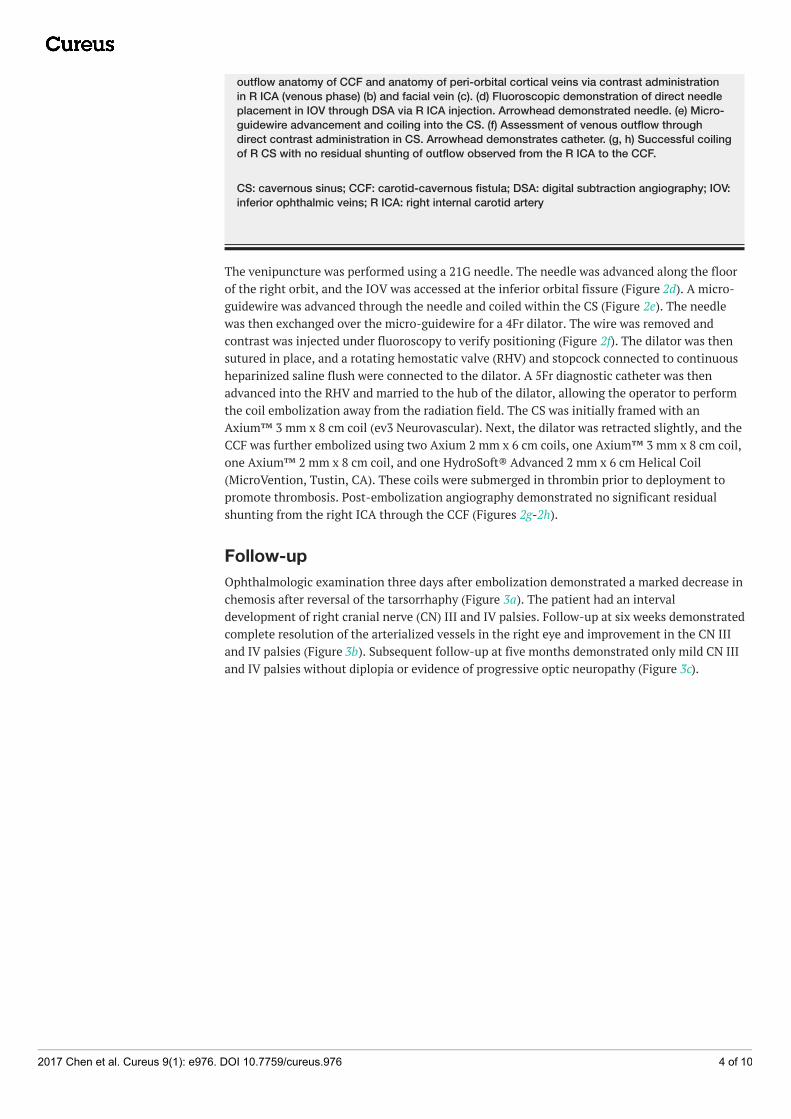
outflow anatomy of CCF and anatomy of peri-orbital cortical veins via contrast administrationin R ICA (venous phase) (b) and facial vein (c). (d) Fluoroscopic demonstration of direct needleplacement in IOV through DSA via R ICA injection. Arrowhead demonstrated needle. (e) Micro-guidewire advancement and coiling into the CS. (f) Assessment of venous outflow throughdirect contrast administration in CS. Arrowhead demonstrates catheter. (g, h) Successful coilingof R CS with no residual shunting of outflow observed from the R ICA to the CCF.
CS: cavernous sinus; CCF: carotid-cavernous fistula; DSA: digital subtraction angiography; IOV:inferior ophthalmic veins; R ICA: right internal carotid artery
The venipuncture was performed using a 21G needle. The needle was advanced along the floorof the right orbit, and the IOV was accessed at the inferior orbital fissure (Figure 2d). A micro-guidewire was advanced through the needle and coiled within the CS (Figure 2e). The needlewas then exchanged over the micro-guidewire for a 4Fr dilator. The wire was removed andcontrast was injected under fluoroscopy to verify positioning (Figure 2f). The dilator was thensutured in place, and a rotating hemostatic valve (RHV) and stopcock connected to continuousheparinized saline flush were connected to the dilator. A 5Fr diagnostic catheter was thenadvanced into the RHV and married to the hub of the dilator, allowing the operator to performthe coil embolization away from the radiation field. The CS was initially framed with anAxium™ 3 mm x 8 cm coil (ev3 Neurovascular). Next, the dilator was retracted slightly, and theCCF was further embolized using two Axium 2 mm x 6 cm coils, one Axium™ 3 mm x 8 cm coil,one Axium™ 2 mm x 8 cm coil, and one HydroSoft® Advanced 2 mm x 6 cm Helical Coil(MicroVention, Tustin, CA). These coils were submerged in thrombin prior to deployment topromote thrombosis. Post-embolization angiography demonstrated no significant residualshunting from the right ICA through the CCF (Figures 2g-2h).
Follow-upOphthalmologic examination three days after embolization demonstrated a marked decrease inchemosis after reversal of the tarsorrhaphy (Figure 3a). The patient had an intervaldevelopment of right cranial nerve (CN) III and IV palsies. Follow-up at six weeks demonstratedcomplete resolution of the arterialized vessels in the right eye and improvement in the CN IIIand IV palsies (Figure 3b). Subsequent follow-up at five months demonstrated only mild CN IIIand IV palsies without diplopia or evidence of progressive optic neuropathy (Figure 3c).
2017 Chen et al. Cureus 9(1): e976. DOI 10.7759/cureus.976 4 of 10

FIGURE 3: Ophthalmology Follow-up Images(a) Three day ophthalmology follow-up reveals improved chemosis and conjunctival injection,along with a steadily improving partial CN III and CN IV palsy. Follow-up at six weeks (b)demonstrated complete resolution of the arterialized vessels in the right eye and improvementin the CN III and IV palsies. Follow-up at five months (c) demonstrated only mild CN III and IVpalsies.
CN: cranial nerve
DiscussionCCFs consist of a group of vascular malformations characterized by an aberrant shunt betweenone or more sources of arterial inflow and the CS; they are subdivided into direct and indirectfistulas [1]. Direct CCFs, which involve a primary connection between the ICA and CS, typicallyresult from trauma or aneurysm rupture and often present with proptosis, chemosis, orbitalbruit, and visual disturbances. Traumatic CCFs are most common in young males. They occur in0.2% of head injuries and in 4% of basilar skull fractures [2]. Following trauma, a CCF maydevelop due to a tear in the carotid artery from an external force or as a result of vessel rupturefrom increased intraluminal pressure combined with compression of downstream vessels [2].
2017 Chen et al. Cureus 9(1): e976. DOI 10.7759/cureus.976 5 of 10

Spontaneous CCFs occur most commonly in older female patients. They can result fromruptured ICA aneurysms as well as genetic conditions, such as fibromuscular dysplasia andEhlers-Danlos syndrome [2]. Indirect CCFs involve fistulous connections between branches ofthe ICA or the external carotid artery (ECA). Their origin is less well understood and is thoughtto be associated with venous outflow obstruction during development. While indirect CCFshave a more indolent course of progression and often present with conjunctival injection,severe ophthalmologic complications may occur in cases with substantial venous outflowobstruction [3]. The Barrow classification scheme is the most widely used system for thecategorization of CCFs (Table 1).
Type Description
A Direct connection between ICA and CS
B Dural shunt (indirect) between meningeal branches of ICA and CS
C Dural shunt (indirect) between meningeal branches of ECA and CS
D Dural shunt (indirect) between meningeal branches of the ICA, ECA, and CS
TABLE 1: Barrow ClassificationICA: internal carotid artery; CS: cavernous sinus; ECA: external carotid artery
Endovascular techniques are the mainstay approach for the treatment of CFFs, andmicrosurgical treatment is currently utilized when the endovascular approach has failed [2]. Tu,et al. presented a series of 78 patients treated with direct surgical obliteration of CCF [4]. Avariety of surgical methods were used, including clipping and sealing the CCF with fascia andacrylate glue. All but one patient in the study had undergone endovascular embolization priorto surgical treatment. The CCF obliteration rate was 100%, and the most common postoperativemorbidity was transient ocular palsy. Day, et al. presented a series of nine patients whounderwent surgery for CCF following the failure of embolization [5]. All patients experiencedsymptomatic relief, there were no deaths, and transient ocular complications resolved by sixmonths follow-up. Stereotactic radiosurgery has also shown promise as both a primary andadjuvant treatment for CCFs. A systematic review by Chen, et al. found that radiosurgicaltreatment of CCFs resulted in a 73% obliteration rate with no post-SRS hemorrhage. The studyalso noted that Barrow Type A CCFs are less amenable to radiosurgical occlusion. Endovasculartreatment proves to be a timely and successful treatment modality for Barrow Type A CCFs,while select patients with Types B, C, and D CCFs may benefit from radiosurgery [6].
Despite successful reports of CCF treatment with surgery and radiosurgery, endovascularembolization remains the first-line therapy. Transarterial approaches are most commonly usedfor direct CCFs, while both transarterial and transvenous approaches are utilized for indirectCCFs. The transarterial approach for direct CCFs involves accessing the fistula via the ICA. Forindirect CCFs, it involves distal access via feeding vessels, usually from the ECA, an approachthat is fraught with difficulty [7]. Transvenous strategies are particularly useful for indirectCCFs with small ICA feeders. The heterogeneity of venous drainage amongst indirect CCFsrequires an adaptive approach, depending on the venous outflow patterns. The inferior petrosalsinus is commonly used to access the venous system, especially in cases with predominantlyposterior drainage. Meyers, et al. reported a 76% success rate when approaching the CS via theinferior petrosal sinus. Similarly, Klisch, et al. were able to completely or partially occlude 60%
2017 Chen et al. Cureus 9(1): e976. DOI 10.7759/cureus.976 6 of 10
of CCFs using this approach. When the inferior petrosal sinus is opacified, thrombosed, orotherwise not angiographically visible, a transvenous approach via the facial vein is analternative. Klisch, et al. reported a 50% treatment success rate when the facial vein was used toaccess the CS [8]. A case series of seven patients by Biondi, et al. analyzed the success ofendovascular treatment following access to the CS via the facial vein [9]. In all seven patients,the CS was successfully accessed through the angular vein and the superior ophthalmic vein.Catheterization through the superior ophthalmic vein failed in one patient. Four patientsdemonstrated complete CCF occlusion, and two additional patients improved clinically.
Despite the success of the above strategies, complex angioarchitecture can precludeconventional transarterial or transvenous approaches. The transorbital approach is a viablealternative for endovascular treatment of CCFs. This technique allows access to the CS throughthe SOV [10-13], IOV [11], or ICA [14-15] or through a direct transorbital puncture of the CS[15-20]. The reported case demonstrates the use of the transorbital approach for coilembolization of a Barrow Type B CCF. A review of the literature identified 12 case reports of thetransorbital approach for CCFs (Table 2). Observed complications included postoperativeptosis, proptosis, chemosis, and CN palsies [10, 12-13, 15-16]. Teng, et al. observed that otherpotential complications include subarachnoid hemorrhage, vision loss, and optic nerve injury[15]. Workman, et al. posit that subarachnoid hemorrhage can be avoided by entering the CSanteriorly, thereby avoiding a breach of the subarachnoid space [16]. Elhammady, et al.reported success using an Onyx® ethylene vinyl alcohol copolymer (EV3 Neurovascular, Irvine,CA) embolization. Onyx allowed the authors to target the posterior compartment of the CCFcontaining the point of fistulization. The use of coils would have likely led tocompartmentalization and incomplete obliteration of the CCF [18]. Mehrzad, et al. alsodemonstrated success using Onyx embolization [10], and Dashti, et al. demonstrated successusing a combination of Onyx and detachable coils [11].
2017 Chen et al. Cureus 9(1): e976. DOI 10.7759/cureus.976 7 of 10

Article Patients(#) Barrow Type Result (#) Complications (#)
Teng, et al. 1995[15] 11 A (11) Complete
obliteration (11) Transient postoperative ptosis (2)
Workman, et al.2002 [16] 1 A (1) Complete
obliterationTransient postoperative ptosis,proptosis, and chemosis
Satchi, et al. 2009[17] 1 D (1) Complete
obliteration None
Elhammady, et al.2011 [18] 1 B(1) Complete
obliteration None
Mehrzad, et al.2011 [10] 1 C (1) Complete
obliterationComplete CN III palsy (resolved at 3months)
Dashti, et al. 2011[11] 2 B (1), D (1) Complete
obliteration (2) None
Luo, et al. 2013 [14] 1 D (1) Completeobliteration None
Pansara, et al. 2013[12] 1 D (1) Complete
obliterationTransient diplopia, proptosis, chemosis,and CN VI palsy
Coumou, et al.2013 [19] 1 N/A (Indirect, low-
flow CCF)Completeobliteration None
Puffer, et al. 2014[20] 1 B (1) Complete
obliteration None
Milburn, et al. 2014[13] 1 D (1) Complete
obliterationTransient CN VI palsy, proptosis,diplopia
TABLE 2: Reports of CCF Treatment Via Transorbital ApproachCCF: carotid-cavernous fistula; CN: cranial nerve; N/A: not available
ConclusionsCCFs are most commonly managed with endovascular embolization. The clinical experience ofthe authors, along with a review of current literature, reveals that, for CCFs, which areinaccessible from a transvenous approach, direct transorbital embolization is a safe andeffective alternative for occlusion of these lesions.
Additional InformationDisclosuresHuman subjects: Consent was obtained by all participants in this study. Animal subjects: Allauthors have confirmed that this study did not involve animal subjects or tissue. Conflicts ofinterest: In compliance with the ICMJE uniform disclosure form, all authors declare the
2017 Chen et al. Cureus 9(1): e976. DOI 10.7759/cureus.976 8 of 10
following: Payment/services info: All authors have declared that no financial support wasreceived from any organization for the submitted work. Financial relationships: All authorshave declared that they have no financial relationships at present or within the previous threeyears with any organizations that might have an interest in the submitted work. Otherrelationships: All authors have declared that there are no other relationships or activities thatcould appear to have influenced the submitted work.
References1. Rhoton AL Jr: The cavernous sinus, the cavernous venous plexus, and the carotid collar .
Neurosurgery. 2002, 51:S1-375-S1-410. 10.1097/00006123-200210001-000102. Ellis JA, Goldstein H, Connolly ES Jr, Meyers PM: Carotid-cavernous fistulas. Neurosurg Focus.
2012, 32:E9. 10.3171/2012.2.FOCUS12233. Debrun GM, Viñuela F, Fox AJ, Davis KR, Ahn HS: Indications for treatment and classification
of 132 carotid-cavernous fistulas. Neurosurgery. 1988, 22:285–89. 10.1227/00006123-198802000-00001
4. Tu YK, Liu HM, Hu SC: Direct surgery of carotid cavernous fistulae and dural arteriovenousmalformations of the cavernous sinus. Neurosurgery. 1997, 41:798–805. 10.1097/00006123-199710000-00006
5. Day JD, Fukushima T: Direct microsurgery of dural arteriovenous malformation type carotid-cavernous sinus fistulas: indications, technique, and results. Neurosurgery. 1997, 41:1119–24.10.1097/00006123-199711000-00017
6. Chen CJ, Lee CC, Ding D, Starke RM, Chivukula S, Yen CP, Moosa S, Xu Z, Pan DH, SheehanJP: Stereotactic radiosurgery for intracranial dural arteriovenous fistulas: a systematic review .J Neurosurg. 2015, 122:353–62. 10.3171/2014.10.jns14871
7. Gemmete JJ, Ansari SA, Gandhi DM: Endovascular techniques for treatment of carotid-cavernous fistula. J Neuroophthalmol. 2009, 29:62–71. 10.1097/WNO.0b013e3181989fc0
8. Klisch J, Huppertz HJ, Spetzger U, Hetzel A, Seeger W, Schumacher M: Transvenous treatmentof carotid cavernous and dural arteriovenous fistulae: results for 31 patients and review of theliterature. Neurosurgery. 2003, 53:836–56. 10.1227/01.NEU.0000083551.26295.AB
9. Biondi A, Milea D, Cognard C, Ricciardi GK, Bonneville F, van Effenterre R: Cavernous sinusdural fistulae treated by transvenous approach through the facial vein: report of seven casesand review of the literature. AJNR Am J Neuroradiol. 2003, 24:1240–46.
10. Mehrzad H, Alam K, Rennie A: The treatment of a dural carotid cavernous fistula (CCF) usingOnyx via a transorbital approach: a technical note. Neuroradiology. 2011, 53:895–98.10.1007/s00234-010-0799-x
11. Dashti SR, Fiorella D, Spetzler RF, Albuquerque FC, McDougall CG: Transorbital endovascularembolization of dural carotid-cavernous fistula: access to cavernous sinus through directpuncture: case examples and technical report. Neurosurgery. 2011, 68:ons75-ons83.10.1227/NEU.0b013e3182073cc5
12. Pansara A, Milburn JM, Perry M, Eubanks B: Clinical images - a quarterly column: transorbitalcoil embolization of a carotid cavernous fistula. Ochsner J. 2013, 13:295–97.
13. Milburn J, Pansara A, Perry M, Vidal G, Eubanks B: E-025 transorbital carotid-cavernousfistula embolization with ruby coils. J NeuroIntervent Surg. 2014, 6:A48-A49.10.1136/neurintsurg-2014-011343.92
14. Luo CB, Teng MMH, Chang FC, Guo WY, Chang CY: Transorbital direct puncture of theposterior cavernous sinus through the internal carotid artery for embolization of isolatedcavernous sinus dural arteriovenous fistula. J NeuroIntervent Surg. 2013, 5:e1.10.1136/neurintsurg-2011-010130
15. Teng MM, Lirng JF, Chang T, Chen SS, Guo WY, Cheng CC, Shen WC, Lee LS: Embolization ofcarotid cavernous fistula by means of direct puncture through the superior orbital fissure.Radiology. 1995, 194:705–11. 10.1148/radiology.194.3.7862966
16. Workman MJ, Dion JE, Tong FC, Cloft HJ: Treatment of Trapped CCF by Direct Puncture of theCavernous Sinus by Infraocular Trans-SOF Approach. Case Report and Anatomical Basis.Interv Neuroradiol. 2002, 8:299–304.
17. Satchi K, Mitchell PJ, McNab AA: Transorbital puncture of the cavernous sinus to treat a duralcarotid-cavernous sinus fistula. Ophthal Plast Reconstr Surg. 2009, 25:54–56.
2017 Chen et al. Cureus 9(1): e976. DOI 10.7759/cureus.976 9 of 10

10.1097/IOP.0b013e318193646f18. Elhammady MS, Peterson EC, Aziz-Sultan MA: Onyx embolization of a carotid cavernous
fistula via direct transorbital puncture. J Neurosurg. 2011, 114:129–32.10.3171/2010.1.jns091433
19. Coumou AD, van den Berg R, Bot JC, Beetsma DB, Saeed P: Direct orbital puncture of thecavernous sinus for the treatment of a carotid-cavernous dural AV fistula with a concomitantvenous/lymphatic malformation. Orbit. 2014, 33:68–71. 10.3109/01676830.2013.844173
20. Puffer RC, Lanzino G, Cloft HJ: Using XperGuide planning software to safely guide catheteraccess to the cavernous sinus via transorbital puncture: a case report. Neurosurgery. 2014,10:e370–73. 10.1227/neu.0000000000000316
2017 Chen et al. Cureus 9(1): e976. DOI 10.7759/cureus.976 10 of 10