technical methods - journal of clinical pathologyjcp.bmj.com/content/jclinpath/32/4/406.full.pdf ·...
TRANSCRIPT

Technical methods
Technical methodsA simplified rapid method forpurification of glomeruliJ. L. PORTIS, S. K. WIKEL, AND F. J. MCATEE USDepartment of Health, Education, and Welfare,Public Health Service, National Institutes of Health,National Institute of Allergy and Infectious Diseases,Rocky Mountain Laboratory, Hamilton, Montana59840, USA
Isolated renal glomeruli have been used in variousimmunological, metabolic, and morphological stud-ies. Several techniques have been described forpurification of glomeruli usually involving gradedsieving or differential density gradient centrifugationand often requiring renal perfusion (reviewed byN0rgaard, 1976). In the current report, we presenta simple technique for purifying glomeruli, whichrequires no special equipment other than a routinelaboratory centrifuge. The technique is rapid (lessthan 30 minutes) and results in ultrastructurallyintact glomeruli without renal tubular or vascularcontamination.
Material and methods
ANIMALSSapphire mink were obtained from the closed colonymaintained at the Rocky Mountain Laboratory(RML). New Zealand white rabbits were obtainedfrom a local source. Hartley guinea pigs, outbredSyrian hamsters, C57BL/10 mice, and Lewis ratswere raised at the RML.
SOLUTIONSSeventy-four percent w/v sucrose, specific gravity1 29 g/ml, was prepared in 0-85% saline and broughtto pH 8-3 to 8-6 with 1 M NaOH. The final dilutionof sucrose to yield a specific gravity of 1-21 to 1-24g/ml was made with 1/15 M phosphate bufferedsaline (PBS), pH 7-2 (34 ml sucrose + 10 ml PBS).
KIDNEYSKidneys from laboratory animals were obtainedfresh at necropsy after exsanguination. The humankidney used in this study was from a patient with ahypernephroma of the superior pole of the kidney.
Received for publication 4 September 1978
The inferior pole appeared grossly and microscopi-cally normal and was frozen and stored at - 90'C.Portions were thawed and used in the glomerularpurification procedure.
ISOLATION OF GLOMERULIWhole kidneys were minced in cold PBS with twoscalpel blades drawn across one another, and thetissue was pressed through a 40 mesh stainlesssteel screen with the plunger from a 5 ml disposablesyringe. The material was then suspended in 30 mlcold PBS and allowed to settle for 5 minutes on ice.The supernate was discarded, and the sediment wasresuspended in PBS and centrifuged at 250 g for 2minutes. After the supernate had been aspirated, thesucrose solution (specific gravity 1 21 to 1 24 g/ml)was added, the pellet dispersed by shaking vigorously,and the suspension centrifuged at 2000 g for 5minutes. The floating debris was then removed withan aspirator, and the pellet consisting of the purifiedglomeruli was suspended in PBS and passed througha 40 mesh stainless steel screen to remove any re-maining tissue fragments.
ELECTRON MICROSCOPYIsolated glomeruli and material before purificationwere fixed in 2% paraformaldehyde, 2-5% glutaralde-hyde, and 0-025% CaCl2 in 0 1 ml cacodylate buffer(pH 7 4) at 40C for 8 hours. The material was washedwith several changes of 0-1 M cacodylate buffer(pH 7-4) at 40C for 18 hours. Post-fixation wascarried out in 1% osmium-0 1 M cacodylate (pH 7 4)for 2 hours at room temperature. Preparations weredehydrated in a graded ethanol series and embeddedin Epon via toluene. Thin sections were cut on aReichert OmU-2 ultramicrotome with a diamondknife, picked up on bare 300 mesh copper grids,stained in uranyl acetate, and counter-stained inlead citrate (Reynolds, 1963). Sections were viewedin an Hitachi HUI1IE-1 electron microscopeoperating at 75 kV. One-micron sections used toorientate the specimen were stained with toluidineblue.
Results and discussion
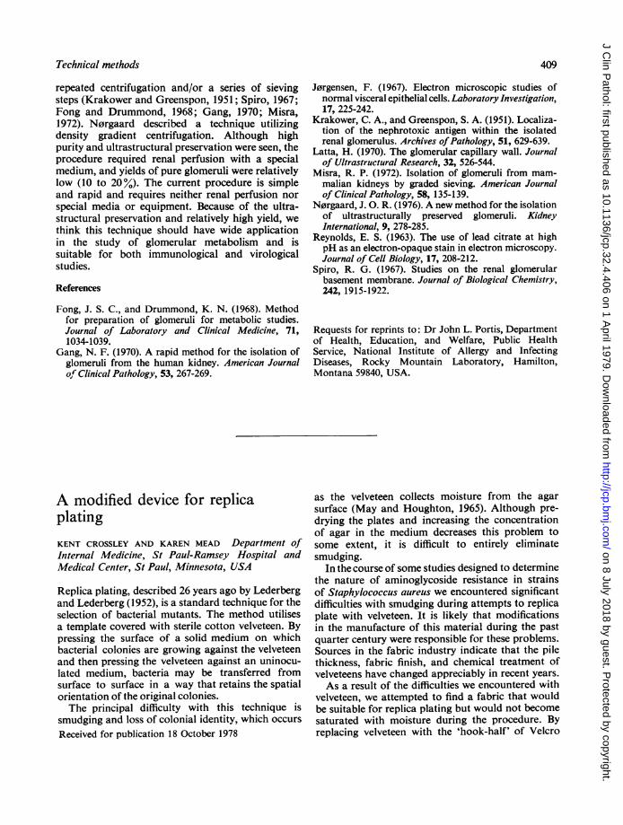
Because of our interest in Aleutian disease of mink,the procedure was initially defined for this species.However, the technique was subsequently found tobe applicable to most common laboratory animals.Figure 1 illustrates the degree of purification achieved
406
406
on 8 July 2018 by guest. Protected by copyright.
http://jcp.bmj.com
/J C
lin Pathol: first published as 10.1136/jcp.32.4.406 on 1 A
pril 1979. Dow
nloaded from

Technical methods
A.....
...*_Be_Mae#F
I, l. l 4
Fig. 1 One-micron section of Epon-embedded isolated mink glomeruli. Most glomeruli have intactBowman's capsules, and there is minimal extraneous debris. ( x 25 toluidine blue)
by pelleting the kidney preparation through sucrose.The abundant cellular debris, along with tubularand vascular fragments seen before centrifugation,are in sharp contrast to the virtually pure glomeruliwith intact Bowman's capsules present in the pellet.The yield of purified mink glomeruli was 40 8
± 6-0% (Table). Optimal recovery was achieved
Table Yield ofglomeruli
Kidney Tube No.t No. of glomeruli x 103* % Yield
Before After
Mink 1 77 18 232 89 42 473 87 43 494 101 44 44
Human 1 9-6 59 732 3-6 3 12 873 1 16 0-87 75
*Glomeruli were counted in the crude kidney suspension (before) andin the suspended pellet after centrifugation through sucrose (after).tSeparate aliquots of the same kidney suspension were run throughthe purification procedure.
with 25 to 50 mg wet kidney per ml of sucrose.Overloading the system decreased the yield andincreased contamination of the pellet with non-glomerular debris.
Isolated mink glomeruli were found by ultra-structural criteria to be generally normal in appear-ance (Jorgensen, 1967; Latta, 1970). Visceralepithelial cells were well preserved in all sectionsexamined (Fig. 2), and the filtration slit membranewas evident between foot processes of these cells(Insert, Fig. 2). The basement membrane was welldefined into the dense inner layer and the lessdense outer layers and was never found to bedisrupted. The only evidence of cellular damage wasfound when endothelial cells along the peripheralareas of the isolated glomeruli were examined.These cells displayed some disruption of the limitingplasma membrane and the formation of smallcytoplasmic vacuoles and had a slightly swollenappearance. These changes were not evident whenendothelial cells were examined in deeper regions ofthe glomerulus. Alterations in the appearance ofperipheral endothelial cells might be attributed to
407
on 8 July 2018 by guest. Protected by copyright.
http://jcp.bmj.com
/J C
lin Pathol: first published as 10.1136/jcp.32.4.406 on 1 A
pril 1979. Dow
nloaded from

Technical Methods
Fig. 2 Electron microscopic appearance of a purified mink glomerulus. Endothelial cell (En), basementmembrane (Bm), and visceral epithelial cell (Ep) show good ultrastructural preservation. Insert: foot processesfrom a visceral epithelial cell; arrows point to filtration slit membrane. ( x 24 750; insert x 49 500)
osmotic changes when the isolated glomeruli were
placed into fixative. Electron microscopic exam-
ination of glomeruli before purification revealedsimilar changes in peripheral endothelial cells;however, the changes observed in purified glomeruliwere slightly more evident.
This technique has been used with similar resultson kidneys from rabbits, guinea pigs, rats, andhamsters. The only laboratory animal for whichthis procedure did not appear to be suitable was themouse; yields were low and contamination was
high. We tested a wide range of specific gravities butnever achieved satisfactory glomerular purification.The purification procedure was modified slightly
for use with human kidney. The initial settling
period was found to be unecessary. By eliminatingthis step, yields were increased without a significanteffect on purity. Otherwise the conditions for optimalyield and purity were identical with those for kidneysof laboratory animals. The yield of human glomeruliis shown in the Table. The majority of purifiedglomeruli were without Bowman's capsules. Thiswas not the result of the sedimentation proceduresince glomeruli had a similar appearance beforecentrifugation through sucrose.
This procedure takes advantage of the high densityof glomeruli, which is probably due to their highratio of blood per gram of tissue. Other techniquesthat have been described require initial renalperfusion to evacuate the glomeruli of blood and
408
on 8 July 2018 by guest. Protected by copyright.
http://jcp.bmj.com
/J C
lin Pathol: first published as 10.1136/jcp.32.4.406 on 1 A
pril 1979. Dow
nloaded from
Technical methods 409
repeated centrifugation and/or a series of sievingsteps (Krakower and Greenspon, 1951; Spiro, 1967;Fong and Drummond, 1968; Gang, 1970; Misra,1972). N0rgaard described a technique utilizingdensity gradient centrifugation. Although highpurity and ultrastructural preservation were seen, theprocedure required renal perfusion with a specialmedium, and yields of pure glomeruli were relativelylow (10 to 20%). The current procedure is simpleand rapid and requires neither renal perfusion norspecial media or equipment. Because of the ultra-structural preservation and relatively high yield, wethink this technique should have wide applicationin the study of glomerular metabolism and issuitable for both immunological and virologicalstudies.
References
Fong, J. S. C., and Drummond, K. N. (1968). Methodfor preparation of glomeruli for metabolic studies.Journal of Laboratory and Clinical Medicine, 71,1034-1039.
Gang, N. F. (1970). A rapid method for the isolation ofglomeruli from the human kidney. American Journalof Clinical Pathology, 53, 267-269.
J0rgensen, F. (1967). Electron microscopic studies ofnormal visceral epithelial cells. Laboratory Investigation,17, 225-242.
Krakower, C. A., and Greenspon, S. A. (1951). Localiza-tion of the nephrotoxic antigen within the isolatedrenal glomerulus. Archives ofPathology, 51, 629-639.
Latta, H. (1970). The glomerular capillary wall. Journalof Ultrastructural Research, 32, 526-544.
Misra, R. P. (1972). Isolation of glomeruli from mam-malian kidneys by graded sieving. American Journalof Clinical Pathology, 58, 135-139.
N0rgaard, J. 0. R. (1976). A new method for the isolationof ultrastructurally preserved glomeruli. KidneyInternational, 9, 278-285.
Reynolds, E. S. (1963). The use of lead citrate at highpH as an electron-opaque stain in electron microscopy.Journal of Cell Biology, 17, 208-212.
Spiro, R. G. (1967). Studies on the renal glomerularbasement membrane. Journal of Biological Chemistry,242, 1915-1922.
Requests for reprints to: Dr John L. Portis, Departmentof Health, Education, and Welfare, Public HealthService, National Institute of Allergy and InfectingDiseases, Rocky Mountain Laboratory, Hamilton,Montana 59840, USA.
A modified device for replicaplatingKENT CROSSLEY AND KAREN MEAD Department ofInternal Medicine, St Paul-Ramsey Hospital andMedical Center, St Paul, Minnesota, USA
Replica plating, described 26 years ago by Lederbergand Lederberg (1952), is a standard technique for theselection of bacterial mutants. The method utilisesa template covered with sterile cotton velveteen. Bypressing the surface of a solid medium on whichbacterial colonies are growing against the velveteenand then pressing the velveteen against an uninocu-lated medium, bacteria may be transferred fromsurface to surface in a way that retains the spatialorientation of the original colonies.The principal difficulty with this technique is
smudging and loss of colonial identity, which occursReceived for publication 18 October 1978
as the velveteen collects moisture from the agarsurface (May and Houghton, 1965). Although pre-drying the plates and increasing the concentrationof agar in the medium decreases this problem tosome extent, it is difficult to entirely eliminatesmudging.
In the course of some studies designed to determinethe nature of aminoglycoside resistance in strainsof Staphylococcus aureus we encountered significantdifficulties with smudging during attempts to replicaplate with velveteen. It is likely that modificationsin the manufacture of this material during the pastquarter century were responsible for these problems.Sources in the fabric industry indicate that the pilethickness, fabric finish, and chemical treatment ofvelveteens have changed appreciably in recent years.As a result of the difficulties we encountered with
velveteen, we attempted to find a fabric that wouldbe suitable for replica plating but would not becomesaturated with moisture during the procedure. Byreplacing velveteen with the 'hook-half' of Velcro
on 8 July 2018 by guest. Protected by copyright.
http://jcp.bmj.com
/J C
lin Pathol: first published as 10.1136/jcp.32.4.406 on 1 A
pril 1979. Dow
nloaded from