tbx1 protein interactions and microrna-96-5p regulation controls
TRANSCRIPT

OR I G INA L ART I C L E
TBX1 protein interactions and microRNA-96-5p
regulation controls cell proliferation duringcraniofacial and dental development: implicationsfor 22q11.2 deletion syndromeShan Gao1, Myriam Moreno2,3, Steven Eliason2,3, Huojun Cao1, Xiao Li2,3,Wenjie Yu2,3, Felicitas B. Bidlack4, Henry C. Margolis5, Antonio Baldini6
and Brad A. Amendt2,3,*1Texas A&M University Health Science Center, Houston, TX, USA, 2Department of Anatomy and Cell Biology,3Craniofacial Anomalies Research Center, The University of Iowa, Iowa City, IA, USA, 4Department of MineralizedTissue Biology, 5Center for Biomineralization, Department of Applied Oral Sciences, The Forsyth Institute,Cambridge, MA, USA and 6Department of Molecular Medicine and Medical Biotechnology, University Federico IIand the Institute of Genetics and Biophysics CNR, Naples, Italy
*To whom correspondence should be addressed. Tel: +1 3193353694; Fax: +1 3193357770; Email: [email protected]
AbstractT-box transcription factor TBX1 is the major candidate gene for 22q11.2 deletion syndrome (22q11.2DS, DiGeorge syndrome/Velo-cardio-facial syndrome), whose phenotypes include craniofacial malformations such as dental defects and cleft palate. Inthis study, Tbx1 was conditionally deleted or over-expressed in the oral and dental epithelium to establish its role inodontogenesis and craniofacial developmental. Tbx1 lineage tracing experiments demonstrated a specific region of Tbx1-positive cells in the labial cervical loop (LaCL, stem cell niche).We found that Tbx1 conditional knockout (Tbx1cKO) mice featuredmicrodontia, which coincides with decreased stem cell proliferation in the LaCL of Tbx1cKO mice. In contrast, Tbx1 over-expression increased dental epithelial progenitor cells in the LaCL. Furthermore, microRNA-96 (miR-96) repressed Tbx1expression and Tbx1 repressed miR-96 expression, suggesting that miR-96 and Tbx1 work in a regulatory loop to maintain thecorrect levels of Tbx1. Cleft palate was observed in both conditional knockout and over-expression mice, consistent with thecraniofacial/tooth defects associated with TBX1 deletion and the gene duplication that leads to 22q11.2DS. The biochemicalanalyses of TBX1 human mutations demonstrate functional differences in their transcriptional regulation of miR-96 andco-regulation of PITX2 activity. TBX1 interacts with PITX2 to negatively regulate PITX2 transcriptional activity and the TBX1N-terminus is required for its repressive activity. Overall, our results indicate that Tbx1 regulates the proliferation of dentalprogenitor cells and craniofacial development through miR-96-5p and PITX2. Together, these data suggest a new molecularmechanism controlling pathogenesis of dental anomalies in human 22q11.2DS.
Received: October 23, 2014. Revised and Accepted: December 30, 2014
© The Author 2015. Published by Oxford University Press. All rights reserved. For Permissions, please email: [email protected]
Human Molecular Genetics, 2015, Vol. 24, No. 8 2330–2348
doi: 10.1093/hmg/ddu750Advance Access Publication Date: 2 January 2015Original Article
2330
Downloaded from https://academic.oup.com/hmg/article-abstract/24/8/2330/652741by gueston 12 February 2018

Introduction22q11.2 deletion syndrome (22q11.2DS) is the unifying term forpatients with a common microdeletion on one of the proximallong arms of chromosome 22. This deletion encompasses thegenes responsible for DiGeorge syndrome (DGS, MIM#188400),velo-cardio-facial syndrome (VCFS, MIM# 192430) and cono-truncal anomaly face syndrome. Characteristic features includecongenital heart defects, hypoplasia or aplasia of the thymusand parathyroid and craniofacial dysmorphisms includingtooth defects (1–3). Three research groups identified Tbx1 asthe candidate gene for 22q11.2DS based on the analyses of seg-mental deletions and single gene knockout mice (4–6). Al-though the extensive evidence gathered from these mousestudies and information on human TBX1 mutations stronglysupport TBX1 as the candidate gene involved in 22q11.2DS,the molecular mechanisms underlying the loss or gain ofTBX1 function in the pathogenesis of 22q11.2DS is not fullyunderstood.
Tbx1 is a member of the T-box gene family, a group of evo-lutionarily conserved transcription factors that share a 180–200amino acid DNA binding domain called the T-box (7). Theexpression pattern of Tbx1 is consistent with the critical roleTbx1 plays during pharyngeal apparatus formation, heartdevelopment and tooth morphogenesis (8–10). Moreover,mouse studies have associated a progressive reduction indosage of the Tbx1 mRNAwith a non-linear increase in severityof the phenotype (11), and an increase in Tbx1 mRNA dosagewith malformations similar to those observed in 22q11.2DSpatients (12,13). Recent studies suggest that Tbx1 plays a rolein the regulation of several myogenic genes associated withcore mesoderm cell survival and fate required for the forma-tion of the branchiomeric muscles (14). Tbx1Cre fate mappingexperiments from E10.5 to E14.5 reveal Tbx1 positive cells intooth buds and surface ectoderm (15). These findings highlightthe need for precise regulation of Tbx1 expression duringembryogenesis.
Because DGS patients have dental anomalies we used Tbx1conditional knockout mice, over-expression mice and Tbx1Cre
mice to determine the molecular basis for dental defects inDGS. The mouse dentition is unique in that the incisor continu-ally grows through the life of the mouse while the molars do not.Each incisor has two cervical loops (CLs), one on the labial side(LaCL) and the other on the lingual side (LiCL). The epithelialstem cells on the incisor reside in the LaCL, which consists ofthe stellate reticulum (SR), the inner enamel epithelium (IEE)and the outer enamel epithelium (OEE). The self-renewing stemcells localize to the SR and these stem cells will give rise totransit-amplifying (T-A) cells that differentiate into matureenamel-secreting ameloblasts. This process is necessary formatrix deposition and subsequent enamel mineralization(16,17). Many 22q11.2DS patients suffer from enamel hypoplasia,hypomineralization, hypodontia, delayed tooth eruption andexcessive dental caries (3). At E11.5, Tbx1 is expressed in the oralepithelium and during early incisor development, it is expressedin the IEE, OEE, CLs and enamel knot (Ek), and at later stages, itis localized to the IEE in molars and incisors (10).
Recent studies have examined the role ofmicroRNAs (miRs) intooth development. Discrete sets ofmiRs are expressed inmolarscompared with incisors, epithelial compared with mesenchymalcompartments of the incisors and differentiated ameloblastscompared with cells of the LaCL (18,19). One study comparedmiRs expressed in the LaCL, the LiCL and enamel-producingameloblasts (20). These studies confirm that discrete cohorts of
miRs regulate the incisor stem cell niche versus ameloblastmaturation.
In this report, we show that Tbx1 and miR-96 interact in anegative regulatory loop to maintain the correct dose of Tbx1 inthe dental epithelium and that Tbx1 regulates the proliferationof epithelial progenitor stem cells in the LaCL. We demonstratenew Tbx1 protein interactions and a molecular basis for humanTBX1mutations in the regulation of tooth and craniofacial devel-opment. This study reveals Tbx1 to play a central role in thepathway that regulates tooth and craniofacial development inadultmice, with changes in Tbx1 dosage in the dental epitheliumaffecting tooth size, molar cusping, ameloblast differentiationand enamel production.
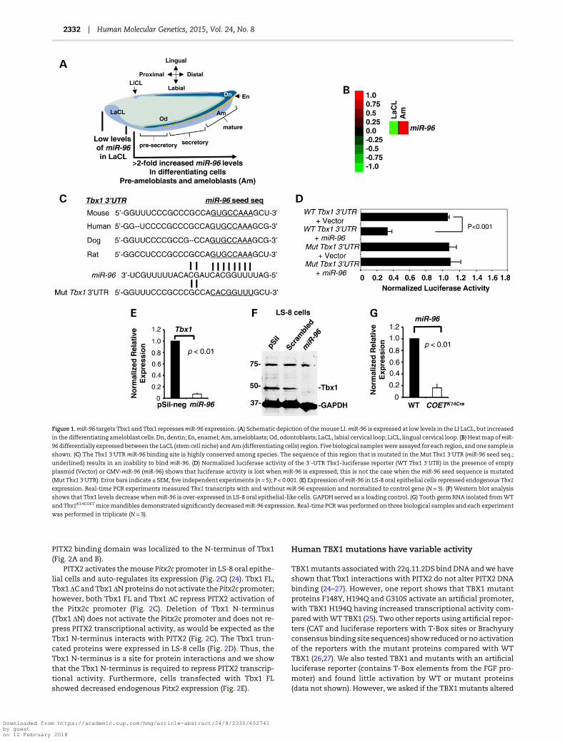
ResultsmiR-96-5p regulates Tbx1 expression in the dentalepithelium
miRs play a critical role in the regulation of tooth stem cell prolif-eration and differentiation (18,20–23). A schematic representa-tion of the mouse lower incisor (LI) and cells that populate thegrowing incisor during tooth development, including the LaCL(the stem cell niche), is shown (Fig. 1A). Themouse dental epithe-lium (light blue, which includes the LaCL and ameloblasts, Am)was extracted and the LaCL cells were isolated from the amelo-blast cells and used to analyze miR expression. Analyses of miRexpression during LI development at P0 showed low miR-96-5pexpression in the LaCL, however,miR-96-5p expression increased>2-fold in the differentiating pre-ameloblast and ameloblast cells(Fig. 1B). The Tbx1 3′UTR contains a highly conserved miR-96binding element, which was cloned into a luciferase reporter toassess miR-96 function (Fig. 1C). miR-96 repressed luciferase ex-pression from the WT Tbx1 3′UTR, but not from a mutated Tbx13′UTR, in LS-8 oral epithelial cells (Fig. 1D). Over-expression ofmiR-96 in LS-8 cells repressed endogenous Tbx1 expression,as shown by real-time PCR (Fig. 1E). Western blots of miR-96transfected LS-8 cells demonstrated decreased Tbx1 protein,while cells transfected with empty vector or a scrambled miRdid not show a change in Tbx1 expression (Fig. 1F). Togetherthese data demonstrate that miR-96 represses Tbx1 andcorrelates with high levels of Tbx1 expression in the LaCL (low le-vels ofmiR-96). Interestingly, a miR screen in Tbx1 over-expressionmice (COETK14Cre) mandibles revealed a decrease in miR-96expression compared with wild-type (WT). Real-time PCR con-firmed decreased miR-96 expression in COETK14Cre mice mandible(Fig. 1G). Thus, we have tentatively identified a Tbx1-miR-96feedback loop, where miR-96 represses Tbx1 and Tbx1 repressesmiR-96 expression.
The Tbx1 N-terminus is required for repressionof PITX2 transcriptional activity
We have previously shown that Tbx1 interacts with the PITX2C-terminus to repress PITX2 transcriptional activity (24). How-ever, the Tbx1 domain for protein interactions was not known.We generated a series of Tbx1 truncated proteins to test forPITX2 interactions and transcriptional activity (Fig. 2A). GST-Tbx1 pull-down experiments demonstrate that PITX2 binds tothe N-terminus of Tbx1 (Fig. 2B). PITX2 protein (500 ng) was incu-bated with GST-Tbx1 FL (full-length) and truncated proteins todetermine the protein interaction domain of Tbx1. PITX2 boundto Tbx1 FL, Tbx1 ΔC and Tbx1 ΔTC, but not to Tbx1 T-box orTbx1 ΔNT. PITX2 did not bind to Tbx1 ΔN (data not shown). The
Human Molecular Genetics, 2015, Vol. 24, No. 8 | 2331
Downloaded from https://academic.oup.com/hmg/article-abstract/24/8/2330/652741by gueston 12 February 2018
PITX2 binding domain was localized to the N-terminus of Tbx1(Fig. 2A and B).
PITX2 activates themouse Pitx2c promoter in LS-8 oral epithe-lial cells and auto-regulates its expression (Fig. 2C) (24). Tbx1 FL,Tbx1 ΔCandTbx1 ΔNproteins do not activate the Pitx2c promoter;however, both Tbx1 FL and Tbx1 ΔC repress PITX2 activation ofthe Pitx2c promoter (Fig. 2C). Deletion of Tbx1 N-terminus(Tbx1 ΔN) does not activate the Pitx2c promoter and does not re-press PITX2 transcriptional activity, as would be expected as theTbx1 N-terminus interacts with PITX2 (Fig. 2C). The Tbx1 trun-cated proteins were expressed in LS-8 cells (Fig. 2D). Thus, theTbx1 N-terminus is a site for protein interactions and we showthat the Tbx1 N-terminus is required to repress PITX2 transcrip-tional activity. Furthermore, cells transfected with Tbx1 FLshowed decreased endogenous Pitx2 expression (Fig. 2E).
Human TBX1 mutations have variable activity
TBX1mutants associatedwith 22q.11.2DS bind DNA andwe haveshown that Tbx1 interactions with PITX2 do not alter PITX2 DNAbinding (24–27). However, one report shows that TBX1 mutantproteins F148Y, H194Q and G310S activate an artificial promoter,with TBX1 H194Q having increased transcriptional activity com-paredwithWTTBX1 (25). Two other reports using artificial repor-ters (CAT and luciferase reporters with T-Box sites or Brachyuryconsensus binding site sequences) show reduced or no activationof the reporters with the mutant proteins compared with WTTBX1 (26,27). We also tested TBX1 and mutants with an artificialluciferase reporter (contains T-Box elements from the FGF pro-moter) and found little activation by WT or mutant proteins(data not shown). However, we asked if the TBX1mutants altered
Figure 1.miR-96 targets Tbx1 and Tbx1 repressesmiR-96 expression. (A) Schematic depiction of themouse LI.miR-96 is expressed at low levels in the LI LaCL, but increased
in the differentiating ameloblast cells. Dn, dentin; En, enamel; Am, ameloblasts; Od, odontoblasts; LaCL, labial cervical loop; LiCL, lingual cervical loop. (B) HeatmapofmiR-
96differentially expressed between the LaCL (stemcell niche) andAm (differentiating cells) region. Five biological sampleswere assayed for each region, and one sample is
shown. (C) The Tbx1 3′UTR miR-96 binding site is highly conserved among species. The sequence of this region that is mutated in the Mut Tbx1 3′UTR (miR-96 seed seq.;
underlined) results in an inability to bind miR-96. (D) Normalized luciferase activity of the 3′-UTR Tbx1-luciferase reporter (WT Tbx1 3′UTR) in the presence of empty
plasmid (Vector) or CMV-miR-96 (miR-96) shows that luciferase activity is lost when miR-96 is expressed; this is not the case when the miR-96 seed sequence is mutated
(Mut Tbx1 3′UTR). Error bars indicate ± SEM, five independent experiments (n = 5); P < 0.001. (E) Expression ofmiR-96 in LS-8 oral epithelial cells repressed endogenous Tbx1
expression. Real-time PCR experiments measured Tbx1 transcripts with and without miR-96 expression and normalized to control gene (N = 3). (F) Western blot analysis
shows that Tbx1 levels decreasewhenmiR-96 is over-expressed in LS-8 oral epithelial-like cells. GAPDH served as a loading control. (G) Tooth germ RNA isolated fromWT
and Tbx1K14COETmicemandibles demonstrated significantly decreasedmiR-96 expression. Real-time PCRwas performed on three biological samples and each experiment
was performed in triplicate (N = 3).
2332 | Human Molecular Genetics, 2015, Vol. 24, No. 8
Downloaded from https://academic.oup.com/hmg/article-abstract/24/8/2330/652741by gueston 12 February 2018

PITX2 transcriptional activity. A schematic representation of theTBX1 gene and mutations is shown (Fig. 3A). TBX1 has three iso-forms, A, B and C with C being the most conserved betweenmice and humans and the most highly expressed in humans(28,29). To determine if these mutants repressedPITX2 transcriptional activity PITX2 was transfected at 1 μg andTBX1 plasmids at 0.25, 0.5 and 1 μg s, respectively. Interestingly,human TBX1 variant C (VC) showed a slight activation of thePitx2c promoter in LS-8 cells (Fig. 3B). However, both TBX1 VCand TBX1 G310S (G-S) mutant proteins repressed PITX2 transcrip-tional activation of the Pitx2c promoter (Fig. 3B). TBX1 G-S andTBX1 H194Q (H-Q)mutant proteins did not activate the Pitx2c pro-moter. Interestingly, the TBX1 H-Q protein did not repress PITX2activation (Fig. 3B). In previous reports, the stability of the TBX1mutant proteins was not analyzed, and we found that both TBX1G-S and H-Q were expressed and stable in the LS-8 cells. BecausePITX2 interacts with the N-terminus of Tbx1 and Tbx1 interactswith the C-terminus of PITX2 the loss of TBX1 repression of
PITX2 with the H-Q mutation suggests other protein functions.Further experiments are required to determine the exactmechan-isms. These data could explain phenotypic variations among22q.11.2DS patients.
Tbx1 binds to the miR-96 promoter and repressesmiR-96 expression
To confirm Tbx1 regulation ofmiR-96, a chromatin immunopreci-pitation (ChIP) assaywas performed to demonstrate endogenousTbx1 binding to the miR-96 chromatin. A Tbx1 binding site wasidentified 3251 base pairs upstream of the miR-96 transcriptionstart site (Fig. 4A). This Tbx1 binding site is similar to a recentreport that identified Tbx1 binding sites by SELEX (27). Asa control, primers were designed to a site upstream of the Tbx1binding element in the miR-96 promoter and this DNA wasnot immunoprecipitated (IP’ed) by IgG serum or Tbx1 antibody(Fig. 4A, lanes 2 and 3, respectively). However, the Tbx1 antibody
Figure 2.The Tbx1N-terminus is required for PITX2 interaction and repression of PITX2 transcriptional activity. (A) A schematic of the Tbx1 truncated proteins used in the
GST pull-down and transfection assays. The black shaded region is the T-box DNA binding domain. (B) GST pull-down using GST-Tbx1 truncated proteins to bind purified
PITX2 protein. The PITX2 bound protein was resolved on 10% PAGE gel transferred to PVDF filters, immunoblotted and detected using PITX2ABCDE antibody (Capra
Science, Sweden) and ECL reagents. PITX2 bound to Tbx1 full-length (FL), Tbx1ΔC (C-terminus deleted) and Tbx1ΔTC (T-box and C-terminus deleted). PITX2 did not
bind to the Tbx1 T-box (Tbx1T-box) or Tbx1 C-terminus (Tbx1ΔNT), also PITX2 did not bind to Tbx1ΔN (data not shown). (C) Tbx1 truncations were tested in
transfection assays to determine their activity and ability to repress PITX2 transcriptional activation of the Pitx2c promoter. As expected Tbx1 activated the Pitx2c
promoter at low levels and the Tbx1ΔC and Tbx1ΔN proteins did not activate the promoter. However, deletion of the Tbx1 N-terminus (Tbx1ΔN) did relieve the
repressive effect of Tbx1 on PITX2 transcriptional activation of the Pitx2c promoter. Luciferase activity is shown as mean-fold activation compared with activity in the
context of the empty expression plasmid (Vector). All luciferase activitieswere normalized to β-galactose expression; five independent experimentswere performed in LS-
8 cells (N = 5). (D) Western blot of transfected cells to show expression in transfected LS-8 cells. Whole-cell lysates (30 μg) were resolved on 10% polyacrylamide gels, and
PITX2 and Tbx1 truncated proteins were detected using an antibody against the Myc tag. All Tbx1 truncated proteins were expressed and denoted by an asterisk. (E) Real-time PCR experiments from cells transfected with Tbx1 or empty vector demonstrate decreased levels of endogenous Pitx2 transcripts by Tbx1 over-expression (N = 3).
Human Molecular Genetics, 2015, Vol. 24, No. 8 | 2333
Downloaded from https://academic.oup.com/hmg/article-abstract/24/8/2330/652741by gueston 12 February 2018

did IP the DNA containing the Tbx1 binding element (Fig. 4A,lane 7).
The miR-96 promoter was cloned (∼5 kb) into the luciferasevector to test for Tbx1 functional regulation. Murine Tbx1,human TBX1 VC and TBX1 G-S all repressed themiR-96 promoter(4-fold or greater, P < 0.05), while TBX1 H-Q had no effect (Fig. 4B).Furthermore, because TBX1 H-Q binds DNA (27), the inability ofthismutant to repress themiR-96 promoter could suggest defect-ive protein interactions, other than PITX2. Thus, Tbx1 directlyrepresses miR-96 expression and this is the first demonstrationof Tbx1 repression of a miR.
Because Tbx1 repressed PITX2 transcriptional activation, wereasoned that miR-96 indirectly modulated PITX2 transcriptionalactivity through the regulation of Tbx1. LS-8 cells co-transfectedwith miR-96 and the Pitx2c promoter revealed no activation;however, co-expression of PITX2 and miR-96 increased PITX2activation of the Pitx2c promoter, compared with PITX2 alone(Fig. 4C). Thus, miR-96 repression of Tbx1 indirectly activatesPITX2 transcriptional activity.
Specific Tbx1 expression in the incisor CL controlsincisor development
The rodent incisor is a uniquemodel for the differentiation of en-amel organ cells from stemcells to enamel-secreting ameloblasts(Fig. 5A). Stem cells located in the LaCL give rise to the pre-secre-tory, secretory and maturation-stage epithelial or ameloblastcells.
Tbx1 was conditionally knocked out using the K14Cre mousecrossed to the Tbx1f/f mouse to generate the Tbx1K14cKO mice. TheK14 promoter is active in surface ectoderm and basal cells fromembryonic day E9.5 in developing hair follicles and tooth epithe-lia (30–32). At P0, the LI LaCL was smaller and disorganized inTbx1K14cKOmice comparedwithWT, and the inner enamel epithe-lial (IEE) cells appeared undifferentiated and not well polarized(Fig. 5B and C). Furthermore, the OEE and stratum intermedium(SI) layer was thin, unorganized and lacked structure (Fig. 5C).
Tbx1 transcripts were specific to the dental epithelium in boththe incisors and molars and were detected from E10.5 to E18.5(10,33,34). Tbx1 protein expression in teeth was confirmed by im-munofluorescence of the LI at E16.5 and Tbx1 was expressed inthemyogenic core of the tongue (t), dental follicle, dental lamina,oral epithelium and in both the LaCL and LiCL (Fig. 5D–I). Tbx1 ex-pression was seen in the IEE, OEE and SR of the LaCL (Fig. 5H andI). However, Tbx1 expression decreased in differentiating amelo-blasts and was not seen in the odontoblasts.
The Tbx1Cre mouse was crossed with the ROSA26LacZ reportermouse to better understand Tbx1 cell fate during incisor develop-ment. Fate mapping of Tbx1-expressing cells revealed Tbx1-expressing cells specifically in the LaCL of the LI. Highmagnificationimaging of the LaCL indicated that Tbx1-expressing cells popu-lated the distal region of the SR, and in the OEE and IEE (Fig. 5J).The stem cells located in the SR compartment intercalate intothe OEE and move around the CL to the IEE, where they divideand migrate to the distal part of the growing incisor (35) (seearrows, Fig. 5J). Interestingly, cells in the proximal SR were Tbx1
Figure 3.TBX1humanmutants differentially regulate PITX2 transcriptional activity. (A) Schematic representation of humanTBX1 isoforms andmutations. TBX1C, TBX1A
and TBX1B aremade by alternative splicing of the last exons and the T-box is shown. (B) TBX1 VC (variant C), TBX1 G310S (G-S) and TBX1 H194Q (H-Q) were transfected or
cotransfected with PITX2 and the Pitx2c promoter. Each transfection used 0.1 µg reporter, 0.1 µg PITX2 and 0.25, 0.05, 0.1 µg of the TBX1 construct. To control for
transfection efficiency, all transfections included the SV-40 β-galactosidase reporter (0.05 μg). Cells were incubated for 24 h and then assayed for luciferase and
β-galactosidase activities. The activities are shown as mean-fold activation compared with the luciferase plasmid with empty vector and normalized to
β-galactosidase activity ±SE from three independent experiments. TBX1 VC slightly activated the Pitx2c promoter (6-fold) and showed a dose-responsive repression of
PITX2 transcriptional activity. TBX1 G-S did not activate the Pitx2c promoter and also showed a dose-responsive repression of PITX2 activity albeit at lower levels
compared with WT TBX1 VC. However, TBX1 H-Q did not activate the Pitx2c promoter or repress PITX2 transcriptional activation of the Pitx2c promoter. (C) Western
blots of transfected LS-8 cells to demonstrate expression levels of TBX1 mutant proteins. Whole-cell lysates (30 μg) were resolved on 10% polyacrylamide gels, and
TBX1 mutant proteins were detected using an antibody against the Myc tag. GAPDH served as a loading control. TBX1 VC, TBX1 G-S and TBX1 H-Q transfected
proteins were expressed in the cells.
2334 | Human Molecular Genetics, 2015, Vol. 24, No. 8
Downloaded from https://academic.oup.com/hmg/article-abstract/24/8/2330/652741by gueston 12 February 2018

negative (Fig. 5J). Tbx1 daughter cells were observed in the differ-entiating ameloblasts or secretory cells and in the SI (Fig. 5K).Overall, these results suggest that Tbx1 marks a specific subsetof dental epithelial stem cells in the LaCL, different and inde-pendent from Sox2 (36).
Abnormal tooth development and amelogeninexpression in Tbx1K14cKO mutant embryosand neonate miceThe differentiation of dental epithelial cells into ameloblastsoccurs through several morphological stages, over this period
the cells become elongated and polarized, features that arerequired for the deposition of enamel (37).
In this report we used Tbx1 conditionally deleted mice, K14Cre
X Tbx1flox/flox(Tbx1K14cKO) embryos to study tooth and craniofacialmorphogenesis. Hematoxylin and eosin (H&E) staining of sagittalsections of the craniofacial region at E16.5 demonstrated a delayin upper incisor (UI) and LI morphogenesis in Tbx1K14cKO mutantmice compared with WT counterparts (data not shown). TheTbx1K14cKO embryos at E18.5 had small LIs (∼25% decrease insize, black bar compared with WT) and small LaCL regions(Fig. 6A and B). Higher magnification of the differentiating dental
Figure 4. Endogenous Tbx1 binds to themiR-96 chromatin. (A) Top panel is a schematic representation of the miR-96 5′-flanking region upstream of pri-miR-96. The Tbx1
binding site is shown and the primer regions used to amplify the chromatin as well as the control primers upstream of the Tbx1 binding site. Bottom panels, ChIP of
endogenous Tbx1 binding to the T-box element upstream of the pri-miR-96 transcript in LS-8 cells, lane 7. IgG did not IP the Tbx1 binding site chromatin, lane 6. IgG
and Tbx1 Ab did not IP the control region (non-specific primers) upstream of the Tbx1 binding sequence, lanes 2 and 3, respectively. Rabbit antisera was used as a
control IP and Tbx1 antibody (Invitrogen) was used to IP Tbx1 binding to the chromatin. The input chromatin is shown as a positive control for the ChIP. (B)Expression plasmids containing the murine Tbx1, human TBX1 variant C, TBX1 G310S and TBX1 H194Q cDNAs were co-transfected into LS-8 cells with the miR-96
5 kb luciferase reporter plasmid. Luciferase activity is shown as mean-fold activation compared with that in the presence of empty mock expression plasmid. All
luciferase activities were normalized to β-galactose expression, three independent experiments (N = 3). (C) Tbx1, PITX2 and miR-96 were co-transfected in LS-8 cells
with the Pitx2c luciferase promoter and luciferase activity measured as in Figure 2. All luciferase activities were normalized to β-galactose expression, three
independent experiments (N = 3).
Human Molecular Genetics, 2015, Vol. 24, No. 8 | 2335
Downloaded from https://academic.oup.com/hmg/article-abstract/24/8/2330/652741by gueston 12 February 2018

epithelium (ameloblasts, Am) and dental mesenchyme (odonto-blasts, Od) revealed only minor defects in both Od and Am polar-ization and differentiation of the LI at E18.5 in the Tbx1K14cKO
embryos (Fig. 6C and D). However, the small tooth size couldhave resulted from a lack of stem cell proliferation in the LaCL.
Ameloblasts are responsible for the secretion of the threestructural enamel matrix proteins, amelogenin, ameloblastinand enamelin. Amelogenin constitutes ∼90% of the enamel or-ganicmatrix and is highly conserved across species (38–40). Ame-logenin and ameloblastin are essential for proper enamelformation (41–44). At P1, amelogenin was decreased in Tbx1K14cKO
LIs comparedwithWT (Fig. 6E–H). At P4, amelogenin levels in theincisors remained low or absent in the mutant mice (data not
shown). The low expression of amelogenin can cause enameldefects.
H&E staining of the lower molars (LMs) at E16.5 revealed adelay in early bell stage morphogenesis in Tbx1K14cKO embryos(data not shown). The Ek, the regionwhere epithelial–mesenchy-mal signaling regulates tooth size and shape, was normal in WTmice, but underdeveloped or absent in Tbx1K14cKO mice (data notshown). The Ek is an organizer of cusp formation and sagittal sec-tions of P0 lowermolars demonstrated defectivemolar cusping ofthe first molar (M1) in the Tbx1K14cKO mice compared with WTmice (Fig. 6I and J). Higher magnification showed defectiveodontoblast (Od) and ameloblast (Am) differentiation in theTbx1K14cKO lower molars (Fig. 6K and L). At P4, amelogenin levels
Figure 5. Tbx1 expressing cells are in a unique region of the LaCL. (A) Schematic representation of depiction of the mouse LI. The LaCL contains dental stem cells that
supply the continuously growing mouse incisor with replacement cells. The pre-secretory, secretory and mature ameloblasts are the components that produce
enamel on the labial side of the incisor. The LiCL provides cells to the growing incisor but these cells do not form ameloblasts or make enamel. Od, odontoblasts.
(B and C) H&E staining of the LIs of P0 WT, and Tbx1K14cKO mice to determine the role of Tbx1 in the LaCL. Mutant mice have smaller CLs and at P0, the cells of both
the IEE and OEE are disorganized compared with their counterparts in WT mice. The SR contains the stem or progenitor cells. The SI is also thinner in the mutant
mice. (D–I) IHC experiments show that Tbx1 is highly expressed in the LaCL and LiCL regions of the E16.5 WT LI, 2-(4-Amidinophenyl)-6-indolecarbamidine
dihydrochloride, 4′,6-Diamidino-2-phenylindole dihydrochloride (DAPI) staining provides contrast. (J) The Tbx1Cre X Rosa26LacZ mouse was analyzed for LacZ staining
and Tbx1 fate mapping experiments. LacZ-positive cells are derived from Tbx1 expressing cells. In panel J, the SR compartment of the LaCL has populations of Tbx1-
negative cells and Tbx1-positive cells. The Tbx1-positive cells are incorporated into the OEE and IEE, as shown by LacZ staining (the arrows denote the direction of
migration of the progenitor cells). (K) The secretory ameloblasts and SI contain cells that expressed Tbx1 and migrated to the distal end of the incisor. These
experiments were repeated three times for each embryonic stage and genotype (N = 3). Abbreviations: t, tongue. Scale bar = 100 μm.
2336 | Human Molecular Genetics, 2015, Vol. 24, No. 8
Downloaded from https://academic.oup.com/hmg/article-abstract/24/8/2330/652741by gueston 12 February 2018
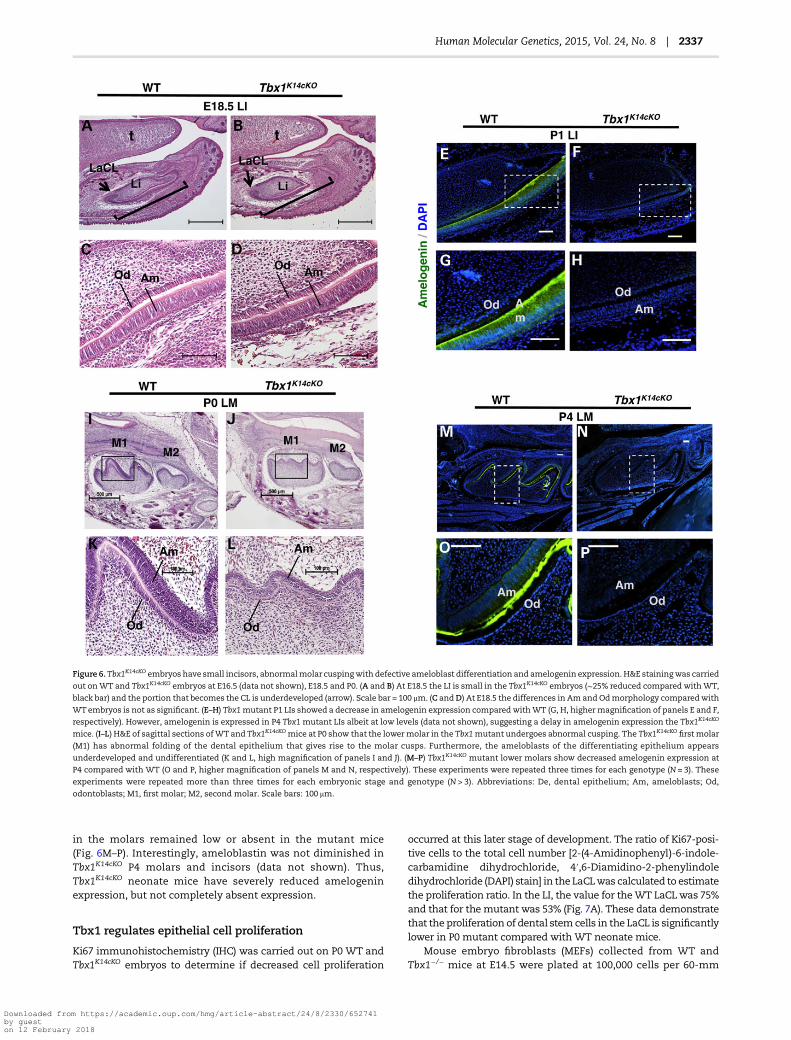
in the molars remained low or absent in the mutant mice(Fig. 6M–P). Interestingly, ameloblastin was not diminished inTbx1K14cKO P4 molars and incisors (data not shown). Thus,Tbx1K14cKO neonate mice have severely reduced amelogeninexpression, but not completely absent expression.
Tbx1 regulates epithelial cell proliferation
Ki67 immunohistochemistry (IHC) was carried out on P0 WT andTbx1K14cKO embryos to determine if decreased cell proliferation
occurred at this later stage of development. The ratio of Ki67-posi-tive cells to the total cell number [2-(4-Amidinophenyl)-6-indole-carbamidine dihydrochloride, 4′,6-Diamidino-2-phenylindoledihydrochloride (DAPI) stain] in the LaCLwas calculated to estimatethe proliferation ratio. In the LI, the value for theWT LaCLwas 75%and that for the mutant was 53% (Fig. 7A). These data demonstratethat the proliferation of dental stem cells in the LaCL is significantlylower in P0 mutant compared withWT neonate mice.
Mouse embryo fibroblasts (MEFs) collected from WT andTbx1−/− mice at E14.5 were plated at 100,000 cells per 60-mm
Figure 6. Tbx1K14cKO embryos have small incisors, abnormalmolar cuspingwith defective ameloblast differentiation and amelogenin expression. H&E stainingwas carried
out onWT and Tbx1K14cKO embryos at E16.5 (data not shown), E18.5 and P0. (A and B) At E18.5 the LI is small in the Tbx1K14cKO embryos (∼25% reduced compared withWT,
black bar) and the portion that becomes the CL is underdeveloped (arrow). Scale bar = 100 μm. (C andD) At E18.5 the differences in Amand Odmorphology comparedwith
WTembryos is not as significant. (E–H) Tbx1mutant P1 LIs showed a decrease in amelogenin expression compared withWT (G, H, highermagnification of panels E and F,
respectively). However, amelogenin is expressed in P4 Tbx1 mutant LIs albeit at low levels (data not shown), suggesting a delay in amelogenin expression the Tbx1K14cKO
mice. (I–L) H&E of sagittal sections ofWT and Tbx1K14cKO mice at P0 show that the lowermolar in the Tbx1mutant undergoes abnormal cusping. The Tbx1K14cKO first molar
(M1) has abnormal folding of the dental epithelium that gives rise to the molar cusps. Furthermore, the ameloblasts of the differentiating epithelium appears
underdeveloped and undifferentiated (K and L, high magnification of panels I and J). (M–P) Tbx1K14cKO mutant lower molars show decreased amelogenin expression at
P4 compared with WT (O and P, higher magnification of panels M and N, respectively). These experiments were repeated three times for each genotype (N = 3). These
experiments were repeated more than three times for each embryonic stage and genotype (N > 3). Abbreviations: De, dental epithelium; Am, ameloblasts; Od,
odontoblasts; M1, first molar; M2, second molar. Scale bars: 100 μm.
Human Molecular Genetics, 2015, Vol. 24, No. 8 | 2337
Downloaded from https://academic.oup.com/hmg/article-abstract/24/8/2330/652741by gueston 12 February 2018

culture dish and counted after 24, 48 and 72 h. At each time point,Tbx1−/−MEFs proliferatedmore slowly thanWTMEFs (Fig. 7B andC). There were no morphological changes in the MEFs betweenWT and Tbx1−/− mice. LS-8 oral epithelial cells were transducedwith Tbx1 or vector only and cell viability/cell growth was re-corded as fold increase compared with non-transduced cells.Tbx1 expressing cells demonstrated an increase in cell viabilitycompared with WT and vector only transduced cells (Fig. 7D).Taken together, these data suggest that Tbx1 regulates cell prolif-eration, and that Tbx1 in the epithelium alone regulates toothsize by controlling cell proliferation and differentiation.
Tooth size, shape and enamel formation are reducedin adult Tbx1K14cKO mice
Skeletal preparations of P14 WT and Tbx1K14cKO mice were made,and scanning electron microscopy (SEM) and microCT (μCT)scans were carried out on the samples. The LIs were shorter inthe mutant compared with the WT mice and the enamel layerthinner (Fig. 8A–D). At a higher magnification, the incisal edgeappeared worn down or chipped in the mutant, indicating apotential decrease in enamel mineralization and/or structuraldefects. SEM imaging of incisors that were fractured perpendicu-lar to the growth axis in the erupted portion of the tooth showedthat the enamel layer was thinner in the mutant than the WTmice (Fig. 8E and F). At higher magnification, it became evident
that the orientation of the enamel crystallite bundles (prisms)is normal in the mutant mice. However, the prisms are lessdensely packed and separate easily from the underlying dentinat the enamel dentine junction (Fig. 8G and H).
At P14, the firstmolar (M1)was smaller in themutant (Fig. 8I–L).Also, the second molar (M2) lacked a distal cusp (Fig. 8N, O, Q, R,arrow). The cusps of the first two molars in the mutant mice werenot as sharp or deep compared with WT mice (Fig. 8Q and R) anddevelopmentof the thirdmolar (M3)wasmoreadvanced in themu-tant compared with WT mice (Fig. 8M–T). Overall, molars of theTbx1K14cKOmice were smaller (Fig. 8J and K) and featured decreasedenamel formation (Fig. 8I, L, M and P), malformed cusping of thefirst two molars and premature growth of the third molar. Theincreased growth of the thirdmolar was surprising, butmay correl-ate to the lack of Pitx2 repression, which initiates dentaldevelopment.
Tbx1 over-expression regulates incisor size and dentalepithelial stem cell proliferation
Analyses of E16.5 LIs of the K14Cre activated Tbx1 over-expressionmouse (COETK14Cre) demonstrated a larger LaCL with less differ-entiated cells (Am) compared with WT mice (Fig. 9A–D). The LIof the COETK14Cre mice is larger than that of WT at P1, with an in-crease in the width and length of the LaCL (Fig. 9E–H). Cell prolif-eration was measured by Ki67 staining of the P1 LI LaCL and
Figure 7. Tbx1 regulates cell proliferation. (A) LIs from WT or Tbx1K14cKOP0 mice were processed, sectioned and stained for Ki67 to examine cell proliferation. Ki67 was
decreased in Tbx1K14cKOtooth sections, and Ki67/DAPI ratio was quantified to determine that the decrease in cell proliferation was statistically significant (P < 0.05). (B)MEFs were collected from WT and Tbx1−/− embryos at E14.5. Growth was analyzed by seeding each well with 100 000 cells, and cells were counted after 24, 48 and 72 h.
MEFs extracted from Tbx1−/− embryos showed decreased cell proliferation comparedwithWT. (C) MEF cell growth was quantitated revealing a decrease in proliferation of
the Tbx1mutant cells. Two different embryos were used for WT and Tbx1−/− and each cell count was done in triplicate. These experiments were repeated three times for
each genotype. (D) LS-8 oral epithelial cells were transduced with a lentivirus expressing Tbx1 or empty vector. Cells were cultured for 48 h and cell growth/viability was
measured every 12 h.
2338 | Human Molecular Genetics, 2015, Vol. 24, No. 8
Downloaded from https://academic.oup.com/hmg/article-abstract/24/8/2330/652741by gueston 12 February 2018

analyses of several mice (N = 3) revealed an increase in cell prolif-eration in the COETK14Cre mice (Fig. 9I–L). These data are consist-ent with loss of Tbx1 function resulting in small incisors anddecreased cell proliferation in the CL regions or stem cell niche.Thus, the expression and dose of Tbx1 control dental stem cellproliferation and differentiation.
Incisor growth, amelogenin expression and enamelformation are increased with Tbx1 over-expression
We asked if Tbx1 over-expression increased amelogenin expres-sion and if it correlates with an increase in enamel formation inthe COETK14Cre mice. At P1, both UIs and LIs showed increasedamelogenin expression in the proximal regions comparedwith WT mice (Fig. 10A–H). These results suggest that Tbx1transcriptional mechanisms may regulate amelogenin or that
Tbx1 expression expands cell proliferation and increased celldifferentiation.
To understand tooth structure and mineralization, we ana-lyzed the incisors and molars of 2-week-old COETK14Cre miceby μCT. Tbx1 over-expression resulted in a larger incisor with in-creased enamel formation compared withWT (Fig. 10I and J; seearrows), and increased enamel formation (red) in the molars(Fig. 10K and L; see arrows). In COETK14Cre mice at 4 weeks ofage, themandibles showed a decrease in alveolar bone develop-ment (Fig. 10M and N; light green arrow and orange bracket). En-amel thickness of the LI was increased in the COETK14Cre mouse(Fig. 10M–P; blue arrow). Development of the third molar(Fig. 10O and P; yellow arrow) was delayed and cortical boneformation was decreased (Fig. 10O and P; white arrow). Theseresults are consistent with a role for Tbx1 in regulating toothsize, shape and dental epithelial cell differentiation, which
Figure 8. Tbx1K14cKO two-week-old mice have dental anomalies and developmentally advanced third molar eruption. Skeletal preparations were performed on WT and
Tbx1K14cKO mice at P14. (A and B) μCT images of mandibles in parasagittal plane show the thinner enamel layer on the LIs (orange arrows), as well as advanced
mineralization of the third molar crown in the Tbx1K14cKO mouse compared with WT (green arrows). (C and D) SEM of the lingual side of LIs reveal smaller incisors
with more wear indicating a difference in biomechanical properties of enamel, compared with WT. Scale bar 100 μm. (E and F) SEM of fracture surfaces of the erupted
portion of the LI show that Tbx1K14cKO enamel is significantly thinner as indicated by blue brackets of identical length. Scale bar 10 μm. (G and H) Mature enamel
fractured at similar positions on the erupted incisor at higher magnification. Bundles of enamel crystallites (prisms) are packed densely in WT enamel but loosely
with spaces between them in the Tbx1K14cKO mouse. Scale bar 10 μm. (I–P) μCT scans at P14 show that mutant mice have a smaller first (M1) and second (M2) molars
and a third (M3) molar that is unusually advanced in its development (green arrows). (I and L) Sections in coronal plane through the center of the distal root of the
first molar, as indicated by the dotted blue line in image J and K, show differences in molar size an enamel development (less enamel mineralization on the incisor
marked by orange arrow) (J and K) Maximum density projection through the mandible in transverse plane shows differences in tooth size. (M and P) Slices in
parasagittal plane through the center of the distal root of the first molar and (N and O) maximum density projection in the sagittal plane show that in the mutant
mice molar cusping is defective (purple arrow) and the third molar is advanced in development (green arrow). (Q and R) SEM images of whole mandibles show clearly
that M2 lacks a distal cusp (purple arrow) and is smaller in themutantmouse and that M3 has erupted and shows advanced development. Furthermore, the enamel ofM1
and M2 shows signs of wear. Scale bar 100 μm. (S and T) SEM images of whole molars dissected out of the mandibles show differences in M2 cusp formation (purple
arrows), similarities in root development and advanced M3 development in the mutant mouse. Scale bar 200 μm. Abbreviations: M1, first molar; M2, second molar;
M3, third molar.
Human Molecular Genetics, 2015, Vol. 24, No. 8 | 2339
Downloaded from https://academic.oup.com/hmg/article-abstract/24/8/2330/652741by gueston 12 February 2018

leads to enamel formation. Interestingly, third molar develop-ment was decreased in the Tbx1 over-expression mouse,which correlates to Tbx1 interaction with PITX2 to repress itstranscriptional gene network required for normal dentaldevelopment.
Tbx1 loss of function and gain of function embryoshave cleft palate
The E16.5 Tbx1K14cKO embryos exhibited a cleft palate. In our Tbx1conditional deletion mice the palatal shelves have elevated butdo not fuse (Fig. 11A and B). Interestingly, Tbx1 over-expressionalso caused cleft palate in COETK14Cre mice (Fig. 11C and D). Thisis consistent with a previous report showing cleft palate in em-bryos over expressing Tbx1 (COETAp2Cre) in the surface ectoderm(45). The palatal shelves appeared to elevate but did not fuse atthe midline.
DiscussionThe cloning and characterization of Tbx1 inmice established thisgene as essential for embryonic development. To understand therole of Tbx1 in odontogenesis, gene andmiR expression was ana-lyzed in WT, Tbx1K14cKO and COETK14Cre embryonic stage-specificmouse mandibles, maxilla and dental epithelial tissue. Bioinfor-matics analyses of the expression data revealed genes and miRsregulated by Tbx1. Comparison of increased gene expressionwithdecreasedmiR expression or the inverse in these tissues revealedtentative correlations of miR-regulated gene expression con-trolled by Tbx1. A regulatory loop was identified between Tbx1and miR-96, which further correlated with their expression pat-terns in the developing incisor epithelium. To further establisha potential link to 22q11.2DS, several TBX1mutants were assayedfor their transcriptional activity and their ability to regulate thegenes and miRs identified in the bioinformatics screens. The
Figure 9. Tbx1 over-expression increases incisor size and dental stem cell proliferation. (A–D) H&E staining ofWTand COETK14Cre E16.5 LIs. The overall size of the incisor is
similar in mice of the two genotypes at this early stage (A and B). Scale bar = 500 μm. However, the LaCL (boxed region) is larger (width and length) with an increase in cell
number in the SR region of the CL and decreased ameloblast differentiation (outlined with white dotted line, highermagnification C and D). Scale bar = 100 μm. (E–H) H&E
staining of WT and COETK14Cre P1 LIs. At this later stage, the COETK14Cre LI is larger (width and length) than itsWT counterpart and the LaCL remains increased in size and
shape (the LaCL is elongated in the COETK14Cre incisor). Scale bar = 500μm. There are more cells in the SR region of the CL in the COETK14Cre incisor (outlined with white
dotted line, higher magnification G and H). Scale bar = 100 μm. (I–L) P1 LIs from WT or COETK14Cre mice were processed, sectioned and stained for Ki67 to assess cell
proliferation. Ki67 expression was higher in the COETK14Cre LaCLs compared with WT, as established by quantifying the Ki67/DAPI ratio; 10 l of pups and 20 mutants
were sectioned, all showed the same phenotype. Abbreviations: Am, ameloblast; t, tongue; LI, lower incisor; LaCL, labial cervical loop.
2340 | Human Molecular Genetics, 2015, Vol. 24, No. 8
Downloaded from https://academic.oup.com/hmg/article-abstract/24/8/2330/652741by gueston 12 February 2018

TBX1 mutations were analyzed in cell-based assays to under-stand their function compared with WT TBX1. The in vivo bio-informatics approach identified direct targets of Tbx1 in mousemodels for 22q11.2DS and these targets could account for themolecular underpinnings of dental and craniofacial anomaliesobserved in DiGeorge patients. Clearly some Tbx1 molecularmechanisms between humans and mouse models are differenthowever using the approach in this report revealed new geneticpathways potentially associated with 22q11.2DS.
TBX1, 22q11.2DS and associated dental anomalies
TBX1 is a candidate gene for 22q11.2DS and is responsible forthe majority of the phenotypes seen in 22q11.2DS patients. Vari-ous clinical studies have shown that 22q11.2DS patients havetooth defects, ranging from hypodontia to enamel defects (3).Independent of the role of TBX1 in the pharyngeal apparatus,epithelial Tbx1 expression in a maturing tooth has been shownto be specific to the IEE, and cells in this region become matureameloblasts that secrete enamel (10).
Dental anomalies such as enamel hypoplasia and hypomi-neralization, hypodontia and aberrant tooth shape are docu-mented in 22q11.2DS patients (3). Tooth defects in 22q11.2DSpatients have been linked to hypocalcemia from hypoplasiaof the parathyroid, and by micrognathia. Traditionally, enameldisturbances in 22q11.2DS patients were thought to be second-ary effects of hypocalcemia caused by hypoparathyroidism.A recent study concluded that a diagnosis of hypoparathyroid-ism did not affect the prevalence of enamel anomalies (46).Thus, our research demonstrates that the dental and craniofa-cial defects in 22q11.2DS patients involve a gene regulatorynetwork modulated by Tbx1 regulating cell proliferation anddifferentiation.
Tbx1–protein interactions regulate development
Pitx2, a bicoid/paired-related homeobox gene, was initially iden-tified as the mutated gene in the autosomal-dominant, haploin-sufficient Axenfeld-Rieger syndrome (47). Patients with thisdisorder display many tooth abnormalities, including dental
Figure 10. Tbx1 over-expression increases amelogenin, enamel formation and a delay in formation of the thirdmolar. (A–D) WT and COETK14Cre P1 UIs were sectioned and
stained for the expression of amelogenin. Amelogenin expressionwas increased in the COETK14CreUI (highermagnification, compare C,WT toD,COETK14Cre sections). (E–H)
Amelogenin expressionwas increased in the COETK14CreLI (highermagnification, compare G,WT to H, COETK14Cre sections). (I–L) μCT imaging shows an increase in the size
and shape of the P14 (2 weeks old) COETK14Cre incisor compared with its WT counterpart (I and J). The enamel on the labial side is thicker in the COETK14Cre incisor (blue
arrow). Imaging of the P14 mandible (left and right sides) shows an increase in molar enamel formation (blue arrow) and a slight loss of alveolar and cortical bone
formation in the COETK14Cre mandible (green color, K, L). (M and N) microCT analyses of P28 mandibles show an increase in enamel formation (blue arrow) in the
incisor of the COETK14Cre mice, coincident with a decrease in alveolar bone formation (light green arrow and orange brackets). (O and P) microCT scans of the
hemimandible show a developmental delay in formation of the third molar (yellow arrow), a decreased in formation of cortical bone (white arrow) and an increase in
formation of incisor enamel in the COETK14Cre incisor (blue arrow).
Human Molecular Genetics, 2015, Vol. 24, No. 8 | 2341
Downloaded from https://academic.oup.com/hmg/article-abstract/24/8/2330/652741by gueston 12 February 2018

hypoplasia, abnormally shaped teeth and anodontia vera.Withinthe craniofacial region, Pitx2 is the earliest detected transcriptionfactor in the oral epithelium, and the expression patterns of Tbx1and Pitx2 overlap during tooth morphogenesis (48,49). Tbx1represses PITX2-mediated activation of the cyclin-dependentkinase inhibitor p21 in teeth by physically interacting with thePITX2 C-terminus, providing a molecular mechanism for theproliferation of dental epithelial cells (24).
A recent study identified a hierarchical network of transcrip-tion factors expressed in the pharyngeal mesoderm that coordi-nates both heart and craniofacial development. This networkincludes genetic interactions between Tbx1, Pitx2, Lhx2, Tcf21and bHLH genes (14,50). This study suggests that Tbx1 levelscan be fine-tuned by interactions with other transcription fac-tors, and that these factors may be modifiers for 22q11.2DS (50).
We have dissected the role of Tbx1 protein domains and theirinteraction with PITX2 to demonstrate that the Tbx1 N-terminusis required for PITX2 binding and repression of PITX2 trans-criptional activity. Tbx1 repression of PITX2 during tooth devel-opment may regulate tooth initiation and the size and shape of
both incisors and molars. We have shown previously thatPitx2−/+/Tbx1−/+ double het mice form an extra premolar, demon-strating that these two factors interact genetically to regulatetooth initiation and formation (24). In this report, we show thatTbx1K14cKO mice have increased third molar development whilethe COETK14Cre mice have decreased third molar development,suggesting that the dose of Tbx1 regulates tooth initiation andthe timing of tooth development. Because TBX1 is a potent regu-lator of PITX2 transcriptional activity,which initiates tooth devel-opment, the TBX1–PITX2 interaction appears to control toothinitiation and patterning. Tbx1 is expressed early during toothdevelopment and is co-expressed with Pitx2 in the developingincisor and molar. Tbx1 and Pitx2 are early regulators of a geneexpression network that define cell proliferation and differenti-ation of several cell types.
Human TBX1 and DGS associated mutations also regulatePITX2 transcriptional activity. Both TBX1 H194Q and TBX1G310S proteins are stable and not degraded in cells. TBX1 G310Srepresses PITX2 activation but at reduced levels compared withWT TBX1; however, TBX1 H194Q has no effect on PITX2 activity.
Figure 11. Tbx1 loss-of-function and gain-of-functionmice have cleft palate. (A and B) At E16.5 Tbx1K14cKOmice have a cleft palate shownby coronal sections of the anterior
palate. (C andD) Coronal sections of COETK14Cremice at E18.5 show that Tbx1 over-expression causes cleft palate. These experiments were repeatedmore than three times
for each embryonic stage and genotype (N > 3). Abbreviations: LI, lower incisor; t, tongue; Ps, palatal shelves; p, palate. Scale bar = 500 μm.
2342 | Human Molecular Genetics, 2015, Vol. 24, No. 8
Downloaded from https://academic.oup.com/hmg/article-abstract/24/8/2330/652741by gueston 12 February 2018

TheTBX1N-terminal tail is highly conserved in all three isoformswhile the C-terminal tail varies (51). Because the interaction be-tween PITX2 andTBX1 is crucial for embryonic craniofacial devel-opment, it makes sense that PITX2 binds to a highly conservedportion of TBX1. Both TBX1 H194Q and G310S mutations werepredicted to affect TBX1 DNA binding and protein stabilization(25), however neither is affected in the mutant proteins, (27)and our study. However, both mutations may alter dimer forma-tion and/or protein function (25). However, these mutations donot appear to change the N-terminal structure of the TBX1 pro-tein, which binds PITX2. Thus other mechanisms are likelyresponsible for their differential transcriptional activity.
Tbx1 in palatogenesis
Craniofacial malformations occur inmore than half of 22q11.2DSpatients, and cleft palate (complete, submucosal and soft) is oneof the most frequent features (52). Tbx1-null mice exhibited ab-normal epithelial adhesion between the palate and the man-dible, leading to clefts similar to those observed in 22q11.2DS(53,54). However, during palate development it was suggestedthat Tbx1-null epitheliumwas hyperproliferative and did not dif-ferentiate and that Tbx1 over-expression inhibited cell growth(53). Funato et al. suggested that Tbx1 regulated the balance be-tween proliferation and differentiation in the epithelium of thepalatal primordial (53). Our studies demonstrate that both lossand gain of Tbx1 function causes clefting supporting the dose-dependent regulation of palatogenesis and development byTbx1. However, during odontogenesis Tbx1 acts to increaseepithelial cell proliferation consistentwith a role for Tbx1 in regu-lating dental epithelial progenitor cells. This is also consistentwith the role of Tbx1 in cardiac progenitor cells where it increasesproliferation and inhibits differentiation (55).
Tbx1 in odontogenesis
The CL regions (stem niche) of both UIs and LIs displayed de-creased proliferation in Tbx1K14cKOmice and increased proliferationin the Tbx1K14COET mice. These data demonstrated that Tbx1 is
essential for maintenance of dental progenitor cells. When Tbx1(Tbx1K14cKO) was conditionally deleted from the oral and dentalepithelia, we observed microdontia, underdeveloped CLs anddefective ameloblast differentiation and these effects can beexplained by a decrease in the proliferation of progenitor cells.
The size of the developed incisors andmolars appeared smal-ler in the Tbx1K14cKOmice at P14 and P20. Inmolars a distal cusp ismissing in the second molar. These abnormalities may beexplained by decreased progenitor cells in the LaCL and delayedformation of the Ek. Both incisors and molars show wear of theenamel layer, and on close examination, the enamel prismsformed are less dense than in WT mice, especially in the regionclosest to the enamel–dentin junction (EDJ). Tbx1 regulates thetiming of ameloblast differentiation and the subsequent produc-tion of enamel proteins, and the effect seen is a delay and not acomplete absence of enamel production.
Tbx1 expression has been shown to be restricted to the IEE atE12.5 and is maintained by mesenchyme-derived Fgf signaling(56). Tbx1 and Fgf8 interact genetically during development(57). However, using Tbx1Cre fate mapping we demonstrate thatTbx1 expressing cells are located in a unique and distinct regionof the LaCL during incisor development. It appears that Tbx1expressing cells are located in a defined region of the SR thatmay define both lineages of the pre-ameloblast cells and cellsof the OEE and SI. Sox2 expressing cells mark a region of theLaCL that appear not to express Tbx1 (36). We define this regionas Tbx1 negative, which suggests that Tbx1 marks a cell lineageseparate from Sox2. However, more experiments are required todetermine the exact differences.
Tbx1 and microRNA regulation
miRs have been shown to be essential regulators of embryogen-esis. Bmp signaling promotes outflow tract (OFT) myocardialdifferentiation by regulating miRs (58). Bmp signals through aconserved Smad-binding element to regulate miR-17-92, whichresults in decreased Tbx1 expression. Smad1 is a critical negativeregulator of SHF proliferation in vivo, and ablation of Smad1 in theSHF enhances cell proliferation (59). Tbx1 binds to Smad1 and
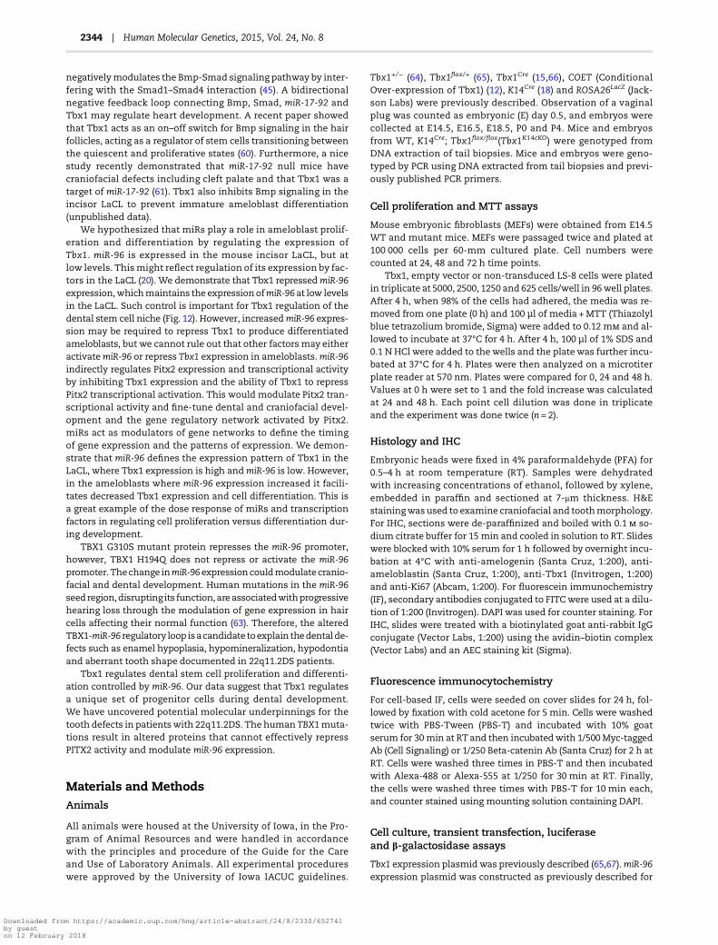
Figure 12.Model for the role of Tbx1 in tooth and craniofacial development. PITX2 and TBX1 are two of the first transcription markers for dental development and both a
co-expressed in the early dental epithelium, dental lamina and oral epithelium. PITX2 is a transcriptional activator, which activates a gene regulatory network required for
dental development (62). TBX1 can repress PITX2 transcriptional activity, but also activate other genes required for cell proliferation. TBX1 is part of a negative feedback
loopwithmiR-96. TBX1 repressesmiR-96 expression andmiR-96 represses TBX1 expression. This feedback loop allows dental epithelial cells to differentiate and produce
ameloblasts, which express amelogenin.
Human Molecular Genetics, 2015, Vol. 24, No. 8 | 2343
Downloaded from https://academic.oup.com/hmg/article-abstract/24/8/2330/652741by gueston 12 February 2018
negativelymodulates the Bmp-Smad signaling pathway by inter-fering with the Smad1–Smad4 interaction (45). A bidirectionalnegative feedback loop connecting Bmp, Smad, miR-17-92 andTbx1 may regulate heart development. A recent paper showedthat Tbx1 acts as an on–off switch for Bmp signaling in the hairfollicles, acting as a regulator of stem cells transitioning betweenthe quiescent and proliferative states (60). Furthermore, a nicestudy recently demonstrated that miR-17-92 null mice havecraniofacial defects including cleft palate and that Tbx1 was atarget of miR-17-92 (61). Tbx1 also inhibits Bmp signaling in theincisor LaCL to prevent immature ameloblast differentiation(unpublished data).
We hypothesized that miRs play a role in ameloblast prolif-eration and differentiation by regulating the expression ofTbx1. miR-96 is expressed in the mouse incisor LaCL, but atlow levels. Thismight reflect regulation of its expression by fac-tors in the LaCL (20). We demonstrate that Tbx1 repressedmiR-96expression,whichmaintains the expressionofmiR-96 at low levelsin the LaCL. Such control is important for Tbx1 regulation of thedental stem cell niche (Fig. 12). However, increasedmiR-96 expres-sion may be required to repress Tbx1 to produce differentiatedameloblasts, but we cannot rule out that other factors may eitheractivatemiR-96 or repress Tbx1 expression in ameloblasts.miR-96indirectly regulates Pitx2 expression and transcriptional activityby inhibiting Tbx1 expression and the ability of Tbx1 to repressPitx2 transcriptional activation. This would modulate Pitx2 tran-scriptional activity and fine-tune dental and craniofacial devel-opment and the gene regulatory network activated by Pitx2.miRs act as modulators of gene networks to define the timingof gene expression and the patterns of expression. We demon-strate that miR-96 defines the expression pattern of Tbx1 in theLaCL, where Tbx1 expression is high andmiR-96 is low. However,in the ameloblasts where miR-96 expression increased it facili-tates decreased Tbx1 expression and cell differentiation. This isa great example of the dose response of miRs and transcriptionfactors in regulating cell proliferation versus differentiation dur-ing development.
TBX1 G310S mutant protein represses the miR-96 promoter,however, TBX1 H194Q does not repress or activate the miR-96promoter. Thechange inmiR-96expressioncouldmodulatecranio-facial and dental development. Human mutations in the miR-96seed region,disrupting its function,areassociatedwithprogressivehearing loss through the modulation of gene expression in haircells affecting their normal function (63). Therefore, the alteredTBX1-miR-96 regulatory loop isacandidate toexplain thedentalde-fects such as enamel hypoplasia, hypomineralization, hypodontiaand aberrant tooth shape documented in 22q11.2DS patients.
Tbx1 regulates dental stem cell proliferation and differenti-ation controlled by miR-96. Our data suggest that Tbx1 regulatesa unique set of progenitor cells during dental development.We have uncovered potential molecular underpinnings for thetooth defects in patientswith 22q11.2DS. The human TBX1muta-tions result in altered proteins that cannot effectively repressPITX2 activity and modulate miR-96 expression.
Materials and MethodsAnimals
All animals were housed at the University of Iowa, in the Pro-gram of Animal Resources and were handled in accordancewith the principles and procedure of the Guide for the Careand Use of Laboratory Animals. All experimental procedureswere approved by the University of Iowa IACUC guidelines.
Tbx1+/− (64), Tbx1flox/+ (65), Tbx1Cre (15,66), COET (ConditionalOver-expression of Tbx1) (12), K14Cre (18) and ROSA26LacZ (Jack-son Labs) were previously described. Observation of a vaginalplug was counted as embryonic (E) day 0.5, and embryos werecollected at E14.5, E16.5, E18.5, P0 and P4. Mice and embryosfrom WT, K14Cre; Tbx1flox/flox(Tbx1K14cKO) were genotyped fromDNA extraction of tail biopsies. Mice and embryos were geno-typed by PCR using DNA extracted from tail biopsies and previ-ously published PCR primers.
Cell proliferation and MTT assays
Mouse embryonic fibroblasts (MEFs) were obtained from E14.5WT and mutant mice. MEFs were passaged twice and plated at100 000 cells per 60-mm cultured plate. Cell numbers werecounted at 24, 48 and 72 h time points.
Tbx1, empty vector or non-transduced LS-8 cells were platedin triplicate at 5000, 2500, 1250 and 625 cells/well in 96well plates.After 4 h, when 98% of the cells had adhered, the media was re-moved from one plate (0 h) and 100 µl of media +MTT (Thiazolylblue tetrazolium bromide, Sigma) were added to 0.12 m and al-lowed to incubate at 37°C for 4 h. After 4 h, 100 µl of 1% SDS and0.1 N HCl were added to thewells and the plate was further incu-bated at 37°C for 4 h. Plates were then analyzed on a microtiterplate reader at 570 nm. Plates were compared for 0, 24 and 48 h.Values at 0 h were set to 1 and the fold increase was calculatedat 24 and 48 h. Each point cell dilution was done in triplicateand the experiment was done twice (n = 2).
Histology and IHC
Embryonic heads were fixed in 4% paraformaldehyde (PFA) for0.5–4 h at room temperature (RT). Samples were dehydratedwith increasing concentrations of ethanol, followed by xylene,embedded in paraffin and sectioned at 7-μm thickness. H&Estainingwas used to examine craniofacial and toothmorphology.For IHC, sections were de-paraffinized and boiled with 0.1 so-dium citrate buffer for 15 min and cooled in solution to RT. Slideswere blocked with 10% serum for 1 h followed by overnight incu-bation at 4°C with anti-amelogenin (Santa Cruz, 1:200), anti-ameloblastin (Santa Cruz, 1:200), anti-Tbx1 (Invitrogen, 1:200)and anti-Ki67 (Abcam, 1:200). For fluorescein immunochemistry(IF), secondary antibodies conjugated to FITC were used at a dilu-tion of 1:200 (Invitrogen). DAPI was used for counter staining. ForIHC, slides were treated with a biotinylated goat anti-rabbit IgGconjugate (Vector Labs, 1:200) using the avidin–biotin complex(Vector Labs) and an AEC staining kit (Sigma).
Fluorescence immunocytochemistry
For cell-based IF, cells were seeded on cover slides for 24 h, fol-lowed by fixation with cold acetone for 5 min. Cells were washedtwice with PBS-Tween (PBS-T) and incubated with 10% goatserum for 30 min at RT and then incubatedwith 1/500Myc-taggedAb (Cell Signaling) or 1/250 Beta-catenin Ab (Santa Cruz) for 2 h atRT. Cells were washed three times in PBS-T and then incubatedwith Alexa-488 or Alexa-555 at 1/250 for 30 min at RT. Finally,the cells were washed three times with PBS-T for 10 min each,and counter stained using mounting solution containing DAPI.
Cell culture, transient transfection, luciferaseand β-galactosidase assays
Tbx1 expression plasmid was previously described (65,67).miR-96expression plasmid was constructed as previously described for
2344 | Human Molecular Genetics, 2015, Vol. 24, No. 8
Downloaded from https://academic.oup.com/hmg/article-abstract/24/8/2330/652741by gueston 12 February 2018

other miRs (68). The pre-miR-96 was cloned into the expressionvector. The Tbx1 3′UTR was cloned after the luciferase gene inpGL3 vector (Promega) (68). Truncations of the mouse Tbx1 genewere made using sequence-specific primers to Tbx1 by PCR withEcoRI and KpnI restriction enzyme sites. Tbx1 FL (full length),Tbx1 ΔC (deletion of C-Terminal tail) and Tbx1 ΔN (deletion ofN-terminus) were cloned into pcDNA3.1. The Pitx2c luciferasepromoter construct has been previously described (24), and the5 kb miR-96 promoter was PCR amplified from mouse genomicDNA and cloned into the luciferase plasmid. All plasmid con-structs were confirmed by DNA sequencing. LS-8 (oral epithelial)cells (69) were cultured in 10% fetal bovine serum (FBS) and 1%penicillin/streptomycin and transfected via electroporation aspreviously described (70). Cells were fed fresh media 24 h beforetransfection, and electroporated with 2.5 μg of expression plas-mid, 5 μg of reporter plasmid and 0.5 μg of SV-40 β-galactosidaseplasmid. Transfected cells were incubated for 48 h in 60-mm cul-ture dishes, lysed and assayed for β-galactosidase activity (TropixInc.), luciferase activity (Promega) and protein content (Bio-Rad).All luciferase activities were normalized to β-galactosidase activ-ity and protein concentration. Experiments were repeated threeto five times and the results are shown ±SEM. Transfection proto-col for miR-96 to knock down endogenous Tbx1 used 1 μg DNA,3 μl of X-tremeGene HP DNA transfection reagent, 200 μl ofserum free media and LS-8 cells in a 6-well plate. Cells were pla-ted at 10–20% confluence and harvested after 48 h and assayedfor endogenous Tbx1 expression by western blot.
Expression and purification of GST-Tbx1 mutantsand GST-PITX2A fusion proteins
Cloning of Tbx1 and PITX2A into pGEX6P-2 GST vector was previ-ously described (24,71). Tbx1 deletion constructs were PCR ampli-fied from a cDNA clone and ligated into pGEX6P-2 GST vector(Amersham Pharmacia Biotech, Piscataway, NJ) using EcoRI andXhoI restriction enzyme sites. The plasmids were confirmed byDNA sequencing and transformed in BL21 cells. Proteinswere ex-tracted as previously described (71,72). PITX2Awas cleaved fromGST moiety using 80 units of PreScission protease (PharmaciaBiotech) per milliliter of glutathione sepharose. Protein concen-trations were quantified with Bradford Reagent (Bio-Rad Labora-tories, Hercules, CA) and stored in 10% glycerol. Commassie Bluestaining of denatured SDS-polyacrylamide gelswas used to verifyproduction of protein.
GST pull-down assays
Immobilized GST-Tbx1, GST-Tbx1ΔNT, GST-Tbx1ΔTC, GST-Tbx1ΔC and GST-Tbx1Tbox fusion proteins were suspended inbinding buffer (20 m HEPES, pH 7.5, 5% glycerol, 50 m NaCl,1 m Ethylenediaminetetraacetic acid, 1 m Dithiothreitol, 1%milk and 400 μg/ml of ethidium bromide). Purified bacteria ex-pressed PITX2A (500 ng) was added to 15 μg immobilized GST-Tbx1 FL and truncated constructs, incubated for 30 min at 4°C.The beads were washed 5× with 200 μl binding buffer. Thebound proteins were eluted by boiling for 5 min in SDS-samplebuffer and separated on a 10% SDS-polyacrylamide gel. The pro-teins were then transferred to polyvinylidene difluoride (PVDF)filters, immunoblotted and detected using PITX2ABCDE antibody(Capra Science, Sweden) and ECL reagents.
Western blotting
Expression of transiently expressed Tbx1 was demonstratedusing a 1:500 dilution of anti-myc antibody (Cell Signaling).
Approximately 15–40 μg of cell lysates were used for sodium do-decyl sulfate gel electrophoresis. The protein was transferred toPVDF filters (Millipore), immunoblotted and detected using spe-cific secondary antibodies and enhanced-chemiluminesceneECL reagents (GE HealthCare).
Quantitative real-time PCR of mRNA and microRNA
Total RNA and miR from MEFs, LS-8, CHO and HEK 293 FT cellswere prepared using the miRNeasy Mini Kit (Qiagen). The quan-tity and integrity of the RNA samples were assessed bymeasure-ments at 260 and 280 nm and verified using gel analysis. LIs andmolars from E18.5 control and Tbx1K14COET were also dissectedand all of the RNA was prepared using the miRNeasy Mini Kit(Qiagen). MicroRNA were reversed transcribed using TaqManmicroRNA assay probes (Applied Biosystems) and the TaqManmicroRNA reverse transcription kit (Applied Biosystems) accord-ing to themanufacturer’s instruction. Quantitative real-time PCR(qPCR) analysis of miRs was performed using TaqManmicroRNAassay probes and normalized using the U6B probe (Taqman Uni-versal PCR mastermix, Applied Biosystems).
Total RNAwas reversed transcribed into cDNAusing oligo (dT)primers according to the manufacturer’s instructions (iScriptSelect cDNA Synthesis Kit, Bio-Rad). The MyiQ single coloredReal-TimeDetection System (Bio-Rad)was used for the reactions,and quantities were analyzed using the MyiQ Optical SystemSoftware 2.0 (Bio-Rad). cDNA levels were normalized to β-actin(F: 5′-GCCTTCCTTCTTGGGTATG-3′ and R: 5′-ACCACCAGACAGCACTGTG-3′). Primers used for qPCR are as follows: Tbx1 (F: 5′-CGACAAGCTGAAACTGACCA-3′ and R: 5′-GTGACTGCAGTGAAGCGTGT-3′) and Sox2 (F: 5′-ATGAGAGCAAGTACTGGCAAG-3′ andR: 5′-TCGGCAGCCTGATTCCAATAA-3′). The thermal cycling profileconsisted of 95°C for 4 min, 40 cycles of denaturation at 95°C for30 s, annealing at 60°C for 30 s and elongation at 72°C for 30 s.Samples were run in triplicate. Melting curves were generatedto confirm the amplification specificity of the PCR products. Inaddition, all of the PCR products were sequenced to verify thatthe correct band was amplified.
Statistical analysis
For each condition, three experiments were performed andthe results are presented as the mean ± SEM. The differencesbetween two groups of conditions were analyzed using an inde-pendent, two-tailed t-test.
SEM imaging and microCT
Hemi-mandibles of littermate WT and Tbx1K14cKO, COET orTbx1Pitx2cKO were dissected, fixed in 4% PFA overnight in 4°C andstored in 70% ethanol. EM images of uncoated specimens weretaken with a Zeiss Evo LS 10 scanning electron microscope (CarlZeiss, Peabody, USA) in secondary electron mode at 7 kV, 3 pAunder high vacuum to assess gross morphology. Subsequentlythe incisors and molars were fractured, mounted on stubs usingadhesive copper tape, and gold coated (Denton V Sputter coater,Denton,Moorestown, NJ). Imageswere then collected in secondaryelectron mode at 10 kV and 8 pA in high vacuum mode and at aworking distance of 5–9 mm. MicroCT samples were analyzed inethanol using a MicroCT 40 (Scanco Medical, Brüttisellen, Switzer-land) at 70 kV, 114 µA, 8 W and 10 µm resolution, with an integra-tion time of 300 ms. All samples to be compared were scanned inone batch to ensure identical conditions and allow for comparisonof mineral densities. The images in DICOM format were processed
Human Molecular Genetics, 2015, Vol. 24, No. 8 | 2345
Downloaded from https://academic.oup.com/hmg/article-abstract/24/8/2330/652741by gueston 12 February 2018

using Fiji imaging software (http://fiji.sc/Fiji) to standardize theorientation of the samples. All sampleswere processed identically,setting an arbitrary, but identical threshold to remove any back-ground and to allow fora comparison ofmineral densities betweensamples in slices and maximum intensity projections.
ChIP assay
ChIP assays were performed as previously described (73,74) usingthe Zymo-Spin ChIP kit (Zymo research) using LS8 cells. SpecificPCR amplification of the putative Tbx1 site in the miR-96 pro-moter was performed using the following primers (-133 and-384 away from the miR-96 predicted start): 5′-TGAAAGGTGGGCTCGG-3′ and 5′-CCTTCTAGGTTTCCTGCT-3′. As a control, PCRanalysis was performed on ChIP samples using primers up-stream of the putative Tbx1 site using the following primers(+384 and +512 from the miR-96 predicted start site): 5′-GGCCAAGGAAGTCAGGAAT-3′ and 5′-TAGTTCCTCGGGTGTGGACT-3′.
AcknowledgementsWe thankmembers of Amendt and Baldini Laboratories for help-ful discussions and Christine Blaumueller for editorial expertise.We thank the Forsyth Institute formicroCT imaging and analysesand Dr James Martin, Baylor College of Medicine, for reagents.
Conflict of Interest statement. None declared.
FundingSupport for this researchwas provided by funds from the Univer-sity of Iowa and grant DE 13941 from the National Institute ofDental and Craniofacial Research to B.A.A.
References
1. Goldberg, R., Motzkin, B., Marion, R., Scambler, P.J. andShprintzen, R.J. (1993) Velo-cardio-facial syndrome: a reviewof 120 patients. Am. J. Med. Genet., 45, 313–319.
2. Shprintzen, R.J., Goldberg, R.B., Lewin, M.L., Sidoti, E.J., Berk-man, M.D., Argamaso, R.V. and Young, D. (1978) A new syn-drome involving cleft palate, cardiac anomalies, typicalfacies, and learning disabilities: velo-cardio-facial syndrome.Cleft Palate J., 15, 56–62.
3. Klingberg, G., Oskarsdottir, S., Johannesson, E.L. and Noren,J.G. (2002) Oral manifestations in 22q11 deletion syndrome.Int. J. Paediatr. Dent., 12, 14–23.
4. Lindsay, E.A. (2001) Chromosomal microdeletions: dissectingdel22q11 syndrome. Nat. Rev. Genet., 2, 858–868.
5. Jerome, L.A. and Papaioannou, V.E. (2001) DiGeorge syndromephenotype in mice mutant for the T-box gene, Tbx1. Nat.Genet., 27, 286–291.
6. Merscher, S., Funke, B., Epstein, J.A., Heyer, J., Puech, A., Lu, M.M., Xavier, R.J., Demay, M.B., Russell, R.G., Factor, S. et al.(2001) TBX1 is responsible for cardiovascular defects invelo-cardio-facial/DiGeorge syndrome. Cell, 104, 619–629.
7. Bollag, R.J., Siegfried, Z., Cebra-Thomas, J.A., Garvey, N.,Davison, E.M. and Silver, L.M. (1994) An ancient family ofembryonically expressed mouse genes sharing a conservedprotein motif with the T locus. Nat. Genet., 7, 383–389.
8. Arnold, J.S.,Werling, U., Braunstein, E.M., Liao, J., Nowotschin,S., Edelmann, W., Hebert, J.M. and Morrow, B.E. (2006) Inacti-vation of Tbx1 in the pharyngeal endoderm results in22q11DS malformations. Development, 133, 977–987.
9. Nowotschin, S., Liao, J., Gage, P.J., Epstein, J.A., Campione, M.and Morrow, B.E. (2006) Tbx1 affects asymmetric cardiacmorphogenesis by regulating Pitx2 in the secondary heartfield. Development, 133, 1565–1573.
10. Zoupa, M., Seppala, M., Mitsiadis, T. and Cobourne, M.T.(2006) Tbx1 is expressed at multiple sites of epithelial–mesenchymal interaction during early development of thefacial complex. Int. J. Dev. Biol., 50, 504–510.
11. Zhang, Z. and Baldini, A. (2008) In vivo response to high-resolution variation of Tbx1 mRNA dosage. Hum. Mol. Genet.,17, 150–157.
12. Vitelli, F., Huynh, T. and Baldini, A. (2009) Gain of function ofTbx1 affects pharyngeal and heart development in themouse. Genesis, 47, 188–195.
13. Liao, J., Kochilas, L., Nowotschin, S., Arnold, J.S., Aggarwal, V.S., Epstein, J.A., Brown,M.C., Adams, J. andMorrow, B.E. (2004)Full spectrum of malformations in velo-cardio-facial syn-drome/DiGeorge syndrome mouse models by altering Tbx1dosage. Hum. Mol. Genet., 13, 1577–1585.
14. Kong, P., Racedo, S.E., Macchiarulo, S., Hu, Z., Carpenter, C.,Guo, T., Wang, T., Zheng, D. and Morrow, B.E. (2014) Tbx1 isrequired autonomously for cell survival and fate in the pha-ryngeal core mesoderm to form the muscles of mastication.Hum. Mol. Genet., 23, 4215–4231.
15. Huynh, T., Chen, L., Terrell, P. and Baldini, A. (2007) A fatemapof Tbx1 expressing cells reveals heterogeneity in the secondcardiac field. Genesis, 45, 470–475.
16. Harada, H., Kettunen, P., Jung, H.S., Mustonen, T., Wang, Y.A.and Thesleff, I. (1999) Localization of putative stem cells indental epithelium and their association with Notch and FGFsignaling. J. Cell Biol., 147, 105–120.
17. Harada, H., Toyono, T., Toyoshima, K., Yamasaki, M., Itoh, N.,Kato, S., Sekine, K. and Ohuchi, H. (2002) FGF10 maintainsstem cell compartment in developing mouse incisors. Devel-opment, 129, 1533–1541.
18. Cao, H., Wang, J., Li, X., Florez, S., Huang, Z., Venugopalan,S.R., Elangovan, S., Skobe, Z., Margolis, H.C., Martin, J.F. et al.(2010) MicroRNAs play a critical role in tooth development.J. Dent. Res., 89, 779–784.
19. Jevnaker, A.M. and Osmundsen, H. (2008) MicroRNA expres-sion profiling of the developing murine molar tooth germand the developing murine submandibular salivary gland.Arch. Oral Biol., 53, 629–645.
20. Jheon, A.H., Li, C.-Y., Wen, T., Michon, F. and Klein, O.D. (2011)Expression of micrornas in the stem cell niche of the adultmouse incisor. PLoS ONE, 6, e24536.
21. Michon, F., Tummers, M., Kyyronen, M., Frilander, M.J. andThesleff, I. (2010) Tooth morphogenesis and ameloblastdifferentiation are regulated by micro-RNAs. Dev. Biol., 340,355–368.
22. Cao, H., Jheon, A., Li, X., Sun, Z., Wang, J., Florez, S., Zhang, Z.,McManus,M.T., Klein, O.D. andAmendt, B.A. (2013) The Pitx2:miR-200c/141:noggin pathway regulates Bmp signaling andameloblast differentiation. Development, 140, 3348–3359.
23. Gay, I., Cavender, A., Peto, D., Sun, Z., Speer, A., Cao, H. andAmendt, B.A. (2013) Differentiation of human dental stemcells reveals a role for microRNA-218. J. Periodontal Res., 49,110–120.
24. Cao, H., Florez, S., Amen, M., Huynh, T., Skobe, Z., Baldini, A.and Amendt, B.A. (2010) Tbx1 regulates progenitor cell prolif-eration in the dental epithelium by modulating Pitx2 activa-tion of p21. Dev. Biol., 347, 289–300.
25. Zweier, C., Sticht, H., Aydin-Yaylagul, I., Campbell, C.E. andRauch, A. (2007) Human TBX1 missense mutations cause
2346 | Human Molecular Genetics, 2015, Vol. 24, No. 8
Downloaded from https://academic.oup.com/hmg/article-abstract/24/8/2330/652741by gueston 12 February 2018

gain of function resulting in the same phenotype as 22q11.2deletions. Am. J. Med. Genet., 80, 510–517.
26. Paylor, R., Glaser, B., Mupo, A., Atallotis, P., Spencer, C.,Sobotka, A., Sparks, C., Choi, C.H., Oghalai, J. and Curran,S., et al. (2006) Tbx1 haploinsufficiency is linked to behav-ioral disorders in mice and humans: implications for22q11 deletion syndrome. Proc. Natl. Acad. Sci. USA, 103,7729–7734.
27. Castellanos, R., Xie, Q., Zheng, D., Cvekl, A. and Morrow, B.E.(2014) Mammalian TBX1 preferentially binds and regulatesdownstream targets via a tandem T-site repeat. PLoS ONE, 9,e95151.
28. Chieffo, C., Garvey, N., Gong, W., Roe, B., Zhang, G., Silver,L., Emanuel, B.S. and Budarf, M.L. (1997) Isolation andcharacterization of a gene from the DiGeorge chromosomalregion homologous to the mouse Tbx1 gene. Genomics, 43,267–277.
29. Gong, W., Gottlieb, S., Collins, J., Blescia, A., Dietz, H.,Goldmuntz, E., McDonald-McGinn, D.M., Zackai, E.H.,Emanuel, B.S., Driscoll, D.A. et al. (2001) Mutation analysisof TBX1 in non-deleted patients with features of DGS/VCFSor isolated cardiovascular defects. J. Med. Genet., 38, E45.
30. Byrne, C., Tainsky, M. and Fuchs, E. (1994) Programminggene expression in developing epidermis. Development, 120,2369–2383.
31. Dassule, H.R., Lewis, P., Bei, M., Maas, R. and McMahon, A.P.(2000) Sonic hedgehog regulates growth and morphogenesisof the tooth. Development, 127, 4775–4785.
32. Takamori, K., Hosokawa, R., Xu, X., Deng, X., Bringas, J. P. andChai, Y. (2008) Epithelial fibroblast growth factor receptor 1regulates enamel formation. J. Dent. Res., 87, 238–243.
33. Mitsiadis, T.A. and Drouin, J. (2008) Deletion of the Pitx1genomic locus affects mandibular tooth morphogenesisand expression of the Barx1 and Tbx1 genes. Dev. Biol., 313,887–896.
34. Caton, J., Luder, H.U., Zoupa, M., Bradman, M., Bluteau, G.,Tucker, A.S., Klein, O. and Mitsiadis, T.A. (2009) Enamel-freeteeth: Tbx1 deletion affects amelogenesis in rodent incisors.Dev. Biol., 328, 493–505.
35. Thesleff, I. and Tummers, M. (2009) ToothOrganogenesis andRegeneration. The Stem Cell Research Community, StemBook,StemBook, ed.
36. Juuri, E., Saito, K., Ahtiainen, L., Seidel, K., Tummers, M.,Hochedlinger, K., Klein, O.D., Thesleff, I. and Michon, F.(2012) Sox2+ stem cells contribute to all epithelial lineagesof the tooth via Sfrp5+ progenitors. Dev. Cell, 23, 317–328.
37. Bei, M. (2009) Molecular genetics of ameloblast cell lineage.J. Exp. Zool. B Mol. Dev. Evol., 312B, 437–444.
38. Fincham, A.G., Moradian-Oldak, J. and Simmer, J.P. (1999) Thestructural biology of the developing dental enamel matrix.J. Struct. Biol., 126, 270–299.
39. Nakahori, Y., Takenaka, O. andNakagome, Y. (1991) A humanX–Y homologous region encodes ‘amelogenin’. Genomics, 9,264–269.
40. Snead, M.L., Lau, E.C., Zeichner-David, M., Fincham, A.G.,Woo, S.L. and Slavkin, H.C. (1985) DNA sequence for clonedcDNA for murine amelogenin reveal the amino acid sequencefor enamel-specific protein. Biochem. Biophys. Res. Commun.,129, 812–818.
41. Gibson, C.W. (2011) The amelogenin proteins and enameldevelopment in humans and mice. J. Oral Biosci., 53, 248–256.
42. Wright, J.T., Hart, T.C., Hart, P.S., Simmons, D., Suggs, C.,Daley, B., Simmer, J., Hu, J., Bartlett, J.D., Li, Y. et al. (2009)Human and mouse enamel phenotypes resulting from
mutation or altered expression of AMEL, ENAM, MMP20 andKLK4. Cells Tissues Organs, 189, 224–229.
43. Gibson, C.W. (1999) Regulation of amelogenin gene expres-sion. Crit. Rev. Eukaryot. Gene Expr., 9, 45–57.
44. Fukumoto, S., Kiba, T., Hall, B., Iehara, N., Nakamura, T.,Longenecker, G., Krebsbach, P.H., Nanci, A., Kulkarni, A.B.and Yamada, Y. (2004) Ameloblastin is a cell adhesionmolecule required for maintaining the differentiation stateof ameloblasts. J. Cell Biol., 167, 973–983.
45. Fulcoli, F.G., Huynh, T., Scambler, P.J. and Baldini, A. (2009)Tbx1 regulates the BMP-Smad1 pathway in a transcription in-dependent manner. PLoS ONE, 4, e6049.
46. Nordgarden, H., Lima, K., Skogedal, N., Følling, I., Storhaug, K.and Abrahamsen, T.G. (2012) Dental developmentaldisturbances in 50 individuals with the 22q11.2 deletion syn-drome; relation to medical conditions? Acta Odontol. Scand.,70, 194–201.
47. Semina, E.V., Reiter, R., Leysens, N.J., Alward, L.M., Small, K.W., Datson, N.A., Siegel-Bartelt, J., Bierke-Nelson, D., Bitoun,P., Zabel, B.U. et al. (1996) Cloning and characterization of anovel bicoid-related homeobox transcription factor gene,RIEG, involved in Rieger syndrome. Nat. Genet., 14, 392–399.
48. Liu, W., Selever, J., Lu, M.F. and Martin, J.F. (2003) Geneticdissection of Pitx2 in craniofacial development uncoversnew functions in branchial archmorphogenesis, late aspectsof toothmorphogenesis and cell migration.Development, 130,6375–6385.
49. Lin, C.R., Kioussi, C., O’Connell, S., Briata, P., Szeto, D., Liu, F.,Izpisua-Belmonte, J.C. and Rosenfeld, M.G. (1999) Pitx2regulates lung asymmetry, cardiac positioning and pituitaryand tooth morphogenesis. Nature, 401, 279–282.
50. Harel, I., Maezawa, Y., Avraham, R., Rinon, A., Ma, H.Y., Cross,J.W., Leviatan, N., Hegesh, J., Roy, A., Jacob-Hirsch, J. et al.(2012) Pharyngeal mesoderm regulatory network controlscardiac and head muscle morphogenesis. Proc. Natl. Acad.Sci. USA, 109, 18839–18844.
51. Yagi, H., Furutani, Y., Hamada, H., Sasaki, T., Asakawa, S.,Minoshima, S., Ichida, F., Joo, K., Kimura, M., Imamura, S.et al. (2003) Role of TBX1 in human del22q11.2 syndrome.Lancet, 362, 1366–1373.
52. Marom, T., Roth, Y., Goldfarb, A. and Cinamon, U. (2012) Headand neck manifestations of 22q11.2 deletion syndromes. Eur.Arch. Otorhinolaryngol., 269, 381–387.
53. Funato, N., Nakamura, M., Richardson, J.A. and Srivastava, D.(2012) Tbx1 regulates oral epithelial adhesion and palataldevelopment. Hum. Mol. Genet., 21, 2524–2537.
54. Goudy, S., Law, A., Sanchez, G., Baldwin, H.S. and Brown, C.(2010) Tbx1 is necessary for palatal elongation and elevation.Mech. Dev., 127, 292–300.
55. Chen, L., Fulcoli, F.G., Tang, S. and Baldini, A. (2009) Tbx1 reg-ulates proliferation and differentiation of multipotent heartprogenitors. Circ. Res., 105, 842–851.
56. Mitsiadis, T.A., Tucker, A.S., De Bari, C., Cobourne, M.T. andRice, D.P.C. (2008) A regulatory relationship between Tbx1and FGF signaling during tooth morphogenesis and amelo-blast lineage determination. Dev. Biol., 320, 39–48.
57. Vitelli, F., Taddei, I., Morishima,M., Meyers, E.N., Lindsey, E.A.and Baldini, A. (2002) A genetic link between Tbx1 and fibro-blast growth factor signaling. Development, 129, 4605–4611.
58. Wang, J., Greene, S.B., Bonilla-Claudio, M., Tao, Y., Zhang, J.,Bai, Y., Huang, Z., Black, B.L., Wang, F. and Martin, J.F. (2010)Bmp signaling regulates myocardial differentiation from car-diac progenitors through a MicroRNA-mediated mechanism.Dev. Cell, 19, 903–912.
Human Molecular Genetics, 2015, Vol. 24, No. 8 | 2347
Downloaded from https://academic.oup.com/hmg/article-abstract/24/8/2330/652741by gueston 12 February 2018

59. Prall, O.W., Menon, M.K., Solloway, M.J., Watanabe, Y.,Zaffran, S., Bajolle, F., Biben, C., McBride, J.J., Robertson, B.R.,Chaulet, H. et al. (2007) An Nkx2.5/Bmp2/Smad1 negativefeedback loop controls heart progenitor specification andproliferation. Cell, 128, 947–959.
60. Chen, T., Heller, E., Beronja, S., Oshimori, N., Stokes, N. andFuchs, E. (2012) An RNA interference screen uncovers a newmolecule in stem cell self-renewal and long-term regener-ation. Nature, 485, 104–108.
61. Wang, J., Bai, Y., Li, H., Greene, S.B., Klysik, E., Yu,W., Schwartz,R.J., Williams, T.J. and Martin, J.F. (2013) MicroRNA-17-92,a direct Ap-2α transcriptional target, modulates T-box factoractivity in orofacial clefting. PLoS Genet., 9, e1003785.
62. Sharp, T., Wang, J., Li, X., Cao, H., Gao, S., Moreno, M. andAmendt, B.A. (2014) A pituitary homeobox 2 (Pitx2):micro-RNA-200a-3p:beta-catenin pathway converts mesenchymecells to amelogenin-expressing dental epithelial cells. J. Biol.Chem., 289, 27327–27341.
63. Mencía, A., Modamio-Høybjør, S., Redshaw, N., Morín, M.,Mayo-Merino, F., Olavarrieta, L., Aguirre, L.A., del Castillo, I.,Steel, K.P., Dalmay, T. et al. (2009)Mutations in the seed regionof humanmiR-96 are responsible for nonsyndromic progres-sive hearing loss. Nat. Genet., 41, 609–613.
64. Lindsay, E.A., Vitelli, F., Su, H., Morishima, M., Huynh, T.,Pramparo, T., Jurecic, V., Ogunrinu, G., Sutherland, H.F.,Scambler, P.J. et al. (2001) Tbx1 haploinsufficieny in theDiGeorge syndrome region causes aortic arch defects inmice. Nature, 410, 97–101.
65. Xu, H., Morishima, M., Wylie, J.N., Schwartz, R.J., Bruneau, B.G., Lindsay, E.A. and Baldini, A. (2004) Tbx1 has a dual role inthe morphogenesis of the cardiac outflow tract. Development,131, 3217–3227.
66. Vitelli, F., Morishima, M., Taddei, I., Lindsay, E.A. and Baldini,A. (2002) Tbx1 mutation causes multiple cardiovasculardefects and disrupts neural crest nerve migratory pathways.Hum. Mol. Genet., 11, 915–922.
67. Amen, M., Liu, X., Vadlamudi, U., Elizondo, G., Diamond, E.,Engelhardt, J.F. and Amendt, B.A. (2007) PITX2 and beta-catenin interactions regulate Lef-1 isoform expression. Mol.Cell Biol., 27, 7560–7573.
68. Zhang, Z., Florez, S., Gutierrez-Hartmann, A., Martin, J.F. andAmendt, B.A. (2010) MicroRNAs regulate pituitary develop-ment, and microRNA 26b specifically targets lymphoidenhancer factor 1 (Lef-1), which modulates pituitary tran-scription factor 1 (Pit-1) expression. J. Biol. Chem., 285,34718–34728.
69. Chen, L.S., Couwenhoven, R.I., Hsu, D., Luo, W. and Snead, M.L. (1992) Maintenance of amelogenin gene expression bytransformed epithelial cells of mouse enamel organ. Archs.Oral Biol., 37, 771–778.
70. Green, P.D., Hjalt, T.A., Kirk, D.E., Sutherland, L.B., Thomas, B.L., Sharpe, P.T., Snead, M.L., Murray, J.C., Russo, A.F. andAmendt, B.A. (2001) Antagonistic regulation of Dlx2 expres-sion by PITX2 andMsx2: implications for tooth development.Gene Expr., 9, 265–281.
71. Amendt, B.A., Sutherland, L.B. and Russo, A.F. (1999) Multi-functional role of the Pitx2 homeodomain protein C-terminaltail. Mol. Cell Biol., 19, 7001–7010.
72. Cox, J.C., Hayhurst, A., Hesselberth, J., Bayer, T.S., Georgiou, G.and Ellington, A.D. (2002) Automated selection of aptamersagainst protein targets translated in vitro: from gene to apta-mer. Nuc. Acids Res., 30, e108.
73. Venugopalan, S.R., Li, X., Amen, M.A., Florez, S., Gutierrez, D.,Cao, H.,Wang, J. andAmendt, B.A. (2011) Hierarchical interac-tions of homeodomain and forkhead transcription factors inregulating odontogenic gene expression. J. Biol. Chem., 286,21372–21383.
74. Li, X., Venugopalan, S., Cao, H., Pinho, F., Paine, M.L.,Snead, M.L., Semina, E.V. and Amendt, B.A. (2013) Amodel for the molecular underpinnings of tooth defectsin Axenfeld-Rieger Syndrome. Hum. Mol. Genet., 23,194–208.
2348 | Human Molecular Genetics, 2015, Vol. 24, No. 8
Downloaded from https://academic.oup.com/hmg/article-abstract/24/8/2330/652741by gueston 12 February 2018