tau protein hyperphosphorylation and aggregation in alzheimer's
TRANSCRIPT

biomolecules
Review
Tau Protein Hyperphosphorylation and Aggregationin Alzheimer’s Disease and Other Tauopathies, andPossible Neuroprotective Strategies
Goran Šimic 1,*, Mirjana Babic Leko 1, Selina Wray 2, Charles Harrington 3, Ivana Delalle 4,Nataša Jovanov-Miloševic 1, Danira Bažadona 5, Luc Buée 6, Rohan de Silva 2,Giuseppe Di Giovanni 7,8, Claude Wischik 3 and Patrick R. Hof 9,10
Received: 2 November 2015; Accepted: 1 December 2015; Published: 6 January 2016Academic Editor: Jürg Bähler
1 Department of Neuroscience, Croatian Institute for Brain Research, University of Zagreb School of Medicine,Zagreb 10000, Croatia; [email protected] (M.B.L.); [email protected] (N.J.-M.)
2 Reta Lila Weston Institute and Department of Molecular Neuroscience, UCL Institute of Neurology,London WC1N 3BG, UK; [email protected] (S.W.); [email protected] (R.S.)
3 School of Medicine and Dentistry, University of Aberdeen, Aberdeen AB25 2ZD, UK;[email protected] (C.H.); [email protected] (C.W.)
4 Department of Pathology and Laboratory Medicine, Boston University School of Medicine,Boston 02118, MA, USA; [email protected]
5 Department of Neurology, University Hospital Center Zagreb, Zagreb 10000, Croatia; [email protected] Laboratory Alzheimer & Tauopathies, Université Lille and INSERM U1172, Jean-Pierre Aubert Research Centre,
Lille 59045, France; [email protected] Department of Physiology and Biochemistry, Faculty of Medicine and Surgery, University of Malta, Msida,
MSD 2080, Malta; [email protected] School of Biosciences, Cardiff University, Cardiff CF10 3AX, UK9 Fishberg Department of Neuroscience, Ronald M. Loeb Center for Alzheimer’s Disease,
Icahn School of Medicine at Mount Sinai, New York, NY 10029, USA; [email protected] Friedman Brain Institute, Icahn School of Medicine at Mount Sinai, New York, NY 10029, USA* Correspondence: [email protected]; Tel.: +385-1-459-6807; Fax: +385-1-459-6942
Abstract: Abnormal deposition of misprocessed and aggregated proteins is a common final pathwayof most neurodegenerative diseases, including Alzheimer’s disease (AD). AD is characterizedby the extraneuronal deposition of the amyloid β (Aβ) protein in the form of plaques and theintraneuronal aggregation of the microtubule-associated protein tau in the form of filaments. Basedon the biochemically diverse range of pathological tau proteins, a number of approaches have beenproposed to develop new potential therapeutics. Here we discuss some of the most promising ones:inhibition of tau phosphorylation, proteolysis and aggregation, promotion of intra- and extracellulartau clearance, and stabilization of microtubules. We also emphasize the need to achieve a fullunderstanding of the biological roles and post-translational modifications of normal tau, as wellas the molecular events responsible for selective neuronal vulnerability to tau pathology and itspropagation. It is concluded that answering key questions on the relationship between Aβ and taupathology should lead to a better understanding of the nature of secondary tauopathies, especiallyAD, and open new therapeutic targets and strategies.
Keywords: Alzheimer’s disease; amyloid β; neurofibrillary degeneration; microtubules;neuropathology; phosphorylation; protein aggregation; protein oligomerization; tauopathies;tau protein
Biomolecules 2016, 6, 6; doi:10.3390/biom6010006 www.mdpi.com/journal/biomolecules

Biomolecules 2016, 6, 6 2 of 28
1. Selective Overview of Major Discoveries on Tau Protein and Tauopathies
1.1. Neurofibrillary Tangles and Paired Helical Filaments
The Bavarian psychiatrist Aloysius (Alois) Alzheimer is credited with the first description ofthe most characteristic pathological brain change—neurofibrillary tangles (NFT)—of a yet-unnameddisease in a 51-year-old woman from Frankfurt am Main, who had developed dementia. That womanwas the first person to receive a diagnosis of the disease for which in 1910 Emil Kraepelin coined theterm Alzheimer’s disease (AD; which he wrongly, albeit cautiously, initially described as “preseniledementia”). Her name was Auguste Deter and she had an early-onset dementia, comorbid withpsychotic features. As she became progressively worse, she had to be admitted to a psychiatric hospitalin November 1901 (where Alzheimer examined her for the first time), where she eventually diedin April 1906. Besides the already known “miliary foci” of extracellular deposits scattered over thecerebral cortex (more commonly later called senile plaques, SP, or neuritic plaques, NP), by using anewly developed silver staining method (20% water solution of silver nitrate, [1]) Alzheimer observeddegenerating cortical neurons with bundles of intracellular fibrils (neurofibrillary tangles, NFT) [2,3].
It was not until 1963 that with the help of electron microscopy, Kidd and Terry independentlyreported NFT to be made up of abnormal filaments alternating between 15 (at their narrowest point)and 30 nm (at their widest point) in width, with a half-periodicity of about 80 nm [4,5]. Becauseit appeared that the two filaments were wound helically around one another, Kidd named thempaired helical filaments (PHF). Also found in NFT of AD, as a minority species, was the so-calledstraight filament (SF), a filament about 15 nm wide that does not exhibit the marked modulationin width shown by the PHF. Due to the fact that PHF were observed to be insoluble in denaturingagents such as sodium dodecyl sulfate (SDS) and urea, despite significant efforts the structural andmolecular composition of PHF (and NFT) was not elucidated until the mid-1980s [6,7]. Morphologicalstudies of fragmentation patterns showed that the PHF actually consists of a left-handed helicalribbon consisting of repeating symmetrical subunits. Using electron diffraction, Crowther and Wischikwere able to establish conclusively that the PHF is made up of a double helical stack of transverselyoriented C-shaped subunits, each of which has three domains. This structure precluded purelydescriptive models available to that point based on rearrangements of preformed cytoskeletal polymersor protofilaments. They concluded that the structure was of a type that might arise from the de novoassembly of a single structural subunit, the biochemical identity of which was then unknown. Laterstudies showed that SF were composed of a similar structural subunit although with a slightly differentrelative arrangement in the two types of filaments [8].
1.2. Tau Protein Isolation and Localization
Tau (tubulin-associated unit) protein was isolated from porcine brain extracts as a heat-stable,highly soluble protein essential for microtubule (MT) assembly [9]. Following the initial discovery oftau, two studies reported the process of tau purification and its physical and chemical properties [10,11],including the ability of tau to become phosphorylated. In 1983, it was discovered that tau could bephosphorylated at multiple sites by various protein kinases, including cyclic-AMP-dependent proteinkinases and casein kinase type-1 [12]. Further studies showed that tau is a phosphoprotein and thatphosphorylation negatively regulates its ability to stimulate MT assembly [13,14].
An immunohistochemical study that compared the localization of tau using the tau-1 antibody(that recognizes all isoforms of tau, see below) with that of microtubule-associated protein 2 (MAP2)and tubulin in human postmortem brain tissue demonstrated that tau protein was primarily localizedto axons [15]. Using the same tau-1 monoclonal antibody and electron microscopy with colloidalgold-labeled secondary antibodies, tau was also found in very low amounts in astrocytes [16]and oligodendrocytes [17], and this was confirmed by tau mRNA expression analysis in themouse brain [18].

Biomolecules 2016, 6, 6 3 of 28
1.3. Tau in Neurofibrillary Tangles
The insolubility of PHF precluded biochemical characterisation of the repeating subunit thatmakes up the structural core of the filament. What was required was a means of solubilising or releasingthe structural subunit as a protein band that could be visualised by gel electrophoresis and linking thisby immuno-electron microscopy to the PHF. Initial attempts based on relatively crude preparationsof NFT were unable to distinguish between proteins copurifying with NFTs due to trapping andloose association within the dense filament bundles, and proteins derived from the structural coreof the PHF. In 1985 Brion and collaborators prepared tau and MAP2 proteins from the adult ratbrain using the microtubule assembly-disassembly method and their property of thermostability;they then generated antisera against tau and MAP2 proteins using polypeptides extracted frompolyacrylamide gels after electrophoretic separation by sodium dodecyl sulfate-polyacrylamidegel electrophoresis (SDS-PAGE) [19]. Antisera were characterized by immunoblotting on purifiedpreparations of tau and MAP2 and found to react with their cognate antigens. These antisera werethen used for immunocytochemistry on tissue sections from control subjects and AD patients: theanti-MAP2 antibody did not label NFT but the anti-tau antibody strongly immunolabelled NFT andabnormal neurites around senile plaques, yielding an immunolabelling indistinguishable from theone obtained with anti-PHF serum [19]. This work therefore established that tau protein was one of anumber proteins associated with NFTs both histologically and in crude NFT extracts. Neurofibrillarytangles can be labeled histologically with antibodies against a variety of other neuronal proteins,including vimentin, actin, ubiquitin, MAP2, and Aβ protein. In crude NFT preparations, isolated NFTcould be labeled with antibodies against MAP2, neurofilament, ubiquitin and tau [19–29].
The proof that tau protein contributes to the structural core of the PHF required preparation offractions highly in enriched in proteolytically stable PHF which retained the subunit structure of thefilament that had been characterised previously. These PHF were solubilised in formic acid and whenexamined by SDS-PAGE gel electrophoresis were found to contain predominantly a 12-kD protein anda corresponding dimer. Surprisingly, this protein was not recognised by an antibody raised againsttau protein. Conversely, a monoclonal antibody raised against the enriched core PHF preparations(mAb 6.423, referred to as MN423) did not recognised purified tau protein [30–33]. Nevertheless,MN423 was shown by immunogold electron microscopy to label the proteolytically stable core ofthe PHF. Furthermore, a ligand related to primulin [34] was used to affinity label the 12-kD species.This ligand, when bound covalently to biotin, was also shown by immunogold electron microscopyto label proteolytically stable core PHF. Therefore, the provenance of the 12-kD protein from thestructural core of the PHF was unequivocally established by two independent approaches. Partialamino acid sequences derived from this band were unrelated to any protein sequence known at thattime. However, when these were used to clone and sequence the corresponding cDNAs from a humanbrain library, the predicted protein was found to be 352 amino acids in length and was found to haveextensive homology to the sequence of the mouse microtubule-associated protein tau isoform that hadjust been published [35,36]. It was concluded that this protein must constitute the human homolog ofmouse tau, and that tau protein therefore must contribute to the structural core of the PHF, and was notsimply a loosely associated protein copurifying with NFT. These data were therefore able to explain theearlier observations linking tau protein with NFT [21,37–41]. In the same year tau cDNA clones wereidentified in the human fetal brain by flow sorting and spot-blot hybridization and later on assigned tothe microtubule-associated protein tau gene (MAPT) on the long arm of the chromosome 17 [42,43].
1.4. Tau Isoforms in the Central Nervous System
In their 1988 paper, Goedert and collaborators also mentioned that they had identified a secondform of tau, with sequence variation in the first repeat, and suggested that tau mRNA was undergoingalternative splicing [34]. This second form was identical to the first, with the exception of an additionalinsert of 31 amino acids in the repeat region. Upon sequencing of genomic clones, the extra repeat wasshown to be encoded by a separate exon, now known as exon 10. This work uncovered the existence

Biomolecules 2016, 6, 6 4 of 28
of at least two types of tau isoforms in the human brain, with three repeats (3R tau) or four repeats(4R tau) of a conserved tubulin-binding motif [44]. Sequencing of a large number of cDNA clonesrevealed the existence of additional tau isoforms with two (29-N1, and 59 amino acid-N2) inserts in theN-terminus region (due to alternative splicing of exons 2 and 3), in combination with both three andfour repeats (Figure 1). With the isoforms described previously, this gave a total of six human braintau isoforms ranging from 352 to 441 amino acids in length [45]. The primary sequence of the longesttau isoform is shown in Figure 2. The most prominent expression of tau was observed during fetaldevelopment, when only the shortest (referred to as fetal tau) isoform (N0R3) is expressed (352 aminoacids with molecular weight of 45 kDa), while the adult human brain expresses all isoforms with R4to R3 ratio equal to 1 [46]. Relative amounts of N0, N1 and N2 tau isoforms are 37%, 54% and 9%,respectively [46]. There is a further larger transcript of tau that encodes for a protein of 110 kDa withan additional 254 amino acids in the N-terminal projection arm, but this protein is generally restrictedto the peripheral nervous system [47].
Biomolecules 2016, 6, 6 5 of 35
shown in Figure 2. The most prominent expression of tau was observed during fetal development,
when only the shortest (referred to as fetal tau) isoform (N0R3) is expressed (352 amino acids with
molecular weight of 45 kDa), while the adult human brain expresses all isoforms with R4 to R3 ratio
equal to 1 [46]. Relative amounts of N0, N1 and N2 tau isoforms are 37%, 54% and 9%, respectively [46].
There is a further larger transcript of tau that encodes for a protein of 110 kDa with an additional 254
amino acids in the N-terminal projection arm, but this protein is generally restricted to the peripheral
nervous system [47].
Figure 1. Chromosomal location of the gene and protein structure for the microtubule-
associated proteins tau, microtubule-associated protein 2 (MAP2) and MAP4. Tau exons 2,
3 and 10 are alternatively spliced, giving rise to six different mRNAs, translated in six
different tau isoforms. Tau isoforms differ by the absence or presence of one or two 29
amino acid inserts encoded by exon 2 (yellow) and 3 (orange) in the N-terminal part, in
combination with either three (R1, R3 and R4) or four (R1-R4) repeat regions in the C-
terminal part. The R2 repeat is encoded by exon 10. The longest 2N4R adult tau isoform
(2+3+10+) has 441 amino acids (aa), followed by 1N4R isoform of 412 aa (2+3�10+),
2N3R isoform of 410 aa (2+3+10�), 0N4R isoform of 383 aa (2�3�10+), 2N3R isoform of
381aa (2+3�10�) and the shortest 0N3R isoform of 352 aa (2�3�10�). The single neuron-
specific promoter of MAPT gene has three binding sites for transcription factors and its
activity increases with axon initiation and outgrowth. The shortest tau isoform is the only
one expressed in the fetal brain (“fetal tau”), while expression of other isoforms begins
postnatally (for a review, see [48]). The MAP2 and MAP4 have comparable repeat domain
sequences in the C-terminus but differ from tau proteins by their longer N-terminal
projection arms.
Figure 1. Chromosomal location of the gene and protein structure for the microtubule-associatedproteins tau, microtubule-associated protein 2 (MAP2) and MAP4. Tau exons 2, 3 and 10 arealternatively spliced, giving rise to six different mRNAs, translated in six different tau isoforms.Tau isoforms differ by the absence or presence of one or two 29 amino acid inserts encoded by exon2 (yellow) and 3 (orange) in the N-terminal part, in combination with either three (R1, R3 and R4) orfour (R1-R4) repeat regions in the C-terminal part. The R2 repeat is encoded by exon 10. The longest2N4R adult tau isoform (2+3+10+) has 441 amino acids (aa), followed by 1N4R isoform of 412 aa(2+3´10+), 2N3R isoform of 410 aa (2+3+10´), 0N4R isoform of 383 aa (2´3´10+), 2N3R isoform of381aa (2+3´10´) and the shortest 0N3R isoform of 352 aa (2´3´10´). The single neuron-specificpromoter of MAPT gene has three binding sites for transcription factors and its activity increases withaxon initiation and outgrowth. The shortest tau isoform is the only one expressed in the fetal brain(“fetal tau”), while expression of other isoforms begins postnatally (for a review, see [48]). The MAP2and MAP4 have comparable repeat domain sequences in the C-terminus but differ from tau proteinsby their longer N-terminal projection arms.
Biomolecules 2016, 6, 6 5 of 28Biomolecules 2016, 6, 6 6 of 35
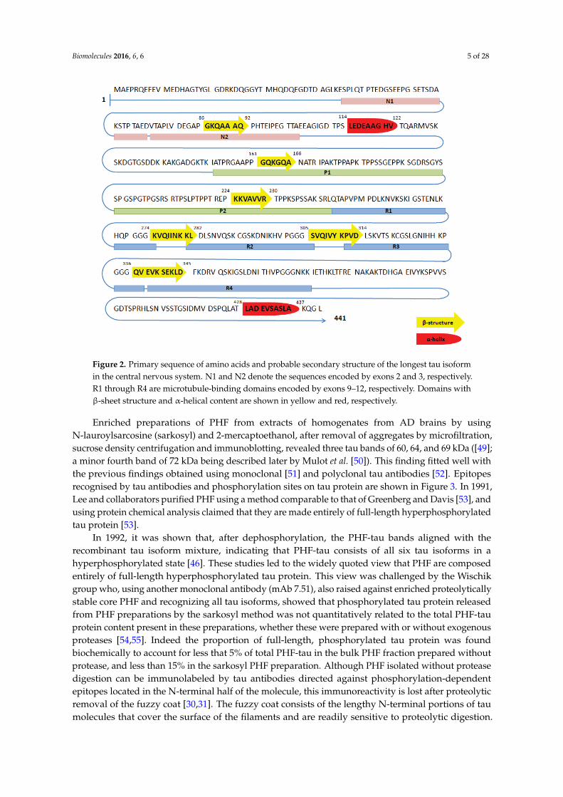
Figure 2. Primary sequence of amino acids and probable secondary structure of the longest
tau isoform in the central nervous system. N1 and N2 denote the sequences encoded by
exons 2 and 3, respectively. R1 through R4 are microtubule-binding domains encoded by
exons 9–12, respectively. Domains with �-sheet structure and �-helical content are shown in
yellow and red, respectively.
Enriched preparations of PHF from extracts of homogenates from AD brains by using
N-lauroylsarcosine (sarkosyl) and 2-mercaptoethanol, after removal of aggregates by microfiltration,
sucrose density centrifugation and immunoblotting, revealed three tau bands of 60, 64, and 69 kDa ([49];
a minor fourth band of 72 kDa being described later by Mulot et al. [50]). This finding fitted well
with the previous findings obtained using monoclonal [51] and polyclonal tau antibodies [52].
Epitopes recognised by tau antibodies and phosphorylation sites on tau protein are shown in Figure 3.
In 1991, Lee and collaborators purified PHF using a method comparable to that of Greenberg and
Davis [53], and using protein chemical analysis claimed that they are made entirely of full-length
hyperphosphorylated tau protein [53].
In 1992, it was shown that, after dephosphorylation, the PHF-tau bands aligned with the recombinant
tau isoform mixture, indicating that PHF-tau consists of all six tau isoforms in a hyperphosphorylated
state [46]. These studies led to the widely quoted view that PHF are composed entirely of full-length
hyperphosphorylated tau protein. This view was challenged by the Wischik group who, using another
monoclonal antibody (mAb 7.51), also raised against enriched proteolytically stable core PHF and
recognizing all tau isoforms, showed that phosphorylated tau protein released from PHF preparations
by the sarkosyl method was not quantitatively related to the total PHF-tau protein content present in
these preparations, whether these were prepared with or without exogenous proteases [54,55]. Indeed
the proportion of full-length, phosphorylated tau protein was found biochemically to account for less
Figure 2. Primary sequence of amino acids and probable secondary structure of the longest tau isoformin the central nervous system. N1 and N2 denote the sequences encoded by exons 2 and 3, respectively.R1 through R4 are microtubule-binding domains encoded by exons 9–12, respectively. Domains withβ-sheet structure and α-helical content are shown in yellow and red, respectively.
Enriched preparations of PHF from extracts of homogenates from AD brains by usingN-lauroylsarcosine (sarkosyl) and 2-mercaptoethanol, after removal of aggregates by microfiltration,sucrose density centrifugation and immunoblotting, revealed three tau bands of 60, 64, and 69 kDa ([49];a minor fourth band of 72 kDa being described later by Mulot et al. [50]). This finding fitted well withthe previous findings obtained using monoclonal [51] and polyclonal tau antibodies [52]. Epitopesrecognised by tau antibodies and phosphorylation sites on tau protein are shown in Figure 3. In 1991,Lee and collaborators purified PHF using a method comparable to that of Greenberg and Davis [53], andusing protein chemical analysis claimed that they are made entirely of full-length hyperphosphorylatedtau protein [53].
In 1992, it was shown that, after dephosphorylation, the PHF-tau bands aligned with therecombinant tau isoform mixture, indicating that PHF-tau consists of all six tau isoforms in ahyperphosphorylated state [46]. These studies led to the widely quoted view that PHF are composedentirely of full-length hyperphosphorylated tau protein. This view was challenged by the Wischikgroup who, using another monoclonal antibody (mAb 7.51), also raised against enriched proteolyticallystable core PHF and recognizing all tau isoforms, showed that phosphorylated tau protein releasedfrom PHF preparations by the sarkosyl method was not quantitatively related to the total PHF-tauprotein content present in these preparations, whether these were prepared with or without exogenousproteases [54,55]. Indeed the proportion of full-length, phosphorylated tau protein was foundbiochemically to account for less that 5% of total PHF-tau in the bulk PHF fraction prepared withoutprotease, and less than 15% in the sarkosyl PHF preparation. Although PHF isolated without proteasedigestion can be immunolabeled by tau antibodies directed against phosphorylation-dependentepitopes located in the N-terminal half of the molecule, this immunoreactivity is lost after proteolyticremoval of the fuzzy coat [30,31]. The fuzzy coat consists of the lengthy N-terminal portions of taumolecules that cover the surface of the filaments and are readily sensitive to proteolytic digestion.

Biomolecules 2016, 6, 6 6 of 28
Such digestion leaves intact the proteolytically stable core structure comprising the left-handedhelical or straight ribbon of repeated C-shaped subunits. In other words, the fuzzy coat comprisingphosphorylated tau does not contribute to the structural core of the PHF. Core PHF have a mass of65 kD/nm, whereas 90% of PHF isolated without proteases is 77 kD/nm, with a further 10% having amass of 110 kD/nm [31]. The core PHF therefore accounts for ~85% of the mass of the filament, with avariable addition of fuzzy coat material which contributes ~12 kD/nm.
Biomolecules 2016, 6, 6 7 of 35
that 5% of total PHF-tau in the bulk PHF fraction prepared without protease, and less than 15% in the
sarkosyl PHF preparation. Although PHF isolated without protease digestion can be immunolabeled by
tau antibodies directed against phosphorylation-dependent epitopes located in the N-terminal half of the
molecule, this immunoreactivity is lost after proteolytic removal of the fuzzy coat [30,31]. The fuzzy
coat consists of the lengthy N-terminal portions of tau molecules that cover the surface of the filaments
and are readily sensitive to proteolytic digestion. Such digestion leaves intact the proteolytically stable
core structure comprising the left-handed helical or straight ribbon of repeated C-shaped subunits. In other
words, the fuzzy coat comprising phosphorylated tau does not contribute to the structural core of the
PHF. Core PHF have a mass of 65 kD/nm, whereas 90% of PHF isolated without proteases is 77 kD/nm,
with a further 10% having a mass of 110 kD/nm [31]. The core PHF therefore accounts for ~85% of
the mass of the filament, with a variable addition of fuzzy coat material which contributes ~12 kD/nm.
Figure 3. Putative phosphorylation sites on tau protein and epitopes specific for major tau
antibodies. Red color denotes amino acids phosphorylated in AD brain, green in both AD
and normal brain, blue in normal brain, while black color means that those phosphorylation
sites have not been fully characterized yet. Tau antibodies specific for phospho-tau epitopes
are given in purple, while pink color denotes antibodies specific for non-phosphorylated
tau epitopes: Alz-50 (aa 2–10, aa 312�342), 43D (aa 1–100), 77E9 (aa 185–195), 39E10
(aa 189–195), Tau-5 (aa 210–230), 5C7 (aa 267–278), Tau-1 (aa 195, 198, 199 and 202),
77G7 (aa 270–375), Tau-46 (aa 404–441), TauC-3 (tau cleaved on aa 421). Red—in the
AD brain; Green—in both the AD and the normal brain; Blue—in the normal brain;
Black—phosphorylation sites that have not been fully characterized yet; Purple—tau antibodies
specific for phospho-tau epitopes; Pink—tau antibodies specific for unphosphorylated tau epitopes.
Figure 3. Putative phosphorylation sites on tau protein and epitopes specific for major tau antibodies.Red color denotes amino acids phosphorylated in AD brain, green in both AD and normal brain,blue in normal brain, while black color means that those phosphorylation sites have not been fullycharacterized yet. Tau antibodies specific for phospho-tau epitopes are given in purple, while pinkcolor denotes antibodies specific for non-phosphorylated tau epitopes: Alz-50 (aa 2–10, aa 312´342),43D (aa 1–100), 77E9 (aa 185–195), 39E10 (aa 189–195), Tau-5 (aa 210–230), 5C7 (aa 267–278), Tau-1(aa 195, 198, 199 and 202), 77G7 (aa 270–375), Tau-46 (aa 404–441), TauC-3 (tau cleaved on aa 421).Red—in the AD brain; Green—in both the AD and the normal brain; Blue—in the normal brain;Black—phosphorylation sites that have not been fully characterized yet; Purple—tau antibodiesspecific for phospho-tau epitopes; Pink—tau antibodies specific for unphosphorylated tau epitopes.
The tau species identified in the 12-kD species isolated from the PHF core were found to bederived from a mixture of fragments originating from both 3- and 4-repeat isoforms, but restrictedto the equivalent of three repeats in length. There are two distinct species originating from the 4Risoform of tau: the fragment derived from repeats 1, 2 and 3 and the fragment derived from repeats2, 3 and 4. There is also a fragment derived from the 3R isoform (which lacks the second repeat),comprising the equivalent of repeats 1, 3 and 4. Since all of these fragments are restricted to theequivalent of three repeats in length, they all have identical gel mobility [56]. Therefore, the PHF iscomposed of a structural core of repeating transverse C-shaped subunits. The only tau protein foundwithin the subunit structure is restricted to the repeat domain of the tau molecule. The contributionof phosphorylated tau is variable and restricted to the fuzzy outer coat of the PHF, accounting forapproximately one in seven of the tau molecules found in the PHF [57].
Biomolecules 2016, 6, 6 7 of 28
1.5. Functions of Tau Protein
Tau protein is most abundantly expressed in axons of central nervous system neurons [13] butcan also be found in the somatodendritic compartment of neurons, oligodendrocytes, and non-neuraltissues [58]. Probably the most important role of tau protein is to promote assembly and stabilityof MT [7,8], although this function is complemented by other MAP (especially by MAP1B), as tauknockout mice are viable, fertile, and relatively normal, with no signs of neurodegeneration. Also,knockdown of tau with small interfering RNA does not kill primary neurons in culture or preventaxon formation [59]. Additionally, MAP1B is probably more important for MT stability than tau itselfbecause knockout of MAP1B results in abnormal brain development and early death, and concurrentknockout of both MAP1B and MAPT worsens the phenotype [60].
The most common post-translational modifications of tau proteins are phosphorylation andO-glycosylation [61]. Phosphorylation changes the shape of tau molecule and regulates its biologicalactivity. Most of the phosphorylation sites are on Ser-Pro and Thr-Pro motives, but a number of siteson other residues have also been reported [62,63]. The majority of tau-based therapeutic strategiesagainst neurodegeneration have focused on modulating tau phosphorylation, given that tau speciespresent within NFT are hyperphosphorylated. O-glycosylation is characterized by the addition ofan O-linked N-acetylglucosamine (O-GlcNAc) on Ser or Thr residues in the vicinity of Pro residues.It is presumed that glycosylation may have a role in subcellular localization and degradation of tauproteins [64]. The recent discovery that tau is also modified by acetylation requires additional researchto provide greater insight into the physiological and pathological consequences of tau acetylation [65].
Tau protein can be divided into two main functional domains: the basic MT binding domain(towards the C-terminus) and the acidic projection domain (towards the N-terminus) [66]. The MTbinding domain regulates the rate of MT polymerization through highly conserved repetitive domainsR1–R4 encoded by exons 9–12 [36]. Adult tau isoforms with 4R (R1–R4) are about 40-fold moreefficient at promoting MT assembly than the fetal isoform that is lacking exon 10 and thus havingonly 3R (Figure 1) [67]. The absence of expression of the R1–R2 inter-repeat region during fetaldevelopment allows for the cytoskeletal plasticity required of growing immature neurons and theirelongating axons [64]. Apart from binding to MT, the repeat domains of tau also bind to tubulindeacetylase, histone deacetylase 6 (HDAC6) [68] and apolipoprotein E (apoE, more with the ε3 thanthe ε4 isovariant [69]).
The projection domain is so called because ultrastructurally it appears as a filamentous “arm”projecting from the wall of the MT. In recent years, many hitherto unknown binding partnersof the projection domain have been identified. The projection domain of tau may be involvedin cell signaling that occurs through the interaction with Lck, Fgr and cSrc (Src-family kinases),growth factor receptor-bound protein 2 (Grb2), phospholipase C-γ [70], phosphatidylinositol andphosphatidylinositol bisphosphate [71,72], peptidyl-prolyl cis/trans isomerase Pin 1, and many others(for review see [73]), making them potential therapeutic targets in tauopathies [74]. In synapses,the projection domain of tau interacts with protein kinase Fyn (plays an important role duringmyelination [75]), postsynaptic density protein 95 (PSD-95) [76], and N-methyl-D-aspartate receptors(NMDAR). Tau knockout mice show that tau is essential for NMDA-dependent long-term potentiation(LTP) and α-amino-3-hydroxy-5-methyl-4-isoxazolepropionic acid (AMPA)-dependent long-termdepression [77–79]. The function of tau protein in the response to heat stress in the cell is also worthnoting. When the heat stress occurs, tau protein binds to DNA and enhances DNA repair [80].An additional “knot” of tau being entangled in epigenetic landscape of neurodegeneration comes fromthe finding that by acting as a HDAC6 inhibitor, tau is being indirectly involved in both (dys)regulationof transcriptional activity and impairment of autophagic clearance by the ubiquitin proteasomesystem [81,82].

Biomolecules 2016, 6, 6 8 of 28
1.6. Amyloid Cascade Theory
The so-called “cholinergic hypothesis of AD” [83,84] dominated the late 1970s and early 1980s,and the “calcium hypothesis” in the late 1980s. After the milestone discovery that cerebrovascularamyloid and NP are composed of Aβ (as they shared the same antigenic determinants; [85,86]) inboth AD and Down syndrome [87], and that the V717I missense “London” mutation in the amyloidprecursor protein gene (APP) on chromosome 21 was found to be causally related to the early-onsetautosomal-dominant familial AD [88], Hardy and colleagues [89,90] proposed the “amyloid cascadehypothesis”, which has become a dominant driver of AD research ever since. According to theamyloid theory, excessive production of Aβ via serial cleavage of the larger amyloid precursor protein(APP) molecule by β-secretase (β-site APP cleaving enzyme, BACE, encoded by the BACE1 gene)and γ-secretase (multiprotein complex now known to consist minimally of four individual proteins:presenilin, nicastrin, anterior pharynx-defective 1, APH-1, and presenilin enhancer 2, PEN-2; [91–93]),is the key pathological event which drives all other pathological changes (astrocytosis, microglialactivation, neuronal death, synaptic loss, and the development of NFTs and dementia) not onlyin early-onset familial cases but also in late-onset, sporadic cases of AD. In 1987 Goldgaber andcollaborators isolated APP and localized its gene to chromosome 21 [94]. It should be noted here thatthe first APP mutation discovered was actually the G to C mutation at codon 693 (APP E693Q) thatwas not associated with AD, but rather with hereditary cerebral hemorrhage with amyloidosis—Dutchtype (HCHWA-D; [95,96]). Interestingly enough, out of four other known mutations within the Aβpart of APP (exons 16 and 17) two also cause fatal hemorrhage due to amyloid angiopathy (APPC692G-Flemish and APP E693K-Italian), while only rare “Arctic” (APP E693G) and Osaka (APP E693∆)mutations cause early-onset AD (EOAD). The well-known fact that many families exist in whichAD has an early onset (before age of 60) and is inherited in an autosomal dominant manner [97]could not be explained by a very small number of AD families in which APP mutations were found.This question was resolved in part by the discovery of mutations in the presenilin 1 gene (PSEN1) onchromosome 14 [98,99], and its homologous presenilin 2 gene (PSEN2) on chromosome 1 [100,101].These mutations of PSEN genes further strengthened the amyloid theory, but the pathogenesis of ADremained elusive. Further research showed that PSEN1 and PSEN2 are part of the γ-secretase complex,which cleaves APP at several points resulting in Aβ of various lengths: the lengths associated withAD are 40 and 42 amino acids long with Aβ42 more likely to aggregate to form SP in the brain thanAβ40. All PSEN mutations lead to an increase in the Aβ42:Aβ40 ratio, although the total quantity ofAβ produced remains constant [102,103]. This can come about by various effects of the mutationsof γ-secretase. Presenilins are also implicated in the processing of notch [104,105], an importantdevelopmental protein (mice that have PS1 knocked out die early in development from developmentalabnormalities similar to those found when notch is disrupted, [106]). APP can also be cleaved byα-secretases such as a disintegrin and metalloproteases domain 10 (ADAM10) and tumor necrosisfactor alpha (TNF-α) converting enzyme (TACE), although this cleavage does not result in Aβ butinstead generates APPs-α, which are thought to be neuroprotective [107].
Collectively, the genetic etiology of AD is very complex: early-onset AD (less than 5% of cases) isoften familial (fAD) with autosomal dominant and fully penetrant inheritance and can be caused byany of more than 200 pathogenic mutations in APP (33 mutations, duplication), PSEN1 (185 mutations)and PSEN2 (13 mutations; http://www.molgen.ua.ac.be/ADmutations). Most AD cases (over 95%)however are sporadic, late-onset (sAD, LOAD) and have less evident genetic components. Theε4 variant of the gene encoding apolipoprotein E (APOE) is known to confer increased risk forLOAD [108,109] with partial penetrance. Based on 320 meta-analyses of 1395 studies in which 695 genesand their 2973 polymorphisms have been tested as late-onset AD candidate genes, over 30 yieldedpositive evidence for association. The number one gene is APOE, with a Bayes factor (BF) > 50. UsingAPOE genotype ε3/ε3 as a neutral benchmark for comparison, individuals with a single copy of theε4 allele manifest a 5 fold increased chance of developing LOAD, while those with two copies havean estimated 20 fold increased risk [110]. It seems that different APOE alleles are not associated with
Biomolecules 2016, 6, 6 9 of 28
an increase in Aβ production, but with a reduced ability to clear Aβ from the brain [111,112]. Thismay be related to decreased production of Aβ auto-antibodies in AD subjects [113]. The next ninegenes with the highest association with LOAD are: BIN1 (BF = 23.4) that encodes several isoformsof a nucleoplasmic adaptor protein, one of which was identified as MYC-interacting protein, CLU(BF = 20.1) that encodes apolipoprotein J, ABCA7 (BF = 18.8) for ATP-binding cassette, subfamily A[ABC1], member 7, CR1 (BF = 18.1) for complement component receptor 1, PICALM (BF = 17.3) forphosphatidylinositol-binding clathrin assembly protein, MS4A6A (BF = 8.7), CD33 (BF = 7.7) for atransmembrane receptor expressed on cells of myeloid lineage-cluster of differentiation 33, MS4A4E(BF = 6.9) coding for protein membrane-spanning 4-domains, subfamily A, member 4E, and CD2AP(BF = 6.6) that codes for a scaffolding molecule that regulates the actin cytoskeleton (according towww.alzgene.org accessed on 11 February 2015). Genetic variants of all of these genes have a relativelyminor influence on AD progression when altered [114] and their influence on the development andcourse of sAD remains largely unknown [115–117]. Most recently, rare mutations of TREM2 [118]and PLD3 [119] have also been discovered to confer a much larger increase in risk for LOAD than theaforementioned common sequence variants [120].
1.7. Staging of Tau Pathology
During the 1990s, the significance of tau pathology for neurodegenerative diseases, in particularfor AD, remained in the shadow of the amyloid theory. However, as the distribution pattern and overallquantity of Aβ turned out to be of limited significance for pathological staging of AD progressionand symptom severity, and after detailed studies of the maturation and distribution of NFTs showingcorrelation with degree of cognitive decline and memory impairment in AD, Braak and Braak proposeda neuropathological staging of the gradual deposition of abnormal tau within vulnerable neuronsin brain areas in the form of either NFT or neuropil threads (NT). At first they used classical silverstaining [66] and later immunohistochemical staining for hyperphosphorylated tau using antibodyAT8 [67]. The finding that NFT provide a better association with cognitive impairment was confirmedby other researchers [121,122], supporting a significant role for tau pathology in the disease. The Braakstaging system classified the topographic progression of AD neurofibrillary degeneration into sixstages, spreading from the transentorhinal region to the hippocampal formation (initial stages I and II,which clinically correlate with subjective or objective impairment of memory for recent events andmild spatial disorientation, but with preservation of general cognitive functioning with or withoutminimum impairment of activities of daily living), then to the temporal, frontal, and parietal neocortex(intermediate stages III and IV, which correlate with impaired recall, delayed word recall and wordfinding difficulties, disorientation in time and space, and impaired concentration, comprehension andconceptualization among other symptoms of dementia), and finally to unimodal and primary sensoryand motor areas of the neocortex (late stages V and VI, which roughly correlate with disturbances inobject recognition, and other perceptual and motor skills).
Besides the fact that Braak and collaborators showed that AD-related pathology proceeds instrictly defined stages, based on the notion that NFT evolve from an accumulation of abnormaltau without PHF formation (described as the “pre-tangle” stage, [123]) they also proposed thatabnormal phosphorylation is a crucial step leading to the formation of both soluble and insoluble taufilaments [124], that neuronal damage in AD actually starts many years before any clinical symptomsand signs and that, unlike Aβ, the distribution of tau pathology is associated with the clinicalprogression of AD. In contrast to the amyloid cascade hypothesis of AD, which implies that taupathology is a secondary, downstream phenomenon, the neuropathological findings of Braak andcollaborators have fueled a significant controversy concerning the importance or contributions of Aβburden in producing damage compared to that caused by tau pathology. Additionally, in AD, thepathological Aβ and tau proteins mutually interact and are influenced by many other contributors,such as inflammatory [125], vascular, and environmental factors, as well as compensatory neuroplastic
Biomolecules 2016, 6, 6 10 of 28
responses to counteract neural injury associated with neurodegenerative processes [126], all of whichmay promote cognitive and behavioral decline.
1.8. Mutations in MAPT Gene and Tauopathies
In the late 1980s and early 1990s, evidence implicating tau pathology in neurodegenerativediseases other than AD began to emerge. As early as 1986, Pollock and colleagues reported that thefilamentous aggregates in Pick’s disease (now part of the group of disorders classes as frontotemporallobar degeneration, FTLD, and called frontotemporal dementia, FTD), progressive supranuclear palsy(PSP) and AD shared the antigenic determinants of tau [127]. While hyperphosphorylation of tau is afeature common to all of these diseases, unlike AD, they lack significant Aβ and α-synuclein pathology.However, biochemical differences in the tau isoforms isolated from preparations of the pathologicalfilaments in various tauopathies were observed. All six tau isoforms are present in sarkosyl extractsin equal ratios of R3 and R4 isoforms is observed in class I tauopathies, which are biochemicallycharacterized by tau triplets of 60, 64 and 69 kDa, and additional minor bands of 72/74 kDa.Such a profile is characteristic for AD, some cases of frontotemporal dementia and parkinsonismlinked to chromosome 17 (FTDP-17), Niemann-Pick disease type C, Down syndrome and dementiapugilistica [128]. On the other side, sarkosyl extracts from the filaments of PSP [129], corticobasaldegeneration (CBD; [130]), argyrophilic grain disease (AgD; [131]), and some cases of FTDP-17, containtau protein that separates as doublets of 64 and 69 kDa and are predominantly composed of tauisoforms with 4R (class II tauopathies), whereas sarkosyl extracts from filaments of Pick’s diseaseare characterized by the presence of pathological tau doublets of 60 and 64 kDa and contain mainly3R tau isoforms (class III tauopathy). Class IV tauopathy is represented by a single neurologicaldisorder—myotonic dystrophy type I (DM1) or Steinert’s disease, in which a major insoluble tau bandof 60 kDa, and minor 64 and 69 kDa bands have been identified [48,61,76,128,132,133]. Despite the factthat these studies showed the filaments contain tau, they did not provide much direct informationabout the relevance of tau dysfunction and filament formation in the neurodegenerative disease process.Although tau involvement in neurodegenerative diseases other than AD attracted wide attention,genetic evidence linking dysfunction of tau protein to neurodegeneration and dementia had beenmissing. In 1994, Wilhelmsen and colleagues reported linkage of an autosomal dominantly inheritedform of FTD with parkinsonism and amyotrophy (disinhibition-dementia-parkinsonism-amyotrophycomplex, DDPAC) to chromosome 17q21.2, the region that contains the MAPT gene [134]. In 1997,Spillantini and colleagues first used the term tauopathy to describe “multiple system tauopathy withpresenile dementia” (MSTD), where tau filaments contain 4R isoforms in absence of 3R tau [135].In parallel, Murrell and colleagues showed that the genetic defect in MSTD mapped to chromosome17q21-22 [136]. At that time, 13 kindreds were considered to have sufficient evidence of linkage to beincluded in what was then named “frontotemporal dementia and parkinsonism linked to chromosome17” (FTDP-17; [137]). The exclusive presence of 4R tau in the MSTD filaments naturally led to anexamination of the isoform composition of the pool of soluble tau and its findings suggested thatincreased splicing of exon 10 of the MAPT gene might be the cause of familial MSTD. Upon DNAsequencing, a guanine (G) to adenine (A) transition at position +3 of the intron following exon 10 wasfound, which segregated with disease [138]. At that time, an additional eight mutations in the MAPTgene had been reported by two other groups: Poorkaj et al. reported two exonic mutations (P301L andV337M) in two families with FTDP-17 [139], while Hutton et al. reported six different mutations in10 families: three of these mutations (G272V, P301L and R406W) were missense mutations in exons,while the other three were in the 5' splice site of exon 10 [140]. Later that year, missense mutationswere shown to reduce the ability of tau to promote microtubule assembly [141,142].
The discovery of these and other subsequent mutations in the MAPT gene finally confirmed thatmolecular tau pathology can give rise to neurodegeneration in the absence of Aβ changes and that tauis in a central position as a key pathological component (leading from normal, soluble tau to abnormal,filamentous tau which causes neurodegeneration and dementia) across many neurodegenerative

Biomolecules 2016, 6, 6 11 of 28
states and disorders, either through mutations in the MAPT gene or the effects of upstream stressorssuch as Aβ or oxidative damage (for review, see [143]). Why particular neurons are susceptible tothe buildup of misfolded tau and tau aggregation remained unanswered, leading to much researchactivity on tau, with the development of transgenic animals and cell lines to model the effects ofexpressing disease-related mutations of the MAPT gene. The level of biochemical diversity and,consequently, complexity of tau pathology is perhaps best illustrated by the simple fact that specificMAPT mutations are associated with specific forms of FTD; in contrast, the very same mutation (suchas TAU P301L) can apparently lead to either CBD or FTDP-17 in the same family, suggesting that otherfactors (genetic, epigenetic, environmental) may influence which neurons are affected and when thisoccurs. Several other findings have further emphasized the importance of tau in neurodegeneration(reviewed in [144]). A PSEN1 mutation causes a Pick’s disease phenotype including FTD tau pathologywithout deposition of Aβ [145]; some MAPT single nucleotide polymorphisms have also been linked tosporadic Parkinson’s disease (PD, [146]); and retarded axonal extension in tau-deficient hippocampalneurons may be due to reduced MT transport by lack of tau-mediated regulation of motor proteinactivities [147].
2. Tau Protein Pathological Changes in Primary and Secondary Tauopathies
2.1. Mechanisms
As mentioned earlier, compelling evidence that tau malfunction or dysregulation alone can besufficient to cause neurodegeneration came in 1998 from the identification of mutations in the MAPTgene on chromosome 17 that causes frontotemporal dementia with parkinsonism (FTDP-17) [140],making cytoskeletal abnormalities a pivotal mechanism in neurodegeneration in AD (mutations inthe MAPT gene cause primary tauopathies, while AD is the most important secondary tauopathywith the MAPT gene itself not being mutated) [143,148]. More specifically, abnormal phosphorylation,aggregation, and proteolysis of the tau protein in a “pre-tangle” stage of neurofibrillary degeneration(Figure 4) has been neuropathologically documented to be an early and crucial event in thepathogenesis of AD, but also other sporadic tauopathies, such as AgD [131] and PSP. Historically, NFTwere considered indicators of cell death, particularly given that since 1995 they have been consistentlyshown to correlate well with the severity of dementia in AD, in contrast to Aβ plaque deposition doesnot [122]. However, which variety of tau is the most toxic (aggregated misfolded/fibrillar, solublehyperphosphorylated/mislocalized, or both) and whether that toxicity represents a gain or loss offunction remains an unanswered question. As there is little direct evidence that tau fibrils themselvesare toxic, the hypothesis that soluble oligomeric forms of tau are more toxic to neuronal and synapticfunction is increasingly gaining favor. The formation of NFT may actually protect neurons acutelyfrom the effects of toxic soluble tau, as shown by Kopeikina and collaborators [149].
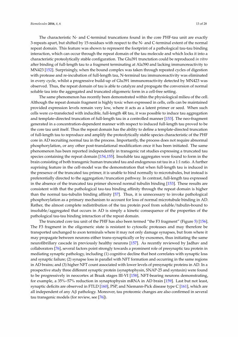
The tau fragment first isolated from the PHF core is approximately 100 amino acids in length.Its N-terminus was defined by sequence analysis [30,56], and its C-terminus was defined by epitopemapping of MN423. Immunoreactivity was shown to depend on a specific C-terminal trunctationat Glu391 [33,150]. Thus, the N-termini of the tau fragments found in the proteolytically stablestructural core of the PHF are located 15-residues C-terminal to the start of the repeats, and havea characteristic C-terminal truncation at position Glu391 which is 15-residues C-terminally to theend of the repeats [151]. These features explained the paradox noted earlier, namely that tau proteinisolated from the core of the PHF is not necessarily recognized by anti-tau antibodies (if these aredirected against epitopes located in the N- or C-terminal portions of the molecule, whether or notphosphorylated), and the monoclonal antibody raised against the PHF core does not recognizednormal full-length tau protein [30].
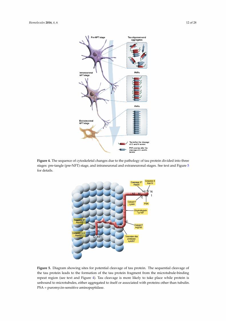
Biomolecules 2016, 6, 6 12 of 28Biomolecules 2016, 6, 6 14 of 35
Figure 4. The sequence of cytoskeletal changes due to the pathology of tau protein divided
into three stages: pre-tangle (pre-NFT) stage, and intraneuronal and extraneuronal stages. See
text and Figure 5 for details.
Figure 5. Diagram showing sites for potential cleavage of tau protein. The sequential cleavage
of the tau protein leads to the formation of the tau protein fragment from the microtubule-
binding repeat region (see text and Figure 4). Tau cleavage is more likely to take place
while protein is unbound to microtubules, either aggregated to itself or associated with
proteins other than tubulin. PSA = puromycin-sensitive aminopeptidase.
Figure 4. The sequence of cytoskeletal changes due to the pathology of tau protein divided into threestages: pre-tangle (pre-NFT) stage, and intraneuronal and extraneuronal stages. See text and Figure 5for details.
Biomolecules 2016, 6, 6 14 of 35
Figure 4. The sequence of cytoskeletal changes due to the pathology of tau protein divided
into three stages: pre-tangle (pre-NFT) stage, and intraneuronal and extraneuronal stages. See
text and Figure 5 for details.
Figure 5. Diagram showing sites for potential cleavage of tau protein. The sequential cleavage
of the tau protein leads to the formation of the tau protein fragment from the microtubule-
binding repeat region (see text and Figure 4). Tau cleavage is more likely to take place
while protein is unbound to microtubules, either aggregated to itself or associated with
proteins other than tubulin. PSA = puromycin-sensitive aminopeptidase.
Figure 5. Diagram showing sites for potential cleavage of tau protein. The sequential cleavage ofthe tau protein leads to the formation of the tau protein fragment from the microtubule-bindingrepeat region (see text and Figure 4). Tau cleavage is more likely to take place while protein isunbound to microtubules, either aggregated to itself or associated with proteins other than tubulin.PSA = puromycin-sensitive aminopeptidase.
Biomolecules 2016, 6, 6 13 of 28
The characteristic N- and C-terminal truncations found in the core PHF-tau unit are exactly3-repeats apart, but shifted by 15-residues with respect to the N- and C-terminal extent of the normalrepeat domain. This feature was shown to represent the footprint of a pathological tau-tau bindinginteraction, which can occur through the repeat domain of the tau molecule and which locks it into acharacteristic proteolytically stable configuration. The Glu391 trunctation could be reproduced in vitroafter binding of full-length tau to a fragment terminating at Ala390 and lacking immunoreactivity toMN423 [152]. Surprisingly, when the bound complex was taken through repeated cycles of digestionwith protease and re-incubation of full-length tau, N-terminal tau immunoreactivity was eliminatedin every cycle, whilst a progressive build-up of Glu391 immunoreactivity detected by MN423 wasobserved. Thus, the repeat domain of tau is able to catalyze and propagate the conversion of normalsoluble tau into the aggregated and truncated oligomeric form in a cell-free setting.
The same phenomenon has recently been demonstrated within the physiological milieu of the cell.Although the repeat domain fragment is highly toxic when expressed in cells, cells can be maintainedprovided expression levels remain very low, where it acts as a latent primer or seed. When suchcells were co-transfected with inducible, full-length 4R tau, it was possible to induce tau aggregationand template-directed truncation of full-length tau in a controlled manner [153]. The neo-fragmentgenerated in a concentration-dependent manner with respect to induced full-length tau proved to bethe core tau unit itself. Thus the repeat domain has the ability to define a template-directed truncationof full-length tau to reproduce and amplify the proteolytically stable species characteristic of the PHFcore in AD recruiting normal tau in the process. Importantly, the process does not require abnormalphosphorylation, or any other post-translational modification once it has been initiated. The samephenomenon has been reported independently in transgenic rat studies expressing a truncated tauspecies containing the repeat domain [154,155]. Insoluble tau aggregates were found to form in thebrain consisting of both transgenic human truncated tau and endogenous rat tau in a 1:1 ratio. A furthersuprising feature in the cell-model was the demonstration that when full-length tau is induced inthe presence of the truncated tau primer, it is unable to bind normally to microtubules, but instead ispreferentially directed to the aggregation/truncation pathway. In contrast, full-length tau expressedin the absence of the truncated tau primer showed normal tubulin binding [153]. These results areconsistent with that the pathological tau-tau binding affinity through the repeat domain is higherthan the normal tau-tubulin binding affinity [57]. Thus, it is unnecessary to invoke pathologicalphosphorylation as a primary mechanism to account for loss of normal microtubule binding in AD.Rather, the almost complete redistribution of the tau protein pool from soluble/tubulin-bound toinsoluble/aggregated that occurs in AD is simply a kinetic consequence of the properties of thepathological tau-tau binding interaction of the repeat domain.
The truncated core tau unit of the PHF has also been termed “the F3 fragment” (Figure 5) [156].The F3 fragment in the oligomeric state is resistant to cytosolic proteases and may therefore betransported unchanged to axon terminals where it may not only damage synapses, but from where itmay propagate between neurons either trans-synaptically or by exosomes, thus initiating the sameneurofibrillary cascade in previously healthy neurons [157]. As recently reviewed by Jadhav andcollaborators [76], several factors point strongly towards a prominent role of presynaptic tau protein inmediating synaptic pathology, including (1) cognitive decline that best correlates with synaptic lossand synaptic failure; (2) synapse loss in parallel with NFT formation and occurring in the same regionsin AD brains; and (3) higher NFT count associated with lower levels of presynaptic proteins in AD. In aprospective study three different synaptic protein (synaptophysin, SNAP-25 and syntaxin) were foundto be progressively in neocortex at Braak stages III-VI [158], NFT-bearing neurons demonstrating,for example, a 35%–57% reduction in synaptophysin mRNA in AD brain [159]. Last but not least,synaptic deficits are observed in FTLD [160], PSP, and Niemann-Pick disease type C [161], which areall independent of any Aβ pathology. Moreover, tau proteomic changes are also confirmed in severaltau transgenic models (for review, see [76]).
Biomolecules 2016, 6, 6 14 of 28
Tau fragments are also able to propagate between neurons trans-synaptically, causing the spreadof neurofibrillary degeneration to post-synaptic neurons [162]. In this case, mutations in the APP,PSEN1 and PSEN2 genes in familial AD only initially compromise endosomal-lysosomal processingand mitochondrial metabolism by altering Aβ clearance thus activating caspases responsible for taucleavage or providing seeding factors required to nucleate pathological aggregation of tau proteinthrough the repeat domain.
2.2. Seeding and Spreading of Tau Proteins
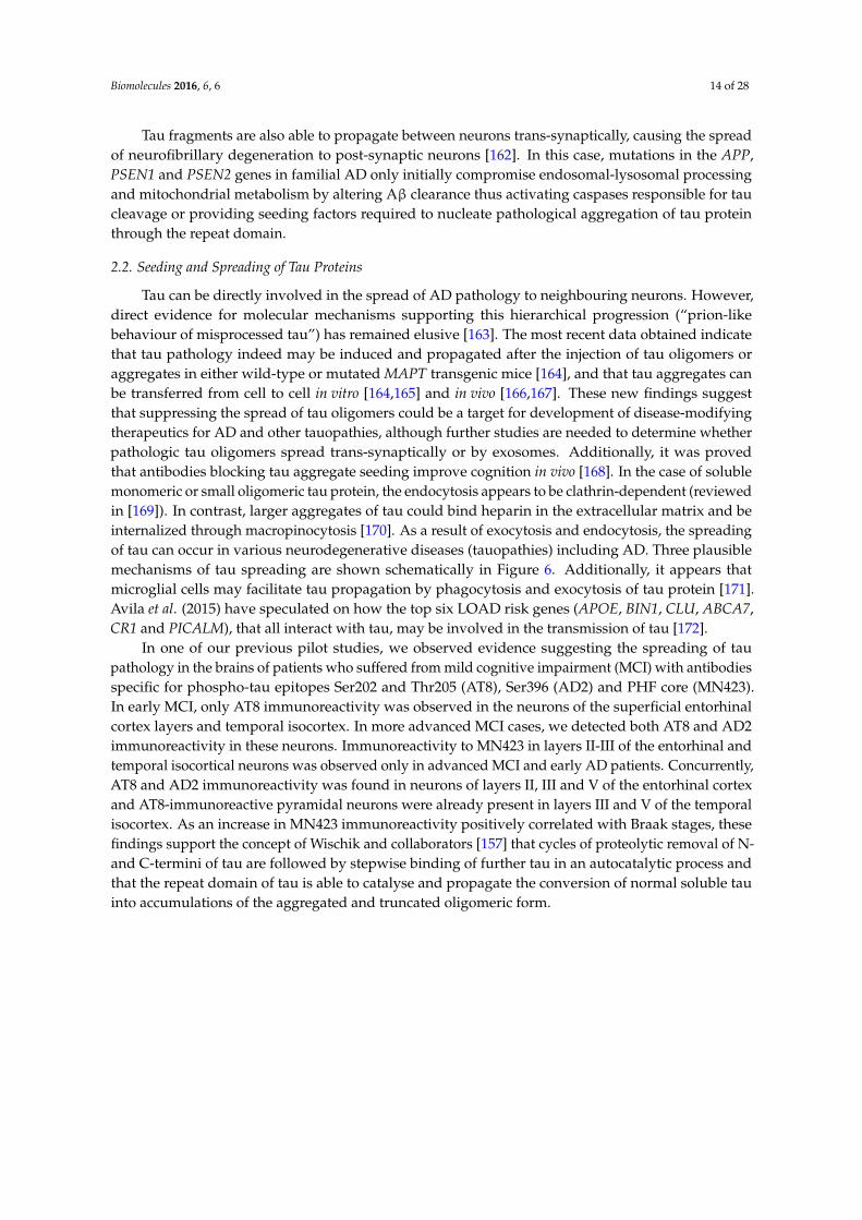
Tau can be directly involved in the spread of AD pathology to neighbouring neurons. However,direct evidence for molecular mechanisms supporting this hierarchical progression (“prion-likebehaviour of misprocessed tau”) has remained elusive [163]. The most recent data obtained indicatethat tau pathology indeed may be induced and propagated after the injection of tau oligomers oraggregates in either wild-type or mutated MAPT transgenic mice [164], and that tau aggregates canbe transferred from cell to cell in vitro [164,165] and in vivo [166,167]. These new findings suggestthat suppressing the spread of tau oligomers could be a target for development of disease-modifyingtherapeutics for AD and other tauopathies, although further studies are needed to determine whetherpathologic tau oligomers spread trans-synaptically or by exosomes. Additionally, it was provedthat antibodies blocking tau aggregate seeding improve cognition in vivo [168]. In the case of solublemonomeric or small oligomeric tau protein, the endocytosis appears to be clathrin-dependent (reviewedin [169]). In contrast, larger aggregates of tau could bind heparin in the extracellular matrix and beinternalized through macropinocytosis [170]. As a result of exocytosis and endocytosis, the spreadingof tau can occur in various neurodegenerative diseases (tauopathies) including AD. Three plausiblemechanisms of tau spreading are shown schematically in Figure 6. Additionally, it appears thatmicroglial cells may facilitate tau propagation by phagocytosis and exocytosis of tau protein [171].Avila et al. (2015) have speculated on how the top six LOAD risk genes (APOE, BIN1, CLU, ABCA7,CR1 and PICALM), that all interact with tau, may be involved in the transmission of tau [172].
In one of our previous pilot studies, we observed evidence suggesting the spreading of taupathology in the brains of patients who suffered from mild cognitive impairment (MCI) with antibodiesspecific for phospho-tau epitopes Ser202 and Thr205 (AT8), Ser396 (AD2) and PHF core (MN423).In early MCI, only AT8 immunoreactivity was observed in the neurons of the superficial entorhinalcortex layers and temporal isocortex. In more advanced MCI cases, we detected both AT8 and AD2immunoreactivity in these neurons. Immunoreactivity to MN423 in layers II-III of the entorhinal andtemporal isocortical neurons was observed only in advanced MCI and early AD patients. Concurrently,AT8 and AD2 immunoreactivity was found in neurons of layers II, III and V of the entorhinal cortexand AT8-immunoreactive pyramidal neurons were already present in layers III and V of the temporalisocortex. As an increase in MN423 immunoreactivity positively correlated with Braak stages, thesefindings support the concept of Wischik and collaborators [157] that cycles of proteolytic removal of N-and C-termini of tau are followed by stepwise binding of further tau in an autocatalytic process andthat the repeat domain of tau is able to catalyse and propagate the conversion of normal soluble tauinto accumulations of the aggregated and truncated oligomeric form.
Biomolecules 2016, 6, 6 15 of 28
Biomolecules 2016, 6, 6 17 of 35
speculated on how the top six LOAD risk genes (APOE, BIN1, CLU, ABCA7, CR1 and PICALM), that
all interact with tau, may be involved in the transmission of tau [172].
In one of our previous pilot studies, we observed evidence suggesting the spreading of tau
pathology in the brains of patients who suffered from mild cognitive impairment (MCI) with
antibodies specific for phospho-tau epitopes Ser202 and Thr205 (AT8), Ser396 (AD2) and PHF core
(MN423). In early MCI, only AT8 immunoreactivity was observed in the neurons of the superficial
entorhinal cortex layers and temporal isocortex. In more advanced MCI cases, we detected both AT8
and AD2 immunoreactivity in these neurons. Immunoreactivity to MN423 in layers II-III of the
entorhinal and temporal isocortical neurons was observed only in advanced MCI and early AD
patients. Concurrently, AT8 and AD2 immunoreactivity was found in neurons of layers II, III and V of
the entorhinal cortex and AT8-immunoreactive pyramidal neurons were already present in layers III
and V of the temporal isocortex. As an increase in MN423 immunoreactivity positively correlated with
Braak stages, these findings support the concept of Wischik and collaborators [157] that cycles of
proteolytic removal of N- and C-termini of tau are followed by stepwise binding of further tau in an
autocatalytic process and that the repeat domain of tau is able to catalyse and propagate the conversion
of normal soluble tau into accumulations of the aggregated and truncated oligomeric form.
Figure 6. Schematic representation of three different ways of anterograde spreading of tau
aggregates by endocytosis, macropinocytosis, and exosomes.
2.3. Therapeutic Approaches Targeting Tau Protein Processing in Tauopathies
A number of neuroprotective strategies have been proposed based on the phosphorylation theory of
tau pathology (Figure 7).
Figure 6. Schematic representation of three different ways of anterograde spreading of tau aggregatesby endocytosis, macropinocytosis, and exosomes.
2.3. Therapeutic Approaches Targeting Tau Protein Processing in Tauopathies
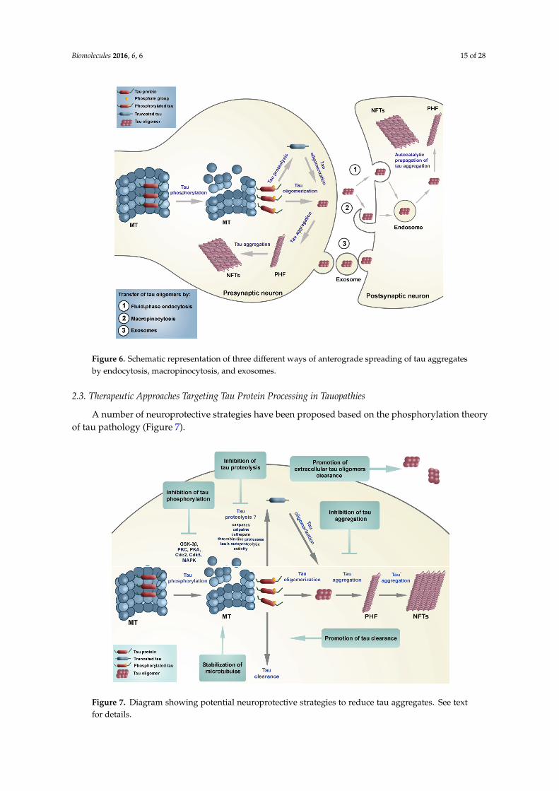
A number of neuroprotective strategies have been proposed based on the phosphorylation theoryof tau pathology (Figure 7).Biomolecules 2016, 6, 6 18 of 35
Figure 7. Diagram showing potential neuroprotective strategies to reduce tau aggregates.
See text for details.
(1) MT-stabilizing agents, which as an approach does not address the accumulation of toxic
tau aggregates.
(2) Modulation of tau phosphorylation has been shown to prevent motor impairments in tau
transgenic mice [173]. Green coffee, a non-toxic small molecule, found to be an inhibitor of protein
phosphatase 2A methylesterase, was shown to improve cognitive and motor performance in mouse
models with tau pathology [174].
(3) A different approach, which does not depend on the phosphorylation theory, is based on
selective inhibition of pathological tau aggregation [157]. The problem in identifying suitable tau
aggregation inhibitors (TAI) is that most assays for tau aggregation are based on fibril formation
(which require relatively high concentrations of tau, tau constructs limited to the MT binding region
with the motif necessary for fibril formation, and a facilitator of aggregation, such as heparin). If
compounds are selected that dissociate preformed large aggregates into smaller (still toxic) oligomers
then they may be detrimental. In these assays, fibril formation is measured by a shift in fluorescence of
an intercalating reporter dye binding to �-sheet structures within tau fibrils.
(4) A more promising approach may be to target tau oligomers, whether these are intracellular or
extracellular. Purified tau oligomer species have been demonstrated to be neurotoxic (they directly
impair synaptic function and long-term potentiation, LTP) in vitro in a dose-dependent manner.
Extracellular tau levels measured in AD are more than four orders of magnitude lower than
intracellular tau concentrations, and as such may represent a more amenable pharmacological target.
(5) A quite different strategy is to target tau clearance—e.g., by rapamycin that induces
macroautophagy [175], inhibitors of Hsp90 chaperone protein that binds to misfolded proteins or by
immunotherapeutic approaches [176].
Figure 7. Diagram showing potential neuroprotective strategies to reduce tau aggregates. See textfor details.

Biomolecules 2016, 6, 6 16 of 28
(1) MT-stabilizing agents, which as an approach does not address the accumulation of toxictau aggregates.
(2) Modulation of tau phosphorylation has been shown to prevent motor impairments in tautransgenic mice [173]. Green coffee, a non-toxic small molecule, found to be an inhibitor of proteinphosphatase 2A methylesterase, was shown to improve cognitive and motor performance in mousemodels with tau pathology [174].
(3) A different approach, which does not depend on the phosphorylation theory, is based onselective inhibition of pathological tau aggregation [157]. The problem in identifying suitable tauaggregation inhibitors (TAI) is that most assays for tau aggregation are based on fibril formation (whichrequire relatively high concentrations of tau, tau constructs limited to the MT binding region with themotif necessary for fibril formation, and a facilitator of aggregation, such as heparin). If compounds areselected that dissociate preformed large aggregates into smaller (still toxic) oligomers then they may bedetrimental. In these assays, fibril formation is measured by a shift in fluorescence of an intercalatingreporter dye binding to β-sheet structures within tau fibrils.
(4) A more promising approach may be to target tau oligomers, whether these are intracellular orextracellular. Purified tau oligomer species have been demonstrated to be neurotoxic (they directlyimpair synaptic function and long-term potentiation, LTP) in vitro in a dose-dependent manner.Extracellular tau levels measured in AD are more than four orders of magnitude lower than intracellulartau concentrations, and as such may represent a more amenable pharmacological target.
(5) A quite different strategy is to target tau clearance—e.g., by rapamycin that inducesmacroautophagy [175], inhibitors of Hsp90 chaperone protein that binds to misfolded proteins or byimmunotherapeutic approaches [176].
(6) Finally, it may be possible to target tau proteolysis directly. Cellular enzymes implicatedin tau proteolysis include caspases, calpains, cathepsins and a thrombin-like protease. Regardlessof the protease, it is reasonable to presume an irreversible loss of normal function of tau once it istruncated. Disulfide-linked oligomers of tau can be observed in AD brains and cerebrospinal fluid (CSF)samples, and show significant fragmentation, making them great potential targets for early diagnosisof AD [177]. The major advantage of targeting tau proteolysis is that it may be more straightforwardto inhibit an enzymatic mechanism than aggregation, provided the proteolytic activity in questioncan be shown to be rate-critical. It is possible that inhibition of truncation could prevent formation ofaggregation-prone fragments and also trans-synaptic/exosomal spread of tau pathology.
Recently, two drugs that targeted tau phosphorylation failed in phase 2 clinical trials [178]. This ledWischik and collaborators [157] to propose that it is not abnormal tau phosphorylation that ought to bereduced by drugs, but tau aggregation. The tau aggregation inhibitor LMTX (leucomethylthioniniumwith a suitable counter-ion, Figure 8) is currently in three parallel Phase III clinical trials, with the firstoutcomes expected in 2016 [179,180]. An older form of the molecule (methylthioninium chloride, MTC)was found to have efficacy in mild/moderate AD in a Phase II clinical trial, in which 90% retardationof disease progression could be demonstrated over 12 months [180]. These investigators stressed thathyperphosphorylation of tau may not play a critical role in aggregation of tau and formation of PHF,and that it may even have an inhibitory effect on tau-tau binding. Thus, it might be more important toclarify proteolysis of tau protein (potentially at position Glu391, although this site may simply reportthe C-terminal extent of the pathological binding domain) that enables the release of the C-terminalfragment. This fragment is the one that appears to be important in the formation or propagation ofproteolytically stable tau oligomers that can spread to neighboring neurons trans-synaptically, furtherpropagate tau pathology and lead ultimately to formation of PHF. Recently, it has been proposed thattau protein acetylation may be responsible for tau aggregation in AD. Grinberg and collaboratorsdetected tau acetylation at Lys274 in all tauopathies (both primary and secondary), except in AgD [181].They hypothesized that tau acetylation could also promote the spreading of tau pathology (whereas inAgD it could have a protective role in this respect).

Biomolecules 2016, 6, 6 17 of 28
Biomolecules 2016, 6, 6 19 of 35
(6) Finally, it may be possible to target tau proteolysis directly. Cellular enzymes implicated in tau
proteolysis include caspases, calpains, cathepsins and a thrombin-like protease. Regardless of the
protease, it is reasonable to presume an irreversible loss of normal function of tau once it is truncated.
Disulfide-linked oligomers of tau can be observed in AD brains and cerebrospinal fluid (CSF) samples,
and show significant fragmentation, making them great potential targets for early diagnosis of AD [177].
The major advantage of targeting tau proteolysis is that it may be more straightforward to inhibit an
enzymatic mechanism than aggregation, provided the proteolytic activity in question can be shown to
be rate-critical. It is possible that inhibition of truncation could prevent formation of aggregation-prone
fragments and also trans-synaptic/exosomal spread of tau pathology.
Recently, two drugs that targeted tau phosphorylation failed in phase 2 clinical trials [178]. This led
Wischik and collaborators [157] to propose that it is not abnormal tau phosphorylation that ought to be
reduced by drugs, but tau aggregation. The tau aggregation inhibitor LMTX (leucomethylthioninium with
a suitable counter-ion, Figure 8) is currently in three parallel Phase III clinical trials, with the first
outcomes expected in 2016 [179,180]. An older form of the molecule (methylthioninium chloride,
MTC) was found to have efficacy in mild/moderate AD in a Phase II clinical trial, in which 90%
retardation of disease progression could be demonstrated over 12 months [180]. These investigators
stressed that hyperphosphorylation of tau may not play a critical role in aggregation of tau and
formation of PHF, and that it may even have an inhibitory effect on tau-tau binding. Thus, it might be
more important to clarify proteolysis of tau protein (potentially at position Glu391, although this site
may simply report the C-terminal extent of the pathological binding domain) that enables the release of
the C-terminal fragment. This fragment is the one that appears to be important in the formation or
propagation of proteolytically stable tau oligomers that can spread to neighboring neurons trans-
synaptically, further propagate tau pathology and lead ultimately to formation of PHF. Recently, it has
been proposed that tau protein acetylation may be responsible for tau aggregation in AD. Grinberg and
collaborators detected tau acetylation at Lys274 in all tauopathies (both primary and secondary),
except in AgD [181]. They hypothesized that tau acetylation could also promote the spreading of tau
pathology (whereas in AgD it could have a protective role in this respect).
Figure 8. Diagram of tau aggregation inhibitor LMTX (leucomethylthioninium with a
suitable counterion), and its presumed mode of action (inhibition of tau aggregation).
Figure 8. Diagram of tau aggregation inhibitor LMTX (leucomethylthioninium with a suitablecounterion), and its presumed mode of action (inhibition of tau aggregation).
The investigators are currently putting most of their efforts into basic, preclinical and clinicaltesting of methylene blue (MB) and its derivatives. MB is a phenothiazine that crosses the blood brainbarrier and acts as a redox cycler. Moreover, besides its beneficial properties as being able to improveenergy metabolism and to act as an antioxidant, it is also able to reduce tau protein aggregation. Howexactly LMTX and MTC exert their neuroprotective effects in vivo is not fully understood. MB (as MTC)is able to reduce the amount of sarkosyl-insoluble tau in Drosophila that express human wild-typetau [182], to disaggregate PHF isolated from AD brain [152] and to block prion-like processing of tauprotein in cell models [153]. Both MTC and LMTX have been shown to reduce tau pathology andreverse behavioural deficits in transgenic mouse models of established pathology based either on therepeat domain fragment or on full-length mutant tau P301S [183]. MB, together with its derivatives(metabolites), azure A and azure B, is able to stimulate protein degradation and inhibit oxidativedamage [184] and also inhibit the activity of caspase-1 and caspase-3 [185]. MB given prior to theonset of tau aggregation was also able to prevent learning and memory deficits in tau transgenicmice [186], suggesting a potential preventative utility. Other possible inhibitors of tau aggregation arerhodanine-based inhibitors, phenylthiazolyl-hydrazide inhibitors, N-phenylamines, phenothiazinesand benzothiazoles, and polyphenols and anthraquinones [187].
3. Conclusions
Although the pathogenic nature of the each type of protein deposit has been a controversial issuefor many years, it is now increasingly accepted that abnormal forms of tau protein are directly involvedin the initiation of neurodegerative processes. This conclusion is based primarily on the discoverythat dominant missense mutations in the MAPT gene are associated with dominant, familial forms ofFTD. Known polymorphisms in MAPT which confer susceptibility not only for AD and FTD, but otherneurodegenerative diseases as well, together with a possible additional novel disease locus near theMAPT gene [188], strongly support the key role of tau protein not only in primary tauopathies butalso in the pathogenesis of LOAD and other secondary tauopathies.
Why disease onset takes decades before symptoms occur remains unclear at present, but currentresults suggest a reduced ability to clear out misfolded, oligomerized and aggregated tau proteinsthat increase with advancing age. As many drug discovery attempts based on the amyloid cascadehypothesis have proved unsuccessful, and due to advances in our understanding of the role for tau inAD pathogenesis [189], it is safe to conclude that tau protein will become an increasingly important

Biomolecules 2016, 6, 6 18 of 28
therapeutic target for the future. The results of clinical trials with LMTX are eagerly awaited to confirmwhether a treatment for tauopathies is viable.
Acknowledgments: This work was supported by The Croatian Science Foundation grant No. IP-2014-09-9730(“Tau protein hyperphosphorylation, aggregation, and trans-synaptic transfer in Alzheimer’s disease:cerebrospinal fluid analysis and assessment of potential neuroprotective compounds”) and European Cooperationin Science and Technology (COST) Action CM1103 (“Stucture-based drug design for diagnosis and treatmentof neurological diseases: dissecting and modulating complex function in the monoaminergic systems of thebrain”). PRH is supported in part by NIH grant P50 AG005138. We also thank Mate Babic for help in preparationof schematics.
Author Contributions: Goran Šimic, Mirjana Babic Leko, Giuseppe Di Giovanni, and Patrick R. Hof conceivedthe review. All authors contributed to drafting the work and revising it critically for important intellectual content.
Conflicts of Interest: Charles Harrington (Chief Scientific Officer) and Claude Wischik (Executive Chairman) areofficers in TauRx Therapeutics Ltd and both are co-inventors on various patents related to tau protein.
Abbreviations
3R tau tau isoforms with three microtubule-binding repeats4R tau tau isoforms with four microtubule-binding repeatsA adenineAβ amyloid β proteinAD Alzheimer’s diseaseAD2 antibody specific for phospho-tau epitope Ser396ADAM10 a disintegrin and metalloprotease domain 10AgD argyrophilic grain diseaseAMPA α-amino-3-hydroxy-5-methyl-4-isoxazolepropionic acidAPH-1 anterior pharynx-defective 1APOE apolipoprotein EAPP amyloid precursor proteinAT8 antibody specific for phospho-tau epitopes Ser202 and Thr205BACE β-site APP cleaving enzymeBF Bayes factorCBD corticobasal degenerationCSF cerebrospinal fluidDDPAC disinhibition-dementia-parkinsonism-amyotrophy complexDM1 myotonic dystrophy type IEOAD early-onset ADfAD familial ADFTD frontotemporal dementiaFTDP-17 frontotemporal dementia and parkinsonism linked to chromosome 17FTLD frontotemporal lobar degnerationG guanineGrb2 growth factor receptor-bound protein 2HCHWA-D hereditary cerebral hemorrhage with amyloidosis-Dutch typeHDAC6 histone deacetylase 6LMTX leucomethylthioniniumLOAD late-onset ADLTP long-term potentiationMAP2 microtubule-associated protein 2MAPT microtubule-associated protein tauMB Methylene blueMCI mild cognitive impairment

Biomolecules 2016, 6, 6 19 of 28
MN423 antibody for PHF coreMSTD multiple system tauopathy with presenile dementiaMT microtubuleNFT neurofibrillary tanglesNMDAR N-methyl-D-aspartate receptorsNT neuropil threadsO-GlcNAc O-linked N-acetylglucosaminePD Parkinson’s diseasePEN-2 presenilin enhancer 2PHF paired helical filamentsPSA puromycin-sensitive aminopeptidasePSEN presenilinPSEN1 presenilin 1PSEN2 presenilin 2PSP progressive supranuclear palsysAD sporadic ADSDS sodium dodecyl sulfateSDS-PAGE sodium dodecyl sulfate-polyacrylamide gel electrophoresisSF straight filamentsSP senile plaquesTACE tumor necrosis factor alpha converting enzymeTau tubulin-associated unitTNF-α tumor necrosis factor alpha
References
1. Bielschowsky, M. Die Silberimprägnation der Achsenzylinder. Neurol. Zentralb. (Leipzig) 1902, 13, 579–584.(In German).
2. Alzheimer, A. Uber eine eigenartige Erkrankung der Hirnrinde. Allg Zeits Psychiatry Psych. Med. 1907, 64,146–148. (In German).
3. Jucker, M.; Beyreuther, K.; Haass, C.; Nitsch, R.; Christen, Y. Alzheimer: 100 Years and Beyond; Springer: Berlin,Germany, 2006.
4. Kidd, M. Paired helical filaments in electron microscopy of Alzheimer’s disease. Nature 1963, 197, 192–193.[CrossRef] [PubMed]
5. Terry, R.D. The fine structure of neurofibrillary tangles in Alzheimer’s disease. J. Neuropathol. Exp. Neurol.1963, 22, 629–642. [CrossRef] [PubMed]
6. Crowther, R.A.; Wischik, C.M. Image reconstruction of the Alzheimer paired helical filament. EMBO J. 1985,4, 3661–3665. [PubMed]
7. Wischik, C.M.; Crowther, R.A.; Stewart, M.; Roth, M. Subunit structure of paired helical filaments inAlzheimer’s disease. J. Cell Biol. 1985, 100, 1905–1912. [CrossRef] [PubMed]
8. Crowther, R.A. Straight and paired helical filaments in Alzheimer disease have a common structural unit.Proc. Natl. Acad. Sci. USA 1991, 88, 2288–2292. [CrossRef] [PubMed]
9. Weingarten, M.D.; Lockwood, A.H.; Hwo, S.Y.; Kirschner, M.W. A protein factor essential for microtubuleassembly. Proc. Natl. Acad. Sci. USA 1975, 72, 1858–1862. [CrossRef] [PubMed]
10. Cleveland, D.W.; Hwo, S.Y.; Kirschner, M.W. Purification of tau, a microtubule-associated protein thatinduces assembly of microtubules from purified tubulin. J. Mol. Biol. 1977, 116, 207–225. [CrossRef]
11. Cleveland, D.W.; Hwo, S.-Y.; Kirschner, M.W. Physical and chemical properties of purified tau factor and therole of tau in microtubule assembly. J. Mol. Biol. 1977, 116, 227–247. [CrossRef]
12. Pierre, M.; Nunez, J. Multisite phosphorylation of tau proteins from rat brain. Biochem. Biophys. Res. Commun.1983, 115, 212–219. [CrossRef]
13. Jameson, L.; Frey, T.; Zeeberg, B.; Dalldorf, F.; Caplow, M. Inhibition of microtubule assembly byphosphorylation of microtubule-associated proteins. Biochemistry 1980, 19, 2472–2479. [CrossRef] [PubMed]

Biomolecules 2016, 6, 6 20 of 28
14. Lindwall, G.; Cole, R.D. Phosphorylation affects the ability of tau protein to promote microtubule assembly.J. Biol. Chem. 1984, 259, 5301–5305. [PubMed]
15. Binder, L.I.; Frankfurter, A.; Rebhun, L.I. The distribution of tau in the mammalian central nervous system.J. Cell Biol. 1985, 101, 1371–1378. [CrossRef] [PubMed]
16. Papasozomenos, S.C.; Binder, L.I. Phosphorylation determines two distinct species of Tau in the centralnervous system. Cell Motil. Cytoskeleton 1987, 8, 210–226. [CrossRef] [PubMed]
17. Migheli, A.; Butler, M.; Brown, K.; Shelanski, M.L. Light and electron microscope localization of themicrotubule-associated tau protein in rat brain. J. Neurosci. 1988, 8, 1846–1851. [PubMed]
18. Couchie, D.; Charrière-Bertrand, C.; Nunez, J. Expression of the mRNA for tau proteins during braindevelopment and in cultured neurons and astroglial cells. J. Neurochem. 1988, 50, 1894–1899. [CrossRef][PubMed]
19. Brion, J.; Passareiro, H.; Nunez, J.; Flament-Durand, J. Mise en évidence immunologique de la protéine tauau niveau des lésions de dégénérescence neurofibrillaire de la maladie d’Alzheimer. Arch. Biol. 1985, 95,229–235. (In French).
20. Anderton, B.H.; Breinburg, D.; Downes, M.J.; Green, P.J.; Tomlinson, B.E.; Ulrich, J.; Wood, J.N.; Kahn, J.Monoclonal antibodies show that neurofibrillary tangles and neurofilaments share antigenic determinants.Nature 1982, 298, 84–86. [CrossRef] [PubMed]
21. Grundke-Iqbal, I.; Iqbal, K.; Quinlan, M.; Tung, Y.C.; Zaidi, M.S.; Wisniewski, H.M. Microtubule-associatedprotein tau. A component of Alzheimer paired helical filaments. J. Biol. Chem. 1986, 261, 6084–6089.[PubMed]
22. Grundke-Iqbal, I.; Iqbal, K.; Tung, Y.C.; Quinlan, M.; Wisniewski, H.M.; Binder, L.I. Abnormalphosphorylation of the microtubule-associated protein tau (tau) in Alzheimer cytoskeletal pathology.Proc. Natl. Acad. Sci. USA 1986, 83, 4913–4917. [CrossRef] [PubMed]
23. Ihara, Y.; Nukina, N.; Miura, R.; Ogawara, M. Phosphorylated tau protein is integrated into paired helicalfilaments in Alzheimer’s disease. J. Biochem. 1986, 99, 1807–1810. [PubMed]
24. Masters, C.L.; Simms, G.; Weinman, N.A.; Multhaup, G.; McDonald, B.L.; Beyreuther, K. Amyloid plaquecore protein in Alzheimer disease and Down syndrome. Proc. Natl. Acad. Sci. USA 1985, 82, 4245–4249.[CrossRef] [PubMed]
25. Masters, C.L.; Multhaup, G.; Simms, G.; Pottgiesser, J.; Martins, R.N.; Beyreuther, K. Neuronal origin of acerebral amyloid: Neurofibrillary tangles of Alzheimer’s disease contain the same protein as the amyloid ofplaque cores and blood vessels. EMBO J. 1985, 4, 2757–2763. [PubMed]
26. Mori, H.; Kondo, J.; Ihara, Y. Ubiquitin is a component of paired helical filaments in Alzheimer’s disease.Science 1987, 235, 1641–1644. [CrossRef] [PubMed]
27. Perry, G.; Friedman, R.; Shaw, G.; Chau, V. Ubiquitin is detected in neurofibrillary tangles and senile plaqueneurites of Alzheimer disease brains. Proc. Natl. Acad. Sci. USA 1987, 84, 3033–3036. [CrossRef] [PubMed]
28. Yen, S.H.; Gaskin, F.; Fu, S.M. Neurofibrillary tangles in senile dementia of the Alzheimer type share anantigenic determinant with intermediate filaments of the vimentin class. Am. J. Pathol. 1983, 113, 373–381.[PubMed]
29. Yen, S.; Dickson, D.; Crowe, A. Alzheimer’s neurofibrillary tangles contain unique epitopes and epitopes incommon with the heat-stable microtubule associated proteins tau and MAP2. Am. J. Pathol. 1987, 126, 81–91.[PubMed]
30. Wischik, C.M.; Novak, M.; Thøgersen, H.C.; Edwards, P.C.; Runswick, M.J.; Jakes, R.; Walker, J.E.; Milstein, C.;Roth, M.; Klug, A. Isolation of a fragment of tau derived from the core of the paired helical filament ofAlzheimer disease. Proc. Natl. Acad. Sci. USA 1988, 85, 4506–4510. [CrossRef] [PubMed]
31. Wischik, C.M.; Novak, M.; Edwards, P.C.; Klug, A.; Tichelaar, W.; Crowther, R.A. Structural characterizationof the core of the paired helical filament of Alzheimer disease. Proc. Natl. Acad. Sci. USA 1988, 85, 4884–4888.[CrossRef] [PubMed]
32. Novak, M.; Wischik, C.M.; Edwards, P.; Pannell, R.; Milstein, C. Characterisation of the first monoclonalantibody against the pronase resistant core of the Alzheimer PHF. Prog. Clin. Biol. Res. 1989, 317, 755–761.[PubMed]
33. Novak, M.; Jakes, R.; Edwards, P.C.; Milstein, C.; Wischik, C.M. Difference between the tau protein ofAlzheimer paired helical filament core and normal tau revealed by epitope analysis of monoclonal antibodies423 and 7.51. Proc. Natl. Acad. Sci. USA 1991, 88, 5837–5841. [CrossRef] [PubMed]

Biomolecules 2016, 6, 6 21 of 28
34. Resch, J.F.; Lehr, G.S.; Wischik, C.M. Design and synthesis of a potential affinity/cleaving reagent forbeta-pleated sheet protein structures. Bioorg. Med. Chem. Lett. 1991, 1, 519–522. [CrossRef]
35. Lee, G.; Cowan, N.; Kirschner, M. The primary structure and heterogeneity of tau protein from mouse brain.Science 1988, 239, 285–288. [CrossRef] [PubMed]
36. Goedert, M.; Wischik, C.M.; Crowther, R.A.; Walker, J.E.; Klug, A. Cloning and sequencing of thecDNA encoding a core protein of the paired helical filament of Alzheimer disease: Identification as themicrotubule-associated protein tau. Proc. Natl. Acad. Sci. USA 1988, 85, 4051–4055. [CrossRef] [PubMed]
37. Delacourte, A.; Defossez, A. Alzheimer’s disease: Tau proteins, the promoting factors of microtubuleassembly, are major components of paired helical filaments. J. Neurol. Sci. 1986, 76, 173–186. [CrossRef]
38. Kosik, K.S.; Joachim, C.L.; Selkoe, D.J. Microtubule-associated protein tau (tau) is a major antigeniccomponent of paired helical filaments in Alzheimer disease. Proc. Natl. Acad. Sci. USA 1986, 83, 4044–4048.[CrossRef] [PubMed]
39. Nukina, N.; Ihara, Y. One of the antigenic determinants of paired helical filaments is related to tau protein.J. Biochem. 1986, 99, 1541–1544. [PubMed]
40. Wood, J.G.; Mirra, S.S.; Pollock, N.J.; Binder, L.I. Neurofibrillary tangles of Alzheimer disease share antigenicdeterminants with the axonal microtubule-associated protein tau (tau). Proc. Natl. Acad. Sci. USA 1986, 83,4040–4043. [CrossRef] [PubMed]
41. Montejo de Garcini, E.; Serrano, L.; Avila, J. Self assembly of microtubule associated protein tau into filamentsresembling those found in Alzheimer disease. Biochem. Biophys. Res. Commun. 1986, 141, 790–796. [CrossRef]
42. Neve, R.L.; Harris, P.; Kosik, K.S.; Kurnit, D.M.; Donlon, T.A. Identification of cDNA clones for thehuman microtubule-associated protein tau and chromosomal localization of the genes for tau andmicrotubule-associated protein 2. Mol. Brain Res. 1986, 1, 271–280. [CrossRef]
43. Donlon, T.; Harris, P.; Neve, R. Localization of microtubule-associated protein tau (MTBT1) to chromosome17q21 (Abstract). Cytogenet. Cell Genet. 1987, 46, 607.
44. Goedert, M.; Spillantini, M.; Potier, M.; Ulrich, J.; Crowther, R. Cloning and sequencing of the cDNA encodingan isoform of microtubule-associated protein tau containing four tandem repeats: Differential expression oftau protein mRNAs in human brain. EMBO J. 1989, 8, 393–399. [PubMed]
45. Goedert, M.; Spillantini, M.G.; Jakes, R.; Rutherford, D.; Crowther, R. Multiple isoforms of humanmicrotubule-associated protein tau: Sequences and localization in neurofibrillary tangles of Alzheimer’sdisease. Neuron 1989, 3, 519–526. [CrossRef]
46. Goedert, M.; Jakes, R. Expression of separate isoforms of human tau protein: Correlation with the tau patternin brain and effects on tubulin polymerization. EMBO J. 1990, 9, 4225–4230. [PubMed]
47. Goedert, M.; Spillantini, M.G.; Crowther, R.A. Cloning of a big tau microtubule-associated proteincharacteristic of the peripheral nervous system. Proc. Natl. Acad. Sci. USA 1992, 89, 1983–1987. [CrossRef][PubMed]
48. Jovanov-Miloševic, N.; Petrovic, D.; Sedmak, G.; Vukšic, M.; Hof, P.R.; Šimic, G. Human fetal tau proteinisoform: Possibilities for Alzheimer’s disease treatment. Int. J. Biochem. Cell Biol. 2012, 44, 1290–1294.[CrossRef] [PubMed]
49. Greenberg, S.G.; Davies, P. A preparation of Alzheimer paired helical filaments that displays distinct tauproteins by polyacrylamide gel electrophoresis. Proc. Natl. Acad. Sci. USA 1990, 87, 5827–5831. [CrossRef][PubMed]
50. Mulot, S.F.; Hughes, K.; Woodgett, J.R.; Anderton, B.H.; Hanger, D.P. PHF-tau from Alzheimer’s braincomprises four species on SDS-PAGE which can be mimicked by in vitro phosphorylation of human braintau by glycogen synthase kinase-3 beta. FEBS Lett. 1994, 349, 359–364. [CrossRef]
51. Wolozin, B.L.; Pruchnicki, A.; Dickson, D.W.; Davies, P. A neuronal antigen in the brains of Alzheimerpatients. Science 1986, 232, 648–650. [CrossRef] [PubMed]
52. Flament, S.; Delacourte, A. Abnormal tau species are produced during Alzheimer’s diseaseneurodegenerating process. FEBS Lett. 1989, 247, 213–216. [CrossRef]
53. Lee, V.M.; Balin, B.J.; Otvos, L.; Trojanowski, J.Q. A68: A major subunit of paired helical filaments andderivatized forms of normal Tau. Science 1991, 251, 675–678. [CrossRef] [PubMed]
54. Lai, R.Y.; Gertz, H.N.; Wischik, D.J.; Xuereb, J.H.; Mukaetova-Ladinska, E.B.; Harrington, C.R.; Edwards, P.C.;Mena, R.; Paykel, E.S.; Brayne, C.; et al. Examination of phosphorylated tau protein as a PHF-precursor atearly stage Alzheimer’s disease. Neurobiol. Aging 1995, 16, 433–445. [CrossRef]
Biomolecules 2016, 6, 6 22 of 28
55. Wischik, C.M.; Edwards, P.C.; Lai, R.Y.; Gertz, H.N.; Xuereb, J.H.; Paykel, E.S.; Brayne, C.; Huppert, F.A.;Mukaetova-Ladinska, E.B.; Mena, R. Quantitative analysis of tau protein in paired helical filamentpreparations: Implications for the role of tau protein phosphorylation in PHF assembly in Alzheimer’sdisease. Neurobiol. Aging 1995, 16, 409–417, discussion 418–431. [CrossRef]
56. Jakes, R.; Novak, M.; Davison, M.; Wischik, C.M. Identification of 3- and 4-repeat tau isoforms within thePHF in Alzheimer’s disease. EMBO J. 1991, 10, 2725–2729. [PubMed]
57. Wischik, C.; Lai, R.; Harrington, C. Modelling prion-like processing of tau protein in Alzheimer’s diseasefor pharmaceutical development. In Brain Microtubule Associated Proteins: Modifications in Disease; Avila, J.,Brandt, R., Kosik, K., Eds.; Harwood Academic Publishers: Amsterdam, The Nederland, 1997; pp. 185–241.
58. Gu, Y.; Oyama, F.; Ihara, Y. Tau is widely expressed in rat tissues. J. Neurochem. 1996, 67, 1235–1244.[CrossRef] [PubMed]
59. Qiang, L.; Yu, W.; Andreadis, A.; Luo, M.; Baas, P.W. Tau protects microtubules in the axon from severing bykatanin. J. Neurosci. 2006, 26, 3120–3129. [CrossRef] [PubMed]
60. Takei, Y.; Teng, J.; Harada, A.; Hirokawa, N. Defects in axonal elongation and neuronal migration in micewith disrupted tau and map1b genes. J. Cell Biol. 2000, 150, 989–1000. [CrossRef] [PubMed]
61. Buée, L.; Bussière, T.; Buée-Scherrer, V.; Delacourte, A.; Hof, P.R. Tau protein isoforms, phosphorylation androle in neurodegenerative disorders. Brain Res. Rev. 2000, 33, 95–130. [CrossRef]
62. Morishima-Kawashima, M.; Hasegawa, M.; Takio, K.; Suzuki, M.; Yoshida, H.; Watanabe, A.; Titani, K.;Ihara, Y. Hyperphosphorylation of tau in PHF. Neurobiol. Aging 1995, 16, 365–371, discussion 371–380.[CrossRef]
63. Bretteville, A.; Ando, K.; Ghestem, A.; Loyens, A.; Bégard, S.; Beauvillain, J.-C.; Sergeant, N.; Hamdane, M.;Buée, L. Two-dimensional electrophoresis of tau mutants reveals specific phosphorylation pattern likelylinked to early tau conformational changes. PLoS ONE 2009, 4, e4843. [CrossRef] [PubMed]
64. Šimic, G.; Diana, A.; Hof, P.R. Phosphorylation pattern of tau associated with distinct changes of the growthcone cytoskeleton. Prog. Mol. Subcell. Biol. 2003, 32, 33–48. [PubMed]
65. Kingwell, K. Neurodegenerative disease: Targeting tau acetylation attenuates neurodegeneration. Nat. Rev.Drug Discov. 2015, 14, 748–749. [CrossRef] [PubMed]
66. Braak, H.; Braak, E. Neuropathological stageing of Alzheimer-related changes. Acta Neuropathol. 1991, 82,239–259. [CrossRef] [PubMed]
67. Braak, H.; Alafuzoff, I.; Arzberger, T.; Kretzschmar, H.; Tredici, K. Staging of Alzheimer disease-associatedneurofibrillary pathology using paraffin sections and immunocytochemistry. Acta Neuropathol. 2006, 112,389–404. [CrossRef] [PubMed]
68. Ding, H.; Dolan, P.J.; Johnson, G.V.W. Histone deacetylase 6 interacts with the microtubule-associated proteintau. J. Neurochem. 2008, 106, 2119–2130. [CrossRef] [PubMed]
69. Huang, D.Y.; Weisgraber, K.H.; Goedert, M.; Saunders, A.M.; Roses, A.D.; Strittmatter, W.J. ApoE3 bindingto tau tandem repeat I is abolished by tau serine262 phosphorylation. Neurosci. Lett. 1995, 192, 209–212.[CrossRef]
70. Reynolds, C.H.; Garwood, C.J.; Wray, S.; Price, C.; Kellie, S.; Perera, T.; Zvelebil, M.; Yang, A.;Sheppard, P.W.; Varndell, I.M.; et al. Phosphorylation regulates tau interactions with Src homology 3domains of phosphatidylinositol 3-kinase, phospholipase Cγ1, Grb2, and Src family kinases. J. Biol. Chem.2008, 283, 18177–18186. [CrossRef] [PubMed]
71. Surridge, C.D.; Burns, R.G. The difference in the binding of phosphatidylinositol distinguishes MAP2 fromMAP2C and Tau. Biochemistry 1994, 33, 8051–8057. [CrossRef] [PubMed]
72. Flanagan, L.A.; Cunningham, C.C.; Chen, J.; Prestwich, G.D.; Kosik, K.S.; Janmey, P.A. The structure ofdivalent cation-induced aggregates of PIP2 and their alteration by gelsolin and tau. Biophys. J. 1997, 73,1440–1447. [CrossRef]
73. Morris, M.; Maeda, S.; Vossel, K.; Mucke, L. The many faces of tau. Neuron 2011, 70, 410–426. [CrossRef][PubMed]
74. Himmelstein, D.S.; Ward, S.M.; Lancia, J.K.; Patterson, K.R.; Binder, L.I. Tau as a therapeutic target inneurodegenerative disease. Pharmacol. Ther. 2012, 136, 8–22. [CrossRef] [PubMed]
75. Klein, C.; Kramer, E.-M.; Cardine, A.-M.; Schraven, B.; Brandt, R.; Trotter, J. Process outgrowth ofoligodendrocytes is promoted by interaction of fyn kinase with the cytoskeletal protein tau. J. Neurosci. 2002,22, 698–707. [PubMed]

Biomolecules 2016, 6, 6 23 of 28
76. Jadhav, S.; Cubinkova, V.; Zimova, I.; Brezovakova, V.; Madari, A.; Cigankova, V.; Zilka, N. Tau-mediatedsynaptic damage in Alzheimer’s disease. Transl. Neurosci. 2015, 6, 214–226. [CrossRef]
77. Mondragón-Rodríguez, S.; Trillaud-Doppia, E.; Dudilot, A.; Bourgeois, C.; Lauzon, M.; Leclerc, N.;Boehm, J. Interaction of endogenous tau protein with synaptic proteins is regulated by N-methyl-D-aspartatereceptor-dependent tau phosphorylation. J. Biol. Chem. 2012, 287, 32040–32053. [CrossRef] [PubMed]
78. Ahmed, T.; van der Jeugd, A.; Blum, D.; Galas, M.-C.; D’Hooge, R.; Buée, L.; Balschun, D. Cognitionand hippocampal synaptic plasticity in mice with a homozygous tau deletion. Neurobiol. Aging 2014, 35,2474–2478. [CrossRef] [PubMed]
79. Regan, P.; Piers, T.; Yi, J.-H.; Kim, D.-H.; Huh, S.; Park, S.J.; Ryu, J.H.; Whitcomb, D.J.; Cho, K. Tauphosphorylation at serine 396 residue is required for hippocampal LTD. J. Neurosci. 2015, 35, 4804–4812.[CrossRef] [PubMed]
80. Sultan, A.; Nesslany, F.; Violet, M.; Bégard, S.; Loyens, A.; Talahari, S.; Mansuroglu, Z.; Marzin, D.;Sergeant, N.; Humez, S.; et al. Nuclear tau, a key player in neuronal DNA protection. J. Biol. Chem.2011, 286, 4566–4575. [CrossRef] [PubMed]
81. Perez, M.; Santa-Maria, I.; de Gomez Barreda, E.; Zhu, X.; Cuadros, R.; Cabrero, J.R.; Sanchez-Madrid, F.;Dawson, H.N.; Vitek, M.P.; Perry, G.; et al. Tau-an inhibitor of deacetylase HDAC6 function. J. Neurochem.2009, 109, 1756–1766. [CrossRef] [PubMed]
82. Leyk, J.; Goldbaum, O.; Noack, M.; Richter-Landsberg, C. Inhibition of HDAC6 modifies tau inclusion bodyformation and impairs autophagic clearance. J. Mol. Neurosci. 2015, 55, 1031–1046. [CrossRef] [PubMed]
83. Davies, P.; Maloney, A.J. Selective loss of central cholinergic neurons in Alzheimer’s disease. Lancet 1976.[CrossRef]
84. Whitehouse, P.J.; Price, D.L.; Clark, A.W.; Coyle, J.T.; DeLong, M.R. Alzheimer disease: Evidence for selectiveloss of cholinergic neurons in the nucleus basalis. Ann. Neurol. 1981, 10, 122–126. [CrossRef] [PubMed]
85. Glenner, G.G.; Wong, C.W. Alzheimer’s disease: Initial report of the purification and characterization of anovel cerebrovascular amyloid protein. Biochem. Biophys. Res. Commun. 1984, 120, 885–890. [CrossRef]
86. Wong, C.W.; Quaranta, V.; Glenner, G.G. Neuritic plaques and cerebrovascular amyloid in Alzheimer diseaseare antigenically related. Proc. Natl. Acad. Sci. USA 1985, 82, 8729–8732. [CrossRef] [PubMed]
87. Glenner, G.G.; Wong, C.W. Alzheimer’s disease and Down’s syndrome: Sharing of a unique cerebrovascularamyloid fibril protein. Biochem. Biophys. Res. Commun. 1984, 122, 1131–1135. [CrossRef]
88. Goate, A.; Chartier-Harlin, M.C.; Mullan, M.; Brown, J.; Crawford, F.; Fidani, L.; Giuffra, L.; Haynes, A.;Irving, N.; James, L. Segregation of a missense mutation in the amyloid precursor protein gene with familialAlzheimer’s disease. Nature 1991, 349, 704–706. [CrossRef] [PubMed]
89. Hardy, J.; Allsop, D. Amyloid deposition as the central event in the aetiology of Alzheimer’s disease.Trends Pharmacol. Sci. 1991, 12, 383–388. [CrossRef]
90. Hardy, J.A.; Higgins, G.A. Alzheimer’s disease: The amyloid cascade hypothesis. Science 1992, 256, 184–185.[CrossRef] [PubMed]
91. Blanquet, V.; Goldgaber, D.; Turleau, C.; Créau-Goldberg, N.; Delabar, J.; Sinet, P.M.; Roudier, M.;de Grouchy, J. The beta amyloid protein (AD-AP) cDNA hybridizes in normal and Alzheimer individualsnear the interface of 21q21 and q22.1. Ann. Génétique 1987, 30, 68–69.
92. Robakis, N.; Wisniewski, H.; Jenkins, E.; Devine-Gage, E.; Houck, G.; Yao, X.; Ramakrishna, N.; Wolfe, G.;Silverman, W.; Brown, W. Chromosome 21q21 sublocalisation of gene encoding beta-amyloid peptide incerebral vessels and neuritic (senile) plaques of people with Alzheimer disease and Down syndrome. Lancet1987, 1, 384–385. [CrossRef]
93. St George-Hyslop, P.H.; Tanzi, R.E.; Polinsky, R.J.; Haines, J.L.; Nee, L.; Watkins, P.C.; Myers, R.H.;Feldman, R.G.; Pollen, D.; Drachman, D.; et al. The genetic defect causing familial Alzheimer’s disease mapson chromosome 21. Science 1987, 235, 885–890. [CrossRef] [PubMed]
94. Goldgaber, D.; Lerman, M.; McBride, W.; Saffiotti, U.; Gajdusek, D. Isolation, characterization, andchromosomal localization of human brain cDNA clones coding for the precursor of the amyloid of brain inAlzheimer’s disease, Down’s syndrome and aging. J. Neural Transm. 1987, 24, 23–28.
95. Van Broeckhoven, C.; Haan, J.; Bakker, E.; Hardy, J.; van Hul, W.; Webnert, A.; der Vegter-Van Vlis, M.;Roos, R. Amyloid beta protein precursor gene and hereditary cerebral hemorrhage with amyloidosis (Dutch).Science 1990, 248, 1120–1122. [CrossRef] [PubMed]

Biomolecules 2016, 6, 6 24 of 28
96. Levy, E.; Carman, M.; Fernandez-Madrid, I. Mutation of the Alzheimer’s disease amyloid gene in hereditarycerebral hemorrhage, Dutch type. Science 1990, 248, 1124–1126. [CrossRef] [PubMed]
97. Lowenberg, K.; Waggoner, R. Familial organic psychosis (Alzheimer’s type). Arch. Neurol. 1934, 31, 737–754.[CrossRef]
98. St George-Hyslop, P.; Haines, J.; Rogaev, E.; Mortilla, M.; Vaula, G.; Pericak-Vance, M.; Foncin, J.F.;Montesi, M.; Bruni, A.; Sorbi, S.; et al. Genetic evidence for a novel familial Alzheimer’s disease locuson chromosome 14. Nat. Genet. 1992, 2, 330–334. [CrossRef] [PubMed]
99. Sherrington, R.; Rogaev, E.I.; Liang, Y.; Rogaeva, E.A.; Levesque, G.; Ikeda, M.; Chi, H.; Lin, C.; Li, G.;Holman, K.; et al. Cloning of a gene bearing missense mutations in early-onset familial Alzheimer’s disease.Nature 1995, 375, 754–760. [CrossRef] [PubMed]
100. Schellenberg, G.D.; Bird, T.D.; Wijsman, E.M.; Orr, H.T.; Anderson, L.; Nemens, E.; White, J.A.;Bonnycastle, L.; Weber, J.L.; Alonso, M.E. Genetic linkage evidence for a familial Alzheimer’s diseaselocus on chromosome 14. Science 1992, 258, 668–671. [CrossRef] [PubMed]
101. Levy-Lahad, E.; Wasco, W.; Poorkaj, P.; Romano, D.M.; Oshima, J.; Pettingell, W.H.; Yu, C.E.; Jondro, P.D.;Schmidt, S.D.; Wang, K.; et al. Candidate gene for the chromosome 1 familial Alzheimer’s disease locus.Science 1995, 269, 973–977. [CrossRef] [PubMed]
102. Czech, C.; Tremp, G.; Pradier, L. Presenilins and Alzheimer’s disease: Biological functions and pathogenicmechanisms. Prog. Neurobiol. 2000, 60, 363–384. [CrossRef]
103. Citron, M.; Westaway, D.; Xia, W.; Carlson, G.; Diehl, T.; Levesque, G.; Johnson-Wood, K.; Lee, M.; Seubert, P.;Davis, A.; et al. Mutant presenilins of Alzheimer’s disease increase production of 42-residue amyloidbeta-protein in both transfected cells and transgenic mice. Nat. Med. 1997, 3, 67–72. [CrossRef] [PubMed]
104. Okochi, M.; Steiner, H.; Fukumori, A.; Tanii, H.; Tomita, T.; Tanaka, T.; Iwatsubo, T.; Kudo, T.; Takeda, M.;Haass, C. Presenilins mediate a dual intramembranous γ-secretase cleavage of Notch-1. EMBO J. 2002, 21,5408–5416. [CrossRef] [PubMed]
105. De Strooper, B.; Iwatsubo, T.; Wolfe, M.S. Presenilins and γ-secretase: Structure, function, and role inAlzheimer Disease. Cold Spring Harb. Perspect. Med. 2012. [CrossRef] [PubMed]
106. Shen, J.; Bronson, R.T.; Chen, D.F.; Xia, W.; Selkoe, D.J.; Tonegawa, S. Skeletal and CNS defects inPresenilin-1-deficient mice. Cell 1997, 89, 629–639. [CrossRef]
107. Van Groen, T. DNA Methylation and Alzheimer’s Disease. In Epigenetics of Aging; Tollefsbol, T., Ed.; Springer:New York, NY, USA, 2010; pp. 315–326.
108. Strittmatter, W.J.; Saunders, A.M.; Schmechel, D.; Pericak-Vance, M.; Enghild, J.; Salvesen, G.S.; Roses, A.D.Apolipoprotein E: High-avidity binding to beta-amyloid and increased frequency of type 4 allele in late-onsetfamilial Alzheimer disease. Proc. Natl. Acad. Sci. USA 1993, 90, 1977–1981. [CrossRef] [PubMed]
109. Saunders, A.M.; Strittmatter, W.J.; Schmechel, D.; George-Hyslop, P.H.; Pericak-Vance, M.A.; Joo, S.H.;Rosi, B.L.; Gusella, J.F.; Crapper-MacLachlan, D.R.; Alberts, M.J. Association of apolipoprotein E alleleepsilon 4 with late-onset familial and sporadic Alzheimer’s disease. Neurology 1993, 43, 1467–1472. [CrossRef][PubMed]
110. Strittmatter, W.J. Old drug, new hope for Alzheimer’s disease. Science 2012, 335, 1447–1448. [CrossRef][PubMed]
111. Mawuenyega, K.G.; Sigurdson, W.; Ovod, V.; Munsell, L.; Kasten, T.; Morris, J.C.; Yarasheski, K.E.;Bateman, R.J. Decreased clearance of CNS beta-amyloid in Alzheimer’s disease. Science 2010. [CrossRef][PubMed]
112. Castellano, J.M.; Kim, J.; Stewart, F.R.; Jiang, H.; DeMattos, R.B.; Patterson, B.W.; Fagan, A.M.; Morris, J.C.;Mawuenyega, K.G.; Cruchaga, C.; et al. Human apoE isoforms differentially regulate brain amyloid-βpeptide clearance. Sci. Transl. Med. 2011. [CrossRef] [PubMed]
113. Qu, B.-X.; Gong, Y.; Moore, C.; Fu, M.; German, D.C.; Chang, L.-Y.; Rosenberg, R.; Diaz-Arrastia, R.Beta-amyloid auto-antibodies are reduced in Alzheimer’s disease. J. Neuroimmunol. 2014, 274, 168–173.[CrossRef] [PubMed]
114. Cacabelos, R. Pharmacogenetic basis for therapeutic optimization in Alzheimer’s disease. Mol. Diagn. Ther.2007, 11, 385–405. [CrossRef] [PubMed]
115. Hollingworth, P.; Harold, D.; Sims, R.; Gerrish, A.; Lambert, J.; Carrasquillo, M.; Abraham, R.; Hamshere, M.;Pahwa, J.; Moskvina, V.; et al. Common variants at ABCA7, MS4A6A/MS4A4E, EPHA1, CD33 and CD2APare associated with Alzheimer’s disease. Nat. Genet. 2011, 43, 429–435. [CrossRef] [PubMed]

Biomolecules 2016, 6, 6 25 of 28
116. Naj, A.; Jun, G.; Beecham, G.; Wang, L.; Vardarajan, B.; Buros, J.; Gallins, P.; Buxbaum, J.; Jarvik, G.;Crane, P.; et al. Common variants at MS4A4/MS4A6E, CD2AP, CD33 and EPHA1 are associated withlate-onset Alzheimer’s disease. Nat. Genet. 2011, 43, 436–441. [CrossRef] [PubMed]
117. Lardenoije, R.; Iatrou, A.; Kenis, G.; Kompotis, K.; Steinbusch, H.W.M.; Mastroeni, D.; Coleman, P.;Lemere, C.A.; Hof, P.R.; van den Hove, D.L. A.; et al. The epigenetics of aging and neurodegeneration.Prog. Neurobiol. 2015, 131, 21–64. [CrossRef] [PubMed]
118. Jonsson, T.; Stefansson, H.; Steinberg, S.; Jonsdottir, I.; Jonsson, P.V.; Snaedal, J.; Bjornsson, S.; Huttenlocher, J.;Levey, A.I.; Lah, J.J.; et al. Variant of TREM2 associated with the risk of Alzheimer’s disease. N. Engl. J. Med.2013, 368, 107–116. [CrossRef] [PubMed]
119. Cruchaga, C.; Karch, C.M.; Jin, S.C.; Benitez, B.A.; Cai, Y.; Guerreiro, R.; Harari, O.; Norton, J.; Budde, J.;Bertelsen, S.; et al. Rare coding variants in the phospholipase D3 gene confer risk for Alzheimer’s disease.Nature 2014, 505, 550–554. [CrossRef] [PubMed]
120. Guerreiro, R.; Wojtas, A.; Bras, J.; Carrasquillo, M.; Rogaeva, E.; Majounie, E.; Cruchaga, C.; Sassi, C.;Kauwe, J.S. K.; Younkin, S.; et al. TREM2 variants in Alzheimer’s disease. N. Engl. J. Med. 2013, 368, 117–127.[CrossRef] [PubMed]
121. Arriagada, P.V.; Growdon, J.H.; Hedley-Whyte, E.T.; Hyman, B.T. Neurofibrillary tangles but not senileplaques parallel duration and severity of Alzheimer’s disease. Neurology 1992, 42, 631–639. [CrossRef][PubMed]
122. Bierer, L.M.; Hof, P.R.; Purohit, D.P.; Carlin, L.; Schmeidler, J.; Davis, K.L.; Perl, D.P. Neocortical neurofibrillarytangles correlate with dementia severity in Alzheimer’s disease. Arch. Neurol. 1995, 52, 81–88. [CrossRef][PubMed]
123. Bancher, C.; Brunner, C.; Lassmann, H.; Budka, H.; Jellinger, K.; Wiche, G.; Seitelberger, F.; Grundke-Iqbal, I.;Iqbal, K.; Wisniewski, H.M. Accumulation of abnormally phosphorylated τ precedes the formation ofneurofibrillary tangles in Alzheimer’s disease. Brain Res. 1989, 477, 90–99. [CrossRef]
124. Braak, H.; Braak, E.; Strothjohann, M. Abnormally phosphorylated tau protein related to the formation ofneurofibrillary tangles and neuropil threads in the cerebral cortex of sheep and goat. Neurosci. Lett. 1994,171, 1–4. [CrossRef]
125. Joshi, Y.B.; Praticò, D. Neuroinflammation and Alzheimer’s disease: Lessons learned from 5-lypoxigenase.Transl. Neurosci. 2014, 5, 197–202. [CrossRef]
126. Wang, H.-Y.; Stucky, A.; Hahn, C.-G.; Wilson, R.; Bennett, D.; Arnold, S. BDNF-trkB signaling in late lifecognitive decline and Alzheimer’s disease. Transl. Neurosci. 2011, 2, 91–100. [CrossRef]
127. Pollock, N.; Mirra, S.; Binder, L.; Hansen, L.; Wood, J. Filamentous aggregates in Pick’s disease, progressivesupranuclear palsy, and Alzheimer’s disease share antigenic determinants with microtubule-associatedprotein, tau. Lancet 1986, 204–205. [CrossRef]
128. Sergeant, N.; Delacourte, A.; Buée, L. Tau protein as a differential biomarker of tauopathies.Biochim. Biophys. Acta 2005, 1739, 179–197. [CrossRef] [PubMed]
129. Flament, S.; Delacourte, A.; Verny, M.; Hauw, J.J.; Javoy-Agid, F. Abnormal Tau proteins in progressivesupranuclear palsy. Similarities and differences with the neurofibrillary degeneration of the Alzheimer type.Acta Neuropathol. 1991, 81, 591–596. [CrossRef] [PubMed]
130. Ksiezak-Reding, H.; Morgan, K.; Mattiace, L.A.; Davies, P.; Liu, W.K.; Yen, S.H.; Weidenheim, K.;Dickson, D.W. Ultrastructure and biochemical composition of paired helical filaments in corticobasaldegeneration. Am. J. Pathol. 1994, 145, 1496–1508. [PubMed]
131. Šimic, G. Pathological tau proteins in argyrophilic grain disease. Lancet Neurol. 2002. [CrossRef]132. Delacourte, A.; Robitaille, Y.; Sergeant, N.; Buée, L.; Hof, P.R.; Wattez, A.; Laroche-Cholette, A.; Mathieu, J.;
Chagnon, P.; Gauvreau, D. Specific pathological tau protein variants characterize Pick’s disease. J. Neuropathol.Exp. Neurol. 1996, 55, 159–168. [CrossRef] [PubMed]
133. Spillantini, M.G.; Goedert, M. Tau protein pathology in neurodegenerative diseases. Trends Neurosci. 1998,21, 428–433. [CrossRef]
134. Wilhelmsen, K.C.; Lynch, T.; Pavlou, E.; Higgins, M.; Nygaard, T.G. Localization ofdisinhibition-dementia-parkinsonism-amyotrophy complex to 17q21–22. Am. J. Hum. Genet. 1994,55, 1159–1165. [PubMed]

Biomolecules 2016, 6, 6 26 of 28
135. Spillantini, M.G.; Goedert, M.; Crowther, R.A.; Murrell, J.R.; Farlow, M.R.; Ghetti, B. Familial multiple systemtauopathy with presenile dementia: A disease with abundant neuronal and glial tau filaments. Proc. Natl.Acad. Sci. USA 1997, 94, 4113–4118. [CrossRef] [PubMed]
136. Murrell, J.R.; Koller, D.; Foroud, T.; Goedert, M.; Spillantini, M.G.; Edenberg, H.J.; Farlow, M.R.; Ghetti, B.Familial multiple-system tauopathy with presenile dementia is localized to chromosome 17. Am. J.Hum. Genet. 1997, 61, 1131–1138. [CrossRef] [PubMed]
137. Foster, N.L.; Wilhelmsen, K.; Sima, A.A.; Jones, M.Z.; D’Amato, C.J.; Gilman, S. Frontotemporal dementiaand parkinsonism linked to chromosome 17: A consensus conference. Conference Participants. Ann. Neurol.1997, 41, 706–715. [CrossRef] [PubMed]
138. Spillantini, M.G.; Murrell, J.R.; Goedert, M.; Farlow, M.R.; Klug, A.; Ghetti, B. Mutation in the tau gene infamilial multiple system tauopathy with presenile dementia. Proc. Natl. Acad. Sci. USA 1998, 95, 7737–7741.[CrossRef] [PubMed]
139. Poorkaj, P.; Bird, T.D.; Wijsman, E.; Nemens, E.; Garruto, R.M.; Anderson, L.; Andreadis, A.; Wiederholt, W.C.;Raskind, M.; Schellenberg, G.D. Tau is a candidate gene for chromosome 17 frontotemporal dementia.Ann. Neurol. 1998, 43, 815–825. [CrossRef] [PubMed]
140. Hutton, M.; Lendon, C.; Rizzu, P.; Baker, M.; Froelich, S.; Houlden, H.; Pickering-Brown, S.; Chakraverty, S.;Isaacs, A.; Grover, A.; Hackett, J.; et al. Association of missense and 5'-splice-site mutations in tau with theinherited dementia FTDP-17. Nature 1998, 393, 702–705. [CrossRef] [PubMed]
141. Hasegawa, M.; Smith, M.J.; Goedert, M. Tau proteins with FTDP-17 mutations have a reduced ability topromote microtubule assembly. FEBS Lett. 1998, 437, 207–210. [CrossRef]
142. Hong, M.; Zhukareva, V.; Vogelsberg-Ragaglia, V.; Wszolek, Z.; Reed, L.; Miller, B.I.; Geschwind, D.H.;Bird, T.D.; McKeel, D.; Goate, A.; et al. Mutation-specific functional impairments in distinct tau isoforms ofhereditary FTDP-17. Science 1998, 282, 1914–1917. [CrossRef] [PubMed]
143. Šimic, G.; Gnjidic, M.; Kostovic, I. Cytoskeletal changes as an alternative view on pathogenesis of Alzheimer’sdisease. Period. Biol. 1998, 100, 165–173.
144. Bhatia, N.; Hall, G.F. Untangling the role of tau in Alzheimer’s disease: A unifying hypothesis.Transl. Neurosci. 2013, 4, 115–133. [CrossRef]
145. Dermaut, B.; Kumar-Singh, S.; Engelborghs, S.; Theuns, J.; Rademakers, R.; Saerens, J.; Pickut, B.A.;Peeters, K.; van den Broeck, M.; Vennekens, K.; et al. A novel presenilin 1 mutation associated withPick’s disease but not beta-amyloid plaques. Ann. Neurol. 2004, 55, 617–626. [CrossRef] [PubMed]
146. Vandrovcova, J.; Pittman, A.M.; Malzer, E.; Abou-Sleiman, P.M.; Lees, A.J.; Wood, N.W.; de Silva, R.Association of MAPT haplotype-tagging SNPs with sporadic Parkinson’s disease. Neurobiol. Aging 2009, 30,1477–1482. [CrossRef] [PubMed]
147. Dawson, H.N.; Ferreira, A.; Eyster, M.V.; Ghoshal, N.; Binder, L.I.; Vitek, M.P. Inhibition of neuronalmaturation in primary hippocampal neurons from tau deficient mice. J. Cell Sci. 2001, 114, 1179–1187.[PubMed]
148. Terry, R.D. The pathogenesis of Alzheimer’s disease: An alternative to the amyloid hypothesis. J. Neuropathol.Exp. Neurol. 1996, 55, 1023–1025. [CrossRef] [PubMed]
149. Kopeikina, K.; Hyman, B.; Spires-Jones, T. Soluble forms of tau are toxic in Alzheimer’s disease.Transl. Neurosci. 2012, 3, 223–233. [CrossRef] [PubMed]
150. Harrington, C.R.; Mukaetova-Ladinska, E.B.; Hills, R.; Edwards, P.C.; de Montejo Garcini, E.; Novak, M.;Wischik, C.M. Measurement of distinct immunochemical presentations of tau protein in Alzheimer disease.Proc. Natl. Acad. Sci. USA 1991, 88, 5842–5846. [CrossRef] [PubMed]
151. Novak, M.; Kabat, J.; Wischik, C.M. Molecular characterization of the minimal protease resistant tau unit ofthe Alzheimer’s disease paired helical filament. EMBO J. 1993, 12, 365–370. [PubMed]
152. Wischik, C.M.; Edwards, P.C.; Lai, R.Y.; Roth, M.; Harrington, C.R. Selective inhibition of Alzheimerdisease-like tau aggregation by phenothiazines. Proc. Natl. Acad. Sci. USA 1996, 93, 11213–11218. [CrossRef][PubMed]
153. Harrington, C.R.; Storey, J.M.D.; Clunas, S.; Harrington, K.A.; Horsley, D.; Ishaq, A.; Kemp, S.J.; Larch, C.P.;Marshall, C.; Nicoll, S.L.; et al. Cellular models of aggregation-dependent template-directed proteolysisto characterize tau aggregation inhibitors for treatment of Alzheimer’s Disease. J. Biol. Chem. 2015, 290,10862–10875. [CrossRef] [PubMed]

Biomolecules 2016, 6, 6 27 of 28
154. Hrnkova, M.; Zilka, N.; Minichova, Z.; Koson, P.; Novak, M. Neurodegeneration caused by expression ofhuman truncated tau leads to progressive neurobehavioural impairment in transgenic rats. Brain Res. 2007,1130, 206–213. [CrossRef] [PubMed]
155. Zilka, N.; Filipcik, P.; Koson, P.; Fialova, L.; Skrabana, R.; Zilkova, M.; Rolkova, G.; Kontsekova, E.; Novak, M.Truncated tau from sporadic Alzheimer’s disease suffices to drive neurofibrillary degeneration in vivo.FEBS Lett. 2006, 580, 3582–3588. [CrossRef] [PubMed]
156. Wang, Y.; Martinez-Vicente, M.; Krüger, U.; Kaushik, S.; Wong, E.; Mandelkow, E.M.; Cuervo, A.M.;Mandelkow, E. Tau fragmentation, aggregation and clearance: The dual role of lysosomal processing.Hum. Mol. Genet. 2009, 18, 4153–4170. [CrossRef] [PubMed]
157. Wischik, C.M.; Harrington, C.R.; Storey, J.M.D. Tau-aggregation inhibitor therapy for Alzheimer’s disease.Biochem. Pharmacol. 2014, 88, 529–539. [CrossRef] [PubMed]
158. Mukaetova-Ladinska, E.B.; Garcia-Siera, F.; Hurt, J.; Gertz, H.J.; Xuereb, J.H.; Hills, R.; Brayne, C.;Huppert, F.A.; Paykel, E.S.; McGee, M.; et al. Staging of cytoskeletal and beta-amyloid changes in humanisocortex reveals biphasic synaptic protein response during progression of Alzheimer’s disease. Am. J. Pathol.2000, 157, 623–636. [CrossRef]
159. Callahan, L.M.; Vaules, W.A.; Coleman, P.D. Progressive reduction of synaptophysin message in singleneurons in Alzheimer disease. J. Neuropathol. Exp. Neurol. 2002, 61, 384–395. [PubMed]
160. Liu, X.; Brun, A. Regional and laminar synaptic pathology in frontal lobe degeneration of non-Alzheimertype. Int. J. Geriatr. Psychiatry 1996, 11, 47–55. [CrossRef]
161. Suzuki, K.; Parker, C.C.; Pentchev, P.G.; Katz, D.; Ghetti, B.; D’Agostino, A.N.; Carstea, E.D. Neurofibrillarytangles in Niemann-Pick disease type C. Acta Neuropathol. 1995, 89, 227–238. [CrossRef] [PubMed]
162. Dujardin, S.; Lécolle, K.; Caillierez, R.; Bégard, S.; Zommer, N.; Lachaud, C.; Carrier, S.; Dufour, N.;Aurégan, G.; Winderickx, J.; et al. Neuron-to-neuron wild-type Tau protein transfer through a trans-synapticmechanism: Relevance to sporadic tauopathies. Acta Neuropathol. Commun. 2014. [CrossRef] [PubMed]
163. Hall, G.F.; Patuto, B.A. Is tau ready for admission to the prion club? Prion 2012, 6, 223–233. [CrossRef][PubMed]
164. Iba, M.; Guo, J.L.; McBride, J.D.; Zhang, B.; Trojanowski, J.Q.; Lee, V.M.-Y. Synthetic tau fibrils mediatetransmission of neurofibrillary tangles in a transgenic mouse model of Alzheimer’s-like tauopathy. J. Neurosci.2013, 33, 1024–1037. [CrossRef] [PubMed]
165. Frost, B.; Jacks, R.L.; Diamond, M.I. Propagation of tau misfolding from the outside to the inside of a cell.J. Biol. Chem. 2009, 284, 12845–12852. [CrossRef] [PubMed]
166. De Calignon, A.; Polydoro, M.; Suárez-Calvet, M.; William, C.; Adamowicz, D.H.; Kopeikina, K.J.; Pitstick, R.;Sahara, N.; Ashe, K.H.; Carlson, G.A.; et al. Propagation of tau pathology in a model of early Alzheimer’sdisease. Neuron 2012, 73, 685–697. [CrossRef] [PubMed]
167. Liu, L.; Drouet, V.; Wu, J.W.; Witter, M.P.; Small, S.A.; Clelland, C.; Duff, K. Trans-synaptic spread of taupathology in vivo. PLoS ONE 2012, 7, e31302. [CrossRef] [PubMed]
168. Yanamandra, K.; Kfoury, N.; Jiang, H.; Mahan, T.E.; Ma, S.; Maloney, S.E.; Wozniak, D.F.; Diamond, M.I.;Holtzman, D.M. Anti-tau antibodies that block tau aggregate seeding in vitro markedly decrease pathologyand improve cognition in vivo. Neuron 2013, 80, 402–414. [CrossRef] [PubMed]
169. Rubinsztein, D.C. The roles of intracellular protein-degradation pathways in neurodegeneration. Nature2006, 443, 780–786. [CrossRef] [PubMed]
170. Holmes, B.B.; Furman, J.L.; Mahan, T.E.; Yamasaki, T.R.; Mirbaha, H.; Eades, W.C.; Belaygorod, L.; Cairns, N.J.;Holtzman, D.M.; Diamond, M.I. Proteopathic tau seeding predicts tauopathy in vivo. Proc. Natl. Acad.Sci. USA 2014, 111, E4376–E4385. [CrossRef] [PubMed]
171. Asai, H.; Ikezu, S.; Tsunoda, S.; Medalla, M.; Luebke, J.; Haydar, T.; Wolozin, B.; Butovsky, O.; Kügler, S.;Ikezu, T. Depletion of microglia and inhibition of exosome synthesis halt tau propagation. Nat. Neurosci.2015, 18, 1584–1593. [CrossRef] [PubMed]
172. Avila, J.; Gómez-Ramos, A.; Bolós, M. AD genetic risk factors and tau spreading. Front. Aging Neurosci. 2015,7, 1088–1093. [CrossRef] [PubMed]
173. Le Corre, S.; Klafki, H.W.; Plesnila, N.; Hübinger, G.; Obermeier, A.; Sahagún, H.; Monse, B.; Seneci, P.;Lewis, J.; Eriksen, J.; et al. An inhibitor of tau hyperphosphorylation prevents severe motor impairments intau transgenic mice. Proc. Natl. Acad. Sci. USA 2006, 103, 9673–9678. [CrossRef] [PubMed]
Biomolecules 2016, 6, 6 28 of 28
174. Varghese, M.; Ho, L.; Wang, J.; Zhao, W.; Levine, S.; Ono, K.; Mannino, S.; Pasinetti, G. Green coffee as anovel agent for Alzheimer’s disease prevention by attenuating diabetes. Transl. Neurosci. 2014, 5, 111–116.[CrossRef]
175. Berger, Z.; Ravikumar, B.; Menzies, F.M.; Oroz, L.G.; Underwood, B.R.; Pangalos, M.N.; Schmitt, I.;Wullner, U.; Evert, B.O.; O’Kane, C.J.; et al. Rapamycin alleviates toxicity of different aggregate-proneproteins. Hum. Mol. Genet. 2006, 15, 433–442. [CrossRef] [PubMed]
176. Troquier, L.; Caillierez, R.; Burnouf, S.; Fernandez-Gomez, F.J.; Grosjean, M.-E.; Zommer, N.; Sergeant, N.;Schraen-Maschke, S.; Blum, D.; Buée, L. Targeting phospho-Ser422 by active Tau Immunotherapy in theTHYTau22 mouse model: A suitable therapeutic approach. Curr. Alzheimer Res. 2012, 9, 397–405. [CrossRef][PubMed]
177. Dawidowitz, E.; Moe, J. Targeting tau for Alzheimer’s disease and related neurodegenerative disorders.Drug Discov. 2012, 3, 16–21.
178. Del Ser, T.; Steinwachs, K.C.; Gertz, H.J.; Andrés, M.V.; Gómez-Carrillo, B.; Medina, M.; Vericat, J.A.;Redondo, P.; Fleet, D.; León, T. Treatment of Alzheimer’s Disease with the GSK-3 Inhibitor Tideglusib: APilot Study. J. Alzheimers. Dis. 2013, 33, 205–215. [PubMed]
179. Baddeley, T.C.; McCaffrey, J.; Storey, J.M.D.; Cheung, J.K.S.; Melis, V.; Horsley, D.; Harrington, C.R.;Wischik, C.M. Complex disposition of methylthioninium redox forms determines efficacy in tau aggregationinhibitor therapy for Alzheimer’s disease. J. Pharmacol. Exp. Ther. 2015, 352, 110–118. [CrossRef] [PubMed]
180. Wischik, C.M.; Staff, R.T.; Wischik, D.J.; Bentham, P.; Murray, A.D.; Storey, J.M.D.; Kook, K.A.;Harrington, C.R. Tau aggregation inhibitor therapy: An exploratory phase 2 study in mild or moderateAlzheimer’s disease. J. Alzheimers. Dis. 2015, 44, 705–720. [PubMed]
181. Grinberg, L.T.; Wang, X.; Wang, C.; Sohn, P.D.; Theofilas, P.; Sidhu, M.; Arevalo, J.B.; Heinsen, H.; Huang, E.J.;Rosen, H.; et al. Argyrophilic grain disease differs from other tauopathies by lacking tau acetylation.Acta Neuropathol. 2013, 125, 581–593. [CrossRef] [PubMed]
182. Mohideen, S.; Yamasaki, Y.; Omata, Y.; Tsuda, L.; Yoshiike, Y. Nontoxic singlet oxygen generator as atherapeutic candidate for treating tauopathies. Sci. Rep. 2015. [CrossRef]
183. Melis, V.; Magbagbeolu, M.; Rickard, J.E.; Horsley, D.; Davidson, K.; Harrington, K.A.; Goatman, K.;Goatman, E.A.; Deiana, S.; Close, S.P.; et al. Effects of oxidized and reduced forms of methylthioninium intwo transgenic mouse tauopathy models. Behav. Pharmacol. 2015, 26, 353–368. [CrossRef] [PubMed]
184. Stack, C.; Jainuddin, S.; Elipenahli, C.; Gerges, M.; Starkova, N.; Starkov, A.; Jové, M.; Portero-Otin, M.;Launay, N.; Pujol, A.; et al. Methylene blue upregulates Nrf2/ARE genes and prevents tau-relatdneurotoxicity. Hum. Mol. Genet. 2014, 23, 3716–3732. [CrossRef] [PubMed]
185. Pakavathkumar, P.; Sharma, G.; Kaushal, V.; Foveau, B.; LeBlanc, A. Methylene blue inhibits caspases byoxidation of the catalytic cysteine. Sci. Rep. 2015. [CrossRef] [PubMed]
186. Hochgräfe, K.; Sydow, A.; Matenia, D.; Cadinu, D.; Könen, S.; Petrova, O.; Pickhardt, M.; Goll, P.; Morellini, F.;Mandelkow, E.; et al. Preventive methylene blue treatment preserves cognition in mice expressing full-lengthpro-aggregant human Tau. Acta Neuropathol. Commun. 2015. [CrossRef] [PubMed]
187. Bulic, B.; Pickhardt, M.; Mandelkow, E.-M.; Mandelkow, E. Tau protein and tau aggregation inhibitors.Neuropharmacology 2010, 59, 276–289. [CrossRef] [PubMed]
188. Jun, G.; Ibrahim-Verbaas, C.A.; Vronskaya, M.; Lambert, J.-C.; Chung, J.; Naj, A.C.; Kunkle, B.W.; Wang, L.-S.;Bis, J.C.; Bellenguez, C.; et al. A novel Alzheimer disease locus located near the gene encoding tau protein.Mol. Psychiatry 2015. [CrossRef] [PubMed]
189. Šimic, G.; Stanic, G.; Mladinov, M.; Jovanov-Miloševic, N.; Kostovic, I.; Hof, P.R. Does Alzheimer’s diseasebegin in the brainstem? Neuropathol. Appl. Neurobiol. 2009, 35, 532–554. [CrossRef] [PubMed]
© 2016 by the authors; licensee MDPI, Basel, Switzerland. This article is an open accessarticle distributed under the terms and conditions of the Creative Commons by Attribution(CC-BY) license (http://creativecommons.org/licenses/by/4.0/).