talin contains three actin-binding sites each of which is
TRANSCRIPT
2715Journal of Cell Science 109, 2715-2726 (1996)Printed in Great Britain © The Company of Biologists Limited 1996JCS4259
Talin contains three actin-binding sites each of which is adjacent to a
vinculin-binding site
L. Hemmings1, D. J. G. Rees2,*, V. Ohanian1,†, S. J. Bolton1,‡, A. P. Gilmore1,§, B. Patel1, H. Priddle1, J. E. Trevithick2, R. O. Hynes2 and D. R. Critchley1,¶
1Department of Biochemistry, University of Leicester, Leicester LE1 7RH, UK2Howard Hughes Medical Institute and Centre for Cancer Research, Department of Biology, Massachusetts Institute ofTechnology, Cambridge, Mass 02139, USA
*Present address: Department of Biochemistry, University of the Western Cape, Private Bag X17, Bellville 7535, South Africa†Present address: Department of Medicine, University of Manchester, Manchester M13 9WL, UK‡Present address: Department of Pharmacology, University of Oxford, Oxford OX1 3QT, UK§Present address: Department of Anatomy and Cell Biology, University of North Carolina at Chapel Hill, NC 27599-7090, USA¶Author for correspondence
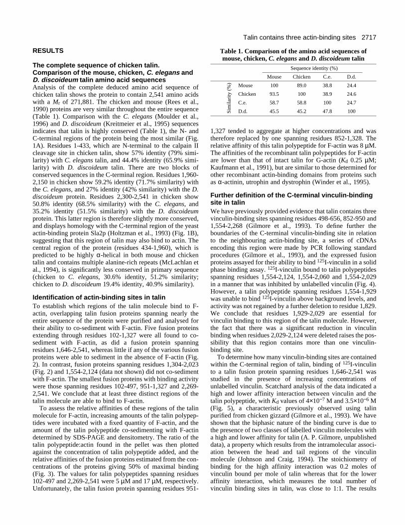
We have determined the sequence of chicken talin (2,541amino acids, Mr 271,881) which is very similar (89%identity) to that of the mouse protein. Alignments with theCaenorhabditis elegans and Dictyostelium discoideum talinsequences show that the N- and C-terminal regions of theprotein are conserved whereas the central part of themolecule is more divergent. By expressing overlappingtalin polypeptides as fusion proteins, we have identified atleast three regions of the protein which can bind F-actin:residues 102-497, 951-1,327 and 2,269-2,541. The N-terminal binding site contains a region with homology tothe ERM family of actin-binding proteins, and the C-terminal site is homologous to the yeast actin-bindingprotein Sla2p. Each of the actin-binding sites is close to, butdistinct from a binding site for vinculin, a protein whichalso binds actin. The Pro1176 to Thr substitution found intalin from Wistar-Furth rats does not destroy the capacity
of this region of the protein to bind actin or vinculin.Microinjection studies showed that a fusion protein con-taining the N-terminal actin-binding site localised weaklyto stress fibres, whereas one containing the C-terminal siteinitially localised predominantly to focal adhesions. Theformer was readily solubilised, and the latter was resistantto Triton extraction. The N-terminal talin polypeptideeventually disrupted actin stress fibres whereas the C-terminal polypeptide was without effect. However, a largerC-terminal fusion protein also containing a vinculin-binding site did disrupt stress fibres and focal adhesions.The results suggest that, although both the N- and C-terminal regions of talin bind actin, the properties of thesetwo regions of the protein are distinct.
Key words: Chicken talin sequence, Actin-binding site, Vinculin,Focal adhesion
SUMMARY
INTRODUCTION
Talin is a cytoskeletal protein localised specifically inadherens-type junctions with the extracellular matrix (Burridgeand Connell, 1983; Geiger et al., 1985), although it is alsofound at points of cell contact between T-helper and antigen-presenting cells (Kupfer et al., 1987). It is thought to be one ofa number of interacting proteins which link the cytoplasmicdomains of integrins to the actin cytoskeleton (reviewed byJockusch et al., 1995). The complete sequences of mouse (Reeset al., 1990) and D. discoideum (Kreitmeier et al., 1995) talinhave been published, and the human gene has been assignedto chromosome 9p (Gilmore et al., 1995). Mouse talin contains2,541 amino acids with a Mr of 269,854. Comparison with theD. discoideum sequence (2,491 amino acids) shows that the N-and C-terminal regions of the protein are conserved (Kreit-meier et al., 1995).
Calpain II cleaves chicken talin between residues 433 and434 to produce a 47 kDa N-terminal fragment and a 190 kDa
C-terminal fragment (Rees et al., 1990). Microinjectionstudies indicate that the N-terminal fragment is important intargeting talin to cell-matrix junctions (Nuckolls et al., 1990).This region of the protein can bind to charged lipids, andappears to contain a membrane binding domain (Niggli et al.,1994). Residues 165-373 show homology with theezrin/radixin/moesin (ERM)/band 4.1 family of cytosketalproteins (Rees et al., 1990; Takeuchi et al., 1994), membersof which also contain an N-terminal membrane-bindingdomain. The 190 kDa C-terminal polypeptide is composed ofa large number of alanine-rich repeats of around 34 aminoacids in length (McLachlan et al., 1994). This region isreported to contain a binding site for the β1 integrin cyto-plasmic domain (Horwitz et al., 1986), and several bindingsites for the cytoskeletal protein vinculin (Lee et al., 1992;Gilmore et al., 1993) which overlap sequences required fortargeting the protein to focal adhesions in cultured cells(Gilmore et al., 1993). Talin is also an actin-binding proteinreported to possess nucleating activity, and able to enhance F-

2716 L. Hemmings and others
actin cross-linking by α-actinin (Muguruma et al., 1990, 1992;Kaufmann et al., 1991). The actin-binding sites in talin havenot been mapped, but the 190 kDa fragment can bind to F-actin, and talin residues 2,300-2,541 show homology to ayeast actin-binding protein Sla2p (Holtzman et al., 1993).Interestingly, talin has recently been reported to bind toresidues close to the C terminus of pp125 focal adhesionkinase (Chen et al., 1995; M. L. Borowsky and R. O. Hynes,unpublished data), a region which also binds paxillin (Hilde-brand et al., 1995).
In D. discoideum, talin accumulates at the tips of filopodiaformed in response to cAMP, consistent with the view that itplays a key role in linking actin to the membrane (Kreitmeieret al., 1995). Talin is recruited to newly forming focaladhesions before vinculin (DePasquale and Izzard, 1991)suggesting that it is involved in the early events leading to focaladhesion assembly. This conclusion is further supported bymicroinjection studies in which a polyclonal antibody to talinwas found to inhibit both cell spreading and cell adhesion onfibronectin (Nuckolls et al., 1992). Down-regulation of talin inHeLa cells using antisense RNA also reduced the rate of cellspreading on fibronectin (Albiges-Rizo et al., 1995).
In the present study, we report the complete sequence ofchicken talin. Alignment of this sequence with that of C.elegans (Moulder et al., 1996) and D. discoideum (Kreitmeieret al., 1995) talin sequences point to a number of blocks ofconserved residues which may help to identify binding sites forproteins which interact with talin. We demonstrate that one ofthese conserved regions (residues 2,300-2,541) at the C-terminal end of the protein contains an F-actin binding site,although at least two other regions of the talin molecule alsobind to F-actin. A second conserved region (residues 1,929-2,029) contains one of the several vinculin-binding sites intalin (Gilmore et al., 1993), each of which is adjacent to anactin-binding site. Finally, by microinjecting purified talinfusion proteins into chicken embryo fibroblasts (CEF), weprovide evidence that the association between the N- and C-terminal regions of talin and the actin cytoskeleton showdifferent characteristics.
MATERIALS AND METHODS
Isolation and sequencing of chicken talin cDNAsThe strategy used to isolate cDNAs encoding the complete sequenceof chicken talin have been described previously (Gilmore et al., 1993).cDNAs were subcloned into either M13 or pBluescript and sequencedon both strands using the dideoxy chain termination method of Sangeret al. (1977). The complete nucleotide sequence was compiled andsequence alignments conducted using the UWGCG suite of pro-grammes.
Purification of talin fusion proteins expressed in E. coliThe chicken talin cDNAs expressed as glutathione-S-transferase(GST) fusion proteins in Escherichia coli have been described previ-ously (Gilmore et al., 1993). Fusion proteins were purified essentiallyas described by Smith and Johnson (1988) using glutathione coupledto agarose beads (Sigma) as an affinity matrix. The fusion proteinswere eluted with 5 mM free glutathione. Concentrations of fusionproteins were determined using the Coomassie protein assay kit(Pierce, Rockford, Ill, USA), and the purity of the proteins analysedby SDS-polyacrylamide gel electrophoresis (SDS-PAGE).
Actin co-sedimentation assayA fixed amount of rabbit muscle G-actin (20 µg) and increasingamounts of the appropriate GST-talin fusion protein were mixed in atotal volume of 100 µl of buffer containing 10 mM Tris-HCl, pH 8.0,0.2 mM ATP, 0.2 mM dithiothreitol, 0.2 mM CaCl2 and 0.02% TritonX-100. Actin polymerisation was initiated by the addition of 100 mMNaCl, and the samples were incubated at room temperature for 60minutes. Samples were centrifuged both with and without actin at75,000 rpm (100,000 g) for 15 minutes. The supernatants wereremoved, the pellet washed carefully with phosphate buffered saline(PBS), and resuspended in 100 µl PBS. Proteins present in equalvolumes of both the supernatant and pellet were resolved by SDS-PAGE, the gels stained with Coomassie blue and analysed using aMolecular Dynamics scanning laser densitometer by volume integra-tion. The ratio of the talin polypeptide:actin (arbitrary units) in thepellet was plotted against the concentration of the talin polypeptideadded, and the binding affinity estimated from the concentration oftalin polypeptide that produced 50% of maximal binding.
Binding of 125I-vinculin to talin fusion proteinsVinculin was purified from chicken gizzard by the method of Evanset al (1984), and 125I-labelled with 0.5 mCi of Bolton and Hunterreagent to a specific activity of approximately 2×10⁷ cpm per µg, asdescribed previously (Gilmore et al., 1993). Purified talin fusionproteins (200 ng) in 25 mM Tris-HCl, pH 7.5/150 mM NaCl (TBS)were adsorbed onto plastic microtitre wells for 4 hours, and excessprotein binding sites blocked with 2% BSA in TBS. 125I-vinculin (4nM) was added to the wells in 100 µl of TBS containing 4% BSA and0.1% Tween-20 with or without unlabelled vinculin. Following anovernight incubation, wells were washed three times in TBS/0.1%Tween-20 and counted in a Beckman 5500 gamma counter.
Microinjection of GST-fusion proteins into chickenembryo fibroblasts (CEF)CEF were grown in Dulbecco’s modified Eagle’s medium (Life Tech-nologies, Paisley, UK) containing 10% tryptose phosphate broth plus5% newborn calf serum and 1% chick serum (Advanced ProteinProducts, West Midlands, UK). Cells were seeded onto glass cover-slips (etched with a cross to aid relocation of the injected cells) at least2 days prior to injection. Approximately 50 cells were microinjected,and the cells returned to the incubator for between 5 and 60 minutesprior to fixing and staining. All injections were carried out with theaid of a Leitz Diavert microscope using a Leitz micromanipulatorconnected to a Narishige microinjector unit. Microinjection needleswere pulled from glass capillaries (Clarke Biomedical Ltd, Slough,Berks., UK) on a microelectrode puller (Campden Instruments Ltd,Sileby, Leics., UK).
Indirect immunofluorescenceThe intracellular localisation of the injected GST-fusion proteins wasdetermined as follows. Injected cells were incubated for 5 minutes at37°C, and cells were then extracted (2 minutes) with 0.5% Triton X-100 in MES buffer (50 mM MES buffer, pH 6.0, 3 mM EGTA, 5mM MgCl2) to remove the cytoplasmic pool of injected protein.Extracted cells were fixed in 3.8% formaldehyde in PBS, and theinjected GST-fusion proteins were detected with a polyclonal rabbitanti-GST antibody (a generous gift from Dr E. Moiseeva, Depart-ment of Biochemistry, University of Leicester) and a Texas Red-labelled anti-rabbit IgG (Amersham). The effects of microinjectedproteins on the actin cytoskeleton were determined after incubatinginjected cells for 30-60 minutes at 37°C. Cells were then fixed in3.8% formaldehyde in PBS, permeabilised with 0.2% Triton X-100in PBS, and stained with FITC-labelled phalloidin (Sigma) accordingto the manufacturers’ instructions. Cells were stained for vinculinusing a mouse monoclonal antibody V284 (Asijee et al., 1990)(Serotec, Kidlington, Oxford, UK) followed by a Texas Red-labelledsheep anti-mouse IgG.
2717Talin contains three actin-binding sites
Table 1. Comparison of the amino acid sequences ofmouse, chicken, C. elegans and D. discoideum talin
Sequence identity (%)
Mouse Chicken C.e. D.d.
Mouse 100 89.0 38.8 24.4
Chicken 93.5 100 38.9 24.6
C.e. 58.7 58.8 100 24.7
D.d. 45.5 45.2 47.8 100Sim
ilari
ty (
%)
RESULTS
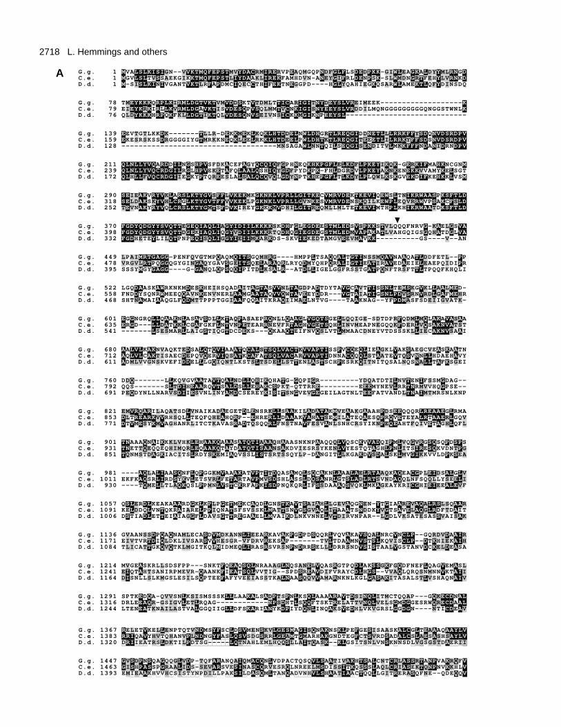
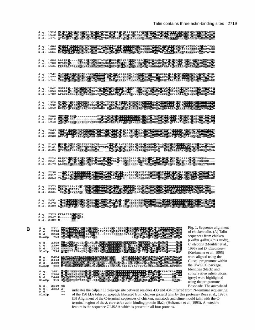
The complete sequence of chicken talin.Comparison of the mouse, chicken, C. elegans andD. discoideum talin amino acid sequencesAnalysis of the complete deduced amino acid sequence ofchicken talin shows the protein to contain 2,541 amino acidswith a Mr of 271,881. The chicken and mouse (Rees et al.,1990) proteins are very similar throughout the entire sequence(Table 1). Comparison with the C. elegans (Moulder et al.,1996) and D. discoideum (Kreitmeier et al., 1995) sequencesindicates that talin is highly conserved (Table 1), the N- andC-terminal regions of the protein being the most similar (Fig.1A). Residues 1-433, which are N-terminal to the calpain IIcleavage site in chicken talin, show 57% identity (79% simi-larity) with C. elegans talin, and 44.4% identity (65.9% simi-larity) with D. discoideum talin. There are two blocks ofconserved sequences in the C-terminal region. Residues 1,960-2,150 in chicken show 59.2% identity (71.7% similarity) withthe C. elegans, and 27% identity (42% similarity) with the D.discoideum protein. Residues 2,300-2,541 in chicken show50.8% identity (68.5% similarity) with the C. elegans, and35.2% identity (51.5% similarity) with the D. discoideumprotein. This latter region is therefore slightly more conserved,and displays homology with the C-terminal region of the yeastactin-binding protein Sla2p (Holtzman et al., 1993) (Fig. 1B),suggesting that this region of talin may also bind to actin. Thecentral region of the protein (residues 434-1,960), which ispredicted to be highly α-helical in both mouse and chickentalin and contains multiple alanine-rich repeats (McLachlan etal., 1994), is significantly less conserved in primary sequence(chicken to C. elegans, 30.6% identity, 51.2% similarity;chicken to D. discoideum 19.4% identity, 40.9% similarity).
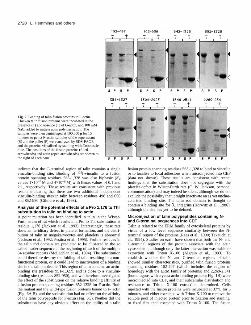
Identification of actin-binding sites in talinTo establish which regions of the talin molecule bind to F-actin, overlapping talin fusion proteins spanning nearly theentire sequence of the protein were purified and analysed fortheir ability to co-sediment with F-actin. Five fusion proteinsextending through residues 102-1,327 were all found to co-sediment with F-actin, as did a fusion protein spanningresidues 1,646-2,541, whereas little if any of the various fusionproteins were able to sediment in the absence of F-actin (Fig.2). In contrast, fusion proteins spanning residues 1,304-2,023(Fig. 2) and 1,554-2,124 (data not shown) did not co-sedimentwith F-actin. The smallest fusion proteins with binding activitywere those spanning residues 102-497, 951-1,327 and 2,269-2,541. We conclude that at least three distinct regions of thetalin molecule are able to bind to F-actin.
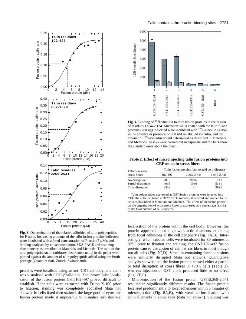
To assess the relative affinities of these regions of the talinmolecule for F-actin, increasing amounts of the talin polypep-tides were incubated with a fixed quantity of F-actin, and theamount of the talin polypeptide co-sedimenting with F-actindetermined by SDS-PAGE and densitometry. The ratio of thetalin polypeptide:actin found in the pellet was then plottedagainst the concentration of talin polypeptide added, and therelative affinities of the fusion proteins estimated from the con-centrations of the proteins giving 50% of maximal binding(Fig. 3). The values for talin polypeptides spanning residues102-497 and 2,269-2,541 were 5 µM and 17 µM, respectively.Unfortunately, the talin fusion protein spanning residues 951-
1,327 tended to aggregate at higher concentrations and wastherefore replaced by one spanning residues 852-1,328. Therelative affinity of this talin polypeptide for F-actin was 8 µM.The affinities of the recombinant talin polypeptides for F-actinare lower than that of intact talin for G-actin (Kd 0.25 µM;Kaufmann et al., 1991), but are similar to those determined forother recombinant actin-binding domains from proteins suchas α-actinin, utrophin and dystrophin (Winder et al., 1995).
Further definition of the C-terminal vinculin-bindingsite in talinWe have previously provided evidence that talin contains threevinculin-binding sites spanning residues 498-656, 852-950 and1,554-2,268 (Gilmore et al., 1993). To define further theboundaries of the C-terminal vinculin-binding site in relationto the neighbouring actin-binding site, a series of cDNAsencoding this region were made by PCR following standardprocedures (Gilmore et al., 1993), and the expressed fusionproteins assayed for their ability to bind 125I-vinculin in a solidphase binding assay. 125I-vinculin bound to talin polypeptidesspanning residues 1,554-2,124, 1,554-2,060 and 1,554-2,029in a manner that was inhibited by unlabelled vinculin (Fig. 4).However, a talin polypeptide spanning residues 1,554-1,929was unable to bind 125I-vinculin above background levels, andactivity was not regained by a further deletion to residue 1,829.We conclude that residues 1,929-2,029 are essential forvinculin binding to this region of the talin molecule. However,the fact that there was a significant reduction in vinculinbinding when residues 2,029-2,124 were deleted raises the pos-sibility that this region contains more than one vinculin-binding site.
To determine how many vinculin-binding sites are containedwithin the C-terminal region of talin, binding of 125I-vinculinto a talin fusion protein spanning residues 1,646-2,541 wasstudied in the presence of increasing concentrations ofunlabelled vinculin. Scatchard analysis of the data indicated ahigh and lower affinity interaction between vinculin and thetalin polypeptide, with Kd values of 4×10⁻⁷ M and 3.5×10⁻⁶ M(Fig. 5), a characteristic previously observed using talinpurified from chicken gizzard (Gilmore et al., 1993). We haveshown that the biphasic nature of the binding curve is due tothe presence of two classes of labelled vinculin molecules witha high and lower affinity for talin (A. P. Gilmore, unpublisheddata), a property which results from the intramolecular associ-ation between the head and tail regions of the vinculinmolecule (Johnson and Craig, 1994). The stoichiometry ofbinding for the high affinity interaction was 0.2 moles ofvinculin bound per mole of talin whereas that for the loweraffinity interaction, which measures the total number ofvinculin binding sites in talin, was close to 1:1. The results
2718 L. Hemmings and others
G.g. 1 MVALSLKISIGN--VVKTMQFEPSTMVYDACRMIRERVPEAQMGQPNDFGLFLSDEDPKK-GIWLEAGKALDYYMLRNGDC.e. 1 MGVLSLTVSSAEKGIKKTMQFEPSTLIYDAAKLIREKFAMHDVN-ANEYGIFRLDENPSK-SLWMDNGRTFEHYLVRNKDD.d. 1 M-SISLKINIVGANTVKTLRFAPDMCIQECCTHIFEKTNEGGPD----HGLYQAHIEGKQSARWLAMEKTLQFYDINSDQ
G.g. 78 TMEYKKKQRPLKIRMLDGTVKTVMVDDSKTVTDMLTTICARIGITNYDEYSLVREIMEEK-------------------KC.e. 79 EIEYKRKIRLLKVRMLDGAVKTISVDESQPVSQLMMTVCNKIGISNYEEYSLVRDDILMQNGGGGGGGGGQNGGSTWNLKD.d. 76 QLDYKKKHRPQKFKLLDGTIKTQLVDESQNVSEIVNSICKKMGIKNPEEYSL----------------------------
G.g. 139 EEVTGTLKKDK-------TLLR-DEKKMEKLKQKLHTDDELNWLDHGRTLREQGIDDNETLLLRRKFFYSDQNVDSRDPVC.e. 159 EKESRSKSSDRGGGGIYGTMRKKNEQKLEELRKKLHTDEELPWLDHTKTLREQGITEEETLILRRKYFFSDSNVDSRDPVD.d. 128 ------------------------------------MNSAGAWLNNTQILSEQGISENDITVLMKKFFFNDANIDRNDPV
G.g. 211 QLNLLYVQARDDILNGSHPVSFDKACEFAGYQCQIQFGPHNEQKHKPGFLELKDFLPKEYIKQK-GERKIFMAHKNCGNMC.e. 239 QLNLLYVQCRDGILRGLHPVEKETAFQLAALQSHIQYGDFPYDKPK-FHLDGRDVLPKEYAKNKENEKKVVAMYKELSGTD.d. 172 QLHLLFVQCRDGIIEGKYPTQREESLALSALQCQVQLGDYNPTKHEPGFLTLKDYLPLQWLKSKGVEKDIFKEHKKLVSM
G.g. 290 SEIEAKVRYVKLARSLKTYGVSFFLVKEKMKGKNKLVPRLLGITKECVMRVDEKTKEVIQEWSLTNIKRWAASPKSFTLDC.e. 318 SELDAKSKYVHLCRGLKTYGVTFFVVKEKLPGKNKLVPRLLGVNKESVMRVDENSKQILKEWPLEQVRRWVPSAKCFSLDD.d. 252 TEVNAKYRYVQLCRSLKTYGMTSFDVKIREYGKKKMVDHILGITREQMLLMLTETKEVIMTHPLKHIKRWAATDKSFTLD
G.g. 370 FGDYQDGYYSVQTTEGEQIAQLIAGYIDIILKKKKSKDHFGLEGDEESTMLEDSVSPKKSTVLQQQFNRVG-KAELGSVAC.e. 398 FGDYQDGYYSVQTTDGEKIAQLIQGYVDIILKKKRTQDHQGIEGDEGSTMLEDMVAPAKATLVAHGQIGSGQHATDGLVAD.d. 332 FGDHETEYLILQTPNPEQISQLIGGYIEIIMKARKDS-SKVIEKEDTAMGVEEVMAVKK----------GS----V--AN
G.g. 449 LPAIMRTGAGG-PENFQVGTMPQAQMQITSGQMHRG----HMPPLTSAQQALTGTINSSMQAVNAAQATLDDFETL--PPC.e. 478 VRGVLRTPQGGQGYGINGAQYGAVSGEITSQELARAQRLRYQDMYQHPQRALIGTIEATIRAVEDAEIELEAEPQIDIPRD.d. 395 SSSYMGYGAGG----G-GANQLQPSQQIPITDLKSALR--ATDLLIGELGGFRSSTGATPQNFTRSFTTLTPQQFKHQLI
G.g. 522 LGQDAASKAWRKNKMDESKHEIHSQADAITAGTASVVNLTAGDPADTDYTAVGCAVTTISSNLTEMSKGVKLLAALMED-C.e. 558 FNDDYSQNRWMEEQQAVNKENVNERLAAMGAATAQVVQWTAVEEYDDR---VGTAIATIGSNLPDVSRNVRDLGAFMEHRD.d. 468 SHTNAMAIAAQGLFQDMTTPPPTGGIAAFQQAITKRAQIIMAELNTVG----TAAKNAG--YFPDMASFSDEIIGVATK-
G.g. 601 EGGNGRQLLQAAKNLASAVSDLLKTAQPASAEPRQNLLQAAGLVGQTSGELLQQIGE-SDTDPRFQDMLMQLAKAVASAAC.e. 635 ERGD---LLDATKKLCGAFGKFLNTVNPETEARRNEVFRTAGHVGETSQHLINVMEAPNEGQQKFDERLVQSAKNVATSTD.d. 541 -------LSESMARLLAIGSTIQGTDCDEKS---QKAAQTEIFNVQSLVTLMMAACDNEYVTDSSSKLLIECAKNVSAAI
G.g. 680 AALVLKAKNVAQKTEDSALQTQVIAAATQCALSTSQLVACTKVVAPTISSPVCQEQLIEAGKLVAKSAEGCVEASKAATNC.e. 712 AQLVLCAKTISAECDEPQVQERVIQSATKCAFATSQLVACARVVAPTIDNNACQQQLSTAATEVTQSVNNLLHDAEHAVYD.d. 611 ADMLVVGNSKVEFIDDELLLGQIQNTLKSTSLTSDELLSTTENLASTSCHPESRKQITNITQSALNQSNALLTAFKSGEI
G.g. 760 DDQ-------LLKQVGVAATAVTQALNDLLQHIKQHATG-GQPIGR---------YDQATDTILNVTENIFSSMGDAG--C.e. 792 QQS-------SLTDIHEAARQVTSALDSLLEHAKCSPKT-QTTRRE---------EEEMYNEVLRRTNRMVVHQGPSE--D.d. 691 PEQDYNLLNARVSDIIESVNLINYAMDCSEREYKISITSNGVEVGEGEILAGTNLTEEFATVANDLTNAIMTMRSNLKNP
G.g. 821 EMVRQARILAQATSDLVNAIKADAEGETDLENSRKLLSAAKILADATAKMVEAAKGAAAHPDSEEQQQRLREAAEGLRMAC.e. 853 DLTREARKVVRHSQLLTEQFQHEAHQRP--EHRERLLDAAAKVAHATSEMILATEQAESQPRQVETEYALRTAAERLGQVD.d. 771 DTVMESYKMVAGHANRLITCTKAVASRADTQSQQRLFNSTNAVFESVANLSNHCRSYIKNPEQEAHTFQIVETAGHLQFL
G.g. 901 TNAAAQNAIKKKLVHKLEHAAKQAAASATQTIAAAQHAAASNKNPAAQQQLVQSCKVVADQIPMLVQGVRGSQSQPDSPSC.e. 931 TNETTQEQQEQHIMQRLEQAAKQTAYDATQTISAANSAKDVIESRSYKENLVYESTQTAGHLPNLITSIRESQKVDNTPGD.d. 851 TQNMSTDAGKIACITSLRDYSKEMIAQVSSLISTSRTSSQYLP-DANGITLLKGAKDVSDALSKLMVGIKKVVLDPKSEA
G.g. 981 ----AQLALIAASQNFLQPGGKMVAAAKATVPTITDQASAMQLSQCAKNLAAALAELRTAAQKAQEACGPLEIDSALGLVC.e. 1011 EKFKAQSRLIRDSYKVLETSVRLFETARTAVPMVSDSHLASSLDQSANRLGTSLADLRTSVNDAQQLNFSQQLLYSEELID.d. 930 ----TQMELLTLAQKQSLPPMNLVSTCKRFAPKISDPNQKQRLIFSSDAAAQSVQKLMKAGEAYKRICGHIEIEEALEVF
G.g. 1057 QSLERDLKEAKAAARDGKLKPLPGETMEKCAQDLGNSTKAVTSAIAHLLGEVAQGNEN-YTGIAAREVAQALRSLSQAARC.e. 1091 KELDDQLVNTQKRAIARELPPIQNATSFSVSSKLMATTSNVGSGVAQLITAAATSNDDKYVGTSAVELAQGLRDFTDAITD.d. 1006 DSTIADLETTEIAIAGGFLDAVSGTTREGAAELLMVAIKDLNKVNNELVTDIRVNPAR--LGDLVKSATESASSVAISAK
G.g. 1136 GVAANSSDPQAQNAMLECASDVMDKANNLIEEARKAVAKPGDPDSQQRLVQVAKAVSQALNRCVNCLP--GQRDVDAAIRC.e. 1171 EIVTVRTDIQLDKLIVSARSVVHESGR-VFDRVREKSAP-------TVLTDAAMNVSTSLKQVISCLP--DTKHIEKAIHD.d. 1084 TLICATTGKQVQTKLMGITKQLMIDMEQLIRASRSVRSNPNDRRSELLLDRRSNDVSISTAALVGSTANVDCKELDEASA
G.g. 1214 MVGEASKRLLSDSFPP---SNKTFQEAQSQLNRAAAGLNQSANELVQASRGTPQDLAKSSGKFGQDFNEFLQAGVEMASLC.e. 1241 EIQTAHTSANIRPMEVR-QAANKFIEATSQLVVTIG--SPDSREAVDIFVRAYCDLHGS--VVAQLQRQSNMNNVKTAIID.d. 1164 DISNLLSLKMGSLESILSQPTEEFAFYVEEIASSTKALNAASQQVVAMARNKNLKGLGASAKITASALSTLVSHAQNAIV
G.g. 1291 SPTKEDQA-QVVSNLKSISMSSSKLLLAAKALSADPTSPNLKSQLAAAARAVTDSINQLITMCTQQAP---GQKECDNALC.e. 1316 DRLEEAQR-GSIGVLETLRQAG------------NPSDHTLSQQFTSKTRELATTVNQLVELSGMDGGESRWQRECDAALD.d. 1244 LTENEATKNAILASTVALGGQIIGLLDFSKARIANYKDPIYDQNLINQAKSVEDHLVKVGRSLGGDGN----NTICDEAV
A
G.g. 1 MVALSLKISIGN--VVKTMQFEPSTMVYDACRMIRERVPEAQMGQPNDFGLFLSDEDPKK-GIWLEAGKALDYYMLRNGDC.e. 1 MGVLSLTVSSAEKGIKKTMQFEPSTLIYDAAKLIREKFAMHDVN-ANEYGIFRLDENPSK-SLWMDNGRTFEHYLVRNKD
G.g. 78 TMEYKKKQRPLKIRMLDGTVKTVMVDDSKTVTDMLTTICARIGITNYDEYSLVREIMEEK-------------------KC.e. 79 EIEYKRKIRLLKVRMLDGAVKTISVDESQPVSQLMMTVCNKIGISNYEEYSLVRDDILMQNGGGGGGGGGQNGGSTWNLK
G.g. 1367 RELETVKELLENPTQTVNDMSYFSCLDSVMENSKVLGESMAGISQNAKNSKLPEFGESISAASKALCGLTEAAAQAAYLVC.e. 1383 RRIQAVYHVTQHANVPLNDNGYFASLQSVSDGSRRLGEAMTGMARHAKGNDTEGFCTSVRDSADALCSLAESASHSAYLVD.d. 1320 DRIIEATRSLDKTILPDTSG-----LQTNAHLEMLHQQSLLAITQASK--KLGSITSNLVNSKNNSDLVGSGSTDAERII
G.g. 1447 GVSDPNSQAGQQGLVDP-TQFARANQAIQMACQNLVDPACTQSQVLSAATIVAKHTSALCNTCRLASSRTANPVAKRQFVC.e. 1463 GISHPASSPGRAALIDS-SEVARSVESINASCQRVESRQLNREELMSDISSITKQSSSLAQLCRIASEKTQNPNVKKHLVD.d. 1393 EMIEAAKHVVHCSISTYNPDILLPAKSILDASQMLTANQADVNHVLSHAATIAACTQQLLGITRERASQFNE--QDEQQV
2719Talin contains three actin-binding sites
G.g. 139 EEVTGTLKKDK-------TLLR-DEKKMEKLKQKLHTDDELNWLDHGRTLREQGIDDNETLLLRRKFFYSDQNVDSRDPVC.e. 159 EKESRSKSSDRGGGGIYGTMRKKNEQKLEELRKKLHTDEELPWLDHTKTLREQGITEEETLILRRKYFFSDSNVDSRDPV
G.g. 211 QLNLLYVQARDDILNGSHPVSFDKACEFAGYQCQIQFGPHNEQKHKPGFLELKDFLPKEYIKQK-GERKIFMAHKNCGNMC.e. 239 QLNLLYVQCRDGILRGLHPVEKETAFQLAALQSHIQYGDFPYDKPK-FHLDGRDVLPKEYAKNKENEKKVVAMYKELSGT
G.g. 290 SEIEAKVRYVKLARSLKTYGVSFFLVKEKMKGKNKLVPRLLGITKECVMRVDEKTKEVIQEWSLTNIKRWAASPKSFTLDC.e. 318 SELDAKSKYVHLCRGLKTYGVTFFVVKEKLPGKNKLVPRLLGVNKESVMRVDENSKQILKEWPLEQVRRWVPSAKCFSLD
G.g. 370 FGDYQDGYYSVQTTEGEQIAQLIAGYIDIILKKKKSKDHFGLEGDEESTMLEDSVSPKKSTVLQQQFNRVG-KAELGSVAC.e. 398 FGDYQDGYYSVQTTDGEKIAQLIQGYVDIILKKKRTQDHQGIEGDEGSTMLEDMVAPAKATLVAHGQIGSGQHATDGLVA
G.g. 449 LPAIMRTGAGG-PENFQVGTMPQAQMQITSGQMHRG----HMPPLTSAQQALTGTINSSMQAVNAAQATLDDFETL--PPC.e. 478 VRGVLRTPQGGQGYGINGAQYGAVSGEITSQELARAQRLRYQDMYQHPQRALIGTIEATIRAVEDAEIELEAEPQIDIPR
G.g. 522 LGQDAASKAWRKNKMDESKHEIHSQADAITAGTASVVNLTAGDPADTDYTAVGCAVTTISSNLTEMSKGVKLLAALMED-C.e. 558 FNDDYSQNRWMEEQQAVNKENVNERLAAMGAATAQVVQWTAVEEYDDR---VGTAIATIGSNLPDVSRNVRDLGAFMEHR
G.g. 601 EGGNGRQLLQAAKNLASAVSDLLKTAQPASAEPRQNLLQAAGLVGQTSGELLQQIGE-SDTDPRFQDMLMQLAKAVASAAC.e. 635 ERGD---LLDATKKLCGAFGKFLNTVNPETEARRNEVFRTAGHVGETSQHLINVMEAPNEGQQKFDERLVQSAKNVATST
G.g. 680 AALVLKAKNVAQKTEDSALQTQVIAAATQCALSTSQLVACTKVVAPTISSPVCQEQLIEAGKLVAKSAEGCVEASKAATNC.e. 712 AQLVLCAKTISAECDEPQVQERVIQSATKCAFATSQLVACARVVAPTIDNNACQQQLSTAATEVTQSVNNLLHDAEHAVY
G.g. 760 DDQ-------LLKQVGVAATAVTQALNDLLQHIKQHATG-GQPIGR---------YDQATDTILNVTENIFSSMGDAG--C.e. 792 QQS-------SLTDIHEAARQVTSALDSLLEHAKCSPKT-QTTRRE---------EEEMYNEVLRRTNRMVVHQGPSE--
G.g. 821 EMVRQARILAQATSDLVNAIKADAEGETDLENSRKLLSAAKILADATAKMVEAAKGAAAHPDSEEQQQRLREAAEGLRMAC.e. 853 DLTREARKVVRHSQLLTEQFQHEAHQRP--EHRERLLDAAAKVAHATSEMILATEQAESQPRQVETEYALRTAAERLGQV
G.g. 901 TNAAAQNAIKKKLVHKLEHAAKQAAASATQTIAAAQHAAASNKNPAAQQQLVQSCKVVADQIPMLVQGVRGSQSQPDSPSC.e. 931 TNETTQEQQEQHIMQRLEQAAKQTAYDATQTISAANSAKDVIESRSYKENLVYESTQTAGHLPNLITSIRESQKVDNTPG
G.g. 981 ----AQLALIAASQNFLQPGGKMVAAAKATVPTITDQASAMQLSQCAKNLAAALAELRTAAQKAQEACGPLEIDSALGLVC.e. 1011 EKFKAQSRLIRDSYKVLETSVRLFETARTAVPMVSDSHLASSLDQSANRLGTSLADLRTSVNDAQQLNFSQQLLYSEELI
G.g. 1057 QSLERDLKEAKAAARDGKLKPLPGETMEKCAQDLGNSTKAVTSAIAHLLGEVAQGNEN-YTGIAAREVAQALRSLSQAARC.e. 1091 KELDDQLVNTQKRAIARELPPIQNATSFSVSSKLMATTSNVGSGVAQLITAAATSNDDKYVGTSAVELAQGLRDFTDAIT
G.g. 1136 GVAANSSDPQAQNC.e. 1171 EIVTVRTDIQLDK
G.g. 1526 QPAKEVANSTANLVKTIKALDGAFNEENRERCRAATAPLIEAVDNLTAFASNPEFATVPAQISPEGRRAMEPIVTSAKTMC.e. 1542 GCAMGVASKTSSLVTAFKDLDRMPDAESR--CTSSASELRQVALQLLHFADKPDFAAIQGSISTEGHSAQQPILQSSREMD.d. 1471 QVRDGIVKSTQQLAHATSSLARAVKSVTSKEPGAKAMISQSLKDLESAINNLLITSSVPASERGIGIADFNKLMSTCRSV
G.g. 1606 LESSAGLIQTARSLAVNPKDPPQWSVLAGHSRTVSDSIKKLITNMRDKAPGQRECDEAIDVLNRCMREVDQASLAAISQQC.e. 1620 LDSSAQMIQTAKSWASAPQDEATWQRMAVNSRQVSDSIMRLVNAIHEAAPGQMELEAAIGRLSALSGQIERSAMDCYASGD.d. 1551 STASSQLIISASSCSQKPKDIELSSILSENAVLMTNSLKDIIKVTSSMMPGVNFCEEAIEIAQRAISDLSSVALSVAVGS
G.g. 1686 LAPREG----ISQEALHNQMITAVQEINNLIEPVASAARAEASQLGHKVSQMAQYFEPLILAAIGAASKTPNHQQQMNLLC.e. 1700 NVQKHGA---NAERQLLQQVQHIASQLEDKVDDLHNAAVEHGERLPKVVQLHCQMVEDLADAACCAAGMTVDSNQQTELFD.d. 1631 FDSSANNKEGLSHVESQERLVDVTKKIGTGINDLLKASRQSPEAIGISAKALSFIAPSLVNTTKPALATAPDADAQNDLV
G.g. 1762 DQTKTLAESALQMLYTAKEAGGNPKQAAHTQEALEEAVQMMKEAVEDLTTTLNEAASAAGVVGGMVDSITQAINQLDEGPC.e. 1777 DKCKTVVEAELAMMVASRESGGNP-NAVHAHANVKDAAGQLKHAIGDMRQTIAKVSSEQGAVQGMVDTISSSIAHTDVAVD.d. 1711 TESKNVGDSILKLCQASLIASSNP--SKETYQIIVNKCVDASEAMSKLVAQISSGVNLYKELDESLDRIRKSVVQTSAKD
G.g. 1842 MGEPEG--TFVDYQTTMVKTAKAIAVTVQEMVTKSTTNPDELGILANQLTNDYGQLAQQAKPAALTAENEEIGSHIKRRVC.e. 1856 SSAHPGS-SFADAQTRMTAYLEDIRRTAIEMPTLNTT---DLGAASLNLSEKYRLVAGDVRQAAGMLPEADIGQRLKLAVD.d. 1789 APKDSENRGYQEYKEELSNLTKNLALSLKTIVATDGNNLVSISTISKDIANYISDIAHVSSAILATTSDQKIRDSIITSS
G.g. 1920 QELGHGCAALVTKAGALQCSPSDAYTKKELIESARKVSEKVSHVLAALQAGNRGTQACITAASAVSGIIADLDTTIMFATC.e. 1932 QKLGTSCIETVTVAGGKRAHPEDERIQRQLSSQAGTVVERVEQVLAALHSASRGTQACINAANTVSGIIGDLDTTIMFATD.d. 1869 RQVIVSTGDIVNHIKVNSTDKANS-SQAKVNDSYRATNDNITRFLQSLKQGAIGEILSDAAIDQIRKVISDLDGYSLFAA
G.g. 2000 AGTLNRENS-----------ETFADHREGILKTAKALVEDTKVLVQNATASQEKLAQAAQSSVSTITRLAEVVKLGAASLC.e. 2012 SGSLNSSDD-----------RKFPSHKEAIVKTAKALVEDTKALVAGAASNQEQLAVAAQNAVRTIVNLSDAVKTGAVSLD.d. 1948 AGQLENDQSSQSTMNEVTKQQHLKNLQKDTITQAKLLIVSSSQLVGSSRGTQEHLGSATTKVANTVSSLVKTAKDIASVL
G.g. 2069 GSEDPETQVVLINAVKDVAKALGDLIGATKAAAGKAGDDPAVYQLKNSAKVMVTNVTSLLKTVKAVEDEATKGTRALEATC.e. 2081 SSENSETQVLVIHAVRDVAAALTSLIQATKNASGLSLQHPAMGHLKDAAKVMVSNVARLLKTVATVEEKNQQGTRAVEAAD.d. 2028 ADTTS--QQDILSASKALSISSQQMVLATKDAQRFKKDATAFRSLGKSAERVAEDVGQFLTSVYTAISDAGKGIKELEKS
G.g. 2149 IEHIRQELAVFSSPVPPAQVS----TPEDFIRMTKGITMATAKAVAAGNSCR-QEDVIATANLSRRAIADMLRACKEAAYC.e. 2161 VDAIGFEMKQFEHDLNEGAAIPTDFRPEHLQQTAEHVSEITKRVMQGADAPQSEEEIIGVANLSRSAVRSLLAVVRTISND.d. 2106 IVQVANYHEKPDTVLSNKDAT-----AEIFAQSARDLAKSSIEIVTSYTSSQ--DSLVKSSQAVVSNVQSFISNSKGVIA
G.g. 2224 HPEVSADVRQRALRFGKECADGYLELLE--HVLVILQKPTHELKQQLAGYSKRVASSVTELIQAAEAMKGTEWVDP----C.e. 2241 DADT-APQRYAVLDSGRDVANNVKSLLVSLHTQMVRNPGQEESRRLLLEASKGVSSALSHLVGLCNEMTGLPHNHEM---D.d. 2179 LLGNGNDLKSKVLENVKQTTGDMLALLQ-----CVKDQDKNGS-TSIADATRSISDCVHSVVTLSKSLPGGQNIVVEEDN
G.g. 2298 --EDPTVIAENELLGAAAAIEAAAKKLEQLKPR---AKPKQADESLDFEEQILEAAKSIAAATSALVKAASAAQRELVAQC.e. 2317 --ESAAAQAENELLGAASSIEAASAKLAELRPRQIVQENTQEIVETEFDDNIIISAKGILHAVHTLMRSASNAQRELAMQD.d. 2253 VLEDLEALAEDELSACARSIEEATAKLIAARPQS--KSKNGKLDAEGVAATIVDASSAIAKAVAKLVNSAAVAQSKRRED
G.g. 2373 GKVGVIPANAVDDGQWSQGLISAARMVAAATNNLCEAANAAVQGHASEEKLISSAKQVAASTAQLLVACKVKADHDSE--C.e. 2395 GRAAAGGTGTY---QWSEGLISAARVVVASVHKLCDAANTLMKGQTTEERLISAAKQVSSSTAQLLVACNVRADPDSQ--D.d. 2331 -QIASGSVYKADP-TWSNGLISAAKGVGAATHRLVEAAMKSATGKAEEEELIATARSVAAATALLVSASRAKSGDDYQSQ
G.g. 2451 -AMKRLQAAGNAVKRASDNLVKAAQKAAAFQDH-DETVVVKEKMVGGIAQIIAAQEEMLRKERELEEARKKLAMIRQQQYC.e. 2470 -ANRRLQAAGQAVRNAAERLVQSAQQEMIARD--DRNIAISDRLVNGIAQVMDAQEEVLRKERELGEARHKLAHLNKARYD.d. 2409 AAHSHLSTAARQVASATSDLVAAAKAATIFDEQQQEEEQEQFNFTGSKVKELEQQMKILKLEKELETARRQMLNSRKQNY
G.g. 2529 KFLPTELRDEEQNC.e. 2547 E------RDGEEAD.d. 2489 N------KN----
G.g. 2311 AAAAIEAAAKKLEQLKPR---AKPKQADESLDFEEQILEAAKSIAAATSALVKAASAAQRC.e. 2330 AASSIEAASAKLAELRPRQIVQENTQEIVETEFDDNIIISAKGILHAVHTLMRSASNAQRD.d. 2268 CARSIEEATAKLIAARPQS--KSKNGKLDAEGVAATIVDASSAIAKAVAKLVNSAAVAQSSla2p 753 TADKIVKSSEHLRVDVPK-----------------PLLSLALMIIDAVVALVKAAIQCQN
G.g. 2368 ELVAQGKVGVIPANAVDDGQWSQGLISAARMVAAATNNLCEAANAAVQGHASE----EKLC.e. 2390 ELAMQGRA---AAGGTGTYQWSEGLISAARVVVASVHKLCDAANTLMKGQTTE----ERLD.d. 2326 KRREDQIAS--GSVYKADPTWSNGLISAAKGVGAATHRLVEAAMKSATGKAEE----EELSla2p 796 EIATTTSIP-LNQFYLKNSRWTEGLISAAKAVAGATNVLITTASKLITSEDNENTSPEQF
G.g. 2424 ISSAKQVAASTAQLLVACKVKADHDSE---AMKRLQAAGNAVKRASDNLVKAAQKAAAFQC.e. 2443 ISAAKQVSSSTAQLLVACNVRADPDSQ---ANRRLQAAGQAVRNAAERLVQSAQQEMIARD.d. 2380 IATARSVAAATALLVSASRAKSGDDYQSQAAHSHLSTAARQVASATSDLVAAAKAATIFDSla2p 855 IVASKEVAASTIQLVAASRVKTSIHSKAQDKLEHCSKDVTDACRSLGNHVMGMIEDDHST
G.g. 2481 DHDETVVVKEKMVGG-IAQIIAAQEEMLRKERELEEARKKLAMIRQQQYKFLPTELRDEEC.e. 2500 D-DRNIAISDRLVNG-IAQVMDAQEEVLRKERELGEARHKLAHLNKARYE-----RDGEED.d. 2440 EQQQEEEQEQFNFTGSKVKELEQQMKILKLEKELETARRQMLNSRKQNYN-----KN---Sla2p 915 SQQQQPLDFTSEHTL-KTAEMEQQVEILKLEQSLSNARKRLGEIRRHAYYN----QDDD-
G.g. 2540 QNC.e. 2553 A-D.d. --Sla2p --
B Fig. 1. Sequence alignmentof chicken talin. (A) Talinsequences from chicken(Gallus gallus) (this study),C. elegans (Moulder et al.,1996) and D. discoideum(Kreitmeier et al., 1995)were aligned using theClustal programme withinthe UWGCG package.Identities (black) andconservative substitutions(grey) were highlightedusing the programmeBoxshade. The arrowhead
indicates the calpain II cleavage site between residues 433 and 434 inferred from N-terminal sequencingof the 190 kDa talin polypeptide liberated from chicken gizzard talin by this protease (Rees et al., 1990).(B) Alignment of the C-terminal sequences of chicken, nematode and slime mould talin with the C-terminal region of the S. cerevisiae actin binding protein Sla2p (Holtzman et al., 1993). A noteablefeature is the sequence GLISAA which is present in all four proteins.
2720 L. Hemmings and others
Fig. 2. Binding of talin fusion proteins to F-actin.Chicken talin fusion proteins were incubated in thepresence (+) and absence (−) of G-actin, and 100 mMNaCl added to initiate actin polymerisation. Thesamples were then centrifuged at 100,000 g for 15minutes to pellet F-actin; samples of the supernatant(S) and the pellet (P) were analysed by SDS-PAGE,and the proteins visualised by staining with Coomassieblue. The positions of the fusion proteins (filledarrowheads) and actin (open arrowheads) are shown tothe right of each panel.
indicate that the C-terminal region of talin contains a singlevinculin-binding site. Binding of 125I-vinculin to a fusionprotein spanning residues 565-1,328 was also biphasic (Kdvalues 1×10⁻⁷ M and 4×10⁻⁶ M) with Bmax values of 0.1 and2:1, respectively. These results are consistent with previousresults indicating that there are two additional independentvinculin-binding sites in talin between residues 498 and 656and 852-950 (Gilmore et al., 1993).
Analysis of the potential effects of a Pro 1,176 to Thrsubstitution in talin on binding to actinA point mutation has been identified in talin in the Wistar-Furth strain of rat which results in a Pro to Thr substitution atresidue 1,176 (Jackson et al., 1993). Interestingly, these ratsshow an heriditary defect in platelet formation, and the distri-bution of talin in megakaryocytes and platelets is abnormal(Jackson et al., 1992; Pestina et al., 1995). Proline residues inthe talin rod domain are predicted to be clustered in the socalled leader sequence at the beginning of each of the multiple34 residue repeats (McLachlan et al., 1994). The substitutioncould therefore destroy the folding of talin resulting in a non-functional protein, or it could lead to inactivation of a bindingsite in the talin molecule. This region of talin contains an actin-binding site (residues 951-1,327), and is close to a vinculin-binding site (residues 852-950), and we therefore investigatedthe effect of the substitution on the relative binding affinity ofa fusion protein spanning residues 852-1328 for F-actin. Boththe mutant and the wild-type fusion proteins bound to F- actin(Fig. 6A,B), and the susbstitution had no effect on the affinityof the talin polypeptide for F-actin (Fig. 6C). Neither did thesubstitution have any obvious affect on the ability of a talin
fusion protein spanning residues 565-1,328 to bind to vinculinor to localise to focal adhesions when microinjected into CEF(data not shown). These results are consistent with recentfindings that the substitution does not segregate with theplatelet defect in Wistar-Furth rats (C. W. Jackson, personalcommunication) and may indeed be silent, although we do notexclude the possibility that it might inactivate an as yet unchar-acterised binding site. The talin rod domain is thought tocontain a binding site for β1 integrins (Horwitz et al., 1986),although the site has yet to be defined.
Microinjection of talin polypeptides containing N-and C-terminal sequences into CEFTalin is related to the ERM family of cytoskeletal proteins byvirtue of a low level sequence similarity between the N-terminal region of the proteins (Rees et al., 1990; Takeuchi etal., 1994). Studies on ezrin have shown that both the N- andC-terminal regions of the protein associate with the actincytoskeleton, although only the latter interaction was stable toextraction with Triton X-100 (Algrain et al., 1993). Toestablish whether the N- and C-terminal regions of talinshowed similar characteristics, purified talin fusion proteinsspanning residues 102-497 (which includes the region ofhomology with the ERM family of proteins) and 2,269-2,541(homologous with a yeast actin-binding protein; Fig. 1B) weremicroinjected into CEF, and their subcellular distribution andresistance to Triton X-100 extraction determined. Cellsinjected with the fusion proteins were incubated at 37°C for 5minutes, and either extracted with Triton X-100 to remove thesoluble pool of injected protein prior to fixation and staining,or fixed first then extracted with Triton X-100. The fusion
2721Talin contains three actin-binding sites
0 5 10 15 20 25 30 35 400.00
0.02
0.04
0.06
0.08
0.10
0.12
0.14
Fusion protein (µM)
Fus
ion
prot
ein
: act
in r
atio
Talin residues
0 2 4 6 8 10 12 140.00
0.05
0.10
0.15
0.20
0.25
Fusion protein (µM)
Fus
ion
prot
ein
: act
in r
atio
Talin residues102-497
2269-2541
0 2 4 6 8 10 12 14 16 18 200.00
0.05
0.10
0.15
0.20
0.25
0.30
0.35
0.40
Fusion protein (µM)
Fus
ion
prot
ein
: act
in r
atio
Talin residues852-1328
Fig. 3. Determination of the relative affinities of talin polypeptidesfor F-actin. Increasing amounts of the talin fusion proteins indicatedwere incubated with a fixed concentration of F-actin (5 µM), andbinding analysed by co-sedimentation, SDS-PAGE and scanningdensitometry as described in Materials and Methods. The ratio of thetalin polypeptide:actin (arbitrary absorbance units) in the pellet wereplotted against the amount of talin polypeptide added using the Profitpackage (Quantum Soft, Zurich, Switzerland).
0
1000
2000
3000
4000
5000
6000
125I
-vin
culin
bou
nd(c
pm)
1 2 3 4 5 6
GST
/155
4-21
24
GST
/155
4-20
60
GST
/155
4-20
29
GST
/155
4-19
29
GST
/155
4-18
29
BS
A
Fig. 4. Binding of 125I-vinculin to talin fusion proteins in the regionof residues 1,554-2,124. Microtitre wells coated with the talin fusionproteins (200 ng) indicated were incubated with 125I-vinculin (4 nM)in the absence or presence of 500 nM unlabelled vinculin, and theamount of 125I-vinculin bound determined as described in Materialsand Methods. Assays were carried out in triplicate and the bars showthe standard error about the mean.
Table 2. Effect of microinjecting talin fusion proteins intoCEF on actin stress fibres
Effect on actin Talin fusion proteins (amino acid co-ordinates)
stress fibres 102-497 2,269-2,541 1,646-2,541
No disruption 28±3 90±6 11±1Partial disruption 49±5 10±6 51±1Total disruption 23±4 0 38±1
Talin polypeptides expressed as GST-fusion proteins were injected intoCEF, the cells incubated at 37°C for 30 minutes, then fixed and stained for F-actin as described in Materials and Methods. The effect of the fusion proteinon the organisation of actin stress fibres is expressed as a percentage (± s.d.)of the total number of cells injected.
proteins were localised using an anti-GST antibody, and actinwas visualised with FITC-phalloidin. The intracellular locali-sation of the fusion protein GST/102-497 proved difficult toestablish. If the cells were extracted with Triton X-100 priorto fixation, staining was completely abolished (data notshown). In cells fixed then stained, the large pool of cytosolicfusion protein made it impossible to visualise any discrete
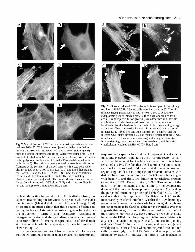
localisation of the protein within the cell body. However, theprotein appeared to co-align with actin filaments extendingfrom focal adhesions at the cell periphery (Fig. 7A,B). Inter-estingly, when injected cells were incubated for 30 minutes at37°C prior to fixation and staining, the GST/102-497 fusionprotein caused disruption of actin stress fibres in most thoughnot all cells (Fig. 7C,D). Vinculin-containing focal adhesionswere similarly disrupted (data not shown). Quantitativeanalysis showed that the fusion protein caused either a partialor total disruption of stress fibres in >70% cells (Table 2),whereas injection of GST alone produced little or no effect(Fig. 7E,F).
Microinjection of the fusion protein GST/2,269-2,541resulted in significantly different results. The fusion proteinlocalised predominantly to focal adhesions within 5 minutes ofmicroinjection (Fig. 8A,B), with weak co-localisation withactin filaments in some cells (data not shown). Staining was

2722 L. Hemmings and others
0 2 4 6 8 10 12 14 16 18 200.00
0.05
0.10
0.15
0.20
0.25
0.30
0.35
0.40
Fusion protein (µM)
Fus
ion
prot
ein
: act
in r
atio
852-1328 Pro1176 Thr
852-1328
>
Fig. 6. The effect of a Pro 1,176 to Thr substitution on binding of atalin polypeptide spanning residues 852-1,328 to F-actin. Platelettalin isolated from the Wistar-Furth (WF) strain of rat contains a Pro1,176 to Thr substitution (Jackson et al., 1993). (A,B) The effect ofthis substitution on binding of increasing concentrations of a chickentalin fusion protein (residues 852-1,328) to a fixed concentration ofF-actin (5 µM) was analysed using a co-sedimentation and SDS-PAGE as described in Materials and Methods. (C) The ratio of thetalin polypeptide:actin (arbitrary absorbance units) in the pellet wasplotted against the amount of talin polypeptide added using the Profitpackage (Quantum Soft, Zurich, Switzerland).
0
0.005
0.01
0.015
0.02B
/F
0 0.5 1
B (mol.mol-1)
Talin residues1646-2541
0
0.01
0.02
0.03
B/F
0 0.5 1 1.5 2 2.5
B (mol.mol-1)
Talin residues565-1328
Fig. 5. Stoichiometry of binding of vinculin to talin fusion proteinsspanning residues 565-1,328 and 1,646-2,541. Microtitre wellscoated with the talin fusion proteins (200 ng) indicated wereincubated with 125I-vinculin (4 nM) in the presence of increasingconcentrations (5 nM - 4,000 nM) of unlabelled vinculin, and theamount of 125I-vinculin bound determined as described in Materialsand Methods. The means of triplicate assays were corrected for non-specific binding to wells coated with BSA. The data are presented asScatchard plots.
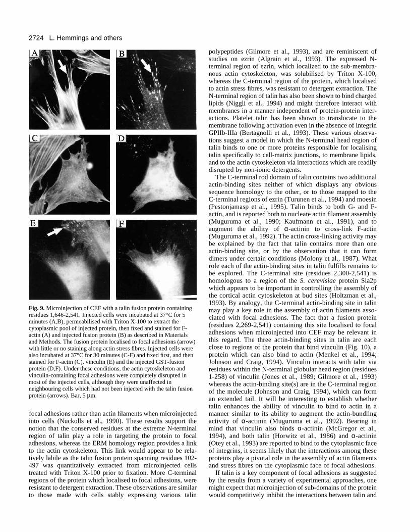
resistant to extraction with Triton X-100. After 30 minutes,very few cells (<20%) showed any signs of stress fibre dis-ruption (Fig. 8C) or loss of vinculin-containing focal adhesions(data not shown). However, the fusion protein was no longerlocalised specifically in focal adhesions and appeared to bemore uniformly distributed throughout the cell with someevidence of filamentous staining (Fig. 8D). A larger C-terminaltalin polypeptide (residues 1,646-2,541) was also seen tolocalise to the ends of actin stress fibres in focal adhesionswithin 5 minutes of injection (Fig. 9A,B), and the protein wasagain resistant to extraction with Triton X-100. However,>80% of cells which were microinjected with GST/1,646-2,541 and incubated for 30 minutes at 37°C showed partial ortotal disruption of actin stress fibres (Fig. 9C,D and Table 2),and loss of vinculin-containing focal adhesions (Fig. 9E,F).
DISCUSSION
Comparisons of the chicken talin sequence reported here with
those of C. elegans (Moulder et al., 1996) and D. discoideum(Kreitmeier et al., 1995) talin sequences show the N- and C-terminal regions of the protein to be conserved. The region N-terminal to the calpain II cleavage site in chicken talin containstwo blocks of conserved sequence; residues 4-129 and 166-422. The intervening region contains an insert in C. elegansand a deletion in D. discoideum talin. There are also twoconserved regions toward the C terminus of the protein, onespanning residues 1,960-2,150, the other residues 2,334-2,524(Fig. 1A,B). We show here that the former contains a vinculin-binding site, and the latter an actin-binding site which displayshomology to the yeast actin-binding protein Sla2p (Holtzmanet al., 1993). However, two other regions of talin also bind toF-actin, namely residues 102-497 and 951-1,327. Interestingly,
2723Talin contains three actin-binding sites
Fig. 7. Microinjection of CEF with a talin fusion protein containingresidues 102-497. CEF were microinjected with the talin fusionprotein GST/102-497 and incubated at 37°C for 5 minutes (A,B)prior to fixation and permeabilisation. Cells were stained for F-actinusing FITC-phalloidin (A) and for the injected fusion protein using arabbit polyclonal antibody to GST and a Texas red-labelled anti-rabbit IgG (B). The fusion protein was weakly associated with actinfilaments at the periphery of the cell (arrows). Injected cells werealso incubated at 37°C for 30 minutes (C,D) and fixed then stainedfor F-actin (C) and the GST/102-497 (D). Under these conditions,the actin cytoskeleton in most injected cells was completelydisrupted, whereas uninjected cells contained numerous actin stressfibres. Cells injected with GST alone (E,F) and stained for F-actin(E) and GST (F) were unaffected. Bar, 5 µm.
Fig. 8. Microinjection of CEF with a talin fusion protein containingresidues 2,269-2,541. Injected cells were incubated at 37°C for 5minutes (A,B), permeabilised with Triton X-100 to extract thecytoplasmic pool of injected protein, then fixed and stained for F-actin (A) and injected fusion protein (B) as described in Materialsand Methods. Under these conditions, the fusion protein waslocalised to focal adhesions (arrows) with little or no staining alongactin stress fibres. Injected cells were also incubated at 37°C for 30minutes (C-D), fixed first and then stained for F-actin (C) and theinjected GST-fusion protein (D). The injected fusion protein (D) wasnow localised in focal adhesions (arrow) and along the actin stressfibres extending from focal adhesions (arrowhead), and the actincytoskeleton remained unaffected (C). Bar, 5 µm.
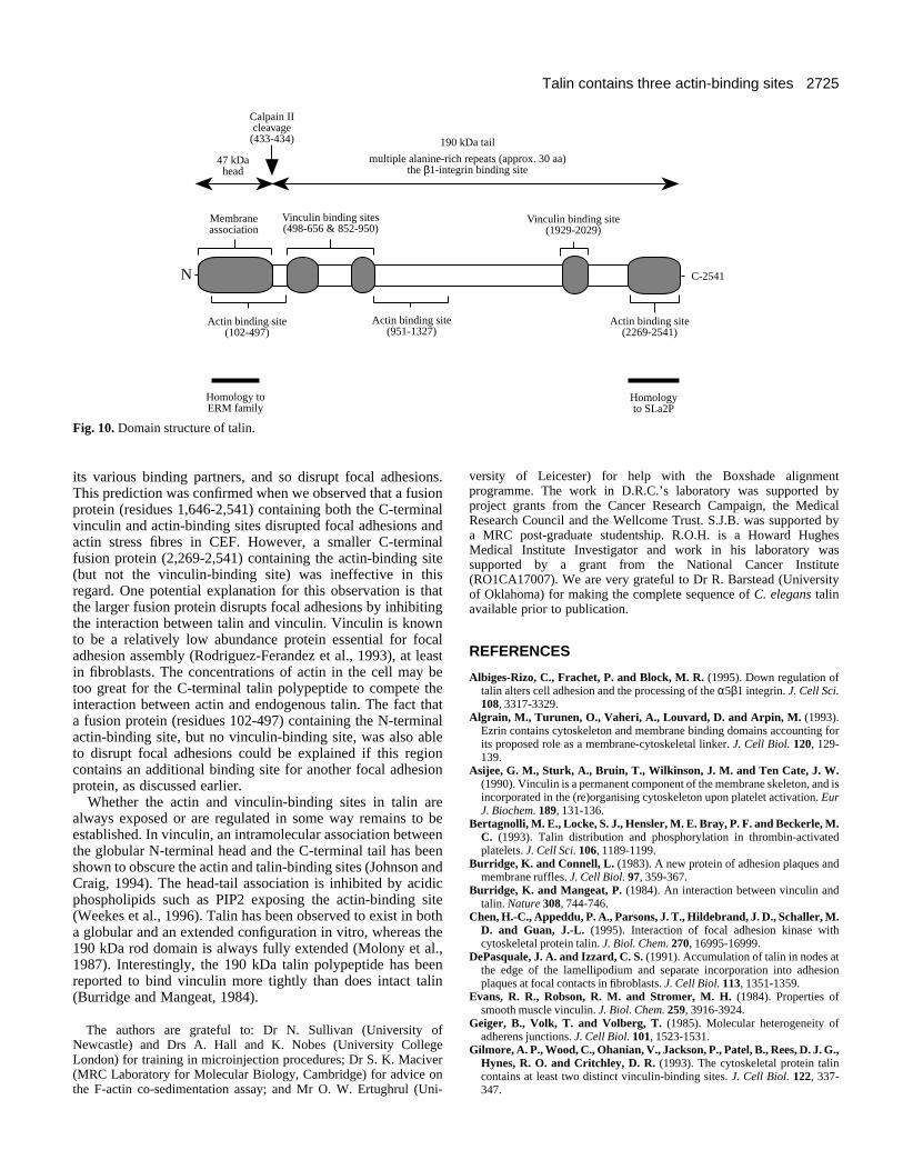
each of the actin-binding sites in talin is distinct from, butadjacent to a binding site for vinculin, a protein which can alsobind to F-actin (Menkel et al., 1994; Johnson and Craig, 1994).Microinjection studies show that those regions of talin con-taining the N- and C-terminal actin-binding sites have distinc-tive properties in terms of their localisation, resistance todetergent extraction and ability to disrupt focal adhesions andactin stress fibres. A schematic representation of the domainstructure of talin which incorporates this new information isshown in Fig. 10.
The microinjection studies of Nuckolls et al. (1990) indicatethat the N- terminal region of talin contains key determinants
responsible for specific localisation of the protein to cell-matrixjunctions. However, binding partners for this region of talinwhich might account for the localisation of the protein haveremained elusive. The fact that the N-terminal region containstwo blocks of conserved residues separated by a non-conservedregion suggests that it is composed of separate domains withdistinct functions. Talin residues 165-373 share homologieswith band 4.1 and the ERM family of cytoskeletal proteins(Rees et al., 1990; Takeuchi et al., 1994). This region of theband 4.1 protein contains a binding site for the cytoplasmicdomain of the transmembrane protein glycophorin C as well asthe peripheral membrane protein p55 (Marfatia et al., 1994),evidence that it is important in localising band 4.1 to themembrane/cytoskeletal interface. Whether the ERM homologyregion in talin contains a binding site for an integral membraneprotein remains to be clarified, although the available evidencesuggests that integrins bind to the C-terminal rod domain ofthe molecule (Horwitz et al., 1986). However, we demonstratehere that the ERM homology region in talin does contain or isadjacent to an actin-binding site, as a fusion protein spanningresidues 102-497 binds actin in vitro, and localises (albeitweakly) to actin stress fibres when microinjected into culturedcells. Interestingly, the 47 kDa N-terminal talin polypeptideliberated by calpain II cleavage (residues 1-433) localized to
2724 L. Hemmings and others
Fig. 9. Microinjection of CEF with a talin fusion protein containingresidues 1,646-2,541. Injected cells were incubated at 37°C for 5minutes (A,B), permeabilised with Triton X-100 to extract thecytoplasmic pool of injected protein, then fixed and stained for F-actin (A) and injected fusion protein (B) as described in Materialsand Methods. The fusion protein localised to focal adhesions (arrow)with little or no staining along actin stress fibres. Injected cells werealso incubated at 37°C for 30 minutes (C-F) and fixed first, and thenstained for F-actin (C), vinculin (E) and the injected GST-fusionprotein (D,F). Under these conditions, the actin cytoskeleton andvinculin-containing focal adhesions were completely disrupted inmost of the injected cells, although they were unaffected inneighbouring cells which had not been injected with the talin fusionprotein (arrows). Bar, 5 µm.
focal adhesions rather than actin filaments when microinjectedinto cells (Nuckolls et al., 1990). These results support thenotion that the conserved residues at the extreme N-terminalregion of talin play a role in targeting the protein to focaladhesions, whereas the ERM homology region provides a linkto the actin cytoskeleton. This link would appear to be rela-tively labile as the talin fusion protein spanning residues 102-497 was quantitatively extracted from microinjected cellstreated with Triton X-100 prior to fixation. More C-terminalregions of the protein which localised to focal adhesions, wereresistant to detergent extraction. These observations are similarto those made with cells stably expressing various talin
polypeptides (Gilmore et al., 1993), and are reminiscent ofstudies on ezrin (Algrain et al., 1993). The expressed N-terminal region of ezrin, which localized to the sub-membra-nous actin cytoskeleton, was solubilised by Triton X-100,whereas the C-terminal region of the protein, which localisedto actin stress fibres, was resistant to detergent extraction. TheN-terminal region of talin has also been shown to bind chargedlipids (Niggli et al., 1994) and might therefore interact withmembranes in a manner independent of protein-protein inter-actions. Platelet talin has been shown to translocate to themembrane following activation even in the absence of integrinGPIIb-IIIa (Bertagnolli et al., 1993). These various observa-tions suggest a model in which the N-terminal head region oftalin binds to one or more proteins responsible for localisingtalin specifically to cell-matrix junctions, to membrane lipids,and to the actin cytoskeleton via interactions which are readilydisrupted by non-ionic detergents.
The C-terminal rod domain of talin contains two additionalactin-binding sites neither of which displays any obvioussequence homology to the other, or to those mapped to theC-terminal regions of ezrin (Turunen et al., 1994) and moesin(Pestonjamasp et al., 1995). Talin binds to both G- and F-actin, and is reported both to nucleate actin filament assembly(Muguruma et al., 1990; Kaufmann et al., 1991), and toaugment the ability of α-actinin to cross-link F-actin(Muguruma et al., 1992). The actin cross-linking activity maybe explained by the fact that talin contains more than oneactin-binding site, or by the observation that it can formdimers under certain conditions (Molony et al., 1987). Whatrole each of the actin-binding sites in talin fulfills remains tobe explored. The C-terminal site (residues 2,300-2,541) ishomologous to a region of the S. cerevisiae protein Sla2pwhich appears to be important in controlling the assembly ofthe cortical actin cytoskeleton at bud sites (Holtzman et al.,1993). By analogy, the C-terminal actin-binding site in talinmay play a key role in the assembly of actin filaments asso-ciated with focal adhesions. The fact that a fusion protein(residues 2,269-2,541) containing this site localised to focaladhesions when microinjected into CEF may be relevant inthis regard. The three actin-binding sites in talin are eachclose to regions of the protein that bind vinculin (Fig. 10), aprotein which can also bind to actin (Menkel et al., 1994;Johnson and Craig, 1994). Vinculin interacts with talin viaresidues within the N-terminal globular head region (residues1-258) of vinculin (Jones et al., 1989; Gilmore et al., 1993)whereas the actin-binding site(s) are in the C-terminal regionof the molecule (Johnson and Craig, 1994), which can forman extended tail. It will be interesting to establish whethertalin enhances the ability of vinculin to bind to actin in amanner similar to its ability to augment the actin-bundlingactivity of α-actinin (Muguruma et al., 1992). Bearing inmind that vinculin also binds α-actinin (McGregor et al.,1994), and both talin (Horwitz et al., 1986) and α-actinin(Otey et al., 1993) are reported to bind to the cytoplasmic faceof integrins, it seems likely that the interactions among theseproteins play a pivotal role in the assembly of actin filamentsand stress fibres on the cytoplasmic face of focal adhesions.
If talin is a key component of focal adhesions as suggestedby the results from a variety of experimental approaches, onemight expect that microinjection of sub-domains of the proteinwould competitively inhibit the interactions between talin and
2725Talin contains three actin-binding sites
Calpain IIcleavage
(433-434)
47 kDahead
Membraneassociation
190 kDa tail
Vinculin binding sites(498-656 & 852-950)
Vinculin binding site(1929-2029)
Actin binding site(2269-2541)
multiple alanine-rich repeats (approx. 30 aa)the β1-integrin binding site
N
Homology toERM family
Homologyto SLa2P
Actin binding site(102-497)
Actin binding site(951-1327)
C-2541
Fig. 10. Domain structure of talin.
its various binding partners, and so disrupt focal adhesions.This prediction was confirmed when we observed that a fusionprotein (residues 1,646-2,541) containing both the C-terminalvinculin and actin-binding sites disrupted focal adhesions andactin stress fibres in CEF. However, a smaller C-terminalfusion protein (2,269-2,541) containing the actin-binding site(but not the vinculin-binding site) was ineffective in thisregard. One potential explanation for this observation is thatthe larger fusion protein disrupts focal adhesions by inhibitingthe interaction between talin and vinculin. Vinculin is knownto be a relatively low abundance protein essential for focaladhesion assembly (Rodriguez-Ferandez et al., 1993), at leastin fibroblasts. The concentrations of actin in the cell may betoo great for the C-terminal talin polypeptide to compete theinteraction between actin and endogenous talin. The fact thata fusion protein (residues 102-497) containing the N-terminalactin-binding site, but no vinculin-binding site, was also ableto disrupt focal adhesions could be explained if this regioncontains an additional binding site for another focal adhesionprotein, as discussed earlier.
Whether the actin and vinculin-binding sites in talin arealways exposed or are regulated in some way remains to beestablished. In vinculin, an intramolecular association betweenthe globular N-terminal head and the C-terminal tail has beenshown to obscure the actin and talin-binding sites (Johnson andCraig, 1994). The head-tail association is inhibited by acidicphospholipids such as PIP2 exposing the actin-binding site(Weekes et al., 1996). Talin has been observed to exist in botha globular and an extended configuration in vitro, whereas the190 kDa rod domain is always fully extended (Molony et al.,1987). Interestingly, the 190 kDa talin polypeptide has beenreported to bind vinculin more tightly than does intact talin(Burridge and Mangeat, 1984).
The authors are grateful to: Dr N. Sullivan (University ofNewcastle) and Drs A. Hall and K. Nobes (University CollegeLondon) for training in microinjection procedures; Dr S. K. Maciver(MRC Laboratory for Molecular Biology, Cambridge) for advice onthe F-actin co-sedimentation assay; and Mr O. W. Ertughrul (Uni-
versity of Leicester) for help with the Boxshade alignmentprogramme. The work in D.R.C.’s laboratory was supported byproject grants from the Cancer Research Campaign, the MedicalResearch Council and the Wellcome Trust. S.J.B. was supported bya MRC post-graduate studentship. R.O.H. is a Howard HughesMedical Institute Investigator and work in his laboratory wassupported by a grant from the National Cancer Institute(RO1CA17007). We are very grateful to Dr R. Barstead (Universityof Oklahoma) for making the complete sequence of C. elegans talinavailable prior to publication.
REFERENCES
Albiges-Rizo, C., Frachet, P. and Block, M. R. (1995). Down regulation oftalin alters cell adhesion and the processing of the α5β1 integrin. J. Cell Sci.108, 3317-3329.
Algrain, M., Turunen, O., Vaheri, A., Louvard, D. and Arpin, M. (1993).Ezrin contains cytoskeleton and membrane binding domains accounting forits proposed role as a membrane-cytoskeletal linker. J. Cell Biol. 120, 129-139.
Asijee, G. M., Sturk, A., Bruin, T., Wilkinson, J. M. and Ten Cate, J. W.(1990). Vinculin is a permanent component of the membrane skeleton, and isincorporated in the (re)organising cytoskeleton upon platelet activation. EurJ. Biochem. 189, 131-136.
Bertagnolli, M. E., Locke, S. J., Hensler, M. E. Bray, P. F. and Beckerle, M.C. (1993). Talin distribution and phosphorylation in thrombin-activatedplatelets. J. Cell Sci. 106, 1189-1199.
Burridge, K. and Connell, L. (1983). A new protein of adhesion plaques andmembrane ruffles. J. Cell Biol. 97, 359-367.
Burridge, K. and Mangeat, P. (1984). An interaction between vinculin andtalin. Nature 308, 744-746.
Chen, H.-C., Appeddu, P. A., Parsons, J. T., Hildebrand, J. D., Schaller, M.D. and Guan, J.-L. (1995). Interaction of focal adhesion kinase withcytoskeletal protein talin. J. Biol. Chem. 270, 16995-16999.
DePasquale, J. A. and Izzard, C. S. (1991). Accumulation of talin in nodes atthe edge of the lamellipodium and separate incorporation into adhesionplaques at focal contacts in fibroblasts. J. Cell Biol. 113, 1351-1359.
Evans, R. R., Robson, R. M. and Stromer, M. H. (1984). Properties ofsmooth muscle vinculin. J. Biol. Chem. 259, 3916-3924.
Geiger, B., Volk, T. and Volberg, T. (1985). Molecular heterogeneity ofadherens junctions. J. Cell Biol. 101, 1523-1531.
Gilmore, A. P., Wood, C., Ohanian, V., Jackson, P., Patel, B., Rees, D. J. G.,Hynes, R. O. and Critchley, D. R. (1993). The cytoskeletal protein talincontains at least two distinct vinculin-binding sites. J. Cell Biol. 122, 337-347.

2726 L. Hemmings and others
Gilmore, A. P., Ohanian, V., Spurr, N. K. and Critchley, D. R. (1995).Localisation of the human gene encoding the cytoskeletal protein talin tochromosome 9p. Hum. Genet. 96, 221-224.
Hildebrand, J. D., Schaller, M. D. and Parsons, J. T. (1995). Paxillin, atyrosine phosphorylated focal adhesion-associated protein binds to thecarboxy terminal domain of focal adhesion kinase. Mol. Biol. Cell 6, 637-647.
Holtzman, D. A., Yang, S., Drubin, D. G. (1993). Synthetic-lethal interactionsidentify 2 novel genes, SLA1 and SLA2, that control membrane cytoskeletonassembly in S. cerevisiae. J. Cell Biol. 122, 635-644.
Horwitz, A., Duggan, K., Buck, C., Beckerle, M. C. and Burridge, K.(1986). Interaction of plasma-membrane fibronectin receptor with talin - atransmembrane linkage. Nature 320, 531-533.
Jackson, C. W., Hutson, N. K., Steward, S. A. and Stenberg, P. E. (1992). Aunique talin antigenic determinant and anomalous megakaryocyte talindistribution associated with abnormal platelet formation in the Wistar-Furthrat. Blood 79, 1729-1737.
Jackson, C. W., Hutson, N. K., Steward, S. A. and Rees, D. J. G. (1993).Detection of a mutation in the cytoskeletal protein talin in the Wistar-Furth(WF) rat - A rat strain with defective platelet formation and a high tumourincidence. Blood 82, 340A.
Jockusch, B. M., Bubeck, P., Giehl, K., Kroemker, M., Moschner, J.,Rothkegel, M., Rudiger, M., Schluter, K., Stanke, G. and Winkler, J.(1995). The molecular architecture of focal adhesions. Annu. Rev. Cell Dev.Biol. 11, 379-416.
Johnson, R. P. and Craig, S. W. (1994). An intramolecular associationbetween the head and tail domains of vinculin modulates talin binding. J.Biol. Chem. 269, 12611-12619.
Jones, P., Jackson, P., Price, G. J., Patel, B., Ohanian, V., Lear, A. L. andCritchley, D. R. (1989). Identification of a talin binding site in thecytoskeletal protein vinculin. J. Cell Biol. 109, 2917-2927.
Kaufmann, S., Piekenbrock, T., Goldmann, W. H., Barmann, M. andIsenberg, G. (1991). Talin binds to actin and promotes filament nucleation.FEBS Lett. 284, 187-191.
Kreitmeier, M., Gerisch, G., Heizer, C. and Muller-Taubenberger, A.(1995). A talin homologue of Dictyostelium rapidly assembles at the leadingedge of cells in response to chemoattractant. J. Cell Biol. 129, 179-188.
Kupfer, A., Swain, S. L. and Singer, S. J. (1987). The specific directinteraction of helper T-cells and antigen presenting B-cells. II. Reorientationof the microtubule organising centre and reorganisation of the membrane-associated cytoskeleton inside the bound helper T-cells. J. Exp. Med. 165,1565-1580.
Lee, S.-W., Wulfkuhle, J. D. and Otto, J. J. (1992). Vinculin binding sitemapped on talin with an anti-idiotypic antibody. J. Biol. Chem. 267, 16355-16358.
Marfatia, S. M., Lue, R. A., Branton, D. and Chisti, A. H. (1994). In vitrobinding studies suggest a membrane-associated complex between erythroidp55, protein 4.1 and glycophorin C. J. Biol. Chem. 269, 8631-8634.
McGregor, A., Blanchard, A. D., Rowe, A. J. and Critchley, D. R. (1994).Identification of the vinculin-binding site in the cytoskeletal protein α-actinin. Biochem. J. 301, 225-233.
McLachlan, A. D., Stewart, M., Hynes, R. O. and Rees, D. J. G. (1994).Analysis of repeated motifs in the talin rod. J. Mol. Biol. 235, 1278-1290.
Menkel, A. R., Kroemaker, M., Bubeck, P., Ronsiek, M., Nikolai, G. andJockusch, B. M. (1994). Characterisation of an F-actin binding domain inthe cytoskeletal protein vinculin. J. Cell Biol. 126, 1231-1240.
Molony, L., McCaslin, D., Abernethy, J., Paschal., B. and Burridge, K.
(1987). Properties of talin from chicken gizzard smooth muscle. J. Biol.Chem. 262, 7790-7795
Moulder, G. L., Huang, M.-M., Waterston, R. H. and Barstead, R. J.(1996). Talin requires β-integrin but not vinculin for its assembly into focaladhesion-like structures in the nematode Caenorhabditis elegans. Mol. Biol.Cell. 7, 1181-1193.
Muguruma, M., Matsumura, S. and Fukazawa, T. (1990). Directinteractions between talin and actin. Biochem. Biophys. Res. Commun. 171,1217-1223.
Muguruma, M., Matsumura, S. and Fukazawa, T. (1992). Augmentation ofα-actinin-induced gelation of actin by talin. J. Biol. Chem. 267, 5621-5624.
Niggli, V., Kaufmann, S., Goldmann, W. H., Weber, T. and Isenberg, G.(1994). Identification of functional domains in the cytoskeletal protein talin.Eur. J. Biochem. 224, 951-957.
Nuckolls, G. H., Turner, C. E. and Burridge, K. (1990). Functional-studiesof the domains of talin. J. Cell Biol. 110, 1635-1644.
Nuckolls, G. H., Romer, L. H., Burridge, K. (1992). Microinjection ofantibodies against talin inhibits the spreading and migration of fibroblasts. J.Cell Sci. 102, 753-762.
Otey, C. A., Vasquez, G. B., Burridge, K. and Erickson, B. W. (1993).Mapping of the α-actinin binding site within the β1-integrin cytoplasmicdomain. J. Biol. Chem. 268, 21193-21197.
Pestina, T. L., Jackson, C. W. and Stenberg, P. E. (1995). Abnormalsubcellular distribution of myosin and talin in Wistar Furth rat platelets.Blood 85, 2436-2446.
Pestonjamasp, K., Amieva, M. R. Strassel, C. P., Nauseef, W. M.,Furthmayr, H. and Luna, E. J. (1995). Moesin, ezrin and p205 are actin-binding proteins associated with neutrophil plasma membranes. Mol. Biol.Cell 6, 247-259.
Rees, D., Ades, S. E., Singer, S. J. and Hynes, R. O. (1990). Sequence anddomain-structure of talin. Nature 347, 685-689.
Rodriguez Fernandez, J. L. R., Geiger, B., Salomon, D., Ben Ze’ev, A.(1993). Suppression of vinculin expression by antisense transfection conferschanges in cell morphology, motility, and anchorage-dependent growth of3T3-cells. J. Cell Biol. 122, 1285-1294.
Sanger, F., Nickel, S. and Coulson, A. R. (1977). DNA sequencing with chainterminating inhibitors. Proc. Nat. Acad. Sci. USA 74, 5463-5466.
Smith, D. B. and Johnson, K. S. (1988). Single step purification ofpolypeptides expressed in E. coli as fusions with glutathione-S-transferase.Gene 67, 31-40.
Takeuchi, K., Kawashima, A., Nagafuchi, A. and Tsukita, S. (1994).Structural diversity of band 4.1 superfamily members. J. Cell Sci. 107, 1921-1928.
Turunen, O., Wahlstrom, T. and Vaheri, A. (1994). Ezrin has a COOH-terminal actin-binding site that is conserved in the ezrin protein family. J.Cell Biol. 126, 1445-1453.
Weekes, J., Barry, S. T. and Critchley, D. R. (1996). Acidic phospholipidsinhibit the intramolecular association between the N- and C-terminal regionsof vinculin exposing actin-binding and protein kinase C phosphorylationsites. Biochem J. 314, 827-832.
Winder, S. J., Hemmings, L., Maciver, S. K., Bolton, S. J., Tinsley, J. M.,Davies, K. E., Critchley, D. R. and Kendrick-Jones, J. (1995). Utrophinactin binding domain: analysis of actin binding and cellular targeting. J. CellSci. 108, 63-71.
(Received 9 May 1996 - Accepted 7 August 1996)