tagged mutagenesis and gene-trap in the moss ... transformation procedure followed schaefer20 with...
TRANSCRIPT

DNA RESEARCH 7. 9 17 (2000)
Tagged Mutagenesis and Gene-trap in the Moss, Physcomitrellapatens by Shuttle Mutagenesis
Tomoaki NlSHlYAMA,1'2 Yuji HIWATASHI,2'3 Keiko SAKAKIBARA,2 '3 Masahiro KATO, 1
and Mitsuyasu H A S E B E 2 1 *
Department of Biological Sciences, Graduate School of Science, the University of Tokyo, Tokyo 113-0033,Japan,1 National Institute for Basic Biology, Okazaki 444-8585, Japan,2 Department of MolecularBiomechanics, The Graduate University for Advanced Studies, Okazaki 444-8585, Japan,3 and PRESTO,Japan Science and Technology Corporation4
(Received 29 November 1999)
AbstractThe moss, Physcomitrella patens has been used as a useful material in many fields, because of its simple
body plan, ease of gene targeting, and other reasons. Although many mutants have been reported, nomethod to isolate the corresponding genes was reported. We developed a gene tagging and gene-trapsystem in P. patens by using the shuttle mutagenesis technique, which has been used in the budding yeast.In 5264 tagged lines, 203 mutants with altered developmental or morphological phenotypes were obtained.In 129 of 4757 gene-trap lines, /3-glucuronidase (GUS) activity was detected in some tissue. Althoughmultiple copies of a tag were detected in many tagged lines by Southern analyses, most copies are likelyintegrated at the same locus according to PCR analyses.Key words: tagging; gene-trap; Physcomitrella patens; homologous recombination; shuttle mutagenesis
1. Introduction
Comparing the developmental mechanisms in modelorganisms is indispensable for understanding the diver-sity and evolution of development. Furthermore, tounderstand the general principles of development, it isnecessary to study different organisms that have spe-cial advantages for developmental studies.1 Many landplants, ranging from angiosperms to bryophytes, havebeen studied as model plants. Some mosses have beenused to study physiology, genetics, and developmentalbiology.2"5 Of these, Physcomitrella patens has receivedspecial attention in the last few years, since a gene-targeting technique ascribed to its high rate of homol-ogous recombination was established.6'7
A number of P. patens mutants that alter their devel-opmental or physiological traits have been described,8~n
but there are no methods to isolate the genes correspond-ing to the mutations. Agrobacterium- or transposon-mediated gene-tagging methods are useful for cloningmutated genes in organisms for which genetic maps arenot available,12'13 such as P. patens. However, neitherinfection with Agrobacterium nor transposition of an en-dogenous or exogenous transposon has been reported
Communicated by Masahiro Sugiura* To whom correspondence should be addressed. Tel./Fax. +81-
564-55-7546, E-mail: [email protected]
in P. patens. Therefore, to establish tagged mutantsof P. patens, we focused on shuttle mutagenesis, whichhas been successfully used in the budding yeast, Sac-charomyces cerevisiae.1416 This method involves threesteps, as shown in Fig. 1. Its essence is the replacementof a P. patens genomic sequence with tagged P. patensDNA sequences by homologous recombination. The highrate of homologous recombination in P. patens is simi-lar to that in the budding yeast,7 which should make itpossible to use this method.
2. Materials and Methods
2.1. Plant material and culture conditionsPhyscomitrella patens (Hedw.) Bruch & Schimp subsp.
Patens Tan17 collected in Gransden Wood, Huntingdon-shire, UK18 was used as the wild-type strain. Protone-mata of P. patens were grown in 9-cm Petri dishes on BC-DATG medium. BCD medium contains 1 mM MgSO*!,10 mM KNO3, 45 /JM FeSO4, 1.8 mM KH2PO4 [pH 6.5adjusted with KOH], and trace element solution (alter-native TES19; 0.22 /iM CuSO4, 0.19 /*M ZnSO4, 10H3BO3, 0.10 /iM Na2MoO4, 2 /iM MnCl2, 0.23CoCl2, and 0.17 /iM KI). BCDATG medium is BCDmedium supplemented with 1 mM CaCl2, 5 mM di-ammonium (+)-tartrate, and 0.5% (w/v) glucose, and so-

10 Tagged Mutagenesis and Gene-trap in Physcomitrella
(1) Generation of P. patens genomic library in E. coli
[Vol.
P.patens genomic DNA
pHSS-Sal
£• con
(2) Insertion of mini-transposon to P. patens genomicDNA in E.coli
P.patens genomic DNA
pHSS-Sal
E.coli
+mini-transposon
juidA nptll |
pHSS-Sa!
E.coli
mini-transposon inserted inP.patens genomic DNA
pHSS-Sal
E.coli
(3) Transformation of P. patens with P. patensgenomic DNA tagged by mini-transposon
P. patensprotoplast
P. patens chromosome taggedby mini-transposon
Figure 1. Schematic diagram of the shuttle mutagenesis ofPhyscomitrella patens. The procedure for shuttle mutagene-sis consists of three steps: (1) generating a P. patens genomiclibrary in E. coli, (2) inserting mini-transposons into the ge-nomic library in E. coli, and (3) transforming P. patens withthe genomic library containing mini-transposon inserts. Theoverall effect is the insertion of the mini-transposons into theP. patens genome.
lidified with 0.8% (w/v) agar (A-9799, SIGMA) (D. Cove,personal communication). The solidified medium wascovered with a layer of cellophane (Futamura ChemicalIndustries Co., Ltd., Nagoya, Japan) to facilitate collec-tion of protonemata from the medium. The dishes wereincubated at 25°C under continuous light (40 /zmol pho-tons m~2s~1). For vegetative propagation, the protone-mata were collected every 5 days, and were ground with aPolytron homogenizer (Kinematica, Littau, Switzerland)
at a speed setting of 4. The ground protonemata weresoaked on the BCDATG medium using the same condi-tions described above.
2.2. Transformation of P. patensThe transformation procedure followed Schaefer20 with
minor modifications. Protoplasts were prepared usingvegetatively propagated protonemata cultured for 4-5days.
The room temperature was kept at 20°C duringthe transformation experiments. Collected protonematawere incubated in 8% (w/v) mannitol and 2% (w/v) Dris-erase (Kyowa Hakko Kogyo Co., Ltd.) for 30 min at 25°Cin the dark with gentle mixing every 5 min. The proto-plasts were filtered through a 50 /im nylon mesh, col-lected by centrifugation at 180 xg with a swinging rotor,and suspended in 8% (w/v) mannitol. This washing pro-cedure was repeated two more times. After counting thedensity of protoplasts with a hemocytometer, the proto-plasts were suspended in MMM solution (8.3% mannitol,0.1% [w/v] MES-KOH [pH 5.6], and 15 mM MgCl2) at1.6xlO6 cells/mL. Thirty microliters of DNA solution inTE (10 mM Tris-HCl [pH 8.0], and 1 mM EDTA) werealiquoted in a polypropylene tube (Falcon 2057; BectonDickinson Labware). Then 300 fiL of the protoplast sus-pension and 300 /zL of PEG/T solution (28.5% polyethy-lene glycol 6000 in 7.2% [w/v] mannitol, 0.1 M CaCl2,and 10 mM Tris-HCl [pH 8.0]) were added. The mixedsuspension was incubated at 45°C for 5 min, and thencooled at 20°C for 10 min in a water bath. The pro-toplasts were diluted stepwise to 8 mL with protoplastliquid culture medium (5 mM Ca(NOs)2, 1 mM MgSC>4,45 fM FeSO4, 0.18 mM KH2PO4 [pH 6.5 adjusted withKOH], the alternative TES, 5 mM ammonium tartrate,6.6% [w/v] mannitol, and 0.5% [w/v] glucose), pouredinto a 6-cm Petri dish, and kept in the dark at 25°C for1 day. The protoplasts were collected by centrifugation at180xg, and suspended in 8 mL of protoplast regenerationmedium for the top layer (PRM/T; BCD medium supple-mented with 5 mM ammonium tartrate, 10 mM CaCb,0.8% [w/v] agar, and 8% [w/v] mannitol). This mediumwas warmed to 45°C to prevent jelling. The protoplastssuspended in PRM/T were poured into four 9-cm dishesthat contained a bottom layer of protoplast regenerationmedium (PRM/B), which was covered with cellophane.PRM/B contains BCD medium supplemented with 5 mMammonium tartrate, 10 mM CaCb, 0.8% (w/v) agar, and6% (w/v) mannitol. After 4 days incubation under con-tinuous light, the cellophane with the PRM/T contain-ing regenerated protoplasts was transferred to BCDATmedium containing 50 mg/L G418 (Life Technologies) toselect transformants. BCDAT medium is BCD mediumcontaining 1 mM CaCk, 5 mM ammonium tartrate, and0.8% (w/v) agar. The plants that survived 3 weeks on theselection medium were transferred to BCDATG medium.

No. 1] T. Nishivama et al. 11
A
mTn-nptll
B
nptll ' ° x P amp1
Physcomitrella cdc2 intron
nptll amp'mTn-4xHA/GUS
3xHA
TR
f6S
loxR | \ nos-ter j \ j\ |oxR
G3 G4 n3 n2 A5A4TR
Physcomitrella cdc2 intron
uidA nptllmTn-3xHA/GUS1
A^ uidA
i T « \
3xHA
TRloxR nos-ter
3 G4 n3 n2 A5A4
D Arabidopsis GPA1 intronXA uidA
mTn-3xHA/GUS2nptll ampf 3xHA
TRloxR I \
G3
nos-ter
G3 G4
resloxP
ri3 n2 TR
Figure 2. Mini-transposons used for mutagenizing Physcomitrellapatens. The figure shows a schematic representation of themini-transposons constructed. Each mini-transposon con-tains a NPTII expression cassette (nptll) and an ampi-cillin-resistance gene (ampr). The arrows within nptlland amp7' indicate their directions. mTn-4xHA/GUS,mTn-3xHA/GUSl, and mTn-3xHA/GUS2 each contain an in-tron, the uidA coding region (thick arrow), and the nopa-line synthase polyadenylation signal (nos-ter). The intron inmTn-4xHA/GUS and mTn-3xHA/GUSl is the intron fromthe P. patens cdc2 gene (K. Fujiwara, personal communica-tion), and the intron in mTn-3xHA/GUS2 is the ArabidopsisGPA1 intron with triple donor and acceptor sites.27 Arrow-heads show the location of the primer annealing sites used forinter-mini-transposon PCR.
incubated for 1 week, and re-inoculated onto the selectionmedium. Those plants that grew on the selection mediumas fast as on the non-selection medium were stored as sta-ble transformants.
2.3. Construction of mini-transposons(1) mTn-nptll
The NPTII expression cassette (nptll), which containsthe cauliflower mosaic virus (CaMV) 35S promoter,21
the nptll gene,22 and a fragment containing the CaMVpolyadenylation signal23 corresponding to 7482-131 ofthe CaMV genome,24 was excised from plasmid pMBL5(provided by D. Cove) as an Xhol-Kpnl fragment,and cloned into the Sma I site of pBluescript SKII+(Stratagene). This plasmid was designated pTN3. A5amHI-//mdIII fragment containing nptll was excised
from pTN3 and cloned between the BarnHl and Hindlllsites of the mini-transposon on the pTn plasmid15 tomake the pTn-nptll plasmid. The mini-transposon con-taining nptll (niTn-nptll; Fig. 2A) was transposed tothe F derivative pOX38 as previously described,15 andthe modified F derivative with the mini-transposon wasnamed pOX38::mTn-nptII.
(2) mTn-4xHA/GUS, mTn-3xHA/GUSl, and mTn-3xHA/GUS2
The mTn-4xHA/GUS mini-transposon (Fig. 2B) wasmade by exchanging the lacZ, URA3, and tetr geneson the mTn-4xHA//acZ25 mini-transposon with the/3-glucuronidase (GUS) gene uidA,26 nptll, and theampr gene, respectively. The plasmid containing mTn-4xHA/lacZ on pHSS614 was kindly provided by M.Snyder. The mini-transposon mTn-4xHA/lacZ containsin order: an HA tag, a loxR site, a lacZ gene, a URA3gene, a res site, and three HA tags, between the terminalinverted repeats. The region from the res site to the loxRsite, which contains the pHSS6 vector, was amplified withprimers having restriction sites at their 5' ends (loxR-r, ATCGATATCgcgtataacttcgtatagcag; res-f, CTAGTC-GACCACGTGcgtaacca; the capitalized nucleotides aresynthetic restriction sites) and self-ligated as pTn-4xHA.The plasmid pTn-4xHA has £coRV, 5a/1, and PmaCIsites between the loxR and res sites. An intron-GUS-nptll fragment prepared in a separate plasmid was clonedbetween the EcoKV and 5a/1 sites, and the amp7' genewas cloned into the PmaCI site. The intron-GUS-nptllfragment and the ampr gene were prepared as follows.A DNA fragment containing the first intron of the P.patens cdc2 gene (kindly provided by K. Fujiwara) wasinserted between the Xba I and -BamHI sites of pBI221,26
bordering the uidA gene. The uidA gene along withthe CaMV 35S promoter, cdc2 intron, and nopaline syn-thase polyadenylation signal (nos-ter) were excised fromthe plasmid as an EcoRl-Hindlll fragment, and clonedbetween the .EcoRI and Hindlll sites of pTN3, whichcontains nptll. An Xba l-Xho I fragment containing thecdc2 intron, uidA, nos-ter, and nptll was excised, andcloned into pTn-4xHA between the EcoTW and Sal Isites. The ampr gene corresponding to coordinates 758-1948 of pUC18 (accession no. L08752) was amplifiedby PCR and cloned into the PmaCI site. The mTn-4xHA/GUS mini-transposon was constructed on pHSS6plasmid.14'25 This mini-transposon was transposed topOX38 as described,25 and the modified F derivativewith the mTn-4xHA/GUS mini-transposon was namedPOX38::mTn-4xHA/GUS.
The mTn-3xHA/GUSl and mTn-3xHA/GUS2 mini-transposons (Fig. 2C, D) were made by modifyingthe mTn-3xHA//acZ mini-transposon25 using the pro-cedures described above. The mTn-3xHA/GUSl mini-transposon contains the P. patens cdc2 intron, as doesthe mTn-4xHA/GUS mini-transposon, while the mTn-

12 Tagged Mutagenesis and Gene-trap in Physcormtrella [Vol.
Table 1. Escherichia coli strains used in this study.
Strain Relevant genotype Reference or supplierXL2 blue MRF' A(mcrA) A(mcrCB-hsdSAlR-mrr)end.41 StratageneDH5a hsdRhsdM+
RDP146 hsdR+
RDP146/pLB101 hsdR+, transposaseRDP146/pOX::mTns mini-transposon on F factor derivativeNS2114Sm Smr, A-creNG135 Smr, resolvase
Life Technologies1515this study15
3xHA/GUS2 mini-transposon contains the Arabidopsisthaliana GPAl intron with triple acceptor sequences.27
2-4- Construction of the mutagenized genomic libraryThe Escherichia coli strains used in this study and
their relevant genotypes are summarized in Table 1.Chemically competent cells of XL2 blue MRF' (Strata-gene) and DH5a (Life Technologies, Inc.) were pre-pared according to Inoue et al.28 Preparation of electro-competent cells of RDP146/pLB101 and electroporationusing an EasyjecT Prima (EquiBio Ltd., Kent, UK) elec-troporator were performed according to the manufac-turer's instructions.
P. patens genomic DNA was isolated from protone-mata by the CTAB method.29 The genomic DNA waspartially digested with Sau3Al, and run on 0.6% (w/v)agarose gels (SeaKem GTG, FMC BioProducts) inlxTAE buffer. The fragments 3-6 kb long were recov-ered by the electroelution method,30 and their ends werepartially filled with dATP and dGTP. These fragmentswere ligated with the Sal I-digested pHSS-Sal vector16
partially filled with dCTP and dTTP. The ligation solu-tion was used to transform E. coli XL2 blue MRF'. Plas-mids extracted from XL2 blue MRF' were subsequentlyused to transform E. coli D H 5 Q . Plasmids extractedfrom DH5a were further used for the transformation ofE. coli RDP146/pLB101. The E. coli RDP146/pLB101containing the P. patens genomic DNA fragments wasmated with E. coli RDP146/pOX38::mTn-nptII to formE. coli RDP146/pLB101/pOX38::mTn-nptII and to mu-tagenize the P. patens genomic library with the mTn-nptll mini-transposon, which was then mated with E.coli NS2114Sm.15 To mutagenize the genomic libraryusing other mini-transposons, E. coli NG135 was usedinstead of NS2114Sm.15'25 The mutagenized library re-covered from NS2114Sm or NG135 was amplified in E.coli XL2 blue MRF', because NS2114Sm and NG135 areendA+ strains. The plasmids extracted from E. coli XL2blue MRF' were digested with Not I, extracted with phe-nol and chloroform, precipitated with ethanol, and di-luted with TE. The concentration of DNA was adjustedto 1.0 //g//ul.
2.5. Histochemical detection of GUS activityThe histochemical GUS activity was assayed accord-
ing to Jefferson et al.31 with slight modifications. Themutated lines were cultured for 14 days on BCDATG orBCDAT medium. The tissues were fixed in a solutionof 0.3% (v/v) formalin, 5.45% (w/v) mannitol, and 0.2%(w/v) MES-KOH (pH 5.6) for 30 min at room temper-ature, washed 3 times with 50 mM NaH2PO4 (pH7.0),infiltrated for 30 min in a substrate solution (50 mMNaH2PO4 [pH7.0], 0.5 mM 5-bromo-4-chloro-3-indolyl 0-D-glucuronide [X-Gluc, Wako Pure Chemical Industries,Osaka, Japan], 0.5 mM K3Fe(CN)6, 0.5 mM K4Fe(CN)6,and 0.3% [v/v] Triton X-100), and then incubated at37°C for 36-48 hr for staining. After the incubation, thetissues were fixed in 5% (v/v) formalin for 10 min, andsoaked in 5% (v/v) acetic acid for 10 min. Then thetissues were dehydrated through an ethanol series. Fi-nally, the stained tissues were observed under a stereomi-croscope and a light microscope. Images of the stainedtissues were digitized with a CCD Camera (FUJIX HC-300Z, Fuji Film).
2.6. Southern hybridizationTwo micrograms of genomic DNA was digested with
an appropriate restriction enzyme, run on a 0.7%(w/v) agarose gel (SeaKem GTG), and transferred toa positively charged nylon membrane (Hybond N + ,Amersham-Pharmacia Biotech). An AlkPhos Direct kit(Amersham-Pharmacia Biotech) was used for labelingDNA probes, hybridization, and detection.
2.7. Northern hybridizationTotal RNA was isolated from each strain as described
by Hasebe et al.32 Poly(A)+ RNA was further purifiedusing oligo-dT magnetic beads (DynaBEADS, Dynal,Oslo, Norway). One microgram of poly(A)+ RNA waselectrophoresed on a denaturing agarose gel (1% [w/v]SeaKem GTG agarose, lxMOPS buffer, and 6.7% [v/v]formaldehyde), and transferred to a Hybond N+ mem-brane by the downward capillary transfer method33 usingI O X S S C . The membrane was fixed with 0.05 M NaOH,washed with lxSSC, and dried at 80°C for 20 min. The

No. 1] T. Nishivama ct al. 13
hybridization was performed in Church buffer3'1 at 65°C.The probes were radiolabeled using a Random PrimerDNA Labeling Kit vcr. 2.0 (Takara) with [Q-:32P] dCTP,and detected by autoradiography.
2.8. PCR to detect the region between the mini-transposon.
To amplify the inter-mini-transposon regions, theprimer sets A5 (5'-TTAATAGACTGGATGGAGGCGGATAAAGTT-3') and n3 (5'-ACGGAAGGAAGGAGGAAGACAAGGAAGGAT-3') or A4 (5'-TAGACTGGATGGAGGCGGATAAAGTTGCAG-3') and n2 (5'-AGGAGGAAGACAAGGAAGGATAAGGTTGCA-3')were used for the mTn-nptll mini-transposon. A5 andG4 (5'-ATTGACCCACACTTTGCCGTAATGAGTGAC-3') or A4 and G3 (5'-TCTTGTAACGCGCTTTCCCACCAACGCTGA-3') were used for the mTn-4xHA/GUS and mTn-3xHA/GUS mini-transposons. EachPCR reaction contained 1 /ih of 10 ng//iL genomic DNA,2 jiL of ExTaq Buffer, 1 /ih each of two 10 fiM primers,2 fiL of 2 mM dNTP, 13 /iL of water, and 0.1 /iL of 5unit/^L ExTaq DNA polymerase (Takara). Each PCRcycle consisted of 94°C for 20 sec and 68°C for 15 min.The cycle was repeated 20 times, preceded by 5min at94°C, followed by 5 min at 68°C. A 50-fold dilution ofthe first PCR product was used as the template for thesecond PCR, which was carried out using the same con-ditions, except for the sample of TNI mutant which wascarried for 15 cycles.
3. Results and Discussion
3.1. Generation of P. patens genomic library in E. coliA P. patens genomic DNA library with insert sizes
from 3 to 6 kb was constructed with the pHSS-Sal16 plas-mid vector, which is suitable for shuttle mutagenesis us-ing E. coli. The size range of the inserted fragments isappropriate for efficient cloning using the pHSS-Sal vec-tor and for homologous recombination of P. patens.
Since P. patens genomic DNA is thought to containmethylated bases like other eukaryotic genomes, we usedthe mcrA~ mcrCB~ mrr~ E. coli strain, XL2-blueMRF' (Stratagene). Since we ultimately introduced thelibrary into an hsdR+ E. coli strain, either NG135 orNS2114Sm, the library was passed through an hsdM+
E. coli strain, DH5a.
3.2. Mini-transposonsIn shuttle mutagenesis of the budding yeast, the LEU2
or URA3 genes are used as a selection marker.16'25 Tofacilitate the selection of P. patens tagged mutants, wegenerated mini-transposons with nptll that confers G418resistance (Fig. 2). For gene-trap, mini-transposons con-taining the uidA gene, which encodes /3-glucuronidase
genomic DNA + mTn-nptllvector + mTn-nptllgenomic DNA
2.2 vector
BPpHB4
PpHB5
PpHB7
PpHB8
PpHB9
i i i i
J I I I
J I
II II
1 kb
Figure 3. Transposition of mini-transposons on plasmids. A, themini-transposon mTn-nptll was transposed to a 4-kb genomicclone. Lane 1, plasmids extracted from E. coli containing the4-kb clone and pLBlOl before the transposition were digestedwith Not I. The 4.4-kb band is pLBlOl. Lane 2, plasmids ex-tracted from a population of E. coli containing a plasmid witha mini-transposon tag derived from the plasmid in lane 1. B,the mini-transposon mTn-nptll was transposed to each PpHBgene. Horizontal line indicates the region of the genomic cloneof each PpHB gene. Vertical bars show the distribution of theinsertion points of the mini-transposons.
(GUS),31 were used as an expression marker instead oflacZ, which is used in the yeast system.
P. patens genes have introns like other plants, whilemost yeast genes lack introns. Either the ArabidopsisGPAl intron27 or the P. patens cdc2 intron was fused tothe 5' end of the uidA gene, so that the expression of atagged P. patens gene could be detected when the mini-transposon was inserted in the intron or coding region.
3.3. Transposition of mini-transposons to P. patens ge-nomic DNA cloned in pHSS-Sal plasmid in E. coli
The donor strain containing the mini-transposon onthe F derivative was mated with the recipient strain con-taining the P. patens genomic DNA library, and themini-transposon was transposed to the P. patens ge-nomic DNA. To test the efficiency of transposition bythis system, eight randomly selected genomic clones of 2to 4 kb were mutagenized with mTn-nptll. Since thereare Not I sites at the boundaries between the pHSS-Salvector and the inserted P. patens genomic DNA frag-ment, plasmids extracted from each mutagenized pop-
14 Tagged Mutagenesis and Gene-trap in Physcomitrella [Vol.
1 2 3 4 5 6 7 w p
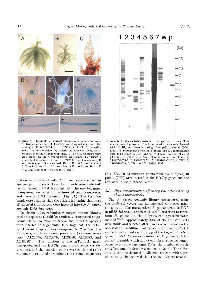
Figure 4. Example of mutant strains and gene-trap lines.A, transformant morphologically indistinguishable from thewild-type (19980725084-3). B (TNI) and C (TN2), morpho-logical mutants obtained by shuttle mutagenesis. D-H, histo-chemical staining of gene-trap lines. D, YH330, mucilage hairsare stained. E, YH78, young leaves are stained. F, YH229, ayoung bud is stained. G and H, YH206, the chloronema (G)and caulonema (H) are stained. Bar in B = 0.5 mm for A andB. Bars in C and D = 0.1 mm. Bar in E = 0.2 mm. Bar in F= 20 /im. Bar in H = 50 /im for G and H.
ulation were digested with Not I, and separated on anagarose gel. In each clone, four bands were detected:vector, genomic DNA fragment with the inserted mini-transposon, vector with .the inserted mini-transposon,and genomic DNA fragment (Fig. 3A). The first twobands were brighter than the others, indicating that mostof the mini-transposons were inserted into the P. patensgenomic DNA fragment.
To obtain a low-redundancy tagged mutant library,mini-transposons should be randomly transposed to ge-nomic DNA. To examine where the mini-transposonswere inserted in a genomic DNA fragment, the mTn-nptll mini-transposon was transposed to P. patens HD-Zip genes, which we cloned previously (accession num-bers: AB28075, AB28076, AB28078, AB28079, andAB28080). The junction of the niTn-nptll mini-transposon and the HD-Zip genomic sequence was de-termined, and the insertion points were confirmed to berandomly distributed throughout the genomic sequences
^ ^ J
Figure 5. Southern hybridization of mutagenized strains. Twomicrograms of genomic DNA from transformants was digestedwith EcoKl, and detected using pTn-nptll probe at 55°C.Lane 1-5, mutagenized with mTn-nptll; lane 6, 7 mutagenizedwith mTn-3xHA/GUSl; lane w, wild-type; lane p, 30 pg ofpTn-nptll digested with Xho I. The strains are as follows: 1,19980725073-2; 2, 19981029021; 3, 1998102902132; 4, TN2; 5,19981029034; 6, TNI; and 7, 19980928037.
(Fig. 3B). Of 53 insertion points from five analyses, 40points (75%) were located in the HD-Zip genes and therest were in the pHSS-Sal vector.
3.4- High transformation efficiency was achieved usingshuttle mutagenesis.
The P. patens genomic library constructed usingthe pHSS-Sal vector was mutagenized with each mini-transposon. The mutagenized P. patens genomic DNAin pHSS-Sal was digested with Not I, and used to trans-form P. patens by the polyethylene glycol-mediatedmethod.20-30 Approximately 30% of the transformantswere stable and selected after 1 week of relaxation on thenon-selection medium. We typically obtained 187±116stable transformants with 30 fig of the tagged P. patensgenomic DNA. When we transformed P. patens with lin-earized plasmids which do not contain a sequence homol-ogous to P. patens genomic DNA, the number of stabletransformants obtained was reduced to 20±7. The differ-ence in the transformation efficiency concurs with a pre-vious study that showed that the homologous recombi-
No. 1 T. Nishivama et al. 15
B
~G3 A4 '
PCR Product
M 1 2 3 4 5 6 7
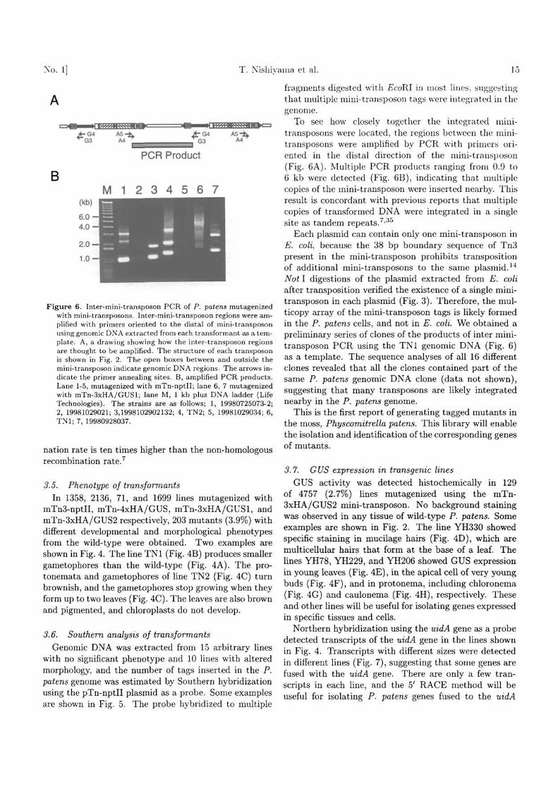
Figure 6. Inter-mini-transposon PCR of P. patens mutagenizedwith mini-transposons. Inter-mini-transposon regions were am-plified with primers oriented to the distal of mini-transposonusing genomic DNA extracted from each transformant as a tem-plate. A, a drawing showing how the inter-transposon regionsare thought to be amplified. The structure of each transposonis shown in Fig. 2. The open boxes between and outside themini-transposon indicate genomic DNA regions. The arrows in-dicate the primer annealing sites. B, amplified PCR products.Lane 1-5, mutagenized with mTn-nptll; lane 6, 7 mutagenizedwith mTn-3xHA/GUSl; lane M, 1 kb plus DNA ladder (LifeTechnologies). The strains are as follows; 1, 19980725073-2;2, 19981029021; 3,1998102902132; 4, TN2; 5, 19981029034; 6,TNI; 7, 19980928037.
nation rate is ten times higher than the non-homologousrecombination rate.7
3.5. Phenotype of transformantsIn 1358, 2136, 71, and 1699 lines mutagenized with
mTn3-nptII, mTn-4xHA/GUS, mTn-3xHA/GUSl, andmTn-3xHA/GUS2 respectively, 203 mutants (3.9%) withdifferent developmental and morphological phenotypesfrom the wild-type were obtained. Two examples areshown in Fig. 4. The line TNI (Fig. 4B) produces smallergametophores than the wild-type (Fig. 4A). The pro-tonemata and gametophores of line TN2 (Fig. 4C) turnbrownish, and the gametophores stop growing when theyform up to two leaves (Fig. 4C). The leaves are also brownand pigmented, and chloroplasts do not develop.
3.6. Southern analysis of transformantsGenomic DNA was extracted from 15 arbitrary lines
with no significant phenotype and 10 lines with alteredmorphology, and the number of tags inserted in the P.patens genome was estimated by Southern hybridizationusing the pTn-nptll plasmid as a probe. Some examplesare shown in Fig. 5. The probe hybridized to multiple
fragments digested with EcoRl in most lines, suggestingthat multiple mini-transposon tags were integrated in thegenome.
To see how closely together the integrated mini-transposons were located, the regions between the mini-transposons were amplified by PCR with primers ori-ented in the distal direction of the mini-transposon(Fig. 6A). Multiple PCR products ranging from 0.9 to6 kb were detected (Fig. 6B), indicating that multiplecopies of the mini-transposon were inserted nearby. Thisresult is concordant with previous reports that multiplecopies of transformed DNA were integrated in a singlesite as tandem repeats.7'35
Each plasmid can contain only one mini-transposon inE. coli, because the 38 bp boundary sequence of Tn3present in the mini-transposon prohibits transpositionof additional mini-transposons to the same plasmid.14
Not I digestions of the plasmid extracted from E. coliafter transposition verified the existence of a single mini-transposon in each plasmid (Fig. 3). Therefore, the mul-ticopy array of the mini-transposon tags is likely formedin the P. patens cells, and not in E. coli. We obtained apreliminary series of clones of the products of inter mini-transposon PCR using the TNI genomic DNA (Fig. 6)as a template. The sequence analyses of all 16 differentclones revealed that all the clones contained part of thesame P. patens genomic DNA clone (data not shown),suggesting that many transposons are likely integratednearby in the P. patens genome.
This is the first report of generating tagged mutants inthe moss, Physcomitrella patens. This library will enablethe isolation and identification of the corresponding genesof mutants.
3.7. GUS expression in transgenic linesGUS activity was detected histochemically in 129
of 4757 (2.7%) lines mutagenized using the mTn-3xHA/GUS2 mini-transposon. No background stainingwas observed in any tissue of wild-type P. patens. Someexamples are shown in Fig. 2. The line YH330 showedspecific staining in mucilage hairs (Fig. 4D), which aremulticellular hairs that form at the base of a leaf. Thelines YH78, YH229, and YH206 showed GUS expressionin young leaves (Fig. 4E), in the apical cell of very youngbuds (Fig. 4F), and in protonema, including chloronema(Fig. 4G) and caulonema (Fig. 4H), respectively. Theseand other lines will be useful for isolating genes expressedin specific tissues and cells.
Northern hybridization using the uidA gene as a probedetected transcripts of the uidA gene in the lines shownin Fig. 4. Transcripts with different sizes were detectedin different lines (Fig. 7), suggesting that some genes arefused with the uidA gene. There are only a few tran-scripts in each line, and the 5' RACE method will beuseful for isolating P. patens genes fused to the uidA

16 Tagged Mutagenesis and Gene-trap in Physcomitrella [Vol. 7,
(kb)
-1.3
(kb)
5.6
3.9
2.0
1.0
3
8wt
(kb)
- 3 . 5
- 2 . 3
- 1 . 0
(kb)
-5 .1
- 2 . 4
Figure 7. Northern blot analyses of the gene-trap lines shown inFig. 4 using the uidA gene probe. Each lane contains 0.5-1 /Jg ofpoly(A)+ RNA. Lane 1, YH330; lane 2, YH78; lane 3, YH229;and lane 4, YH206.
gene.Acknowledgements: We would like to thank P.
Ross-Macdonald (Yale Univ.), M. Snyder (Yale Univ.),A. Tone (Univ. Tokyo), K. Fujiwara (Univ. Tokyo), D. G.Schaefer (Univ. Lausanne), R. Kofuji, and K. Yamaguchi(Kanazawa Univ.) for kindly supplying materials used inthe yeast system or valuable technical suggestions, andT. Fujita for careful reading of this manuscript. The pro-tocol for the Physcomitrella culture was kindly providedby D. Cove (Univ. Leeds). Dr. J. Machuka constructedpMBL5 as part of "The Physcomitrella EST Programme(PEP)" at the University of Leeds (UK) and Washing-ton University in St. Louis (USA). We thank FutamuraChemical Industries Co., Ltd. for providing cellophaneand Kyowa Hakko Kogyo Co., Ltd. for Driserase. We aregrateful to M. Umeda, C. Ono, and Y. Bitou for theirtechnical help. T. N. is a research fellow of the JapanSociety for the Promotion of Science. This study waspartly supported by grants from the Ministry of Edu-cation, Science, Sports and Culture of Japan (TN, MK,and MH), and PRESTO, Japan Science and TechnologyCorporation (MH).
References
1. Walpert, L., Beddington, R., Brockes, J., Jessell, T.,Lawrence, P., and Meyerowitz, E. 1998, Principles of De-velopment, Oxford University Press, London.
2. Cove, D. J., Knight, C. D., and Lamparter, T. 1997,Mosses as model systems, Trends Plant Sci., 2, 99-105.
3. Schumaker, K. S. and Dietrich, M. A. 1997, Programmedchanges in form during moss development, Plant Cell, 9,1099-1107.
4. Reski, R. 1998, Development, Genetics and MolecularBiology of Mosses, Bot. Ada, 111, 1-15.
5. Schumaker, K. S. and Dietrich, M. A. 1998, Hormone-induced signaling during moss development, Annu. Rev.Plant Physiol. Plant Mol. Bioi, 49, 501-523.
6. Kammerer, W. and Cove, D. J. 1996, Genetic analysisof the effects of re-transformation of transgenic lines ofthe moss Physcomitrella patens., Mol. Gen. Genet., 250,380-382.
7. Schaefer, D. G. and Zryd, J.-P. 1997, Efficient gene tar-geting in the moss Physcomitrella patens, Plant J., 11,1195-1206.
8. Courtice, G. R. M. and Cove, D. J. 1983, Mutants ofthe moss, Physcomitrella patens which produce leaves ofaltered morphology, J. Bryol., 12, 595-605.
9. Cove, D. J. and Knight, C. D. 1987, In: Thomas,H., Grierson, D. (eds) Development Mutants of HigherPlants, Cambridge Univ. Press, Cambridge, pp. 181-196.
10. Featherstone, D. R., Cove, D. J., and Ashton, N. W.1990, Genetic analysis by somatic hybridization of cy-tokinin overproducing mutants of the moss, Mol. Gen.Genet., Ill, 217-224.
11. Ashton, N. W., Grimsley, N. H., and Cove, D. J.1979, Analysis of gametophytic development in the moss,Physcomitrella patens, using auxin and cytokinin resis-tant mutants., Planta, 144, 427-435.
12. Koncz, C, Nemeth, K., Redei, G. P., and Schell, J. 1992,T-DNA insertional mutagenesis in Arabidopsis thaliana,Plant Mol. Bioi, 20, 963-976.
13. Chuck, G., Robbins, T., and Nijiar, C. 1993, Taggingand cloning of a petunia flower color gene with the maizetransposable element, Plant Cell, 5, 371-378.
14. Seifert, H. S., So, M., and Heffron, F. 1986, In: Setlow, J.K., Hollaender, A. (eds) Genetic Engineering: Principlesand methods, Plenum Press, New York, pp. 123-133.
15. Seifert, H. S., Chen, E. Y., So, M., and Heffron, F. 1986,Shuttle mutagenesis: A method of transposon mutagen-esis for Saccharomyces cerevisiae, Proc. Natl. Acad. Sci.USA, 83, 735-739.
16. Burns, N., Grimwade, B., Ross-Macdonald, P. B. et al.1994, Large-scale analysis of gene expression, protein lo-calization, and gene disruption in Saccharomyces cere-visiae, Genes Dev., 8, 1087-1105.
17. Tan, B. C. 1979, A new classification for the genusPhyscomitrella B.S.G., J. Hattory Bot. Lab., 46, 327-336.
18. Ashton, N. W. and Cove, D. J. 1977, The isolation andpreliminary characterisation of auxotrophic and analogueresistant mutants in the moss Physcomitrella patens, Mol.Gen. Genet, 154, 87-95.
19. Knight, C. D., Cove, D. J., Boyd, P. J., and Ashton, N.W. 1988, In: Glime, J. M. (ed) Methods in Bryology,Hattori Botanical Laboratory, Miyazaki, Japan, pp. 47-58.
20. Schaefer, D. 1994, Molecular genetic approaches to thebiology of the moss Physcomitrella patens [PhD Thesis],University of Lausanne, (http://www.unil.ch/lpc/docs/DSThesis.htm)
21. Odell, J. T., Nagy, F., and Chua, N.-H. 1985, Identi-fication of DNA sequences required for activity of thecauliflower mosaic virus 35S promoter, Nature, 313, 810-812.
22. Beck, E., Ludwig, G., Auerswald, E. A., Reiss, B., and

No. 1] T. Nishiyama et al. 17
Schaller, H. 1982, Nucleotide seq\icnce and exact localiza-tion of the neomycin phosphotransferase gene from trans-poson Tn5, Gene, 19, 327-33G.
23. Guerineau, F., Brooks, L., Meadows, J., Lucy, A.,Robinson, C , and Mullineaux, P. 1990, Sulfonamide re-sistance gene for plant transformation, Plant Mol. Bioi,15, 127-136.
24. Franck, A., Guilley, H., Jonard, G., Richards, K., andHirth, L. 1980, Nucleotide sequence of cauliflower mosaicvirus DNA, Cell, 21, 285-294.
25. Ross-Macdonald, P., Sheehan, A., Roeder, G. S., andSnyder, M. 1997, A multipurpose transposon system foranalyzing protein production, localization, and functionin Saccharomyces cerevisiae, Proc. Natl. Acad. Sci. USA,94, 190-195.
26. Jefferson, R. A. 1987, Assaying chimeric genes in plants:the GUS gene fusion system, Plant Mol. Biol. Rep., 5,387-405.
27. Sundaresan, V., Springer, P., Volpe, T. et al. 1995, Pat-terns of gene action in plant development revealed by en-hancer trap and gene trap transposable elements, GenesDev., 9, 1797-1810.
28. Inoue, H., Nojima, H., and Okayama, H. 1990, High effi-ciency transformation of Escherichia coli with plasmids,
Gene, 96, 23-28.29. Murray, M. G. and Thompson. W. F. 1980, Rapid iso-
lation of high molecular weight plant DNA, Nucl. AcidsRes., 8, 4321-4325.
30. Sainbrook, J., Fritch, E. F., and Maniatis, T. 1989,Molecular cloning: A Laboratory Manual, 2nd Ed., ColdSpring Harbor Laboratory, Cold Spring Harbor, NewYork.
31. Jefferson, R. A., Kavanagh, T. A., and Bevan, M. W.1987, GUS fusions: /3-glucuronidase as a sensitive andversatile gene marker in higher plants, EMBO J., 6,3901-3907.
32. Hasebe, M., Wen, C.-K., Kato, M., and Banks, J. A.1998, Characterization of MADS homeotic genes in thefern Ceratopteris richardii, Proc. Natl. Acad. Sci. USA,95, 6222-6227.
33. Chomczynski, P. 1992, One-hour downward alkaline cap-illary transfer for blotting of DNA and RNA, Anal.Biochem., 201, 134-139.
34. Church, G. M. and Gilbert, W. 1984, Genomic sequenc-ing., Proc. Natl. Acad. Sci. USA, 81, 1991-1995.
35. Schaefer, D., Zryd, J.-P., Knight, C. D., and Cove, D. J.1991, Stable transformation of the moss Physcomitrellapatens, Mol. Gen. Genet., 226, 418-424.
