t2 & t2# maps: sequence development and clinical impact on … · submitted to epos by third...
TRANSCRIPT

Page 1 of 12
T2 & T2# maps: Sequence Development and Clinical Impacton Joint Study
Poster No.: C-2444
Congress: ECR 2012
Type: Scientific Exhibit
Authors: C. Sirignano1, E. Soscia1, D. Iodice2, S. Innocenti3, G. Palma1, D.
Greco3, L. Balbi3, B. Alfano1, M. Salvatore2; 1Napoli (NA), italia/IT,2Napoli/IT, 3Genova (GE)/IT
Keywords: Transplantation, Athletic injuries, Arthritides, Diagnostic procedure,Image manipulation / Reconstruction, Musculoskeletal joint
DOI: 10.1594/ecr2012/C-2444
Any information contained in this pdf file is automatically generated from digital materialsubmitted to EPOS by third parties in the form of scientific presentations. Referencesto any names, marks, products, or services of third parties or hypertext links to third-party sites or information are provided solely as a convenience to you and do not inany way constitute or imply ECR's endorsement, sponsorship or recommendation of thethird party, information, product or service. ECR is not responsible for the content ofthese pages and does not make any representations regarding the content or accuracyof material in this file.As per copyright regulations, any unauthorised use of the material or parts thereof aswell as commercial reproduction or multiple distribution by any traditional or electronicallybased reproduction/publication method ist strictly prohibited.You agree to defend, indemnify, and hold ECR harmless from and against any and allclaims, damages, costs, and expenses, including attorneys' fees, arising from or relatedto your use of these pages.Please note: Links to movies, ppt slideshows and any other multimedia files are notavailable in the pdf version of presentations.www.myESR.org

Page 2 of 12
Purpose
T2- and T2 #- maps are quantitative images that, by the measurement of relaxation times,give the amount of water content of tissue. In the specific case of cartilage, the presenceof water has an inverse correlation with the presence of proteoglicans, the fundamentalconstituents of cartilage itself.The value of T2-maps on high-field MRI apparati hasalready been assessed in literature.
We tested a 3D Steady State and a 3D Gradient echo on a low field dedicated MRIequipment and correlated our observations with 3D dedicated commercial sequencesand with the results obtained by the arthroscopic evaluation.
Methods and Materials
We studied 36 patients scheduled for arthroscopy on a dedicated MRI system(EsaoteG-Scan - 0.25 T) with two optimized Steady-State Free-Precession (Sharc and SST1)sequences (fig.1). The related signal equations were inverted voxel-by-voxel to obtainquantitative T1- and T2-maps (fig.2-3) of the whole joint.
The T2#-map of the joint was derived as harmonic mean of T1 and T2. Sharc, T2- andT2#-maps were evaluated in consensus by two radiologists, on an Osirix DICOM PACSworkstation, at fixed range scales to find cartilage defects.
The number of focal or diffuse cartilage alterations detected by MRI images by meansof each kind of map was compared to the number cartilage defects found by Sharcsequence and the alterations found by surgeons during arthroscopy.
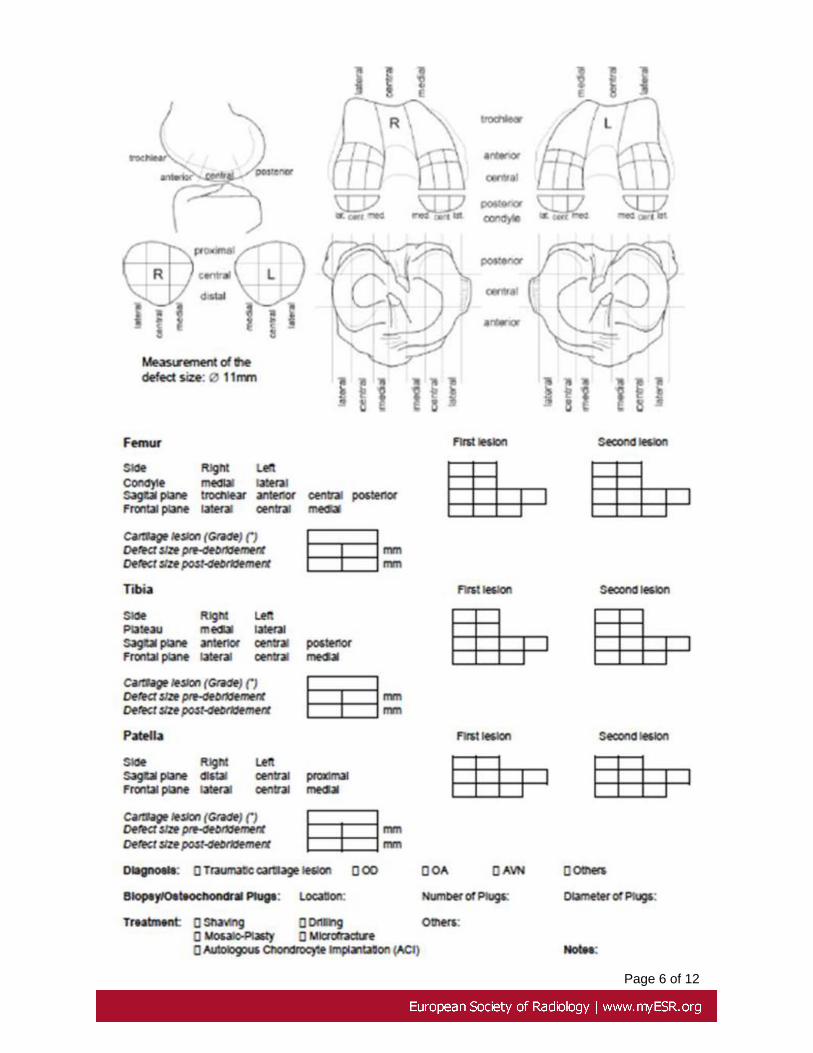
To standardize the evaluation and the localization of alteration found, radiologists andorthopedics compiled a standardized evaluation form (table 1). In this kind of form the jointcartilage is subdivided in sectors and the defects is quantified in extension and tipology(e.g. full thickness defect, superficial fibrillation, etc.).
Images for this section:

Page 3 of 12
Fig. 1: 3D SHARC

Page 4 of 12
Fig. 2: T2 MAP

Page 5 of 12
Fig. 3: T2rho MAP
Page 6 of 12

Page 7 of 12
Table 1: Evaluation form

Page 8 of 12
Results
As for the evaluation of diffuse cartilage alterations, Sharc, T2-maps and T2#-maps werepositive in 8, 41 and 35 cases, respectively. Moreover Sharc revealed 16, T2-mapsrevealed 68 and T2#-maps revealed 71 focal cartilage alterations.
The arthroscopy surgeons found 73 focal cartilage lesions, so the correlation was poorfor the SHARC images (20% of agreement) and good with T2- and T2 #-maps (81- 95%of agreement).
Moreover, when a the lesion was found, there was a good correlation in the spacelocalization between MRI (both Sharc and maps) and arthroscopy.
Conclusion
Our initial results show that both T2- and T2#-maps provide good extra information on thehealth status of knee cartilage, even in absence of morphological evidence of alterationon native 3D images; in particular, T2#-maps appeared more sensitive than T2-ones.
Our data, confirmed by orthopaedics' arthroscopy, open new functional possibility forthe dedicated MRI equipments in the cartilage defects evaluation, allowing an earlydiagnosis and a accurate follow up with new therapeutic opportunities. In addition theinformation given by the MRI, can selectively guide the orthopaedic to test a specificregion of cartilage and, eventually, to repair it.
Images for this section:

Page 9 of 12
Fig. 4: 3D SHARC
Fig. 5: T2 MAP

Page 10 of 12
Fig. 6: T2rho

Page 11 of 12
References
• Astrid Pinzano, Pierre Ruaud, Pierre Olivier. Effect of ProteoglycanDepletion on T2 Mapping in Rat Patellar Cartilage. Radiology 2005;234:162-170
• Iwan Van Breuseghem, Hilde T. C. Bosmans, Guy J. Marchal. T2 Mappingof Human Femorotibial Cartilage with Turbo Mixed MR Imaging at 1.5 T:Feasibility. Radiology 2004; 233:609-614
• Tallal C. Mamisch , Siegfried Trattnig , Goetz H. Welsch. Control Cartilageand Cartilage Repair Tissue in the Knee with Unloading. Radiology2010;354:818-826
• Dardzinski Bernard, Mosher Timothy, Li Shizhe et al. Spatial Variation of T2in human articular cartilage. Radiology 1997; 205:546-550
• Liess C., Lusse S. , Karger N., Heller M et al. Detection of changes incartilage water content using MRI T2-mapping in vivo Osteoarthritis andCartilage 2002; 10: 907-913
• Gold Garry, Reeder Scott, Yu Huanzhou et al. Articular Cartilage of theKnee: Rapid Three-dimensional MR Imaging at 3.0 T with IDEAL BalancedSteady-State Free Precession-Initial Experience. Radiology 2006; 240:546-551
• Mosher Timothy, Dardzinski Bernard, Smith Michael. Human articularcartilage: Influence of aging and early symptomatic degeneration on thespatial variation of T2- preliminary fingings at 3T. Radiology 2000; 214:259-266.
• Deoni Sean, Rutt Brian, Peters Terry. Rapid combined T1 and T2 mappingusing gradient recalled acquisition in the steady state. Magnetic Resonancein Medicine 2003 49:515-526
• Timothy Dunn, Ying Lu, Hua Jin et al. T2 relaxation time of cartilage at MRImaging:Comparison with severity of knee osteoarthritis. Radiology 2004;232:592-598
• Li Xiaojuan, Ma Benjamin, Link Thomas et al. In vivo T1Rho and T2mapping of articular cartilage in osteoarthritis of the knee using 3 tesla MRI.Osteoarthritis Cartilage. 2007 July ; 15: 789-797
• Michel Crema, Frank Roemer, Monica Marra et al. Articular cartilage in theknee: Current MR Imaging techniques and applications in clinical practiceand research. RadioGraphics 2011; 31:37-62
Personal Information
Cesare Sirignano,Ernesto Soscia, Delfina Iodice, Giuseppe Palma, Bruno Alfano,Marco Salvatore.

Page 12 of 12
Istituto di Biostrutture e Bioimmagini CNR e Dipartimento di Diagnostica per Immagini,Università degli Studi di Napoli "Federico II". Via S. Pansini n°5. 80131 Napoli.
Mail to: [email protected]
Stefania Innocenti, Danilo Greco, Luca Balbi.
Esaote S.p.A., Via Angelo Siffredi 58, 16153 Genova, Italy.