synthesis, structural study and spectroscopic ...file.scirp.org/pdf/ojsta_2013012810182997.pdfopen...
TRANSCRIPT

Open Journal of Synthesis Theory and Applications, 2013, 2, 8-32 http://dx.doi.org/10.4236/ojsta.2013.21002 Published Online January 2013 (http://www.scirp.org/journal/ojsta)
Synthesis, Structural Study and Spectroscopic Characterization of a Quinolin-8-Yloxy Derivative with
Potential Biological Properties
Elida Romano1, María V. Castillo1, Jorgelina L. Pergomet2, Juan Zinczuk2, Silvia A. Brandán1* 1Faculty of Biochemistry, Chemistry and Pharmacy of the National University of Tucumán, Tucumán, R. Argentina
2Chemistry Rosario Institute (CONICET-UNR), Faculty of Biochemistry and Pharmacists Sciences, Rosario, R. Argentina Email: *[email protected]
Received November 14, 2012; revised December 16, 2012; accepted December 25, 2012
ABSTRACT
We have prepared the (5-chloro-quinolin-8-yloxy) acetic acid and characterized it by using infrared, Raman and multi- dimensional nuclear magnetic resonance spectroscopies. The density functional theory (DFT) together with the 6-31G* and 6-311++G** basis sets were used to study its structure and vibrational properties. Three stable conformations of the compound were theoretically determined in gas phase and probably these conformations are present in the solid phase. The harmonic vibrational wavenumbers for the optimized geometries were calculated at the same theory levels. For a complete assignment of the observed bands in the vibrational spectra, the DFT calculations were combined with Pulay’s scaled quantum mechanical force field (SQMFF) methodology in order to fit the theoretical wavenumber values to the experimental ones. Besides, the force constants of the three conformers of (5-chloro-quinolin-8-yloxy) acetic acid were calculated and compared with those obtained by us for the 2-(quinolin-8-yloxy) acetic acid. In addition, the characteris- tics of the electronic delocalization of those structures were performed by using natural bond orbital (NBO), while the corresponding topological properties of electronic charge density are analysed by employing Bader’s atoms in mole- cules theory (AIM). Keywords: (5-Chloro-Quinolin-8-Yloxy) Acetic Acid; Vibrational Spectra; Molecular Structure; Force Field; DFT
Calculations
1. Introduction
Heterocyclic compounds that contain the (quinolin-8- yloxy) moiety exhibit a wide range of biological proper- ties [1-5], such as the 2-(quinolin-8-yloxy) acetohydra- zones that have antiamoebic activities [5] for which, the structural and vibrational study of these types of com- pounds are of great chemical and pharmaceutical impor- tance. Recently, a complete vibrational analysis of all observed bands in the vibrational spectra of 2-(quinolin- 8-yloxy)-acetic acid [6] was performed by means of the DFT calculations combined with Pulay’s scaled quantum mechanical force field (SQMFF) methodology [7-9]. In that study, the normal mode calculations were accom- plished by using a generalized valence force field (GVFF) [8,9] considering three different structures for the anhy- drous and monohydrated compounds. In the present work, we report an experimental and theoretical vibrational study of (5-chloro-quinolin-8-yloxy) acetic acid (CQA) by means of the B3LYP calculations using 6-31G* and
6-311++G** basis sets. For a complete assignment of the compound, the DFT calculations were combined with the SQMFF methodology [7-9] in order to fit the theoretical wavenumber values to the experimental ones. So far, the crystal and molecular structure of (5-chloro-quinolin-8- yloxy) acetic acid has not been determined. First, we have synthesized and characterized the compound and then, the optimized geometries and frequencies for the normal modes of vibration of CQA considering three different stable structures were calculated in order to carry out a complete assignment of all observed bands in the infrared and Raman spectra. Here, the normal mode calculations and the force fields for those structures were obtained by using the transferable scaling factors of Rauhut and Pulay [9] while the harmonic force constants were subsequently scaled to reproduce as well as possi- ble the experimental frequencies. Thus, a complete as- signment of the compound in terms of the potential en- ergy distribution was performed. Furthermore, when the forces constants values of this compound were compared with those corresponding to the 2-(quinolin-8-yloxy)- *Corresponding author.
Copyright © 2013 SciRes. OJSTA

E. ROMANO ET AL. 9
acetic acid [6] numerous changes in the structural, topo- logical and vibrational properties attributed to the chloro atom were observed. Additionaly, the nature of the dif- ferent rings and bonds of the three studied structures of (5-chloro-quinolin-8-yloxy) acetic acid were analyzed by means of the NBO studies [10-12] while the topological properties of electronic charge density were determined employing the Bader’s atoms in molecules theory (AIM) [13].
2. Experimental Methods
2.1. Synthesis
(5-chloroquinolin-8-yloxy) acetic acid was obtained ac- cording to Cho et al. [1] using the following procedure.
2.1.1. (5-Chloroquinolin-8-Yloxy) Acetic Acid Methyl Ester
A mixture of 5-chloro 8-hydroxyquinoline (3.6 g, 20 mmol), methyl bromoacetate (3.7 g, 24 mmol) and K2CO3 (5.52 g, 40 mmol) in acetone (50 mL) was heated for 3 h under reflux, filtered and concentrated in vacuo. The residue was partitioned between ethyl acetate and brine, and the organic layer was dried with MgSO4, fil- tered, and concentrated in vacuo. The residue was puri- fied by crystallization from isopropyl ether to give (5-chloroquinolin-8-yloxy) acetic acid methyl ester 3584 g (71%) m.p. 102.1˚C - 102.3˚C.
2.1.2. (5-Chloroquinolin-8-Yloxy) Acetic Acid (CQA) A mixture of ester ((1.26 g, 5 mmol) and LiOH·H2O (352 mg, 8.40 mmol) in 75 mL of THF/CH3OH/H2O (1:1:1, 6) was stirred and heated at reflux for 1 h and concentrated. The aqueous layer was washed with ether and adjusted to pH 3 with 1 N HCl. The precipitate was filtered and dried to give (5-chloroquinolin-8-yloxy) acetic acid (1085 g, 91%) m.p. 219.5˚C - 221˚C.
2.2. NMR Spectra
1H NMR (300 MHz, CDCl3) δ: 4.92 (2H, s, O-CH2); 7.08 (1H, d, J = 8.48, H7), 7.64 (1H, d, J = 8.48, H6); 8,47 (1H, d, J = 8.58, H4); 7.70 (1H, dd, J = 4.18, J = 8.58, H3); 8.94 (1H, d, J = 4.18, H2).
13C NMR (75 MHz, CDCl3) δ: 65.94 (CH2); 110.54 (C7); 121.69 (C5); 123.53 (C3); 126.66 (C4a); 127.07 (C6); 132.64 (C4); 140.55 (C8a); 150.28(C2); 153.54 (C8); 170.34 (CO2H) (Atom numbering according to naphtha- lene).
2.3. Infrared and Raman Spectra
The infrared spectrum of the solid substance was re- corded in KBr pellets in the wavenumbers range from 4000 to 400 cm−1 with a FT-IR Perkin Elmer spectrome-
ter, provided with a Globar source and a DGTS detector. FT-Raman spectrum of the crystalline solid was recorded on a Bruker RFA 106/S FT-Raman instrument using the 1064 nm excitation line from an Nd: YAG laser in the region of 4000 - 0 cm−1. Two hundred scans were accu mulated at 4 cm−1 resolution using a laser power of 150 mW.
3. Computational Details
The potential energy curves associated with the internal rotation described by the C21-C18-O17-C10 dihedral angle for CQA were studied at the B3LYP/6-31G* and 6-311++G** theory levels. With both calculations, two conformations stable, named, CI and CII, according to the position of the OH group in relation to the N atom of the ring were obtained. Another plane structure was also considered (CIII) in agreement with the experimental structure of the 8-(carboxymethoxy) quinolinium nitrate monohydrate compound [14]. The structures and labell- ing of the atoms for all conformers of CQA can be seen in Figure 1. The electronic charge density topological analysis for those structures were performed by using the AIM200 program package [15] while the NBO calcula- tions were obtained by means of the NBO 3.1 [16] pro- gram, as implemented in the GAUSSIAN 03 package [17]. The natural internal coordinates for the compound have been defined as those reported in the literature [7,8,18] and are listed in the Supporting Material as Ta- ble S1. The resulting force fields were transformed to “natural” internal coordinates by using the MOLVIB program [19,20]. Then, following the SQMFF procedure [7-9,21], the harmonic force fields for those structures were evaluated at the B3LYP/6-31G* level. The poten- tial energy distribution components (PED) higher than or equal to 10% were subsequently calculated with the re- sulting SQMFF. The nature of all vibration modes was carried out by means of the Gauss View program [22].
Figure 1. Theoretical structures and labelling of the atoms for the stable CI, CII and CIII conformers of (5-chloro-qui- nolin-8-yloxy) acetic acid.
Copyright © 2013 SciRes. OJSTA

E. ROMANO ET AL. 10
4. Results and Discussion
4.1. Geometry
Table S2 (Supporting Material) shows the comparison of the total energies for the compound, and the correspond- ing dipole moment values for the CI, CII and CIII con- formers of C1 symmetries. With both methods, the en- ergy of the CI is lower than the other ones, as was ob- served in the compound without chlorine [6], and the potential energy difference between the CI and CII forms, by using the B3LYP/6-31G* and B3LYP/6-311++G** methods, are −43.80 and −46.43 kJ/mol, respectively. When the CI conformer is compared with the planar ones the potential energy difference values slightly by using both basis sets decrease up to −27.80 and −32.36 kJ/mol, respectively.
Also, in this case, the high values of the dipolar mo- ments for the CI structures could probably explain its stabilities, as was observed in similar molecules [6,23-25].
Table 1 shows a comparison of the calculated ge- ometrical parameters for all structures of CQA, with the ones corresponding to that observed from X-ray diffrac- tion for the 8-(carboxymethoxy) quinolinium nitrate monohydrate compound [14] by means of the root mean of square deviations (rmsd) values. Note that the 6-311++G** basis set reproduce reasonably well the theoretical bond lengths for the CI structure (0.019 Å) and the bond angles for the CIII structure (2.00˚), as is expected because this last conformer is planar as that compared compound [14]. Besides, the calculation pre- dicts that the CI and CIII conformers are the most stable and probably both are present in the crystalline state.
The stabilities of the CII and CIII structures of CQA, in relation to the CI structure, were investigated by using the natural atomic charges [26-29] and the results are given in Table S3. Note that the natural charges values (NPA) corresponding to the C21 atoms have the most positive values in all structures while the most negative values correspond to the O23 atoms. In this compound, as in 2-(quinolin-8-yloxy)-acetic acid [6], the higher NPA charges observed on the O17 and O22 atoms in the CII structure justified the repulsion between those atoms. This way, the CII structure is more unstable than the cor- responding to the CI and CIII conformers. The bond or- ders, expressed by Wiberg’s index for all conformers of CQA are given in Table S4. The bond order values for the O23 atoms in all conformers have the lowest values (belong to the COO groups) than the other ones (the O17 and O22 atoms belongs to the heterocyclic rings), while the bond orders for the N16 atoms have higher values.
4.2. NBO Study
The stability of the three structures of CQA was also
investigated by means of NBO calculations [10-12]. The second order perturbation energies E(2) (donor accep- tor) that involve the most important delocalization all conformers of CQA are given in Tables S5, S6. The contributions of the stabilization energies for the ET* charge transfers are similar to those obtained for the 2-(quinolin-8-yloxy)-acetic acid compound [6].
Here, the delocalizations ETLP* for the CII structure has lower values than the ET* delocalizations, and the calculated total energy values favours to the CI and CIII conformers, which structures are the most stable in the gas phase. Thus, the total energy values clearly show the higher stability of the CI conformer however, by us- ing the 6-311++G** basis set, the CII and CIII conform- ers have the same ones approximately total energy values.
4.3. AIM Analysis
Furthermore, the three structures of CQA were analysed by means of Bader’s charge electron density topological analysis [13]. The calculated electron density, () and the Laplacian values, 2(r) in the bond critical points (BCPs) and ring critical points (RCPs) for those struc- tures are shown in Table S7. The BCP has the typical properties of the closed–shell interaction and for this, the value of ρ(r) is relatively low, the relationship |1|/3 is <1 and 2(r) is positive indicating that the interaction is dominated by the charge contraction away from the in- teratomic surface toward each nucleus. Note that the re- sults are clearly dependent of the size basis set. Thus, the analysis shows two BCPs and RCPs in the CI structure when the 6-31G* basis set is employed but, only one BCP and RCP when the 6-311++G** basis set is used. On the contrary, for the CII structure with both basis sets only one BCP and RCP are observed while for the CIII structure there is one BCP with the 6-31G* basis set. These results, togheter to the NBO analysis, justify the stabilities of the CI and CII structures due to the presence of short intramolecular O17-H24 and N16-H24 bonds, in the first case and only a N16-H19 bonds in the sec- ond one, as observed in Table S7.
4.4. Vibrational Analysis
The Figures 2, 3 show the registered infrared and Raman spectra for the compound in solid phase. In this study, in accordance with the NBO and AIM results, the three structures for the compound in gas phase were consid- ered. The CQA’s structures have C1 symmetries and 66 normal vibration modes, all active in the infrared and Raman spectra. Probably, the three species are present in the solid phase because the comparison of each vibra- tional spectrum with the corresponding experimental one is very different among them, as observed in Figure 4, however, a comparison between the average calculated
Copyright © 2013 SciRes. OJSTA

E. ROMANO ET AL.
Copyright © 2013 SciRes. OJSTA
11
Table 1. Calculated geometrical parameters for the conformers of (5-chloro-quinolin-8-yloxy) acetic acid.
a6-31G* a6-311++G** Parameter
CI CII CIII CI CII CIII
bExp.
Bond length (Å)
C3-C4 1.432 1.437 1.434 1.430 1.434 1.432 1.402 (3)
C4-N16 1.362 1.363 1.357 1.360 1.365 1.354 1.368 (2)
C5-N16 1.319 1.318 1.318 1.317 1.318 1.315 1.317 (2)
C4-C10 1.429 1.431 1.439 1.427 1.431 1.437 1.411 (3)
C9-Cl6 1.758 1.762 1.763 1.758 1.758 1.763
C10-O17 1.367 1.374 1.357 1.367 1.377 1.356 1.362 (2)
C18-O17 1.433 1.413 1.407 1.432 1.412 1.406 1.424 (2)
C18-C21 1.537 1.531 1.520 1.535 1.530 1.518 1.498 (3)
C21-O22 1.210 1.202 1.203 1.205 1.202 1.196 1.203 (2)
C21-O23 1.331 1.362 1.358 1.329 1.363 1.359 1.303 (2)
RMSD 0.020 0.027 0.025 0.019 0.027 0.025
Bond angle (degrees)
C4-N16-C5 118.7 118.4 118.2 118.9 118.2 118.5 122.98 (19)
C4-C10-C12 119.9 120.1 119.8 119.9 120.3 119.7 118.90 (19)
C4-C10-O17 117.6 121.8 115.3 117.8 121.8 115.5 114.39 (17)
C12-C10-O17 122.4 117.9 124.9 122.2 117.7 124.8 126.71 (18)
C10-O17-C18 119.2 118.5 117.8 119.7 118.4 118.0 117.04 (15)
O17-C18-C21 112.5 112.3 108.6 113.1 112.4 109.0 108.78 (16)
C18-C21-O23 117.4 114.0 108.8 117.7 113.9 108.7 110.57 (19)
C18-C21-O22 119.3 125.2 127.2 119.2 125.3 127.4 125.03 (19)
O22-C21-O23 123.3 120.8 124.0 123.0 120.8 123.9 124.40 (2)
RMSD 4.05 4.65 2.01 4.24 4.71 2.00
Dihedral angles (degrees)
C21-C18-O17-C10 −112.8 −76.7 −180.0 −112.4 −76.7 −180.0 −178.52 (17)
C4-C10-O17-C18 110.4 −63.4 180.0 107.8 −62.0 180.0 176.72 (17)
C12-C10-O17-C18 −73.2 121.9 0.0 −76.4 123.3 0.0 −3.3 (3)
RMSD 67.3 167.0 2.8 69.4 166.7 2.8
infrared spectra (from B3LYP/6-31G* level for the CI, CII and CIII conformers) by using average wavenumbers
and intensities with the corresponding experimental one demonstrate a good correlation, as observed in Figure S1.
E. ROMANO ET AL. 12
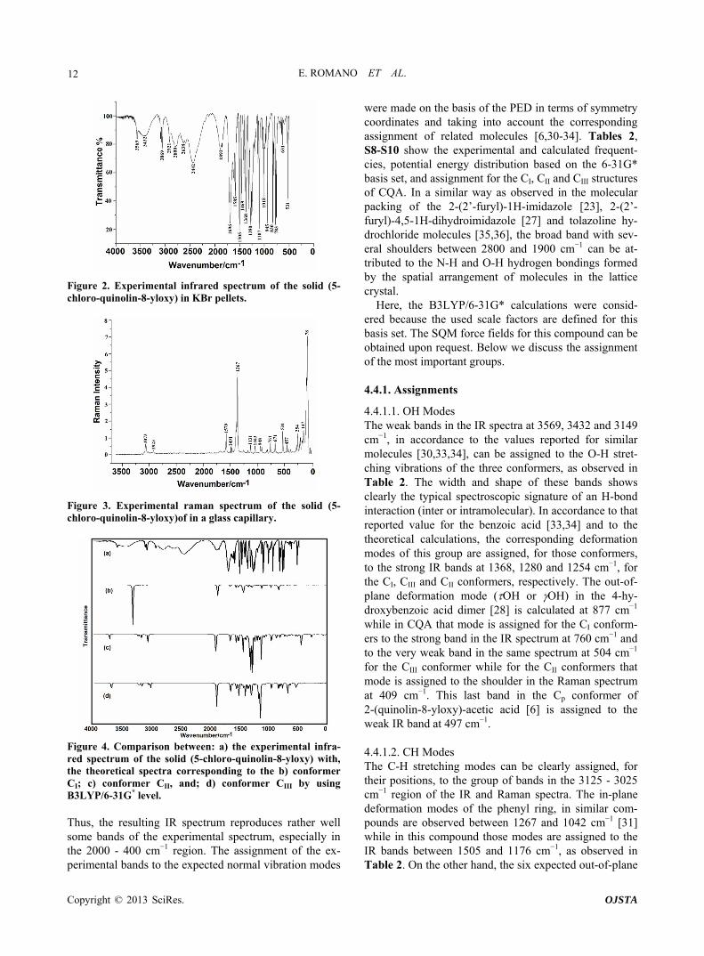
Figure 2. Experimental infrared spectrum of the solid (5- chloro-quinolin-8-yloxy) in KBr pellets.
Figure 3. Experimental raman spectrum of the solid (5- chloro-quinolin-8-yloxy)of in a glass capillary.
Figure 4. Comparison between: a) the experimental infra- red spectrum of the solid (5-chloro-quinolin-8-yloxy) with, the theoretical spectra corresponding to the b) conformer CI; c) conformer CII, and; d) conformer CIII by using B3LYP/6-31G* level. Thus, the resulting IR spectrum reproduces rather well some bands of the experimental spectrum, especially in the 2000 - 400 cm−1 region. The assignment of the ex- perimental bands to the expected normal vibration modes
were made on the basis of the PED in terms of symmetry coordinates and taking into account the corresponding assignment of related molecules [6,30-34]. Tables 2, S8-S10 show the experimental and calculated frequent- cies, potential energy distribution based on the 6-31G* basis set, and assignment for the CI, CII and CIII structures of CQA. In a similar way as observed in the molecular packing of the 2-(2’-furyl)-1H-imidazole [23], 2-(2’- furyl)-4,5-1H-dihydroimidazole [27] and tolazoline hy- drochloride molecules [35,36], the broad band with sev- eral shoulders between 2800 and 1900 cm−1 can be at- tributed to the N-H and O-H hydrogen bondings formed by the spatial arrangement of molecules in the lattice crystal.
Here, the B3LYP/6-31G* calculations were consid- ered because the used scale factors are defined for this basis set. The SQM force fields for this compound can be obtained upon request. Below we discuss the assignment of the most important groups.
4.4.1. Assignments
4.4.1.1. OH Modes The weak bands in the IR spectra at 3569, 3432 and 3149 cm−1, in accordance to the values reported for similar molecules [30,33,34], can be assigned to the O-H stret- ching vibrations of the three conformers, as observed in Table 2. The width and shape of these bands shows clearly the typical spectroscopic signature of an H-bond interaction (inter or intramolecular). In accordance to that reported value for the benzoic acid [33,34] and to the theoretical calculations, the corresponding deformation modes of this group are assigned, for those conformers, to the strong IR bands at 1368, 1280 and 1254 cm−1, for the CI, CIII and CII conformers, respectively. The out-of- plane deformation mode (OH or OH) in the 4-hy- droxybenzoic acid dimer [28] is calculated at 877 cm−1 while in CQA that mode is assigned for the CI conform- ers to the strong band in the IR spectrum at 760 cm−1 and to the very weak band in the same spectrum at 504 cm−1 for the CIII conformer while for the CII conformers that mode is assigned to the shoulder in the Raman spectrum at 409 cm−1. This last band in the Cp conformer of 2-(quinolin-8-yloxy)-acetic acid [6] is assigned to the weak IR band at 497 cm−1.
4.4.1.2. CH Modes The C-H stretching modes can be clearly assigned, for their positions, to the group of bands in the 3125 - 3025 cm−1 region of the IR and Raman spectra. The in-plane deformation modes of the phenyl ring, in similar com- pounds are observed between 1267 and 1042 cm−1 [31] while in this compound those modes are assigned to the IR bands between 1505 and 1176 cm−1, as observed in Table 2. On the other hand, the six expected out-of-plane
Copyright © 2013 SciRes. OJSTA

E. ROMANO ET AL.
Copyright © 2013 SciRes. OJSTA
13
Table 2. Observed and calculated wavenumbers (cm−1) and assignment for the (5-chloro-quinolin-8-yloxy) acetic acid.
Experimentala CI CII CIII
IR solid Raman solid SQMb Assignment SQMb Assignment SQMb Assignment
3569 w 3558 (O23-H24)
3432 w,br 3541 (O23-H24)
3149 vvw 3145 vvw 3175 (O23-H24)
3125 vvw 3130 vvw 3100 (C2-H8) 3109 (C12-H14) 3102 (C12-H14)
3100 w 3096 (C13-H15) 3097 (C2-H8) 3096 (C2-H8)
3098 vvw 3085 (C1-H7) 3092 (C13-H15) 3086 (C13-H15)
3069 w 3073 w 3081 (C12-H14) 3079 (C1-H7) 3076 (C1-H7)
3057 w 3056 (C5-H11)
3025 vvw 3028 vw 3036 (C5-H11) 3037 (C5-H11)
3006 vvw 3006 a CH2
2981 vw 2974 a CH2
2966 vw 2946 s CH2
2943 vvw 2931 s CH2 2931 a CH2
2921 vw 2923 vw 2895 s CH2
1892 m,br 1823 (C21-O22)
1854 m 1814 (C21-O22)
1696 s 1687 vw 1792 (C21-O22)
1626 m 1646 vw 1613 (C2-C3) 1613 (C2-C3) 1614 (C2-C3)
1608 m 1610 vw 1599 (C1-C2) 1600 (C1-C2) 1601 (C13-C9)
1585 m 1579 m 1575 (C13-C9) 1570 (C13-C9) 1572 (C13-C12)
1505 vs 1491 vw 1509 C1-H7 1508 C1-H7 1513 C1-H7
1469 s 1470 vw 1482 C5-H11 1480 C5-H11 1485 C5-H11
1431 vw 1446 CH2 1453 CH2
1423 sh 1433 CH2 1431 wag CH2
1417 m 1408 C2-H8 1408 C2-H8
1395 sh 1398 m 1402 (C5-N16) 1403 (C5-N16) 1407 (C10-C4)
1386 m 1386 m 1374 wag CH2 1390 wag CH2 1405 C2-H8
1368 s 1367 s 1353 OH 1350 (C3-C4), (C12-C10)
1324 s 1340 (C3-C4) 1336 (C3-C4) 1320 (C10-O17)
1317 vvw 1305 (C12-C10)
1280 s 1278 vvw 1305 (C12-C10) 1279 CH2 1289 OH
1254 s 1255 vw 1269 CH2 1254 OH 1259 (C4-N16)
1251 sh 1253 w 1251 (C4-N16) 1250 (C4-N16) 1252 1251
(C5-N16) CH2
1238 sh 1238 (C10-C4) 1234 (C10-C4)
1219 vw 1221 (C21-O23) 1212 C13-H15

E. ROMANO ET AL. 14
Continued
1202 w 1202 vvw 1207 C13-H15, (C3-C9)
1207 C13-H15, (C3-C9)
1176 vw 1174 vw 1166 C12-H14 1167 C12-H14 1180 C12-H14
1133 m 1131 w 1131 R 1 (A2) 1136 R 1 (A2) 1143 (C1-C2)
1107 s 1105 vvw 1083 (C13-C12) 1112 1086
(C21-O23) (C13-C12)
1128
1104
(C13-C12), OH,
(C21-O23)
1042 vw 1043 w 1037 (C1-C5) 1036 (C1-C5) 1035 (C1-C5)
1018 m 1012 vvw 1029 (C18-O17) 1029 (C18-O17)
996 w 998 (C18-O17)
981 w 993 C1-H7 991 C2-H8 991 C2-H8
954 sh 967 sh 963 C5-H11 967 w CH2 962 w CH2
948 sh 948 vw 961 w CH2 960 C5-H11 959 C5-H11
945 s 937 C13-H15 939 C12-H14 926 R1 (A2)
913 vw 919 vw 925 (C9-Cl6) 921 R 1 (A1) 915 C13-H15
836 sh 851 (C18-C21) 847 C13-H15 884 (C18-C21)
830 s 833 vw 839 C12-H14 838 (C18-C21) 818 R3 (A2)
820 m 817 vw 817 R 1 (A1) 816 R 3 (A2) 818 C12-H14
806 m 812 C2-H8 809 C1-H7 809 C1-H7
783 s 785 vw 778 R1 (A2) 774 R1 (A2) 777 R1 (A1)
760 s 761 w 737 OH 742 R 1 (A1)
701 vw 720 (C10-O17)
680 vw 699 (C10-O17)
671 w 665 R 2 (A1) 666 C10-O17 655 R 2 (A1)
654 vw 655 vvw 649 C10-O17 650 R 2 (A1)
631 w 633 vvw 624 COO 624 R1 (A1) 632 620
R1 (A2) COO
613 vw 613 vvw 619 COO
606 vw 600 vvw 601 R1 (A1) 595 C9-Cl6 597 R 2 (A2)
590 vw 572 R 3 (A2) 569 C10-O17, R 2 (A2)
585 C10-O17
531 m 531 m 534 COO 544 COO
520 vw 522 sh 528 R 2 (A2) 522 R 2 (A2) 526 (C3-C9)
504 vw 503 R 3 (A1) 490 COO 508 495
R 3 (A1) OH
488 vvw 469 R3 (A1) 480 R 3 (A1)
457 m 457 R2 (A1) 460 R2 (A1) 463 R2 (A2)
431 vw 415 R3 (A2) 439 COO
409 sh 416 R3 (A2) 399 OH 418 R3 (A2)
386 vw 388 C9-Cl6 385 (C9-Cl6) 378 (C9-Cl6)
356 vvw 335 COO 330 COO 346 C9-Cl6
278 w 312 C10-O17 314 CCO
Copyright © 2013 SciRes. OJSTA

E. ROMANO ET AL.
Copyright © 2013 SciRes. OJSTA
15
Continued
254 s 240 CCO 254 COC 252 C9-Cl6
216 w 225 C9-Cl6 219 C9-Cl6 223 C10-O17
208 m 207 COC 196 C10-O17 199 CCO
173 w 168 Butt 174 Butt 188 Butt
147 m 135 R2 (A2) 123 R2 (A2) 135 R3 (A1)
92 s 88 CCOC,
w COO 102 R3 (A1)
116 109
w Ring R2 (A1)
78 vs 87 w COO 54 w COO 80 COC
48 w Ring 36 CCOC 33 CCOC
20 vw 22 CCOC 24 w Ring 25 w COO
deformations of C-H group for the three conformers of CQA are assigned to the IR and Raman bands located between 981 and 806 cm−1.
4.4.1.3. CH2 Modes The group of bands in the 3006 - 2923 cm−1 region of the Raman spectra are assigned to the antisymmetric and symmetric stretching modes of this group in agreement to similar compounds [26,32]. The scissoring modes for the three conformers of CQA can be assigned to the shoulder in the IR spectrum and to the weak Raman band respectively at 1423 and 1431 cm−1. The shoulder in the IR spectrum and the weak Raman band at 1423 cm−1 and the band of medium intensity at 1386 cm−1 are assigned to the wagging modes of this group, as predicted by cal- culation, while the strong bands and the shoulder at 1280, 1254 and 1251 cm−1 are assigned to the rocking modes. The twisting mode for all CQA conformers are associ- ated with the shoulders in the IR spectrum at 954 and 948 cm−1 as was observed in the 2-(quinolin-8-yloxy)-acetic acid [6].
4.4.1.4. COO Modes The C=O stretching mode, for the CI conformer of CQA is calculated at 1792 cm−1, it is at lower wavenumber than the other conformers (1823 and 1814 cm−1, CII and CIII conformers, respectively). On the other hand, for the benzoic acid the C=O stretching mode is predicted be- tween 1786 and 1608 cm−1 while the C-O stretching mode are assigned between 1359 and 1334 cm−1 [33,34]. Hence, the IR bands of the media intensities and the strong IR band at 1892, 1854 and 1696 cm−1 are clearly assigned to the C=O stretching modes, as predicted by the calculations while the the Raman and IR bands re- spectively at 1219 and 1107 cm−1 are assigned to the C-O stretching mode corresponding to the three conformers of CQA. In the 4-hidroxybenzoic acid dimer [30] the two (COO) modes are predict at 772 cm−1, whereas the two (COO) modes at 771 and 620 cm−1. Here, those modes
are predict at different wavenumbers, hence the IR bands at 613 and 531 cm−1 are associated with the (COO) modes while the IR bands at 631 and 504 cm−1 are as- signed to the (COO) modes, as observed in Table 2. The rocking modes of COO groups are predicted be- tween 439 and 335 cm−1, as in similar compounds [30,33,34], hence, they are assigned to the IR bands at 431 and 356 cm−1. The SQM clearly predict the twisting modes between 87 and 25 cm−1, accordingly, these modes are associated to the very strong and very weak Raman bands at 78 and 20 cm−1, respectively.
4.4.1.5. Skeletal Modes Here, the skeletal stretching modes in the three CQA’s conformers are predicted strongly mixed among them (Tables S8-S10). In agreement to the values reported for similar molecules [30-34] and our theoretical results, the IR bands of the media intensities at 1626, 1608 and 1585 cm−1 are mainly associated to a C=C stretchings. Also, the IR bands at 1368, 1324, 1202, 1133, 1107, 1042, 913 and 830 cm−1, and the shoulders in the same spectrum at 1395, 1238 and 836 cm−1 are associated to the C-C stretching modes, as observed in Table 2. The strong band and the shoulders in the IR spectrum respectively at 1254, 1395 and 1251 cm−1 are associated to the C-N stretching modes. A very important observation in this compound is related to the C-Cl stretchings mode be-cause for the CI conformer is predicting at higher wavenumber than the other ones. Thus, the IR band at 813 cm−1 is assigned to that mode for the CI conformer while the very weak Raman band at 386 cm−1 are as- signed to those modes for the remains conformers. The six phenyl ring deformations and torsions modes for the three CQA’s conformers, as the calculation predicts and taking into account the assignments for similar molecules [30-34], are assigned in the expected regions, as ob- served in Table 2. Finally, the butterfly modes are pre- dicted by the calculations between 188 and 168 cm−1, for this, they are assigned to the weak Raman band at 173

E. ROMANO ET AL. 16
cm−1, as observed in Table 2.
4.5. Force Field
The calculated forces constants for the three CQA’s conformers are given in Table 3 and they were calcu- lated by means of the scaling procedure of Pulay et al. [7,8] with the MOLVIB program [19,20]. The C=O stretching force constants values for the CII and CIII con- formers (14.16 and 14.10 mdyn·Å−1, respectively) are higher than the corresponding to the CI structure (13.62 mdyn·Å−1) because the calculated C=O distances in those conformers are lower (1.202 and 1.203 Å, respectively) than the corresponding to CI conformer (1.210 Å). Also, these same reasons justify the higher f(C-O) force con- stant value in the CI structure (6.33 mdyn·Å−1) in relation to the others (see Table 1). The formation of a H bond (OH-O) in the CI structure through the OH group and the lower C-O-H angle value in this structure (112.4˚) justi-fies the lower f(OH) force constant value in this struc-ture (6.11 mdyn·Å−1) in reference to the others. The dif-ferences between the f(O=C=O), f(H-C-H) and f(C-O-H) force constant values for the CI structure in relation to the others are also attributed to the geometri- cal parameters. On the other hand, the analysis of the force constants for all conformers of this compound with the values for 2-(quinolin-8-yloxy) acetic acid [6] show that the values Table 3. Comparison of scaled internal force constants for the three conformers of (5-chloro-quinolin-8-yloxy) acetic acid.
B3LYP/6-31G*
(5-chloro-quinolin-8-yloxy) acetic acid
2-(8-quinolinyloxy) acetic acidb Force constant
CI CII CIII CI CII
f(C=O) 13.62 14.16 14.10 12.53 13.07
f(C-O) 6.33 5.93 6.15 6.73 5.50
f(O-H) 6.11 7.71 7.65 5.55 7.10
f(C-H2) 5.31 5.23 5.10 4.87 4.80
f(C-H) 5.68 5.67 5.66 5.17 5.16
f(C-Cl) 3.51 3.49 3.42 - -
f(C-N) 7.90 7.77 7.84 7.26 7.12
f(C-C) 6.65 6.59 6.61 6.22 6.17
f(O=C=O) 1.33 1.24 1.26 1.33 1.23
f(H-C-H) 0.91 0.87 0.88 0.84 0.80
f(C-O-H) 1.01 0.79 0.77 0.91 0.70
f(C-O-C) 0.99 1.16 1.46 0.98 1.18
Units are mdyn·Å−1 for stretching and stretching/stretching interaction and mdyn·Å·rad−2 for angle deformations; aThis work; bAnhydrous from Ref [6].
are higher in CQA, with exeption of the f(C-O) force constant. An explanation can be probably due to that the chloro atom increase the topological properties of the RCPs in the CI conformer and as conesquence decrease the O-H distance increasing the C-O distance in this conformer in relation to the 2-(quinolin-8-yloxy) acetic acid [6].
4.6. HOMO-LUMO Energy Gap
The frontier molecular HOMO and LUMO orbitals were calculated for 2-(quinolin-8-yloxy) acetic acid and com- pared with the corresponding values for the (5-chloro- quinolin-8-yloxy) acetic acid [37], as observed in Table S11. The results show clearly that both orbitals are mainly localized on the rings, indicating that the HOMO- LUMO are mostly the π-antibonding type orbitals and that the values of the energy separation between those orbitals are higher in the 2-(quinolin-8-yloxy) acetic acid than in CQA. This large HOMO-LUMO gap for the 2- (quinolin-8-yloxy) acetic acid automatically means high excitation energies for many excited states, a good stabil- ity and a high chemical hardness. For these reasons, the presence of a Cl atom in CQA increases the reactivity compared to the 2-(quinolin-8-yloxy) acetic acid.
4.7. NMR Analysis
Tables 4, 5 show a comparison between the experimen- tal and calculated chemical shifts for the 1H and 13C nu- clei, respectively. The calculated chemical shifts for the H nuclei show a reasonable agreement in relation to ex- perimental values with observed RMSD values between 0.578 and 0.174 ppm, while the chemical shifts for the carbon nuclei show higher RMSD values (7.325 and 0.254 ppm). The calculated 1H chemical and 13C shifts show a good concordance for the three conformers when the 6-311++G** basis set is used. Thus, Table 4 shows that the calculated 13C chemical shifts with the GIAO method using the 6-311++G** basis set are in accor- dance with the experimental values. In general, the cal- culated shifts for the 13C nuclei are higher than the cor- responding experimental values.
5. Conclusion
The (5-chloro-quinolin-8-yloxy) acetic acid was synthe- sized and characterized by infrared, Raman and NMR spectroscopic techniques. The presence of CI, CII and CIII conformers was detected in both spectra, and a complete assignment of the vibrational modes was accomplished. The B3LYP/6-31G* and B3LYP/6-311++G** calcula- tions suggest the existence of three conformers for CQA in the gas phase and, probably, the three are present in the solid state. An SQM/B3LYP/6-31G* force field was
Copyright © 2013 SciRes. OJSTA

E. ROMANO ET AL. 17
obtained for the three structures of CQA after adjusting the force constants obtained theoretically to minimize the difference between the observed and calculated wave- numbers. Also, the principal force constants for the stretching and deformation modes of CQA were deter- mined. The NBO and AIM analysis confirm the O-H and N-H bonds in the three conformers of CQA while the HOMO-LUMO study shows that the Cl atom increases the reactivity of CQA, as compared with 2-(quino- lin-8-yloxy) acetic acid.
6. Acknowledgements
This work was supported with grants from CIUNT (Consejo de Investigaciones, Universidad Nacional de Tucumán) and CONICET (Consejo Nacional de Inves- tigaciones Científicas y Técnicas, R. Argentina). The authors thank Prof. Tom Sundius for his permission to use MOLVIB and the Dr. Jorge Güida for the Raman spectrum.
REFERENCES [1] S. Y. Cho, J. H. Ahn, J. D. Ha, S. K. Kang, J. Y. Baek, S.
S. Han, E. Y. Shin, S. S. Kim, K. R. Kim, H. G. Cheon and J. K. Choi, “Protein Tyrosine Phosphatase 1B Inhibi-tors: Heterocyclic Carboxylic Acids,” Bulletin of the Ko-rean Chemical Society, Vol. 24, No. 10, 2003, pp. 1455-1464. doi:10.5012/bkcs.2003.24.10.1455
[2] B. P. Kennedy and C. Ramachandran, “Protein Tyrosine Phosphatase-1B in Diabetes,” Biochemical Pharmacology, Vol. 60, No. 7, 2000, pp. 877-883. doi:10.1016/S0006-2952(00)00305-1
[3] N. Moller, L. Iversen, H. Andersen, J. McCormack, “Protein Tyrosine Phosphatases (PTPs) as Drug Targets: Inhibitors of PTP-1 for the Treatment of Diabetes,” Current Opinion in Drug Discovery & Development, Vol. 3, No. 5, 2000, pp. 527-540.
[4] A. Hubele, “Use of Quinoline Derivatives for the Protec-tion of Cultivated Plants from Herbicides,”US Patent No. 4902340, 1990.
[5] F. Hayat, A. Salahuddin, J. Zargan and A. Azam, “Synthe- sis, Characterization, Antiamoebic Activity and Cytotox- icity of Novel 2-(Quinolin-8-Yloxy) Acetohydrazones and Their Cyclized Products (1,2,3-Thiadiazole and 1,2,3-Selena- diazole Derivatives),” European Journal of Medicinal Che- mistry, Vol. 45, No. 12, 2010, pp. 6127-6134. doi:10.1016/j.ejmech.2010.09.066
[6] G. R. Argañaraz, E. Romano, J. Zinczuk and S. A. Bran- dán, “Structural and Vibrational Study of 2-(Quinolin- 8-yloxy)-Acetic Acid Based on FT-IR-Raman Spectros- copy and DFT Calculations,” Journal of Chemistry and Chemical Engineering, Vol. 5, No. 8, 2011, pp. 747-758.
[7] P. Pulay, G. Fogarasi, F. Pang and E. J. Boggs, “Systema- tic ab Initio Gradient Calculation of Molecular Geome- tries, Force Constants, and Dipole Moment Derivatives,” Journal of the American Chemical Society, Vol. 101, No. 10, 1979, pp. 2550-2560. doi:10.1021/ja00504a009
[8] P. Pulay, G. Fogarasi, G. Pongor, J. E. Boggs and A. Vargha, “Combination of Theoretical ab Initio and Expe- rimental Information to Obtain Reliable Harmonic Force Constants. Scaled Quantum Mechanical (QM) Force Fields for Glyoxal, Acrolein, Butadiene, Formaldehyde, and Ethylene,” Journal of the American Chemical Society, Vol. 105, No. 24, 1983, pp. 7037-7047. doi:10.1021/ja00362a005
[9] G. Rauhut and P. Pulay, “Transferable Scaling Factors for Density Functional Derived Vibrational Force Fields,” The Journal of Physical Chemistry, Vol. 99, No. 39, 1995, pp. 3093-3100. doi:10.1021/j100039a056
[10] A. E. Reed, L. A. Curtis and F. Weinhold, “Intermolecu- lar Interactions from a Natural Bond Orbital, Donor-Acce- ptor Viewpoint,” Chemical Review, Vol. 88, No. 6, 1988, pp. 899-926. doi:10.1021/cr00088a005
[11] J. P. Foster and F. J. Weinhold, “Natural Hybrid Orbi- tals,” Journal of the American Chemical Society, Vol. 102, No. 24, 1980, pp. 7211-7218. doi:10.1021/ja00544a007
[12] A. E. Reed and F. Weinhold, “Natural Localized Mo- lecular Orbitals,” Chemical Physics, Vol. 83, No. 4, 1985, pp. 1736-1740.
[13] R. F. W. Bader, “Atoms in Molecules: A Quantum The- ory,” Oxford University Press, Oxford, 1990.
[14] F. Sun, L. Chen, H. C. Fang, X. M. Lin and Y. P. Cai, “8-(Carboxymethoxy)Quinolinium Nitrate Monohydrate,” Acta Crystallographica, Vol. 64, 2008, p. 1641.
[15] F. Biegler-Köning, J. Schönbohm and D. Bayles, “AIM 2000—A Program to Analyze and Visualize Atoms in Molecules,” Journal of Computational Chemistry, Vol. 22, No. 5, 2001, pp. 545-559.
[16] E. D. Glendening, J. K. Badenhoop, A. D. Reed, J. E. Carpenter and F. Weinhold, “Natural Bond Orbital: NBO 3.1,” University of Wisconsin, Madison, 1996.
[17] M. J. Frisch, G. W. Trucks, H. B. Schlegel, G. E. Scuseria, M. A. Robb, J. R. Cheeseman, J. A. Montgomery, T. Vreven, K. N. Kudin, J. C. Burant, J. M. Millam, S. S. Iyengar, J. Tomasi, V. Barone, B. Mennucci, M. Cossi, G. Scalmani, N. Rega, G. A. Petersson, H. Nakatsuji, M. Hada, M. Ehara, K. Toyota, R. Fukuda, J. Hasegawa, M. Ishida, T. Nakajima, Y. Honda, O. Kitao, H. Nakai, M. Klene, X. Li, J. E. Knox, H. P. Hratchian, J. B. Cross, C. Adamo, J. Jaramillo, R. Gomperts, R. E. Stratmann, O. Yazyev, A. J. Austin, R. Cammi, C. Pomelli, J. W. Ochterski, P. Y. Ayala, K. Morokuma, G. A. Voth, P. Salvador, J. J. Dannenberg, V. G. Zakrzewski, S. Dap- prich, A. D. Daniels, M. C. Strain, O. Farkas, D. K. Ma- lick, A. D. Rabuck, K. Raghavachari, J. B. Foresman, J. V. Ortiz, Q. Cui, A. G. Baboul, S. Clifford, J. Cioslowski, B. B. Stefanov, G. Liu, A. Liashenko, P. Piskorz, I. Ko-maromi, R. L. Martin, D. J. Fox, T. Keith, M. A. Al-Laham, C. Y. Peng, A. Nanayakkara, M. Challacombe, P. M. W. Gill, B. Johnson, W. Chen, M. W. Wong, C. Gonzalez and J. A. Pople, “Gaussian 03, Revision B.01,” Gaussian, Inc., Pittsburgh, 2003.
[18] G. Fogarasi, X. Zhou, P. Taylor and P. Pulay, “The Calculation of Ab Initio Molecular Geometries: Efficient Optimization by Natural Internal Coordinates and Empi- rical Correction by Offset Forces,” Journal of the Ameri-
Copyright © 2013 SciRes. OJSTA

E. ROMANO ET AL.
Copyright © 2013 SciRes. OJSTA
18
can Chemical Society, Vol. 114, No. 21, 1992, pp. 8191-8201. doi:10.1021/ja00047a032
[19] T. Sundius, “Molvib—A Flexible Program for Force Field Calculations,” Journal of Molecular Structure, Vol. 218, No.1-2, 1990, pp. 321-326. doi:10.1016/0022-2860(90)80287-T
[20] T. Sundius, “Scaling of Ab Initio Force Fields by MOLVIB,” Vibrational Spectroscopy, Vol. 29, No. 1-2, 2002, pp. 89-95. doi:10.1016/S0924-2031(01)00189-8
[21] F. Kalincsák and G. Pongor, “Extension of the Density Functional Derived Scaled Quantum Mechanical Force Field Procedure,” Spectrochimica Acta Part A: Molecular and Biomolecular Spectroscopy, Vol. 58, No. 5, 2002, pp. 999-1011. doi:10.1016/S1386-1425(01)00572-8
[22] A. B. Nielsen and A. J. Holder, “Gauss View, User’s Re- ference,” GAUSSIAN, Inc., Pittsburgh, 1997-1998.
[23] A. E. Ledesma, S. A. Brandán, J. Zinczuk, O. Piro, J. J. L. González, A. B. Altabef, “Structural and Vibrational Study of 2-(2’-Furyl)-1H-Imidazole,” Journal of Physical Organic Chemistry, Vol. 21, No. 12, 2008, pp. 1086-1097. doi:10.1002/poc.1449
[24] E. Romano, A. B. Raschi, A. Benavente and S. A. Brandán, “Structural Analysis, Vibrational Spectra and Coordinated Normal of 2R-(−)-6-Hydroxytremetone,” Spec- trochimica Acta Part A: Molecular and Biomolecular Spec- troscopy, Vol. 84, No. 1, 2011, pp. 111-116. doi:10.1016/j.saa.2011.09.011
[25] S. A. Brandán, G. Benzal, et al., “Theoretical and Expe- rimental Atudy of the Vibrational Apectra of 1,5-Sime- thylcytosine,” Vibrational Spectroscopy, Vol. 46, No. 2, 2008, pp. 89-99. doi:10.1016/j.vibspec.2007.11.001
[26] A. E. Ledesma, J. Zinczuk, A. B. Altabef, J. J. López-González and S. A. Brandán, “Synthesis and Vi- brational Analysis of N-(2’-Furyl)-Imidazole,” Journal of Raman Spectroscopy, Vol. 40, No. 8, 2009, pp. 1004- 1010.
[27] J. Zinczuk, A. E. Ledesma, S. A. Brandán, O. E. Piro, J. J. López-González, A. B. Altabef, “Structural and Vibra- tional Study of 2-(2’-Furyl)-4,5-1H-Dihydroimidazole,” Journal of Physical Organic Chemistry, Vol. 22, No. 12, pp. 1166-1167. doi:10.1002/poc.1572
[28] S. A. Brandán, E. Erog˘lu, A. E. Ledesma, O. Oltulu and O. B. Yalçınkaya, “A New Vibrational Study of Aceta- zolamide Compound Based on Normal Coordinate Anal- ysis and DFT Calculations,” Journal of Molecular Struc- ture, Vol. 993, No. 1-3, 2011, pp. 225-231. doi:10.1016/j.molstruc.2010.11.012
[29] L. C. Bichara, H. E. Lanús, C. G. Nieto and S. A. Brandán,
“Density Functional Theory Calculations of the Molecu- lar Force Field of l-Ascorbic Acid, Vitamin C,” The Jour- nal of Physical Chemistry A, Vol. 114, No. 14, 2010, pp. 4997-5004. doi:10.1021/jp912251g
[30] S. A. Brandán, F. M. López and M. Montejo, J. J. L. González, A. B. Altabef, “Theoretical and Experimental Vibrational Spectrum Study of 4-Hydroxybenzoic Acid as Monomer and Dimer,” Spectrochimica Acta Part A: Molecular and Biomolecular Spectroscopy, Vol. 75, No. 5, 2010, pp. 1422-1434. doi:10.1016/j.saa.2010.01.012
[31] A. E. Ledesma, C. Contreras, J. Svoboda, A. Vektariane and S. A. Brandán, “Theoretical Structures and Expe- rimental Vibrational Spectra of Isomeric Benzofused thieno [3,2-b] Furan Compounds,” Journal of Molecular Structure, Vol. 967, No. 1-3, 2010, pp. 159-165. doi:10.1016/j.molstruc.2009.12.050
[32] C. D. Contreras and M. Montejo, J. J. L. González, J. Zinczuk and S. A. Brandán, “Structural and Vibrational Analyses of 2-(2-Benzofuranyl)-2-Imidazoline,” Journal of Raman Spectroscopy, Vol. 42, No. 1, 2011, pp. 108-116. doi:10.1002/jrs.2659
[33] J. Antony, G. V. Helden, G. Meijer and B. Schmidt, “Anharmonic Midinfrared Vibrational Spectra of Benzoic Acid Monomer and Dimer,” Journal of Chemical Physics, Vol. 123, No. 1, 2005, pp. 14305-14311. doi:10.1063/1.1947191
[34] C. D. Contreras, A. E. Ledesma, H. E. Lanús, J. Zinczuck and S. A. Brandán, “Hydration of L-Tyrosine in Aqueous Medium. An Experimental and Theoretical Study by Mix- ed Quantum Mechanical/Molecular Mechanics Methods,” Vibrational Spectroscopyc, Vol. 57, No. 1, 2011, pp. 108- 115.
[35] S. Ghose and J. K. Dattagupta, “Crystal Structure of Tolazoline Hydrochloride (Priscoline), an α-Adrener- gic Antagonist,” Journal of the Chemical Society, Per- kin Transactions 2, Vol. 200, No. 1-2, 1989, pp. 599-601.
[36] C. D. Contreras, A. E. Ledesma, J. Zinczuk and S. A. Brandán, “Vibrational Study of Tolazoline Hydrochloride by Using FTIR-Raman and DFT Calculations,” Spectro- chimica Acta Part A: Molecular and Biomolecular Spec- troscopy, Vol. 79, No. 5, 2011, pp. 1710-1714. doi:10.1016/j.saa.2011.05.041
[37] E. Romano, M. V. Castillo, J. L. Pergomet, J. Zinczuk and S. A. Brandán, “Synthesis and Structural and Vibrational Analysis of (5,7-Dichloro-Quinolin-8-Yloxy) Acetic Acid,” Journal of Molecular Structure, Vol. 1018, No. 27, 2012, pp. 149-155. doi:10.1016/j.molstruc.2012.03.013

E. ROMANO ET AL. 19
Supplement
Table S1. Definition of natural internal coordinates for (5-chloro-quinolin-8-yloxy) acetic acid compound.
Mode Internal coordinate Definition
S1 r (O23-H24) (O23-H24)
S2 r (C1-H7) (C1-H7)
S3 r (C2-H8) (C2-H8)
S4 r (C5-H11) (C5-H11)
S5 r (C9-Cl6) (C9-Cl6)
S6 r (C12-H14) (C12-H14)
S7 r (C13-H15) (C13-H15)
S8 r (C18-H19) + r (C18-H20) s CH2
S9 r (C18-H19) − r (C18-H20) a CH2
S10 r (C4-N16) (C4-N16)
S11 (C5-N16) (C5-N16)
S12 r (C21-O23) (C21-O23)
S13 r (C21-O22) (C21-O22)
S14 r (C10-O17) (C10-O17)
S15 r (C18-O17) (C18-O17)
S16 r (C1-C5) (C1-C5)
S17 r (C1-C2) (C1-C2)
S18 r (C2-C3) (C2-C3)
S19 r (C3-C4) (C3-C4)
S20 r (C3-C9) (C3-C9)
S21 r (C13-C9) (C13-C9)
S22 r (C13-C12) (C13-C12)
S23 r (C12-C10) (C12-C10)
S24 r (C10-C4) (C10-C4)
S25 r (C18-C21) (C18-C21)
S26 (C21-O23-H24) OH
S27 (C10-O17-C18) COC
S28 5 (H19-C18-H20) + (C21-C18-H20) CH2
S29 (H11-C5-C1) − (H11-C5-N16) C5-H11
S30 (H7-C1-C2) − (H7-C1-C5) C1-H7
S31 (H8-C2-C3) − (H8-C2-C1) C2-H8
S32 (Cl6-C9-C13) − (Cl6-C9-C3) C9-Cl6
S33 (H15-C13-C12) − (H15-C13-C9) C13-H15
S34 (H14-C12-C10) − (H14-C12-C13) C12-H14
S35 2 (O23-C21-O22) − (O23-C21-C18) − (O22-C21-C18) COO
Copyright © 2013 SciRes. OJSTA

E. ROMANO ET AL. 20
Continued
S36 (O23-C21-C18) − (O22-C21-C18) COO
S37 5 (C21-C18-H20) + (H19-C18-H20) CCO
S38 (C4-C10-O17) − (C12-C10-O17) C10-O17
S39 (C4-C10-O17-C18) + (C12-C10-O17-C18) w Ring
S40 (H19-C18-O17) + (H20-C18-C21) − (H20-C18-O17) − (H19-C18-C21) wag CH2
S41 (H19-C18-O17) + (H20-C18-O17) − (H20-C18-C21) − (H19-C18-C21) CH2
S42 (H19-C18-O17) + (H19-C18-C21) − (H20-C18-O17) − (H20-C18-C21) w CH2
S43 6−1/2[ (C3-C9-C13) + (C13-C12-C10) + (C10-C4-C3) − (C9-C13-C12) − (C12-C10-C4)
− (C4-C3-C9)] R 1 (A1)
S44 12−1/2[2 (C3-C9-C13) − (C9-C13-C12) − (C13-C12-C10) + 2 (C12-C10-C4) − (C10-C4-C3)
− (C4-C3-C9)] R 2 (A1)
S45 S50 = 1/2[ (C9-C13-C12) − (C13-C12-C10) + (C10-C4-C3) − (C4-C3-C9)] R 3 (A1)
S46 6−1/2[ (C1-C2-C3) + (C3-C4-N16) + (N16-C5-C1) − (C2-C3-C4) − (C4-N16-C5) − (C5-C1-C2)] R 1 (A2)
S47 12−1/2[2 (C1-C2-C3) − (C2-C3-C4) − (C3-C4-N16) + 2 (C4-N16-C5) − (N16-C5-C1) − (C5-C1-C2)] R 2 (A2)
S48 S50 = 1/2[ (C2-C3-C4) − (C3-C4-N16)+ (N16-C5-C1) − (C5-C1-C2)] R 3 (A2)
S49 (H14-C12-C13-C10) C12-H14
S50 (H15-C13-C9-C12) C13-H15
S51 (Cl6-C9-C3-C13) C9-Cl6
S52 (H8-C2-C1-C3) C2-H8
S53 (H7-C1-C5-C2) C1-H7
S54 (H11-C5-N16-C1) C5-H11
S55 (C18-C21-O22-O23) COO
S56 (O17-C10-C12-C4) C10-O17
S57 6−1/2[ (C9-C3-C4-C10) − (C4-C10-C12-C13) + (C12-C13-C9-C3) − (C3-C4-C10-C12) +
(C10-C12-C13-C9) − (C13-C9-C3-C4)] R1 (A1)
S58 1/2[− (C4-C10-C12-C13) + (C13-C9-C3-C4) − (C9-C3-C4-C10) + (C10-C12-C13-C9)] R2 (A1)
S59 12−1/2[− (C9-C3-C4-C10) + 2 (C3-C4-C10-C12) − (C4-C10-C12-C13) − (C10-C12-C13-C9) + 2
(C12-C13-C9-C3) − (C13-C9-C3-C4)] R3 (A1)
S60 6−1/2[ (C2-C1-C5-N16) − (C5-N16-C4-C3) + (C4-C3-C2-C1) − (C1-C5-N16-C4) + (N16-C4-C3-C2) −
(C3-C2-C1-C5)] R1 (A2)
S61 1/2[− (C5-N16-C4-C3) + (C4-C3-C2-C1) − (C2-C1-C5-N16) + (N16-C4-C3-C2)] R2 (A2)
S62 12−1/2[− (C2-C1-C5-N16) + 2 (C1-C5-N16-C4) − (C5-N16-C4-C3) − (N16-C4-C3-C2) + 2
(C4-C3-C2-C1) − (C3-C2-C1-C5)] R3 (A2)
S63 (O23-C21-C18-O17) + (O23-C21-C18-H19) + (O23-C21-C18 -H20) + (O22-C21-C18-O17) +
(O22-C21-C18-H19) + (O22-C21-C18-H20) w COO
S64 (C21-C18-O17-C10) CCOC
S65 (N16-C4-C3-C9) + (C10-C4-C3-C2) Butt
S66 (H24-O23-C21-O22) + (H24-O23-C21-C18) OH
Abbreviations:, stretching; deformation in the plane; deformation out of plane; wag, wagging; torsion; R, deformation ring; R, torsion ring; , rock- ing; twis, twisting; , angular deformation; , deformation; a, antisymmetric; s, symmetric; A1, Ring 1; A2, Ring 2.
Copyright © 2013 SciRes. OJSTA

E. ROMANO ET AL. 21
Table S2. Calculated total energy (E) and dipolar moments for the structures of (5-chloro-quinolin-8-yloxy) acetic acid at different theory levels.
(5-chloro-quinolin-8-yloxy) acetic acid
CI CII CIII
Method E1 (Hartrees) (D) E2 (Hartrees) (D)
E = E1 − E2
(kJ/mol) E3 (Hartrees) (D)
E = E1 − E3
(kJ/mol)
B3LYP/6-31G* −1164.6146 5.56 −1164.5979 4.19 −43.80 −1164.6040 1.99 −27.80
B3LYP/6-311++G** −1164.8428 6.07 −1164.8251 4.38 −46.43 −1164.8305 2.08 −32.26
Table S3. Atomic charges (NPA) for the conformers of (5-chloro-quinolin-8-yloxy) acetic acid at different theory levels.
B3LYP METHOD
CI CII CIII Atoms
6-31G* 6-311++G** 6-31G* 6-311++G** 6-31G* 6-311++G**
1 C −0.268 −0.234 −0.275 −0.241 −0.275 −0.233
2 C −0.162 −0.125 −0.167 −0.131 −0.167 −0.142
3 C −0.103 −0.125 −0.106 −0.112 −0.106 −0.108
4 C 0.159 0.160 0.145 0.134 0.145 0.142
5 C 0.064 0.101 0.046 0.082 0.046 0.076
6 Cl 0.004 0.012 −0.006 0.002 −0.006 −0.004
7 H 0.253 0.220 0.247 0.216 0.247 0.214
8 H 0.260 0.228 0.256 0.226 0.256 0.224
9 C −0.030 0.004 −0.032 −0.024 −0.032 −0.037
10 C 0.319 0.323 0.313 0.337 0.313 0.364
11 H 0.236 0.197 0.224 0.185 0.224 0.187
12 C −0.262 −0.220 −0.237 −0.205 −0.237 −0.296
13 C −0.230 −0.215 −0.231 −0.203 −0.231 −0.200
14 H 0.256 0.224 0.267 0.234 0.267 0.214
15 H 0.261 0.227 0.259 0.225 0.259 0.222
16 N −0.479 −0.488 −0.464 −0.468 −0.464 −0.418
17 O −0.556 −0.576 −0.531 −0.552 −0.531 −0.503
18 C −0.215 −0.127 −0.222 −0.142 −0.222 −0.129
19 H 0.236 0.202 0.245 0.219 0.245 0.202
20 H 0.250 0.212 0.246 0.210 0.246 0.202
21 C 0.796 0.772 0.794 0.769 0.794 0.786
22 O −0.596 −0.590 −0.562 −0.552 −0.562 −0.554
23 O −0.709 −0.696 −0.700 −0.677 −0.700 −0.698
24 H 0.517 0.513 0.488 0.469 0.488 0.488
Copyright © 2013 SciRes. OJSTA

E. ROMANO ET AL.
Copyright © 2013 SciRes. OJSTA
22
Table S4. Wiberg index bond for the conformers of (5-chloro-quinolin-8-yloxy) acetic acid at different theory levels.
B3LYP METHOD
CI CII CIII Atoms
6-31G* 6-311++G** 6-31G* 6-311++G** 6-31G* 6-311++G**
1 C 3.941 3.958 3.942 3.959 3.942 3.960
2 C 3.931 3.950 3.933 3.952 3.933 3.954
3 C 3.991 3.994 3.992 3.992 3.992 3.993
4 C 3.984 3.984 3.987 3.991 3.987 3.997
5 C 3.913 3.930 3.925 3.943 3.925 3.944
6 Cl 1.211 1.224 1.203 1.215 1.203 1.207
7 H 0.937 0.954 0.940 0.956 0.940 0.957
8 H 0.935 0.951 0.936 0.952 0.936 0.953
9 C 4.010 4.010 4.008 4.011 4.008 4.008
10 C 3.919 3.915 3.920 3.914 3.920 3.904
11 H 0.946 0.963 0.952 0.968 0.952 0.968
12 C 3.943 3.960 3.939 3.958 3.939 3.954
13 C 3.939 3.959 3.939 3.957 3.939 3.959
14 H 0.936 0.952 0.930 0.948 0.930 0.957
15 H 0.933 0.951 0.934 0.952 0.934 0.953
16 N 3.118 3.124 3.083 3.090 3.083 3.106
17 O 2.067 2.067 2.088 2.096 2.088 2.178
18 C 3.768 3.807 3.786 3.823 3.786 3.826
19 H 0.947 0.963 0.943 0.957 0.943 0.963
20 H 0.940 0.958 0.941 0.959 0.941 0.963
21 C 3.842 3.854 3.829 3.844 3.829 3.848
22 O 2.035 2.036 2.074 2.084 2.074 2.081
23 O 1.985 2.002 1.966 1.986 1.966 1.965
24 H 0.737 0.741 0.763 0.785 0.763 0.766

E. ROMANO ET AL. 23
Table S5. Main delocalization energy (in kJ/mol) for the CI conformer of (5-chloro-quinolin-8-yloxy) acetic acid at different theory levels.
B3LYP METHOD
CI Delocalization
6-31G* 6-311++G**
C1-C2 LP(1)C3 165.24 165.90
C1-C2 * C5-N16 108.47 107.93
C5-N16 LP*(1)C4 171.67 171.97
C5-N16 * C1-C2 45.10 43.93
C9-C13 LP(1)C3 159.26 159.80
C9-C13 * C10-C12 63.91 64.41
C10-C12 LP*(1)C4 171.21 171.59
C10-C12 * C9-C13 81.38 80.47
ET* 966.25 966.00
LP(1)C3 * C1-C2 234.21 238.13
LP(1)C3 * C9-C13 244.49 250.21
LP(1)C4 * C5-N16 241.14 244.40
LP(1)C4 * C10-C12 228.94 227.35
LP(3)Cl6 * C9-C13 51.75 53.50
LP(1)N16 * O23-H24 83.68 80.59
LP(2)O22 * C18-C21 91.17 83.31
LP(2)O22 * C21-O23 129.91 122.18
LP(2)O23 * C21-O22 114.53 192.70
ETLP * 1419.82 1492.39
E Total 2386.07 2458.38
Copyright © 2013 SciRes. OJSTA

E. ROMANO ET AL. 24
Table S6. Main delocalization energy (in kJ/mol) for the CII and CIII conformers of (5-chloro-quinolin-8-yloxy) acetic acid at different theory levels.
B3LYP METHOD
CII CIII Delocalization
6-31G* 6-311++G** 6-31G* 6-311++G**
C1-C2 * C3-C4 63.08 63.79 63.08 66.59
C1-C2 * C5-N16 104.75 104.17 104.75 98.27
C3-C4 * C1-C2 74.49 75.78 74.49 73.69
C3-C4 * C5-N16 62.99 63.87 62.99 64.29
C3-C4 * C9-C13 69.68 71.31 69.68 69.85
C3-C4 * C10-C12 64.62 63.70 64.62 70.35
C5-N16 * C1-C2 46.94 45.77 46.94 50.41
C5-N16 * C3-C4 81.47 83.01 81.47 88.16
C9-C13 * C3-C4 68.72 69.30 68.72 67.47
C9-C13 * C10-C12 65.00 65.33 65.00 56.97
C10-C12 * C3-C4 73.40 74.07 73.40 58.19
C10-C12 * C9-C13 83.43 82.64 83.43 83.89
ET * 858.57 862.75 858.57 848.12
LP(1)Cl6 * C9-C13 49.87 51.50 49.87 50.75
LP(1)N16 * C1-C5 42.51 39.08 42.51 40.30
LP(1)N16 * C3-C4 43.35 40.30 43.35 41.80
LP(2)O17 * C10-C12 52.21 52.38 52.21 135.81
LP(2)O22 * C18-C21 89.66 84.73 89.66 86.90
LP(2)O22 * C21-O23 147.39 142.96 147.39 145.05
LP(2)O23 * C21-O22 186.14 170.42 186.14 176.90
ETLP * 611.12 1352.77 611.12 1402.72
E Total 1469.69 2215.53 1469.69 2250.85
Copyright © 2013 SciRes. OJSTA

E. ROMANO ET AL. 25
Table S7. An analysis of the bond and ring critical points (BCP, RCP) for the conformers of (5-chloro-quinolin-8-yloxy) ace- tic acid at different theory levels.
B3LYP METHOD
CI CII CIII
6-31G* 6-311++G** 6-31G* 6-311++G** 6-31G* Parameter (a.u.)
N16-H24 O17-H24 RCP1 RCP2 N16-H24 RCP1 N16-H19 RCP1 N16-H19 RCP1 N16-H1
9 RCP1
(r) 0.0347 0.0183 0.0146 0.0182 0.0378 0.0142 0.0167 0.0119 0.0165 0.0120 0.0167 0.0119
2 (r) 0.0939 0.0760 0.0504 0.0889 0.0961 0.0481 0.0556 0.0581 0.0568 0.0616 0.0556 0.0582
1 −0.0511 −0.0201 −0.0139 −0.0183 −0.0584 −0.0129 −0.0169 −0.0060 −0.0164 −0.0056 −0.0169 −0.0060
2 −0.0491 −0.0070 0.0158 0.0093 −0.0557 0.0129 −0.0155 0.0151 −0.0148 0.0173 −0.0155 0.0151
3 0.1941 0.1031 0.0485 0.0979 0.2102 0.0481 0.0880 0.0491 0.0881 0.0499 0.0880 0.0491
1/3 0.2632 0.1950 0.2860 0.1873 0.2777 0.2684 0.1925 0.1234 0.1863 0.1114 0.1925 0.1234
Distance (Å) 3.930 2.187 3.883 2.308 2.298 4.598
Copyright © 2013 SciRes. OJSTA

E. ROMANO ET AL. 26
Table S8. Observed and calculated wavenumbers (cm−1), potential energy distribution and assignment for the CI conformer of (5-chloro-quinolin-8-yloxy) acetic acid.
Mode IRa Solid Ramana solid Calculatedb SQMc IR int.d Raman act.e PED (10%)
1 3149 vvw 3145 vvw 3312 3175 1074.3 294.7 S1 (100)
2 3125 vvw 3130 vvw 3234 3100 6.5 150.3 S3 (75) S2 (23)
3 3100 w 3230 3096 1.3 143.2 S7 (81) S6 (18)
4 3098 vvw 3218 3085 7.7 135.6 S2 (70) S3 (24)
5 3069 w 3073 w 3214 3081 4.8 67.3 S6 (81) S7 (18)
6 3057 w 3188 3056 10.6 112.3 S4 (93)
7 3006 vvw 3136 3006 13.5 85.8 S9 (92)
8 2966 vw 3074 2946 18.4 72.1 S8 (92)
9 1696 s 1687 vw 1863 1792 366.8 19.3 S13 (76)
10 1626 m 1646 vw 1661 1613 11.2 4.7 S17 (18) S18 (11) S48 (11)
11 1608 m 1610 vw 1647 1599 41.8 1.8 S23 (24) S21 (16)
12 1585 m 1579 m 1621 1575 2.9 102.9 S22 (11) S19 (11) S16 (11) S21 (10)
13 1505 vs 1491 vw 1549 1509 61.1 14.2 S16 (16) S22 (11) S30 (11)
14 1469 s 1470 vw 1512 1482 57.3 9.4 S29 (15) S30 (13) S18 (10)
15 1423 sh 1494 1433 21.7 12.5 S28 (78)
16 1417 m 1438 1408 81.3 89.2 S33 (23) S31 (18) S20 (12)
17 1395 sh 1398 m 1431 1402 95.6 3.7 S29 (22) S11 (21) S10 (10)
18 1386 m 1386 m 1416 1374 242.4 37.9 S41 (76) S28 (11)
19 1368 s 1367 s 1390 1353 11.1 94.9 S26 (24) S12 (17) S19 (10)
20 1324 s 1382 1340 9.1 10.1 S19 (23) S26 (11)
21 1280 s 1278 vvw 1343 1305 54.8 11.2 S23 (10)
22 1254 s 1255 vw 1302 1269 37.7 14.6 S40 (52)
23 1251 sh 1253 w 1277 1251 44.7 2.2 S10 (17) S29 (13) S40 (10)
24 1238 sh 1273 1238 50.3 0.7 S14 (14) S24 (12) S43 (11) S18 (11) S11 (10)
25 1219 vw 1264 1221 22.8 5.4 S12 (27) S26 (26) S25 (13)
26 1202 w 1202 vvw 1234 1207 30.6 1.6 S33 (27) S20 (19)
27 1176 vw 1174 vw 1187 1166 19.4 5.0 S30 (21) S31 (16) S17 (14) S34 (10)
28 1133 m 1131 w 1157 1131 12.8 6.1 S46 (17) S22 (16)
29 1107 s 1105 vvw 1112 1083 104.0 5.9 S22 (19) S15 (13) S46 (10)
30 1042 vw 1043 w 1071 1037 21.6 10.6 S16 (53)
31 996 w 1038 998 6.1 1.4 S15 (46) S46 (17)
32 981 w 1024 993 23.3 8.8 S53 (39) S52 (39) S60 (11) S54 (10)
33 954 sh 967 sh 1003 963 0.5 0.3 S54 (65) S52 (19)
34 948 sh 948 vw 974 961 0.4 1.8 S42 (50) S55 (15)
35 945 s 952 937 63.1 3.9 S50 (41) S49 (38)
Copyright © 2013 SciRes. OJSTA

E. ROMANO ET AL. 27
Continued
36 913 vw 919 vw 946 925 9.7 1.9 S5 (16) S44 (13)
37 836 sh 876 851 21.2 6.8 S25 (24) S12 (14) S50 (14) S49 (13)
38 830 s 833 vw 852 839 18.5 2.3 S49 (28) S50 (25) S25 (11)
39 820 m 817 vw 834 817 38.3 1.8 S48 (24) S46 (13) S43 (13)
40 806 m 828 812 8.3 3.5 S53 (21) S52 (20) S60 (19) S54 (12)
41 783 s 785 vw 818 778 118.6 6.8 S60 (33) S57 (24) S61 (13)
42 760 s 761 w 798 737 28.1 2.8 S66 (81)
43 701 vw 737 720 15.9 17.0 S37 (12) S47 (11)
44 671 w 676 665 13.8 6.6 S47 (22) S44 (13) S60 (11)
45 654 vw 655 vvw 661 649 4.1 1.0 S60 (27) S56 (19) S37 (10)
46 631 w 633 vvw 634 624 10.8 0.8 S35 (32) S37 (12)
47 606 vw 600 vvw 622 601 2.7 2.2 S57 (24) S51 (23) S59 (13)
48 590 vw 582 572 2.1 5.9 S38 (27) S48 (12) S47 (11)
49 531 m 531 m 553 534 15.8 2.1 S55 (41) S42 (19)
50 520 vw 522 sh 536 528 5.7 9.1 S47 (16) S32 (16) S38 (10)
51 504 vw 510 503 6.3 8.1 S45 (35) S36 (14) S48 (11)
52 488 vvw 484 469 1.0 0.9 S61 (24) S59 (15)
53 457 m 472 457 0.6 1.7 S58 (21) S62 (17) S61 (10)
54 409 sh 430 416 1.4 1.7 S62 (44)
55 386 vw 398 388 0.01 2.5 S62 (15) S5 (13) S51 (11) S58 (11)
56 356 vvw 338 335 4.7 2.3 S36 (17) S37 (13)
57 278 w 308 312 3.2 1.7 S38 (21) S37 (18) S39 (14) S64 (13)
58 254 s 242 240 4.1 1.1 S37 (41) S38 (14) S36 (13)
59 216 w 228 225 4.8 4.6 S32 (30) S27 (11) S63 (10)
60 208 m 212 207 6.2 1.7 S27 (15) S32 (15) S63 (14) S61 (10)
61 173 w 179 168 4.6 0.2 S65 (31) S62 (13) S56 (13) S59 (11)
62 147 m 140 135 3.1 2.3 S61 (27) S63 (13) S66 (11) S58 (11)
63 92 s 99 88 2.2 1.3 S64 (34) S63 (32)
64 78 vs 92 87 2.3 0.4 S63 (43) S66 (19) S39 (12)
65 53 48 0.1 3.8 S39 (39) S66 (16) S63 (12)
66 20 vw 25 22 7.0 2.2 S63 (40) S64 (39) S39 (18)
aThis work; bDFT B3LYP/6-31G*; cFrom scaled quantum mechanics force field; dUnits are km·mol−1; eRaman activities in Å4 (amu)−1.
Copyright © 2013 SciRes. OJSTA

E. ROMANO ET AL. 28
Table S9. Observed and calculated wavenumbers (cm−1), potential energy distribution and assignment for the CII conformer of (5-chloro-quinolin-8-yloxy) acetic acid.
Mode IRa Solid Ramana Solid Calculatedb SQMc IR int.d Raman act.e PED (10%)
1 3569 w 3711 3558 28.6 69.0 S1 (100)
2 3125 vvw 3130 vvw 3243 3109 0.7 148.4 S6 (88) S7 (11)
3 3100 w 3231 3097 6.1 123.9 S3 (83) S2 (16)
4 3098 vvw 3225 3092 3.1 79.7 S7 (87) S6 (12)
5 3069 w 3073 w 3212 3079 10.8 142.3 S2 (80) S3 (17)
6 3025 vvw 3028 vw 3167 3036 29.0 144.0 S4 (96)
7 2981 vw 3103 2974 4.6 63.1 S9 (99)
8 2943 vvw 3058 2931 53.6 125.6 S8 (99)
9 1892 m,br 1896 1823 158.1 6.6 S13 (82)
10 1626 m 1646 vw 1661 1613 8.1 5.8 S17 (19) S18 (11) S48 (10)
11 1608 m 1610 vw 1649 1600 38.9 0.8 S23 (24) S21 (17)
12 1585 m 1579 m 1616 1570 3.2 89.6 S22 (13) S21 (11) S16 (11)
13 1505 vs 1491 vw 1548 1508 52.4 11.6 S16 (17) S30 (14) S22 (12)
14 1469 s 1470 vw 1514 1480 38.7 4.6 S29 (18) S30 (10)
15 1431 vw 1504 1446 30.0 15.3 S28 (80)
16 1417 m 1437 1408 8.7 86.9 S33 (21) S31 (18) S20 (12)
17 1395 sh 1398 m 1430 1403 90.3 4.9 S29 (19) S11 (18) S41 (10)
18 1386 m 1386 m 1404 1390 33.3 4.7 S41 (60)
19 1324 s 1383 1336 35.6 107.3 S19 (29) S21 (14) S20 (13)
20 1317 vvw 1345 1305 15.9 4.6 S11 (10) S23 (10)
21 1280 s 1278 vvw 1318 1279 134.9 16.0 S40 (63)
22 1254 s 1255 vw 1298 1254 202.9 27.9 S26 (30) S12 (17) S40 (11)
23 1251 sh 1253 w 1274 1250 0.1 2.1 S10 (23) S29 (14) S43 (12) S31 (11)
24 1238 sh 1271 1234 202.1 2.2 S14 (12) S24 (11) S26 (10)
25 1202 w 1202 vvw 1233 1207 47.3 0.9 S33 (29) S20 (21)
26 1176 vw 1174 vw 1189 1167 37.2 4.9 S30 (20) S31 (16) S17 (12) S34 (12)
27 1133 m 1131 w 1165 1136 0.4 12.5 S15 (15) S46 (11)
28 1107 s 1105 vvw 1154 1112 39.8 2.8 S12 (31) S26 (27)
29 1107 s 1105 vvw 1121 1086 137.4 2.9 S15 (24) S22 (20)
30 1042 vw 1043 w 1072 1036 14.8 7.6 S16 (56)
31 1018 m 1012 vvw 1062 1029 11.6 4.6 S15 (30) S46 (25)
32 981 w 1033 991 13.6 0.7 S52 (42) S53 (38) S60 (10)
33 954 sh 967 sh 1001 967 0.6 0.3 S42 (51) S55 (17)
34 948 sh 948 vw 971 960 0.1 2.8 S54 (68) S52 (17)
35 945 s 949 939 1.0 2.6 S49 (42) S50 (41)
Copyright © 2013 SciRes. OJSTA

E. ROMANO ET AL. 29
Continued
36 913 vw 919 vw 946 921 59.2 4.2 S5 (18) S44 (14) S46 (12) S43 (10)
37 836 sh 866 847 7.5 8.9 S50 (37) S49 (36) S56 (10)
38 830 s 833 vw 857 838 25.9 2.2 S25 (52) S37 (14)
39 820 m 817 vw 829 816 0.4 0.9 S48 (28), S43 (16), S46 (15), S45 (10), S24 (10)
40 806 m 825 809 14.5 1.5 S53 (23) S52 (21) S60 (20) S54 (12)
41 783 s 785 vw 796 774 26.0 2.1 S60 (38) S57 (23) S61 (13)
42 680 vw 716 699 19.7 17.1 S47 (20) S19 (11) S14 (11)
43 671 w 678 666 14.9 3.2 S60 (22) S56 (15) S27 (12) S35 (10)
44 654 vw 655 vvw 660 650 5.5 3.4 S47 (21) S44 (17) S5 (13)
45 631 w 633 vvw 639 624 7.1 1.2 S60 (25) S57 (22) S56 (10)
46 606 vw 600 vvw 616 595 5.5 1.3 S51 (24) S57 (16) S59 (15)
47 590 vw 578 569 6.1 6.2 S38 (29) S47 (17)
48 531 m 531 m 572 544 6.9 1.2 S55 (45) S42 (24)
49 520 vw 522 sh 530 522 9.8 10.3 S47 (21) S32 (20) S38 (11)
50 504 vw 499 490 8.7 2.1 S45 (26) S35 (13) S48 (11)
51 488 vvw 494 480 6.8 4.9 S61 (12) S48 (11) S45 (10) S59 (10)
52 457 m 475 460 6.1 1.3 S58 (27) S61 (19) S62 (16)
53 431 vw 437 415 85.7 1.5 S62 (50) S65 (9)
54 409 sh 429 399 0.5 2.8 S66 (61) S55 (17)
55 386 vw 396 385 0.8 3.6 S62 (13) S5 (12) S66 (11)
56 356 vvw 337 330 1.8 2.6 S51 (12) S61 (10) S36 (10)
57 278 w 320 314 2.6 1.2 S37 (18) S38 (16) S44 (10)
58 254 s 257 254 20.9 4.2 S36 (25) S27 (17) S37 (13) S61 (11)
59 216 w 221 219 1.4 3.0 S32 (72)
60 208 m 200 196 2.0 1.3 S37 (27) S38 (25) S36 (10)
61 173 w 187 174 3.8 0.5 S65 (42) S62 (17) S51 (12)
62 147 m 127 123 3.3 2.5 S61 (46) S58 (18) S62 (10)
63 92 s 107 102 4.0 2.0 S59 (40) S64 (16) S39 (15)
64 78 vs 60 54 7.2 1.1 S63 (49) S39 (16)
65 38 36 0.9 2.9 S63 (39) S64 (22) S27 (13)
66 20 vw 28 24 4.9 3.6 S39 (57) S64 (15) S38 (12)
aThis work; bDFT B3LYP/6-31G*; cFrom scaled quantum mechanics force field; dUnits are km·mol−1; eRaman activities in Å4 (amu) −1.
Copyright © 2013 SciRes. OJSTA

E. ROMANO ET AL. 30
Table S10. Observed and calculated wavenumbers (cm−1), potential energy distribution and assignment for the CIIÏ con-for- mer of (5-chloro-quinolin-8-yloxy) acetic acid.
Mode IRa Solid Ramana Solid Calculatedb SQMc IR int.d Raman act.e PED (10%)
1 3432 w,br 3694 3541 69.2 191.2 S1 (100)
2 3125 vvw 3130 vvw 3236 3102 8.0 143.8 S6 (80), S7 (19)
3 3100 w 3230 3096 7.4 117.4 S3(84), S2 (14)
4 3098 vvw 3219 3086 7.1 66.8 S7 (80), S6 (19)
5 3069 w 3073 w 3209 3076 14.0 155.6 S2 (81), S3 (15)
6 3025 vvw 3028 vw 3168 3037 27.3 148.6 S4 (95)
7 2943 vvw 3057 2931 17.9 49.6 S9 (100)
8 2921 vw 2923 vw 3020 2895 40.2 110.8 S8 (99)
9 1854 m 1888 1814 228.8 5.0 S13 (81)
10 1626 m 1646 vw 1662 1614 10.1 5.3 S17 (19), S18 (11), S48 (10)
11 1608 m 1610 vw 1649 1601 66.6 2.2 S21 (24), S23 (17), S17 (11)
12 1585 m 1579 m 1617 1572 17.1 93.9 S22 (13)
13 1505 vs 1491 vw 1553 1513 61.4 14.1 S16 (18), S30 (12)
14 1469 s 1470 vw 1521 1485 2.6 7.4 S30 (13), S29 (12)
15 1431 vw 1508 1453 117.4 8.5 S28 (56), S41 (22)
16 1423 sh 1456 1431 20.1 17.1 S41 (37), S28 (34), S25 (12)
17 1395 sh 1398 m 1438 1407 5.6 98.8 S29 (29), S11 (11), S24 (10)
18 1386 m 1386 m 1426 1405 51.8 1.2 S31 (23), S33 (22), S20 (13)
19 1368 s 1367 s 1398 1350 120.6 151.8 S19 (22), S21 (18), S20 (13), S23 (13)
20 1324 s 1359 1320 43.3 7.0 S14 (16), S34 (14), S19 (13)
21 1280 s 1278 vvw 1337 1289 44.9 4.3 S26 (32), S12 (17), S35 (14), S41 (13)
22 1254 s 1255 vw 1295 1259 184.9 0.7 S10 (24)
23 1251 sh 1253 w 1281 1252 4.2 4.3 S11 (18), S43 (16), S14 (10)
24 1251 sh 1253 w 1269 1251 1.8 11.4 S40 (98)
25 1219 vw 1238 1212 15.4 2.7 S33 (30), S20 (20), S18 (13)
26 1176 vw 1174 vw 1206 1180 6.8 2.7 S30 (14), S46 (12), S31 (12), S34 (10), S15 (10)
27 1133 m 1131 w 1173 1143 135.2 3.2 S15 (19), S30 (16), S17 (11)
28 1107 s 1105 vvw 1163 1128 46.3 2.0 S22 (21), S26 (15), S12 (14)
29 1107 s 1105 vvw 1143 1104 376.5 1.4 S12 (29), S26 (21), S22 (10)
30 1042 vw 1043 w 1070 1035 9.7 9.2 S16 (59)
31 1018 m 1012 vvw 1054 1029 14.8 2.5 S15 (30), S46 (28)
32 981 w 1053 991 0.1 0.1 S53 (41), S52 (35), S54 (12), S60 (11)
33 954 sh 967 sh 1001 962 0.5 0.3 S42 (75), S55 (21)
34 948 sh 948 vw 970 959 0.2 2.3 S54 (66), S52 (23)
35 945 s 947 926 54.2 2.5 S5 (18), S46 (17), S43 (15), S44 (12)
Copyright © 2013 SciRes. OJSTA

E. ROMANO ET AL. 31
Continued
36 913 vw 919 vw 924 915 0.3 2.4 S50 (59), S49 (27)
37 836 sh 910 884 4.5 15.5 S25 (46), S37 (16), S27 (13)
38 830 s 833 vw 833 818 2.0 1.1 S48 (27), S46 (15), S43 (14), S24 (11)
39 820 m 817 vw 830 818 45.4 0.6 S49 (46), S50 (15), S56 (12)
40 806 m 824 809 0.4 2.6 S53 (23), S52 (23), S60 (16), S54 (12)
41 783 s 785 vw 799 777 27.0 1.6 S60 (41), S57 (22), S61 (14)
42 760 s 761 w 762 742 44.6 10.1 S43 (13)
43 671 w 673 655 98.1 1.3 S47 (40), S44 (21)
44 631 w 633 vvw 665 632 18.0 11.2 S60 (37), S57 (23), S56 (23)
45 631 w 633 vvw 649 620 7.3 0.9 S35 (28), S48 (17), S5 (12), S45 (11)
46 613 vw 613 vvw 630 619 16.4 0.5 S66 (42), S55 (34), S42 (14)
47 606 vw 600 vvw 610 597 0.3 2.9 S38 (26), S35 (11), S47 (11), S48 (10)
48 590 vw 604 585 6.3 1.5 S51 (26), S59 (18), S65 (12), S56 (12), S57 (10)
49 520 vw 522 sh 535 526 8.5 12.5 S47 (29), S32 (15), S44 (11), S20 (10)
50 504 vw 530 508 28.4 6.8 S45 (32), S35 (15), S48 (14)
51 504 vw 514 495 27.1 6.8 S66 (45), S55 (35), S42 (14)
52 457 m 478 463 0.02 0.3 S61 (30), S58 (22), S59 (11), S62 (10)
53 431 vw 444 439 6.8 2.4 S36 (28), S45 (21), S27 (13)
54 409 sh 434 418 0.1 3.0 S62 (57), S65 (13)
55 386 vw 387 378 3.0 2.5 S5 (28), S44 (20), S27 (10)
56 356 vvw 354 346 0.3 0.7 S51 (26), S58 (16), S62 (16), S61 (13)
57 254 s 256 252 0.5 5.0 S32 (21), S27 (12), S38 (10)
58 216 w 225 223 0.5 3.8 S36 (20), S37 (19), S38 (15), S32 (13), S45 (10)
59 208 m 203 199 0.4 0.5 S32 (35), S37 (17)
60 173 w 202 188 1.1 1.5 S65 (38), S51 (16), S39 (15)
61 147 m 143 135 2.9 0.6 S61 (26), S59 (25), S62 (13), S39 (12)
62 92 s 127 116 2.4 1.4 S39 (22), S64 (22), S63 (16), S59 (15)
63 92 s 114 109 4.5 1.2 S58 (33), S61 (28)
64 78 vs 80 80 1.7 0.2 S27 (34), S37 (30), S38 (17)
65 36 33 0.4 2.7 S64 (53), S39 (38)
66 20 vw 28 25 1.2 0.4 S63 (55), S64 (14), S55 (11)
aThis work; bDFT B3LYP/6-31G*; cFrom scaled quantum mechanics force field; dUnits are km·mol−1; eRaman activities in Å4 (amu) −1.
Copyright © 2013 SciRes. OJSTA

E. ROMANO ET AL.
Copyright © 2013 SciRes. OJSTA
32
Table S11. The frontier molecular HOMO and LUMO orbitals for both conformers of (5-chloro-quinolin-8-yloxy) acetic acid.
2-(quinolin-8-yloxy) acetic acida
CI CII CIII Orbital
6-31G* 6-311++G** 6-31G* 6-311++G** 6-31G* 6-311++G**
HOMO (53) −0.23736 −0.25313 −0.22271 −0.23632 −0.21430 −0.22804
LUMO (54) −0.07379 −0.08994 −0.05684 −0.07351 −0.04592 −0.06262
GAP (a.u.) −0.16357 −0.16319 −0.16587 −0.16281 −0.16838 −0.16542
GAP (eV) −4.45090 −4.44056 −4.51349 −4.43022 −4.58179 −4.50124
(5-chloro-quinolin-8-yloxy) acetic acidb
CI CII CIII Orbital
6-31G* 6-311++G** 6-31G* 6-311++G** 6-31G* 6-311++G**
HOMO (61) −0.24054 −0.25452 −0.22653 −0.23905 −0.22653 −0.23103
LUMO (62) −0.08310 −0.09854 −0.06683 −0.08283 −0.06683 −0.07230
GAP (a.u.) −0.15744 −0.15598 −0.15970 −0.15622 −0.15970 −0.15873
GAP (eV) −4.28410 −4.24443 −4.34566 −4.25096 −4.34570 −4.31926
aThis work, bFrom Ref [37].
Figure S1. Comparison between the infrared experimental spectrum of (5-chloro-quinolin-8-yloxy) acetic acid (upper) with the calculated infrared spectra (bottom) for CI, CII and CIII conformers from B3LYP/6-31G* wavenumbers and intensities using Lorentzian band shapes (for a population relation CI, CII and CIII of 1:1:1 for each conformer).