synthesis of noble metal nanoparticles and their non- ordered superstructures by nadja c. bigall,...
TRANSCRIPT
Synthesis of noble metal nanoparticles and their non-ordered superstructures
by Nadja C. Bigall, and Alexander Eychmüller
Philosophical Transactions AVolume 368(1915):1385-1404
March 28, 2010
©2010 by The Royal Society

(a) Size distributions of four different batches of nanoparticles.
Nadja C. Bigall, and Alexander Eychmüller Phil. Trans. R. Soc. A 2010;368:1385-1404
©2010 by The Royal Society

(a) Experimentally determined extinction spectra of the platinum particles in aqueous solution.
Nadja C. Bigall, and Alexander Eychmüller Phil. Trans. R. Soc. A 2010;368:1385-1404
©2010 by The Royal Society

Scanning electron microscope (a,b) and transmission electron microscope images (c and d) of aerogels from platinum nanoparticles de-stabilized from solution by the addition of ethanol.
Nadja C. Bigall, and Alexander Eychmüller Phil. Trans. R. Soc. A 2010;368:1385-1404
©2010 by The Royal Society

Photograph of (a) a gold–silver hydrogel, (b) a piece of the corresponding aerogel (scale bar, 1 mm) and (c) EDX mapping of the aerogel showing equal distributions of gold and silver (scale
bar, 1.2 μm).
Nadja C. Bigall, and Alexander Eychmüller Phil. Trans. R. Soc. A 2010;368:1385-1404
©2010 by The Royal Society

Scanning electron micrographs of platinum–silver aerogels at different magnifications (a,b), as well as TEM micrographs of (c) platinum–silver hydrogel and (d) aerogel.
Nadja C. Bigall, and Alexander Eychmüller Phil. Trans. R. Soc. A 2010;368:1385-1404
©2010 by The Royal Society

(a) Scanning electron micrograph taken in the backscattering mode of a critical point dried gold–Penicillium citreonigrum hybrid structure after several months of growth; gold appears as bright
areas (scale bar, 20 μm).
Nadja C. Bigall, and Alexander Eychmüller Phil. Trans. R. Soc. A 2010;368:1385-1404
©2010 by The Royal Society

Colour photographs of (a) gold–Penicillium citreonigrum hybrid structure in a gold nanoparticle solution and (b) after critical point drying (scale bar, 0.1 cm).
Nadja C. Bigall, and Alexander Eychmüller Phil. Trans. R. Soc. A 2010;368:1385-1404
©2010 by The Royal Society
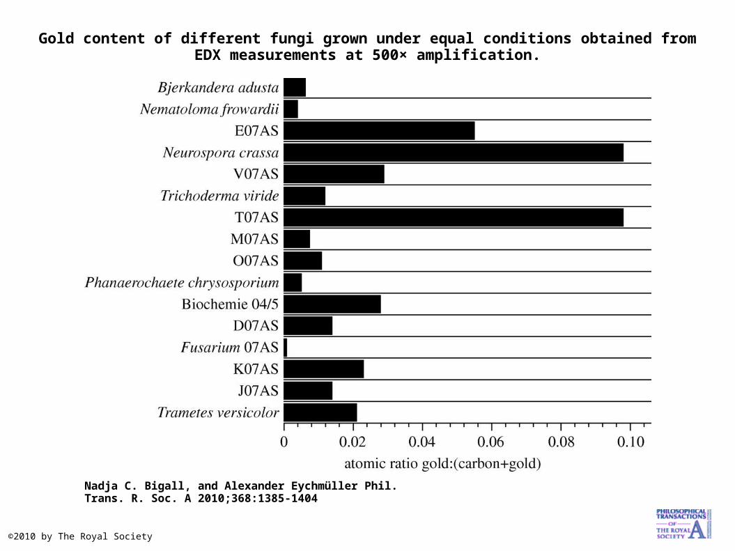
Gold content of different fungi grown under equal conditions obtained from EDX measurements at 500× amplification.
Nadja C. Bigall, and Alexander Eychmüller Phil. Trans. R. Soc. A 2010;368:1385-1404
©2010 by The Royal Society

(a) Single gold nanoparticles assembled on the hyphae of Neurospora crassa shown by scanning electron microscopy in the backscattering mode (scale bar, 250 nm).
Nadja C. Bigall, and Alexander Eychmüller Phil. Trans. R. Soc. A 2010;368:1385-1404
©2010 by The Royal Society

Different metal affinities (obtained from EDX analysis) of seven different fungi grown for two months in identically prepared colloidal solutions.
Nadja C. Bigall, and Alexander Eychmüller Phil. Trans. R. Soc. A 2010;368:1385-1404
©2010 by The Royal Society
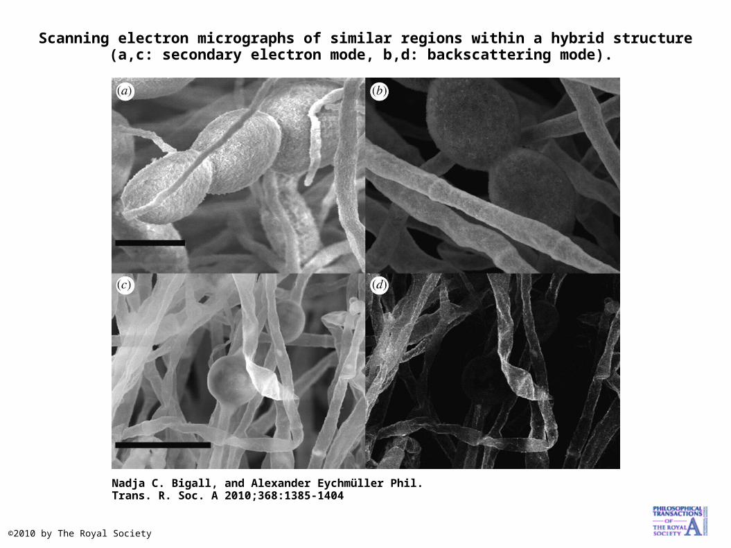
Scanning electron micrographs of similar regions within a hybrid structure (a,c: secondary electron mode, b,d: backscattering mode).
Nadja C. Bigall, and Alexander Eychmüller Phil. Trans. R. Soc. A 2010;368:1385-1404
©2010 by The Royal Society