synthesis of controlled-size silver nanoparticles for the
TRANSCRIPT

RSC Advances
PAPER
Ope
n A
cces
s A
rtic
le. P
ublis
hed
on 1
2 M
arch
202
0. D
ownl
oade
d on
12/
29/2
021
2:10
:07
PM.
Thi
s ar
ticle
is li
cens
ed u
nder
a C
reat
ive
Com
mon
s A
ttrib
utio
n-N
onC
omm
erci
al 3
.0 U
npor
ted
Lic
ence
.
View Article OnlineView Journal | View Issue
Synthesis of cont
aDepartment of Inorganic Chemistry, Faculty
Granada, Spain. E-mail: marisarozalen@ugbDepartment of Genomic Medicine, GENYO,
University of Granada & Andalusian Regiona
Ilustracion 114, Granada 18016, Spain
† Electronic supplementary informa10.1039/c9ra08657a
Cite this: RSC Adv., 2020, 10, 10646
Received 22nd October 2019Accepted 17th February 2020
DOI: 10.1039/c9ra08657a
rsc.li/rsc-advances
10646 | RSC Adv., 2020, 10, 10646–
rolled-size silver nanoparticles forthe administration of methotrexate drug and itsactivity in colon and lung cancer cells†
M. Rozalen, *a M. Sanchez-Polo,a M. Fernandez-Perales,a T. J. Widmannb
and J. Rivera-Utrillaa
A controlled synthesis of methotrexate (MTX) silver nanoparticles (AgNPs-MTX) using borohydride and
citrate as reduction and reduction/capping agents, respectively, was performed in order to obtain
AgNPs-MTX conjugates with a narrow size distribution. Their characterization showed polydispersed
spherical shape nanoparticles with a mean size around 13 nm and distribution range between 7–21 nm.
The presence of MTX was confirmed by FTIR and EDX analysis. Spectroscopic determinations suggest
the chemisorption of MTX through a carboxylic group (–COOH) onto AgNPs via the exchange with
a citrate molecule. Drug loading capacities calculated for AgNPs synthesized using different amounts of
MTX were 28, 31 and 40%. In vitro drug release tests depicted similar release profiles for all conjugated
amounts releasing between 77 to 85% of the initial MTX loaded into the AgNPs. With respect to free
MTX, the addition of the nanocarrier delayed its release and also changed its pharmacokinetics. Free
MTX is released after 3 hours following a first order kinetic model, whereas in the presence of AgNPs,
a fast initial release is observed during the first 5 hours, followed by a plateau after 24 hours. In this case,
AgNPs-MTX fitted a Higuchi model, where its solubilization is controlled by a diffusion process. Results
obtained from flow cytometry of different cell lines treated with AgNPs-MTX demonstrated the
combined anticancer effect of both reagents, decreasing the percentage of living cells in a colon cancer
cell line (HTC-116) down to 40% after 48 hours of exposure. This effect was weaker but still significant
for a lung cancer cell line (A-549). Finally, a zebrafish assay with AgNPs-MTX did not show any significant
cytotoxic effect, confirming thereby the reduction of systemic drug toxicity achieved by coupling MTX to
AgNPs. This observed toxicity reduction in the zebrafish model implies also a probable improvement of
the usage of AgNPs-MTX in chemotherapy against human cancers.
1. Introduction
The emergence of nanotechnology has had a deep impact onclinical therapeutics in the last two decades. Compared withconventional chemotherapeutic agents, nanoscale drug carriershave been demonstrated to possess the capacity to improve treat-ment efficacy while avoiding toxicity in healthy cells due to featuressuch as high accumulation in tumors via the enhanced perme-ability and retention (EPR) effect and active cellular uptake.1–3
Among numerous drug delivery systems, silver nanoparticles(AgNPs) have been used as a novel platform for various therapeuticpurposes. AgNPs exhibit small size and large surface area as well as
of Science, University of Granada, 18071
r.es
Centre for Genomics & Oncology (Pzer -
l Government), PTS Granada, Avda. de La
tion (ESI) available. See DOI:
10660
antimicrobial activity, which is benecial for maintaining formu-lation sterility for extended periods of time.3 In addition, they alsopresent anticancer effects in different human cancer cell lines,including lung broblast (IMR-90) and glioblastoma cells (U295),4
endothelial5 and MDA-MB-231 breast cancer cells.6
Methotrexate (MTX), a chemotherapeutic folic acid analog, isan anionic anticancer drug well known for its effectiveness inthe treatment of certain cancers such as breast, head and neckcancer, leukemia, lymphoma, lung cancer and osteosarcoma.7,8
However, the very short plasma half-life and high efflux rate ofMTX compared to the inux rate requires a high administrationdose, as well as overcoming drug resistance,9 causing manyrestrictions on its clinical application.
MTX therapy optimization had been proposed using poly-ethylene glycol-coated silver nanoparticles10 and AgNPsembedded in graphene oxide (GO)11 as promising deliverysystems that can improve anticancer efficacy and decrease MTXside effects. Cytotoxicity was also tested against breast cancerMCF-7 and hepatoblastoma HepG2 cells11 pointing out that theconjugation GO-MTX avoids ROS (Reactive Oxygen Species)
This journal is © The Royal Society of Chemistry 2020

Paper RSC Advances
Ope
n A
cces
s A
rtic
le. P
ublis
hed
on 1
2 M
arch
202
0. D
ownl
oade
d on
12/
29/2
021
2:10
:07
PM.
Thi
s ar
ticle
is li
cens
ed u
nder
a C
reat
ive
Com
mon
s A
ttrib
utio
n-N
onC
omm
erci
al 3
.0 U
npor
ted
Lic
ence
.View Article Online
generated by AgNPs alone, causing cellular apoptosis as evi-denced by an apoptosis assay (Annexin V/Flow cytometer).(Muhammad et al., 2016)10 included PEG (Poly Ethylene Glycol)in the formulation, because it is capable of reducing the opso-nization process in which nanoparticles are directed to the liverby the help of macrophages. They tested the cytotoxicity in theMCF-7 breast cancer cell line revealing its anticancer activity.
Despite the fact that numerous studies have been investi-gating the effect of the colloidal surface properties undervarious environmental conditions on the stability or theaggregation potential of various nanoparticles, there is littleinformation available with regard to AgNPs. Toxicologicalstudies by ref. 12 in aquatic plant duckweed (Spirodela poly-rhiza) found out that particle size and surface charge of AgNPshave a signicant impact on the response against microorgan-isms. Moreover, the development of physiologically realisticsynthetic microvascular networks (SMNs) have been employedto investigate the effect of size or surface chemistry aloneallowing rapid screening of cancer drug delivery systems.13,14
Recently the results obtained by Khor et al.15 demonstrate thatcarboxylic acid groups decorated nanoparticles bind relativelyweakly to HUVEcs (human umbilical vein endothelial cells) andas such, larger-sized particles (70–130 nm) are strongly affectedby the drag force leading to removal from cell surface.
For this reason, in this study we have decided to use the co-reduction method employing two different reductants (NaBH4
and trisodium citrate (TSC)), which offers a better control ofnucleation and growth of nanoparticles.16,17 The dual thermaltreatment (between 60–90 �C) enhances fast nucleation fol-lowed by a controlled growth at approximately the same rate,resulting in relatively monodispersed nanoparticles.18 Alsoa variation of the pH of the reaction medium helps tuning themorphology of particles from quasi-spherical to nearly spher-ical shape.19 Moreover, previous studies have demonstratedtheir antimicrobial nature to be size20 and shape dependent,21
with smaller nanoparticles displaying a better antimicrobialactivity.18 Even though triangular nanoparticles appeared to bemore effective for microbial killing, spherical ones are consid-ered to be the best-suited for practical applications.22–24
With this background, the main objective of the present studywas to synthesize and characterize discrete and controlled sizemethotrexate silver nanoparticles (AgNPs-MTX), using as reduc-tion agent the combination of borohydride and citrate, in order tobe used as nanocarriers of MTX. Aer material characterization,their anticancer activity was tested studying in vitro cytotoxicity
Table 1 Experimental conditions used to synthesize AgNPs-MTX nanop
AgNO3 (mmol) MTX (mmol) NaBH4 (mmol) TCS (m
0.01 — 0.225 0.090.01 0.0020 0.225 0.090.01 0.0030 0.225 0.090.01 0.0040 0.225 0.09
This journal is © The Royal Society of Chemistry 2020
on lung and colon cancer cell lines. In order to test theirbiocompatibility, a zebrash assay was used as in vivo model.Moreover, we have added new datasets on controlledMTX releaseand tted them with a kinetic model with the aim to understandhow to maintain MTX concentration in blood or in target tissues.
2. Materials and methods2.1. Materials
The reagents: silver nitrate (AgNO3), trisodium citrate dehydrate(TSC), sodium borohydride (NaBH4), and methotrexate (MTX)were of analytical grade and used without further purication.
2.2. Synthesis of silver nanoparticles (AgNPs) andmethotrexate silver nanoparticles (AgNPs-MTX)
Silver nanoparticles have been synthesized following a chemicalreduction method, using sodium borohydride as primaryreductant and trisodium citrate as secondary reductant as wellas stabilizing agent. In order to obtain monodispersed sphericalnanoparticles with a narrow size distribution, we have followedthe procedure described in ref. 18. The temperature during thereduction process was set up at 60 and 90 �C and nal pH wasxed around 10.5. Subsequently, the related/correspondingconjugates using methotrexate were obtained.
2.2.1. Silver nanoparticles (AgNPs). 45 mL of freshlyprepared aqueous solutions containing 5 mM and 2 mM ofNaBH4 and TSC, respectively were mixed and heated to 60 �C inthe dark under vigorous stirring to ensure a homogeneoussolution. Then, 5 mL of 2 mM AgNO3 solution was addeddropwise. Subsequently, temperature was further raised to90 �C and the solution pH was adjusted to 10.5 adding 2 mL of0.1 M NaOH. The intense yellow solution obtained was keptunder vigorous stirring during 30 min. Finally, the resultingcolloid was cooled at room temperature, centrifuged at12 000 rpm during 15 min and kept at �80 �C for 48 hours forfurther freeze-drying.
2.2.2. Synthesis of methotrexate silver nanoparticles(AgNPs-MTX). Initially, we prepared a 10 mM MTX solution in1 mM K2CO3 and added different amounts to the fresh madeAgNPs solutions in order to determine an optimal ratio. Solu-tions were added drop by drop and kept under stirring foranother 30 min. Finally, the resulting suspension was cooled atroom temperature, centrifuged at 12 000 rpm during 15 minand kept at �80 �C for 48 hours for further freeze-drying.
articles
mol)
Ratio
AgNO3 MTX TCS NaBH4 pH
1 — 9 22.5 10.51 0.2 9 22.5 11.381 0.3 9 22.5 11.361 0.4 9 22.5 11.36
RSC Adv., 2020, 10, 10646–10660 | 10647

RSC Advances Paper
Ope
n A
cces
s A
rtic
le. P
ublis
hed
on 1
2 M
arch
202
0. D
ownl
oade
d on
12/
29/2
021
2:10
:07
PM.
Thi
s ar
ticle
is li
cens
ed u
nder
a C
reat
ive
Com
mon
s A
ttrib
utio
n-N
onC
omm
erci
al 3
.0 U
npor
ted
Lic
ence
.View Article Online
In order to determine an optimal ratio for AgNPs and MTX,different concentrations of AgNO3, MTX, TSC, and NaBH4 weretested and are showed in Table 1.
2.3. Characterization of AgNPs and AgNPs-MTX
UV-visible spectroscopy was performed with a VWR UV-1600PCUV/Vis spectrophotometer. Samples were analyzed in the400–900 nm spectral range. The XRD pattern of the sampleswere recorded by a Bruker D8 Discover Diffractometer (Ka Cu)with a PILATUS3R 100K-A detector and equipped with a Cu Ka(l ¼ 1.5406 A) radiation lter. The chemical structure andfunctional groups of the AgNPs-MTX were analyzed by FourierTransform Infrared Spectroscopy (FT-IR, JASCO FT/IR 6300spectrometer) and recorded in absorbance mode in the4000–400 cm�1 range with a spectral resolution of 4 cm�1 fromthe average of 100 spectra. Samples were prepared in KBrpressed pellets. Light scattering analysis was conducted usinga ZETASIZER (Nano ZS; ZEN3600, Malvern) at 25 �C, witha He–Ne laser operating at a wavelength of 633 nm. Theintensity size distributions were obtained from analysis of thecorrelation functions using the Multiple Narrow Modes algo-rithm in the instrument soware. The samples (AgNPs, AgNPs200, 300 and 400) were diluted 1 : 20 in distilled water toeliminate the primary charge effect and ultrasound during10 min to avoid agglomeration. A sample volume of 2 mL wasused in 10 mm-diameter disposable cuvettes. At least 3 repeatmeasurements on each sample were taken to check for resultrepeatability. The assembly and morphology of AgNPs andAgNPs-MTX were studied by high-resolution transmissionelectron microscopy (HRTEM) images, obtained using a FEITitan, operated at 300 kV. SAED patterns were collected usinga 10 mm aperture allowing collection of diffraction data froma circular area. Compositional maps of selected areas wereacquired in scanning transmission electron microscopy (STEM)mode using a Super X EDX detector (FEI), formed by fourwindowless SSD detectors. STEM images were collected witha high angle annular dark eld (HAADF) detector.
2.4. Stability test
1 mL of silver colloid solution was mixed with 2 mL of PBSbuffer (pH 7.4), DMEM, Milli-Q water containing 10% FBS,DMEM containing 10% FBS, PBS containing 10% FBS. Aer theincubation at 37 �C for 1, 6, 12 and 24 hours the changes of themaximum absorption wavelength were determined by UV-visspectroscopy.
2.5. In vitro drug release and drug encapsulation efficiency
The release proles of AgNPs-MTX were studied by dialysis withPBS (10 mmol L�1, pH ¼ 7.4) as release media. 10 mg ofnanoparticles were placed into a dialysis membrane bag(MWCO ¼ 8000–14 000) with 2 mL of PBS and closed with clips.The bags were immersed in 78 mL of PBS. The system wasshaken at a speed of 150 rpm at 37 �C. At desired time intervals,3 mL of sample was withdrawn, MTX concentration measuredby UV-Vis spectroscopy at lmax ¼ 304 nm, and poured back into
10648 | RSC Adv., 2020, 10, 10646–10660
the solution. As a control experiment, the release of free MTXfrom the dialysis bag was also measured.
Once the experiments were nished, we opened the dialysisbags and added 5 mg of proteinase K enzyme, a protease thatcleaves MTX amine bonds, and incubated during 20 min at37 �C. The nanoparticles were then separated from the free MTXby centrifugation (15 000 rpm) and the absorbance of thesupernatant was measured at 304 nm. A similar drug releasequantication strategy was reported in ref. 25.
2.6. Cell viability assays
In vitro cell viabilities of colorectal cancer (HTC-116) (ECACC no.91091005 (lot no. 05K025) and human lung carcinoma (A-549)(ATCC no. CCL-185 (lot no. 3624224) cell lines, from the CICcell bank of the University of Granada, were determinedthrough ow cytometry using a simultaneous double-stainingprocedure with uorescein diacetate (FDA) and propidiumiodide (PI) in the presence of free MTX, AgNPs and AgNPs-MTX.
40 000 cells were separately incubated and distributed in 12-well plates for further 24 h incubation at 37 �C in a humidatmosphere enriched with 5% CO2. The medium was removed,and fresh medium was added together with free MTX (atconcentrations of 45.4, 454, 909, 2272 and 4544 mg mL�1),AgNPs and AgNPs-MTX (38, 76, 152, 253, 380 and 760 mg mL�1).Aer 12, 24, and 48 hours of treatment 100 mL per well of pro-pidium iodide solution (100 mg mL�1) was added and incubatedfor 10 min at 28 �C in darkness. Aerwards, 100 mL per well ofuorescein diacetate (100 ng mL�1) was added and incubatedunder the same aforementioned conditions. Finally, the cellswere recovered by centrifugation at 1500 rpm for 10 min and theprecipitate was washed with PBS. Flow cytometric analyses wereperformed with a FACS Vantage™ ow cytometer (BectonDickinson). The percentage of viability was calculated incomparison to the control culture. The IC50 was calculatedusing linear-regression analysis from the Kc values at theemployed concentrations using the soware GraphPad Prism 6.
2.7. Confocal microscopy
The cellular imaging of AgNPs and AgNPs-MTX was recorded byusing an inverted laser scanning confocal microscope (LeicaDMI6000) equipped with an Argon optical system. Samples wereimaged in reectance mode at lexc ¼ 561 nm. Cells morphologywere analyzed by recording differential interference contrast(DIC) snapshot in a transmission mode.
2.8. In vivo study/zebrash assay
Zebrash assays were done in collaboration with researchersfrom GENYO (Pzer-University of Granada-Junta de AndalucıaCentre for Genomics and Oncological Research). Zebrashembryos were collected from the zebrash aquarium of CIC-UGR (Scientic Instrumentation Center of the University ofGranada) and staged according to standard procedures. Fortoxicity studies, 10 healthy embryos at shield stage (gastrula-tion, 6–7 hours post fertilization, hpf) were transferred to eachwell of a 24-well plate along with 1 mL of zebrash E3 medium.Different concentrations of free MTX (10, 15, 30, 50 and 100 mg
This journal is © The Royal Society of Chemistry 2020

Paper RSC Advances
Ope
n A
cces
s A
rtic
le. P
ublis
hed
on 1
2 M
arch
202
0. D
ownl
oade
d on
12/
29/2
021
2:10
:07
PM.
Thi
s ar
ticle
is li
cens
ed u
nder
a C
reat
ive
Com
mon
s A
ttrib
utio
n-N
onC
omm
erci
al 3
.0 U
npor
ted
Lic
ence
.View Article Online
mL�1), AgNPs and AgNPs-MTX 400 (100, 50 and 25 mg mL�1)were added to the wells and embryos were incubated for72 hours at 28.5 �C. All tests were repeated three times(60 embryos per concentration). Using a stereomicroscope,morphological changes, heart rate measurements, andmortality of the embryos were annotated aer 24, 48 and 72 hpf(hours post fertilization). Mortality rate was expressed as thetotal number of dead embryos aer 72 hpf. Heart rate wasrecorded using a stopwatch by direct observation under themicroscope. Moreover, morphological changes such as peri-cardial edema or morphological defects, such as bent andtwisted notochord, reduced number of somites, abnormal yolkextension or delayed growth were recorded at every stage.
2.9. Statistical analysis
Statistical analysis was performed by using the Graph Pad Primv.6 soware. The one-way analysis of variance (ANOVA) statis-tical method was used to evaluate the signicance of theexperimental data. A value of p < 0.05 was considered statisti-cally signicant.
3. Results and discussion3.1. Characterization of AgNPs and AgNPs-MTX
Optical properties of AgNPs and AgNPs-MTX conjugates weremeasured by UV-Vis adsorption and are showed in Fig. 1. Theposition of the plasmon resonance peak at 394 nm conrms theformation of spherical AgNPs.26 A bathochromic shi of theplasmon band is observed when MTX is added (404, 403 and405 nm) due to the environmental changes around the silvernanoparticles when MTX replace citrate molecules.27 Further-more, two bands were observed at 258 and 305 nm that corre-spond to the MTX absorption bands, which are conjugated tothe AgNPs (Fig. 1a and b). Color changes are also observed:AgNPs presented a bright yellow color that turns darker asadded MTX concentration increases (Fig. 1b).
Fig. 1 (a) Comparative UV-Vis absorption spectra of free MTX, AgNO3
AgNPs-MTX with different concentrations of MTX.
This journal is © The Royal Society of Chemistry 2020
AgNPs and AgNPs-MTX (200, 300, and 400) samples revealedthe same typical XRD pattern characterized by 4 diffractionpeaks located at 38.48, 44.25, 64.72 and 77.40� (Fig. 2) corre-sponding to the (111), (200), (220), and (311) crystal planes of Ag(Ag XRD ref no. 01-087-0719), respectively. The XRD patternsuggested the formation of crystalline Ag with a face-centeredcubic (fcc) structure.28
FTIR spectra of free MTX and AgNPs-MTX 200, 300, and 400are illustrated in Fig. 3. The peak at 3394 cm�1 indicates thepresence of a –NH group. Absorptions at 3059 and 2951 cm�1
point out to the existence of carboxylic acid for free MTX(Fig. 3a). Peaks in the region of 1500–1700 cm�1 can be attrib-uted to C–N or NH2 vibrations, while peaks at 1490 and1209 cm�1 represent the stretching vibration of the C–C or C–Hbonds.29 In contrast with free MTX, the FTIR spectrum ofAgNPs-MTX conjugates exhibits few characteristics peaks asa consequence of the interaction between AgNPs and MTX. Thepresence of peaks at 1507 and 1398 cm�1, assigned to thestretching mode of the amide II C–N of MTX and the in-planebending of O–H in carboxyl groups, indicated the successfulloading of MTX into the silver nanoparticles.29
Dynamic light scattering (DLS) measurements were used todetermine the particle size of the synthesized AgNPs in aqueoussolution. As it is shown in Table 2, AgNPs showed an averagesize of 11.2 nm, and it increased as MTX was present. As theamount of MTX increases from AgNPs-MTX 200 to AgNPs-MTX400 Z-potential became more negative, from �23.8 to �31.7 �2.0 mV (Table 2). Moreover, the values of polydispersity index(PDI) decreased, pointing out the efficiency of MTX as a cappingmaterial, stabilizing the nanoparticles by providing intensivenegative charges that keep all the particles away from eachother.
The morphology of synthesized AgNPs and AgNPs-MTX wasinspected by HRTEM analysis. Images conrmed the formationof spherical and highly dispersed nanoparticles, as showed inFig. 4. The average size and distribution were calculated basedon the diameter measurement of 200 nanoparticles, obtainingan average size close to the values obtained with DLS (Table 2).
and AgNPs (b) comparative UV-Vis absorption spectra of AgNPs and
RSC Adv., 2020, 10, 10646–10660 | 10649

Fig. 2 XRD diffractograms of AgNPs and AgNPs-MTX400.
Fig. 3 FTIR spectra of (a) free MTX, (b) AgNPs-MTX 200, (c) AgNPs-MTX 300, and (d) AgNPs-MTX 400.
Table 2 Zeta potential and effective diameter of synthesized silver nano
Sample DLS diameter (nm) PDI
AgNPs 14.7 � 2.7 0.155AgNPs-MTX 200 17.3 � 7.9 0.360AgNPs-MTX 300 18.0 � 4.7 0.228AgNPs-MTX 400 21.9 � 8.7 0.155
10650 | RSC Adv., 2020, 10, 10646–10660
RSC Advances Paper
Ope
n A
cces
s A
rtic
le. P
ublis
hed
on 1
2 M
arch
202
0. D
ownl
oade
d on
12/
29/2
021
2:10
:07
PM.
Thi
s ar
ticle
is li
cens
ed u
nder
a C
reat
ive
Com
mon
s A
ttrib
utio
n-N
onC
omm
erci
al 3
.0 U
npor
ted
Lic
ence
.View Article Online
For instance, Fig. 4a and b showed the histograms obtained forAgNPs and AgNPs-MTX 400. The average size calculated forAgNPs was 11.13 � 2.27 nm with a distribution range between 6and 19 nm, and as the amount of MTX increases the averagesize also increases from 12.5 to 15.2 nm: even tough differencesin size are not signicant, polydispersity index and Z-potentialmeasurements conrmed the improvement of AgNPs-MTX 400.
EDX analysis (Fig. 5d) showed the presence of silver andnitrogen, as an indicator of the presence of MTX. The chemicalelement mapping analysis (Fig. 5c) showed the distribution ofelements for a representative group of AgNPs-MTX nano-particles, pointing out a homogeneous distribution of Ag and Nin the sample with predominance of a silver crystalline phase,conrmed by SAED image (Fig. 5b). Nitrogen was not distrib-uted in layers but along the sample (Fig. 5c), which suggestedthe formation of AgNPs-MTX conjugates. Moreover, the latticefringe images (Fig. 6) showed that some AgNPs and conjugateswith MTX are made up of ve domains, which also conrms the
particles with methotrexate
HRTEM diameter (nm)Z-Potential(mV)
labs(nm)
11.13 � 2.3 �20.8 39412.5 � 2.8 �23.1 40414.6 � 6.8 �30.0 40315.2 � 3.9 �31.7 405
This journal is © The Royal Society of Chemistry 2020

Fig. 4 Particle size distribution histogram obtained by HRTEM imagesmeasurements for: (a) AgNPs and (b) AgNPs-MTX 400.
Paper RSC Advances
Ope
n A
cces
s A
rtic
le. P
ublis
hed
on 1
2 M
arch
202
0. D
ownl
oade
d on
12/
29/2
021
2:10
:07
PM.
Thi
s ar
ticle
is li
cens
ed u
nder
a C
reat
ive
Com
mon
s A
ttrib
utio
n-N
onC
omm
erci
al 3
.0 U
npor
ted
Lic
ence
.View Article Online
polycrystalline nature of the particles. The average inter-planardistance determined from the HRTEM image is 0.231 nm cor-responding to the (111) lattice.
In order to provide evidence for the chemisorption of MTXon the surface of AgNPs, we obtained the XP spectra of MTX ina pure solid form as well as of MTX adsorbed on AgNPs. Thesurface scan spectra showed the presence of Ag, C, O, and Natoms according to their binding energies. For solid MTX, in theC 1s region, four peaks are identied at: (i) 284.3 eV associatedwith sp2 hybridization of C]C, (ii) 285.4 eV corresponding withsp3 hybridization of C–C and C–N, (iii) 286.6 eV associated withC–O in hydroxyl groups, and (iv) 288.4 eV corresponding with–COOH in carboxylic groups. The most prominent signal in the
This journal is © The Royal Society of Chemistry 2020
XPS spectrum is the Ag 3d consisting of two spin–orbitcomponents at 368.8 (Ag 3d5/2) and 374.8 (Ag 3d3/2) eV andseparated by 6.0 eV (Fig. 7). Moreover, the deconvolution of Ag(3d) doublet revealed an asymmetric peak shape. These twocharacteristics indicate that Ag exists in the metallic form.Energy loss features at 371.9 and 378.0 eV are observed at thehigher binding energy side of each spin–orbit component for Ag3d metal. These results are in good agreement with the XRDcharacterization. XPS high-resolution scan for the C 1s corelevel (Fig. 7) showed the presence of four different peaks: themain peak centered at 285.56 eV was attributed to C–C (sp3),while the peaks at 286.3, 288.0, and 288.8 eV were attributed toC–O, C]O, and C–O–Ag, respectively. The doublet for O 1s at531.7 and 533.3 eV was assigned to oxygen atoms in the ringcarbonyl (pC]O) and the carboxyl moieties, respectively. Theseresults are in good agreement with the study of Chen et al.27 forgold nanoparticles suggesting that MTX can be directly boundonto AgNPs through a carboxylic group (–COOH) via theexchange with a citrate molecule. Covalent bonding of thera-peutic agents on nanocarriers is usually favored because thebond strength makes the NPs drug conjugates highly stable andtherefore is most likely to be disrupted only under harsh envi-ronments inside lysosomes.30 MTX-conjugated NPs are believedto be taken up to a higher amount into cells by the human folatereceptor than free MTX.27
3.2. Drug loading capacity and in vitro drug release
Drug loading capacities calculated for synthesized silver nano-particles are 28.3, 31.48, and 40.4% w/w for AgNPs-MTX 200,AgNPs-MTX 300 and AgNPs-MTX 400, respectively. Typical invitro release proles were investigated and illustrated in Fig. 8for free MTX (control experiment) and AgNPs-MTX. The controlexperiment showed a complete MTX diffusion across the dial-ysis membrane within 180 min (Fig. 8a). Nonetheless, as it wasexpected, when MTX is conjugated with AgNPs the systemexhibited a slower release compared with the control (Fig. 8b).
Fig. 8b depicts similar release proles for the synthesizedAgNPs-MTX conjugates, releasing between 77 to 85% of theinitially MTX loaded on the silver nanoparticles, aer 24 hours.A rapid and short release of MTX is observed during the rst 5hours, followed by a stable plateau prole. Such sustained two-step release behavior could maintain the drug level withina therapeutic window.31 Regarding the initial MTX concentra-tion added, there are no signicant differences between drugrelease proles.
3.3. Kinetic modeling
The mechanism of MTX release was studied applying differentkinetic models, in order to t the experimental cumulative drugrelease data (Table 3). The release of free MTX tted a rst-ordermodel represented by the following equation:
log C ¼ log Co � k
2:303t (1)
where Co is the initial concentration of drug, k is the rst orderrate constant and t is the experiment time. The data obtained
RSC Adv., 2020, 10, 10646–10660 | 10651

Fig. 5 (a) HAADF image of a representative group of AgNPs-MTX. (b) SAED image showing the presence of a silver nanocrystalline phase (c) mapsfor relative distribution of the elements (pink¼ Ag and green¼ N) jointly shown (d) EDX analysis confirming the relative proportions of Ag and N.
RSC Advances Paper
Ope
n A
cces
s A
rtic
le. P
ublis
hed
on 1
2 M
arch
202
0. D
ownl
oade
d on
12/
29/2
021
2:10
:07
PM.
Thi
s ar
ticle
is li
cens
ed u
nder
a C
reat
ive
Com
mon
s A
ttrib
utio
n-N
onC
omm
erci
al 3
.0 U
npor
ted
Lic
ence
.View Article Online
were plotted as log cumulative (%) of drug remaining versustime, obtaining a value of k ¼ 0.0026 min�1 with a regressioncoefficient of r2 ¼ 0.988.
The presence of AgNPs as nanocarriers modied MTXpharmacokinetics, as expected, tting better to a Higuchi model(eqn (2)), expressed by the following simplied equation:
Q ¼ KHt1/2 (2)
Fig. 6 HRTEM lattice fringe image of AgNPs-MTX 400 showingmultiple domains.
10652 | RSC Adv., 2020, 10, 10646–10660
where Q is the amount of drug released as a function of time perunit of area, KH is the Higuchi rate constant and t1/2 is thesquare root time. This model is applied to study the release ofwater soluble and low soluble drugs incorporated in semi-solidand/or solid matrices.
The values of the Higuchi rate constant obtained were KH ¼35.62 h�0.5 (r2 ¼ 0.932), 31.32 h�0.5 (r2 ¼ 0.982) and 37.15 h�0.5
(r2 ¼ 0.966) for AgNPs-MTX 200, 300, and 400, respectively. Thismodel describes drug release as a diffusion process based onFick's law, which depends on the square root of time (Table 3).According to the results obtained, we selected the sampleAgNPs-MTX 400 to carry out the in vitro anticancer activity tests.
3.4. Stability test
We investigated the short term stability (around 24 hours) indifferent media and long term storage stability (one month) ofprepared silver nanoparticles conjugated with differentamounts of methotrexate, in the darkness and refrigerated at4 �C. According with the position of the maximum absorptionwavelength of solutions showed in Fig. S1 (ESI†) none of thesamples showed signicant difference aer one month.
Particles not stable when diluted by only phosphate bufferedsolution (PBS), while in the presence of fetal bovine serum (FBS)they were stable at least for 24 hours without the addition ofDMEM. This means AgNPs and AgNPs-MTX synthetized aresensitive to high salts contents, but FBS can stabilize nano-particles and keep their dispersity. Moreover, aer one month
This journal is © The Royal Society of Chemistry 2020
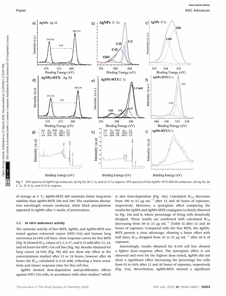
Fig. 7 XPS spectra of AgNPs (g) widescan; (a) Ag 3d; (b) C 1s, and (c) O 1s regions. XPS spectra of the AgNPs-MTX 400 (h) widescan; (d) Ag 3d; (e)C 1s; (f) O 1s, and (i) N 1s regions.
Paper RSC Advances
Ope
n A
cces
s A
rtic
le. P
ublis
hed
on 1
2 M
arch
202
0. D
ownl
oade
d on
12/
29/2
021
2:10
:07
PM.
Thi
s ar
ticle
is li
cens
ed u
nder
a C
reat
ive
Com
mon
s A
ttrib
utio
n-N
onC
omm
erci
al 3
.0 U
npor
ted
Lic
ence
.View Article Online
of storage at 4 �C, AgNPs-MTX 400 maintain better long-termstability than AgNPs-MTX 300 and 200. The maximum absorp-tion wavelength remain unaltered, while black precipitationappeared in AgNPs aer 3 weeks of preservation.
3.5. In vitro anticancer activity
The cytotoxic activity of free MTX, AgNPs, and AgNPs-MTX wastested against colorectal cancer (HTC-116) and human lungcarcinoma (A-549) cell lines. Dose response curves for free MTX(Fig. 9) showed IC50 values of 2.3, 0.37, and 0.15mM aer 12, 24,and 48 hours for HTC-116 cell line (Fig. 9a). Results obtained forlung cancer (A-549) (Fig. 9b) did not show any effect at theconcentrations studied aer 12 or 24 hours, however aer 48hours the IC50 calculated is 0.10 mM, reecting a lower sensi-tivity and slower response time for this cell line.
AgNPs showed dose-dependent anti-proliferative effectsagainst HTC-116 cells, in accordance with other studies,6 which
This journal is © The Royal Society of Chemistry 2020
is also time-dependent (Fig. 10c). Calculated IC50 decreasesfrom 186 to 63 mg mL�1 aer 12 and 48 hours of exposure,respectively. Moreover, a synergistic effect comparing theresults for AgNPs and AgNPs-MTX conjugates is clearly observedin Fig. 10a and b, where percentage of living cells drasticallydropped. These results are conrmed with calculated IC50
decreasing from 88 to 23 mg mL�1 (Table 4) aer 12 and 48hours of exposure. Compared with the free MTX, the AgNPs-MTX present a clear advantage, showing a faster effect withhalf dose; IC50 dropped from 45 to 23 mg mL�1 aer 48 h ofexposure.
Interestingly, results obtained for A-549 cell line showeda lighter dose–response effect. The synergistic effect is notobserved and even for the highest dose tested, AgNPs did notshow a signicant effect decreasing the percentage live cellsfrom 93 to 84% aer 12 and 48 hours of exposure, respectively(Fig. 11a). Nevertheless, AgNPs-MTX showed a signicant
RSC Adv., 2020, 10, 10646–10660 | 10653

Fig. 8 (a) Release profile of free MTX. (b) Release profiles for MTX fromsilver nanoparticles prepared using different initial MTX/Ag ratios andnamed as AgNPs-MTX 200, 300, and 400.
RSC Advances Paper
Ope
n A
cces
s A
rtic
le. P
ublis
hed
on 1
2 M
arch
202
0. D
ownl
oade
d on
12/
29/2
021
2:10
:07
PM.
Thi
s ar
ticle
is li
cens
ed u
nder
a C
reat
ive
Com
mon
s A
ttrib
utio
n-N
onC
omm
erci
al 3
.0 U
npor
ted
Lic
ence
.View Article Online
cytotoxic activity decreasing the percentage of live cells from 69to 36% aer 12 and 48 hours of exposure.
Finally, similar concentrations of AgNPs-MTX, whencompared with AgNPs, aer 48 hours of exposure showed that
Table 3 Calculated parameters and correlation coefficients (r2) of diffeconjugates synthesized in this study
Model First order Higuchi
Parameters r2 KH (h�1) r2
Free MTX 0.988 0.909 0.976AgNPs-MTX 200 0.908 0.315 0.932AgNPs-MTX 300 0.964 0.260 0.982AgNPs-MTX 400 0.945 0.295 0.966
Fig. 9 (a) Free MTX dose response curve calculated for HTC-116 cell lin
10654 | RSC Adv., 2020, 10, 10646–10660
the antiproliferative effect is strengthened with increase in theconcentration, as stated in Fig. 11c.
Considering that the percentage of MTX on AgNPs variesbetween 20 and 40% and their IC50 decreases more than anorder of magnitude compared with free MTX, it is clear thateffective doses of AgNPs-MTX are lower than those corre-sponding to free MTX doses for lung and colon cancer cell lines,although the effect is more pronounced for the colon cancer cellline (HTC-116).
3.6. Cellular imaging of HTC-116 and A-549 lines with AgNPsand MTX
The cellular uptake of AgNPsMTX conjugates was conrmedusing confocal microscopy. Images were taken aer incubatingcells with AgNPs and AgNPsMTX at selected concentrations,based on the ow cytometry results. Representative results areshown in Fig. 12 and 13 for HTC-116 and A-549 cell linesrespectively.
As expected, control cells showed high conuency aer 48 hincubation for colon cancer line (HTC116) (Fig. 12a). Cellstreated with AgNPs showed a signicant amount of red uo-rescence, increasing when treated with AgNPs-MTX conjugates.
rent models of release kinetics of AgNPs and the corresponding MTX
Korsmeyer–Peppas
KH (h�0.5) r2 KH (h�n) Value “n”
69.18 0.980 0.145 0.95435.62 0.950 0.159 0.75131.32 0.960 0.167 0.71937.15 0.907 0.267 0.707
e. (b) Free MTX dose response curve calculated for A-549 cell line.
This journal is © The Royal Society of Chemistry 2020

Fig. 10 Flow cytometry analysis of cell viability in HTC-116 cell line after treatment with (a) AgNPs, and (b) AgNPs-MTX. (c) Comparative bar graphillustrating the mean percentages of live cells for control, AgNPs, and AgNPs-MTX after 12, 24, and 48 hours of exposure.
Table 4 Calculated IC50 for HTC-116 cell line
Nanoparticles
IC50 (mg mL�1)
12 h 24 h 48 h
AgNPs 186 98 63AgNPs-MTX 400 88 38 23
This journal is © The Royal Society of Chemistry 2020
Paper RSC Advances
Ope
n A
cces
s A
rtic
le. P
ublis
hed
on 1
2 M
arch
202
0. D
ownl
oade
d on
12/
29/2
021
2:10
:07
PM.
Thi
s ar
ticle
is li
cens
ed u
nder
a C
reat
ive
Com
mon
s A
ttrib
utio
n-N
onC
omm
erci
al 3
.0 U
npor
ted
Lic
ence
.View Article Online
These results support the conjugated effect of MTX and AgNPsobserved by ow cytometry.
However, for lung cancer cell line (A-549) images showedsimilar effects for AgNPs and AgNPs-MTX conjugates (Fig. 13).This could be related to the presence of folate receptors in lineHTC-116 on the contrary than line A-549, supporting thespecic uptake of folate-conjugated AgNPs-MTX by folatereceptor positive tumor cells.
RSC Adv., 2020, 10, 10646–10660 | 10655

Fig. 11 Flow cytometry analysis of cell viability in A-549 cell line after treatment with different concentrations of (a) AgNPs and (b) AgNPs-MTX.(c) Comparative bar graph illustrating the dose effect for AgNPs and AgNPs-MTX after 48 hours of exposure.
RSC Advances Paper
Ope
n A
cces
s A
rtic
le. P
ublis
hed
on 1
2 M
arch
202
0. D
ownl
oade
d on
12/
29/2
021
2:10
:07
PM.
Thi
s ar
ticle
is li
cens
ed u
nder
a C
reat
ive
Com
mon
s A
ttrib
utio
n-N
onC
omm
erci
al 3
.0 U
npor
ted
Lic
ence
.View Article Online
3.7. Zebrash assay
Reported results relating to the toxicity assessment of MTX inzebrash demonstrated that high doses (0.2–1.5 mM ¼ 91–681mg mL�1) cause disruption of the folate pathway, leading todevelopmental defects in early zebrash stages.32,33 Our resultsshowed a dose dependent toxicity of MTX in embryos aerconstant exposure (from 6 hpf onwards) to lower MTX concen-trations ranging from 10 to 30 mg mL�1, increasing embryonicmortality rate from 13 to 20% at 24 hpf. Moreover, phenotypicchanges as yolk defects and less somites were observed ((Fig.
10656 | RSC Adv., 2020, 10, 10646–10660
14I–L), compared with wild type embryos in (Fig. 14A–D)) . Up to30% of the embryos presented pericardial edema. Aer 48 hpf,the hatching rate decreased from 70 to 56%, as MTX concen-tration increased from 10 to 30 mg mL�1. Around 10% of theembryos presented yolk defects, less somites and degenerationof body parts, as bent or shortened tails. No signicant heartedema was noticed any more aer 48 hpf, but a few embryospresented a lower heart beat rate (between 3 and 6%).
Between 66 and 80% of the embryos hatched aer 72 h andno signicant phenotypic changes were observed, besides
This journal is © The Royal Society of Chemistry 2020

Fig. 12 Representative bright field and fluorescence images of HTC-116 cells incubated for 48 h with AgNPs (b and e) and AgNPs-MTX (c and f)compared with a control (a and c).
Fig. 13 Representative bright field and fluorescence images of A-549 cells incubated for 48 h with AgNPs (b and e) and AgNPs-MTX (c and f)compared with a control (a and c).
This journal is © The Royal Society of Chemistry 2020 RSC Adv., 2020, 10, 10646–10660 | 10657
Paper RSC Advances
Ope
n A
cces
s A
rtic
le. P
ublis
hed
on 1
2 M
arch
202
0. D
ownl
oade
d on
12/
29/2
021
2:10
:07
PM.
Thi
s ar
ticle
is li
cens
ed u
nder
a C
reat
ive
Com
mon
s A
ttrib
utio
n-N
onC
omm
erci
al 3
.0 U
npor
ted
Lic
ence
.View Article Online

Fig. 14 Microscopic images of control zebrafish embryos, zebrafish embryos treated with AgNPs-MTX, and zebrafish embryos treated with freeMTX at different developmental stages (6 hpf, 24 hpf, 48 hpf, and 72 hpf, stages according to ref. 34). (A–D) Wild type control embryos showednormal embryonic development: (A) embryo enclosed in chorion at 6 hpf, (B) embryo enclosed in chorion at 24 hpf, (C and D) hatched larvae at48 and 72 hpf. (E–H) Some embryos treatedwith AgNPs-MTX (AgNps alone images not shown) presented verymild toxicity phenotypes: (E and F)embryos with light heart edema at 24 hpf, (G) delay in hatching at 48 hpf, and (H) delayed growth and bent tail. (I–L) A higher percentage ofembryos treated with free MTX showed more severe effects as: (I) chorion with cloudy appearance resembling dead embryos at 24 hpf, (J)delayed growth andmorphological defects after 24 hpf, (K) larvae with twisted tail at 48 hpf, and (L) delayed growth and bent tail/notochord at 72hpf.
RSC Advances Paper
Ope
n A
cces
s A
rtic
le. P
ublis
hed
on 1
2 M
arch
202
0. D
ownl
oade
d on
12/
29/2
021
2:10
:07
PM.
Thi
s ar
ticle
is li
cens
ed u
nder
a C
reat
ive
Com
mon
s A
ttrib
utio
n-N
onC
omm
erci
al 3
.0 U
npor
ted
Lic
ence
.View Article Online
a growth delay in around 10%. Consequently, later stages ofembryos were found to be more resistant to MTX treatment.Although MTX concentrations tested in this study did not showa pronounced toxicity, Sun et al. showed that zebrash embryosexposed to a 1.5 mM (681 mg mL�1) MTX during 6–10 hpf failedto form a normal cardiovascular system. MTX induced suchmalformations by inhibiting the dihydrofolate reductase(DHFR), an enzyme that is critical for nucleotide synthesis andmethylation, both of which play essential roles in embryonicdevelopment.33
AgNPs-treated embryos did not show dose-dependenttoxicity under laboratory conditions, with an overall mortalitylower than 4% aer 24, 48, and 72 hpf. However, a few embryosexhibited some phenotypic changes characterized by bent andtwisted tails, pericardial edema and delayed growth at the rststage observed (24 hpf). Also, a mild transient pericardial edemais noted in up to a 66% of the embryos for 100 mg mL�1 AgNPs,but disappeared aer 48 and 72 hpf, where the hatching ratereached almost 100% of the embryos (images not shown, asphenotypeare similar to those of AgNPs-MTX treated embryos).
Finally, the AgNPs-MTX conjugates studied (200, 300, and 400)(Fig. 14E–H) showed a dose-dependent mortality rate reachinga 16% aer 24 hours for higher concentrations (100 mg mL�1). Asit was noted in early stages (24 hpf) for AgNPs alone, light peri-cardial edema reached up to 80% of the embryos for 100 mg mL�1
AgNPs-MTX 400, but it disappeared at 48 hours, allowinga hatching rate between 90 and 100% aer 72 hours. As observedin Fig. 15, the mortality rate (in %) in free MTX (10 and 30 mgmL�1) treated zebrash embryos is notably higher than themortality rate in AgNPs or AgNPs-MTX conjugate treated embryos,pointing out the reduced toxicity of MTX attached to nanocarriers.
As shown in Fig. 15, heart rate of embryos at 72 hpf wasunaffected by AgNPs and AgNPs-MTX treatments. On the other
10658 | RSC Adv., 2020, 10, 10646–10660
hand, the comparative mortality percentage for differentconcentrations of MTX and 100 mg mL�1 of AgNPs and AgNPs-MTX 400 showed no toxicity for AgNPs and AgNPs-MTX equiv-alent doses tested on cancer cells.
On the contrary, a remarkable toxic effect of free MTX, 30mg mL�1, is observed for the development of zebrash (asreported in the literature33). AgNPs alone showed a mild effectinducing delayed growth and weak morphological defects.However, when methotrexate is conjugated to AgNPs, there isno general cytotoxic effect in zebrash, AgNPs-MTX rathershow similar side effects as AgNPs alone. AgNPs-MTX,however, are effective against colon and lung cancer cells,as shown in the cytotoxicity assay. Taken together, theseresults point towards a strong reduction of systemic drugtoxicity of MTX when conjugated to silver nanoparticles andtherefore an expected improvement in the outcomes of AgNP-MTX when used in chemotherapy.
4. Conclusions
Polydisperse AgNPs-MTX of controlled size have been success-fully synthesized by controlling temperature and pH, and usingas reducing agents borohydride and citrate.
Polydispersed spherical shape silver nanoparticles witha mean size around 13 nm and a distribution range between 7and 21 nm were obtained. HRTEM mapping showed a homo-geneous distribution of Ag and N (as an indicator of the pres-ence of MTX) with prevalence of a silver nanocrystalline phase(determined by SAED images), which suggested the formationof an Ag-MTX conjugate. Spectroscopic determinations indi-cated the chemisorption of MTX through a carboxylic group(–COOH) onto AgNPs via exchange with a citrate molecule.
This journal is © The Royal Society of Chemistry 2020

Fig. 15 (A) Average heart beat rate of embryos at 72 hpf. (B)Comparative mortality (in percent) for different concentrations of MTXand 100 mg mL�1 of AgNPs and AgNPs-MTX 400.
Paper RSC Advances
Ope
n A
cces
s A
rtic
le. P
ublis
hed
on 1
2 M
arch
202
0. D
ownl
oade
d on
12/
29/2
021
2:10
:07
PM.
Thi
s ar
ticle
is li
cens
ed u
nder
a C
reat
ive
Com
mon
s A
ttrib
utio
n-N
onC
omm
erci
al 3
.0 U
npor
ted
Lic
ence
.View Article Online
Drug loading capacities calculated for synthesized AgNPsusing different amounts of MTX were 28, 31, and 40%. In vitrodrug release tests depicted similar release proles for all drug-conjugated AgNPs releasing between 77 and 85% of the initialMTX loaded onto AgNPs. The implementation of a kinetic modelpointed out that the free MTX dataset tted a rst order model,whereas the AgNPs-MTX dataset tted a simplied Higuchimodel, in whichMTX release is controlled by diffusion, with drugrelease being slowed down when compared to free MTX.
Anticancer activity testing in colon and lung cancer cellssuggested the effectiveness of drug-conjugated AgNPs: conju-gation is enhancing the therapeutic effect of MTX and it is at thesame time decreasing the effective doses of MTX required forachieving this effect.
Finally, an in vivo zebrash assay did not show a remarkabletoxicity or malformation induction by AgNPs-MTX, providinga rationale for further progress on the therapeutic use of AgNPs-MTX nanoparticles. MTX is used in chemotherapy in humancancers, such as acute lymphocytic leukemia, osteosarcoma,non-Hodgkin lymphoma, head and neck cancer and breastcancer.35 However, its therapeutic window is very narrow and itstoxicity is high. AgNPs-MTX with their higher efficiency againstcancerous cells could help to decrease the required drug intake,direct the therapy more specically to cancerous cells andreduce overall MTX toxicity to healthy body cells.
This journal is © The Royal Society of Chemistry 2020
Ethical statement
All animal procedures were performed in accordance with theGuidelines for Care and Use of Laboratory Animals of GranadaUniversity and experiments were approved by the Animal EthicsCommittee. The approval was registered by the Department ofAgriculture and Fishing of the local Andalusian Government.
Conflicts of interest
There are no conicts to declare.
Acknowledgements
The authors are grateful for the nancial support of theMinistryof Science and Innovation (CTQ2016-80978-C2-1-R). We alsothank Dr Nieves Rodrıguez Cabezas and Dr Jaime Lazuen Alcon(from the CIC-UGR) for helpful advice during cytotoxicity assaysdesign. The authors are further grateful for being able to do thein vivo tests on zebrash embryos (0–5 dpf) provided by theanimal house of the CIC-UGR. The animal house has allnecessary permissions for zebrash maintenance. Finally, wethank Dr Jorge Fernandez-Sanchez, from Inorganic ChemistryDepartment at UGR, for technical and scientic advice duringDLS and Z-meter measurements.
References
1 H. Koo, M. S. Huh, I.-C. Sun, S. H. Yuk, K. Choi, K. Kim, et al.,In vivo targeted delivery of nanoparticles for theranosis, Acc.Chem. Res., 2011, 44(10), 1018–1028.
2 H. Maeda, J. Wu, T. Sawa, Y. Matsumura and K. Hori, Tumorvascular permeability and the EPR effect in macromoleculartherapeutics: a review, J. Controlled Release, 2000, 65(1), 271–284.
3 M. Rai, A. Yadav and A. Gade, Silver nanoparticles as a newgeneration of antimicrobials, Biotechnol. Adv., 2009, 27(1),76–83.
4 P. V. AshaRani, G. Low Kah Mun, M. P. Hande andS. Valiyaveettil, Cytotoxicity and Genotoxicity of SilverNanoparticles in Human Cells, ACS Nano, 2009, 3(2), 279–290.
5 K. Kalishwaralal, S. BarathManiKanth, S. R. K. Pandian,V. Deepak and S. Gurunathan, Silver nano—a trove forretinal therapies, J. Controlled Release, 2010, 145(2), 76–90.
6 S. Gurunathan, K.-J. Lee, K. Kalishwaralal, S. Sheikpranbabu,R. Vaidyanathan and S. H. Eom, Antiangiogenic properties ofsilver nanoparticles, Biomaterials, 2009, 30(31), 6341–6350.
7 A. Kim, J.-E. Lee, W.-S. Jang, S.-J. Lee, S. Park, H. J. Kang,et al., A combination of methotrexate and irradiationpromotes cell death in NK/T-cell lymphoma cells via down-regulation of NF-kB signaling, Leuk. Res., 2012, 36(3), 350–357.
8 Z. Pan, G. Yang, H. He, G. Zhao, T. Yuan, Li Yu, et al.,Concurrent radiotherapy and intrathecal methotrexate fortreating leptomeningeal metastasis from solid tumors with
RSC Adv., 2020, 10, 10646–10660 | 10659

RSC Advances Paper
Ope
n A
cces
s A
rtic
le. P
ublis
hed
on 1
2 M
arch
202
0. D
ownl
oade
d on
12/
29/2
021
2:10
:07
PM.
Thi
s ar
ticle
is li
cens
ed u
nder
a C
reat
ive
Com
mon
s A
ttrib
utio
n-N
onC
omm
erci
al 3
.0 U
npor
ted
Lic
ence
.View Article Online
adverse prognostic factors: a prospective and single-armstudy, Int. J. Cancer, 2016, 139(8), 1864–1872.
9 Y. Pan, N. G. Sahoo and L. Li, The application of grapheneoxide in drug delivery, Expet Opin. Drug Deliv., 2012, 9(11),1365–1376.
10 Z. Muhammad, A. Raza, S. Ghafoor, A. Naeem, S. S. Naz,S. Riaz, et al., PEG capped methotrexate silvernanoparticles for efficient anticancer activity andbiocompatibility, Eur. J. Pharm. Sci., 2016, 91, 251–255.
11 R. K. Thapa, J. H. Kim, J.-H. Jeong, B. S. Shin, H.-G. Choi,C. S. Yong, et al., Silver nanoparticle-embedded grapheneoxide-methotrexate for targeted cancer treatment, ColloidsSurf., B, 2017, 153, 95–103.
12 H.-S. Jiang, M. Li, F.-Y. Chang, W. Li and L.-Y. Yin,Physiological analysis of silver nanoparticles and AgNO3
toxicity to Spirodela polyrhiza, Environ. Toxicol. Chem.,2012, 31(8), 1880–1886.
13 B. Prabhakarpandian, M.-C. Shen, J. B. Nichols, C. J. Garson,I. R. Mills, M. M. Matar, et al., Synthetic tumor networks forscreening drug delivery systems, J. Contr. Release, 2015, 201,49–55.
14 Y. Tang, F. Soroush, J. B. Sheffield, B. Wang,B. Prabhakarpandian and M. F. Kiani, A BiomimeticMicrouidic Tumor Microenvironment PlatformMimicking the EPR Effect for Rapid Screening of DrugDelivery Systems, Sci. Rep., 2017, 7(1), 9359.
15 S. Y. Khor, M. N. Vu, E. H. Pilkington, A. P. R. Johnston,M. R. Whittaker, J. F. Quinn, et al., Elucidating theInuences of Size, Surface Chemistry, and Dynamic Flowon Cellular Association of Nanoparticles Made byPolymerization-Induced Self-Assembly, Small, 2018, 14(34),1801702.
16 X. Dong, X. Ji, H. Wu, L. Zhao, J. Li and W. Yang, ShapeControl of Silver Nanoparticles by Stepwise CitrateReduction, J. Phys. Chem. C, 2009, 113(16), 6573–6576.
17 R. Ma, C. Levard, S. M. Marinakos, Y. Cheng, J. Liu,F. M. Michel, et al., Size-Controlled Dissolution of Organic-Coated Silver Nanoparticles, Environ. Sci. Technol., 2012,46(2), 752–759.
18 S. Agnihotri, S. Mukherji and S. Mukherji, Size-controlledsilver nanoparticles synthesized over the range 5–100 nmusing the same protocol and their antibacterial efficacy,RSC Adv., 2014, 4(8), 3974–3983.
19 Y. Ma and Y. Qu, A simple approach towards uniformspherical Ag-like nanoparticles, Nanoscale, 2012, 4(10),3036–3039.
20 J. R. Morones, J. L. Elechiguerra, A. Camacho, K. Holt,J. B. Kouri, J. T. Ramırez, et al., The bactericidal effect ofsilver nanoparticles, Nanotechnology, 2005, 16(10), 2346–2353.
21 S. Pal, Y. K. Tak and J. M. Song, Does the antibacterialactivity of silver nanoparticles depend on the shape of thenanoparticle? A study of the Gram-negative bacteriumEscherichia coli, Appl. Environ. Microbiol., 2007, 73(6),1712–1720.
22 S. Agnihotri, S. Mukherji and S. Mukherji, Immobilizedsilver nanoparticles enhance contact killing and show
10660 | RSC Adv., 2020, 10, 10646–10660
highest efficacy: elucidation of the mechanism ofbactericidal action of silver, Nanoscale, 2013, 5(16), 7328–7340.
23 S. Agnihotri, S. Mukherji and S. Mukherji, Antimicrobialchitosan–PVA hydrogel as a nanoreactor and immobilizingmatrix for silver nanoparticles, Appl. Nanosci., 2012, 2(3),179–188.
24 S. Mukherji, J. Ruparelia and S. Agnihotri AntimicrobialActivity of Silver and Copper Nanoparticles: Variation inSensitivity Across Various Strains of Bacteria and Fungi, inNano-Antimicrobials: Progress and Prospects, ed. N. Cioffiand M. Rai, Berlin, Heidelberg, Springer Berlin Heidelberg,2012, pp. 225–251, DOI: 10.1007/978-3-642-24428-5_8.
25 K. Nathan, C. Sun, A. Fichtenholtz, J. Gunn, F. Chen andM. Zhang, Methotrexate-Immobilized Poly(ethylene glycol)Magnetic Nanoparticles for MR Imaging and DrugDelivery, Small, 2006, 2(6), 785–792.
26 J. J. Mock, M. Barbic, D. R. Smith, D. A. Schultz andS. Schultz, Shape effects in plasmon resonance ofindividual colloidal silver nanoparticles, J. Chem. Phys.,2002, 116(15), 6755–6759.
27 Y.-H. Chen, C.-Y. Tsai, P.-Y. Huang, M.-Y. Chang,P.-C. Cheng, C.-H. Chou, et al., Methotrexate Conjugated toGold Nanoparticles Inhibits Tumor Growth in a SyngeneicLung Tumor Model, Mol. Pharm., 2007, 4(5), 713–722.
28 K. Shameli, M. Bin Ahmad, E. A. Jaffar Al-Mulla,N. A. Ibrahim, P. Shabanzadeh, A. Rustaiyan, et al., Greenbiosynthesis of silver nanoparticles using Callicarpamaingayi stem bark extraction, Molecules, 2012, 17(7),8506–8517.
29 S. Ayyappan, N. Sundaraganesan, V. Aroulmoji, E. Muranoand S. Sebastian, Molecular structure, vibrational spectraand DFT molecular orbital calculations (TD-DFT and NMR)of the antiproliferative drug methotrexate, Spectrochim.Acta, Part A, 2010, 77(1), 264–275.
30 H. Nosrati, M. Salehiabar, S. Davaran, H. Danafar andH. K. Manjili, Methotrexate-conjugated L-lysine coated ironoxide magnetic nanoparticles for inhibition of MCF-7breast cancer cells, Drug Dev. Ind. Pharm., 2018, 44(6), 886–894.
31 Z.-L. Liu, D.-Y. Tian, S.-P. Li, X.-D. Li and T.-H. Lu, MTX/LDHs hybrids synthesized from reverse microemulsions:particle control and bioassay study, Int. J. Pharm., 2014,473(1), 414–425.
32 M. S. Lee, J. R. Bonner, D. J. Bernard, E. L. Sanchez,E. T. Sause, R. Prentice, et al., Disruption of the folatepathway in zebrash causes developmental defects, BMCDev. Biol., 2012, 12(1), 12.
33 S. Sun, Y. Gui, Y. Wang, L. Qian, X. Liu, Q. Jiang, et al., Effectsof methotrexate on the developments of heart and vessel inzebrash, Acta Biochim. Biophys. Sin., 2009, 41(1), 86–96.
34 C. B. Kimmel, W. W. Ballard, S. R. Kimmel, B. Ullmann andT. F. Schilling, Stages of embryonic development of thezebrash, Dev. Dynam., 1995, 203(3), 253–310.
35 J. Jolivet, K. H. Cowan, G. A. Curt, N. J. Clendeninn andB. A. Chabner, The Pharmacology and Clinical Use ofMethotrexate, N. Engl. J. Med., 1983, 309(18), 1094–1104.
This journal is © The Royal Society of Chemistry 2020