synthesis of bispecific antibodies using genetically ...junaxup.com/publications/bifab.pdf ·...
TRANSCRIPT

Synthesis of Bispecific Antibodies using Genetically EncodedUnnatural Amino AcidsChan Hyuk Kim,⊥,§ Jun Y. Axup,⊥,§ Anna Dubrovska,⊥,† Stephanie A. Kazane,⊥ Benjamin A. Hutchins,⊥,‡
Erik D. Wold,⊥ Vaughn V. Smider,*,¶ and Peter G. Schultz*,⊥
⊥Department of Chemistry and the Skaggs Institute for Chemical Biology, ¶Department of Molecular Biology, The Scripps ResearchInstitute, 10550 North Torrey Pines Road, La Jolla, California 92037, United States
*S Supporting Information
ABSTRACT: Bispecific antibodies were constructedusing genetically encoded unnatural amino acids withorthogonal chemical reactivity. A two-step processafforded homogeneous products in excellent yield. Usingthis approach, we synthesized an anti-HER2/anti-CD3bispecific antibody, which efficiently cross-linked HER2+cells and CD3+ cells. In vitro effector-cell mediatedcytotoxicity was observed at picomolar concentrations.
Recently, there has been considerable interest in thegeneration of bispecific antibodies that simultaneously
bind two different antigens.1−4 Indeed, several clinical successesof bispecific antibody therapies have been reported, and in2009, catumaxomab (Removab) became the first approvedbispecific antibody drug for the treatment of malignant ascites.5
A number of recombinant strategies have been developed tosynthesize bispecific antibodies, which include single chainvariable fragment (scFv)-derived formats such as diabodies,6
tandem diabodies,7 BiTEs (bispecific T-cell engager),8 andDARTs (Dual Affinity Re-Targeting),9 as well as immunoglo-bulin G (IgG)-based formats such as Triomab,10 DVD-Ig (DualVariable Domain antibodies),11 and two-in-one antibodies.12 Inaddition, a number of chemical approaches have beendeveloped which largely exploit the reactivity of lysine orcysteine residues within the antibody.13,14 However, lysinemodification often yields heterogeneous products due tomultiple reactive surface lysines in an antibody, and whilecysteine-based approaches are more selective, the reaction iscomplicated due to multiple disulfide bonds in the antibodymolecule.15 Recently a novel chemical strategy has beenreported in which heterodimeric peptides with a branchedazetidinone linker were fused to the antibody in a site-specificmanner.16,17 In this approach, however, antigen-specific ligandsmust first be developed, rather than directly utilizing the diversepool of existing selective, high-affinity monoclonal antibodies.Herein we report a simple, high yield, and general method togenerate chemically defined homogeneous bispecific antibodies.Our strategy takes advantage of genetically encoded
unnatural amino acids with orthogonal chemical reactivityrelative to the canonical 20 amino acids to site-specificallymodify antibody fragments.18 Specifically, we used an evolvedtRNA/aminoacyl-tRNA synthetase pair to site-specificallyincorporate p-acetylphenylalanine (pAcF, Figure 1A) at definedsites in each of two Fab fragments19 in response to an amber
nonsense codon. The mutant Fab fragments were thenselectively coupled by a stable oxime bond to bifunctionallinkers with an alkoxy-amine on one terminus and an azide orcyclooctyne group at the other (Figure 1B). In a second step,the two Fab-linker conjugates were linked to obtain theheterodimer through a copper-free [3 + 2] Huisgen cyclo-addition (“Click” reaction) (Figure 1C).20−22 This approachhas a number of advantages over recombinant technologies andconventional coupling chemistries. For example, the use ofbioorthogonal chemistries produces homogeneous, chemicallydefined products; variable linker lengths and conjugation siteson the antibody can be easily optimized to ensure flexibility andgood efficacy for each specific application; sequences fromexisting monoclonal antibodies can be directly adopted; and themodular approach easily and rapidly allows for combinatorialgeneration of diverse heterodimers (antibodies, enzymes,cytokines, etc.).To test the feasibility of this approach, we first synthesized a
homodimer of the Fab fragment of the HER2-specific antibody,trastuzumab (Herceptin; Roche/Genentech).23 The surfaceexposed residue Ser202 in the anti-HER2 Fab light chain (LC)was selected for pAcF incorporation because this residue isdistal to the antigen-binding site and is not expected to affectbinding or cross-linking. An LC-Ser202 amber mutant of the
Received: April 23, 2012Published: May 26, 2012
Figure 1. (A) Structure of p-acetylphenylanine (pAcF, 1). (B)Structure of bifunctional ethylene glycol linkers. (C) General schemefor the generation of bispecific antibodies.
Communication
pubs.acs.org/JACS
© 2012 American Chemical Society 9918 dx.doi.org/10.1021/ja303904e | J. Am. Chem. Soc. 2012, 134, 9918−9921
anti-HER2 Fab gene was inserted into the pBAD plasmid andcotransformed into an E. coli DH10B strain with a plasmidcontaining a M. jannaschii mutant tRNA/aminoacyl-tRNAsynthetase pair specific for pAcF (pEVOL-pAcF). Cells weregrown in Luria−Bertani (LB) medium supplemented with 1mM pAcF at 37 °C and induced with 0.2% arabinose. The Fabmutants were purified by protein G chromatography (GEHealthcare) and analyzed by SDS-PAGE and ESI-MS(expected 47858 Da; observed 47855 Da. See SupportingInformation (SI) for detailed expression and purificationprocedures). The mutant protein yielded 2 mg/L in shakeflasks and 500 mg/L by high-density fermentation. The bindingactivity was comparable to that of wild type Fab as confirmedby enzyme-linked immunosorbent assay (ELISA) with theextracellular domain of HER2 (Fc conjugate) and detectionwith antihuman-kappa-HRP.Water-soluble, flexible bifunctional cross-linkers were de-
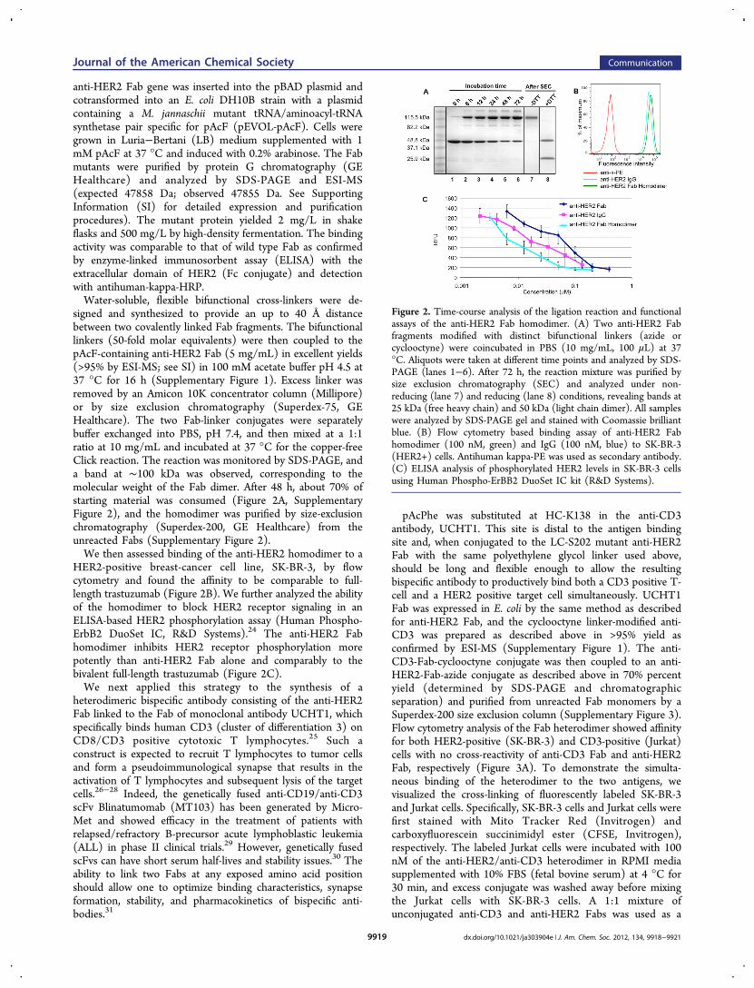
signed and synthesized to provide an up to 40 Å distancebetween two covalently linked Fab fragments. The bifunctionallinkers (50-fold molar equivalents) were then coupled to thepAcF-containing anti-HER2 Fab (5 mg/mL) in excellent yields(>95% by ESI-MS; see SI) in 100 mM acetate buffer pH 4.5 at37 °C for 16 h (Supplementary Figure 1). Excess linker wasremoved by an Amicon 10K concentrator column (Millipore)or by size exclusion chromatography (Superdex-75, GEHealthcare). The two Fab-linker conjugates were separatelybuffer exchanged into PBS, pH 7.4, and then mixed at a 1:1ratio at 10 mg/mL and incubated at 37 °C for the copper-freeClick reaction. The reaction was monitored by SDS-PAGE, anda band at ∼100 kDa was observed, corresponding to themolecular weight of the Fab dimer. After 48 h, about 70% ofstarting material was consumed (Figure 2A, SupplementaryFigure 2), and the homodimer was purified by size-exclusionchromatography (Superdex-200, GE Healthcare) from theunreacted Fabs (Supplementary Figure 2).We then assessed binding of the anti-HER2 homodimer to a
HER2-positive breast-cancer cell line, SK-BR-3, by flowcytometry and found the affinity to be comparable to full-length trastuzumab (Figure 2B). We further analyzed the abilityof the homodimer to block HER2 receptor signaling in anELISA-based HER2 phosphorylation assay (Human Phospho-ErbB2 DuoSet IC, R&D Systems).24 The anti-HER2 Fabhomodimer inhibits HER2 receptor phosphorylation morepotently than anti-HER2 Fab alone and comparably to thebivalent full-length trastuzumab (Figure 2C).We next applied this strategy to the synthesis of a
heterodimeric bispecific antibody consisting of the anti-HER2Fab linked to the Fab of monoclonal antibody UCHT1, whichspecifically binds human CD3 (cluster of differentiation 3) onCD8/CD3 positive cytotoxic T lymphocytes.25 Such aconstruct is expected to recruit T lymphocytes to tumor cellsand form a pseudoimmunological synapse that results in theactivation of T lymphocytes and subsequent lysis of the targetcells.26−28 Indeed, the genetically fused anti-CD19/anti-CD3scFv Blinatumomab (MT103) has been generated by Micro-Met and showed efficacy in the treatment of patients withrelapsed/refractory B-precursor acute lymphoblastic leukemia(ALL) in phase II clinical trials.29 However, genetically fusedscFvs can have short serum half-lives and stability issues.30 Theability to link two Fabs at any exposed amino acid positionshould allow one to optimize binding characteristics, synapseformation, stability, and pharmacokinetics of bispecific anti-bodies.31
pAcPhe was substituted at HC-K138 in the anti-CD3antibody, UCHT1. This site is distal to the antigen bindingsite and, when conjugated to the LC-S202 mutant anti-HER2Fab with the same polyethylene glycol linker used above,should be long and flexible enough to allow the resultingbispecific antibody to productively bind both a CD3 positive T-cell and a HER2 positive target cell simultaneously. UCHT1Fab was expressed in E. coli by the same method as describedfor anti-HER2 Fab, and the cyclooctyne linker-modified anti-CD3 was prepared as described above in >95% yield asconfirmed by ESI-MS (Supplementary Figure 1). The anti-CD3-Fab-cyclooctyne conjugate was then coupled to an anti-HER2-Fab-azide conjugate as described above in 70% percentyield (determined by SDS-PAGE and chromatographicseparation) and purified from unreacted Fab monomers by aSuperdex-200 size exclusion column (Supplementary Figure 3).Flow cytometry analysis of the Fab heterodimer showed affinityfor both HER2-positive (SK-BR-3) and CD3-positive (Jurkat)cells with no cross-reactivity of anti-CD3 Fab and anti-HER2Fab, respectively (Figure 3A). To demonstrate the simulta-neous binding of the heterodimer to the two antigens, wevisualized the cross-linking of fluorescently labeled SK-BR-3and Jurkat cells. Specifically, SK-BR-3 cells and Jurkat cells werefirst stained with Mito Tracker Red (Invitrogen) andcarboxyfluorescein succinimidyl ester (CFSE, Invitrogen),respectively. The labeled Jurkat cells were incubated with 100nM of the anti-HER2/anti-CD3 heterodimer in RPMI mediasupplemented with 10% FBS (fetal bovine serum) at 4 °C for30 min, and excess conjugate was washed away before mixingthe Jurkat cells with SK-BR-3 cells. A 1:1 mixture ofunconjugated anti-CD3 and anti-HER2 Fabs was used as a
Figure 2. Time-course analysis of the ligation reaction and functionalassays of the anti-HER2 Fab homodimer. (A) Two anti-HER2 Fabfragments modified with distinct bifunctional linkers (azide orcyclooctyne) were coincubated in PBS (10 mg/mL, 100 μL) at 37°C. Aliquots were taken at different time points and analyzed by SDS-PAGE (lanes 1−6). After 72 h, the reaction mixture was purified bysize exclusion chromatography (SEC) and analyzed under non-reducing (lane 7) and reducing (lane 8) conditions, revealing bands at25 kDa (free heavy chain) and 50 kDa (light chain dimer). All sampleswere analyzed by SDS-PAGE gel and stained with Coomassie brilliantblue. (B) Flow cytometry based binding assay of anti-HER2 Fabhomodimer (100 nM, green) and IgG (100 nM, blue) to SK-BR-3(HER2+) cells. Antihuman kappa-PE was used as secondary antibody.(C) ELISA analysis of phosphorylated HER2 levels in SK-BR-3 cellsusing Human Phospho-ErBB2 DuoSet IC kit (R&D Systems).
Journal of the American Chemical Society Communication
dx.doi.org/10.1021/ja303904e | J. Am. Chem. Soc. 2012, 134, 9918−99219919
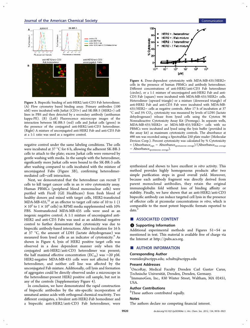
negative control under the same labeling conditions. The cellswere incubated at 37 °C for 6 h, allowing the adherent SK-BR-3cells to attach to the plate; excess Jurkat cells were removed bygentle washing with media. In the sample with the heterodimer,significantly more Jurkat cells were bound to the SK-BR-3 cellsafter washing compared to cells incubated with the mixture ofunconjugated Fabs (Figure 3B), confirming heterodimer-mediated cell−cell interaction.Next, we demonstrated that the heterodimer can recruit T
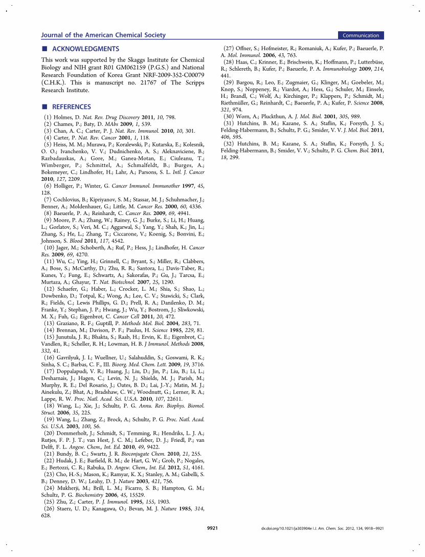
cells to kill target cancer cells in an in vitro cytotoxicity assay.Human PBMCs (peripheral blood mononuclear cells) werepurified with Ficoll (GE Healthcare) from fresh blood ofhealthy donors and mixed with target cells, HER2-transfectedMDA-MB-435,32 at an effector to target cell ratio of 10 to 1 (1× 10⁵ to 1 × 10⁴ cells) in RPMI media supplemented with 10%FBS. Nontransfected MDA-MB-435 cells were used as anisogenic negative control. A 1:1 mixture of unconjugated anti-HER2 and anti-CD3 Fabs was used as an additional negativecontrol to further demonstrate that cytotoxicity was due tobispecific antibody-based interactions. After incubation for 16 hat 37 °C, the amount of LDH (lactate dehydrogenase) wasmeasured from lysed cells as an indicator of cytotoxicity.9 Asshown in Figure 4, lysis of HER2 positive target cells wasobserved in a dose dependent manner only when theconjugated anti-HER2/anti-CD3 heterodimer was present;the half maximal effective concentration (EC50) was ∼20 pM.HER2-negative MDA-MB-435 cells were not affected by theheterodimer, and neither cell line was affected by theunconjugated Fab mixture. Additionally, cell lysis and formationof aggregates could be directly observed under a microscope inthe heterodimer-present HER2 positive cell sample, but not inany of the controls (Supplementary Figure 4).In conclusion, we have demonstrated the rapid construction
of bispecific antibodies by the site-specific incorporation ofunnatural amino acids with orthogonal chemical reactivity. Twodifferent conjugates, a bivalent anti-HER2-Fab homodimer anda bispecific anti-HER2/anti-CD3 Fab heterodimer, were
synthesized and shown to have excellent in vitro activity. Thismethod provides highly homogeneous products after twosimple purification steps in good overall yield. Moreover,because each antibody fragment was directly derived fromparent monoclonal antibodies, they retain the originalimmunoglobulin fold without loss of binding affinity orstability. Finally, we have shown that an anti-HER2/anti-CD3bispecific antibody can induce targeted cell lysis in the presenceof effector cells at picomolar concentrations in vitro, which iscomparable to the most potent bispecific formats reported todate.2
■ ASSOCIATED CONTENT
*S Supporting InformationAdditional experimental methods and Figures S1−S4 asmentioned in text. This material is available free of charge viathe Internet at http://pubs.acs.org.
■ AUTHOR INFORMATION
Corresponding [email protected]; [email protected]
Present Addresses†OncoRay, Medical Faculty Dresden Carl Gustav Carus,Technische Universitat, Dresden, Dresden, Germany.‡ImmunoGen, Inc., 830 Winter Street, Waltham, MA 02451,USA.
Author Contributions§These authors contributed equally.
NotesThe authors declare no competing financial interest.
Figure 3. Bispecific binding of anti-HER2/anti-CD3 Fab heterodimer.(A) Flow cytometry based binding assay. Primary antibodies (100nM) were incubated with Jurkat (CD3+) and SK-BR-3 (HER2+) celllines in PBS and then detected by a secondary antibody (antihumankappa-PE). (B) (Left) Fluorescence microscope images of theinteraction between SK-BR-3 (red) cells and Jurkat cells (green) inthe presence of the conjugated anti-HER2/anti-CD3 heterodimer.(Right) A mixture of unconjugated anti-HER2 Fab and anti-CD3 Fabat a 1:1 ratio was used as a negative control.
Figure 4. Dose-dependent cytotoxicity with MDA-MB-435/HER2+cells in the presence of human PBMCs and antibody heterodimer.Different concentrations of anti-HER2/anti-CD3 Fab heterodimer(circles), or a 1:1 mixture of unconjugated anti-HER2 Fab and anti-CD3 Fab (square) were incubated with MDA-MB-435/HER2+ cells.Heterodimer (upward triangle) or a mixture (downward triangle) ofanti-HER2 Fab and anti-CD3 Fab were incubated with MDA-MB-435/HER2− cells as negative controls. After 17 h of incubation at 37°C and 5% CO2, cytotoxicity was measured by levels of LDH (lactatedehydrogenase) release from lysed cells using the Cytotox 96Nonradioactive Cytotoxicity Assay Kit (Promega). In separate wells,MDA-MB-435/HER2+ or MDA-MB-435/HER2− cells with noPBMCs were incubated and lysed using the lysis buffer (provided inthe assay kit) as maximum cytotoxicity controls. The absorbance at490 nm was recorded using a SpectraMax 250 plate reader (MolecularDevices Corp.). Percent cytotoxicity was calculated by % Cytotoxicity= (Absorbanceexpt − Absorbancespontaneous average)/(Absorbancemax average− Absorbancespontaneous average).
Journal of the American Chemical Society Communication
dx.doi.org/10.1021/ja303904e | J. Am. Chem. Soc. 2012, 134, 9918−99219920
■ ACKNOWLEDGMENTS
This work was supported by the Skaggs Institute for ChemicalBiology and NIH grant R01 GM062159 (P.G.S.) and NationalResearch Foundation of Korea Grant NRF-2009-352-C00079(C.H.K.). This is manuscript no. 21767 of The ScrippsResearch Institute.
■ REFERENCES(1) Holmes, D. Nat. Rev. Drug Discovery 2011, 10, 798.(2) Chames, P.; Baty, D. MAbs 2009, 1, 539.(3) Chan, A. C.; Carter, P. J. Nat. Rev. Immunol. 2010, 10, 301.(4) Carter, P. Nat. Rev. Cancer 2001, 1, 118.(5) Heiss, M. M.; Murawa, P.; Koralewski, P.; Kutarska, E.; Kolesnik,O. O.; Ivanchenko, V. V.; Dudnichenko, A. S.; Aleknaviciene, B.;Razbadauskas, A.; Gore, M.; Ganea-Motan, E.; Ciuleanu, T.;Wimberger, P.; Schmittel, A.; Schmalfeldt, B.; Burges, A.;Bokemeyer, C.; Lindhofer, H.; Lahr, A.; Parsons, S. L. Intl. J. Cancer2010, 127, 2209.(6) Holliger, P.; Winter, G. Cancer Immunol. Immunother 1997, 45,128.(7) Cochlovius, B.; Kipriyanov, S. M.; Stassar, M. J.; Schuhmacher, J.;Benner, A.; Moldenhauer, G.; Little, M. Cancer Res. 2000, 60, 4336.(8) Baeuerle, P. A.; Reinhardt, C. Cancer Res. 2009, 69, 4941.(9) Moore, P. A.; Zhang, W.; Rainey, G. J.; Burke, S.; Li, H.; Huang,L.; Gorlatov, S.; Veri, M. C.; Aggarwal, S.; Yang, Y.; Shah, K.; Jin, L.;Zhang, S.; He, L.; Zhang, T.; Ciccarone, V.; Koenig, S.; Bonvini, E.;Johnson, S. Blood 2011, 117, 4542.(10) Jager, M.; Schoberth, A.; Ruf, P.; Hess, J.; Lindhofer, H. CancerRes. 2009, 69, 4270.(11) Wu, C.; Ying, H.; Grinnell, C.; Bryant, S.; Miller, R.; Clabbers,A.; Bose, S.; McCarthy, D.; Zhu, R. R.; Santora, L.; Davis-Taber, R.;Kunes, Y.; Fung, E.; Schwartz, A.; Sakorafas, P.; Gu, J.; Tarcsa, E.;Murtaza, A.; Ghayur, T. Nat. Biotechnol. 2007, 25, 1290.(12) Schaefer, G.; Haber, L.; Crocker, L. M.; Shia, S.; Shao, L.;Dowbenko, D.; Totpal, K.; Wong, A.; Lee, C. V.; Stawicki, S.; Clark,R.; Fields, C.; Lewis Phillips, G. D.; Prell, R. A.; Danilenko, D. M.;Franke, Y.; Stephan, J. P.; Hwang, J.; Wu, Y.; Bostrom, J.; Sliwkowski,M. X.; Fuh, G.; Eigenbrot, C. Cancer Cell 2011, 20, 472.(13) Graziano, R. F.; Guptill, P. Methods Mol. Biol. 2004, 283, 71.(14) Brennan, M.; Davison, P. F.; Paulus, H. Science 1985, 229, 81.(15) Junutula, J. R.; Bhakta, S.; Raab, H.; Ervin, K. E.; Eigenbrot, C.;Vandlen, R.; Scheller, R. H.; Lowman, H. B. J Immunol. Methods 2008,332, 41.(16) Gavrilyuk, J. I.; Wuellner, U.; Salahuddin, S.; Goswami, R. K.;Sinha, S. C.; Barbas, C. F., III. Bioorg. Med. Chem. Lett. 2009, 19, 3716.(17) Doppalapudi, V. R.; Huang, J.; Liu, D.; Jin, P.; Liu, B.; Li, L.;Desharnais, J.; Hagen, C.; Levin, N. J.; Shields, M. J.; Parish, M.;Murphy, R. E.; Del Rosario, J.; Oates, B. D.; Lai, J.-Y.; Matin, M. J.;Ainekulu, Z.; Bhat, A.; Bradshaw, C. W.; Woodnutt, G.; Lerner, R. A.;Lappe, R. W. Proc. Natl. Acad. Sci. U.S.A. 2010, 107, 22611.(18) Wang, L.; Xie, J.; Schultz, P. G. Annu. Rev. Biophys. Biomol.Struct. 2006, 35, 225.(19) Wang, L.; Zhang, Z.; Brock, A.; Schultz, P. G. Proc. Natl. Acad.Sci. U.S.A. 2003, 100, 56.(20) Dommerholt, J.; Schmidt, S.; Temming, R.; Hendriks, L. J. A.;Rutjes, F. P. J. T.; van Hest, J. C. M.; Lefeber, D. J.; Friedl, P.; vanDelft, F. L. Angew. Chem., Int. Ed. 2010, 49, 9422.(21) Bundy, B. C.; Swartz, J. R. Bioconjugate Chem. 2010, 21, 255.(22) Hudak, J. E.; Barfield, R. M.; de Hart, G. W.; Grob, P.; Nogales,E.; Bertozzi, C. R.; Rabuka, D. Angew. Chem., Int. Ed. 2012, 51, 4161.(23) Cho, H.-S.; Mason, K.; Ramyar, K. X.; Stanley, A. M.; Gabelli, S.B.; Denney, D. W.; Leahy, D. J. Nature 2003, 421, 756.(24) Mukherji, M.; Brill, L. M.; Ficarro, S. B.; Hampton, G. M.;Schultz, P. G. Biochemistry 2006, 45, 15529.(25) Zhu, Z.; Carter, P. J. Immunol. 1995, 155, 1903.(26) Staerz, U. D.; Kanagawa, O.; Bevan, M. J. Nature 1985, 314,628.
(27) Offner, S.; Hofmeister, R.; Romaniuk, A.; Kufer, P.; Baeuerle, P.A. Mol. Immunol. 2006, 43, 763.(28) Haas, C.; Krinner, E.; Brischwein, K.; Hoffmann, P.; Lutterbuse,R.; Schlereth, B.; Kufer, P.; Baeuerle, P. A. Immunobiology 2009, 214,441.(29) Bargou, R.; Leo, E.; Zugmaier, G.; Klinger, M.; Goebeler, M.;Knop, S.; Noppeney, R.; Viardot, A.; Hess, G.; Schuler, M.; Einsele,H.; Brandl, C.; Wolf, A.; Kirchinger, P.; Klappers, P.; Schmidt, M.;Riethmuller, G.; Reinhardt, C.; Baeuerle, P. A.; Kufer, P. Science 2008,321, 974.(30) Worn, A.; Pluckthun, A. J. Mol. Biol. 2001, 305, 989.(31) Hutchins, B. M.; Kazane, S. A.; Staflin, K.; Forsyth, J. S.;Felding-Habermann, B.; Schultz, P. G.; Smider, V. V. J. Mol. Biol. 2011,406, 595.(32) Hutchins, B. M.; Kazane, S. A.; Staflin, K.; Forsyth, J. S.;Felding-Habermann, B.; Smider, V. V.; Schultz, P. G. Chem. Biol. 2011,18, 299.
Journal of the American Chemical Society Communication
dx.doi.org/10.1021/ja303904e | J. Am. Chem. Soc. 2012, 134, 9918−99219921