synthesis, characterization and toxicity studies of schiff ... · synthesis, characterization and...
TRANSCRIPT
Digest Journal of Nanomaterials and Biostructures Vol. 7, No. 4, October-December 2012, p. 1843-1857
SYNTHESIS, CHARACTERIZATION AND TOXICITY STUDIES OF SCHIFF BASES [2-(2, 2-DIPHENYLETHYLIMINO)METHYL)PHENOLS]
ANCHORED SILVER NANOPARTICLES
SHANMUGAVEL SUJARANIA, THANGAMUTHU ANITHA SIRONMANIb*, ANDY RAMUa aSchool of Chemistry, Madurai Kamaraj University, Madurai – 625021, Tamilnadu. bSchool of Biotechnology, Madurai Kamaraj University, Madurai – 625021, Tamilnadu Diphenylethylamine is one of the morphine like drugs. A new series of biocompatible compounds have been prepared by Schiff base condensation reaction using 2,2-diphenylethylamine with 4-OH / 4-OMe substituted salicylaldehydes. The solid derivatives have been isolated and characterized by using IR, NMR and UV-Visible spectral techniques. Silver nano particles were synthesized by using starch as reducing and capping agent. The functionalized silver nanomaterials with Schiff base ligand anchored with fluorescent marker Rhodamine B was characterized using UV-Visible, IR, Fluorescent and AFM. Toxicity studies carried out in vivo (mice) and in vitro showed silver nano particles anchored 2-((2,2-dipenylethylimino)methyl)phenols to be bio-compatible without any apparent toxicity. A salicylaldehyde group on a definite position of an analgetically active molecule frequently causes an increase in the activity and an enhancement of binding to the opiate receptor. The primary reason behind the effort for their utility in biomedicine and therapy is their unique plasmonic properties and easy surface chemistry for a variety of functionalizations. (Received October 22, 2012; Accepted November 23, 2012) Keywords: Silver-2-((2,2-dipenylethylimino)methyl)phenols,salicylaldehyde, Diphenylethylamine, Schiff base condensation, Toxicity studies.
1. Introduction Schiff bases are an important class of compounds in medicinal and pharmaceutical field.
Also Schiff bases and their complexes have been used as biological models to understand the structures of biomolecules and biological processes [1a,b]. Schiff bases have often been used as chelating ligands in coordination chemistry[2a,b]. Schiff base with donors (N, O, S, etc) have structure similarities with neutral biological systems and due to presence of imine group, Schiff bases are utilized in elucidating the mechanism of transformation of rasemination reaction in biological system [3 a-c]. They show biological activities including antibacterial antifungal[4 a-c,1a,b] antidiabetic [4c,5a-g] antitumor [6a-d] antiproliferative [7a,b], anticancer [8a,b], herbicidal [9], anticorrosion and anti-inflammatory activities [10,1a]. Schiff bases represent an important class of compounds because they are utilized as starting materials in the synthesis of industrial products [10]. Moreover, Schiff bases are regarded as privileged ligands [11]. Due to their capability to form complexes with different transition metals can act as catalysts for many different reactions [12a-j].
New areas of researches, which mainly focuse on the synthesis specific, highly functional metal drug complexes, have drawn considerable attention. Transitional metal complexes are
*Corresponding author: [email protected] / [email protected]

1844 appealing candidates in the search for new diagnostic and therapeutic agents. They represent a uniquely modular system, wherein the metal centre holds its ligands in a precisely defined three-dimensional structure. These ligands can be varied relatively easily, in order to selectively change the characteristics of the complex in either subtle or dramatic fashion. Transition metal complexes also offer rich photophysical and photochemical properties, expanding their utility beyond structural recognition.
Morphine, a narcotic analgesic is a potent opiate drug that has a number of physiological effects. In 1943, Dodds, Lawson and Williams have published a note on the morphine-like properties of diphenylethylamine. Many active compounds, some of them much more potent than morphine, were synthesized on the basis of this structural design. It is well-known that the introduction of a salicylaldehyde group at a definite position on an analgetically active molecule frequently causes an increase in the activity and an enhancement of binding to the opiate receptor [13a,b].
Salicylaldehyde compound is a bidentate ligand and has several applications in chemistry and medicine [14]. Also the ligand has a good ability to form large numbers of chelates with most transition and non- transition metal ions of periodic table [15a,b]; Metal chelation is one of the excellent ways to increase the lipophilic character of the organic moiety. In fact, on coordination, ligands might improve their bioactivity profiles, while some inactive ligands may acquire pharmacological properties. Activity of various anti-inflammatory drugs existing in market has enhanced after complexation with transition metal ions [16a-d]. Silver nanoparticles have been of great interest due to their distinctive properties such as catalytic, optical, magnetic, electrical and due to its additional antibacterial properties [17a-c].
Diamino tetradentate Schiff bases and their complexes have been used as biological models to understand the structures of biomolecules and biological processes [1a,b]. To design effective chemotherapeutic agents and better anticancer drugs, it is essential to explore the interactions of metal with Schiff bases. In the present article, we report the synthesis and characterization of Schiff base derived from the ligand L (E) -2-((2,2- diphenylethylimino)methyl)phenol and its metal complexes to gain more information about related structural and spectral properties as well as their antimicrobial activities. In order to investigate the influence of substituents on the analgesic activity, 4 hydroxy /4 methoxy salicylaldehyde were synthesized.
2. Methods 2.1. Synthesis of the Schiff base ligand (E)-2-((2,2-iphenylethylimino)methyl ) Phenol The ligand L (E) -2-((2,2-diphenylethylimino)methyl)phenol: To 2.5 g of anhydrous
Na2SO4 suspended in 20mL of CH2Cl2, (1mmole) of 2,2-diphenyl ethyl amine was added followed by 1mmole of substituted salicylaldehyde with 4-OH/4-OME modification. The reaction mixture was refluxed for 2h until a yellowish colour appeared and then the solution was filtered, washed with 2 x 5 ml portions of CH2Cl2 and the volatile components in the combined solution were removed by evaporation in vacuum. The substituted Diphenylethylamines and Diphenylethanolamines were prepared as per earlier works [18a,b]. The crude product was obtained as a yellow powder, which was pure enough as inferred from the H1 NMR spectrum, and was used without further purification. The melting points of the compounds were determined on an XT4-100X microscopic melting point apparatus (Beijing, China).
2.2. Synthesis of silver nanoparticles Synthesis of silver (Ag) nanoparticles, were done by reduction and capping of 1 mM
solution of silver nitrate with starch as described in our earlier report [19]. Preparation of metal complexes Starch capped silver nanoparticles were mixed with ligand attached with Rhodamine B in 1: 5 or 2: 5 ratio (V/V ligand in chloroform and silver nanoparticle in distilled water respectively). The reaction was carried out by simple mixing at room temperature for one hour. The chemical structures of the Schiff-base ligand and its metal complexes were confirmed by various spectroscopic studies.
1845
2.3. UV-Visible- Spectroscopy Studies UV-Visible- Spectroscopy measurements (JascoV-550 spec) were carried out at a
resolution of 1nm. 2.4. Transmission Electron Microscopy (TEM) Measurements A drop of nanoparticle solution was loaded on carbon-coated copper grids and solvent was
allowed to evaporate. TEM measurements were performed on Philips model CM 200 instrument operated at an accelerating voltage at 200 kV.
2.5. Atomic Force Microscope AFM characterization was done by Advanced Physics and Engineering Research
Aloo SGS AFM. 2.6. Fourier Transform Infrared (FTIR) Spectroscopy Measurements FTIR spectroscopic analyses were carried out using a Jasco Fourier Transform Infrared
Spectrometer 410. FTIR spectrophotometer was connected to a photoacoustic cell in the spectral range from 4000 to 400 cm-1.
2.7. Fluorescence spectral studies Fluorescence spectra were recorded using RF-5301PC spectrofluorophotometer
(Shimadzu, Japan) with a 1 cm quartz cell. Both the excitation and emission bandwidths were 10 nm. All the experiments were measured after 5 min at a constant room temperature.
2.8. UV- visible absorption spectroscopy The UV-Visible spectrum was measured in a JASCO V 650 spectrophotometer containing
double beam in identical compartments each for reference and test solution from 200 nm to 900 nm.
2.9. NMR studies 1H NMR and 13C NMR spectra (at room temperature) (in CHCl3) were recorded on a
Bruker AVANCE II 300 DRX or average 400 DRX spectrometer with reference to Me4Si (0.0 ppm).
2.10. Confocal fluorescence microscopy Images obtained from the Confocal fluorescence microscopic images for the silver schiff
base [Ligand dpea-sal-OCH3] attached with rhodamine B were obtained using Carl Zeiss LSM510 MEPA instrument. Excitation with an Ar-ion laser at 488 nm, emission through a 505- to 530-nm filter reflected from a 545-nm dichroic mirror was monitored. The images of 2D and 3D orientation were obtained.
2.11. DNA cleavage study The DNA cleavage of the three nano preparations was examined by agarose gel
electrophoresis and CD spectrum by gel Electrophoresis. 2.11.1. Agarose Gel electrophoresis Calf thymus genomic DNA at a concentration of 10ug was treated with the complexes in
Tris buffer (50 mM Tris–acetate, 18 mM NaCl buffer, pH 7.2), and the contents were incubated for 1 h at room temperature. The samples were electrophoresed on 0.8% agarose gel in Tris–acetate buffer. After electrophoresis, the gel was stained with 1 mg/ml Ethidium bromide and photographed under UV light. To determine the extent of DNA cleavage, the results were compared with standard DNA marker.
1846
2.11.2 Circular Dichroism measurements CD spectra were taken using Circular dichroic spectrums of DNA were obtained by using
JASCO J-810 spectropolarimeter. All experiments were done using a quartz cell of 1 or 0.2 cm path length. Each CD spectrum was collected after averaging over at least 2 accumulations using a scan speed of 100 nm min_1 and a 1s response time. Machine plus cuvette baselines were subtracted and the resultant spectrum zeroed 50 nm outside the absorption bands.
2.12. In vitro hemolytic activity The lytic activity of all three the silver nanoparticle - Schiff base [ Ligand (E) -2-((2,2-
diphenylethylimino)methyl)phenol] were tested using Sheep RBC and trypan blue staining for viability testing. The dead cells were counted under the microscope.
2.13. In vitro apoptosis assay The growth inhibitory effect of metal complexes on the tumor cells was measured by the
microculture tetrazolium [3-(4, 5-dimethylthiazol-2-yl)-2,5- diphenyltetrazolium bromide, MTT] assay [20]. After incubation with the metal complex for 24 hours cells were exposed to 20 ul MTT solutions (5 mg/ml) and further cultivated for 4 h. The media with MTT were removed, and 100 ul of DMSO was added to dissolve formazan crystal at room temperature for 30 min. The absorbance of each cell at 450 nm was determined. Photographs were taken before the addition of DMSO under fluorescence microscope.
2.14. In vitro cytotoxicity assay In another experiment the effect on cell growth for the complexes (5mM) was studied by
culturing the cells in medium alone for 24 h. The viable cells remaining at the end of treatment period were determined by trypan blue exclusion assay and calculated as % of control, treated cell lysis.
2.15. Toxicity of the silver nanoparticle- [Ligand (E) -2-((2, 2-diphenylethylimino)
methyl) phenol]. The toxicity of the silver nanoparticles complexed with all three ligands was evaluated
using mice. The mice were injected with 50ug/ml of the silver nanoparticles - ligand complex subcutaneously. After four days the whole body X-Ray was taken. Blood and other organs were dissected and screened for the presence of silver nano - ligand complex.
2.16. Pharmacology The silver nanoparticles-[Ligand (E) -2-((2, 2-diphenylethylimino) methyl) phenol] were
evaluated for in vivo analgesic and anti-inflammatory activity. Acetic acid-induced rat paw edema method for evaluating the analgesic activity and anti-inflammatory activity [21].
2.17. Antifungal activity and antibacterial activity The antimicrobial tests were performed by the standard disc diffusion method [22]. The
complexes were screened for their antifungal activity against fungi viz. Aspergillus niger and anti bacterial activity against Escherischia Coli.
3. Results and discussion Due to their good complexing properties, biological activity and analytical application, the
Schiff bases of different denticity, as well as their metal complexes, have been subject of many studies. In the present paper, Schiff bases derived from substituted salicylaldehyde and 2, 2 – diphenylethylamine, complexed with silver nanoparticles were synthesized and characterized. Also ligands with simple substitutions, e.g. of hydroxy and methoxyl groups on the side chain, have been examined. The toxicity, immunological activities and their biological applications were studied.
1847
3.1. Biosynthesis and characterization of silver nano particles The silver nanoparticles were synthesized by the reduction and capping of starch.
Formation of silver nanoparticles was confirmed by using UV–Visible spectral analysis, TEM, etc [19]. The same batch of nanoparticles was used after functionalization with ligands. Silver nanoparticles have free electrons, which give rise to a surface plasmon resonance (SPR) absorption band [23]at 420 nm due to the combined vibration of electrons of metal nanoparticles in resonance with the light wave. More uniform shape (spherical), nanoparticles with a size of 20 nm were obtained (Data not shown).
3.2. Synthesis 3.2.1. Synthesis of the Schiff base [ Ligand (E) -2-((2,2- diphenylethylimino)methyl)phenol] A new series of bio-compatible compounds [ ligand L (E) -2-((2,2-
diphenylethylimino)methyl)phenol, L1, and L2] have been prepared by Schiff base condensation reaction using 2,2-diphenylethylamine with 4-OH / 4-OMe substituted salicylaldehydes (dpea-sal , dpea-sal-OH and dpea-sal-OCH3) as described in the methods. Salicylaldehydes are known to form simple neutral complexes coordinating as bidentate ligands in the mono-anionic form, adopting square-planar geometries with divalent transition metals, as observed for [Zn(salo)2], [Zn(5-Br-salo)2], [Cu(5-Br-salo)2], or octahedral [VO(3-OCH3- salo)2(H2O)][24].The newly synthesized Schiff bases acted as bidendate ligands.The metal ion is coordinated through the azomethine nitrogen and phenolic oxygen atoms via deprotonation. The bonding of ligand to metal ion is confirmed by the analytical and spectral studies. To enable pharmoco dynamic studies and other binding and metastasis in to cells both in vivo / in vitro, the ligands were attached with Rhodamine B prior to complexing with silver nanoparticles.
3.3. Characterization of Ligand Schiff base ligand (E) -2-((2,2-diphenylethylimino)methyl)phenol were subjected to CHN
analysis. The synthesized Schiff bases were characterized by 1H, 13C NMR spectra to ensure ligand purity in solution and elucidate the differently positioned proton and carbon. In 1H NMR spectra of the ligand complexes (Table 1), the signal due to azomethine proton of Schiff bases (9.42–9.1 ppm) appears in the region 10.2–9.8 ppm indicating that the coordination of the azomethine nitrogen to the metal ion. This downfield shift is due to deshielding of the N=CH proton. Also the slight downfield shift is observed for aromatic and aliphatic protons. The resonance due to phenolic –OH around 14.7–14.3 ppm in the Schiff bases disappears in the spectrum of complexes; this confirms that, the hydroxyl group reacted with metal ion via deprotonation. All these observations were confirmed by IR inferences. Azomethine carbon atom was observed at 151.69 ppm and the phenolic carbon was seen at 169.88 ppm while the carboxylic carbon was observed at 194.08 ppm [25][60].Melting temperatures were found to be 89, 93, and 97 for dpea-sal, dpea-sal-OH and dpea-sal-OCH3 respectively. The transition temperature of melting point for dpea-sal, dpea-sal-OH and dpea-sal-OCH3 was 4°C.

1848
Table 1. 1H, 13C NMR spectral data of Schiff base ligand (E) -2-((2,2- diphenylethylimino) methyl)phenol in CHCl3
Position of Proton/ CHCl3 CHCl3 Carbon 1H 13C
CH 4.1ppm 52ppm
CH2 4.3ppm 64ppm
OH 11.07ppm 165ppm
CH=N 8.268ppm 160ppm
Aromatic 6.7to 8.2ppm 116-142ppm protons /carbon
3.4. Characterization of Silver nanoparticles functionalized with ligand The Schiff base derivatives with fluorescent marker Rhodamine B was anchored on to
silver nanoparticles as described in methods. The resulting ligand anchored silver nanomaterials were characterized by using UV-Visible, IR, Fluorescent and AFM. Toxicity studies were carried out in vivo and in vitro on animal models.
3.4.1. Atomic Force Microscopic study Atomic force microscopy (AFM) was employed to characterize the size of the silver
particles and was found to vary between 20–60 nm (Fig.1). Aggregation and increase in size was observed in silver nanoparticles with ligands with methyl and methoxyl group substitutions on the side chain.

1849
Fig.1. Atomic Force Microscopy image of Schiff base ligand (E) -2-((2,2- diphenylethylimino) methyl)phenol anchored silver nanoparticles. 1. Ag- dpeasal
2. Ag-dpea-sal-OH and 3.Ag-dpea-sal-OCH3
3.4.2. Confocal fluorescence microscopy Images obtained from the confocal fluorescence microscope for the silver schiff base
[Ligand dpea-sal-OCH3] attached with rhodamine B are shown in Fig.2. We imaged spherical and rod-shaped silver nanoparticles, heterogeneously dispersed on the glass. A single confocal image was found to be sufficient for determining the 2D orientation of a metallic nanorod. Further, comparisons of simulations with observations indicated that it is possible to determine the orientation of metallic nanorods in 3D. Heterogeneous regions of high intensity are clearly
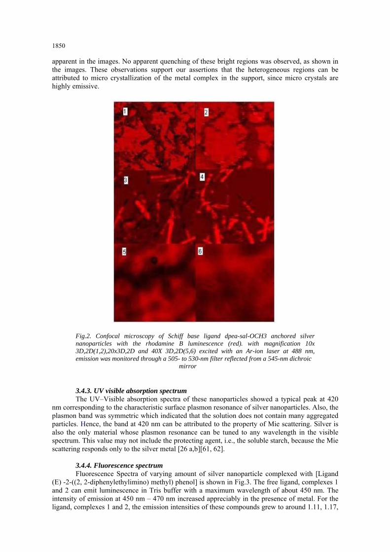
1850 apparent in the images. No apparent quenching of these bright regions was observed, as shown in the images. These observations support our assertions that the heterogeneous regions can be attributed to micro crystallization of the metal complex in the support, since micro crystals are highly emissive.
Fig.2. Confocal microscopy of Schiff base ligand dpea-sal-OCH3 anchored silver nanoparticles with the rhodamine B luminescence (red). with magnification 10x 3D,2D(1,2),20x3D,2D and 40X 3D,2D(5,6) excited with an Ar-ion laser at 488 nm, emission was monitored through a 505- to 530-nm filter reflected from a 545-nm dichroic mirror 3.4.3. UV visible absorption spectrum The UV–Visible absorption spectra of these nanoparticles showed a typical peak at 420
nm corresponding to the characteristic surface plasmon resonance of silver nanoparticles. Also, the plasmon band was symmetric which indicated that the solution does not contain many aggregated particles. Hence, the band at 420 nm can be attributed to the property of Mie scattering. Silver is also the only material whose plasmon resonance can be tuned to any wavelength in the visible spectrum. This value may not include the protecting agent, i.e., the soluble starch, because the Mie scattering responds only to the silver metal [26 a,b][61, 62].
3.4.4. Fluorescence spectrum Fluorescence Spectra of varying amount of silver nanoparticle complexed with [Ligand
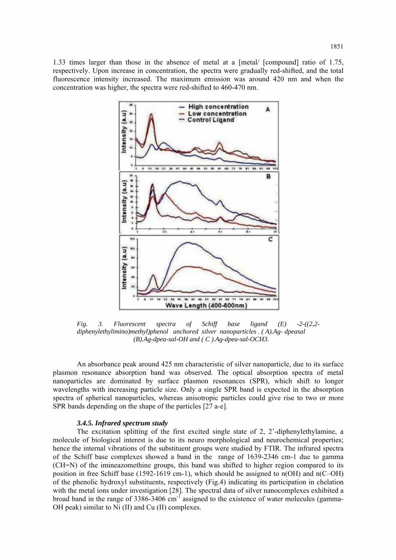
(E) -2-((2, 2-diphenylethylimino) methyl) phenol] is shown in Fig.3. The free ligand, complexes 1 and 2 can emit luminescence in Tris buffer with a maximum wavelength of about 450 nm. The intensity of emission at 450 nm – 470 nm increased appreciably in the presence of metal. For the ligand, complexes 1 and 2, the emission intensities of these compounds grew to around 1.11, 1.17,
1851
1.33 times larger than those in the absence of metal at a [metal/ [compound] ratio of 1.75, respectively. Upon increase in concentration, the spectra were gradually red-shifted, and the total fluorescence intensity increased. The maximum emission was around 420 nm and when the concentration was higher, the spectra were red-shifted to 460-470 nm.
Fig. 3. Fluorescent spectra of Schiff base ligand (E) -2-((2,2- diphenylethylimino)methyl)phenol anchored silver nanoparticles . ( A).Ag- dpeasal (B).Ag-dpea-sal-OH and ( C ).Ag-dpea-sal-OCH3.
An absorbance peak around 425 nm characteristic of silver nanoparticle, due to its surface
plasmon resonance absorption band was observed. The optical absorption spectra of metal nanoparticles are dominated by surface plasmon resonances (SPR), which shift to longer wavelengths with increasing particle size. Only a single SPR band is expected in the absorption spectra of spherical nanoparticles, whereas anisotropic particles could give rise to two or more SPR bands depending on the shape of the particles [27 a-e].
3.4.5. Infrared spectrum study The excitation splitting of the first excited single state of 2, 2’-diphenylethylamine, a
molecule of biological interest is due to its neuro morphological and neurochemical properties; hence the internal vibrations of the substituent groups were studied by FTIR. The infrared spectra of the Schiff base complexes showed a band in the range of 1639-2346 cm-1 due to gamma (CH=N) of the imineazomethine groups, this band was shifted to higher region compared to its position in free Schiff base (1592-1619 cm-1), which should be assigned to n(OH) and n(C–OH) of the phenolic hydroxyl substituents, respectively (Fig.4) indicating its participation in chelation with the metal ions under investigation [28]. The spectral data of silver nanocomplexes exhibited a broad band in the range of 3386-3406 cm-1 assigned to the existence of water molecules (gamma-OH peak) similar to Ni (II) and Cu (II) complexes.

1852
Fig.4. FTIR spectra of Schiff base ligand (E) -2-((2,2-diphenylethylimino)methyl)phenol anchored silver nanoparticles (a) Ligand alone (b) Low concentration and (c ) Ligand at
high concentration. 1.Ag- dpea-sal 2.Ag-dpea-sal-OH and 3.Agdpea- sal-OCH3.
The out-of-plane and in-plane C-N bending vibrations for DPEA were assigned at 818 and
634 cm-1, respectively. The same spectra showed bands at 462 – 482 and 641- 667 cm-1 due to (M-N) and (M-O) vibrations [29 a,b]. The appearance of these bands supports the involvement of azomethine and hydroxyl groups in coordination with silver nanoparticles [15a,b].
3.4.6. DNA Cleavage study DNA-binding studies are important for the rational design and construction of new and
more efficient drugs targeted to DNA. To design effective chemotherapeutic agents and better anticancer drugs, it is essential to explore the interactions of metal complexes with DNA. These studies are also important to understand the toxicity of drugs containing metal ions. The gel after electrophoresis clearly revealed that, Schiff base ligand (E) -2-((2,2-diphenylethylimino)methyl)phenol complex with silver nanoparticles have acted on DNA as there was no visible band in the treated DNA samples but a strong band was observed in the control (data not shown). This shows that the silver nanocomplexes showed full cleavage of E. coli DNA. As the compound was observed to cleave the DNA, it may be concluded that the compound inhibits the growth of the pathogenic organism by cleaving the genome.
3.4.7. Circular Dicharism In order to confirm any structural perturbation in the native structure of the DNA after the
adsorption of DNA onto the surface of nano preparations, we carried out circular dichroism (CD) studies. As shown in Fig.5, the CD spectrum revealed that the DNA used in our studies was in a B-form, as evidenced by a negative band at 248 nm and a positive band at 280 nm. The CD spectrum showed some broadening of the negative band at 248 nm, a slight shift of the positive band at 280 nm and a considerable absorption in the longer wavelength (>300 nm) indicating some perturbation of the DNA structure. A solution of CT-DNA exhibited a positive band (275 nm) from base stacking interactions and a negative band (245 nm) from the right-handed helicity of DNA [30]. The result implies that Ag- dpea-sal and Ag-dpea-sal-OH can interact with DNA in different mode. No appreciable spectral changes were observed in the presence of Ag-dpea-sal-OCH3 (Fig. 5) indicating that there was no binding with DNA.

1853
Fig.5. Circular dichroism (CD) spectra of native DNA and DNA in the Schiff base ligand (E) -2-((2,2-diphenylethylimino)methyl)phenol anchored silver
nanoparticles conjugate
The hyperchromic effect may be due to the electrostatic interaction between positively charged complexes and the negatively charged phosphate backbone at the periphery of the double helix DNA [31] similar to copper (II) complexes containing 1, 10-phenanthroline and L-phenylalanine ligands [32a-c]. The cationic charge of the complex could exert a strong electrostatic attraction to the anionic phosphate backbone of DNA, thus the metal complexes bind to DNA by non-covalent interactions such as electrostatic binding, groove binding and intercalative binding [33a,b]. Among these interactions, intercalation is one of the most important DNA binding modes, which was related to the antitumor activity of the compound [34]. It has been reported that the intercalating ability of the complex depends on the planarity of ligands, the coordination geometry, ligand donor atom type and the metal ion type [35].
3.5. In vivo animal study Schiff bases are an important class of compounds in medicinal and pharmaceutical fields.
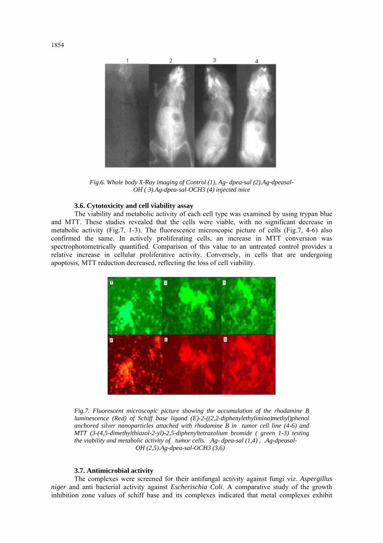
They show biological applications including antibacterial, antifungal, antitumor activity [36a-c] etc. Hence Schiff base ligand (E) -2- ((2,2-diphenylethylimino)methyl)phenol complex with silver nanoparticles were used to understand their interactions and functions in mammalian cells both in vivo and in vitro. Swiss mice were given subcutaneous injection and after 4 days the whole body X-ray was taken (Fig.6). The results compared with the control showed a broad tissue distribution except the brain and most of the nanocomplexes were observed in the abdomen. The in vivo and in vitro tissue studies showed the biodistribution in liver, lungs and intestine. Diphenylethylamine, with the side-chain-CH2CH(C6H5)NH2, differs widely from benzedrine, with the side-chain -CH2CH(CH3)NH2, in its actions on the central nervous system, on blood pressure and on smooth muscle in various organs. The substitution in this position of a phenyl for a methyl group, therefore, completely alters the physiological action [37a-c].
1854
Fig.6. Whole body X-Ray imaging of Control (1), Ag- dpea-sal (2).Ag-dpeasal- OH ( 3).Ag-dpea-sal-OCH3 (4) injected mice
3.6. Cytotoxicity and cell viability assay The viability and metabolic activity of each cell type was examined by using trypan blue
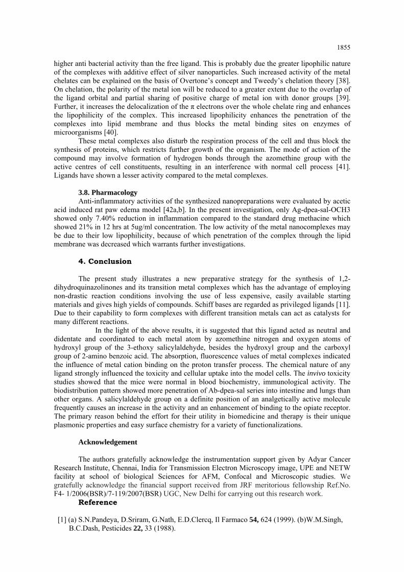
and MTT. These studies revealed that the cells were viable, with no significant decrease in metabolic activity (Fig.7, 1-3). The fluorescence microscopic picture of cells (Fig.7, 4-6) also confirmed the same. In actively proliferating cells, an increase in MTT conversion was spectrophotometrically quantified. Comparison of this value to an untreated control provides a relative increase in cellular proliferative activity. Conversely, in cells that are undergoing apoptosis, MTT reduction decreased, reflecting the loss of cell viability.
Fig.7. Fluorescent microscopic picture showing the accumulation of the rhodamine B luminescence (Red) of Schiff base ligand (E)-2-((2,2-diphenylethylimino)methyl)phenol anchored silver nanoparticles attached with rhodamine B in tumor cell line (4-6) and MTT (3-(4,5-dimethylthiazol-2-yl)-2,5-diphenyltetrazolium bromide ( green 1-3) testing the viability and metabolic activity of tumor cells. Ag- dpea-sal (1,4) , .Ag-dpeasal- OH (2,5).Ag-dpea-sal-OCH3 (3,6) 3.7. Antimicrobial activity The complexes were screened for their antifungal activity against fungi viz. Aspergillus
niger and anti bacterial activity against Escherischia Coli. A comparative study of the growth inhibition zone values of schiff base and its complexes indicated that metal complexes exhibit
1855
higher anti bacterial activity than the free ligand. This is probably due the greater lipophilic nature of the complexes with additive effect of silver nanoparticles. Such increased activity of the metal chelates can be explained on the basis of Overtone’s concept and Tweedy’s chelation theory [38]. On chelation, the polarity of the metal ion will be reduced to a greater extent due to the overlap of the ligand orbital and partial sharing of positive charge of metal ion with donor groups [39]. Further, it increases the delocalization of the π electrons over the whole chelate ring and enhances the lipophilicity of the complex. This increased lipophilicity enhances the penetration of the complexes into lipid membrane and thus blocks the metal binding sites on enzymes of microorganisms [40].
These metal complexes also disturb the respiration process of the cell and thus block the synthesis of proteins, which restricts further growth of the organism. The mode of action of the compound may involve formation of hydrogen bonds through the azomethine group with the active centres of cell constituents, resulting in an interference with normal cell process [41]. Ligands have shown a lesser activity compared to the metal complexes.
3.8. Pharmacology Anti-inflammatory activities of the synthesized nanopreparations were evaluated by acetic
acid induced rat paw edema model [42a,b]. In the present investigation, only Ag-dpea-sal-OCH3 showed only 7.40% reduction in inflammation compared to the standard drug methacine which showed 21% in 12 hrs at 5ug/ml concentration. The low activity of the metal nanocomplexes may be due to their low lipophilicity, because of which penetration of the complex through the lipid membrane was decreased which warrants further investigations.
4. Conclusion The present study illustrates a new preparative strategy for the synthesis of 1,2-
dihydroquinazolinones and its transition metal complexes which has the advantage of employing non-drastic reaction conditions involving the use of less expensive, easily available starting materials and gives high yields of compounds. Schiff bases are regarded as privileged ligands [11]. Due to their capability to form complexes with different transition metals can act as catalysts for many different reactions.
In the light of the above results, it is suggested that this ligand acted as neutral and didentate and coordinated to each metal atom by azomethine nitrogen and oxygen atoms of hydroxyl group of the 3-ethoxy salicylaldehyde, besides the hydroxyl group and the carboxyl group of 2-amino benzoic acid. The absorption, fluorescence values of metal complexes indicated the influence of metal cation binding on the proton transfer process. The chemical nature of any ligand strongly influenced the toxicity and cellular uptake into the model cells. The invivo toxicity studies showed that the mice were normal in blood biochemistry, immunological activity. The biodistribution pattern showed more penetration of Ab-dpea-sal series into intestine and lungs than other organs. A salicylaldehyde group on a definite position of an analgetically active molecule frequently causes an increase in the activity and an enhancement of binding to the opiate receptor. The primary reason behind the effort for their utility in biomedicine and therapy is their unique plasmonic properties and easy surface chemistry for a variety of functionalizations.
Acknowledgement The authors gratefully acknowledge the instrumentation support given by Adyar Cancer
Research Institute, Chennai, India for Transmission Electron Microscopy image, UPE and NETW facility at school of biological Sciences for AFM, Confocal and Microscopic studies. We gratefully acknowledge the financial support received from JRF meritorious fellowship Ref.No. F4- 1/2006(BSR)/7-119/2007(BSR) UGC, New Delhi for carrying out this research work.
Reference
[1] (a) S.N.Pandeya, D.Sriram, G.Nath, E.D.Clercq, Il Farmaco 54, 624 (1999). (b)W.M.Singh, B.C.Dash, Pesticides 22, 33 (1988).

1856 [2] (a) Y.Shibuya,K.Nabari,M.Kondo,S.Yasue,K.Maedo, F.Uchida,H.Kawaguchi , Chem.Lett. 37, 78 (2008) .(b) A.Roth, J.Becher, C.Herrmann, H.Gorls, G.Vaughn, M.Reiher, D.Klemm,W.Plass, Inorg.Chem. 45, 10066 (2006). [3] (a) E.Keskioglu, A.B.Gunduzalp, S.Cete, F.Hamurcu, B.Erk, Spectrochim.Acta A70 634 (2008) .(b) J.Z.Wu, L.Yuan, J.Inorg. Biochem. 98, 41 (2004). (c) K.P.Bala- subramanian, K.Parameswari, V.Chinnusamy, R.Prabhakaran, K.Natarajan. Spectrochim Acta A 65, 678 (2006). [4] (a) P.G.More, R.B.Bhalvankar, S.C.Pattar, J.Indian Chem.Soc.78, 474 (2001). (b) A.H.El-Masry, H.H.Fahmy, S.H.A.Abdelwahed, Molecules 5 1429 (2000). (c) M.A.Baseer, V.D.Jadhav, R.M.Phule, Y.V.Archana, Y.B.Vibhute, Orient J. Chem. 16,553 (2000).(d) D.P.Singh, K.Kumar, C.Sharma, Eur.J. Med.Chem. 44, 3299 (2009). [5] (a) R.S.Kumar, S.Arunachalam, Eur.J. Med.Chem. 44, 1878 (2009). (b)A.Kulkarni, S.A.Patil, P.S.Badami, Eur.J.Med.Chem. 44, 2904 (2009). (c) G.B.Bagihalli, P.G.Avaji, S.A.Patil, P.S.Badami, Eur.J. Med.Chem.43, 2639 (2008).(d)K.Singh, M.S.Bharwa, P.Tyagi, Eur. J. Med.Chem. 42, 394 (2007). (e) K.Singh, M.S.Bharwa, P.Tyagi, Eur.J. Med.Chem. 41,147 (2006). (f)R.Ramesh, S.Maheswaran, J.Inorg. Biochem. 96, 457 (2003) (g)J.Vanco, J.Marek,Z.Travnicek, E.Racanska, J.Muselik, O.Svajlenova,J.Inorg. Biochem. 102, 595 (2008). [6] (a) V.C.Silveira, J.S.Luz, C.C.Oliveira, I.Graziani,M.R.Ciriolo,A.M.C.Ferreira, J.Inorg. Biochem. 102, 1090 (2008).(b) M.P.Sathisha, U.N.Shetti, V.K.Revankar, K.S.R.Pai, Eur.J. Med. Chem.43, 2338 (2008).(c)S.A.Galal, K.H.Hegab, A.S.Kassab, M.L.Rodriguez, S.M.Kervin, A.M.A.El-Khamry, H.I.El-Diwani, Eur.J. Med. Chem. 44, 1500 (2009).(d) X.Zhong, J.Yi, J.Sun, H.L.Wei, W.S.Liu, K.B.Yu, Eur. J. Med. Chem.41, 1090 (2006). [7] (a) A.T.Chaviara, P.J.Cox, K.H.Repana, R.M.Papi, K.T.Papazisis, D.Zambouli,A.H.Kortsaris, D.A.Kyriakidis, C.A.Bolos, J. Inorg. Biochem. 98, 1271 (2004).(b) N.A.Illan-Cabeza, F.Hueso-Urena, M.N.Moreno-Carretero, J.M.Martinez-Martos, M.J.Ramirez-Exposito, J.Inorg.Biochem. 102, 647 (2008). [8] (a) S.B.Desai, P.B.Desai, K.R.Desai, Heterocycl.Commun. 7, 83 (2001). (b)P.Pathak, V.S.Jolly, K.P.Sharma, Orient.J.Chem. 16, 161 (2000). [9] S.Samadhiya, A.Halve, Orient.J.Chem. 17, 119 (2001). [10] S.Allah, A.M.Abid, Phosphorus, Sulfur Silicon Relat. Elem. 170, 75 (2001). [11] T.P.Yoon, E.N.Jacobsen, Science 299, 1691 (2003). [12] (a) T.Yamada, T.Ikeno, Y.Ohtsuka, S.Kezuka, M.Sato, I.Iwakura, Sci.Technol.Adv. Mater. 7, 184 (2006).(b)Y.P.Cai, C.Y.Su, A.W.Xu, B.S.Kang, H.Q.Liu, S.Jie, Polyhedron 20, 657 (2001).(c). W.Zeng, J.Li, S.Qin, Inorg.Chem. Commun. 9, 10 (2006).(d) X.Wang, J.Ding, J.J.Vittal, Inorg. Chim. Acta. 359, 3481 (2006).(e) G.Wu, X.Wang, J.Li, N.Zhao, W.Wei, Y.Sun, Catal.Today 131, 402 (2008).(f) G.Romanowski, E.Kwiatkowski, W.Nowicki, M.Kwiatkowski, T.Lis,Polyhedron 27, 1601 (2008).(g)S.Rayati, N.Torabi, A.Ghaemi, S.Mohebbi, A.Wojtczak, A.Kozakiewicz,Inorg. Chim. Acta 361, 1239 (2008).(h) V.Mirkhani, M.Moghadam, S.Tangestaninejad, I.Mohammadpoor- Baltork,E.Shams, N.Rasouli, Appl. Catal.A 334, 106 (2008).(i) V.Mirkhani, M.Moghadam, S.Tangestaninejad, I.Mohammadpoor-Baltork,N.Rasouli, Catal.Commun. 9,219 (2008) (j) Y.Chen, J.V.Ruppel, X.P.Zhang, J. Am.Chem.Soc. 129, 12074 (2007). [13] (a) C.C.Chien, G.W.Pasternak, Neurosci. Lett.190(2), 137 (1995).(b) T.Koch, V.Höllt, Pharmacol.Ther.117(2), 199 (2008). [14] W.T.Angew, Journal of Chemistry Education 35, 2708 (1996). [15] (a) S.M.Ben-Saber, A.A.Maihub, S.S.Hudere, M.M.El-ajaily, American Microchemical Journal 81, 191 (2005).(b) A.A.Maihub, M.M.El-ajaily, A.N.El-tajoury,The Egyptian Science Magazine 2(4), 83 (2005). [16] (a) M.Roch-Arveiller, V.Revelant, D.Pham Huy, L.Maman, J.Fontagne, J.R.J.Sorenson, J.P.Giroud, Inflamm.Res. 31, 1 (1990).(b) M.Konstandinidou, A.Kourounakis, M.Yiangou, L.Hadjipetrou, D.Kovala-Demertzi, S.Hadjikakou, M.Demertzis, J. Inorg. Biochem.70, 63 (1998).(c) A.Singh, R.Jain, A.Kumarsingla,
1857
Application number: EP19980610026,Filing date: 08/17/1998. (d) P.Lay, T.Hambley, B.Kennedy, Y.Morgan, Patent application 20090042848 Filing date: 24/03/2005. [17] (a) P.Mukherjee, A.Ahmad, D.Mandal, S.Senapati, R.Sainkar Sudhakar, M.I.Khan, et al. Nano.Lett.1, 515 (2001).(b) I.Sondi, S.S.Branka, J.Colloid Interface Sci.275, 177 (2004).(c) X.Chen, H.J.Schluesener, Toxicol.Lett. 176, 1 (2008). [18] (a) Dodds, Lawson, Williams. Nature 161, 614 (1943).(b)Dodds, Lawson, Williams. Nature 154, 514 (1944). [19] S.C.G.Kiruba Daniel, T.Anitha Sironmani, V.Tharmaraj, K.Pitchumani, Bull. Mater. Sci. 34(1), 1 (2011). [20] P.Moongkarndi, A.Srivattana, N.Bunyapraphatsara, S.Puthong, K.Laohathai, Mahidol University Journal of Pharmaceutical Sciences 18, 25 (1991). [21] C.A.Winter, E.A.Risley, G.W.Nuss, Exp.Biol.Med.111, 544 (1962). [22] W.L.Drew, A.L.Barry, R.O’Toole, J.C.Sherris, Appl.Environ.Microbiol. 24, 240 (1972). [23] K.Nakamoto, Infrared and Raman spectra of inorganic and coordination compounds 4th edition (John Wiley & Sons, New York, 1986. [24] N.Raman, Res.J. Chem. Environ. 4, 9 (2005). [25] K.Mounika et al. J.Sci. Res. 2(3), 513 (2010). [26] (a) W.Kleemann, Int. J. Mod. Phys. B 7, 2469 (1993).(b) K.Aoki, J.Chen, N.Yang, H.Nagasawa, Langmuir 19, 9904 (2003). [27] (a) M.A.Noginov, G.Zhu, M.Bahoura, J.Adegoke, C.Small, B.A.Ritzo, V.P.Drachev, V.M.Shalaev, Appl.Phys.B 86, 455 (2007).(b) S.S.Nath, D.Chakdar, G.Gope,Nanotrends-J.Nanotechnol.Appl.2, (2007).(c) I.O.Sosa, C.Noguez, R.G.Barrera, J.Phys.Chem. B 107, 6269 (2003).(d)A.Caceres, B.R.Lopez, M.A.Giron, H.Logemann, J. Ethnopharmacol.31, 263 (1991). (e) A.Caceres, H.Menendez, E.Mendez, E.Cohobon, B.E.Samayoa, E.Jauregui, E.Peralta, G.Carrillo, J. Ethnopharmacol. 48, 85 (1995). [28] MM.El-ajaily, A.A.Maihub, Jerash for research and studies 8(1), 7 (2003). [29] (a) Y.M.Issa, A.L.Al-ansary, O.E.Sherif, M.M.El- ajaily, Transition Metal Chemistry 22, 441 (1997).(b) M.M.El-ajaily, S.Ben-Gweirif, A.A.Maihub, A.N.El-tajouri, Basic Science and its application Journal 1(1), 196 (2006). [30] P.Lincoln, E.Tuite, B.Norden, J. Am. Chem. Soc.119, 1454 (1997). [31] Y.N.Xiao, C.X.Zhan, J. Appl. Polym. Sci. 84, 887 (2002). [32] (a) R.S.Kumar, S.Arunachalam, Eur. J. Med. Chem. 44, 1878 (2009). (b)N.Shahabadi, S.Kashanian, F.Darabi, DNA Cell Biol. 28, 1 (2009). (c) C.F.Jordan, LS.Lerman, J.H.Venable, Nature New Biol. 236, 67 (1982). [33] (a) J.Cowan, Curr. Opin. Chem. Biol. 5,634 (2001).(b).S.Mathur, S.Tabassum, Cent.Eur. J. Chem. 4, 502 (2006). [34] J.Tan, B.Wang, L.Zhu, Bioorg. Med. Chem. 17, 614 (2009). [35] H.Xu, K.C.Zheng, H.Deng, L.J.Lin, Q.L.Zhang, L.N.Ji, T.Dalton, 3, 2260 (2003). [36] (a) P.G.More, R.B.Bhalvankar, S.C.Pattar, J. Indian Chem. Soc.78, 474 (2001).(b) D.P.Singh, R.Kumar, J.Singh, Eur. J. Med. Chem. 44, 1731 (2009). (c)S.A.Galal, K.H.Hegab, A.S.Kassab, M.L.Rodriguez, S.M.Kervin, A.M.A.El-Khamry, H.I.El-Diwani, Eur. J. Med. Chem. 44, 1500 (2009). [37] (a) J.A.Gunn, Brit. Med. J. 2, 155 (1939).(b)A.Chistoni, E.Beccari, Arch. Ital. Sci.Farmac. 9, 1 (1940).(c) J.A.Gunn, M.R.Gurd, J. Phy.Biol. 97, 453 (1940). [38] B.G.Tweedy, Phyto. Pathology 55, 910 (1964). [39] K.Kralova, K.Kissova, O.Svajlenova, J.V.J.Parekh, P.Inamdhar, R.Nair, S.Baluja, S.Chanda, J. Serb. Chem. Soc. 70, 1161 (2005). [40] Y.Vaghasia, R.Nair, M.Soni, S.Baluja, S.N.Raman, Res.J. Chem. Environ.4, 9 (2005). [41] P.Dharamraj, Viswanathanmurthi, K.Natarajan, Trans. Met. Chem. 26, 105 (2001). doi:10.1023/A:1007132408648. [42] (a) R.Vinegar, J.F.Truax, J.L.Selph, Fed. Proc.35, 2447 (1976).(b) M.Di Rosa, J.P.Giroud, D.A.Willoughby, J. Pathol. 104, 15 (1971).