synthesis and development of diagnostic tools for medical ... · synthesis and development of...
TRANSCRIPT
General rights Copyright and moral rights for the publications made accessible in the public portal are retained by the authors and/or other copyright owners and it is a condition of accessing publications that users recognise and abide by the legal requirements associated with these rights.
Users may download and print one copy of any publication from the public portal for the purpose of private study or research.
You may not further distribute the material or use it for any profit-making activity or commercial gain
You may freely distribute the URL identifying the publication in the public portal If you believe that this document breaches copyright please contact us providing details, and we will remove access to the work immediately and investigate your claim.
Downloaded from orbit.dtu.dk on: Jan 14, 2020
Synthesis and Development of Diagnostic Tools for Medical Imaging
Schaarup-Jensen, Henrik
Publication date:2017
Document VersionPublisher's PDF, also known as Version of record
Link back to DTU Orbit
Citation (APA):Schaarup-Jensen, H. (2017). Synthesis and Development of Diagnostic Tools for Medical Imaging. DTUChemistry.

Synthesis and Development of Diagnostic
Tools for Medical Imaging
PhD thesis
Henrik Schaarup-Jensen
Kgs. Lyngby 2017
Department of Chemistry
Technical University of Denmark
1


Abstract
The need for novel diagnostic tools in medical imaging is increasing since they can improve
the positive therapeutic outcome as well as patient compliance. In this thesis different di-
agnostic tools were developed within an interdisciplinary project, whereas the main work
reported in this thesis was the synthesis of different materials.
The first project introduces the development of injectable fiducial markers within the
field of image-guided radiotherapy. Fiducial markers for computed tomography (CT)-
imaging are today needed in order to correlate the positioning of the tumor to provide
a more precise and improved radiation, as tumors rarely display a fixed position during
radiotherapy. A fiducial marker based on encapsulated gold nanoparticles within the gela-
tion matrix of sucrose acetate isobutyrate (SAIB) was developed and tested in vivo. The
scientific objective was to provide sufficient surface engineering of the gold nanoparticles
that will allow full dispersion of AuNPs within the hydrophobic environment of the SAIB
matrix. As stabilizing coating-materials PEG, PNIPAM polymers and a dithiolane SAIB
derivative were tested.
The unique gelation properties of the SAIB matrix led to the second project of injectable
fiducial tissue markers for surgical guidance of non-palpable tumors and brachytherapy.
As radioactive tracer, radioiodinated SAIB-derivatives were developed based on the re-
gioselective ipso-iodination of aryl-TMS moieties. Radioiodination was conducted under
carrier free conditions in high radiochemical yields by Tl(OOCCF3)3 and [125]NaI. The
application of the radiolabeled 125I-SAIB derivative was tested in vivo as a tissue marker
for surgical guidance and evaluated in terms of dosimetry.
The third project involved the synthesis of iodide-based contrast agents designed for re-
mote loading of liposomes. Long circulating contrast agents for blood pool imaging by
CT-imaging are of interest due to the current limitations of short retention times and the
considerable amounts needed to achieve a proper contrast. A small library of contrast
agents designed for remote loading of liposomes was synthesized. Remote loading of one
candidate was successful; however, the proper contrast level was not sufficient to be visible
by CT-imaging.
Another diagnostic tool for blood pool imaging is DOTA-modified pluronic/cyclodex-
trin (CD)-based polyrotaxanes (PRs). With the previously reported chelation of Gd and
the prolonged retention time of Gd-chelated Pluronic/CD PRs, the aim was to extend
the use of DOTA-modified Pluronic/CD-based PRs as positron emitting agents by chela-
tion of 64Cu. Pluronic/CD-based PRs grafted with DOTA was synthesized in the fourth
project. The last project deals with the site-specific radioiodination of peptides and pro-
teins. To achieve a conclusive outcome in radioimmunoassays as well as retaining a high
binding affinity of receptor binging peptides, the regioselective radioiodination is crucial.
Therefore, a TMS-substituted tyrosine was synthesized via the Negishi coupling to test
i

0. Abstract
if regioselective iodination could be obtained. The tyrosine derivative was used in the
synthesis of dipeptides of phenylalanine, tyrosine and tryptophan respectively in order to
evaluate the selectivity towards the ipso-substitution of the TMS in the iodination reac-
tion. First proof-of-concept experiments using aryl-TMS as placeholder in the site-specific
iodination of peptides and proteins have been demonstrated.
ii
Resumé
Behovet for nye redskaber inden for medicinsk billedbaseret diagnosticering har igennem
de sidste år været tiltagende grundet ønsket om at forbedre de terapeutiske resultater
samt patienternes tryghed under behandlingen. Denne afhandlingen beskriver tilblivelsen
af flere forskellige nye billedbaserede diagnosticerings metoder, som alle er udviklet i et in-
terdisciplinært projekt kaldt Nanoguide. Hovedvægten i rapporteringen ligger på syntese-
beskrivelsen af de forskellige materialer, der indgår i disse metoder.
Det første projekt beskriver udviklingen af en injicerbar markør inden for billede-guidet
radioterapi. Der er i dag et behov for markører der er synlig via CT billeddannelse og som
kan indsættes i tumorer som et reference punkt til en mere præcis og forbedret strålebehan-
dling under et radioterapi forløb. Dette er med formål at tilvejebringe at positioneringen
af tumorer varierer under et radioterapi forløb. En markør baseret på sterisk stabiliseret
guld nanopartikler (AuNPs) formuleret i en geleringsmatrix bestående af sucrose acetate
isobutyrat (SAIB) blev udviklet og testet in vivo. Den videnskabelig udfordring var at
overfladestabilisere AuNPs hvormed fuld blandbarhed i SAIB matricen kunne opnås. De
stabiliserende overfladematerialer der blev anvendt var polymerer af PEG og PNIPAM
samt et SAIB derivat der blev syntetiseret fra sucrose over fire trin.
De unikke geleringsegenskaber af SAIB matricen førte ligeledes til udviklingen af en
injicerbar vævsmarkør til kirurgisk vejledning af ikke mærkbare tumorer inden for
brystcancer samt brachyterapi. Som del af denne vævsmarkør blev et 125I mærket SAIB
derivat syntetiseret ud fra et TMS baseret SAIB forgangsmateriale via en regioselektiv
ipso-substitutering. Ioderingen blev gennemført uden brug af ikke-radioaktiv Jod i et
høj radiokemisk udbytte ved brug af Tl(OOCCF3)3 og [125I]NaI. Det 125I mærket SAIB
derivat blev formuleret og testet in vivo for at undersøge egnetheden som vævs markør
inden for kirurgisk vejledning. Dosimetri blev ligeledes beregnet for brug i patienter.
Det tredje projekt involverede syntesen af jod-baserede kontraststoffer konstrueret til at
undergå loading af liposomer ved brug af en gradient. Langtids cirkulerende kontrast-
stoffer til billeddannelse af det kardiovaskulære system er interessante på grund af de
nuværende begrænsninger i form af kort opholdstider og de betragtelige mængder af kon-
straststof nødvendig for at kunne opnå en tilstrækkelig konstrast af det cardiovaskulære
system. Et mindre bibliotek af kontraststoffer blev syntetiseret der er i stand til at kan
loades i liposomer ved brug af en gradient. Gradient medieret loading af liposomer var
succesfuldt for en af de syntetiserede kandidater. Desværre viste studiet at mængden af
kontraststof ikke var tilstrækkelig til at opnå en tilfredsstillende konstrast der var synbar
via CT-billeddannelse.
Et andet diagnostisk værktøj til visualisering af det cardiovaskulære system er DOTA-
iii

0. Resumé
modificerede pluronic/cyclodextrin-baseret polyrotaxaner (PRs). Med den tidligere rap-
porterede chelering af Gd og den forlængede retentionstid i blodet for Gd-chelaterede
Pluronic/CD PRs, var målet at udvide brugen af DOTA-modificerede Pluronic / CD-
baserede PRs til at chelere positron emitterende isotoper som 64Cu. DOTA modificerede
Pluronic/CD-baserede PRs blev syntetiseret i dette projekt.
Det sidste projekt beskæftiger sig med den regiospecifikke radioiodinering af peptider
og proteiner. For at opnå et konkluderbart og præcist radioimmunassay samt at
fastholde en høj affinitet for receptor bindende peptider efter radiomærkning, regioselektiv
radioiodinering er altafgørende. Derfor blev et TMS-substitueret tyrosin derivat
syntetiseret via Negishi-koblingen for at teste, om der kunne opnås regioselektiv iodinering
af peptider og proteiner. Tyrosin-derivatet blev anvendt i syntesen af dipeptiderne af
phenylalanin, tyrosin og tryptophan for at evaluere selektiviteten for ipso-iodineringen af
TMS-gruppen i iodineringsreaktionen. De første beviser på at aryl-TMS kan anvendes i
regioselektive iodineringer af peptider og proteiner blev demonstreret.
iv

Preface
The work presented in this PhD thesis is the result of three years of research conducted
from March 2013 to May 2016 at Center of Nanomedicine and Theranostics, Department
of Chemistry at the Technical University of Denmark. The Danish Council for Strategic
Research (Nanoguide, application no. 0603-00442B) and the Department of Chemistry
gratefully the PhD project. The work was conducted under the supervision of Prof. Mads
H. Clausen as main supervisor and Prof. Thomas L. Andresen as co-supervisor.
A three months external stay was conducted in the group of David H. Thompson at
the Department of Chemistry, Purdue University, Indianapolis, USA.
Appended to this thesis is the scientific paper including results presented in chapter ??.
The paper was published during the PhD study.
Injectable Colloidal Gold for Use in Intrafactional 2D Image-Guided Radiation
Therapy. Rasmus. I. Jølck, Jonas S. Rydhög, Anders N. Christensen, Anders E. Hansen,
Linda M. Bruun, Henrik Schaarup-Jensen, Asger Stevner von Wenck, Betina Børresen,
Annemarie T. Kristensen, Mads H. Clausen, Andreas Kjær, Knut Conradsen, Rasmus
Larsen, Per Munck af Rosenschöld Thomas L. Andresen. Adv. Healthcare Mater. 2015,
4, 856-863. included as appendix.
Publications based on results presented in remaining chapters are currently on-going.
v


Acknowledgements
There are numerous people that have contributed and being involved in the projects I
have been involved in during my PhD.
First and foremost, I would sincerely like to thank Prof. Mads. H. Clausen for ac-
cepting me as a Ph.D. student in his group and allowing me the opportunity to develop
as a person. I can not put into words what a learning experience this has been to me. I
started as a rookie to end up as being a promising researcher. I am grateful for such an
extraordinary guidance and useful discussions. It has been a privilege. I would likewise
like to thank Prof. Thomas L. Andresen for his visionary ideas; your approach to solve
scientific problems is a true inspiration. All collaborators of the Nanoguide project are
gratefully acknowledged for their work and contributions in order to realize the projects.
It has been a true learning experience dealing with scientific problems of high complex-
ity in a context beyond the field of organic chemistry. Part of the Nanoguide project I
would especially like to thank Dr. Rasmus I. Jølck, Dr. Anders E. Hansen, Associate
Prof. Jonas R. Henriksen, Gokce Engudar, Linda M. Bruun, Dr. Andreas I. Jensen and
Associate Prof. Henrik H. El-Ali.
I would also like to thank former and present members of the Mads H. Clausen group
as well as colleagues of DTU Chemistry for a good company. Especially I would like to
thank Dr. Beatrice Bonora, I could not have wished for a better lab-mate and friend.
I would also like to thank Dr. Peter Hammershøj for the daily supervision and useful
discussions. Jorge Peiro, you will always remain as my Real Madrid nemesis but your
positive attitude and friendship mean a lot to me. I would like to commend Dr. Casper
Hoeck for useful NMR discussions and second-opinions. For synthetic discussions, I am
thankful to especially Kim T. Mortensen, Dr. Thomas Flagstad and Dr. Lasse B. Olsen.
The late night hard-working company of Christine Kinneart and Kim T. Mortensen was
truly appreciated.
I would like to express my deepest gratitude to Prof. David H. Thompson for my ex-
ternal stay. Thank you for your guidance and encouragement. As a person, you are a
true inspiration. I would especially like to thank Dr. Seok-Hee Hyun for hosting me dur-
ing my stay together with the rest of the group of Prof. David H. Thompson especially
Bradley Loren, Vivek Badvaik and Christopher Collins for an extraordinary friendship
and company. I would like to thank the financial support of Frants Allings Legat and
Oticon Fonden during my external stay at Purdue University.
The finalization of this thesis was not possible without the tedious and extraordinary
proof-readings of Dr. Irene Boos and Dr. Shahid I. Awan. I would like to declare a
personally and profound acknowledgement to Irene Boos for sparing, useful discussions
and helping out the last months of this thesis.
Finally, I would like to thank my family for you encouragement, and support throughout
vii

0. Acknowledgements
this thesis, without you, this would not be possible. You are my solid ground. Especially,
I would like to thank my mom and dad, This thesis is to you, I owe you everything.
viii

Table of contents
Abstract i
Resumé iii
Preface v
Acknowledgements vii
1 Synthesis of Nanoparticle-based Fiducial Markers for Image-Guided Radiotherapy 1
1.1 Image-guided radiotherapy . . . . . . . . . . . . . . . . . . . . . . . . . . . . 1
1.2 Use of radiopaque fiducial markers in IGRT . . . . . . . . . . . . . . . . . . 3
1.3 Problem statement . . . . . . . . . . . . . . . . . . . . . . . . . . . . . . . . 4
1.4 Design and theoretical deliberations . . . . . . . . . . . . . . . . . . . . . . 4
1.5 Contrast properties of AuNPs . . . . . . . . . . . . . . . . . . . . . . . . . . 5
1.6 Sucrose acetate isobutyrate . . . . . . . . . . . . . . . . . . . . . . . . . . . 5
1.7 The first generation - SAIB-AuNPs based liquid fiducial radiopaque marker 7
1.8 The chemistry of sucrose . . . . . . . . . . . . . . . . . . . . . . . . . . . . . 10
1.9 Design of the dithiolane-functionalized SAIB derivative . . . . . . . . . . . . 15
1.10 The synthesis of dithiolane functionalized SAIB derivative . . . . . . . . . . 16
1.11 Surface functionalization of AuNP . . . . . . . . . . . . . . . . . . . . . . . 17
1.12 Discussion and future perspectives . . . . . . . . . . . . . . . . . . . . . . . 18
1.13 PNIPAM and PEG coated AuNPs in SAIB formulation . . . . . . . . . . . 21
1.14 Conclusion . . . . . . . . . . . . . . . . . . . . . . . . . . . . . . . . . . . . 23
1.15 Iodine based SAIB derivative for IGRT . . . . . . . . . . . . . . . . . . . . . 24
2 Injectable fiducials for pre-operative localization of tumors in breast cancer 27
2.1 Design and theoretical deliberations . . . . . . . . . . . . . . . . . . . . . . 31
2.2 The β-cationic effect of trimethylsilanes in electrophilic substitutions . . . . 32
2.3 Synthesis of SAIB-based precursor for radioiodination . . . . . . . . . . . . 33
2.4 Radioiodination of SAIB precursor . . . . . . . . . . . . . . . . . . . . . . . 35
2.5 TMS-BioXmark . . . . . . . . . . . . . . . . . . . . . . . . . . . . . . . . . . 39
2.6 Conclusion . . . . . . . . . . . . . . . . . . . . . . . . . . . . . . . . . . . . 50
3 Synthesis of Contrast Agents for Active Loading in Liposomes 53
3.1 Contrast agent for blood pool imaging . . . . . . . . . . . . . . . . . . . . . 53
3.2 Liposomes . . . . . . . . . . . . . . . . . . . . . . . . . . . . . . . . . . . . . 55
3.3 Liposomal formulation of CT contrast agents . . . . . . . . . . . . . . . . . 56
3.4 Remote loading of active compounds in liposomes . . . . . . . . . . . . . . . 57
3.5 Problem statement . . . . . . . . . . . . . . . . . . . . . . . . . . . . . . . . 59
3.6 Design of contrast agents . . . . . . . . . . . . . . . . . . . . . . . . . . . . 60
3.7 Synthesis of iohexol derivative for mediated loading . . . . . . . . . . . . . . 60
3.8 Synthesis of diatrizoic acid derivative for mediated loading . . . . . . . . . . 61
ix

TABLE OF CONTENTS
3.9 Building blocks for development of new CT contrast agents . . . . . . . . . 62
3.10 Phosphoplipid based contrast agents . . . . . . . . . . . . . . . . . . . . . . 63
3.11 Glucosamine based contrast agents . . . . . . . . . . . . . . . . . . . . . . . 64
3.12 Mediated loading of contrast agents . . . . . . . . . . . . . . . . . . . . . . 66
3.13 Optimized loading using ammonium sulfate gradient . . . . . . . . . . . . . 66
3.14 Mediated loading using citrate buffer gradient . . . . . . . . . . . . . . . . . 67
3.15 In vivo performance of mediated loaded liposomes . . . . . . . . . . . . . . 67
3.16 Discussion and future perspectives . . . . . . . . . . . . . . . . . . . . . . . 67
3.17 Conclusion . . . . . . . . . . . . . . . . . . . . . . . . . . . . . . . . . . . . 68
4 Cyclodextrin-pluoronic based polyrotaxanes for PET-imaging 69
4.1 Niemann Pick disease Type C . . . . . . . . . . . . . . . . . . . . . . . . . . 71
4.2 Threading/inclusion complex formation of CDs . . . . . . . . . . . . . . . . 72
4.3 In vivo bio-distributions studies of CD/pluoronic PRs . . . . . . . . . . . . 72
4.4 CD/pluoronic PRs as contrast agents . . . . . . . . . . . . . . . . . . . . . . 73
4.5 Problem statement . . . . . . . . . . . . . . . . . . . . . . . . . . . . . . . . 75
4.6 Design and theoretical deliberation of PET HP-β-CD/pluoronic PRs . . . . 75
4.7 Synthesis of the CD/pluoronic based polyrotaxanes . . . . . . . . . . . . . . 76
4.8 Discussion . . . . . . . . . . . . . . . . . . . . . . . . . . . . . . . . . . . . . 81
4.9 Conclusion . . . . . . . . . . . . . . . . . . . . . . . . . . . . . . . . . . . . 81
5 Regioselective iodination of peptides and proteins 83
5.1 Iodination by electrophilic aromatic substitution . . . . . . . . . . . . . . . 86
5.2 Iodination by iodine monochloride . . . . . . . . . . . . . . . . . . . . . . . 87
5.3 Iodination by Chloramine T . . . . . . . . . . . . . . . . . . . . . . . . . . . 87
5.4 Iodination by Iodogen . . . . . . . . . . . . . . . . . . . . . . . . . . . . . . 88
5.5 Iodination by use of prosthetic groups . . . . . . . . . . . . . . . . . . . . . 88
5.6 Miscellaneous techniques for Phe iodination . . . . . . . . . . . . . . . . . . 89
5.7 Problem statement . . . . . . . . . . . . . . . . . . . . . . . . . . . . . . . . 90
5.8 Retrosynthetic analysis for the synthesis of the TMS derived Tyrosine . . . 91
5.9 Synthesis of amino acids using β-iodo-alanine derivatives . . . . . . . . . . . 94
5.10 Synthesis of a TMS-derived tyrosine derivative by Negishi coupling . . . . . 95
5.11 Formation of dipeptides . . . . . . . . . . . . . . . . . . . . . . . . . . . . . 99
5.12 Hydrogenolysis of dipeptides . . . . . . . . . . . . . . . . . . . . . . . . . . 100
5.13 Iodination of dipeptides . . . . . . . . . . . . . . . . . . . . . . . . . . . . . 102
5.14 Discussion . . . . . . . . . . . . . . . . . . . . . . . . . . . . . . . . . . . . . 106
5.15 Future perspectives . . . . . . . . . . . . . . . . . . . . . . . . . . . . . . . . 108
5.16 Conclusion . . . . . . . . . . . . . . . . . . . . . . . . . . . . . . . . . . . . 108
6 Experimental 111
7 Concluding remarks 129
Bibliography 131
x

Synthesis of Nanoparticle-basedFiducial Markers for Image-Guided
Radiotherapy 11.1 Image-guided radiotherapy
External beam radiotherapy (EB-RT) remains today as one of the most promising and
used treatments for cancer [1]. It is reported that approximately 50% of all cancer patients
undergo radiotherapy treatment [1]. Likewise, it is estimated that usage of radiation ther-
apy is cost-efficient and constitutes only 5% of the total cost of cancer treatment [1].
The technical term tumor control used in EB-RT treatment is stated to be proportional to
the dose of radiation delivered to the tumor [2]. This is described via the linear-quadratic
model presented in Brenner et al., correlating the therapeutic radiation dose to cell/tumor
viability [3]. The main reason that limits the of dose to malignant tissue, is the increased
risk of harming or damaging nearby vital organs or surrounding healthy tissue [2]. This
emphasize that the current issue within EB-RT is related to the current standards of pre-
cision of high radiation doses to a defined target.
Figure 1.1: Schematic representation of the different kinds of target volume.
As a common procedure in the planning of EB-RT, a gross target volume (GTV) is
1

1. Synthesis of Nanoparticle-based Fiducial Markers for Image-Guided Radiotherapy
outlined corresponding to the tumor volume (Figure 1.1). GTV plus the surrounding
area within the risk of microscopy spread in total constitute the clinical target volume
(CTV) [2]. To compensate for daily positioning errors and internal motion of organs, an
additional technical margin is added to CTV to afford the total planned target volume
(PTV) [2], confer Figure 1.1. The technical margin is inferred as tumors rarely display
a fixed position during radiation treatment. Tumor motion is clearly illustrated in the
example shown below (Figure 1.2).
1.2b
Figure 1.2: CT images showing a change in volume and position of adenocarcinoma of
the right lung during radiotherapy. The white line indicates the gross tumor volume
(GTV) (white line missing for image c). a) Delineation of gross target volume from the
original planning CT scan. b) CT scan conducted after 24 days. c) megavoltage CT scan
conducted after 60 days. d) kilovoltage scan conducted after 67 days [4].
The dynamic of the irradiation process is balanced by the clinical evaluation of gaining
tumor control - also referred to as tumor control probability (TCP) - versus the risk of
complicating normal healthy tissue known as the normal tissue complication probability
(NTCP) [2].
To illustrate the impact of the addition of margins to assure full irradiation of a target,
consider spherical tumor with a diameter of 4 cm. With an additional 3 mm standard
deviation in order to assure full coverage (derived from systematic and random deviation)
leads to a 6 cm diameter volume to be irradiated which roughly corresponds to three times
the volume of the initial tumor [5]. Alternatively, a reduction of 1.5 mm in safety margin
of a 5.0 cm spherical tumor reduce the irradiated volume including healthy tissue from 316
cm3 to 48 cm3. To further underline the difficulties ensuring tumor control within EB-RT,
2

1. Synthesis of Nanoparticle-based Fiducial Markers for Image-Guided Radiotherapy
the minimum radiation dose should ideally be homogenously distributed throughout the
PTV [5]. This is not the case in many situations as the distribution of dose is heteroge-
neous and as a consequence, the PTV has to be increased [5]. As described by Jaffray et
al. − "If all regions of a tumor are considered to be equal in their radiation response, the
dose that determines tumor control is the minimum dose to the tumor" [5].
Improved accuracy and precision of radiotherapy are required to enhance tumor control [].
Therefore, in order to improve and specify the therapeutic radiation dose to the tumor, the
strategy is to provide a conformal dose distribution, corresponding to the target volume.
Conformal dose distribution in accordance to the target volume is achieved by advanced
computerized planning systems [4]. Among initiatives to decrease the PTV, the imple-
mentation of frequent imaging during treatment, referred to as image-guided radiotherapy
(IGRT) has been developed. IGRT provides knowledge of the exact tumor position and
elucidates the dynamic behavior of organ motions and tumors during radiotherapy [2].
The main contribution of IGRT is usually in repositionings if gross misalignments are
observed due to unpredictable tumor or organ motion [4]. In addition, IGRT has enabled
the implementation of intensity modulated radiotherapy (IMRT). In IMRT, the inten-
sity of the radiation dose is modulated (a non-uniform dose) to conform more precisely
throughout the PTV. This is in accordance to the prerequisite for IMRT of high control
of geometric uncertainties as a steep decline between target delineation and healthy tissue
is required [4]. This enables potential dose escalation to the tumor without irradiation of
nearby tissues [2]. Being able to customize and adapt the dose more directly towards the
treated volume imply a higher acceptance of risk as high conformal radiation doses will
lead to control of tumor and lowering the possibility to damaging healthy tissue, NTCP [5].
The toxicity of radiotherapy can be obtained, as fewer fractionations are needed to achieve
therapeutic effect. As an example, front-line radiotherapy using conformal radiotherapy
and IMRT, has facilitated high-dose conformal radiotherapy of unresectable tumors in liver
cancer which previously was not suitable for radiotherapy [4]. Image guidance is usually
referred to as either online or offline. Online, images are recorded before each fractions of
radiation doses to assess simple positioning corrections. In the case of offline image guid-
ance, images are recorded after several fractions without intervention by which statistical
derivations are computed to check for systematic geometric errors. Online image guid-
ance has proven to reduce the number of geometric errors and to be more accurate when
the surrounding tissue is vital or high dose radiation is given in one or few fractions [4].
Adjusting the radiation to the specific situation provide adaptive radiotherapy [4].
1.2 Use of radiopaque fiducial markers in IGRT
In those cases of cancers where the anatomical tumor position cannot be accurately
identified or correlated to anatomical reference points, like parts of the skeleton,
radiopaque fiducials markers are inserted in or nearby the tumor [4]. A radiopaque fiducial
marker is defined as a material possessing radioopacity which makes it possible to use
as a reference point [4]. Inserting these markers in tumors improves the contrast and
tumor localization and allows for fluoroscopic real-time tumor tracking. This has proven
to increase the accuracy of treatment for moving tumors [6–8]. The radiopaque fiducial
3

1. Synthesis of Nanoparticle-based Fiducial Markers for Image-Guided Radiotherapy
markers used today are typical solid metal-based implants of gold seeds with rather large
physical dimensions, typically in the range of 1 mm x 5 mm (diameter x length) [6].
Gold fiducial markers have been implemented in the location of tumors within prostate,
lung, liver, pancreatic, and paraspinal cancers [4]. Internal markers have proven to be
a necessity for accurate tumor correlation in gated radiotherapy used in lung cancer and
have been shown to provide a tumor correlation than external/superficial markers [7,9,10] .
Moreover, the implantation procedure requires complicated insertion procedures typically
via percutaneous insertion using 18 G needles. Especially in the case of fiducial marker
insertion in lung tumors, this can lead to complications in form of pneumothorax (33-68%
of the cases) [11–13]. Bleeding is likewise also reported [11–13]. Besides these adverse
effects, solid markers also have a tendency to migrate from their original position [13,14].
1.3 Problem statement
In order to enhance the precision of radiotherapy towards a defined target volume and the
use of fiducial markers to enhance contrast and correlation of tumors, it was envisioned
within the Nanoguide project, that a nanoparticle-based liquid fiducial radiopaque marker
for CT/fluoroscopy could be developed for IGRT. A nanoparticle based fiducial marker
should encompass an improved quality of image-guided radiotherapy by providing better
tumor coverage, including better tumor correlation and online tracking. The intended
outcome should hopefully be a more favorable conformation of the radiation dose and
improved intensity modulated radiotherapy leading to better tumor control. In the more
advanced setups like gated radiotherapy (in lung cancers) or if the tumor is inaccessible,
where the radiation dose has to conform tightly with the PTV, it was envisioned that the
design would improve irradiation compared to the current fiducial markers. Consequently,
a new nanoparticle-based liquid fiducial radiopaque marker will provide a safer and
convenient insertion and meet higher patient compliance.
1.4 Design and theoretical deliberations
As mentioned above, considerable complications belong to insertion procedures of fiducial
markers particularly within lung cancers. The nanoparticle-based fiducial marker could
overcome this problem and be compatible with CT imaging modalities. The design was
based on the intention of developing a liquid radiopaque fiducial marker that will possess
sol-gel properties similar to the lower critical solution temperature (LCST) properties of
many polymeric systems and proteins.The sol-gel property will ensure that the marker
will gel up upon injection. Liquefaction of the fiducial marker will entail percutaneously
insertions (injections) using hyper- and thin hypodermic needles within the range of
25 G needles.This will reduce the dimensions of the the current standards of 18 gauge
needles used today for percutaneous insertions, thus affording improved usability in clinical
routines and patient compliance. As contrast property (radioopacity), it was reasoned
that use of gold nanoparticles (AuNPs) could be sufficiently suspended within a proper
sol-gel system. The premise will be based on the AuNPs being sterically stabilized towards
aggregation and leakage of AuNPs from the gel-system will be kept at a minimum. AuNPs
have likewise been proven stable during irradiation with X-rays [15].
4

1. Synthesis of Nanoparticle-based Fiducial Markers for Image-Guided Radiotherapy
1.5 Contrast properties of AuNPs
In order to develop a proper contrast agent for computed tomography (CT), heavy atoms
that possess high X-ray attenuation and contribute with a high mass fraction of the
contrast agents have to be located at the desired targets [16]. The most common heavy
atoms used today in contrast agents are mainly based on gold or iodine. Gold possess
higher X-ray attenuation than iodine (at 100 KeV: Au - 5.16 cm2 g−1, I - 1.97 cm2 g−1,
soft tissue - 0.169 cm2 g−1, bone - 0.186 cm2 g−1) [17]. In accordance to Lambert Beer’s
law, attenuation is most efficiently achieved by a material with high absorption coefficient
or by increasing the thickness of the material [18]. As gold is a more dense material than
iodine X-ray attenuation is higher [18]. This difference was illustrated in the comparison of
PEGylated AuNPs and the iodine-based Ultravist contrast agent (iopromide). PEGylated
AuNPs with a size of 30 nm and at a concentration of 33 mg AuNPs mL−1 provided a
contrast of 838 Hounsfield Units (HU), as opposed to Ultravist, where a concentration of
407 mg mL−1 Auravist is needed to afford the same contrast level. Per weight iodine (mg I
mL−1), this would correspond to 189 mg I mL−1 which gives a ratio of 5.6 higher of contrast
of AuNPs compared to iodine in this particular case [19]. The study also confirmed that
biocompability and non-toxicity of PEGylated AuNPs [19]. The was further substantiated
in the study of Hainfeld et al., where AuNPs provided longer cardiovascular circulation
times than the corresponding Omnipaque (Iohexol). In addition, The AuNPs was not
taken up by the liver or spleen but cleared renally. Intravenous injection likewise indicated
a biphasic clearance profile and the LD50 for mice was reported to be 3.2 g Au kg−1 [17].
1.6 Sucrose acetate isobutyrate
Almost a decade ago, an attractive gelling system was published based on the sucrose
acetate isobutyrate (SAIB) for the sustained release of risperidone [20]. SAIB is a fully
esterified derivative of sucrose. The esters are mixed acetyl - and isobutyryl esters in a ratio
of six isobutyryl - to two acetyl groups. The acetate groups are positioned at the primary
positions while the remaining primary - and secondary hydroxy-groups are isobutyrylated.
The main isomer 1.1a of SAIB has the 6-OH and 6’-OH positions acetylated (Figure 1.3).
The isomers 1.1b and 1.1c differ in the acetylation pattern of the primary hydroxy groups.
O11
44
55O
OO
66
22
333'3'
5'5'
4'4'
O
6'6' OR3
OR21'1'
O
O
2'2'O
OR1
O
O
O
O
O
R1 = Ac, R2 = i-Pr(C=O)-, R3 = AcR1 = i-Pr(C=O)-, R2 = Ac, R3 = AcR1 = Ac, R2 = Ac, R3 = i-Pr(C=O)-
1.1a1.1b1.1c
Figure 1.3: The general structure of sucrose acetate isobutyrate.
Due to the mixture of regioisomers with regards to the positioning of the acetates, SAIB is
provided as a highly viscous liquid with a reported viscosity of 100.000 Pa·s [20]. Dilution
of SAIB with common solvents like EtOH or NMP in the range of 15-35%, change the
viscosity drastically into a Newtonian liquid with a viscosity in the range of 50-200 Pa·s,
which makes it suitable for percutaneous injections through hypodermic needles [20].
In vitro release studies indicate that EtOH is the best solvent - together with poly(lactic
5

1. Synthesis of Nanoparticle-based Fiducial Markers for Image-Guided Radiotherapy
acid) (PLA) as excipient - to mediate a sustained release of risperidone, as the burst
release is kept at a minimum [20]. The sustained flux of risperidone is characterized by
the modulated release described by Higuchi diffusion mass-transfer equations [20,21]. An
interesting property of the EtOH diluted SAIB mixtures is the change observed upon
hydration conditions (submersion to a hydrophilic environment). Under these conditions,
an efflux of EtOH occurs, by which the corresponding hardening of a SAIB-based gel-like
implant occur, leaving a water insoluble, amorphous gel-like implant of high viscosity [6].
The process occurs by the non-solvent induced phase separation (NIPS), a term usually
applied within membrane preparation. It refers to the behavior of a three-component
system: 1) A polymer material (in this case SAIB) that does not dissolve in a hydration
media (non-solvent), 2) a non-solvent providing hydration also referred to coagulation
bath, and 3) a solvent excipient (in this case EtOH) which acts as solvent for polymer
material solution and is partly or fully miscible in the non-solvent hydration media [20,22].
Upon immersion in the coagulation bath (non-solvent), exchange of the non-solvent and
the solvent excipient occurs. This result in an efflux of the solvent excipient (EtOH) from
the polymer solution towards the non-solvent (hydration media) and a corresponding
hydration of the polymer (SAIB) affording a membrane-like settling according to the
immiscibility of the non-solvent and the polymer [20, 22]. As the in situ settling is not
occurring immediately, a burst release from gel system is observed and the hardening
process is seen as a development over time (Figure 1.4) [20] .
Figure 1.4: Schematic representation of the non-solvent induced phase separation of the
SAIB formulation. From left: the SAIB formulation (with EtOH) as a viscous liquid.
Upon hydration, an efflux of EtOH is taking place (middle figure). The figure to the right
illustrates the settling process of the SAIB-deposit. The settling process is taking hours
illustrated by the graduate change in color.
Based on the inferred findings of the gelation-properties of SAIB and the contrast
properties of AuNPs, it was envisioned that combining AuNPs with the in situ gelation
of SAIB could constitute the liquid radiopaque fiducial marker. The underlying challenge
would be to ensure compatibility between the SAIB matrix and AuNPs that will ensure
the stabilization and encapsulation of AuNPs within the hydrophobic environment of the
6

1. Synthesis of Nanoparticle-based Fiducial Markers for Image-Guided Radiotherapy
SAIB gel.
1.7 The first generation - SAIB-AuNPs based liquid
fiducial radiopaque marker
As presented in Joelck et al., the first generation of an injectable fiducial marker was
developed by other members of the Nanoguide-project [6]. The AuNPs were synthesized
according to the Turkewich/Frens method using a seeding protocol where tetrachloroauric
acid (HAuCl4) is reduced by sodium citrate as a mild reducing agent [23, 24]. The size
of the AuNPs was controlled by the addition of HAuCl4. The synthesized AuNPs were
subsequently changed from being electrostatically stabilized by citrate to be sterically
stabilized by PEGylation by ligand exchange using a MeO-PEG5000-SH polymer with the
thiol as anchoring group (Scheme 1.1).
Scheme 1.1: AuNP synthesis and sterically stabilization using MeO-PEG5000-SH.
Size distribution and ζ-potential by DLS measurements were analyzed together with
surface characterization by UV-VIS. The characterization of the AuNPs is depicted in
Figure 1.5 (ζ-potential not shown). UV-VIS spectroscopy confirmed that the surface
plasmon resonance (SPR) property was intact upon the steric stabilization of the
synthesized AuNPs. The hydrodynamic diameter increased likewise in accordance with
the introduction of polymer coating. In order to tune the proper contrast to the intended
level, the contrast in HU was correlated to the concentration of AuNPs.
7

1. Synthesis of Nanoparticle-based Fiducial Markers for Image-Guided Radiotherapy
1.10
Figure 1.5: Characterization of synthesized AuNPs. a) UV-Vis spectrum showing the
surface plasmon resonance effect. b) DLS measurements of synthesized AuNPs. c) TEM
imaging of the quasi-spherical morphology of the synthesized PEGylated AuNPs. d)
Calibration of AuNPs concentration versus CT-contrast in Hounsfield Units (HU). Upper
Left corner and proceeding clockwise; MQ-H2O, 1.0, 2.5, 5.0, 7.5, 10.0, 15.0, 20.0 mg
AuNP mL−1. [6]
In order to achieve a proper contrast level, 10 mg AuNPs mL−1 was chosen for the in
vivo experiments. Advantageously, the sterically stabilized AuNPs could be re-dispersed
in other solvents by centrifugation of the AuNPs and decantation of the aqueous solution.
Final compositions of AuNPs and SAIB formulation were chosen to be 10 mg AuNPs mL−1
of either SAIB/EtOH (80:20 vol%) formulation or SAIB/EtOH/PLA (75:20:5 vol%) for-
mulation which were tested in vitro and in vivo.
The in vitro burst release analysis of AuNPs is depicted in Figure 1.6.
8

1. Synthesis of Nanoparticle-based Fiducial Markers for Image-Guided Radiotherapy
Figure 1.6: In vitro burst release analysis. a) Standard curve of PEGylated AuNPs in
PBS-buffer. b) In vitro release of PEGylated AuNPs in PBS-buffer [6].
In vitro studies indicated a quite significant burst release of AuNPs in the range of 6%
from the SAIB/EtOH deposit formulation [6]. The burst release seemed to be controlled
in the other deposit formulation of SAIB/EtOH/PLA (75:20:5 vol%), which was explained
by the presence of 5% PLA as part of the formulation. Accordingly to the sustained re-
lease study of risperidone using the SAIB deposit, it was proposed that the addition of
PLA forms a diffusional membrane at the outer rim of gel. Upon contact with release me-
dia, the PLA polymer undergoes precipitation as the solvent composition changes in the
interface between the gel and release media [20]. This is further substantiated by Fick’s
law of diffusion, as increasing the thickness dx of the membrane will have the effect of
lowering the flux. The decrease in flux is likely also due to the alteration in the partition
coefficient of the AuNPs in the PLA-layer.
The maximum loading of AuNPs within the SAIB/EtOH/PLA (75:20:5 vol%) formulation
was found to be 30 mg AuNPs mL−1. This was obtainable but led to a drastic burst release
in the range of 20%, which could be lowered by changing the amount of EtOH [6]. The in
vivo performance of the 10 mg AuNPs mL−1 SAIB/EtOH/PLA (75:20:5 vol%) formulation
was analyzed by the subcutaneous injection in mice using thin 25 G needles. The deposit
was visible on micro-CT and afforded a contrast enhancement of 200 HU. The average
contrast level decreased 24% over 12 weeks post-injection. Homogeneity analysis revealed
in-homogeneities within the gel as AuNPs accumulated at the rim of the gel-deposit [6],
9
1. Synthesis of Nanoparticle-based Fiducial Markers for Image-Guided Radiotherapy
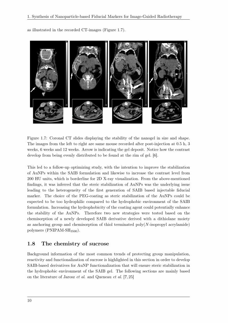
as illustrated in the recorded CT-images (Figure 1.7).1.10
Figure 1.7: Coronal CT slides displaying the stability of the nanogel in size and shape.
The images from the left to right are same mouse recorded after post-injection at 0.5 h, 3
weeks, 6 weeks and 12 weeks. Arrow is indicating the gel deposit. Notice how the contrast
develop from being evenly distributed to be found at the rim of gel. [6].
This led to a follow-up optimizing study, with the intention to improve the stabilization
of AuNPs within the SAIB formulation and likewise to increase the contrast level from
200 HU units, which is borderline for 2D X-ray visualization. From the above-mentioned
findings, it was inferred that the steric stabilization of AuNPs was the underlying issue
leading to the heterogeneity of the first generation of SAIB based injectable fiducial
marker. The choice of the PEG-coating as steric stabilization of the AuNPs could be
expected to be too hydrophilic compared to the hydrophobic environment of the SAIB
formulation. Increasing the hydrophobicity of the coating agent could potentially enhance
the stability of the AuNPs. Therefore two new strategies were tested based on the
chemisorption of a newly developed SAIB derivative derived with a dithiolane moiety
as anchoring group and chemisorption of thiol terminated poly(N -isopropyl acrylamide)
polymers (PNIPAM-SH3500).
1.8 The chemistry of sucrose
Background information of the most common trends of protecting group manipulation,
reactivity and functionalization of sucrose is highlighted in this section in order to develop
SAIB-based derivatives for AuNP functionalization that will ensure steric stabilization in
the hydrophobic environment of the SAIB gel. The following sections are mainly based
on the literature of Jarosz et al. and Queneau et al. [?, 25]
10

1. Synthesis of Nanoparticle-based Fiducial Markers for Image-Guided Radiotherapy
1.8.1 Sucrose
Sucrose is a naturally occurring disaccharide consisting of β-d-fructofuranosyl-(2→1)-α-d-
glucopyranoside. It is a non-reducing sugar as both reducing ends of the glucopyranoside
and fructofuranosyl units are bonded to each other. Thus, the hemiacetal chemistry of the
anomeric center is replaced by the bis-acetalization chemistry of the glycosidic linkage [?].
The glycosidic linkage of sucrose is stable in basic environment but labile under acidic
conditions [25]. Beside the glycosidic linkage, sucrose possesses three primary hydroxy
groups and five secondary hydroxy groups. All hydroxy groups show similar reactivity,
but differentiation of the primary and secondary hydroxy groups is achievable using bulky
reagents. The secondary hydroxy groups can furthermore be differentiated but not with-
out difficulties [25].
Sucrose possess interesting conformations as shown in Figure 1.8. In the solid state, two
hydrogen bonds exist; one in-between the 6’-OH and the pyranosidic endocyclic oxygen
while the second hydrogen bonding occur between 2-OH and 1’-OH (1.2a) [?, 25]. Upon
solvation, only the hydrogen bonding between the 2-OH and 3’-OH or 1’-OH are occurring,
1.2b and 1.2c respectively. This hydrogen bonding together with the neopentyl reactivity
of 1’-OH underpin the less accessibility and reactivity compared to 6-OH and 6’-OH.
O11
44
55HO
HO
O
1'1'
66
22
3'3'33
5'5'
2'2'
4'4'
O
OH6'6'
OH
O
OH
O
OH
HH
O11
44
55HO
HO
O
66
22
333'3'
5'5'
2'2'
4'4'O
OH
OH
O
OH
1'1'
OH
6'6'
H
O11
44
55HO
HO
O
66
22
333'3'
5'5'
2'2'
4'4'O
OH
OH
O
OH
1'1'
OH
6'6'O
H
H
OHin solutionin solid state
1.2a 1.2b 1.2c
Figure 1.8: Structural conformations of sucrose. The conformation of 1.2a shows the two
hydrogen bonding present in the solid state. 1.2b and 1.2c show the equilibrium states
of the two types of hydrogen bonding present in solution [26]
The most acidic hydroxy-groups of sucrose have been estimated to be those nearest the
glycosidic linkage, thus 2-OH, 1’-OH and 3’-OH [?]. This property has been used to
regioselectively modify the 2-OH position of unprotected sucrose e.g. by using benzyl
bromide and NaH in DMF yielding 2-O-benzyl-sucrose as the main species [?]. A common
strategy to allow regioselective modification of sucrose at the primary positions is to
protect the primary hydroxy group(s) of interest - using a bulky protecting group - followed
by orthogonal protection of the remaining hydroxy-groups. Subsequent deprotection of the
primary position(s) afford the hydroxy-group(s) of interest available for modification [25].
In terms of the use of ester-type protection groups at the secondary/remaining positions,
the deprotection of the bulky protecting group at the primary position(s) can lead to acyl-
migration of the neighboring acyl group. Migration is usually facilitated by acidic or basic
conditions, which is usually a requirement for the deprotection of the bulky protecting
groups of silylating agents or trityl moieties.
11

1. Synthesis of Nanoparticle-based Fiducial Markers for Image-Guided Radiotherapy
1.8.2 Ether modification of sucrose
The bulky protecting groups used to differentiate between the primary and the secondary
hydroxy groups are usually tert-butyldiphenylsilyl (TBDPS) or triphenylmethyl (Trt).
The reactivity of the primary hydroxy-groups differentiates, based upon the different
reagents used. The overall order of reactivity observed is usually 6-OH>6’-OH>1’-OH.
1’-OH is less reactive due to its accessibility and neopentyl-like reactivity. The reaction
pattern of unprotected sucrose 1.3 towards bulky protecting groups are illustrated in
Scheme 1.2. Regarding tritylation, excess use of TrtCl (3.6 equiv.) in basic conditions
at elevated temperature afford the 6,1’,6’-tri-O-trityl-sucrose 1.4 as the main derivative.
Carrying out the same reaction using similar equivalents at room temperature afford be-
side 1.4, 6,6’-di-O-tritylsucrose 1.5. Lowering the amount of TrtCl to 2.0 equiv. provide
a mixture of mono-tritylated sucrose, 6-O-trityl-sucrose 1.6 and 6’-O-trityl-sucrose 1.7.
Lowering the amount of TrtCl to 1.2 equiv, afford the same mixture of monotritylated
sucrose derivatives in lower yields [25].
OHOHO
HOO
OH
OH
OH
OH
O
OH
OHOHO
HOO
OR3
OR2
OH
OH
O
OR1
a), b) or c)
R1 = R2 = R3 = TrtR1 = R3 = Trt, R2 = H
R1 = Trt, R2 = R3 = H
R1 = H, R2 = Trt, R3 = H
OHOHO
HOO
OH
OH
OH
OH
O
OH
OHOHO
HOO
OR3
OR2
OH
OH
O
OR1
a) pyridine, DMAP, TBDPSCl (4.6 equiv.), 55-60 °C, 24 h
b) pyridine, DMAP, TBDPSCl (3 equiv.), 55-60 °C, 24 h
c) pyridine, DMAP, TBDPSCl (1.1 equiv.), rt, 4 h
R1 = R2 = R3 = TBDPSR1 = R3 = TBDPS, R2 = H
R1 = R2 = H, R3 = TBDPS
a)b)
c)
TrtCl
1.3
1.31.4
1.5
1.6
1.7
1.8
1.9
1.10
Scheme 1.2: The reaction pattern of sucrose 1.3 using TrtCl and TBDPSCl [25,27]
Application of the bulky silyl group of TBDPS enables improved selectivity between the
primary hydroxy groups of sucrose. Reactions performed using a 1.1 equiv. of TBDPSCl
in pyridine and 4-dimethylamino-pyridine (DMAP) at room temperature provide the 6’-
O-TBDPS-sucrose 1.10 as the main species together with the formation of 6,6’-di-O-
12

1. Synthesis of Nanoparticle-based Fiducial Markers for Image-Guided Radiotherapy
TBDPS-sucrose 1.9 in trace amounts [25, 27]. Using TBDPS as the bulky protecting
group inverts the reactivity towards the regioisomer of the 6’-OH position compared to
the use of Trt. Providing excess amount of TBDPSCl (4.6 equiv.) at elevated temperature
show similar reactivity and afford the fully 6,1’,6’-tri-O-TBDPS-sucrose 1.8 while lowering
to 3.0 equiv. affording a 1:3 ratio of 1.9 and 1.8 [25].
Studies using 2,3,3’,4,4’-penta-O-benzylsucrose 1.11 as model substrate have shown that
the primary hydroxy groups can be further discriminated through additional protecting-
group manipulations (scheme Figure 1.9) [25].
OBnOBnO
BnOO
OH
OH
OBn
OBn
O
OH
p-nitrobenzoic acid,PPh3, DEAD,
pyridine, 20°C
OBnOBnO
BnOO
OBz-pNO2
OH
OBn
OBn
O
OBz-pNO2
a) MOMCl, DIPEAb) BOMCl, pyridine
OBnOBnO
BnOO
OBz-pNO2
OR1
OBn
OBn
O
OBz-pNO2
R1 = MOM
R1 = BOM
a)
b)
OBnOBnO
BnOO
OH
OR1
OBn
OBn
O
OH
R1 = MOM
R1 = BOM
a)
b)
OBnOBnO
BnOO
OTBDPS
OBOM
OBn
OBn
O
OH
CH3ONa,CH3OH/THF
TBDPSCl (1 equiv.), CH2Cl2, DMAP, DIPEA
TBDPSCl (3 equiv.), DMF, NaH
OBnOBnO
BnOO
OTBDPS
OBOM
OBn
OBn
O
OTBDPS
HF·pyridine
OBnOBnO
BnOO
OH
OBOM
OBn
OBn
O
OTBDPS
(Only )
(Only )
1.11 1.12 1.131.14
1.15
1.16
1.16
1.16
1.17
1.18
1.19
Figure 1.9: Further discrimination of the primary hydroxy groups starting from 1.11 [25].
Employing Mitsunobu conditions using p-nitrobenzoic acid as the nucleophile, afford 6,6’-
diester of 1.12 which subsequently can lead to regioselective protection of the 1’-OH
position affording 1.13 or 1.14 using MOM or BOM as protecting group, respectively. The
6,6’-diol of 1.15 or 1.16 can subsequently be re-established using Zempleen conditions.
Further discriminations between the 6-OH and 6’-OH can be made, because TBDPS-
protection (1 equiv.) will block the 6-OH position available (1.17), in line with the
reactivity described previously. The 6’-OH position can be achieved by fully di-TBDPS
protection of 1.18 followed by subsequent regioselective mono-desilylation using 1 equiv.
of HF·pyridine liberating the 6’-OH position (1.19) available for further modification [25].
1.8.3 Ester modifications of sucrose
Applying Mitsunobu conditions have also been exerted to perform ester-linkages of differ-
ent carboxylic acids. Mitsunobu-conditions on native sucrose 1.3 yielded the 6-O-acyl-
sucrose using a slight excess of a fatty acid derivative, whereas two equiv. provide the 6,6’-
di-O-acyl-sucrose [25]. In terms of ester protections of the primary hydroxy-groups, the
13

1. Synthesis of Nanoparticle-based Fiducial Markers for Image-Guided Radiotherapy
common pivaloylation, benzoylation and acetylation have been conducted on sucrose [25].
Pivaloylation showed similar trend of reactivity on the primary hydroxy-groups as men-
tioned in section 1.8.2, while benzoylation mainly occur on 6-OH compared to 1’-OH and
6’-OH. Regioselective acetylation of primary hydroxy groups has been conducted using
either kinetic conditions to afford 6-O-acetylsucrose while using DABCO and 3-acyl-5-
methyl-1,3,4-thiadiazole-2(3H)-thiones afford the regioisomeric 6’-O-acyl-sucrose [25].
1.8.4 Acetalization of sucrose
Sucrose 1.3 undergoes isopropylidene acetalization with the diols of 4-OH and 6-OH and
furthermore at the 1’-OH and 2-OH. As acid is required for the acetalization, the choice
of acid has proven to be a key factor for the outcome of the acetalization. Using mineral
acids and acetone led to decomposition of sucrose while organic acids like p-toluenesulfonic
acid and 2-methoxypropene in slight excess provides the 4,6-O-isopropylidene-sucrose in
decent yield. Formation of the corresponding benzylidene acetal is likewise reported but
in low yield. The benzylidene acetal offers the possibility of regioselective ring-opening
of the acetal that would provide either 4-O or 6-O functionalized derivatives of sucrose [25].
Stannylene-acetals are well known in carbohydrate chemistry and are widely used. Using
(n-Bu)2SnO with native sucrose afford the dimer of 4,6-O-stannyleneacetal-sucrose 1.20
(Scheme 1.3) [25, 28].
Bu2SnO,CH3OH, 65 °C
OOHO
HOO
OH
OH
OH
OH
O
OSn
Bu
Bu
O
O Sn
O
HO
OHOHO
Bu
Bu
O
OHHO
OH
OHOHO
HOO
OH
OH
OH
OH
O
OCOR
RCOX, DMF,Et3N, 20°C
1.3
1.20 1.21
Scheme 1.3: Stannylene acetylation of sucrose [28].
The formation of this acetal increases the nucleophilicity of 6-O position which in the
presence of electrophilic agents undergo regioselective nucleophilic substitution affording
the 6-O-acyl-sucrose 1.21 [25, 29]. Likewise, the formation of 5-membered cyclic
stannylene acetals have been reported on the 1.4. Stannylene acetal formation occurred
between the trans-diol of 3’-OH and 4’-OH of 1.4. Depending on the solvent applied,
mesylation or triflation at the 3’-OH or 4’-OH position of 1.4 was achievable, respectively
[30].
Another convenient transformation used on sucrose 1.3 is the Appel reaction converting
the primary positions of 6-OH and 6’-OH into the corresponding dihalo derivatives
of sucrose using standard conditions of tetrahalomethane (chloro or bromo) and
triphenylphosphine (PPh3) [25].
A wide range of enzymes have also been employed to afford sucrose derivatives.
14
1. Synthesis of Nanoparticle-based Fiducial Markers for Image-Guided Radiotherapy
Peracetylated sucrose 1.22 can be regioselectively deacetylated liberating the given
hydroxy group of interest for further modifications. An overview of the different enzymes
and their deacetylation position is illustrated in figure Scheme 1.4 [25].
OAcOAcO
AcOO
OH
OH
OAc
OAc
O
OAc
OHOAcO
AcOO
OAc
OAc
OAc
OAc
O
OAc
OAcOAcO
AcOO
OH
OAc
OAc
OAc
O
OAc
OAcOAcO
AcOO
OAc
OH
OAc
OH
O
OAc
OAcOAcO
AcOO
OAc
OH
OAc
OAc
O
OAc
OAcOAcO
AcOO
OAc
OAc
OAc
OAc
O
OH
OAcOAcO
AcOO
OAc
OAc
OAc
OAc
O
OH
OAcOAcO
AcOO
OAc
OAc
OAc
OH
O
OAc
OAcOAcO
AcOO
OAc
OAc
OAc
OAc
O
OAc
1.
4.
1. Alkylase or Protease N2. Chymotrypsin3. lipase MY ( Candida cylindracea)4. lipase AP6 ( Aspergillus niger)5. lipase OF ( Candida cylindracea)6. lipase Candida ( Candida cylindracea)
2.
3.5.
6.
1.22
Scheme 1.4: Different options of using enzymes to achieve regioselective deacetylation of
peracetylated sucrose 1.22 [25].
1.9 Design of the dithiolane-functionalized SAIB
derivative
The design of a SAIB-based ligand for AuNP functionalization was based upon the "like
dissolves like principle". Being as similar as possible in structure to SAIB 1.28 - could
potentially encompass enhanced stability within the SAIB formulation. In a retrosynthetic
perspective, such a ligand could be achieved in few steps, in accordance with the synthetic
trends and strategies of sucrose described in section 1.8. Several studies have shown that
the dithiolane group provide enhanced stability in AuNP functionalization when compared
to thiol conjugation [31–33]. Since the lipoic acid is commercially available, and thiols
commonly tend to form disulfides it was decided to use lipoic acid as anchoring group.
Having the butyl linker in-between the dithiolane group and the carboxylic acid will act
as a spacer.
Functionalization of AuNps with carbohydrates have been reported previously within
the field of Au glyconanoparticles. Glyconanoparticles have been used within the area
of carbohydrate research to gain knowledge about how carbohydrates are involved in
signaling and carbobydrate-based interactions in cellular functioning []. The carbohydrate
based ligands developed within this field are of unprotected sugars, which was expected
not be compatible with the hydrophobic environment of SAIB 1.28.
15

1. Synthesis of Nanoparticle-based Fiducial Markers for Image-Guided Radiotherapy
1.10 The synthesis of dithiolane functionalized SAIB
derivative
The dithiolane functionalized SAIB derivative was synthesized in four steps starting from
sucrose 1.3 (Scheme 1.5).
TBDPSCl DMAP
pyridinei) 3 h, 70 °C
ii) 24 h, 20 °C
DMAPpyridine20 °C
O
O
O
THF20 °C
TBAFacetic acid
EDAC·HCl,DMAP, DMF
20 °C
O
OHSS
OHOHO
HOO
OH
OH
OH
OH
O
OH
OHOHO
HOO
O
OH
OH
OH
O
OH
TBDPS
OOO
OO
O
O
O
O
O
O
TBDPSO
OO
O
O
O
O
OOO
OO
OH
O
O
O
O
O
O
OO
O
O
O
O OOO
OO
O
O
O
O
O
O
O
OO
O
O
O
O
O
S S
40% 80%
97%
86%
1.3 1.23 1.24
1.25 1.26
Scheme 1.5: Overall scheme for the synthesis of 1.26 [6].
Regioselective tert-butyldiphenylsilyl (TBDPS) protection of sucrose afforded the 6’-TBDPS-
O-sucrose 1.23 using 1.2 equiv. TBDPSCl in pyridine. The mono silylated sucrose 1.23
was subsequently isobutyrylated at the remaining positions using isobutyric anhydride
and DMAP conducted in pyridine affording the 6’-O-TBDPS-hepta-isobutyryl sucrose
1.24 in 80% yield. The position of the TBDPS of 1.3 was verified by 2D-NMR exper-
iments of 1.24 and was in accordance with literature [25, 27]. The isobutyrylation of
1.24 was confirmed to be fully isobutyrylated by the presence of seven carbonyl ester car-
bon resonances in 13C NMR together with HMBC correlations to the backbone of sucrose.
The TBDPS group was attempted removed using 1.2 equiv. of tetra-N -butylammonium
fluoride (TBAF) in THF. These conditions were apparently too harsh in terms of basicity
as - besides the deprotection of the TBDPS 1.25 - this also led to the de-isobutyrylation
at the 4’-OH position affording the diol 1.27 (Scheme 1.6).
THF, 20 °C
OOO
OO
OTBDPS
O
O
O
O
O
O
OO
O
O
O
O OOO
OO
OH
O
O
O
O
O
O
OO
O
O
O
OTBAF OOO
OO
OH
O
O
OH
O
O
O
O
O
O
O
O+
1.24 1.25 1.27
Scheme 1.6: Deprotection of 1.24 using TBAF.
16
1. Synthesis of Nanoparticle-based Fiducial Markers for Image-Guided Radiotherapy
With the isolation of this side-product 1.27 could indicate that migration occurred to the
neighboring 6’-OH position upon silyl-deprotection followed by subsequent hydrolysis. In
order to control and keep the migration to a minimum, the deprotection was optimized
by adding acetic acid in slight excess (1.5 equiv.) compared to TBAF (1.2 equiv). This
gave the desilylated product 1.25 in almost quantitative yield (97%), as shown in the
Scheme 1.5. The dithiolane moiety was introduced by the formation of the activated ester
using N -dimethylaminopropyl-N’ -ethyl carbodiimide HCl (EDC·HCl) and DMAP. The
esterification afforded the dithiolane derivative 1.26 in 86% yield.
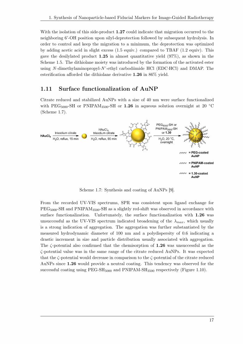
1.11 Surface functionalization of AuNP
Citrate reduced and stabilized AuNPs with a size of 40 nm were surface functionalized
with PEG5000-SH or PNIPAM3500-SH or 1.26 in aqueous solution overnight at 20 ◦C
(Scheme 1.7).
Scheme 1.7: Synthesis and coating of AuNPs [9].
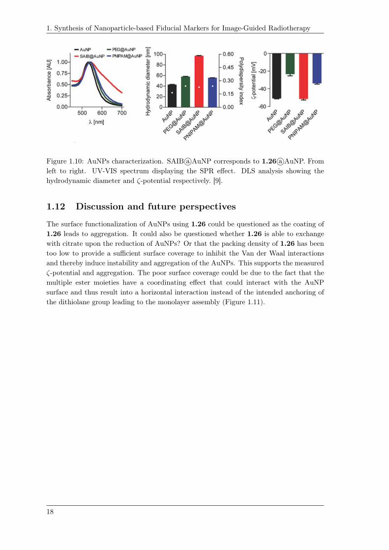
From the recorded UV-VIS spectrums, SPR was consistent upon ligand exchange for
PEG5000-SH and PNIPAM3500-SH as a slightly red-shift was observed in accordance with
surface functionalization. Unfortunately, the surface functionalization with 1.26 was
unsuccessful as the UV-VIS spectrum indicated broadening of the λmax, which usually
is a strong indication of aggregation. The aggregation was further substantiated by the
measured hydrodynamic diameter of 100 nm and a polydispersity of 0.6 indicating a
drastic increment in size and particle distribution usually associated with aggregation.
The ζ-potential also confirmed that the chemisorption of 1.26 was unsuccessful as the
ζ-potential value was in the same range of the citrate reduced AuNPs. It was expected
that the ζ-potential would decrease in comparison to the ζ-potential of the citrate reduced
AuNPs since 1.26 would provide a neutral coating. This tendency was observed for the
successful coating using PEG-SH5000 and PNIPAM-SH3500 respectively (Figure 1.10).
17
1. Synthesis of Nanoparticle-based Fiducial Markers for Image-Guided Radiotherapy
1.17
Figure 1.10: AuNPs characterization. SAIB a○AuNP corresponds to 1.26 a○AuNP. From
left to right. UV-VIS spectrum displaying the SPR effect. DLS analysis showing the
hydrodynamic diameter and ζ-potential respectively. [9].
1.12 Discussion and future perspectives
The surface functionalization of AuNPs using 1.26 could be questioned as the coating of
1.26 leads to aggregation. It could also be questioned whether 1.26 is able to exchange
with citrate upon the reduction of AuNPs? Or that the packing density of 1.26 has been
too low to provide a sufficient surface coverage to inhibit the Van der Waal interactions
and thereby induce instability and aggregation of the AuNPs. This supports the measured
ζ-potential and aggregation. The poor surface coverage could be due to the fact that the
multiple ester moieties have a coordinating effect that could interact with the AuNP
surface and thus result into a horizontal interaction instead of the intended anchoring of
the dithiolane group leading to the monolayer assembly (Figure 1.11).
18

1. Synthesis of Nanoparticle-based Fiducial Markers for Image-Guided Radiotherapy
Figure 1.11: Schematic representation of types of interaction between AuNPs and
1.26. a)Schematic representation of the intended mono-layer assembly of 1.26 using
the dithiolane as anchoring group. b) Schematic representation illustrates horizontal
interaction of 1.26 via carbonyl groups.
As stated, the chemisorption/ligand exchange was conducted in aqueous solution which
was a sufficient solvent for the polymeric coatings of PEG5000-SH and PNIPAM3500-SH.
The choice of performing the coating of AuNPs with 1.26 in aqueous solution was expected
to be insufficient in regards to the solubility 1.26 in aqueous solution.
Phase transfer of citrate reduced and stabilized AuNPs to chloroform have been reported
in the literature [33]. This method encompasses the use of cetrimonium bromide (CTAB)
as a surfactant and the use of a highly hydrophobic dithiolane functionalized oleyl based
ligand [33]. Phase transfer has been reported for AuNPs for sizes in the range between
5-70 nm. Retro-perspectively, optimization by use of a phase transfer agent in order
to transfer the as-synthesized AuNPs to an organic solvent, could potentially overcome
the solvent limitation of 1.26, by which AuNPs functionalization potentially could be
achieved.
In the study of Hurst et al. [34], the assembly of DNA onto the surface of AuNPs was
analyzed as quantitation method. Likewise, it was analyzed how the assembly of DNA
fragments can be improved by the presence of NaCl. The study also emphasized the
importance of the length of the linker between the anchoring alkanethiol and the DNA
fragment, and how this influenced the loading onto the AuNPs. The PEG spacer seemed
to lead to a higher loading compared to the nucleotide-base based spacers of A10 and T10,
which could be argued to be reasoned that the bulky DNA moiety is moved further away
from the particle surface by which the sterically crowding and interstrand repulsion is
minimized [34].
Hill et al highlighted how the size and curvature of AuNPs influence surface charac-
teristics in terms of surface coverage of AuNPs [35]. As an indirect measure of the spatial
19
1. Synthesis of Nanoparticle-based Fiducial Markers for Image-Guided Radiotherapy
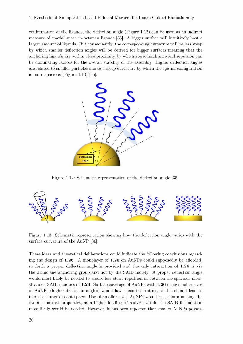
conformation of the ligands, the deflection angle (Figure 1.12) can be used as an indirect
measure of spatial space in-between ligands [35]. A bigger surface will intuitively host a
larger amount of ligands. But consequently, the corresponding curvature will be less steep
by which smaller deflection angles will be derived for bigger surfaces meaning that the
anchoring ligands are within close proximity by which steric hindrance and repulsion can
be dominating factors for the overall stability of the assembly. Higher deflection angles
are related to smaller particles due to a steep curvature by which the spatial configuration
is more spacious (Figure 1.13) [35].
Figure 1.12: Schematic representation of the deflection angle [35].
replacements
Figure 1.13: Schematic representation showing how the deflection angle varies with the
surface curvature of the AuNP [36].
These ideas and theoretical deliberations could indicate the following conclusions regard-
ing the design of 1.26. A monolayer of 1.26 on AuNPs could supposedly be afforded,
so forth a proper deflection angle is provided and the only interaction of 1.26 is via
the dithiolane anchoring group and not by the SAIB moiety. A proper deflection angle
would most likely be needed to assure less steric repulsion in-between the spacious inter-
stranded SAIB moieties of 1.26. Surface coverage of AuNPs with 1.26 using smaller sizes
of AuNPs (higher deflection angles) would have been interesting, as this should lead to
increased inter-distant space. Use of smaller sized AuNPs would risk compromising the
overall contrast properties, as a higher loading of AuNPs within the SAIB formulation
most likely would be needed. However, it has been reported that smaller AuNPs possess
20
1. Synthesis of Nanoparticle-based Fiducial Markers for Image-Guided Radiotherapy
higher X-ray attenuation [37, 38]. The use of higher concentration of AuNPs could be
limited by the cytotoxicity of AuNPs at increased concentrations [15,37].
Introducing a longer spacer between the dithiolane anchoring group and the SAIB moiety
of 1.26 would afford that the SAIB moiety would have been moved further away from
the AuNPs surface. This could potentially increase the spatial surroundings leading to
less steric hindrance. It was decided not to continue optimizing the functionalization of
AuNPs using 1.26.
1.13 PNIPAM and PEG coated AuNPs in SAIB
formulation
30 mg mL−1 PEG5000-SH or PNIPAM3500-SH coated AuNPs in SAIB/EtOH/PLA (75:20:5
vol%) were tested in vitro in MQ-H2O at 37 ◦C. A significant burst release was observed
of the PEG coated AuNPs while no burst release of the PNIPAM coated AuNPs was
observed. The enhanced stability of PNIPAM coated AuNPs and high loading is assumed
to be due to the fact that PNIPAM is more hydrophobic, but this could also be explained
by the hydrogen bonding of the amide [9]. The SAIB/EtOH/PLA formulation containing
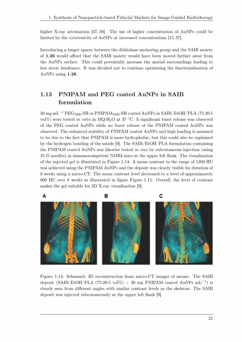
the PNIPAM coated AuNPs was likewise tested in vivo by subcutaneous injection (using
25 G needles) in immunocompetent NMRI-mice at the upper left flank. The visualization
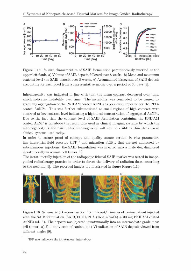
of the injected gel is illustrated in Figure 1.14. A mean contrast in the range of 1200 HU
was achieved using the PNIPAM AuNPs and the deposit was clearly visible for duration of
8 weeks using a micro-CT. The mean contrast level decreased to a level of approximately
800 HU over 8 weeks as illustrated in figure Figure 1.15. Overall, the level of contrast
makes the gel suitable for 2D X-ray visualization [9].
Figure 1.14: Schematic 3D reconstruction from micro-CT images of mouse. The SAIB
deposit (SAIB/EtOH/PLA (75:20:5 vol%) + 30 mg PNIPAM coated AuNPs mL−1) is
clearly seen from different angles with similar contrast levels as the skeleton. The SAIB
deposit was injected subcutaneously at the upper left flank [9].
21
1. Synthesis of Nanoparticle-based Fiducial Markers for Image-Guided Radiotherapy
1.19
Figure 1.15: In vivo characteristics of SAIB formulation percutaneously inserted at the
upper left flank. a) Volume of SAIB deposit followed over 8 weeks. b) Mean and maximum
contrast level the SAIB deposit over 8 weeks. c) Accumulated histogram of SAIB deposit
accounting for each pixel from a representative mouse over a period of 30 days [9].
Inhomogeneity was indicated in line with that the mean contrast decreased over time,
which indicates instability over time. The instability was concluded to be caused by
gradually aggregation of the PNIPAM coated AuNPs as previously reported for the PEG-
coated AuNPs. This was further substantiated as small regions of high contrast were
observed at low contrast level indicating a high local concentration of aggregated AuNPs.
Due to the fact that the contrast level of SAIB formulation containing the PNIPAM
coated AuNP is far above the resolutions used in clinical imaging systems by which the
inhomogeneity is addressed, this inhomogeneity will not be visible within the current
clinical systems used today.
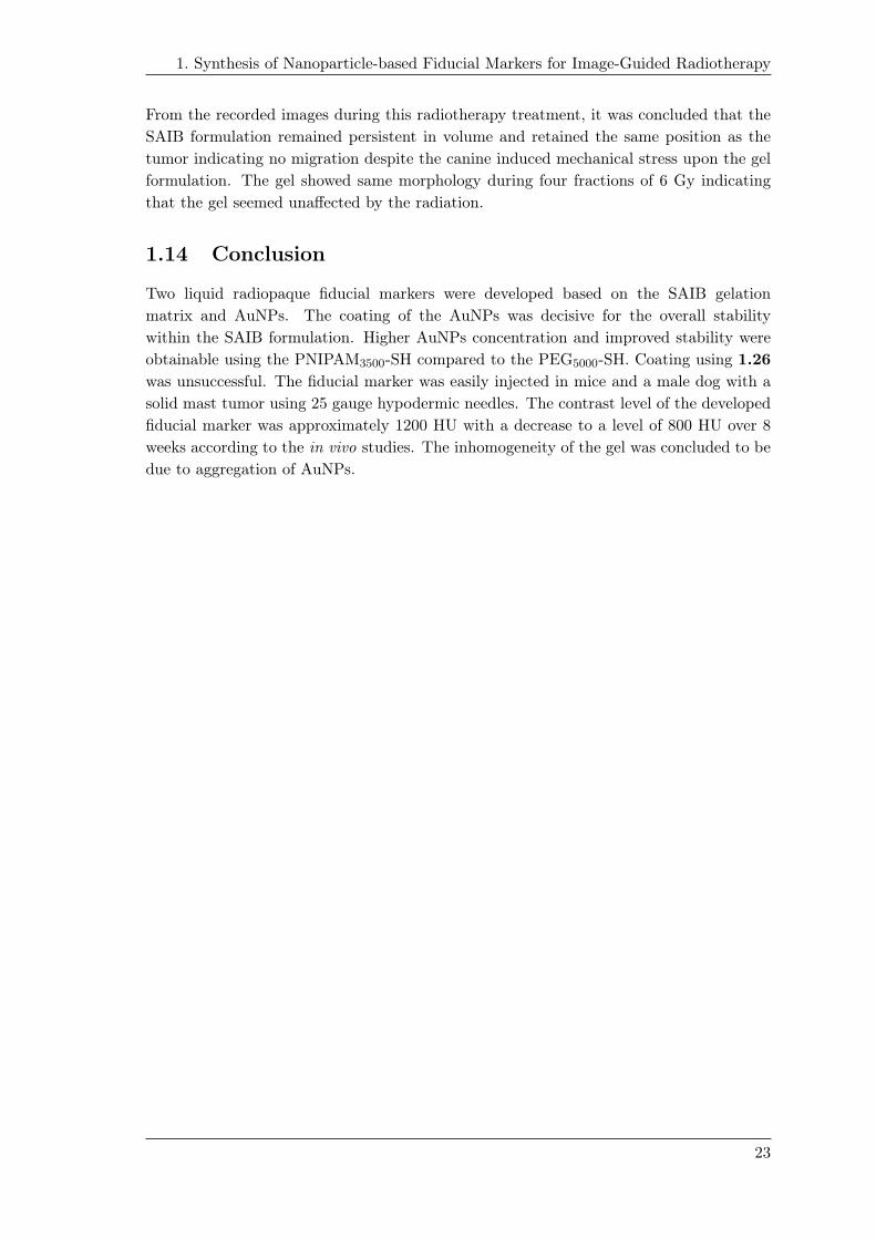
In order to assure proof of concept and quality assure certain in vivo parameters
like interstitial fluid pressure (IFP)1 and migration ability, that are not addressed by
subcutaneous injections, the SAIB formulation was injected into a male dog diagnosed
intratumorally in a mast cell tumor [9].
The intratumorally injection of the radiopaque fiducial SAIB marker was tested in image-
guided radiotherapy practice in order to direct the delivery of radiation doses according
to the position [9]. The recorded images are illustrated in figure Figure 1.16
Figure 1.16: Schematic 3D reconstruction from micro-CT images of canine patient injected
with the SAIB formulation (SAIB/EtOH/PLA (75:20:5 vol%) + 30 mg PNIPAM coated
AuNPs mL−1). The deposit was injected intratumorally into an intermediate-grade mast
cell tumor. a) Full-body scan of canine, b-d) Visualization of SAIB deposit viewed from
different angles [9].
1IFP may influence the intratumoral injectablity.
22
1. Synthesis of Nanoparticle-based Fiducial Markers for Image-Guided Radiotherapy
From the recorded images during this radiotherapy treatment, it was concluded that the
SAIB formulation remained persistent in volume and retained the same position as the
tumor indicating no migration despite the canine induced mechanical stress upon the gel
formulation. The gel showed same morphology during four fractions of 6 Gy indicating
that the gel seemed unaffected by the radiation.
1.14 Conclusion
Two liquid radiopaque fiducial markers were developed based on the SAIB gelation
matrix and AuNPs. The coating of the AuNPs was decisive for the overall stability
within the SAIB formulation. Higher AuNPs concentration and improved stability were
obtainable using the PNIPAM3500-SH compared to the PEG5000-SH. Coating using 1.26
was unsuccessful. The fiducial marker was easily injected in mice and a male dog with a
solid mast tumor using 25 gauge hypodermic needles. The contrast level of the developed
fiducial marker was approximately 1200 HU with a decrease to a level of 800 HU over 8
weeks according to the in vivo studies. The inhomogeneity of the gel was concluded to be
due to aggregation of AuNPs.
23

1. Synthesis of Nanoparticle-based Fiducial Markers for Image-Guided Radiotherapy
1.15 Iodine based SAIB derivative for IGRT
Contrary to the AuNPs based SAIB formulation developed in chapter 1, it was decided
within the Nanoguide-project to see if the level of contrast and homogeneity could be
enhanced by developing an iodine-based contrast derivative. Iodide derivatives are widely
used as CT-agents due to the attractive X-ray attenuation coefficients [].
A common trend for the iodine-based contrast agents used today is the use of the 2,4,6-
triiodoaryl moiety. This is used, because it introduces three iodide atoms with relatively
stable carbon-iodide bonds. Examples where this core structure have been used are iohexol
1.29, diatrizoic acid 1.30, iopromide 1.31 and metrizamide 1.32 among many others.
Iodide based contrast agents are further highlighted in chapter 3.
1.15.1 BioXmark
In the Nanoguide project it was decided that a triiodophenol SAIB derivative (TIP-SAIB)
could be synthesized from the similar synthetic strategy, outlined in section 1.8, used in the
synthesis of 1.26. TIP-SAIB 1.33 was synthesized by other project partners (Scheme 1.8).
The diol 1.34 could be obtained by TBDPS protection of sucrose 1.3 affording 1.9, fol-
lowed by isobutyrylation to 1.35 and TBDPS-deprotection affording 1.34. This was
conducted in a 51% yield over three steps. The 2-(2,4,6-triiodophenoxy)-acetic acid 1.36
was made from the reaction of 2,4,6-triiodophenol 1.37 and tert-butyl 2-bromoacetate fol-
lowed by acidic hydrolysis of the tert-butyl ester affording the acid 1.36 in 85% over two
steps. Conjugation to the SAIB diol 1.34 was mediated through standard ester coupling
by activation of 1.36 using EDC·HCl and DMAP providing 1.33 in 83% yield.
TBDPSCl DMAP
pyridinei) 3 h, 70 °C
ii) 24 h, 20 °C
DMAPpyridine20 °C
O
O
O
THF20 °C
TBAFacetic acid
EDC·HCl,DMAP, DMF
20 °C
OHOHO
HOO
OH
OH
OH
OH
O
OH
OHOHO
HOO
O
OH
OH
OH
O
OTBDPS
TBDPS
OOO
OO
O
O
O
OO
OTBDPS
TBDPSO
O
O
O
O
OOO
OO
OH
O
O
O
O
OH
O
OO
O
O
O OOO
OO
O
O
O
O
O
O
O
OO
O
O
O
O
O
O
O
65% Quantiative
79% 83%
O
I
I I
I
I
I
OHI
II
OI
II
OH
O
1) Tert-butyl 2-bromoacetate, K2CO3, DMF, 20°C2) TFA, CH2Cl2, 20°C
85% over 2 steps
1.3 1.9
1.331.34
1.35
1.35
1.361.37
Scheme 1.8: The scheme for the synthesis of 1.33 [39].
24

1. Synthesis of Nanoparticle-based Fiducial Markers for Image-Guided Radiotherapy
1.15.2 BioXmark as fiducial marker in lung cancer
The visibility of TIP-SAIB 1.33 was determined and compared to the current standard
of solid markers used in clinics [40]. Four different concentrations were tested, where the
highest concentration of the BioXmark formulation showed comparable radiopacity to the
standards used today. The quality of CT-images was improved, since less CT-artifacts
were reported. The artifacts from BioXmark was though more pronounced accordingly to
the difference in physical size [40].
The in vivo stability of BioXmark was tested during radiotherapy as a liquid fiducial
marker for locally advanced non small cell lung cancer in patients. The formulation of 1.33
was readily inserted using 25G hypodermic needles. Variations of the BioXmark deposit
diverging from the gross target volume (GTV) was evaluated during inter-fractional
radiotherapy. The BioXmark deposit displayed a mean contrast of 991 ± 219 HU. The
volume of the BioXmark deposit decreased to a level of 24% relative to the initial deposit.
The relative deviation in distance (3D vector) between the GTV and BioXmark deposit
was reported to be -1.5±2.3 mm. The clarification of these characteristics are further
elaborated in [40,41].
25


Injectable fiducials for pre-operativelocalization of tumors in breast
cancer 2With the increasing quality of mammographic imaging and the introduction of screen-
ings, non-palpable tumors in breast cancer are detected to a much higher degree and in
an earlier stage [42–44]. Almost one fourth of all breast lesions today are detected as non-
palpable [45]. With the identification of the non-palpable tumors in the early stages and
the use of neoadjuvant systemic therapy, breast conserving treatments like lumpectomy
have become an attractive option compared to mastectomy [42]. Combined lumpectomy
and external radiotherapy as treatment, elaborated in the context of long-term survival,
shows similar survival rates when compared to mastectomy [46]. Despite this attractive
future prospective of survival rates, the number of recurrences in lumpectomy are reported
higher than in mastectomy [46]. This drawback is related to the complexity of the ex-
cision procedure of non-palpable tumors, which is highly dependent on accurate target
delineation (prelocalization) needed for surgical guidance [42, 43]. This has highlighted
the need for improved pre-operative image-guided localization for surgical guidance, in
order to lower the number of resections and recurrence rates [42, 47]. The underlying
main criteria to improve the quality of lumpectomy will be that the specimen is excised
at the interface between healthy and malignant tissue with an additional margin to pro-
vide a negative margin [44]. A positive margin is defined as tumor tissue present at the
boundary-site (colored with ink) by the Society of Surgical Oncology and American So-
ciety for Radiation Oncology (SSO-ASTRO) [46]. Adequate margins are therefore added
from the site of the positive margin in order to remove tissue with chance of microscopic
spread of cancer [?]. This requisite of a negative margin is emphasized since recurrences of
malignant tissue is related to positive margins [46]. The margins reported in many cases
are in-sufficient, as margins today are reported positive in up to 50% of the cases [44].
The outcome of the lumpectomy rely to a great extent on the pre-operative image guided
localization which is conducted as surgical guidance so the surgeon will locate the non-
palpable lesion for excision. The pre-operative image guided localization is conducted in
accordance to the preceding mammographic images [44].
The current technologies used in clinical routines today for preoperative localization
are wire-guided localization (WGL), radioactive occult lesion localization (ROLL) and
radioactive seed localization (RSL).
27

2. Injectable fiducials for pre-operative localization of tumors in breast cancer
Figure 2.2: Mammographic image confirming the insertion of the pre-operative localization
using a radioactive seed [48].
WGL has been the gold standard for many years and encompasses a hooked wire, which
is inserted in the breast with the terminal point of the hooked wire pointing towards the
tumor localization (Figure 2.1) [44]. WGL was introduced by Dodd et al. [49]. WGL have,
beside the high rate of positive margins (13-58%) a number of other limitations [42, 43].
The overall procedure of WGL requires first of all the localization of the tumor followed
by the insertion of the wire, which is conducted by a radiologist. The surgical removal is
then conducted subsequently by the surgeon accordingly to the pre-localization performed
by the radiologist . Logistically, this usually turns out to be problematic, as the surgery
is not carried out immediately after the WGL. This can cause a number of difficulties e.g.
the inserted wire have to be maintained in the correct position until surgery [44, 47, 50].
Additionally, the WGL conducted by the radiologist may not be in accordance with the
preferred incision point of the surgeon to achieve the best cosmetic outcome. Other issues
encountered are wire misplacement or transection of the wire [50]. In terms of patient
compliance, WGL is perceived as unpleasant, painful and in some cases traumatic to
patients [42, 44]. The high rate of reported positive margins and the above-mentioned
drawbacks of using WGL has led to the search of alternative methods like ROLL and RSL
with the hope of potentially reducing the number of positive margins [43].
ROLL was first reported by Luini et al. at the European Institute of Oncology (Figure 2.3)
[51, 52]. The technology uses 99mTc labeled colloidal albumin particles, which are in-
jected intratumorally. The pre-operative localization is confirmed from the corresponding
gamma-probe trace of the gamma-emitting 99mTc. ROLL has been reported to be easier
to perform by the radiologist and surgeons with less severe pain to patients [44]. 99mTc
labeled colloidal albumin particles are also used in the standard procedure of sentinel
lymph node (SLN) mapping and the corresponding biopsy (Figure 2.4).
28

2. Injectable fiducials for pre-operative localization of tumors in breast cancer
Figure 2.3: Imaging using ROLL, a) Preceding mammographic imaging, b) Lateral
scintigraphic imaging upon injection of 99mTc and c) overlay of images a and b [52].
ROLL uses the same source of identification as
Figure 2.4: schematic representation of
the sentinel lymph node [53]
in SLN mapping, which have been stated to be
advantageous or disadvantageous depending on
the study [54].
One advantage of ROLL would be that sur-
geons are already experienced in using 99mTc
for the SLN mapping, and biopsy, moreover
only one type of injection is needed. A ma-
jor drawback of ROLL is that the insertion of99mTc cannot be post-confirmed by mammo-
graphic imaging (ROLL is visible using scinti-
graphic imaging) as opposed to RSL where the
seed is clearly visible (described below) [43].
Another limitation of ROLL as a reference point of radioactivity is the diffusive behav-
ior of 99mTc labeled colloidal albumin particles into the surrounding tumor tissue from
the insertion point [42, 44]. This is a consequence of the ability of the small-sized 99mTc
labeled albumin particles to penetrate into the surrounding tissue by diffusion [43]. Due
to the diffusive tendency healthy tissue might be recognized as tumor tissue by which
unnecessary tissue is excised during the lumpectomy.
The half-life of 99mTc (6 h) makes the time-frame for surgery short from the time of injec-
tion [47, 50]. Intratumoral injections are additionally not preferable according to the ab-
sence of lymphatic system within the tumor and the high intratumorally pressure. [54,55].
Intratumorally injections is confined by the high interstitial fluid pressure in tumors, mak-
ing injections difficult [9].
29

2. Injectable fiducials for pre-operative localization of tumors in breast cancer
In RSL, a radioactive seed coated with radioactive iodide 125I (typical dimensions 0.8 x 4.5
mm) is used with an activity ranging from 3.7 to 10.7 MBq [56]. The seed is percutaneously
implanted using 18 G needles in the non-palpable lesion (Figure 2.5) [44, 56]. RSL was
first presented by Gray et al. [57]. The position of the specimen for excision is given by
the gamma probe signal intensity (Figure 2.5) [50].
1.13
Figure 2.5: Schematic illustration of a) the detection of the radioactive seed by a gamma
probe and b) the guided lumpectomy using guidance from the radioactive seed [58]
RSL have been attractive logistically owing to the half-life of 125I (60 days). This enables
the opportunity that the radioactive seed can be inserted up to 5 days before surgery,
which provides flexibility in surgery planning [50, 59]. Advantageously, the seed can be
post-confirmed upon insertion by mammographic imaging [43]. RSL in combination with
SLN mapping render possible a dual trace system where both sources are gamma-emitting.
They are differentiable as the emission energies are different (125I, 30 KeV, 99mTc 140
KeV). The study of Pouw et al. illustrated the combinatorial use of 125I coated seeds
together with the 99mTc labeled particles [45, 48]. This study indicated that the use of
high activity 99mTc (100 MBq, corresponding to a one-day protocol by which the patient
proceeds directly to surgery) can lead to considerable emission energies that will be de-
tectable within the detection window of the 125I emissions due to Compton scattering. A
potential limitation of RSL have been concerned migration of the inserted seed. Though,
it has been reported that clinically seed migration has been observed in less than 1% of
the patients [43, 50]. As RSL is a rather new methodology compared to WGL, it is still
within the process of implementation which require training and handling of radioactive
material as well as safe disposal.
Overall clinically, when comparing the different technologies in terms of clinical
performances, ROLL and RSL have been reported to lower the rate of positive margins and
resections in randomized studies [42,47]. In the study of Gray et al., the number of patients
in need of additional resections was reported lower for RSL than for WGL [57]. Most
studies indicate no significant difference (in margins) on the number of positive margins
between WGL and RSL or ROLL though [44, 46, 47, 60, 61]. No significant differences in
30

2. Injectable fiducials for pre-operative localization of tumors in breast cancer
terms of positive margins and re-excisions were found when comparing ROLL to RSL [43].
It has been stated in several studies that using radioactive tracers as reference for the
localization facilitates reorientation and real-time feedback for the surgeon in regards of
localization [?, 56]. This allow for a better assurance that the entire lesion is removed as
intended [?].
In terms of patient compliance and logistics, RSL have been superior compared to WGL
and ROLL [43]. In a study regarding patient compliance, less severe pain and improved
convenience were deduced by patients treated with RSL compared with patients treated
with WGL [60].
2.0.3 Problem statement
Taking the unique gelation properties of the SAIB formulation into account (section 1.6),
the potential application as a tissue marker for surgical guidance of non-palpable lesions
in breast cancer was devised. The aim of this project was therefore to develop a liquid
radioactive fiducial tissue marker - based on SAIB - that will function as a radioactive
seed and will be injectable using hypodermic needles.
To develop a radioactive fiducial tissue marker for surgical guidance of non-palpable tu-
mors in breast cancer, a radioactive source have to be associated with the tissue marker.
As radioactive source, it was decided to develop a radioiodinated SAIB derivative that will
provide the radioactive point source. Deriving a SAIB precursor for radioiodination will
offer that all attractive isotopes of iodide can be used like the positron emitting isotope124I (τ1/2 = 4.2 days) - enabling e.g. CT/PET imaging in combination with the developed
TIP-SAIB 1.33 derivative - or the gamma-emitting isotopes of 123I (τ1/2 = 13.2 h) or125I (τ1/2 = 60 days), or the beta-emitting isotope 131I (τ1/2 = 8 days). Additionally, the
radioactivity of the developed tissue marker can be custom-made to the given application
as this is given by the concentration of the radiolabeled SAIB derivative.
It was decided that the developed radioactive fiducial tissue marker should be based on
TIP-SAIB 1.33 reasoned its unique contrast properties and its use as injectable radiopaque
fiducial marker within IGRT of lung cancer patients [40,41]. The use of 1.33 will benefit
visualization by CT imaging as well as biodegradability and biocompatibility and easy
percutaneous insertion using hypodermic needles. Combining 1.33 with a radioiodinated
SAIB derivative will render the novel injectable seed which will be applicable as a tissue
marker for surgical guidance of non-palpable tumors.
Such a radioactive fiducial tissue marker could potentially also find use within the field
of brachytherapy (BT) as similar seeds used in RSL are employed in BT for therapeutic
treatment in various type of cancers mainly prostate and cervix cancer [].
2.1 Design and theoretical deliberations
Building on the already established experience of synthesizing SAIB derivatives, it
was envisioned that the radiolabeled SAIB derivative of interest can be synthesized in
accordance to the synthetic strategies highlighted in section 1.8. As described in the
introduction, the 125I isotope is attractive due to its half-life (60 days). The limitation of
31

2. Injectable fiducials for pre-operative localization of tumors in breast cancer
using radioactive isotopes of iodide, is the issue regarding up-take of iodide in the thyroid
gland. Considerable uptake in thyroid gland of radioactive iodide is a potential adverse side
effect. The extent of uptake will have to be evaluated. The requirement would therefore
be a long term stable iodinated SAIB derivative that will not undergo deiodination. As
a placeholder and directing group for regioselective iodinations, activated trimethylsilyl
(TMS) aryl moieties have been used in multiple studies [62]. This is further elucidated in
following section 2.2.
2.2 The β-cationic effect of trimethylsilanes in
electrophilic substitutions
The following section describe the ipso-substitution of trimethylsilyl (TMS) aryl moieties
and the β-cationic effect. The following section is based on the review of Chan [63].
Silanes is usually referred to as super - or a big proton []. It is considered as a hard
electrophile that forms strong bonds with hard nucleophiles (flouride, oxygen, chloride) [].
The C-Si bond is considered more polarized than the C-H bond [63].
Aryl-TMS moieties are known to undergo electrophilic aromatic ipso-substitution
(Scheme 2.1).
SiMe3EE
SiMe3ENu
ESiMe3
E
coincidingorbital overlap
1.7
Scheme 2.1: The electrophilic aromatic ipso-substitution of aryl-TMS. Scheme adapted
from [64].
Beside the intended ipso-substitution of the TMS group, the competing substitution of
hydrogen in accordance to the substitution pattern (activated vs de-activated) of the
given aryl-TMS is a possibility [?]. The reason ipso-substitution is typically favored
can be explained by the stabilized carbocation formed, as depicted in Scheme 2.1. The
electrophilic addition to the ipso-carbon takes place so the orientation of the formed
empty p-orbital in the β-position is in alignment with the C-Si σ-binding orbital. This
coinciding overlap of the empty p-orbital and C-Si σ-binding orbital induces an inductive
effective to the bonding electrons of C-Si σ-bond, which leave the C-Si bond weakened. The
electrophilicty of (CH3)3Si- is likewise increased and makes it prone to nucleophilic attack.
This is known as the β-cation stabilizing effect. This electrophilic aromatic substitution
is completed as the TMS group is eliminated in the presence of weak nucleophile by which
the aromaticity is regained. The electrophilic aromatic ipso-substitution is more prone to
undergo reaction in activated arene systems.
It should be mentioned that another β-cation can be formed which would correspond to
the β-cation when electrophilic substitution occurs in the meta-position in regard to aryl
silane (meta-directing, Scheme 2.2).
32

2. Injectable fiducials for pre-operative localization of tumors in breast cancer
SiMe3 SiMe3
E HE
SiMe3
C-Si bonding orbital is ortho-gonal to the empty p orbital
E
Scheme 2.2: The non meta-directing effect of aryl-TMS. Scheme adapted from [64].
In this case, the C-Si bond remains sp2 hybridized and thereby in the plane of the aromatic
system and perpendicular to the empty p orbital. In this case, no overlap is possible which
explains why aryl silanes are not meta-directing. The β-cationic effect is also observed in
case of vinylic, allylic and alkynylic TMS compounds.
2.3 Synthesis of SAIB-based precursor for radioiodination
.
As the prototype to establish proof of concept, it was inferred that the SAIB precursor
for radioiodinations could be obtained from the intermediate 1.25, used in section 1.10,
and 2-(p-TMS-phenoxy)-acetic acid 2.1 (Scheme 2.3).
EDC·HCl, DMAPDMF, 20 °C
OOO
OO
OH
O
O
O
O
O
O
OO
O
O
O
O OOO
OO
O
O
O
O
O
O
O
OO
O
O
O
O
O
O
82%
TMS
OOH
O
TMS
1.25
2.1
2.2
Scheme 2.3: The scheme for the synthesis of precursor 2.2.
2-(p-TMS-phenoxy)acetic acid 2.1 was obtained in three steps starting from p-
bromophenol 2.3 (Scheme 2.4).
n-BuLi TMSCl
THF, -78 °C CH3OH, 20 °C
78% 77%
OH
Br
OH
TMS
Br
O
O O
O
O
TMSK2CO3,
acetone, reflux
KOH O
O
OH
TMS
90%
2.12.3 2.4 2.5
Scheme 2.4: The scheme for the synthesis of 2-(p-TMS-phenoxy)acetic acid 2.1.
Metal-halogen exchange of the bromide of 2.3 using n-BuLi followed by addition of TMSCl
afforded p-TMS-phenol 2.4 in 78% yield. The phenol was O-alkylated using methyl bro-
moacetate and K2CO3 giving 2.5 in 90% yield. The methyl ester of 2.5 was subsequently
33
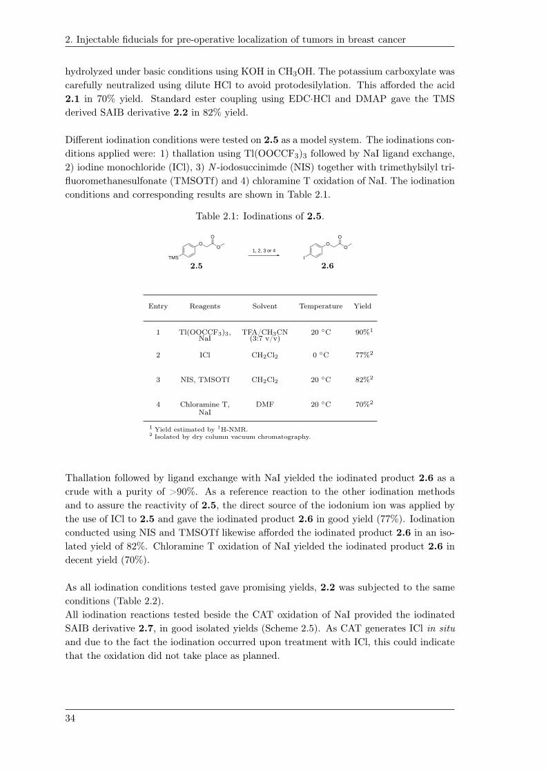
2. Injectable fiducials for pre-operative localization of tumors in breast cancer
hydrolyzed under basic conditions using KOH in CH3OH. The potassium carboxylate was
carefully neutralized using dilute HCl to avoid protodesilylation. This afforded the acid
2.1 in 70% yield. Standard ester coupling using EDC·HCl and DMAP gave the TMS
derived SAIB derivative 2.2 in 82% yield.
Different iodination conditions were tested on 2.5 as a model system. The iodinations con-
ditions applied were: 1) thallation using Tl(OOCCF3)3 followed by NaI ligand exchange,
2) iodine monochloride (ICl), 3) N -iodosuccinimde (NIS) together with trimethylsilyl tri-
fluoromethanesulfonate (TMSOTf) and 4) chloramine T oxidation of NaI. The iodination
conditions and corresponding results are shown in Table 2.1.
Table 2.1: Iodinations of 2.5.
OO
O
TMS
1, 2, 3 or 4O
O
O
I
1.21
2.5 2.6
Entry Reagents Solvent Temperature Yield
1 Tl(OOCCF3)3, TFA/CH3CN 20 ◦C 90%1
NaI (3:7 v/v)
2 ICl CH2Cl2 0 ◦C 77%2
3 NIS, TMSOTf CH2Cl2 20 ◦C 82%2
4 Chloramine T, DMF 20 ◦C 70%2
NaI
1 Yield estimated by 1H-NMR.2 Isolated by dry column vacuum chromatography.
Thallation followed by ligand exchange with NaI yielded the iodinated product 2.6 as a
crude with a purity of >90%. As a reference reaction to the other iodination methods
and to assure the reactivity of 2.5, the direct source of the iodonium ion was applied by
the use of ICl to 2.5 and gave the iodinated product 2.6 in good yield (77%). Iodination
conducted using NIS and TMSOTf likewise afforded the iodinated product 2.6 in an iso-
lated yield of 82%. Chloramine T oxidation of NaI yielded the iodinated product 2.6 in
decent yield (70%).
As all iodination conditions tested gave promising yields, 2.2 was subjected to the same
conditions (Table 2.2).
All iodination reactions tested beside the CAT oxidation of NaI provided the iodinated
SAIB derivative 2.7, in good isolated yields (Scheme 2.5). As CAT generates ICl in situ
and due to the fact the iodination occurred upon treatment with ICl, this could indicate
that the oxidation did not take place as planned.
34

2. Injectable fiducials for pre-operative localization of tumors in breast cancer
Table 2.2: Iodinations of 2.2.
OOO
OO
O
O
O
O
O
O
O
OO
O
O
O
O
OO
I
OOO
OO
O
O
O
O
O
O
O
OO
O
O
O
O
OO
TMS
1, 2, 3 or 4
2.2 2.7
Entry Reagents Solvent Temperature Yield1
1 Tl(OOCCF3)3, TFA/CH3CN 20 ◦C 97%NaI (3:7 v/v)
2 ICl CH2Cl2 0 ◦C 93%
3 NIS, TMSOTf CH2Cl2 20 ◦C 81%
4 Chloramine T, DMF 20 ◦C -NaI
1 Isolated by dry column vacuum chromatography.
2.4 Radioiodination of SAIB precursor
From the preliminary iodinations performed, it was decided to perform the radio-
iodination of 2.2 using Tl(OOCCF3)3 followed by ligand exchange with NaI (Scheme 2.5).
The radioiodination was conducted by other project members of the Nanoguide project.
[125I]-
OOO
OO
O
O
O
O
O
O
O
OO
O
O
O
O
OO
125I
OOO
OO
O
O
O
O
O
O
O
OO
O
O
O
O
OO
TMS
Tl(OOCCF3)3,
Na[125I]I
TFA/CH3CN (3:7 v/v)
20°C
RCY: 95%
2.2 2.7
Scheme 2.5: The radioiodination of 2.2 using Tl(OOCCF3)3.
The choice of Tl(OOCCF3)3 as iodination reagent was, despite the toxicity, owed to
the mild conditions of the thallation and the experience within the group of Mads
Clausen using Tl(OOCCF3)3 [62]. Moreover, since the commercially available source of
radioactive iodine is NaI, it can be directly used with the thallated intermediate without
any preceding transformations and proceeds under carrier free conditions. The iodination
was designed to ensure no thallated intermediates would be part of the radioiodinated
product [125I]-2.7. The radioactive iodide was therefore first allowed to react with the
thallated intermediate before excess of non-radioactive iodine was added. Surprisingly,
35

2. Injectable fiducials for pre-operative localization of tumors in breast cancer
it was observed that 90% of the radioactivity was incorporated in the SAIB derivative
[125I]-2.7 within a reaction time of 15 min, before addition of non-radioactive NaI.
According to the study of Mckillop et al. and the previous experience with iodinations
by thallation in the group, this was at first thought a bit surprising as the iodide is
believed to enter the reaction in two equivalents upon the formation of aryl-Tl(OOCCF3)2to prompt the ligand exchange [62, 65]. Subsequently, the ‘reductive elimination like’
reaction yields the aryl iodide together with Tl(I)-I. In accordance with this reaction
pattern, the maximum radiochemical yield is 50%, which is not in agreement with the
observed >90% yield. Mckillop et al. also reported the observation when using the
iodide in trace amount or < 1 equiv. Under these conditions the aryl iodide was also
found in nearly quantitative yields. It was proposed by McKillop et al., that the unstable
aryl-Tl(III)-I-(OOCCF3) intermediate is formed leading to the aryl-iodide together with
Tl(I)-(OOCCF3). Following addition of non-radioactive NaI and SEP-PAK cartridge
purification, [125I]-2.7 was isolated in a radiochemical yield of 83%. All fractions from
the SEP-PAK cartridge purification was analyzed by ICP-EOS measurements. No traces
of thallium was found in the fractions of [125I]-2.7. All thallium used (>98%) in the
radiolabeling was pre-eluted using aq. diethylenetriaminepentaacetic acid (DTPA) prior
to the elution of [125I]-2.7.
2.4.1 In vivo performance of [125I]-2.7
The combined formulation of [125I]-2.7 and 1.33 was tested in vivo using an NMRI-mice
model. This study was conducted by other project partners of the Nanoguide-project. A
deposit of the gel-like implant, corresponding to a 50 µL percutaneous injection (27 G
hypodermic needles) was inserted in the right flank of the NMRI-mice. CT images of the
gel-like implant was recorded at 1, 24 and 168 h respectively, and the 125I-biodistribution
analysis was obtained by well-counting of the deposit as well of the tissues/organs of
interest. From the recorded CT-images recorded afer 1 h, 24 h, and 168 h respectively,
the implant is clearly visible displaying a contrast level >1500 HU (Figure 2.6).
36

2. Injectable fiducials for pre-operative localization of tumors in breast cancer
Figure 2.6: CT images recorded after 1 h (A and B), 24 h (C and D) and 168 h (E and F)
respectively of the combined formulation of [125I]-2.7 and 1.33 percutaneously injected
in NMRI mice. The CT images was recorded from two different angles, axial (A, C and
E) and coronal (B, D and F). The scale bar in G indicates contrast in hounsfield units
(HU).
Beside the expected reduction in volume of the gel-like implant, which was in accordance
to previously observed experiments when using 1.33 in SAIB, the volume of the implant
remained stable within the time-frame analyzed of 168 h (Figure 2.7).
Figure 2.7: Volume changes over time.
The 125I-seeds used in clinic today consist of a 125I-coated silver wire which is encapsulated
within a titanium shell []. This type of system is considered as a closed radioactive
37
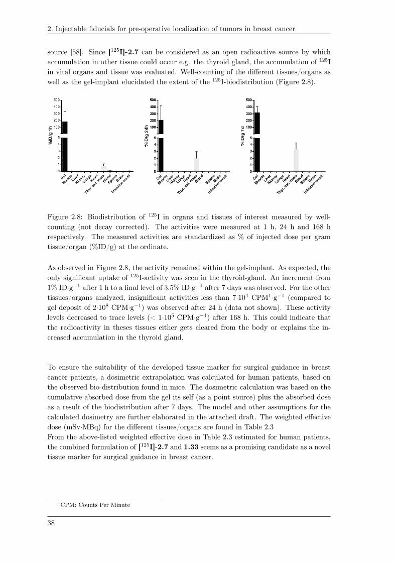
2. Injectable fiducials for pre-operative localization of tumors in breast cancer
source [58]. Since [125I]-2.7 can be considered as an open radioactive source by which
accumulation in other tissue could occur e.g. the thyroid gland, the accumulation of 125I
in vital organs and tissue was evaluated. Well-counting of the different tissues/organs as
well as the gel-implant elucidated the extent of the 125I-biodistribution (Figure 2.8).
1.24
Figure 2.8: Biodistribution of 125I in organs and tissues of interest measured by well-
counting (not decay corrected). The activities were measured at 1 h, 24 h and 168 h
respectively. The measured activities are standardized as % of injected dose per gram
tissue/organ (%ID/g) at the ordinate.
As observed in Figure 2.8, the activity remained within the gel-implant. As expected, the
only significant uptake of 125I-activity was seen in the thyroid-gland. An increment from
1% ID·g−1 after 1 h to a final level of 3.5% ID·g−1 after 7 days was observed. For the other
tissues/organs analyzed, insignificant activities less than 7·104 CPM1·g−1 (compared to
gel deposit of 2·108 CPM·g−1) was observed after 24 h (data not shown). These activity
levels decreased to trace levels (< 1·105 CPM·g−1) after 168 h. This could indicate that
the radioactivity in theses tissues either gets cleared from the body or explains the in-
creased accumulation in the thyroid gland.
To ensure the suitability of the developed tissue marker for surgical guidance in breast
cancer patients, a dosimetric extrapolation was calculated for human patients, based on
the observed bio-distribution found in mice. The dosimetric calculation was based on the
cumulative absorbed dose from the gel its self (as a point source) plus the absorbed dose
as a result of the biodistribution after 7 days. The model and other assumptions for the
calculated dosimetry are further elaborated in the attached draft. The weighted effective
dose (mSv·MBq) for the different tissues/organs are found in Table 2.3
From the above-listed weighted effective dose in Table 2.3 estimated for human patients,
the combined formulation of [125I]-2.7 and 1.33 seems as a promising candidate as a novel
tissue marker for surgical guidance in breast cancer.
1CPM: Counts Per Minute
38

2. Injectable fiducials for pre-operative localization of tumors in breast cancer
Table 2.3: The weighted absorbed dose for the organs/tissues of interest and the effectivedose in mSv·MBq−1.
Tissue/Organ Weighted effective dose(mSv·MBq−1)
Gel-Implant 5.91·10−1
Thyroid 4.15·10−2
Brain 4.58·10−6
Lungs 1.07·10−2
Heart 4.88·10−3
Liver 5.35·10−4
Spleen 8.70·10−5
Kidneys 1.42·10−5
Intestine 4.78·10−6
Muscle 1.00·10−4
Effective dose Equivalent 1.84(EDE)
Effective dose 6.56·10−1
2.5 TMS-BioXmark
Based on the convincing results of 1.33 as radiopaque fiducial marker for IGRT together
with the promising results of [125I]-2.7 formulated in the TIP-SAIB formulation, it was
desirable to develop a TMS derivative of 1.33 that upon radioiodination will be identical
to 1.33. As BioXmark has been authorized for clinical use as a medical device by the
Danish Health Authority, the 125I-labeled BioXmark [125I]-1.33 could add-on application
within the field of surgical guidance and brachytherapy. It would likewise enable bio-
distribution and stability studies of 1.33.
The challenging moiety to construct synthetically, would be the core structure of diiodo-
TMS-phenol. The positioning of the TMS, whether it would be positioned in the ortho or
para-position was thought at this stage not to be decisive for the outcome of the radio-
iodination. The synthetic challenge would be to introduce TMS without compromising
the already installed iodides. Having the p-TMS-phenol building block 2.4, used in the
development of [125I]-2.7, it was considered that the iodides could be installed by regios-
elective electrophilic aromatic iodination or ortho-lithiation.
In the case of ortho-lithiation, a directing group would have to be introduced to 2.4 as
39

2. Injectable fiducials for pre-operative localization of tumors in breast cancer
a handle that upon the installation of the iodides in the ortho-positions is readily removed.
2.5.1 Electrophilic aromatic iodination
Electrophilic aromatic bromination of the activated m-TMS-anisole and m-TMS-phenol
have been reported to take place in the corresponding para or ortho-positions with
preservation of the TMS-group [63,66].
For the electrophilic aromatic iodination of 2.4, it was devised that with the formation
of the corresponding phenoxide of 2.4, the nucleophilicity of the ortho-positions could
be improved and thereby circumvent the competing ipso-substitution of the TMS-moiety.
Treatment of 2.4 with KOH and NIS and TMSOTf in a mixture of CH3CN and H2O
led to a mixture of mono, di and tri-iodinations including ipso-substitution, thus without
preservation of the TMS-group. Deprotonation by NaH in THF followed by iodination
using NIS and TMSOTf was likewise tested but without any signs of selective ortho-
iodinations. From these experiments, it was inferred that the combinatorial effect of
having the TMS in in the activated para-position was too strong to allow regioselective
ortho-iodination.
2.5.2 Directed ortho metalation of p-TMS-phenol
One common way to add functional groups to an aromatic system is by ortho-lithiation also
known as directed ortho metalation. Aromatic hetero-atom-containing functional groups
allow directed metalation adjacent to the aromatic functional group (ortho-position).
The overall ortho-lithiation reaction constitute first bringing the lithiating species into
proximity, by complexation, followed by deprotonation [67].
Some of the most common groups showing directing ability in directed ortho metalation
is the methyl, methoxymethyl (MOM) and benzyloxymethyl (BOM) ethers of the
corresponding phenol. Since the removal of the methyl or MOM require harsh acidic
conditions (refluxing acid), these conditions could lead to protodesilylation. Clayden
ranked the directing group ability of certain functional groups (Figure 2.9) [67].
40
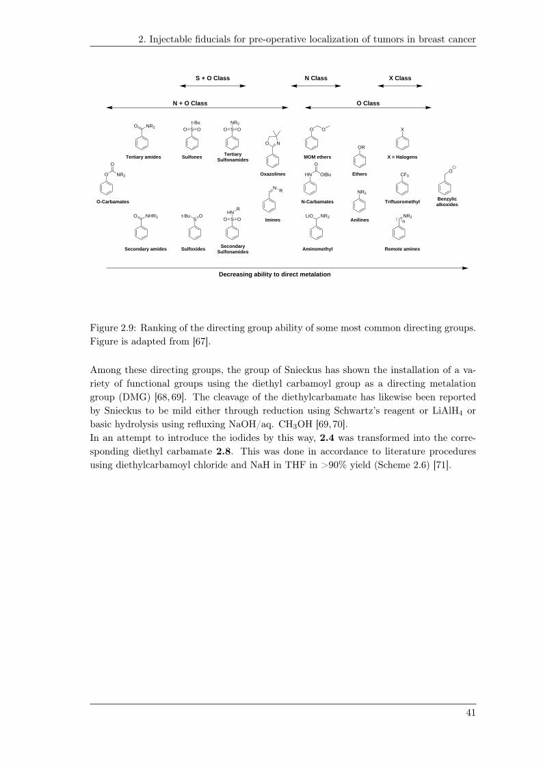
2. Injectable fiducials for pre-operative localization of tumors in breast cancer
O NR2
O
NR2O
NHR2O
St-Bu
O O
St-Bu O
SNR2
O O
SHN
O O
R
NO
NR
O O
HN OtBu
O
LiO NR2
OR
NR2
X
NR2
CF3
n
O
O-Carbamates
Tertiary amides
Secondary amides
Sulfones
Sulfoxides
TertiarySulfonamides
SecondarySulfonamides
Oxazolines
Imines
MOM ethers
N-Carbamates
Aminomethyl
Anilines
Ethers
X = Halogens
Trifluoromethyl
Remote amines
Benzylicalkoxides
Decreasing ability to direct metalation
N + O Class
S + O Class X ClassN Class
O Class
Figure 2.9: Ranking of the directing group ability of some most common directing groups.
Figure is adapted from [67].
Among these directing groups, the group of Snieckus has shown the installation of a va-
riety of functional groups using the diethyl carbamoyl group as a directing metalation
group (DMG) [68, 69]. The cleavage of the diethylcarbamate has likewise been reported
by Snieckus to be mild either through reduction using Schwartz’s reagent or LiAlH4 or
basic hydrolysis using refluxing NaOH/aq. CH3OH [69,70].
In an attempt to introduce the iodides by this way, 2.4 was transformed into the corre-
sponding diethyl carbamate 2.8. This was done in accordance to literature procedures
using diethylcarbamoyl chloride and NaH in THF in >90% yield (Scheme 2.6) [71].
41

2. Injectable fiducials for pre-operative localization of tumors in breast cancer
Et2NCOCl, NaH
THF0 °C - 20 °C
TMS
OH
TMS
O N
O 1) TMEDA, s-BuLi, THF, -78 -> 20 °C2) I2 in THF, -78 °C
TMS
O N
O
I
TMS
O
N
OI
TMS
O
N
O
a) 1) TMEDA, s-BuLi, THF, -78 -> 20 °C 2) I2 in THF, -78 °C
b) 1) LDA, THF, -78 °C 2) I2 in THF, -78 °C
1) TMEDA, s-BuLi, THF, -78 -> 20 °C2) TMSCl, -78°C3) s-BuLi, -78 -> 20 °C4) I2 in THF, -78 °C
I TMS
I
1) TMEDA, s-BuLi (2 equiv.), THF, -78 -> 20 °C2) I2 (2 equiv.) in THF, -78 °C
TMS
O NI
O
TMS
O
N
OTMS
TMS
OH O
N
Anionic Ortho-Fries rearrangement
side-product
+TMS
- 0%
- XX%
- 57%
- 90% - XX%
observed
1) TMEDA, s-BuLi, THF, -78 -> 20 °C2) TMSCl, -78 °C
TMS
O
N
OTMS
TMS
OHTMS
+
O
N
path a
path b
path d
path c
2.7
2.4
2.8
2.8 2.9
2.10 2.11
2.12
2.12
2.12
2.12
2.13
2.13
2.14
2.14
2.15
2.15
Scheme 2.6: Scheme showing different attempts of ortholithiations.
Different ortho-lithiation strategies were attempted in order to try to afford the 2,6-diiodo-
4-TMS-phenyl-(diethyl)-carbamate 2.9. Firstly, it was ensured that the lithiation condi-
tions would enable ortho-lithiation according to literature [68]. Providing a slight excess of
s-BuLi at -78 ◦C to a pre-cooled solution of tetra-methylethylenediamine (TMEDA) and
2.8 followed by treatment of TMSCl gave, beside residues of 2.8, isomers of the intended
TMS-substituted derivative according to LC-MS analysis (path a). It was hypothesized
that the two isomers were the product 2.10 and 2.11 (the anionic ortho-Fries-rearranged
isomer), respectively. Changing the electrophile to I2 in THF could indicate whether
the iodide could be introduced without ipso-substituting the TMS in the para-position
(path b). Same lithiation-conditions was applied, beside that the I2 in THF was added
upon the ortho-lithiation. The ortho-iodinated product 2.12 was observed together with
considerable formation of the anionic ortho-Fries rearranged isomer 2.13 and residues
of starting material 2.8 (Scheme 2.6). The starting material of 2.8 and ortho-iodinated
product 2.12 were hardly separable by chromatography, unless full conversion of 2.8. The
ortho-lithiated product of 2.12 was isolated in a decent yield. Noticeable, traces of what
to be believed as 2.9 was observed according to LC-MS analysis.
42

2. Injectable fiducials for pre-operative localization of tumors in breast cancer
These experiments indicated that the reaction temperature and the stoichiometric amounts
of s-BuLi were important to control in order to achieve the intended substituted product
in a clean fashion. Elevation of temperature (to room temperature) is likewise reported
to promote the formation of the [1-3] acyl migration of the diethylcarbamoyl-group [69].
Following these one-step lithiations, it was attempted to perform sequential one-pot or-
tho-lithiations, as it was reported in study of Reed et al., the sequential insertion of first
a TMS followed by iodide in the other ortho-position [68]. This would yield the 2-iodo-
4,6-di-TMS-phenyl diethylcarbamate 2.14 which potentially would be of value, as only
one TMS would be needed to be regio-selectively substituted by iodide, prospectively
(Scheme 2.6). To a cooled solution of 2.8 and TMEDA, the order of addition was: 1)
s-BuLi, 2) TMSCl, 3) s-BuLi and 4) I2 in THF (path c), in accordance to the original
procedure. Surprisingly this afforded the 2,4,6-tri-TMS-phenyl diethylcarbamate 2.15 as
the only species, isolated in 57% yield. The formation of 2.15 raised the questioning about
the stoichiometric amounts of s-BuLi and TMSCl added. The addition of I2 in THF was
discontinued as the reaction mixture turned yellow upon addition of the first droplets of
I2. Since the iodide was not introduced, this could suggest that the TMS was installed at
both ortho-positions – as a result of either the sequential addition of s-BuLi, or due to the
di-lithiated anion was formed directly during the first addition of s-BuLi. In context of the
bulky behavior of the diethylcarbamoyl group, it could be questioned whether the DMG
group enhances the complex induced proximity effect of the second ortho-position, due
to steric effects of the firstly introduced TMS, making the second lithiation more prone
to occur during the addition of the second equivalent of s-BuLi. The introduction of the
second TMS-group is under the assumption that TMSCl is present during the second ad-
dition of s-BuLi - before the addition of I2 in THF.
Sequential one-pot ortho-iodinations were also performed on 2.8 with the repeating ad-
dition of s-BuLi and I2 in THF (Scheme 2.6). This afforded only 2-iodo-4-TMS-phenyl
diethylcarbamate 2.12. The observation of the mono-iodinated product 2.12, after two
sequential iodinations, could indicate that the directed metal-halogen exchange occurred
of the already installed iodide from the first lithiation. This was further investigated by
monitoring the ortho-lithiation of 2.12 by quenching the lithiated intermediate of 2.12
using D2O as the electrophile. As there was no sign of the deuterium introduced in the
second ortho-position of 2.12, the deuterium experiment indicated, that the iodide of
2.12 more likely undergoes directed metal-halogen exchange. It was therefore tried to
promote a kinetic deprotonation using a steric base that will not be prone to undergo
metal-halogen exchange (from path b). Treatment of 2.12 with lithium diisopropylamide
(LDA) in THF at -78 ◦C followed by addition of I2 in THF showed no formation of 2.9
(Scheme 2.6).
As a final attempt it was tried to apply 2 equiv. of s-BuLi to 2.8 to see whether the
dianionic species could be formed followed by the addition of 2 equiv. of I2 in THF (path
d, Scheme 2.6). This experiment only led to formation of 2.12 in a more clean fashion.
Hoppe et al. presented the use of an isopropyl carbamoyl directing group as an alternative
to diethylcarbamoyl moiety [72]. The isopropyl carbamoyl group should be easier to cleave
43

2. Injectable fiducials for pre-operative localization of tumors in breast cancer
after use according to conditions reported in [73]. In addition, Hoppe likewise reported
the mono magnesium-iodide exchange of the 2,6-diiodophenyl isopropylcarbamate using i -
PrMgCl [73]. Based on these findings, it was considered that the mono magnesium iodine
exchange could be conducted on the substrate 2,4,6-triiodophenyl isopropylcarbamate
2.17 affording the 2,4-diiodo-6-TMS-phenyl isopropylcarbamate 2.16. The corresponding
isopropyl carbamate 2.17 was therefore constructed from the isopropyl isocyanate and
2,4,6-triiodophenol 1.37 in quantiative yields (Scheme 2.7).
DMAP,THF reflux
I
OH
I
O
1) TBSOTf, TMEDA, Et2O, 20 °C2) i-PrMgCl·LiCl, THF, -30 °C3) TMSCl, -30 °CI I
IIO
NHN
CO
I
OMgCl·LiClIO
NTBS
I
OTMSIO
NH
- 99%
2.161.37
2.17
2.17
Scheme 2.7: Selective magnesium-iodine exchange attempted on 2.17.
Pretreatment with TBSOTf of 2.17 was conducted for the in situ N -TBS protection of the
isopropyl carbamate hydrogen. i -PrMgCl·LiCl was added at -30 ◦C followed by addition
of TMSCl after 30 min. Unfortunately there was no signs of the TMS insertion. It was at
first believed, that the use of i -PrMgCl·LiCl (turbo Grignard [74]) would be compatible
within directed ortho-lithiations. By second thought, the use of i -PrMgCl·LiCl could be
questioned in terms of the outcome of the reaction, as the LiCl could compete in terms of
coordination with the DMG of 2.16.
The strategy was then changed from trying to introduce the iodides on the aryl
TMS, to introducing the TMS-group having the iodides already set e.g. from phenol
1.37. The group of Hiyama reported the Pd-catalyzed insertion of TMS using 2-
(diphenylphosphaneyl)phenol 2.18 as ligand and hexamethyldisilane (TMS-TMS) as TMS
source, respectively [75]. Within the scope of this reaction, the presence of ester moieties as
well as di- and tri-brominated arenes - as non-participating group - were tested compatible
to the reaction conditions. The Pd-catalyzed TMS insertion was therefore conducted on
the methyl and tert-butyl esters of 1.37. The methyl ester 2.19 and tert-butyl ester 2.20
was conducted by O-alkylation using K2CO3 and the methyl or tert-butyl bromoacetate,
respectively (Scheme 2.8).
44

2. Injectable fiducials for pre-operative localization of tumors in breast cancer
K2CO3,DMF, 20 °C
I
[PdCl(allyl)]2 TMS-TMS, NaOH
R = Me R = t-Bu
OH
I
IBr
O
OR
I
O
I
I
O
OR
R1
O
R2
R3
O
OR
OHPPh2
THF / H2O (1:1 v/v),100 °C
R = Me R1 = TMS, R2 =I, R3 = I, R1 = I, R2 = TMS, R3 = I, R1 = I, R2 = I, R3 = TMS,
R = t-Bu R1 = TMS, R2 =I, R3 = I, R1 = I, R2 = TMS, R3 = I, R1 = I, R2 = I, R3 = TMS,
- XX%- XX%
R = Me
R = t-Bu
1.37
2.18
2.19
2.19
2.20
2.20
Scheme 2.8: Selective magnesium-iodine exchange attempted on 2.32.
This gave 2.19 and 2.20 in excellent yields, respectively. The Pd-catalyzed insertion of
the TMS was not observed in 2.19 and 2.20 using the conditions reported in Hiyama et
al. (Scheme 2.8). On the other hand, hydrolysis of the ester 2.19 was observed due to
the basic conditions.
Metal-halogen exchange was then attempted, to see if the TMS-group could be introduced
by exchanging with an already fixed iodide of 2,4,6-triiodophenol or derivatives hereof.
Metal-halogen exchange was carried out using varying equivalents and types (n or s) of
BuLi and i -PrMgCl·LiCl. Metal-halogen exchange of 1.37 only led to de-iodinations.
In order to simplify the metal-halogen exchange of 1.37, it was inferred that a bulky
protecting group introduced at the oxygen position of 1.37 could aid steric hindrance
and screen of the iodides in the ortho-positions hopefully leading to regioselective metal-
halogen exchange in the para-position. Phenol 1.37 was transformed into the TBPDS
ether 2.21 using TBDPSCl, and imidazole as base. The reaction was performed in DMF
and afforded 2.21 in 65% yield. Metal halogen exchange was then performed using 1
or 2 equiv. of n-BuLi (Scheme 2.9). LC-MS analysis indicated the formation of an
apolar species that was not ionizable. 1H-NMR of the crude reaction mixture indicated
of what to be a TMS resonance in the area of 0.5-(-0.5)ppm. The crude mixture was
therefore attempted purified by chromatography but due to the very apolar character of
the components and instability on silica gel, isolation of what to be the expected TMS
substituted 2.22 was unsuccessful. Different conditions were therefore applied in order
to deprotect what was expected to be the TMS-substituted di-iodophenyl-TBDPS-ether
2.22. In order not to compromise the installed TMS-group, traditional fluoride based
deprotection could be challenging in terms of selectivity, as this type of deprotection show
poor selectivity []. Therefore, alternative milder conditions would have to be applied.
Wang et al. reported the deprotection of aryl silyl-ethers using LiOAc in a mixture of
DMF and H2O [76]. Another attractive removal of aryl silyl ethers was developed by the
group of Kim et al. using DBU in CH3CN/H2O [77].
45

2. Injectable fiducials for pre-operative localization of tumors in breast cancer
K2CO3,DMF, 20 °C
IOH
I
I
TBDPSCl,imidazole I
OTBDPSI
I
THF, -78 °C
IOTBDPS
I
TMS
Expected productof metal-halogen exchange
a, b or c
a) TBAF, THF, 20 °Cb) LiOAc, DMF/H2O (50:1 v/v), 20 °Cc) DBU, CH3CN/H2O (19:1 v/v), 20 °C
c)
n-BuLi,TMSCl TBDPS
OHI
I
LiO
I
I
SiPh
Pht-Bu
I
I
SiPh
Pht-Bu
OI
I
SiPhPh
t-BuOSi
a, b or c
TMSCl
n-BuLi
- 65% a) b)
1.37
2.17
2.21
2.21
2.22
2.232.23
2.23
2.23
2.24
Scheme 2.9: Synthesis and metal halogen exchange of 2.21.
As a control, TBAF deprotection was attempted (Scheme 2.9). Treatment gave one main
species, which did not correspond to the intended product according to LC-MS analysis.
The following LiOAc and DBU deprotection showed the same tendency in reaction pro-
file with one main species (??). This compound was isolated and NMR analysis did not
indicate any sign of a TMS-group in the of compound. On the other hand, the TBDPS
was still present and the phenolic hydroxy-group was also confirmed. Further 2D-NMR
analysis revealed that this side-product surprisingly was 2-TBDPS-4,6-diiodophenol 2.23
in accordance with the observed m/z by LC-MS analysis. With the isolation of 2.23, this
could indicate, that 2.21, upon addition of n-BuLi, led to metal-halogen exchange in the
ortho-position followed by reverse Brook-rearrangement. Upon the addition of TMSCl
the rearranged lithium 4,6-diiodo-2-TBDPS-phenoxide 2.24 gets re-protected as the TMS
phenyl ether. The liability of the aryl TMS ethers explains the suspicious behavior of 2.24
during the isolation as well as during the applied deprotection conditions with immediate
reaction.
As recently published, and inspired from the experiments above, it was reasoned that
the TMS-group could be introduced via the reverse Brook- rearrangement by the
formation of 2,4,6-triiodophenyl TMS ether 2.25 followed by lithiation in the ortho-
position of 2.25 [78]. As a simplified model system to gain experience, the reverse Brook-
rearrangement was conducted on 2-iodo-phenol using n-BuLi and TMSCl. The reverse
Brook-rearrangement of 2-iodo-phenol was feasible in promising yield according to LC-MS
analysis. Based on this procedure, reverse Brook-rearrangement was carried out on 1.37.
The phenol 1.37 was first protected using TMSCl and Et3N in THF. Upon workup of
the TMS-protection, crude 1H-NMR confirmed the formation of the phenyl TMS ether of
2.25 (Figure 2.10).
46

2. Injectable fiducials for pre-operative localization of tumors in breast cancer
8.0 7.5 7.0 6.5 6.0 5.5 5.0 4.5 4.0 3.5 3.0 2.5 2.0 1.5 1.0 0.5 0.0 ppm
9.0
0
1.8
7
Figure 2.10: 1H-NMR of the crude 2.25.
To our satisfaction, subjecting the crude 2.25 to s-BuLi at -78 ◦C yielded the rearranged
product 2.26 in 72% yield (Scheme 2.10).
IOH
I
I
TMSCl, Et3N,THF, 20 °C I
OI
I
OI
I
SiSi
SiOLi
I
I
LiO
I
I
Si
TMSOH
I
I
s-BuLi, THF,-78 °C -> 20 °C
- 72%
1.37 2.25
2.26
2.26
Scheme 2.10: Scheme for the synthesis of 2.26.
2,4-Diiodo-6-TMS-phenol 2.26 was then O-alkylated using methyl bromoacetate and
K2CO3 in refluxing acetone. This afforded the methyl ester 2.27 in 96% yield
(Scheme 2.11). It was believed that ester hydrolysis should be fairly convenient using
KOH in CH3OH. Due to low solubility of 2.27 in CH3OH, hydrolysis was conducted in
a mixture of CH3OH and THF. The product isolated from hydrolysis was unfortunately
the proto-desilylated product 2.28 in 81% yield. This was either attributed to be due to
the basic conditions or the acidification during workup that led to the protodesilylation.
Concurrently to this project, the methyl ester of 2.29 (section 5.10) had to be hydrolyzed.
It was reported that the methyl ester of 2.29 could be transformed to the corresponding
47

2. Injectable fiducials for pre-operative localization of tumors in breast cancer
acid using aq. Na2CO3 in CH3CN [79]. These conditions were applied to 2.27 followed
by drop-wise acidification during workup, which yielded the corresponding acid 2.30 in
90% yield (Scheme 2.11).
TMS
OHI
I K2CO3,,Acetone, reflux
a) KOH in CH3OH, THF, 20 °Cb) 3% Na2CO3 / CH3CN (1:1 v/v), 20 °C
BrO
O
TMS
OI
I
O
O
R
OI
I
OH
O
R = H 81%
R = TMS 90%
- 96% a)
b)
R = H
R = TMS
a)
b)
2.20
2.26
2.27
2.27 2.28
2.28
2.30
2.30
Scheme 2.11: Scheme for the synthesis of 2.30.
The assembly of the TMS-precursor of BioXmark 2.31 was constructed using standard
ester coupling of 1.34 and 2.30 mediated by EDC·HCl and DMAP (Scheme 2.12). The
6,6’-diol 1.34 was kindly afforded by NCK A/S, the company appointed for the scale-up
synthesis of 1.33. Previous attempts of TBDPS-protection afforded crude mixtures of 6,6’-
diol and 1’,6’-diol. Therefore, in order to assure that the final labeled version of BioXmark
would be identical with non-labeled BioXmark, the diol 1.34 provided by NCK A/S was
used. This afforded the TMS derived BioXmark precursor 2.31 for radioiodination in 83%
yield.
EDC·HCl, DMAPDMF, 20 °C
OOO
OO
OH
O
O
O
O
OH
O
OO
O
O
O
OOO
OO
O
O
O
O
O
O
O
OO
O
O
O
O
O
O
O
83%
I
OOH
O
I
TMS
I
I
TMS
I
I
TMS
1.34
2.1
2.30
2.31
Scheme 2.12: Scheme for the synthesis of 2.31.
2.5.3 Iodination of TMS-BioXmark precursor 2.31
Iodinations of 2.31 using Tl(OOCCF3)3 and NaI did not proceed as expected. Iodination
was only observed in trace levels. By monitoring the reaction by LC-MS analysis, an
unknown side product with an observed m/z of 1664 was formed during the thallation,
prior to the addition of NaI. Optimization of the thallation reaction was attempted.
The temperature was raised from 20 ◦C to 37 ◦C and the stoichiometric amounts of
Tl(OOCCF3)3 (10 equiv.) and NaI (20 equiv.) were increased2. Prolonged reaction
time were conducted. Changing these parameters had the only affect of increasing the
2Standard conditions developed in the group of Mads Clausen are Tl(OOCCF3)3 (2 equiv.) and NaI(4 equiv.) in TFA/CH3CN (3:7 v/v) at 20 ◦C.
48

2. Injectable fiducials for pre-operative localization of tumors in breast cancer
formation of the side-product with the observed m/z of 1664 in almost quantiative amount.
LC-MS analysis indicated even that the side product was formed almost quantitatively
prior to the ligand exchange with NaI. Moreover, the side-product remained unaffected
upon the addition of NaI. This unexpected side-product could indicate that thallation of
the 2-(2,4-diiodo-6-TMS-phenoxy)acetyl moiety is more prone to undergo this unknown
side-reaction than substituting the TMS as intended. The reactivity/nucleophilicity, due
to the mesomeric/inductive effect of the two iodides already present of the 2-(2,4-diiodo-
6-TMS-phenoxy)acetyl moiety, could question the nucleophlicity of the aryl-TMS.
To assure that 2.31 could undergo electrophilic aromatic iodination, 2.31 was subjected
to ICl as the direct source of the iodonium ion (??). LC-MS analysis indicated after 18 h
the formation of the mono-iodnated intermediate 2.32 and the di-iodinated product 2.33
of 2.34 (??). Further optimization is needed to assure full iodination of 2.31. Additionally
[125I]-1.33 formulated in 1.33 would have to be tested in vivo.
49

2. Injectable fiducials for pre-operative localization of tumors in breast cancer
CH2Cl2, 0 -> 20 °C
OOO
OO
O
O
O
O
O
O
O
OO
O
O
O
O
O
O
O
I
R1
I
I
R2
I
OOO
OO
O
O
O
O
O
O
O
OO
O
O
O
O
O
O
O
I
TMS
I
I
TMS
I
ICl
R1 = TMS, R2 = I
R1 = I, R2 = TMS
R1 = I, R2 = I
2.31 2.32
2.33
(b) Iodination of 2.31 using ICl.
Time-0.00 0.25 0.50 0.75 1.00 1.25 1.50 1.75 2.00 2.25 2.50 2.75 3.00 3.25 3.50 3.75 4.00 4.25 4.50 4.75 5.00
AU
0.0
5.0
1.0e+1
1.5e+1
2.0e+1
2.5e+1
3.0e+1
3.5e+1
4.0e+1
4.5e+1
5.0e+1
5.5e+1
6.0e+1
6.5e+1
7.0e+1
7.5e+1
MHC_HSJ_ICl_18h_BioXmark_120516 3: Diode Array Range: 8.658e+13.95
3.94
0.41
0.19 0.21
0.41
0.48
0.59
0.64
2.243.39
4.11
4.35
4.58
(d) UV-chromatogram of the iodination of 1.33 using ICl after 18 h of reaction. The peak eluting at 3.95min has a m/z of 1808.65 corresponding to the [M+Na]+ adduct of 2.33. The peak eluting at 4.11 minhas a m/z of 1754.88 corresponding to the [M+Na]+ adduct of 2.32. The peak eluting at 4.35 min has am/z of 1701.18 corresponding to the [M+Na]+ adduct of 2.34.
Figure 2.11: Reaction scheme & LC-MS analysis of the iodination of 2.31 using ICl in
CH2Cl2.
2.6 Conclusion
A radioactive fiducial tissue marker for surgical guidance has been developed based
on the fiducial marker of 1.33 and the synthesis of [125I]-2.7. Radiolabeling of 2.2
using Tl(OOCCF3)3 and NaI gave [125I]-2.7 in high radiochemical yield (83%) with a
radiochemical purity of 95% under carrier free conditions. The corresponding gel implant
of the formulation containing 1.33 and [125I]-2.7 was deposited in vivo in the right flank
of NMRI mice. The implant remained stable during the seven days of investigation. The
50

2. Injectable fiducials for pre-operative localization of tumors in breast cancer
biodistribution study of [125I]-2.7 indicated only limited uptake in the thyroid gland to
a level of 3.5% ID·g−1 The dosimetry analysis indicated overall that the vital organs and
tissue would not be affected by the radiation dose corresponding to the uptake in the
respective organs/tissue. A SAIB-based precursor 2.31 for radio-iodination, that upon
radio-iodination will be identical with 1.33, was developed. Iodination via thallation of
2.31 was unsuccessful, though iodination using ICl seemed to be successful. The iodination
of 2.31 and its in vivo performance are currently under investigation.
51


Synthesis of Contrast Agents forActive Loading in Liposomes 3
3.1 Contrast agent for blood pool imaging
X-ray computed tomography (CT) imaging is regarded as a non-invasive imaging technol-
ogy which inter alia led to the award of the Nobel Prize in 1979 to G. Hounsfield and A.
McCormack [80]. The use of CT imaging in angiography or blood pool imaging can help
visualize various cardiovascular diseases including discovery of tumors in early stages and
how tumors respond during therapy [81]. This is further substantiated as the process of
angiogenesis is related to tumor growth [81]. Blood pool imaging likewise aid delineation
of abnormalities e.g. thrombi or atherosclerotic lesions [81].
The blood pool remains a challenge to clearly visualize using the CT modality, as the
X-ray attenuation of soft tissues are not sufficiently distinctive in order to achieve a proper
contrast [80]. With the different mass X-ray attenuations of e.g. bone and soft tissue,
a distinctive contrast is achievable [?]. Soft tissues like fat and muscle, or organs and
tumor, with similar densities is not clearly separable by CT-imaging [?]. This has induced
the use/need of contrast agents to delineate soft tissues and the cardiovascular system to
enhance the contrast of anatomic compartments [80]. The common contrast agents used
in CT imaging today should - beside possessing high X-ray attenuation (radioopacity) -
be compatible with the serum of blood plasma. Additionally, as highlighted by Hallouard
et al., the main challenges can be summed up to be: "to provide the highest contrast pos-
sible within the blood that will remain stable over time and allow for imaging, and when
combined with either passive or active targeting will potentially accumulate at the site
of interest" [80]. The use of iodinated contrast agents within cardiovascular CT-imaging,
has been developed to aid proper contrast. One of the first examples reported was the
use of the salt NaI [82]. The recently employed types of CT contrast agents within car-
diovascular CT imaging are derivatives of 2,4,6-triiodo-benzoic acids and other arenes as
the core-moiety. An overview of the iodinated contrast agents are depicted in Figure 3.1.
53

3. Synthesis of Contrast Agents for Active Loading in Liposomes
I I
I
N
O
O
HN
OHOH
O
HN
OHHO
OHOH
IohexolOmnipaque
I I
INH
Ioxaglic Acid
OHO
I I
I
NH
O
O
HN
OHOH
O
NOH
HO
O
IopromideUltravist
I I
INN
H
Metrizoic Acid
OHO
O O
OHOHO
NH
OH
OHO
I
I
I
NH
ONHO
Metrizamide
I I
INH
NH
Diatrizoic Acid
OHO
O OHN
OHO
OHN
O
I
I
IHN
O
N
O
I
I I
N
O NHOH
OH
O
HN
OHOH
I
II
N
OHNHO
HO
O
HN
OHHO
OHOO
IodixanolVisipaque
I I
I
HN
O
HN OH
O
HNHO
IopamidolNiopam
O
(S)(S)
HO OH
HO
I
I I
N
O NHOH
OH
O
HN
OHOH
I
II
N
OHNHO
HO
O
HN
OHHO
IotrolIsovist
OO
OH OH
OHHO
2.24
1.30 3.1 3.2
1.29 1.31 3.3
1.323.4 3.5
Figure 3.1: Examples of some of the most used iodinated contrast agents used in clinics
today.
The primarily ionic character of the iodinated benzoic acid derivatives (diatrizoic and
metrizoic acid, 1.30 and 3.1 respectively) ensures solubility in blood plasma but admin-
istration of these type of derivatives indicated that the ionic character was a limitation in
terms of osmolality. These contrast agents have proved to be highly osmolar (diatrizoicate
1.30: 1570 mOsm·kg−1, ioxaglic acid 3.2: 560 mOsm·kg−1 given at concentration of 300
mg I·L−1 [82]). In those cases where the osmolality of the contrast agent surpasses the
osmolality of the blood plasma (300 mOsm·kg−1 [83]), a net flow of free/unbound water in
plasma occurs to neutralize the concentration difference between the compartments. This
can cause adverse side-effects as it can lead to an increase in blood viscosity, which can
further lead to e.g. endothelial damage, hypervolaemia and etc. [82, 83]. Concentrations
up to 1.5 m of diatrizoicate adducts (sodium or meglumine) have likewise been reported,
having in mind the molality of 0.16 m of body fluids [82,83].
This led to the improved developments of the non-ionic (monomeric as well as dimeric)
iodide-based contrast agents like iohexol 1.29 (trade name: Omnipaque, GE Healthcare,
USA), iopromide 1.31 (trade name: Ultravist, Bayer Healthcare, Germany) iodixanol
3.3 (trade name: Visipaque, Ge Healthcare, USA), metrizamide 1.32 and iopamidol 3.4
(trade name: Niopam, Bracco Diagnostics, Italy) (Figure 3.1) [84]. These contrast agents
have displayed improved compatibility in terms of osmolality, as the osmolality have been
reduced by approximately 50% [82]. The LD50 of the non-ionic contrast agents are likewise
reported to be lower than the ionic-based contrast agents. Additionally, the pharmacoki-
54

3. Synthesis of Contrast Agents for Active Loading in Liposomes
netic parameters have been characterized and indicated a higher tolerance, limited plasma
protein interactions and being inert to biotransformations [83]. As seen from Figure 3.1,
a general trend in the structures is the poly-hydroxylyated side-chains of the tri-iodoaryl
core structure which presumably assure sufficient solubility in the blood serum plasma.
Despite the improvement of lowering the osmolality by using non-ionic contrast agents, a
general limitation of the both ionic and non-ionic contrast agents are the high viscosity
due to the high concentrations administered. As an example, ionic monomeric contrast
agent at concentration level of 370 mg I·mL−1 possess a density corresponding to 1.43
g·mL−1 [82]. Additionally, this class of iodinated contrast agents display lack of reten-
tion time within the blood pool as they are rapidly cleared in vivo by renal excretion [?].
This led to the use of formulations of contrast agents with prolonged retention time in the
blood. Among the typical formulations for CT contrast agents are nanoparticle-based sys-
tems like nano-emulsions, polymeric nanoparticles or liposomes [80]. As the scope of this
project was the development of contrast agents for active/mediated loading of liposomes,
the following sections are focused on the liposomal formulation of CT contrast agents.
3.2 Liposomes
Liposomes are defined as being microscopic lipid vesicles possessing a bilayer structure [85].
The bilayer often consist of phospholipids that enclose an inner aqueous compartment
(interior).
This assembly has been used as drug carrier for various hydrophilic and hydrophobic drugs
that otherwise would cause severe side effects if administered without any drug delivery
formulations [86]. Liposomes have even been assessed to be the closest drug delivery sys-
tem to Paul Ehrlich’s "magic bullet", when combined with targeting [85]. Liposomes can
be designed to vary in size (20 nm - 1 µm) and lipid composition. Additionally, liposomes
can be customized to contain either one bilayer (unilammellar) or several (multilamellar),
thereby containing more than one aqueous compartment (Onion layer construct) [85,86].
With the implementation of extrusion in the preparation of the liposomes, mono-disperse
liposomes can be refined in size with a narrow vesichle-distribution [85]. Liposomes are
classified by size and whether they are unilamellar or multilamellar. The most known
types of liposomes are multilamellar vesichles (MLV, size range >0.5 µm), large unilamel-
lar vesichles (LUV, size range >100 nm) and small unilamellar vesichles (SUV, size range
20-100 nm) according to [86].
Despite bio-compatibility, liposomes were discovered to be opsonized and cleared by the
reticuloendothelial system [85]. The clearance was proved to be dependent of the size
of the liposomes. Large multilamellar liposomes are rapidly removed while liposomes <
100 nm are retained in the blood stream as they are more slowly taken up by the retic-
uloendothelial system. Further improvements in the circulation time was achieved by
employing cholesterol as an additive, resulting in an increased rigidity of the bilayer by
which opsonin-tagging will be kept to a minimum. Together with the use of steric sta-
55

3. Synthesis of Contrast Agents for Active Loading in Liposomes
bilization using mainly PEG-segments (also referred to as stealth formulation), an outer
hydrophilic protective shield is formed that prevent disintegration by plasma proteins and
opsonins (Figure 3.2). Particular, this development made liposomes long-circulating as
macrophage uptake decreased significantly compared to liposomes without PEG on the
surface [85]. With the discovery of what is known as the Enhanced Permeability and
Retention effect (EPR effect) of liposomes, (Figure 3.3), enhanced accumulation in defec-
tive perforated vasuclature, typically found in inflamed and cancer tissues, was observed
(normal vessels pore cut-off: 6-7 nm, subcutaneously tumor pore cut-off: 200 nm - 1.2
µm [80]). This has emphasized liposomes as one of the most potentially encapsulating
drug carriers [85].
2.21
Figure 3.2: Schematic representa-
tion of liposomes and its different
coatings [86].
Fig. 2. Blood circulation kinetics of conventional liposomes and long-circulating (PEGylated) liposomes after intravenous
Figure 3.3: Blood circulation kinet-
ics of different types of 67Ga-labeled
liposomes in rats [86].
3.3 Liposomal formulation of CT contrast agents
Liposomal formulation of contrast agents like LUVs and MLVs of diatrizoicate 1.30 and the
corresponding labeled [125I]-1.30, LUVs of metrizamide 1.32, LUVs of iopromide 1.31,
and MLVs of radioiodinated iotrol [125I]-3.5 have been conducted previously [87–91]. The
overall in vivo performance was likewise analyzed in the above-mentioned studies. Typi-
cally, the concentration of iodide was in the range between 25-110 mg·mL−1 [80]. As the
size of liposomes used in these studies was constructed as rather large vesicles, the in vivo
performance revealed mainly increased uptake and contrast enhancement in the reticu-
loendothelial system and the corresponding tissue (liver, spleen). Increment of contrast in
the blood was also observed. Overall, the general tendency was a contrast enhancement
in the range of 30-100 HU of various tissues. Prolonged circulation times were generally
observed for all liposome formulation of contrast agents. The lipid composition seemed to
influence the leakage behavior of the [125I]-1.30 [87].
Liposomal stealth formulations of iohexol 1.29 and iopromide 1.31 have likewise been
reported with a narrow vesicle distribution (extrusion applied) [81, 92, 93]. The encapsu-
lation efficiency reported for 1.31 was in the range of 40-45% [93]. In all the reported
56

3. Synthesis of Contrast Agents for Active Loading in Liposomes
liposomal formulation, the osmolality was not addressed.
The current limitation of liposomal formulation of contrast agents is stated to be the
relative low concentration of mg I ·mL−1 in the range of 70-80 mg I·mL−1 suspension [90].
The low concentration entails that a large volume of contrast agents has to be administered
to enable a proper contrast enhancement. As deduced in the above mentioned examples
as well as the study of liposomal formulation of iohexol 1.29, up to 14 mL of the contrast
formulation had to be injected in a rat (2.2 kg) corresponding to 14% of the blood plasma
volume [81]. This could potentially lead to severe side effects since liposomal formulation
of contrast agents usually possess a lower LD50 in rats and mice than the contrast agent
itself. As example, the LD50 in rats measured for the non-formulated iopromide 1.31 is
11 g I·kg−1 while LD50 of the corresponding stealth liposomal formulation of 1.31 is 4.5
g I·kg−1 [80]. Administration of large amount of lipids may potentially lead to severe side
effects []. The side-effect of lipid toxicity states that the concentration of contrast agent
have to be increased. It has likewise been proposed that the proper iodide/lipid ratio
should at least be in the range of 1:1 to be clinical applicable [94]. In the above-mentioned
studies encapsulation efficiencies did not exceed 30-40% and to the knowledge of the
authors, the contrast agents were encapsulated during the preparation of the liposomes
(passive loading). Optimization of loading efficiencies has been looked into and inferred to
be dependent on various parameters like contrast agent concentration, lipid composition,
liposome concentration and manufacturing process like extrusion [80]. Likewise encapsula-
tion of polar species and species with same charge as the head-group/surface of the bilayer
(repulsive behavior) are stated to have lower encapsulation upon the hydration-process of
the lipids [86].
3.4 Remote loading of active compounds in liposomes
Improvements especially within encapsulation methodologies of liposomes was first
demonstrated by Nicols and Deamer, who showed that preparing liposomes in citrate
buffer at pH 5 (establishing pH 5 in the water compartment) and subsequently changed
the exterior solution to pH 8 provided a transmembrane gradient of three pH units [85,95].
This transmembrane gradient was used to mediate loading of catecholamines. The
equilibrium and state of the pH-exchangeable amine will affect the permeability coefficient,
which is defined as the ability to permeate and cross the bilayer of the membrane. Applying
the intended amine in the range of the pkaH will afford the un-protonated amine, which
can permeate through the bilayer and into the liposome. Upon permeation to the aqueous
compartment, the amine will be protonated due to the pH 5 of the interior, by which the
induced charge (protonation) affect the permeability coefficient of the amine. This renders
the entrapment of the given amine/target compound (Figure 3.4).
57

3. Synthesis of Contrast Agents for Active Loading in Liposomes
Intraliposomal aqueous phase
(NH4)2 SO4
2 NH3
A.
2 NH3 + 2H+-
2 NH4 + SO42-
(D-NH2)SO4 D-N 2D-N + 2H+
2D-NH+ + 2CI-
2D-NHCI
External medium
3.2
B.
External medium
D-COO- + H+Intraliposomal aqueous phase
Ca (AcH)2
Ca(D-COO-)2
Ca2+ + 2Ac-
H2O
Ca2+
D-COO- + H2O
OH- + AcH
D-COOH + OH- D-COOH
AcH
Figure 3.4: Schematic representation of the mechanism of loading. The illustration to the
left accounts for amines while the illustration to the right accounts for loading of weak
acids [85]
Usually, as the concentration of the amine in the aqueous inner compartment exceeds the
solubility, this can cause precipitation of the amine salt of the corresponding conjugated
base of the buffer used. As an example anthracycline citrate salts possess low solubility and
are often reported to precipitate affording these coffee bean like liposomes (Figure 3.5) [85].
The state of the active species (solute or solid)
200 nm
Figure 3.5: Cryo-TEM of precipitated
idarubicin in liposomes [85].
can be dependent on the drug to lipid ratio
w/w (D/L). At low ratios, the active species
will usually be solubilized while at higher ra-
tios, the active species is most likely found in
a solid state [85]. D/L employed in a drug de-
livery context have found great use, due to the
reason that liposomes possess the EPR effect.
With the induced precipitation in the interior
of the liposomes, leakage from liposomes is re-
duced and dose release is sustained, as the ac-
tive species has to pass from solid state to sol-
ubilized form before permeating through the
bilayer. This has lowered the leakage behavior
of the active species from the inner compart-
ment and emphasized why this state of CT con-
trast agent within the interior of the liposome
is preferable.
This technology has among other things led to the first FDA approved liposomal for-
mulation of doxorubicin (Doxil) []. Doxorubicin is one of the most known and used drugs
in chemotherapy. Doxorubicin interfers with the DNA by which transcription and repli-
cation of DNA is stopped. Since Doxorubicin is non-specific towards any DNA, the use
of Doxorubicin cause severe side-effects by affecting healthy cells during treatment. Using
liposomes as a drug carrier, targeting towards cancer tissue can be applied due to the EPR
effect with less side-effects. This class of drugs can be actively loaded with a D/L ratio of
0.3 with encapsulation efficiencies close to 100% in liposomes [85]. Beside using a buffer
mediated pH gradient to achieve an inbalanced system for active loading, Haran et al.
58

3. Synthesis of Contrast Agents for Active Loading in Liposomes
employed (NH4)2SO4 to generate what is referred to as a pH gradient. With the influx of
the amine of interest and the corresponding protonation inside the aqueous compartment,
neutral NH3 is liberated and efflux of NH3 through the bilayer takes place [85]. The group
of Barenholz have further looked into determining multiple parameters that influence the
mediated loading of liposomes [96]. The mediated loading of weak acids has likewise been
demonstrated. A schematic representation of the mechanism of action for the mediated
loading is depicted in Figure 3.4.
Beside the mediated loading of doxorubicin, Zhiageltsev et al. reported the mediated
loading of the water insoluble drug docetaxel 3.6. Docetaxel 3.6 was successfully loaded
into liposomes with an encapsulation efficiency of 100% [85,97]. In order to achieve active
loading, the prerequisite would be that the active specie possesses an amphiphilic nature
as a function of pH. Therefore 3.6 was derivatized as a N -methyl periparazinyl butanoic
acid derivative 3.7 (Scheme 3.1) with a weakly basic character, that enabled mediated
loading with 100% encapsulation efficiencies in D/L up to 0.4 [97].
Water Insoluble
OHBocHN O
O
OHOOH
OOAcBzOHO
NN
O OBocHN O
O
OHOOH
OOAcBzOHO
N
NO
OH
N Cl
Et3N, CH2Cl2, 20 °C
3.6 3.7
Scheme 3.1: N -methyl periparazinyl butanoic acid modification of docetaxel 3.6 that
enabled mediated loading and led to 100% encapsulation efficiency [97].
3.5 Problem statement
Based on the above-mentioned concepts, the intention was to develop iodide based contrast
agents for the application of CT blood pool imaging, which will facilitate mediated loading
in liposomes. Providing higher loading efficiencies will potentially enable that a higher
concentration of contrast can be achieved, which would lower the volume of contrast
media needed to obtain a significant contrast. Liposomal formulation will likewise provide
prolonged circulation time. The hydrophilic/hydrophobic balance of the contrast agent,
together with the proper D/L ratio and salt combination are crucial parameters for
the outcome of the mediated loading. These parameters will influence whether induced
precipitation of the contrast agent in the aqueous interior of the liposome is feasible.
The contrast property of the corresponding precipitates will be promising as the X-ray
attenuation of solid compound will be high and the gradient towards the interior will be
infinite from an equilibrium standpoint.
59

3. Synthesis of Contrast Agents for Active Loading in Liposomes
3.6 Design of contrast agents
In the development of contrast agents for mediated loading it would be necessary to in-
troduce e.g. a weakly basic functional group that will be amphiphilic upon change in pH.
According to the great success of using amine moieties in mediated loadings, the aim was
to introduce amine functionalities to triiodoaryl-based contrast agents. In order to achieve
highest contrast possible the final molecular weight of the derived contrast agent should
be as low as possible by which the iodide mass contribution is as possible (wiodide·w−1
total).
As proof of concept that mediated loading is achievable, two widely used contrast agents
like iohexol 1.29 and diatrizoic acid 1.30 were chosen as starting points. As a rough and
indicative measure to predict the polarity of the contrast agents, the calculated logP. Be-
sides this, the intention was to show the flexibility and opportunity of providing building
blocks of the 2,4,6-tri-iodophenol 1.37. Using 1.37 as scaffold (Scheme 3.6), the phenol
can be O-alkylated with a variety of functionalities. This will show the potential of how
1.37 can be transformed into novel contrast agents for CT imaging. To compensate for
the hydrophobic nature of the triiodo-moiety of 1.37, the transformation of 1.37 should
be hydrophilic in order to provide a adequate aqueous solubility. From these building
blocks, it was thought that a library of novel contrast agents could be prepared where
hopefully the right candidate would induce precipitation upon loading.
3.7 Synthesis of iohexol derivative for mediated loading
Inspired by the synthesis of iohexol 1.29 from the corresponding 1-chloro-2,3-dihydroxypropane
and 5-acetamido-N,N -bis(2,3-dihydroxypropyl)-2,4,6-triiodoisophtalamide 3.8, it was in-
ferred that the acetamide moiety of 3.8 could be functionalized in accordance to literature
(Scheme 3.2) []. Treatment of the commercially available 3.8 with NaOH in 2-methoxy-
ethanol selectively deprotonates the acetamide proton (estimated pKa = 11.5) and forms
the corresponding sodium acetamidate that undergoes N -alkylation in the presence of a
proper electrophile.
I I
I
N
O
O
HN
OHOH
O
HN
OHHO
OHOH
N-alkylationI I
I
NH
O
O
HN
OHOH
O
HN
OHHO
OHHO
Cl
+
(Commercial available)
2.11
1.29 3.8
Scheme 3.2: Retrosynthetic analysis of iohexol 1.29 [].
Using 2-(Boc-amino)ethyl bromide as electrophile afforded the N -alkylated compound 3.9
(Scheme 3.3). Purification of 3.9 was conducted by dry column vacuum chromatography
using H2O/CH3CN linear increments. The presence of the ethyl 2-(Boc amino) group was
mainly confirmed by multiplicity-edited HSQC-NMR together with the presence of the
tert-butyl resonance of the Boc-moiety. The following boc-deprotection was conducted
60
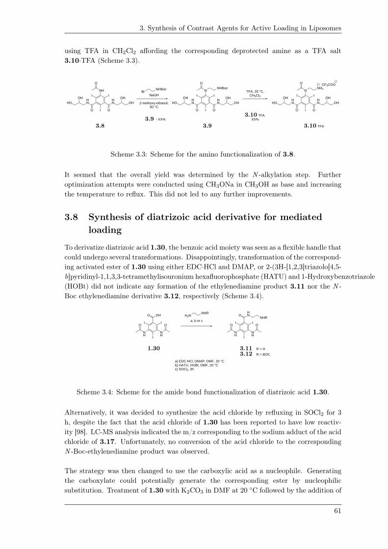
3. Synthesis of Contrast Agents for Active Loading in Liposomes
using TFA in CH2Cl2 affording the corresponding deprotected amine as a TFA salt
3.10·TFA (Scheme 3.3).
I I
I
NH
O
O
HN
OHOH
O
HN
OHHO
I I
I
N
O
O
HN
OHOH
O
HN
OHHO
NHBocNHBocBr
NaOH
2-methoxy-ethanol,50 °C
- XX%
I I
I
N
O
O
HN
OHOH
O
HN
OHHO
NH3
· TFA
CF3COO
· TFAXX%
TFA, 20 °C,CH2Cl2
3.8
3.9
3.9
3.10
3.10
Scheme 3.3: Scheme for the amino functionalization of 3.8.
It seemed that the overall yield was determined by the N -alkylation step. Further
optimization attempts were conducted using CH3ONa in CH3OH as base and increasing
the temperature to reflux. This did not led to any further improvements.
3.8 Synthesis of diatrizoic acid derivative for mediated
loading
To derivatize diatrizoic acid 1.30, the benzoic acid moiety was seen as a flexible handle that
could undergo several transformations. Disappointingly, transformation of the correspond-
ing activated ester of 1.30 using either EDC·HCl and DMAP, or 2-(3H-[1,2,3]triazolo[4,5-
b]pyridinyl-1,1,3,3-tetramethylisouronium hexafluorophosphate (HATU) and 1-Hydroxybenzotriazole
(HOBt) did not indicate any formation of the ethylenediamine product 3.11 nor the N -
Boc ethylenediamine derivative 3.12, respectively (Scheme 3.4).
I I
INH
NH
OO
OHO
I I
INH
NH
OO
HNO
NHR
R = H
a) EDC·HCl, DMAP, DMF, 20 °C b) HATU, HOBt, DMF, 20 °Cc) SOCl2, 3h
a, b or c
H2NNHR
R = BOC
1.30 3.11
3.12
Scheme 3.4: Scheme for the amide bond functionalization of diatrizoic acid 1.30.
Alternatively, it was decided to synthesize the acid chloride by refluxing in SOCl2 for 3
h, despite the fact that the acid chloride of 1.30 has been reported to have low reactiv-
ity [98]. LC-MS analysis indicated the m/z corresponding to the sodium adduct of the acid
chloride of 3.17. Unfortunately, no conversion of the acid chloride to the corresponding
N -Boc-ethylenediamine product was observed.
The strategy was then changed to use the carboxylic acid as a nucleophile. Generating
the carboxylate could potentially generate the corresponding ester by nucleophilic
substitution. Treatment of 1.30 with K2CO3 in DMF at 20 ◦C followed by the addition of
61
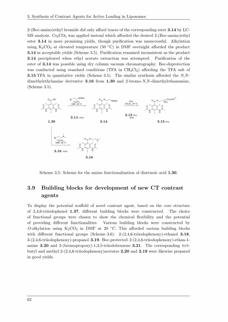
3. Synthesis of Contrast Agents for Active Loading in Liposomes
2-(Boc-amino)ethyl bromide did only afford traces of the corresponding ester 3.14 by LC-
MS analysis. Cs2CO3 was applied instead which afforded the desired 2-(Boc-amino)ethyl
ester 3.14 in more promising yields, though purification was unsuccessful. Alkylation
using K2CO3 at elevated temperature (50 ◦C) in DMF overnight afforded the product
3.14 in acceptable yields (Scheme 3.5). Purification remained inconsistent as the product
3.14 precipitated when ethyl acetate extraction was attempted. Purification of the
ester of 3.14 was possible using dry column vacuum chromatography. Boc-deprotection
was conducted using standard conditions (TFA in CH2Cl2) affording the TFA salt of
3.15·TFA in quantiative yields (Scheme 3.5). The similar synthesis afforded the N,N -
dimethylethylamine derivative 3.16 from 1.30 and 2-bromo-N,N -dimethylethanamine,
(Scheme 3.5).
I I
INH
NH
NHBocBr
- XX%
· TFA
· TFA XX%
TFA,CH2Cl2, 20 °C
OO
OHO
I I
INH
NH
OO
OONHBoc
I I
INH
NH
OO
OONH3
CF3COOK2CO3,
DMF, 50 °C
I I
INH
NH
OO
OON
NBr
K2CO3,,DMF, 50 °C
- XX%
2.30
1.30 3.14
3.143.15
3.15
3.16
3.16
Scheme 3.5: Scheme for the amino functionalization of diatrizoic acid 1.30.
3.9 Building blocks for development of new CT contrast
agents
To display the potential scaffold of novel contrast agent, based on the core structure
of 2,4,6-triiodophenol 1.37, different building blocks were constructed. The choice
of functional groups were chosen to show the chemical flexibility and the potential
of providing different functionalities. Various building blocks were constructed by
O-alkylation using K2CO3 in DMF at 20 ◦C. This afforded various building blocks
with different functional groups (Scheme 3.6): 2-(2,4,6-triiodophenoxy)-ethanol 3.18,
3-(2,4,6-triiodophenoxy)-propanol 3.19, Boc-protected 2-(2,4,6-triiodophenoxy)-ethan-1-
amine 3.20 and 2-(bromopropoxy)-1,3,5-triiodobenzene 3.21. The corresponding tert-
butyl and methyl 2-(2,4,6-triiodophenoxy)acetates 2.20 and 2.19 were likewise prepared
in good yields.
62
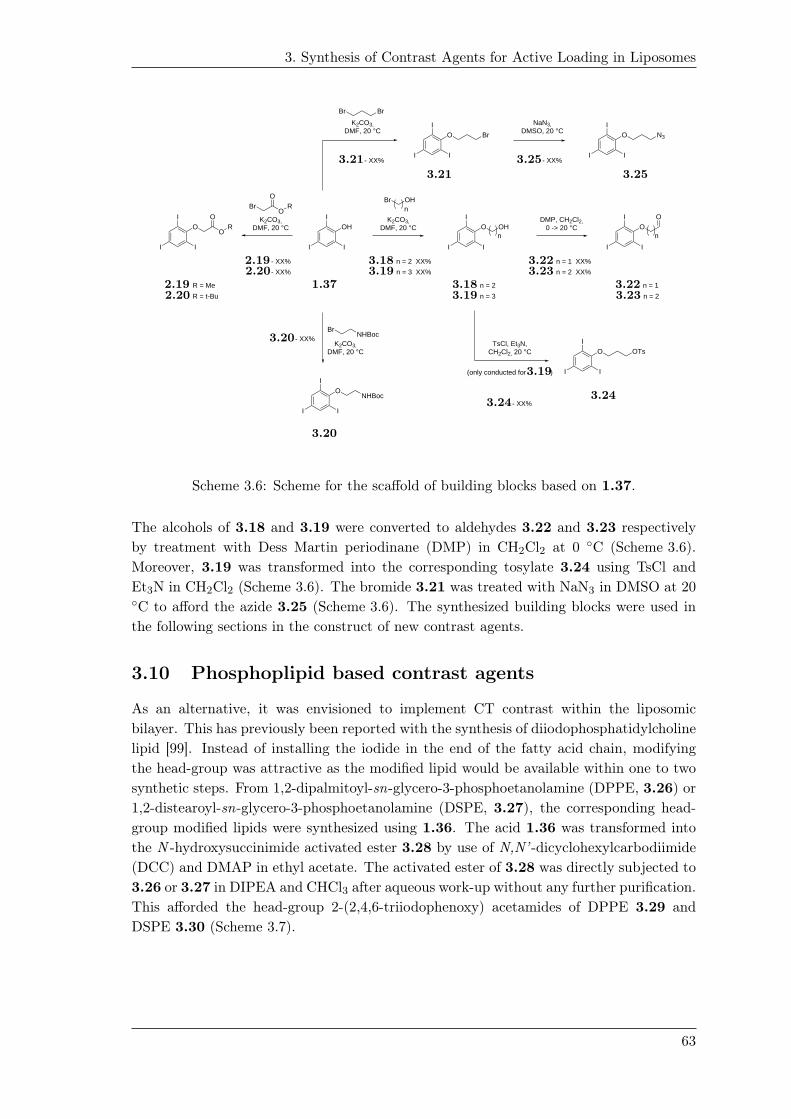
3. Synthesis of Contrast Agents for Active Loading in Liposomes
n = 2
K2CO3,DMF, 20 °C
DMP, CH2Cl2,0 -> 20 °C
I
II
OHI
II
O OHn
I
II
OO
I
II
O OTs
I
II
O BrI
II
O N3
OHBrn
K2CO3,DMF, 20 °C
Br Br
NaN3,DMSO, 20 °C
I
II
ONHBoc
K2CO3,DMF, 20 °C
BrNHBoc
TsCl, Et3N,CH2Cl2, 20 °C
I
II
OO
OR
K2CO3,DMF, 20 °C
BrO
OR
R = Me
n = 2 XX% - XX%
- XX%
- XX% - XX%
- XX%
n = 3 XX%
n = 1 XX%
n = 2 XX%
n
- XX%
n = 3 R = t-Bu
(only conducted for )
n = 1
n = 2
1.37
2.19
2.19
2.20
2.20
3.18
3.18
3.19
3.19
3.19
3.20
3.20
3.21
3.21
3.22
3.22
3.23
3.23
3.243.24
3.25
3.25
Scheme 3.6: Scheme for the scaffold of building blocks based on 1.37.
The alcohols of 3.18 and 3.19 were converted to aldehydes 3.22 and 3.23 respectively
by treatment with Dess Martin periodinane (DMP) in CH2Cl2 at 0 ◦C (Scheme 3.6).
Moreover, 3.19 was transformed into the corresponding tosylate 3.24 using TsCl and
Et3N in CH2Cl2 (Scheme 3.6). The bromide 3.21 was treated with NaN3 in DMSO at 20◦C to afford the azide 3.25 (Scheme 3.6). The synthesized building blocks were used in
the following sections in the construct of new contrast agents.
3.10 Phosphoplipid based contrast agents
As an alternative, it was envisioned to implement CT contrast within the liposomic
bilayer. This has previously been reported with the synthesis of diiodophosphatidylcholine
lipid [99]. Instead of installing the iodide in the end of the fatty acid chain, modifying
the head-group was attractive as the modified lipid would be available within one to two
synthetic steps. From 1,2-dipalmitoyl-sn-glycero-3-phosphoetanolamine (DPPE, 3.26) or
1,2-distearoyl-sn-glycero-3-phosphoetanolamine (DSPE, 3.27), the corresponding head-
group modified lipids were synthesized using 1.36. The acid 1.36 was transformed into
the N -hydroxysuccinimide activated ester 3.28 by use of N,N’ -dicyclohexylcarbodiimide
(DCC) and DMAP in ethyl acetate. The activated ester of 3.28 was directly subjected to
3.26 or 3.27 in DIPEA and CHCl3 after aqueous work-up without any further purification.
This afforded the head-group 2-(2,4,6-triiodophenoxy) acetamides of DPPE 3.29 and
DSPE 3.30 (Scheme 3.7).
63

3. Synthesis of Contrast Agents for Active Loading in Liposomes
O
O(R)(R)
OP
O
ONH2
HO
O
OHn
n O
O(R)(R)
OP
O
OHN
HO
O
OHn
nO
O
I I
I
OOH
OI
II
OOI
II
n = 11, XX%
DIPEA,CHCl3, 20 °C
NHS,DCC, DMAPEtOAc, 20 °C
DPPE, n = 11
ON
O
O
DSPE, n = 13
n = 11
n = 13
n = 13, XX%
1.36
3.20
3.26
3.27
3.28
3.29
3.29
3.30
3.30
Scheme 3.7: Head-group modification of the phospholipids 3.26 and 3.27 using 2-(2,4,6-
triiodophenoxy)acetic acid 1.36.
3.11 Glucosamine based contrast agents
To compensate for the hydrophobic nature of the synthesized tri-iodophenoxy building
blocks, the building blocks should be combined with hydrophilic counterparts to facilitate
the proper permeability through the bilayer of the liposome. d-Glucosamine 3.31 was
chosen for conjugation to the tri-iodophenoxy building blocks. Blocking the hemiacetal
chemistry of the anomeric position by Fischer glycosylation followed by the installation
of the 2,4,6-triiodophenoxy unit at the 2-amino position will leave the hydroxyl-groups
of 3-OH, 4-OH and 6-OH available, which was thought as a handle for further modifica-
tions to control and customize the polarity of the synthesized contrast agents. In order
to provide weak basic character for mediated loading the primary amine of glucosamine
3.31 could be converted into the secondary amine through reductive amination from the
corresponding aldehyde building blocks of 3.22 and 3.23. The secondary amine could
likewise be achieved through nucleophilic substitution of the corresponding bromide of
3.21 or tosylate 3.24, respectively. The amide could also be constructed from the 1.36
and glucosamine 3.31.
According to literature, d-glucosamine hydrochloride 3.31·HCl can be transformed into
the methyl 2-deoxy-2-amino-d glucopyranoside hydrochloride 3.32·HCl by peracetylation
to give 3.33 followed by a Fischer glycosylation (Scheme 3.8) [100]. Peracetylation was
conducted using acetic anhydride in pyridine at 20 ◦C. This gave the peracetylated prod-
uct 3.33 (α/β = 20:1). The Fischer glycosylation of 3.33 was performed using refluxing
5% HCl in CH3OH (Scheme 3.8). Prolonged reaction time was applied to make sure the
acetamide was hydrolyzed. Concentration in vacuo afforded 3.32·HCl in an α/β = 6:1
(Scheme 3.8). The ratio of α/β-isomers was concluded not to be decisive for the contrast
properties nor the mediated loading properties, 3.32·HCl was therefore used without fur-
ther purifications.
The reductive amination was performed using 3.32·HCl and the aldehyde 3.22 (Scheme 3.8).
Compound 3.22 was chosen over 3.23 in order to avoid the potential side-reaction of
β-elimination of the aldehyde 3.23. Sodium triacetoxyborohydride (NaBH(OAc)3) was
64

3. Synthesis of Contrast Agents for Active Loading in Liposomes
chosen as reducing agent over the corresponding sodium cyanoborohydride (NaBH3CN)
due to easier handling. The experimental procedure was conducted according to litera-
ture [101]. The hydrochloride salt of 3.32·HCl was neutralized using Et3N prior to the
formation of the imine-intermediate. The reduction of the imine of 3.32 and 3.22 gave
the N -alkylated 3.34 as the main species. The reductive aminated product 3.34 was
isolated by dry column vacuum chromatography. The reductive amination was later on
repeated and up-scaled. The upscaling was inconsistent as competing reduction of the
aldehyde 3.22 to the corresponding alcohol 3.18 was observed. Therefore a disappointing
yield was achieved. The observation of a low yield was believed to be due to the attempt
of improving the yield by pre-mixing the amine of 3.32 and 3.22 in order to form the
imine-intermediate prior to the addition of NaBH(OAc)3. Since 3.34 only appeared in
low yields, this could indicate that the imine-formation was formed inefficient.
The amide of 3.35 was obtained from the acid 1.36 and 3.32·HCl, using EDC·HCl and
DMAP (Scheme 3.8). Beside the intended product formation, a significant amount side-
product of the migrated N -acylurea product of the activated ester was observed (according
to LC-MS analysis). The presence of the N -acylurea side-product made purification by
dry column vacuum chromatography difficult as similar retention factors of 3.35 and
the side-product were observed. Applying an acidic wash (0.1 m HCl) during work-up, re-
moved exclusively all of N -acylurea side-product by which isolation afforded the amidated
product 3.35.
· HCl
OHOHO
NH3
OH
OH
Cl
OOO
NH
O
O
O
O
O O O
· HCl
OHOHO
NH3
OH
OCl
(Ac2)Opyridine, 20 °C
5% HCl / CH3OH,reflux
I
II
OO
Et3N, (AcO)3BH, THF20 °C
OHOHO
NH
OH
O
O I
I
I
OHOHO
NH
OH
OO
O I
I
I
I
II
OOH
O
EDC·HCl, DMAP,Et3N, DMF
XX%(α:β) = 20:1
· HCl XX%
(α:β) = 6:1XX%
isolated as α-anomer only
XX%(α:β) = 6:1
1.36
3.19
3.31
3.32
3.32
3.33
3.33
3.34
3.35
3.35
Scheme 3.8: Synthesis of glucosamine-derived CT-contrast agents 3.34 and 3.35.
65

3. Synthesis of Contrast Agents for Active Loading in Liposomes
3.12 Mediated loading of contrast agents
Mediated liposome loading was demonstrated for contrast agents 3.10 and 3.15. This
was conducted by project partners of the Nanoguide-project. Liposomes with the lipid
composition of HSPC/MPEG-2000-DSPE/cholesterol (3:1:1) (Tm = 55 ◦C, MW = 750
g·mol−1) were prepared. 0.2 mol% of PE Lissammine Rhodamine B was added to the lipid
composition in order to provide UV-Vis detection (λmax = 560 nm). The lipid mixture
was first lyophilized followed by rehydration using 120 mm (NH4)2SO4 (pH 5.2) at 65◦C for 1 h. The size of the LMV liposomes was downsized and changed to LUVs by
extrusion. Extrusion was conducted using 100 nm pore sized polycarbonate membrane
and was repeated 21 times at 65 ◦C. This afforded LUVs with average size of 116 ± 0.29
nm with a narrow poly-distribution of PDI = 0.057.
The transmembrane (NH4)2SO4 gradient was established by iso-osmotic dialysis against
a 25 mm HEPES, 150 mm NaCl, pH 7.4 solution.The mediated loading was conducted at
65 ◦C. A solution of 3.10·TFA or 3.15·TFA in the given buffer with a D/L ratio = 0.1
were added to the liposome mixture. Encapsulation efficiencies of 3.10 did not exceed
10% while 3.15 was loaded to a level of 70% after 1 h at 65 ◦C. It was reasoned that
the loading of 3.10 was insufficient due to the hydrophilic nature of 3.10. No further
optimization of the loading of 3.10 was pursued. The loading efficiency of 3.15 decreased
over time, indicating a leaky behavior of 3.15 from the aqueous inner compartment. The
reduction in loading over time was reasoned to be a consequence of the active loading
being performed above the transition temperature (Tm) of the lipid-mixture constituting
the bilayer of the liposomes.
Parameters like choice of external buffer solution, pH, mediated loading temperatures,
size of liposome and D/L ratio were analyzed to optimize the loading of 3.15.
3.13 Optimized loading using ammonium sulfate gradient
The loading temperature was analyzed at three different temperatures (40 ◦C , 55 ◦C or 65◦C) around the Tm of the lipid mixture using a pH 7.4 HEPES buffer. The highest loading
was observed after 1 h at 65 ◦C corresponding to 72-73%. Similar loading efficiencies were
seen after 24 h at 55 ◦C. The optimal D/L ratio were likewise found. Mediated loading
of 3.15 using different D/L of 0.1 to 0.8 were analyzed. Using a D/L ratio of 0.1 yielded
the best loading after 3 h.
The size of the liposomes were likewise analyzed. Two different sizes of 140 nm and
180 nm were tested. The highest loading was achieved using the 140 nm liposomes after
3 h while the same loading efficiency for the 180 nm liposomes was obtained after 20 h.
Several buffers with different pH were tested for the mediated loading of 3.15. The buffers
and pH tested were 1) a 25 mm HEPES, 150 mm NaCl, pH 7.4 solution, 2) a 25 mm Tris,
150 mm NaCl, pH 8.1 solution, 3) a 25 mm MES, 150 mm NaCl solution and 4) a 25 mm
PBS buffer, 150 mm NaCl, pH 7.0 solution. Beside the obtained loading using the HEPES
buffer at pH 7.4, the best achievable mediated loading was achieved using the 25 mm PBS
phosphate buffer, 150 mm NaCl. This afforded the encapsulated 3.15 liposomes (size 140
nm) with a 78% loading efficiency after 20 h according to UV-Vis measurements after spin
column filtration. The measured osmolality of the liposome mixture was determined to
66

3. Synthesis of Contrast Agents for Active Loading in Liposomes
be 332 mOsm·kg−1.
3.14 Mediated loading using citrate buffer gradient
Lipsomes based on the same lipid mixture as described above was used. Lyophilization
and hydration using a 300 mm citrate buffer (pH 4, 576 mOsm·kg−1) during heating at
65 ◦C for 1 h was performed. The size of the liposomes were increased as it was reasoned
that the size of the liposome could be decisive for the mediated loading (bigger liposome,
increased encapsulation efficiencies). Extrusion using 400 nm and 200 nm polycarbonate
filters were used to afford LUV liposomes of 176±0.901 nm with a PDI = 0.071±0.032.
The gradient was established by adjusting the pH of the liposome mixture to pH 7.0
using 1 m Na2CO3. The contrast agent 3.15·TFA was directly added to the mixture
as a solid. The mediated loading was conducted at 55 ◦C for 3 h. This successfully
afforded the loading of 3.15 with an encapsulation efficiency of 96% according to UV-Vis
measurements. ICP measurements gave a loading efficiency of 91%.
With the promising loading efficiencies using the citrate buffer, the mediated loading was
repeated in order to lower the osmolality of the liposome mixture for an in vivo study in
mice, mainly to check whether the loaded 3.15 would provide a sufficient visible contrast.
A 200 mm citrate buffer was used during the hydration affording the liposomes with a
size of 169.7±2.4 nm and a PDI 0.063±0.021. The mediated loading was conducted by
adjusting the pH using 1 m Na2CO3 solution. The final concentration of the loaded
liposomes with a loading efficiency of 85%/73% (UV-Vis/ICP-MS) was determined to be
53 mm corresponding to a 3.15 concentration of 8.7 mg·mL−1 (5.01 mg I·mL−1). The
liposomes was further concentrated to a final iodine concentration of 10 mg I·mL−1.
3.15 In vivo performance of mediated loaded liposomes
Liposome formulation of 3.15 was injected in the of NMRI mice from which CT scans
was recorded. There was no visible contrast from the loaded liposomes.
3.16 Discussion and future perspectives
From the obtained mediated loadings and encapsulation efficiencies, it was clear that it
would be difficult to achieve the previously reported concentrations of contrast agents by
mediated loading. The current limitation to achieve the concentration corresponding to
a sufficient contrast, is mainly proposed to be a consequence of the encapsulated amount
of the buffer (citrate, (NH4)2SO4 etc.). The amount of buffer encapsulated within the
interior of the liposome after purification is estimated to be in range of 10% correspond-
ing to a concentration of buffer in the range of 20-30 mol·L−1 solution liposome [102].
The concentration of buffer within the aqueous inner compartment would still correspond
to the concentration used during the hydration-procedure (200-300 mm). Though, the
concentration of buffer (20-30 mm) after purification puts the limit of the gradient and
thus the extent of loading. An intuitive idea would be to increased the buffer-capacity
of the buffer used. This will have the effect of providing a higher gradient implying that
more material can be loaded. The limitation of increasing the buffer-capacity would be
the corresponding increment in osmolality. A higher osmolality will lead to an increased
67
3. Synthesis of Contrast Agents for Active Loading in Liposomes
osmotic pressure across the membrane of the bilayer, by which in-stability of the liposomes
is suspected to occur, due to uptake of solvent e.g. unbound water of the blood plasma
leading to increased blood viscosity or even burst of the liposomes.
Another option would be to improve the size of the liposomes as an increased aque-
ous compartment would enable increased encapsulation of buffer during the hydration.
Based on extensive experience, LUV liposomes are hardly reproducible in size [102]. In
perspective: as the mediated loading have been applied to various drugs e.g. Doxorubicin
where the therapeutic effect needed is low, mediated loading of contrast agents - where
high concentrations are needed - is not suitable, unless precipitation inside the interior of
the liposome is achievable.
Prospectively, it would therefore be of interest to conduct active loading of radioiodinated
derivatives of 3.15 due to the sensitivity of radioactive compounds. This will likewise
benefit the preparation method of liposomal formulations of radiolabeled contrast agents.
According to the attractive isotopes of iodide and the EPR effect of liposomes, potentially
novel liposomal contrast agents can be visualized by different imaging-modalities (SPECT
and PET). This is at the moment being undertaking by project partners in the Nanoguide
project.
3.17 Conclusion
Five new contrast agent (3.10, 3.15, 3.16 3.34 and 3.35) for CT imaging have been
synthesized for mediated loading in liposomes. 3.10 and 3.15 were loaded in liposomes
where 3.15 showed the most promising results. An encapsulation efficiency of 85%
corresponding to a 5.01 mg I·mL−1 was achieved. The in vivo injection in a mouse
model confirmed that this concentration was in-sufficient for obtaining contrast. It was
concluded that the gradient established was not sufficient to loading the amount needed
to achieve a proper CT contrast.
68

Cyclodextrin-pluoronic basedpolyrotaxanes for PET-imaging 4
An interesting supramolecular construct with application within nanomedicine and drug
delivery is polyrotaxanes. Polyrotaxanes (PRs) are the term and collective name of the
supramolecular construct of “threaded macrocyclics” onto an axle (usually a polymer).
The intermediate assembly is referred to as the pseudorotaxanes where disassociation (de-
threading) is still optional. The polyrotaxanes are formed when the pseudorotaxane is
subsequently endcapped with a bulky endgroup with the intended purpose of trapping the
macrocyclic threaded onto the polymer. When retained on the polymer, the macrocyclic
possesses, at this state, only rotational and longitudinal motions until slippage of the
macrocyclic occurs or detachment by either macrocyclic opening or the endcaps are
liberated from the PRs. The driving force for the assembly is mainly based on non-
covalent interaction. A schematic representation is shown in Figure 4.1.
Figure 4.1: Schematic representation of the concept and synthesis of polyrotaxanes.
One example of a PR system is the cyclodextrin (CD)/pluoronic-based PRs. Pluoronic
polymers are categorized as being the biocompatible and less toxic tri-block polymer
of PEG-PPG-PEG. The pluoronic polymers vary in size as well as in the total ratio
of the PEG and PPG segments. The most common pluoronic polymers are listed in
Table 4.1. As seen from the table the size (1.9 – 12.5 kDa) of polymers as well as the
hydrophilic/hydrophobic balance (HLB) vary for the different polymers. The HLB is
defined as an empirical numerical scale, used today to rank polymers and surfactants by
their polarity (higher the value, the more polar the polymer/surfactants will be) [].
69

4. Cyclodextrin-pluoronic based polyrotaxanes for PET-imaging
Table 4.1: Table listing the different pluoronic polymers and the composition and
properties.
3.23
Cyclodextrins are defined as being cyclic oligosaccharides consisting of repeating α-1,4-d-
glucopyranoside units (figure Figure 4.2). The most common CDs are α, β and γ CDs with
a six, seven and eight glucopyranoside units, respectively []. The conformational struc-
ture of CDs are similar to a truncated cone by which the hydroxy-groups are oriented
towards the exterior such that the interior is relatively hydrophobic similar to a mixture
of EtOH and H2O (Figure 4.2) []. The hydrophobic cavity allows for host guest inclusion
complexes with small hydrophobic molecules or segments of polymers. Cyclodextrins have
found several applications like drug delivery, solubility enhancement and mediated drug
release [103,104].
OH
OHOH OH OH OH
OH
OH
OH-6 primary hydroxyl groups
a
OH-2, OH-3 secondary hydroxyl groupsHydrophobic cavity
6
6
5
54
4 3
322 1
1
b
1.30
Figure 4.2: a) glycosidic linkage b) the conformational structure of CDs
The prerequisites for the rotaxanation reactions with CDs and pluoronic ploymers are
among other parameters influenced by physical constraints. The cross-sectional area of
the polymer must be of the proper dimension to induce a favorable inclusion complex
with the CD. The solvent chosen for the rotaxanation must provide solubility but not be
competitive in regard to the inclusion complex formation. The pluoronic polymer should
be functional at the terminal ends for the endcapping reaction. Likewise, the endcapping
70
4. Cyclodextrin-pluoronic based polyrotaxanes for PET-imaging
reaction has to be orthogonal/selective to the hydroxy-group chemistry of the CDs and
be fast in order to capture as many CDs as possible. The review of Loethen et al. cover
the common trends of choice of endcaps as well as the common post-modification options
of threaded CDs [105].
4.1 Niemann Pick disease Type C
The group of Thompson as well as the Harada group are among the main frontiers within
the field of CD/pluoronic PRs. CD/pluoronic PRs have found used with the Niemann
Pick Type C (NPC) disease, drug delivery of gene therapy and contrast agents [].
The NPC disease is a not-tissue-specific genetic disorder owing to mutations in genes
encoding for efflux proteins, which are located in the membranes as well as within the
aqueous compartment of the late endosome/lysosome (LE/LY) [106, 107]. Normally, the
role of these proteins is to control the regulation and efflux of un-esterified cholesterol.
The deficiency of these proteins, due to mutations, leads to aberrant accumulation of un-
esterified cholesterol, which is characterized as a lysosomal storage disease. The build-up
of cholesterol in the lysosomes trigger a number of severe effects like lysosomal swelling,
upregulation of genes controlling cholesterol synthesis, and low density lipoproteins uptake
as well as neurological effects like demyelination of brain cell axons together with death
of Purkinje cells [107] and release of cytokines in regard to macrophage activity [107].
These effects compiled lead to cell death with the symptoms taking shape as neurological,
hepatic and/or pulmorary symptons that will become intensified over time [107]. At the
severe stages, this can lead to dysarthria, hysphagia, ataxia as examples of neurological
disorders caused by NPC [107].
In vivo studies have shown that CDs have been able to mobilize accumulated cholesterol
from the late endosomes/lysosomes due to the inclusion complex ability of CDs [106]. The
response of using 2-hydroxypropyl (HP)-β-CD is a normalization in intracellular levels of
un-esterified cholesterol as well as down-regulation of the intrinsic cholesterol synthesis
and activation of various related metabolic-genes [107]. Despite these promising signs of
response using HP-β-CD, at this given point, mice studies have indicated that rather high
doses in range of 4 g HP-β-CD·kg−1 body weight and even higher doses is expected to be
needed as the clearance from the bloodstream is rapid in accordance to the water solubility
and the relative low molecular weight of CDs [106]. Likewise, it has been reported that
re-accumulation of cholesterol in the LE/LY was observed, which could indicate a post-
treatment up-regulation of cholesterol upon treatment [107]. This means that treatment
using HP-β-CD has to be mediated by a sustained dosage by continuous administration of
HP-β-CD by multiple/repeating doses [107]. This clearly states the drug delivery issue of
CDs in accordance to the high doses needed and the continuous therapeutic dose treatment
of NPC.
CD-based PRs become therefore useful as an efficient carrier of CDs, as multiple units of
CDs can be threaded onto the polymer/axle. Likewise applying a long-time circulation
property could improve a more persistent treatment and delivery of CDs to NPC affected
cells.
The group of Thompson recently published the construction of HP-β-CD/pluronic PRs.
HP-β-CD was chosen over β-CD due to the higher water solubility of HP-β-CD (650
mg·mL−1) as opposed to native β-CD (18 mg·mL−1) which also is reported to be
71

4. Cyclodextrin-pluoronic based polyrotaxanes for PET-imaging
parenterally nephrotoxic. The study of Harada et al. showed how the dimensions of
cavity of CDs has a significant influence on the overall threading []. Harada et al. showed
that α-CDs are easily threaded onto PEG while β-CD are not threaded. β-CD is on the
other hand preferably threaded on PPG. This is exemplified in the preceding polyrotaxane
assemblies. The coverage of the PPG has been verified by 2D-NOESY NMR experiment
where a spatial correlation between the methyl group of PPG as structural backbone of the
CDs [106]. Various derivatives of CDs have likewise been threaded on pluoronic polymers.
4.2 Threading/inclusion complex formation of CDs
Usually the inclusion complex formation is conducted in a polar solvent, by which the
CDs will be soluble while the guest molecule will be less soluble. Using this setup, the
solvent can be used to drive the inclusion complex formation – based on the principles
of the hydrophobic effect - as the cavity would act as hydrophobic pocket where the
hydrophobic guest molecule can reside. Together with the entropic release of the high
energetic restricted water molecules residing inside the cavity, these aspects are some of
the most important effects influencing the dynamic of the inclusion complex formation.
Normally in CD-based PRs formation, the entropy will be disfavored due to the loss of
conformational flexibility of the polymer. As the CDs likewise become more ordered with
only longitudinal – and rotational movement, this will contribute to an entropic disfavor.
The enthalpy contribution are mainly dominated by the hydrophobic effect of the inclusion
complex formation as well as the solvent relaxation upon threading [105]. According to the
recent papers within Thompsons group, threading conducted in apolar solvents have led
to higher threading efficiencies of CDs. The rationale for the higher threading efficiencies
would be that multiple CDs would tend to form aligned channels/tubings in a hydrophobic
environment, by which the pluoronic polymer is easily threaded []. When entering the
aligned arrangement of cavities, the pluoronic polymers is more easily directed through
the alignment of multiple CDs resulting in a higher loading/threading.
4.3 In vivo bio-distributions studies of CD/pluoronic PRs
A study conducted in the group of Thompson showed the synthesis of HP-β-CD/pluronic
PRs with trinitrobenzene as endcapping group [106], and various pluoronic polymers
was threaded using HP-β-CDs. The threading was conducted in hexane which afforded
promising threading efficiencies. The PRs were tested in vitro at a buffered pH of 5.5 and
7.4, displaying prolonged stability. The synthesized PRs were likewise tested in vitro on
npc2−/− fibroblast cell line and showed a remarkable reduction of aberrant un-esterified
cholesterol according to filipin staining, which is commonly used for the quantification of
cholesterol (Figure 4.3). The mechanism of mobilization and corresponding uptake of the
cholesterol remain unknown.
72

4. Cyclodextrin-pluoronic based polyrotaxanes for PET-imaging
Figure 4.3: In vitro evaluation of cholesterol-uptake by CD/pluoronic based PRs. A) The
filipin stain of npc2−/− fibroblasts treated with CD/pluoronic based PRs (entry 11HP.L81
and 07.HPF127) and HP-β-CD as a function of time. When compared to the control, the
intensity of stain is significantly lowered, indicating that the amount of cholesterol have
decerased. B) direct quantification of the effect the CD/pluoronic based PRs to lower the
amount cholesterol.
4.4 CD/pluoronic PRs as contrast agents
HP-β-CD/ pluoronic PRs have likewise found use as MRI contrast agent for blood pool
imaging. PRs conjugated with GD3+ chelates (T1 paramagnetic relaxation enhancement)
would provide the property of enhancing the relaxation rate of T1 of coordinated H2O-
molecules. As highlighted in the study in Thompson group, the attractable features of
using PRs as contrast agents contrary to clinical approved contrast agents in current use,
would be prolonged circulation times as well as higher Gd3+ loading per molecule. More-
over, other nanomaterials used as contrast agents, suffer from poor renal filtration as well
as hepatobiliary uptake and in vivo accumulation. Additionally, the use of acyclic chelates
can cause leakage of GD by which these chelates are referred as being kinetically labile.
Leakage of Gd from the contrast agent can lead to nephrogenic systemic fibrosis.
73
4. Cyclodextrin-pluoronic based polyrotaxanes for PET-imaging
As stated, a polyrotaxane scaffold-based contrast agent will offer the benefit of higher
Gd-loading - as multiple Gd chelates can be appended per polymer - and prolonged cir-
culation time according to the flexible rod-like structure of PRs []. Several studies have
indicated that the morphology has an influence on the macrophage uptake, i.e. spherical
nanoparticles have proved to be taken up by macrophages and HeLa cells at higher rate
than rod-like nanoparticles [].
The study concerning HP-β-CD/pluoronic PRs as MRI contrast agents was based on
the Pluoronic F127 polymer with a 46% HP-β-CD threading efficiency and cholesterol
as endcapping groups. The macrocyclic chelation unit of 1,4,7,10 tetraazacyclododecane-
1,4,7-triacetic acid (DO3A) was further introduced in a post-modification strategy of the
PRs. The hydroxy shell of the HP-β-CD of the PR was first functionalized with 1,8-
diamino-3,6-dioxooctane (EDA) via a CDI activation followed by nucleophilic addition of
p-thioisocyanate-benzyl-DO3A. 14 units of DO3A were appended to the CD/pluoronic
PRs and loaded with 14 Gd3+ ions and this construct was characterized in terms of a
standardized relaxation of H2O per Gd concentration (r1). The MRI properties is depicted
in figure ??.
A proportional tendency of increasing relaxitivity with increasing concentration of Gd was
observed for both T1 - as well as T2 relaxation. In accordance to the higher loading of
Gd per PR, the relaxation rate of the bound H2O was observed to be faster than the
corresponding DO3A-HP-β-CD monomer. This was further supported by the in vivo per-
formance. Enhanced contrast was observed of the cardiovascular system (blood pool) for
prolonged time with subsequently clearance. These findings were promising as no persis-
tent in vivo accumulation of the PRs was observed.
This was further substantiated in the follow up structure-property relationship study of
the class of HP-β-CD/pluoronic PRs with cholesterol as endcaps. The aim of this study
was to analyze the in vivo distribution, pharmacokinetics, clearance and the interaction
with the protein corona with common proteins found in the blood plasma [108].
This study indicated that a tendency between the threading efficiency of the PRs and the
overall in vivo performance. The corresponding PRs made from the L35 and L81 pluoronic
polymers possessed the highest threading efficiencies but also exhibited longest circulation
time in BALB/c mice [108]. The overall observed trend was a fast kinetic phase along
with a biphasic clearance [108]. In both cases, it was reasoned that the flexible rod-like
morphology was determining the outcome of the in vivo clearance. As the PPG-segment
of L35 and L81 constitute the main part of these pluoronic polymers, high threading
efficiencies make the PRs of L35 and L81 adopt a rod-like structure [108].
In terms of the bio-distribution, the highly threaded PRs showed preferentially uptake in
the liver while less PRs were accumulating in the lungs. The blood compatibility of PRs
was likewise evaluated. No evident kidney toxicity nor liver damage or signs of hemolysis
were observed. As expected, the interaction of PRs with the protein corona was mainly
constituted by lipoproteins which was in accordance to the design of PRs with cholesterol
endcaps. As part of the design in the above-mentioned PR for MRI contrast enhancement,
the choice of cholesterol as endcapping group was based on the choice of a drug delivery
tag. As cholesterol is transported by several lipoproteins to the LE, the concentration
of the PR could hopefully be enhanced at the LE/LY site [108]. The relaxitivity of this
74

4. Cyclodextrin-pluoronic based polyrotaxanes for PET-imaging
family of PRs with cholesterol was likewise characterized and compared to DOTA chelated
Gd (DOTAREM). In comparison to the previously developed PRs with TNTS endcaps,
the cholesterol endcapped PRs showed similar relaxitivities, which was concluded to be in
the range of 2-4 fold higher than the monomeric DOTAREM [108].
4.5 Problem statement
Having the positron emitting 64Cu cyclotron disposable at the Heavesy laboratory at
the Center of Nuclear Technologies, Technical University of Denmark, the 64Cu-chelated
PRs could be synthesized. A collaboration was therefore established between the group
of Professor Thompson and DTU Chemistry. To enable a more sensitive imaging of the
CD/Pluoronic-based PRs in vivo, and obtain improved information of the bio-distribution,
CD/Pluoronic-based PRs able of chelating 64Cu would have to be synthesized.
4.6 Design and theoretical deliberation of PET
HP-β-CD/pluoronic PRs
With the well-established and documented use of HP-β-CD/pluoronic PRs as contrast
agents within the MRI modality, PET imaging would offer a higher sensitivity and poten-
tially more clearly elucidation of the in vivo performance and biodistribution of tissues
with low accumulation of PRs. One issue that remains an open question would be whether
PRs are able to cross the blood brain barrier? This question could potentially be addressed
by the development of PRs for 64Cu chelation.
In order to correlate the already achieved MRI imaging of CD/pluoronic based PRs with
the PET-imaging the construct of the PRs for PET imaging should not deviate too far
from the CD/pluoronic based PRs core-structure used in the MRI imaging study. In ac-
cordance with the previous studies of the cholesterol endcapped HP-β-CD/pluoronic PRs,
the PRs of L35 and L81 showed the longest circulation times. It was therefore decided
to base the PRs on the pluoronic polymers of L35 and L81. CD-based PRs generally
possess poor solubility hypothesized by the conformational orientation of the CDs that
provide hydrogen bonding in-between the repeating units of CDs. This has the effect that
the pluoronic polymer and threaded CDs have a confined/restricted interaction with the
solvent through hydrogen bonding [105]. In order to improve the solubility of this class/-
type of PRs for PET imaging, the threading experiments were conducted using a mixture
of HP-β-CD and the commercially available Captisol. Captisol is a CD derivative with
randomly ether-modified butyl sodium sulfonates which by the polar sulfonates enhance
solubility compared to the native CD []. The average degree of modification of sulfonates
is stated to be approximately 6.6.
In terms of applying functionalities of PRs two strategies are often used. The most com-
monly way is post-modification of the hydroxyl shell of CDs. This is determined by the
hydroxy-group chemistry. This method usually lacks control as the modification takes
place randomly. Improved control can be achieved by threading functionalized CD deriva-
tives. Various mono-functionalized CD-derivatives are achievable from the mono-tosylated
β-CD. The mono-tosylated β-CD can then easily be exchanged with i.e. azide that offer
the feasible click chemistry of the Cu catalyzed azide alkyne cycloaddition (CuAAC) with
75
4. Cyclodextrin-pluoronic based polyrotaxanes for PET-imaging
a corresponding alkyne.
Based on the previous designs, the employed linkage was based on carbamates. The car-
bamates are hypothesized to undergo hydrolysis by enzymatic cleavage and was proven
to be stable at pH of 5.5 and 7.4 corresponding to pH levels found in the blood and LE/LY.
A secondary objective of this project was to develop a PR-based contrast agent for
dual modality imaging. Besides introducing the DOTA moiety for chelation it would
be interesting to see if an aryl-TMS moiety could be appended/grafted to PRs as a
precursor for radioiodination. Gd chelation combined with the radioactive isotopes of
iodine would offer a dual imaging modality. Inspired of the post-modifications strategies of
dextran-polymers using CD derivatives [109], it was envisioned that the aryl-TMS could be
grafted onto the CD-PRs through the ring-opening of the corresponding (p-TMS)-phenyl
glycidyl ether (keeping in mind that β-elimination could be a potentially side-reaction) or
through click chemistry having threaded N3-B-CD followed by a subsequent CuAAC with
p-ethynylphenyltrimethylsilane. The triazole would potentially induce an inductive effect
on the activation of the p-ethynylphenyltrimethylsilane according to the three nitrogen
atoms constituting the ring. It was hypothesized that the mesomeric effect would be
dominating and thus provide the activation of the aryl-TMS to undergo electrophilic
aromatic ipso-substitution with the iodonium ion.
4.7 Synthesis of the CD/pluoronic based polyrotaxanes
The pluronic polymers of L81 4.1 and L35 4.2 was first functionalized at the end
terminals with the tris(2-aminoethyl)amine (TAEA) using a CDI mediated activation of
the terminal hydroxyl groups. Prior to the reaction, the pluoronic polymers was azeotropic
concentrated in vacuo using toluene. The pluoronic polymers was then pre-activated
using CDI in CH3CN followed by the addition of tris(2-aminoethyl)amine (TAEA). TAEA
modification of L81 4.3 was afforded in a low yield of 13%. but repetition of the TAEA
modification afforded the TAEA modified L81 pluoronics polymer 4.3 in 79%. The TAEA
modification of L35 4.4 was synthesized in 62% yield (Scheme 4.1)
76

4. Cyclodextrin-pluoronic based polyrotaxanes for PET-imaging
HOO
OO
OO
OO
OH
x y z
1. CDI, Et3N, CH3CN, 20 °C, 5h2. Tris(2-aminoethyl)amine
OO
OO
OO
OO
O NH
O
NH
ON N
H2N
H2N
NH2
NH2
x y z
X=Z=3, Y=43
X=Z=10, Y=16
X=Z=3, Y=43
X=Z=10, Y=16
13%, 79%
62%
4.1
4.2
4.3
4.3
4.4
4.4
Scheme 4.1: TAEA modification of the pluoronic polymers of 4.1 and and 4.2 respectively.
The polymers of 4.3 and 4.4 was then threaded using a CD feed ratio of 70/30 of HP-
β-CD/captisol, respectively. The total CD amount used was based on the theoretical
amount of threaded PPGs units of the respective pluoronic polymers. As an estimate, it
was assumed that β-CDs cover the PPG-segment with an average coverage of 2 PPG/CD.
This will give a maximum of threaded CDs possible of 8 and 21 CDs for L35 4.2 and
L81 4.2, respectively. Before threading, 4.3 and 4.4 were vigorously suspended in hexane
overnight. HP-β-CD and captisol was added as fine-grinded mixed solid to the hexane
solutions of 4.4 or 4.3, respectively. Bath and probe sonication were then conducted
as sonication has proven to improve the threading efficiencies in previous studies [].
Sonication is reasoned to disrupt any micelle formation that is believed to be the state
of supramolecular assembly above the level of the aggregation concentration []. The
threading reaction was then conducted for 72 h. The solvent was then changed from
hexane to CH2Cl2 and the formed pseudorotaxanes was then endcapped using cholesteryl
chloroformate and Et3N. Subsequent dialysis and lyophillization afforded the threaded
L35 and L81 HP-β-CD/Captisol PRs, 4.5 and 4.6 respectively (Scheme 4.2).
77

4. Cyclodextrin-pluoronic based polyrotaxanes for PET-imaging
OO
OO
OO
OO
O NH
O
NH
O
N N
H2N
H2N
NH2
NH2
1. HP-β-CD (70% Feed), Captisol (30% Feed),
hexane, 72 h, 20 °C
2. Cholesteryl chloroformate,
Et3N, CH2Cl2, 24 h, 20 °C
X=Z=3, Y=43
X=Z=10, Y=16
OO
OO
OO
OO
O NH
O
NH
O
N N
NH
NH
HN
HN
O
O
R
O
O R
O
O
R
O
O
R
R: CholesterolBlue: HP-B-CD Red: Captisol
X=Z=3, Y=43
X=Z=10, Y=16
XX%
XX%
O
OH
O
SO3
-
x y z
x y z
3.27
4.3
4.4
4.5
4.5
4.6
4.6
Scheme 4.2: The threading and formation of CD/Pluoronic-based PRs of 4.5 and 4.6,
respectively
Other CD feed ratios of 50/30/20 and 65/30/5 of HP-β-CD/Captisol/N3-β-CD were
threaded respectively. According to previous studies, 6-monodeoxy-6-monoazido-β-
CD (N3-β-CD, 4.7) has been successfully threaded onto pluoronics []. N3-β-CD was
synthesized according to literature (Scheme 4.3). 6-O-mono-tosyl-β-CD (TsO-β-CD, 4.8)
was first synthesized from the inclusion complex of native β-CD 4.9 and TsCl followed by
deprotonation using concentrated NaOH. This afforded 4.8 as the mono-tosylated main
compound (Scheme 4.3). Recrystallization afforded the pure monotosylated 4.8. The
tosyl group was exchanged with the azide using NaN3 in DMF at 70 ◦C. This afforded
the 6-monodeoxy-6-monoazido-β-CD (N3-β-CD) (Scheme 4.3).
78

4. Cyclodextrin-pluoronic based polyrotaxanes for PET-imaging
TsClNaOH
H2O, 0 °C
NaN3
DMF, 80 °C
HO TsO N3
4.84.94.10
Scheme 4.3: The synthesis of 4.7 via the monotosylated CD-derivative 4.8.
The threading efficiencies was roughly estimated based on 1H-NMR. By comparing the
integral of CH3-group resonance of the PPG to the integral of the anomeric protons of
the threaded CDs, the threading efficiency can be estimated accordingly to the theoretical
threading efficiency, defined previously. The ratio of HP-B-CD/captisol was determined
by comparing the signal of the butyl-linker to the number anomeric protons representing
the number of total CDs. The difference between the number of CDs and the number
of captisol will determine the number of HP-β-CDs or HP-β-CD + N3-β-CD. The first
threading experiment 4.5 conducted of 4.3 was considered questionable according to the
NMR analysis. This was reasoned as the number of β-CDs exceeded the number theoret-
ical β-CDs possible. It was concluded that the threading was incomplete or impure with
the presence of free CDs. Re-dialysis of 4.5 gave no further improvement of the purity
by which it was concluded that 4.5 was low threaded. Repeating the threading of 4.3
afforded gave expected threading efficiencies. 4.4 was successfully threaded with a thread-
ing efficiency of the threading efficiencies of 4.6 and 4.5 are in accordance with previously
reported. The estimated masses calculated are accordingly to the previously measured
GPC measurements of similar constructs []. The PRs of 4.6 and 4.5 was then post-
modified using 1,8-diamino-3,6-dioxooctane (EDA) and CDI in dry DMSO (Scheme 4.4).
The polyrotaxane of 4.6 and 4.5 were respectively pre-activated for 24 h with CDI and
Et3N in DMSO prior to the addition of EDA. In order to ensure that no crosslinking
occurred, excess of EDA and CDI were used according to procedure. This afforded the
amino-functionalized PRs 4.11 and 4.10 Attempt to lower the equiv. used of EDA and
CDI was tried but was not concluded successful as the DMSO used had a high water
content (bubble formation during addition of CDI).
DOTA was appended to 4.11 and 4.10 in freshly distilled DMSO by the nucleophilic addi-
tion of DOTA-NCS, respectively (Scheme 4.4). The grafting of DOTA was designed with
the aim of one DOTA unit per CD for 4.11 and two DOTA units per CD for 4.10. This
afforded of what to be believed at the DOTA appended PRs 4.13 and 4.12, respectively.
79
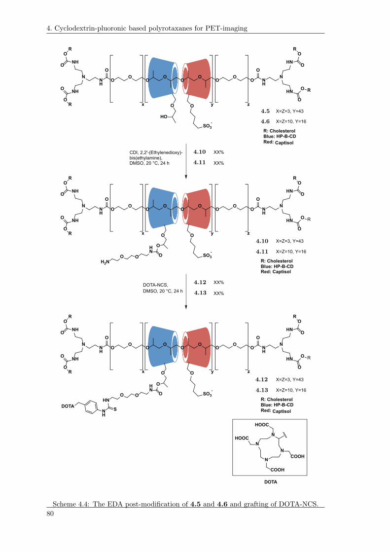
4. Cyclodextrin-pluoronic based polyrotaxanes for PET-imaging
OO
OO
OO
OO
O NH
O
NH
O
N N
NH
NH
HN
HN
O
O
R
O
O R
O
O
R
O
O
R
CDI, 2,2'-(Ethylenedioxy)-bis(ethylamine),DMSO, 20 °C, 24 h
OO
OO
OO
OO
O NH
O
NH
O
N N
NH
NH
HN
HN
x y z
O
O
R
O
O R
O
O
R
O
O
R O
OHN
OOOH2N
R: CholesterolBlue: HP-B-CD
Red: Captisol
X=Z=3, Y=43
X=Z=10, Y=16
R: CholesterolBlue: HP-B-CDRed: Captisol
X=Z=3, Y=43
X=Z=10, Y=16
O
HO
O
SO3
-
O
SO3
-
XX%
XX%
DOTA-NCS,
DMSO, 20 °C, 24 h
XX%
XX%
OO
OO
OO
OO
O NH
O
NH
O
N N
NH
NH
HN
HN
O
O
R
O
O R
O
O
R
O
O
R O
OHN
OOOHN R: Cholesterol
Blue: HP-B-CD
Red: Captisol
X=Z=3, Y=43
X=Z=10, Y=16
O
SO3
-
SNH
DOTA
N
N
N
N
HOOC
HOOC
COOH
COOH
DOTA
x y z
x y z
1.32
4.5
4.6
4.10
4.10
4.11
4.11
4.12
4.12
4.13
4.13
Scheme 4.4: The EDA post-modification of 4.5 and 4.6 and grafting of DOTA-NCS.
80

4. Cyclodextrin-pluoronic based polyrotaxanes for PET-imaging
4.8 Discussion
At the moment, the successful grafting of DOTA is currently under investigation. The
presence of DOTA is questionable as it is hardly detectable by 1H-NMR as the resonance
signals are not significant enough compared to the remaining backbone of 4.13 and 4.12.
HSQC-NMR analysis of 4.12 was conducted in order to verify the presence of the aromatic
resonances of the benzylic moiety. No correlation was detected for 4.12, unfortunately.
Despite these 1H-NMR analysis, 4.12 was successfully Gd-loaded to a Gd content of 9%
wt, which on the other hand could verify the presence of the DOTA-moiety. Supporting
analysis in needed to ensure the presence of the DOTA moiety.
Grafting experiments of the aryl TMS moieties of 2.4, 2.1 and 4.14 are still on-going.
The precursors of 2.4 and 2.1 were synthesized according to the procedures developed in
chapter ??. The epoxide of 4.14 was conducted by O-alkylation of 2.4 using epichloro-
hydrin and K2CO3 (Scheme not shown). The grafting of 2.4 was mediated using CDI in
DMSO on 4.10 while the grafting of 2.1 was mediated using the activated ester made
from EDC·HCl. The basic ring-opening of 4.14 was attempted by treating 4.15 with
NaOH prior to the addition of 4.14.
All TMS-aryl grafting has been carried out but was not conclusive as these experiments
were conducted and the end of the time-frame of this project. The TMS-aryl grafting is
still currently on-going.
4.9 Conclusion
PRs of based on the construct illustrated in Scheme 4.4 were synthesized. The presence
of the DOTA moiety was questionable according to NMR analysis. Before loading with
the PET-isotope of 64Cu, improved structural characterization would be needed.
81


Regioselective iodination of peptidesand proteins 5
Small synthetic receptor binding peptides are today favored over proteins and antibodies as
peptide-based radiopharmaceuticals in the field of tumor-targeting and receptor-imaging
[110]. This tendency is due to the availability of small peptides possessing a wide range of
biological activities, unique receptor-recognition and the ease of gaining information from
these systems [110]. Moreover, small peptides possess smaller size, easier preparation, and
are in general a simplification of its corresponding complex proteins and antibodies. A
number of natural occurring peptides functioning as hormones, neurotransmitters etc. are
displaying a higher affinity than antibody fragments [111,112]. Due to the fact that tumor
cells also possess receptors that are unique to the type of cancer and are over-expressed at
the surface of the cells, this has led to the in vivo imaging and use of numerous bioactive
peptides with high affinity for these receptors [110]. Labeling of peptides with specific
binding to over-expressed receptors allow the visualization of tumor lesions by different
imaging modalities or even provide the option of using these peptides as therapeutic
radiopharmaceutical agents [110,112].
Small peptides also possess attractive pharmacokinetics such as uptake at target site, and
rapid clearance from not-target related tissues. Moreover, they are feasible in chemical
modification and radiolabeling [110]. With solid phase peptide synthesis (SPPS), peptides
are easily affordable and the corresponding labeling has developed into a strongly evolving
area [].
As molecular recognition is confined to a certain region of the sequence, most peptides can
be extended in sequence or modified for radiolabeling without alteration of the receptor
binding site nor the biological activity (chemical flexibility) [111]. Examples of these type
of peptides are Glucagon-like peptide-1, bombesin 5.1, somatostatin 5.2 and analogs like
octreotide 5.3 (??) [113]. The current use of receptor binding peptides in clinical research
(??) is depicted in Figure 5.1.
83

5. Regioselective iodination of peptides and proteins
ONH2
O
NH
O
NH
S
O
HN
H2N
O
HN
O NH2
O
NH
O
NH
O
HN
NHO
NH
NH2
O
HN
HO
O
HN
O
NHHO
O
NHHO
O
HN
S
OH
Somatostatin
HNNH
O
HN
O
NHNH
O
O NHNH2
O
HNO O
NH2
O
HN
NHO
O NH
HNO
OHN
HNN
NHO
O
NH
HN
H2NO
O
NH
NH2
SHN
H2N
O
O
Bombesin
OH
OH
NH
O
SS
NHO
NH2
O
NH
O NH
NH
O
HN
NH2
O
HNOH
O NH
Octreotide
4.3
5.1 5.2 5.3
(b) Structure of receptor binding peptides.
Target receptor
in vitro
evaluation
patient
studies
clinical
application
status of probe development
Melanocortin-1 receptor
(MCR-1)
Vasoactive intestinal
peptide receptor (VPAC-1)
Neurotensin receptor (NTR1)
Cholecystokinin-B/Gastrin
receptor (CCK-B)
Gastrin releasing peptide
receptor (GRP-R/BB2)
αvβ3 integrin
Somatostatin receptor (sst)
Neuropeptide Y receptor (Y1)
Glucagon-like peptide-1
receptor (GLP-1)
Gonadotropin releasing
hormone receptor (GnRH-I)
Chemokine receptor 4 (CXCR4)
1
2
3-5
6-10
11-15
16-25
> 61
26-35
36-60
Biodistribution study
in animal model
in vivo imaging
in animal model
3.14
(d) Different types of receptor binding peptides and the current state in clinical research measured by thenumber of publications. The grey scale in the box indicates the number of studies published for the givenreceptor binding peptide [113].
Figure 5.1: Examples of receptor binding peptides.
Beside the application of in vivo imaging, radiolabeling of peptides and proteins are like-
wise central in radioimmunoassays (RIAs). RIAs are considered as being the current
workhorse in the accurate in vitro screening of antibodies (at pico mol level). Within
84
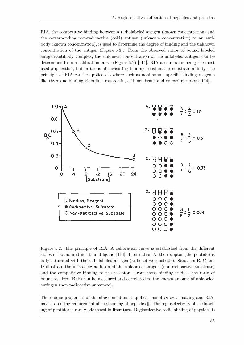
5. Regioselective iodination of peptides and proteins
RIA, the competitive binding between a radiolabeled antigen (known concentration) and
the corresponding non-radioactive (cold) antigen (unknown concentration) to an anti-
body (known concentration), is used to determine the degree of binding and the unknown
concentration of the antigen (Figure 5.2). From the observed ratios of bound labeled
antigen-antibody complex, the unknown concentration of the unlabeled antigen can be
determined from a calibration curve (Figure 5.2) [114]. RIA accounts for being the most
used application, but in terms of measuring binding constants or substrate affinity, the
principle of RIA can be applied elsewhere such as nonimmune specific binding reagents
like thyroxine binding globulin, transcortin, cell-membrane and cytosol receptors [114].
Figure 5.2: The principle of RIA. A calibration curve is established from the different
ratios of bound and not bound ligand [114]. In situation A, the receptor (the peptide) is
fully saturated with the radiolabeled antigen (radioactive substrate). Situation B, C and
D illustrate the increasing addition of the unlabeled antigen (non-radioactive substrate)
and the competitive binding to the receptor. From these binding-studies, the ratio of
bound vs. free (B/F) can be measured and correlated to the known amount of unlabeled
antingen (non radioactive substrate).
The unique properties of the above-mentioned applications of in vivo imaging and RIA,
have stated the requirement of the labeling of peptides []. The regioselectivity of the label-
ing of peptides is rarely addressed in literature. Regioselective radiolabeling of peptides is
85
5. Regioselective iodination of peptides and proteins
preferable as the number of sites for labeling are limited compared to the number of sites
in the radiolabeling of proteins [111]. Likewise, with the high potency of receptor binding
peptides and low tissue occurrence of corresponding receptors, the specific activity of the
receptor binding peptide is crucial [111]. As highlighted by Okarvi: "an ideal labeling
procedure is one in which the receptor binding properties and biological activity of the
peptide remains unaffected and the peptide remains active throughout the imaging" [110].
Additionally, non-specific labeling can influence the conclusive outcome of the RIA, as the
labeling of the antigen can lead to a mixture of different isomers. While the intended
isomer displays the intended binding, other isomers may display (dramatically) different
binding constants as a consequence of labeling near the binding site []. This scenario
was highlighted in the work of Sharma et al., where Chloramine T radioiodination of an
α-melanotropin hormone led to complete loss of biological activity, while radioiodination
of an adrenocorticotropin hormone resulted in misleading results in a radio receptor bind-
ing assay [115]. It has likewise been specified that the labeling of endogenous peptides
has to be in tight structural and stereochemical accordance in order not to alter or lose
potency or selectivity towards a given receptor subgroup [115]. This was substantiated
by the labeling of the opoid peptide analog [d-Pen2,d-Pen5]enkephalin (5.4, DPDPE)
(Figure 5.3); labeling DPDPE at the Tyr residue resulted in a 10-100-fold loss of potency
while Phe-labeling improved the selectivity towards the receptor without decreasing the
potency [115].
The most common way to radiolabel peptides is by
[D-Pen2,5]-Enkephalin
HO
H2N
O
HN
S
NH
OO
HN
OHN
S
O
OH
5.4
Figure 5.3: Structure of DPDPE
5.4.
synthetic post-modification; either by direct label-
ing of the peptide/protein or by the introduction of
prosthetic group that has been pre-labeled. Exam-
ples of prosthetic groups are bifunctional chelating
agents able to chelate metals of interest or a moiety
with the radioactive species covalently attached [].
As an example, site-specific bio-conjugation during
SPPS and introducing the prosthetic group of a
[19F]-p-fluorobenzaldehyde via oxime or hydrazone
ligation have been reported [116,117].
Receptor binding peptides have been labeled with various isotopes. The most common
ones are 111In, 99mTc, 18F, 64Cu, 68Ga and 123I [110, 113]. One of the most used and
attractive isotopes for peptide and protein labeling is iodide []. In vitro radioiodination
of peptides and proteins has been favored over the corresponding labeling methods due
to feasibility and easier labeling with proteins, higher specific activities and advantageous
detection of e.g. gamma irradiation [118]. The scope of this study deals with the site
specific iodination of peptides.
5.1 Iodination by electrophilic aromatic substitution
Iodination is commonly conducted by electrophilic aromatic substitution. Since the com-
mercial available radioactive iodine is usually NaI, the iodide will have to undergo oxida-
86

5. Regioselective iodination of peptides and proteins
tion. The appealing advantages by electrophile aromatic iodination are the simplicity and
accessibility.
The iodination by electrophilic aromatic substitution is not without challenge, as other
aromatic amino acids can undergo electrophilic aromatic iodination like Phe, Tyr, Trp
and His. Tyrosine is according to its activation/nucleophilicity most prone to undergo
iodination. Iodination of Trp is possible because of the electron rich indole. However, ox-
idation of Trp have likewise been reported [119]. Histidine likewise undergoes iodination
in the presence of iodonium ions while Phe display the lowest reactivity of the aromatic
amino acids [].
As the key-step/challenge in iodination of peptides remains the formation of the iodonium
ion, the following sections describe the most common ways to generate iodonium ions in
situ as well as examples of prosthetics groups used in radioiodination of peptides and
methods to achieve iodination of Phe-residues and other aromatic amino acids.
5.2 Iodination by iodine monochloride
Iodination using ICl was developed by McFarlane as a consequence of low radiochemical
yields using molecular iodine I2 as the radioactive source, since radioactive I2 suffer from
the fact of being a carrier method [120]. The radioactive ICl was made by isotope exchange
of ICl with radioactive NaI. Due to the electron-density on chloride, ICl undergoes
electrophilic aromatic iodination on activated ring systems but electrophilic addition is also
reported for double bonds [121]. The method of McFarlane was considered as more efficient
and easier to handle (less hazardous) than the previous radioiodination using the highly
energetic KI method developed by Yalow and Berson for the iodination of insulin [122].
Though, ICl still remained a carrier method since dilution with non-radioactive iodine
monochloride was needed resulting in low specific activities [118].
5.3 Iodination by Chloramine T
One of the most well-known and established methods was developed by Hunter and col-
leagues, also referred to as the Chloramine T method [118, 123, 124]. The method con-
sists of in situ generation of ICl by the oxidation of radioactive NaI using N -chloro-p-
toluenesulfonamide (Chloramine T) (Scheme 5.1) and remains as the most cited method
[118]. This approach has been applied to several peptides and proteins []. The chloramine
T method is attractive as the source of radioactive iodide formed is under carrier-free
conditions. Though a limitation of using chloramine T is the oxidation, which can lead
to oxidation of side-groups of certain amino acids e.g. the oxidation of methionine to
methionine sulfoxide or sulfone [125]. Trp is likewise prone to undergo oxidation under
acidic conditions [119]. The oxidative damage of proteins has led to the development of
solid-supported Chloramine T by which oxidative damage during iodination should be
limited.
87

5. Regioselective iodination of peptides and proteins
SO
ON
Cl
H OH
SO
ONH
Cl
OH
SO
ONH
ClO
H
SO
ONH2H2O
ClO
H
Na
I NaI Cl
OH
Iodine monochloride ClO
H OH
OH
NaI
4.7
Scheme 5.1: Proposed mechanism for the in situ formation of ICl by the chloramine T
oxidation of NaI [].
5.4 Iodination by Iodogen
To keep undesired oxidation at a minimum the use of 1,3,4,6-
N N
N N
O
O
Cl
Cl
Cl
Cl
Iodogen
5.5
Figure 5.4: Structure of
iodogen 5.5.
tetrachloro-3α,6α-diphenyl glycoluril (5.5, iodogen), was pro-
posed (Figure 5.4) [125,126]. Due to its hydrophobic nature,
iodogen is water-insoluble and will be deposited at the walls
of the vial. The reaction is similar to the chloramine T oxi-
dation but is taking place at the site of the local deposit of
5.5 reducing the risk of performing oxidative damages to the
peptide/protein [126,127].
5.5 Iodination by use of prosthetic groups
As alternative to the in situ oxidation and corresponding electrophilic aromatic iodina-
tion, prelabeled aromatic prosthetic moieties have been developed [128–134]. The bio-
conjugation of the aromatic prosthetic group is based on the nucleophilic substitution of
the N -hydroxysuccinimidyl ester part of the radioiodinated aromatic prosthetic group.
This method therefore relies on nucleophilic functional groups e.g. the ǫ-amino group of
Lys residues or the N-terminus of the peptide/protein.
The first prosthetic group developed was the Bolton-Hunter reagent, the N -succinimidyl 3-
(4-hydroxy 3-iodo-phenyl)propionate 5.6 (Figure 5.5) [128]. Other similar constructs 5.7-
5.10 have been developed diverging from the core-structure of an iodinated phenol in order
to enhance the in vivo stability towards deiodination by deiodinases. In vivo instability is
an observed trend for Tyr-iodinated-peptides explained by the action of deiodinases found
e.g. in the thyroid. Zalutsky and colleagues have developed several prosthetic groups that
have been applied in the bio-conjugation of peptides (Figure 5.5) [129–134]. Examples of
prosthetic moieties that have been developed is based on limiting the structural recognition
by deiodinases (Figure 5.5). Moreover, the activated ester of tetrafluorophenyl-5-iodo-4-
88

5. Regioselective iodination of peptides and proteins
pentenoate 5.11 has been used as a prosthetic group (Figure 5.5) [135].
O ON OO
*I
O ON OO
*IOH*I
OON OO
O ON OO
*IOMe
OMe
N
O ON OO
*I
*I O
O
F
FF
F
5.6 5.7 5.8 5.9 5.10 5.11
Figure 5.5: Various prosthetic groups that have been prelabeled with radioactive iodide
prior to the post-modification of peptides. The post-modification is based on the not
site-specific conjugation to a peptide via Lys residues or the N-terminus [].
In addition, the structural resemblance of the iodinated aromatic group has been studied
in a structure-property/activity relationship [136]. This study tested the deiodinating
activity of enzymes from the thyroid and liver microsomes towards analogs of iodinated
tyrosine. Interestingly, 3-iodo-l-Tyr underwent almost full deiodination while analogs
of d-iodo-tyrosine, α-methyl-dl-iodotyrosines and, 3,5-diiodo-4-hydroxyphenyl-dl-lactic
acid were reported to be stable against deiodination. It was inferred from this assay that
the enzymatic site seemed to be dependent on carboxyl, amino and phenol groups to exert
the deiodination [136].
5.6 Miscellaneous techniques for Phe iodination
The low in vivo stability of peptides/proteins labeled at the Tyr residue, has led to the
iodination of other amino acids like Phe. The labeling of Phe has been based on the
strategy of directing the iodination (regioselectivity) using an in-direct functional group
as placeholder (precursor) - that is part of the peptide/protein and will be equivalent to
the radioiodinated peptide upon iodination. As an example, Escher illustrated the io-
dination of p-amino-phenylalanine by the Sandmeyer reaction; diazotiation followed the
Cu-mediated insertion of the iodide [137]. The main limitation of the reaction is the rel-
atively low radiochemical yield of 1-2% under carrier free conditions [137]. The presence
of a crown ether enhanced yields to approximately 25% [115].
In the study of Wilson et al., Ac-Phe(4-TMS)-OMe 5.12 was developed using TMS as
directing group in the iodination (Scheme 5.2) [138]. Iodination was conducted using
AgBF4 and I2 and the need for the activating effect of silyl substitution was demon-
strated by the unsuccessful iodination of Ac-Phe-OMe [138]. A limited scope of sensitive
substrates (methyl- and ethyl esters) was tested in this study [138]. Nevertheless, the se-
lectivity of 5.12 as part of a peptide sequence with multiple aromatic amino acids was not
evaluated, 5.12 was only tested as part of N -formyl-Phe(4-TMS)-Gly-OEt dipeptide [138].
Wilbur et al. published the enantiomerselective formation of Boc-Phe(4-Sn(Bu)3)-tetrafluorophenyl
ester 5.13 (Scheme 5.2) and the corresponding iodination [139]. This derivative was pre-
89

5. Regioselective iodination of peptides and proteins
pared in order to conduct radioiodination of 5.13 prior to the following peptide synthesis.
The stannyl derivative of 5.13 was designed for a Merrifield approach to SPPS using
the Boc/Bn protecting group strategy [139]. Iodination was conduction under carrier
conditions using a mixture of radioactive NaI and nonradioactive NaI together with N -
chlorosuccinimide.
The development of the stannyl derivative 5.13 was reasoned the milder radioiodination
conditions used for organostannyl compounds, which proved to be compatible with the
activated tetrafluorophenyl ester of 5.13 [139]. The iodination conditions used in the io-
dination of 5.12 was believed according to Wilbur et al. not to be applicable to 5.13 due
to the presence of an activated ester like 5.13.
AcHN
TMS
COOMe
BocHN
Sn(Bu)3
O
OF
F
FF
4.9
5.12 5.13
Scheme 5.2: Structures for the 5.12 and 5.13 [138,139].
To the knowledge of the author, no other in-direct/temporary placeholder-system - as
exemplified above – has been combined with other amino-acids like for instance tyrosine.
5.7 Problem statement
In order to provide an improved labeling of peptides, group-members of Mads Clausen
have previously been working on using a TMS-group as a placeholder for the iodina-
tion [62]. As described in section 2.2, aryl-TMS groups undergo ipso-substitution and
it was envisioned that the TMS-group can be used as a precursor to direct the desired
iodination of peptides as also illustrated before [138]. This will allow regioselective iod-
inations and potentially afford more precise and conclusive RIA studies and ensure that
the labeling does not takes place at the active site responsible for the biological receptor
activity. Beside these attractive features, the labeling will remain direct compared to the
introduction of a prosthetic group.
Previous results include synthesis of Boc-(4-TMS)Phe-OBn [140]. To evaluate the selec-
tivity, the corresponding dipeptides of Phe (5.15), His (5.16), Tyr (5.17) and Trp (5.18)
were synthesized, respectively. Iodination by thallation Tl(OOCCF3)3 indicated predom-
inantly ipso-substitution of the TMS of (4-TMS)Phe with no observed iodination of the
corresponding His and Phe moieties of dipeptides 5.19 and 5.20, respectively (Table 5.1).
The dipeptide of Tyr however did not display any selectivity, as products corresponding
to mono-, di- and tri-iodination were observed when Tl(OOCCF3)3 (2 equiv.) and NaI (4
equiv.) were used in an excess. Lowering the amount of Tl(OOCCF3)3 (0.5 equiv.) and
NaI (1 equiv.) indicated that the TMS was substituted prior to the iodination of the Tyr
90

5. Regioselective iodination of peptides and proteins
occurred in 5.21 (Table 5.1). Iodination of the dipeptide of Trp 5.22 was not successful
as it was inferred that oxidation of Trp under acidic conditions occurred (Table 5.1) [140].
Table 5.1: Selectivity in the iodination reaction of Phe(4-TMS)
O
NH2 ONH
OHR
I
O
NH2 ONH
OHR
TMSTl(OOCCF3)3 (2 equiv).
NaI (4 equiv.)
TFA/CH3CN(3:7 v/v), 20 °C
- - 5.15 5.18 5.19 5.22
Dipeptide 5.15 5.16 5.17 5.18
R NHN OH
HN
Iodinated dipeptide 5.19 5.20 5.21 5.22
Selectivity + + (+)1-
1 Selectivity was observed when 0.5 equiv. Tl(OOCCF3)3 and1. equiv NaI was used.
From these findings with predominant selectivity for the ipso-substitution of the TMS-
group in the iodination reactions of 5.15 and 5.16, it was deduced that improved
selectivity can be achieved from the corresponding Tyr(3-TMS). With the increased
nucleophilicity of the phenol of Tyr and placing the TMS in the reactive ortho-position
to the hydroxy-group, it was predicted that this electronic effect could improve the
desired selectivity. Despite the low in vivo stability of Tyr-iodinated peptides, iodinated
Tyr peptides/proteins will still find use in the in vitro screening of RIA. As previously
mentioned, enhanced in vivo stability can be achieved by certain changes in the design
of the amino-acid construct e.g. 1) N -alkylation of the amide bond, 2) using the
corresponding d-isomer and 3) employing peptoids [110]. Given that a Tyr(3-TMS) can
be synthesized, selectivity over His, Phe, Trp and Tyr will have to be tested. Likewise,
a corresponding Fmoc-(3-TMS)Tyr(tBu)-OH derivative is a necessity to obtain so that
this derived iodination strategy is applicable for Fmoc-based SPPS. With the excellent
radiochemical yield and purity achieved for the iodination of 2.2 using Tl(OOCCF3)3under carrier free conditions, it was hypothesized that high specific activities can be
obtained in radioiodination of peptides with this method.
5.8 Retrosynthetic analysis for the synthesis of the TMS
derived Tyrosine
Several strategies were devised within this project to install the TMS unit within a limited
number of steps starting from the commercial available H-l-Tyr(3-I)-OH 5.23 and Boc-
l-Tyr(t-Bu)-OMe 5.24.
91

5. Regioselective iodination of peptides and proteins
One comprehensive strategy to install the TMS by metal halogen exchange, (Scheme 5.3)
would first involve iodination of 5.24 followed by reduction of the methyl ester that
will afford the intermediate of the amino alcohol of 5.25. Additional protecting group
manipulations and with the cancellation of the acidity-issue of the α-hydrogen will allow
metal halogen exchange of the iodide by which the TMS can be introduced. Reduction
of the carboxylic acid of 5.23 and protecting group manipulations will likewise afford the
amino alcohol of 5.25. In both cases, several steps upon the introduction of the TMS
will be needed, as the reduced amino alcohol first will need to undergo oxidation to the
corresponding carboxylic acid. Following the oxidation, will be exchange of protecting
groups that will be compatible with the Fmoc based-strategy of peptide synthesis.
Another limitation - beside the number of steps - of this strategy could potentially be
the compatibility, selectivity and orthogonality of the protecting groups involved in this
strategy. This synthetic strategy was discarded according to the number of step involved
to get the TMS substituted Tyr derivative 5.26.
COOMeBocHN
O
TMSBocN
O
I
COOMeBocHN
O
1) Iodination2) Ester reduction3) Protecting group manipulations
Metal HalogenExchange
Commercial availableOPGPG
COOHH2N
OH
I
Commercial available
PG = Protecting Group
BocN
O
TMS
OPGPG
1) Acid reduction2) Protecting group manipulations
1) Protecting group manipulations 2) Alcohol oxidation3) Protecting group manipulations
4.10
5.23
5.245.255.26
Scheme 5.3: Retrosynthetic analysis based on the formation of the protected iodinated
amino Tyr alcohol 5.25 for metal halogen exchange.
An elegant and shortened/more direct synthetic strategy that will afford the TMS-
substituted Tyr derivative of 5.27, from 5.23, is via a reverse-Brook rearrangement of
the TMS protected phenol of 5.30 (Scheme 5.4). Another option would be to perform
metal halogen exchange of 5.28 to afford 5.26 (Scheme 5.5). Both strategies will rely
on the metal halogen exchange of the iodide at -78 ◦C to be faster than the competitive
α-hydrogen abstraction or NH abstraction of the Boc-carbamate. The latter could be
avoided by the formation of N(Boc)2 protection of the α-amino group or in situ protection
of the α-NH-carbamate. The subsequent deprotection of the N(Boc)2 has though been
reported to be challenging due to observed epimerization, which is hypothesized to be in
accordance with the increased acidity of the α-hydrogen [141]. This favors the strategy of
in situ protection of the α-amino-NH-carbamate e.g. silyl protection.
92

5. Regioselective iodination of peptides and proteins
COOMeBocHN
OH
TMS
COOMeBocHN
OH
I
COOHH2N
OH
I
Protecting groupmanipulations
Reverse Brookrearrangement
Commercial available
5.235.27 5.30
Scheme 5.4: The retrosynthetic analysis for the TMS derived Tyr-derivative 5.27 starting
from the commercially available 5.23.
COOMeBocHN
O
RCOOMeBocHN
O
I
COOMeBocHN
OIodination
Metal halogen exchangeor
Stannanoyl insertion
Commercial available
R = Si(Me)3
R = Sn(Bu)3
5.245.26 5.28
5.29
Scheme 5.5: The retrosynthetic analysis for the TMS derived Tyr-derivative 5.26 starting
from the commercially available 5.24.
Whether the metal halogen exchange or reverse Brook-rearrangement were achievable
directly on Tyr was tested on 5.28 and 5.30 (Scheme 5.6 and Scheme 5.7). The mono-
iodinated tyrosine 5.28 was assembled from the commercial available 5.24 which under-
went mono-iodination using ICl in CH2Cl2 affording 5.28 (Scheme 5.6). Applying 2 equiv.
of TMSCl followed by the addition of 2 equiv. n-BuLi at -78 ◦C only provided a mix-
ture of 5.28 and the corresponding deiodinated derivative 5.24, thus no TMS insertion
was observed. Compound 5.30 was synthesized from 5.23 in a one-pot fashion by first
forming the methyl ester using SOCl2 in CH3OH followed by the Boc-protection of the
amine using Boc2O in dioxane according to literature (Scheme 5.7) [142]. The formation
of the TMS ether of 5.30 was successfully achieved by using 2. equiv. TMSCl and Et3N
in dry THF. Applying n-BuLi at -78 ◦C gave no signs of the rearranged product 5.27,
only deiodination was observed according to LC-MS analysis.
1. TMSCl, THF, 20 °C2. n-BuLi, THF, -78 °C
COOMeBocHN
O
COOMeBocHN
O
I
COOMeBocHN
O
TMS
ICl, CH2Cl2, 20 °C
5.24 5.265.28
Scheme 5.6: Iodination and metal halogen exchange of 5.24.
93

5. Regioselective iodination of peptides and proteins
1. TMSCl, Et3N, THF, 20 °C2. n-BuLi, THF, -78 °C
COOHH2N
OH
I
COOMeBocHN
OH
I
COOMeBocHN
OH
TMS
1. SOCl2, CH3OH, 0-20 °C2. Boc2O, Et3N, dioxane, 20 °C
5.23 5.275.30
Scheme 5.7: Reverse-Brook rearrangement of 5.30.
Another alternative that will allow insertion of a placeholder would be to perform a cross-
coupling reaction at the already introduced iodide of 5.28. Despite the previous nega-
tive experience with the Pd-mediated TMS insertion (2.5.2), and despite the toxicity of
organo-stannanes, they are more readily introduced by Pd-chemistry and more established
in modern chemistry as they are used in numerous examples of the Stille coupling and as
precursors in radio-iodinations. It was suspected that Sn(CH3)- would display the same
β-cationic effect and undergo ipso-substitution in an electrophilic aromatic iodination.
Preliminary experiments indicated that the tributylstannane derivative of 5.28 could be
synthesized. This was not further pursued as the synthesis of the TMS-substituted Tyr
derivative via the Negishi-coupling was conducted concurrently, as described in the fol-
lowing sections.
.
5.9 Synthesis of amino acids using β-iodo-alanine
derivatives
Jackson and co-workers reported the use of organozinc reagents based on Boc-β-iodo-
Ala-OBn 5.31 to synthesize various analogs of Phe-derivatives via the Negishi-coupling
[143,144]. Jackson and co-workers likewise indicated that the homoenolate of the organoz-
inc reagent remained stable and did not undergo epimerization [143]. The enantiomeric
control was further substantiated by the formation of the Mosher-ester of the formed
phenylalanine derivatives which supported that the Negishi-coupling gave the phenylala-
nine derivative in an enantiomerically pure fashion [143]. The yields reported were mod-
erate [144]. This led to further optimization and screening in search of the proper cat-
alyst/ligand system and likewise the tolerance/scope of functional groups (Scheme 5.8).
This led to the use of Pd2dba3 and SPhos 5.32 in DMF by which the yields of the cross
coupled product using 5.33 were improved [145]. Beside the catalytic system, the im-
proved Negishi reaction was also reasoned to be due to improved zinc-insertion [145].
94

5. Regioselective iodination of peptides and proteins
COOMeBocHN1. Zn*, I2, DMF2. Pd2dba3, , DMF
COOMeBocHN
I
X
X = Br, I
RR
R = -CH3, -OH, -F, -Cl, -NO2, -NH2
R = -CH3, -OH, -F, -Cl, -NO2, -NH2
PCy2
OMeMeO
SPhos5.32
5.32
5.33
Scheme 5.8: Optimized conditions for the Negishi-Coupling of Boc-β-iodo-Ala-OMe and
various aryl-iodides/bromides using Pd2dba3 and Sphos 5.32 [145].
The strategy of Jackson was applied to afford the Boc-Phe(4-TMS)-OBn derivative 5.14
conducted by previous members of the group of Mads Clausen (Scheme 5.9). The TMS
derived phenylalanine was assembled from the corresponding 4-bromo-1-TMS-benzene
5.34, though yields were not high (best achievable yield, 58%). The highest yield was
achieved from 4-iodo-1-TMS-benzene 5.35 (69% yield).
COOBnBocHN1. Zn*, I2, DMF2. Pd2dba3, , DMF
COOBnBocHN
I
X
X = Br
TMS
X = I
TMS
5.14
5.31
5.32
5.34
5.35
Scheme 5.9: The synthesis of 5.14 by Negishi coupling.
5.10 Synthesis of a TMS-derived tyrosine derivative by
Negishi coupling
Concurrently to the previous mentioned direct strategies involved in this project, the target
molecule of 5.26 was pursued by the Negishi-coupling. The building block 1-tert-butoxy-
2-TMS-4-bromobenzene 5.36 was made in the group of Mads Clausen (Scheme 5.10). 2,4-
Dibromophenol 5.37 was first tert-butyl O-alkylated to 5.38 followed by regioselective
metal halogen exchange in the 2-position to afford building block 5.36.
95

5. Regioselective iodination of peptides and proteins
OHBr
Br
OBr
Br
OTMS
Br
CF3SO3H
CH2Cl2, -78 °C Et 2O, -78 °C
n-BuLi, TMSCl
5.33
5.365.37 5.38
Scheme 5.10: The synthesis of building block 5.36.
The choice of tert-butyl as protecting group was based on the compatibility with the
Fmoc-based SPPS strategy and since it is the most common protecting group of tyro-
sine. However this strategy could potentially come to the limitation within a Fmoc-
based strategy, as side-group protection and cleavage from beads/resin usually require
harsh acidic conditions which could lead to proto-desilylation of the TMS-group. This
could imply that the radioiodination will have to be carried out on the resin or that
the fully protected peptide will have to be cleaved from the resin in order to undergo
subsequent iodination in solution. The cleavage of the fully protected peptide can be
achieved by applying extreme acid-labile - or base sensitive linkers like the Cl-trityl linker
(cleavage: hexafluoroisopropanol/CH2Cl2 (1:4 v/v) [146]), xanthenyl linker appended to
4-methylbenzhydrylamine resin (cleavage: 1% TFA/CH2Cl2 [147]), SASRIN linker resin
(cleavage: 0.5%-1% TFA/CH2Cl2 or Hexafluoroisopropanol/CH2Cl2 (1:4 v/v) [148]), or
the base labile HMBA linker (cleavage: 0.1 m NaOH [140, 149, 150]) would have to be
applied. Cleavage from the resin will enable the option of conducting the radioiodination
on the fully protected peptide in solution followed by deprotection of side-groups in a
post-treatment fashion with e.g. TFA upon iodination. In order to display the situation
where a given peptide, synthesized using a Fmoc-based strategy and cleaved from a resin,
the tert-butyl was chosen as protecting group for the phenol of the TMS substituted Tyr
derivative.
In order to get an personal impression of the Negishi-coupling and its sensitivity, ex-
perience was obtained by the formation of the Boc-Phe-OMe from 5.33 and iodobenzene.
The organozinc reagent of 5.33 was made from activated Zn dust and together with
iodobenzene, Pd2dba3 and SPhos in DMF, Boc-Phe-OMe was afforded in moderate yields
according to LC-MS analysis.
For the intended Negishi cross-coupling between 5.36 and 5.33 (Path A), the organozinc
reagent of 5.33 was formed from activated Zn dust (<10 µm) followed by mixing with
SPhos, Pd2dba3 and 5.36. This afforded product 5.26 in low yields (23%, Scheme 5.11).
The low reactivity was reasoned the use of the aryl-bromide 5.36, which was in accordance
with similar results reported in the group [140].
96

5. Regioselective iodination of peptides and proteins
OTMS
ITHF, -78 °C
OTMS
Br
n-BuLi,I2 in THF
O
NHBocI OR
O
NHBocOR
TMS
O
O
NHBocI OCH3
67%
69%
23%
R = CH3
R = Bn
1. Zn*, I2, DMF2. Pd2dba3, , DMF
1. Zn*, I2, DMF2. Pd2dba3, , DMF
R = CH3
R = Bn
85%
Path A
Path B
5.26
5.31
5.32
5.32
5.33
5.33
5.36 5.40
5.41
Scheme 5.11: The synthesis of metal halogen exchanged building block 5.40 and the
corresponding Negishi synthesis. Note that the asterisk of the Zn refer to that Zn
underwent activation in 0.1 m HCl prior to use.
The group of Buchwald reported the Cu-catalyzed halogen exchange of aryl halides also
referred to as an aromatic Finkelstein type reaction [151]. The Cu-catalyzed halide
exchange rely on the equilibrium between the aryl halide and a second halide salt in which
the position of the equilibrium – among other parameters - is influenced by the solubility
of the given halide salt [151]. Applying the conditions of CuI (5 mol%), rac-trans-N,N’ -
dimethyl-1,2 cyclohexanediamine 5.39 (10 mol %) and NaI (2 equiv.) in refluxing dioxane
to 5.36 gave what was a presumed mixture of the bromide 5.36 and the iodide 5.40 in
an approximately ratio of 1:1 ratio by LC-MS analysis (Scheme 5.12).
HN
NH
CH3
CH3
CuI, NaI (2 equiv.)Dioxane, 110 °C
O
TMS
Br
O
TMS
I
5.36
5.39
5.40
Scheme 5.12: The Cu-catalyzed halogen exchange of 5.36.
Due to the expected difficulty isolating the aryl-iodide of 5.40 from the corresponding
bromide 5.36 it was concluded that the crude mixture of the bromide and the iodide
would have to be applied further on in the Negishi cross-coupling. Concurrently to the
aromatic Finkelstein reaction developed by Buchwald, the bromide was successfully ex-
changed with iodide using n-BuLi followed by the addition of I2 in THF. This afforded
the iodo-substituted aryl derivative 5.40 in 67% yield (Path B, Scheme 5.11).
Exchanging the halide from the bromide to the iodide of 5.40 had a significant impact of
97

5. Regioselective iodination of peptides and proteins
tripling the yield of the Negishi-coupling by the formation of 5.26 (69%) and 5.41 (85%)
(Scheme 5.11).
The next step was the chemoselective Boc-deprotection of the α-amino-group without
compromising the tert-butyl nor the aryl TMS. To elucidate the acid liability of the dif-
ferent groups and to see if Boc-group could be selectively removed in TFA, the stability
in various ratios of TFA and Et3SiH in CH2Cl2 were tested. Unfortunately no selectivity
was observed for the Boc-group could be achieved.
Deprotection of N -Boc protected amines in the presence of the t-Bu-phenol ether has been
reported according to literature using mild conditions of TMSOTf and 2,6-lutidine [152].
Alternatively, AlCl3 has likewise been reported as a mild method for Boc-deprotection
[153]. TMSOTf and 2,6-lutidine in CH2Cl2 exclusively removed the Boc-group in quanti-
tative yields (Scheme 5.13) affording the amine of 5.42 and 5.43 respectively.
CH2Cl2, 0 → 20 °C
TMSOTf,2,6-lutidine
O
NH2
ORTMS
O
R = CH3
O
NHBocOR
TMS
O
R = CH3
R = Bn
R = Bn
95%
99%
5.18
5.26
5.41
5.42
5.42
5.43
5.43
Scheme 5.13: Boc deprotection of 5.42 and 5.43 using TMSOTf and 2,6-lutidine.
The successful Boc-deprotection allowed the transformation of 5.42 into the Fmoc-protected
5.44 using Fmoc-OSu and Et3N in CH2Cl2 in 75% yield (Scheme 5.14). The ester of 5.44
was hydrolyzed using Na2CO3 in H2O/CH3CN according to literature [154]. The con-
ditions was reported to be compatible with the Fmoc-group where common hydrolytic
conditions using strong bases like hydroxides are reported to lead to the (partial) loss
of the Fmoc-group [154].1 It was likewise reported that the stereochemistry was not af-
fected during the hydrolysis [154]. The carboxylic acid 5.45 was isolated in 73% yield
(Scheme 5.14), though 1H-NMR surprisingly indicated an impure product.
1In the previous study of Phe(4-TMS), the Bn-ester was successfully removed in the presence ofFmoc using hydrogenolysis, despite that Fmoc likewise have been reported to undergo hydrogenolysis ina mixture of CH3OH and CH3CN [155]. As an alternative strategy, Fmoc-protection of 5.43 followed byhydrogenolysis was envisioned to potentially afford 5.45.
98
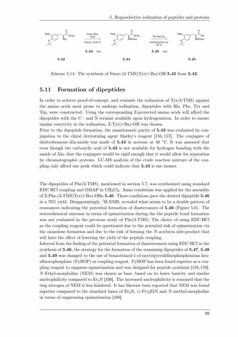
5. Regioselective iodination of peptides and proteins
CH2Cl2, 0-20 °C
Fmoc-OSu,Et3N
O
NHFmocOCH3
TMS
O
75%
O
NH2
OCH3TMS
OH2O/CH3CN (1:1)
20 °C
3% Na2CO3
O
NHFmocOH
TMS
O
73%
5.42 5.44
5.44
5.45
5.45
Scheme 5.14: The synthesis of Fmoc-(3-TMS)Tyr(t-Bu)-OH 5.45 from 5.42.
5.11 Formation of dipeptides
In order to achieve proof-of-concept, and evaluate the iodination of Tyr(3-TMS) against
the amino acids most prone to undergo iodination, dipeptides with His, Phe, Tyr and
Trp, were constructed. Using the corresponding Z-protected amino acids will afford the
dipeptides with the C - and N termini available upon hydrogenation. In order to ensure
similar reactivity in the iodination, Z-Tyr(t-Bu)-OH was chosen.
Prior to the dipeptide formation, the enantiomeric purity of 5.43 was evaluated by con-
jugation to the chiral derivatizing agent Marfey’s reagent [156, 157]. The conjugate of
dinitrobenzene-Ala-amide was made of 5.43 in acetone at 40 ◦C. It was assessed that
even though the carboxylic acid of 5.43 is not available for hydrogen bonding with the
amide of Ala, that the conjugate would be rigid enough that it would allow for separation
by chromatographic systems. LC-MS analysis of the crude reaction mixture of the cou-
pling only afford one peak which could indicate that 5.43 is one isomer.
The dipeptides of Phe(3-TMS), mentioned in section 5.7, was synthesized using standard
EDC·HCl coupling and DMAP in CH2Cl2. Same conditions was applied for the assembly
of Z-Phe-(3-TMS)Tyr(t-Bu)-OBn 5.46. These conditions gave the desired dipeptide 5.46
in a 76% yield. Disappointingly, 1H-NMR, revealed what seems to be a double pattern of
resonances indicating the potential formation of diasteromers of 5.46 (Figure 5.6). The
stereochemical outcome in terms of epimerization during the the peptide bond formation
was not evaluated in the previous study of Phe(3-TMS). The choice of using EDC·HCl
as the coupling reagent could be questioned due to the potential risk of epimerization via
the oxazolone formation and due to the risk of forming the N -acylurea side-product that
will have the effect of lowering the yield of the peptide coupling.
Inferred from the finding of the potential formation of diastereomers using EDC·HCl in the
synthesis of 5.46, the strategy for the formation of the remaining dipeptides of 5.47, 5.48
and 5.49 was changed to the use of benzotriazol-1-yl-oxytripyrrolidinophosphonium hex-
afluorophosphate (PyBOP) as coupling reagent. PyBOP has been found superior as a cou-
pling reagent to suppress epimerization and was designed for peptide synthesis [158,159].
N -Ethyl-morpholine (NEM) was chosen as base, based on its lower basicity and similar
nucleophilicity compared to Et3N [160]. The increased nucleophilicity is reasoned that the
ring nitrogen of NEM is less hindered. It has likewise been reported that NEM was found
superior compared to the standard bases of Et3N, (i -Pr)2EtN and N -methyl-morpholine
in terms of suppressing epimerization [160].
99

5. Regioselective iodination of peptides and proteins
The reaction conditions for the dipeptide synthesis are summarized in Table 5.2. Us-
ing PyBOP and NEM afforded the dipeptides in acceptable yields. Dry column vacuum
chromatography purification of Z-His-(3-TMS)-Tyr(t-Bu)-OBn 5.49 was unsuccessful by
which 5.49 was purified by PREP-HPLC explaining the low yield. 1H-NMR of the dipep-
tides indicated the formation of the single diastereomer for 5.47, 5.48 and 5.49.
5.21
Figure 5.6: Selected 1H-NMR resonances of the synthesized dipeptides of 5.46 (red), 5.48
(blue) and 5.47 (green) indicating the presence of diastereomers of 5.46.
5.12 Hydrogenolysis of dipeptides
What seemed on the drawing board to be a promising step – the hydrogenolysis of the
benzyl-ester and concurrently removal of the Cbz of 5.46-5.49 – turned out to be chal-
lenging. This strategy was based on the similar approach conducted for Phe(4-TMS).
Standard hydrogenolysis conditions using Pd/C in CH3OH or EtOH, disappointingly led
to what was believed to be the reductive aminated product of the methylated or ethy-
lated N -terminus, respectively. This has previously been reported and it was speculated
to be due to the formation of formaldehylde or acetaldehyde during hydrogenolysis, re-
spectively [161]. Due to the formation of this side product, the solvent was changed to
EtOAc or THF. Unfortunately, beside the formation of the linear dipeptides 5.50-5.53,
a significant amount of the corresponding cyclized diketopiperazine side-product 5.54-
100

5. Regioselective iodination of peptides and proteins
Table 5.2: Peptide couplings of 5.43.
OH2N
O
OBnTMS O
CbzHN ONH
O
OBnTMSR
- 5.43 5.46 5.49
Entry Amino acid Coupling Reagent Base solvent (R) Yield1
1 Z-Phe-OH EDC·HCl2 DMAP2 CH2Cl2 5.46 (76%)
2 Z-Tyr(t-Bu)-OH PyBOP3 NEM3 DMFO-tBu
5.47 (76%)
3 Z-Trp-OH PyBOP3 NEM3 DMF HN5.48 (78%)
4 Z-His-OH PyBOP3 NEM3 DMF NHN
5.49 (36%)4
1 Isolated by dry column vacuum chromatography unless otherwise stated.2 2 equiv. EDC·HCl & 2.1 equiv. DMAP at 20 ◦C3 1.2 equiv. PyBOP & 6 equiv. NEM at 20 ◦C4 Isolated by Preparative HPLC
5.57 was formed concurrently. The formation of the diketopiperazine side-product was
more pronounced in EtOAc than in THF, why the final hydrogenation was conducted in
THF. The formation of the diketopiperazine side-product lowered the yields dramatically
(Table 5.3). the up-scale hydrogenolysis of 5.49 was unsuccessful and was not further
pursued in this project due to time-frame of this project.
It was hypothesized that the formation of diketopiperazine could be explained by the hy-
drogenolysis of Cbz of the α-amino group taking place prior to the hydrogenolysis of the
Bn-ester. With the liberation of the α-amino group, cyclization by nucleophilic substitu-
tion at the C-terminus optionally speculated to be mediated by coordination of Pd, could
take place. On the other hand, the liberation of the corresponding benzylate could be
questioned due to its poor leaving group ability.
The hydrogenolysis was likewise sluggish, which was presumed to be due to catalyst pol-
lution by the α-amino group of the N-terminal. The coordination of amino-groups to Pd
is commonly known and have even led to the chemoselectivity in hydrogenations when
using a Pd/C-ethylenediamine complex [162].
Hydrogenolysis of the Phe-dipeptide 5.46 revealed, what was indicated by 1H-NMR, that
the dipeptide was a mixture of diastereomers. Dry column vacuum chromatography using
H2O/CH3CN as gradient allowed separation of the diasteromers. Disappointingly, 1H-
NMR revealed that the diastereomers remained mixtures.
101

5. Regioselective iodination of peptides and proteins
Table 5.3: Hydrogenolysis of dipeptides 5.46-5.49
-
O
NH2 ONH
O
OHTMSRO
CbzHN ONH
O
OBnTMSR
ONH
OHN
O
TMS
R
+Pd/C, H2
THF, 20 °C
- - 5.46 5.49 5.50 5.53 5.54 5.57
Entry side-chain yield (linear) yield (cyclic)
R (%) (%)
1 5.50 (44%)1 5.54 (7%)
2O-tBu
5.51 (32%) 5.55 (34%)
3 HN5.52 (47%) 5.56 (29%)
4 NHN
5.53 (-)2 5.57 (-)2
1 The other diastereomer of 5.50 was isolated in 27%yield.
2 Not available, hydrogenolysis of 5.49 was unsuccessful.
5.13 Iodination of dipeptides
The iodination of the linear dipeptides 5.50-5.52 were performed by thallation using
Tl(OOCCF3) and NaI. Iodination by thallation was chosen since oxidative methods like
lactoperoxidase and chloramine T has been reported to led to oxidation of Trp in acidic
conditions [?]. Previous thallation-results of H-Trp-Phe(4-TMS)-OH 5.18 indicated that
thallation was not successful using Tl(OOCCF3)3 (2. equiv) and NaI (4. equiv.) in a
mixture of TFA/CH3CN (3:7 v/v). Based on this, thallation of 5.52 was envisioned to be
feasible by lowering the amount of TFA.
With the acid-liability stability test of 5.26 in TFA/Et3SiH/CH2Cl2, the iodination re-
action was tested on 5.26 and the diketopiperazines of 5.55 and 5.56 by varying the
amount of TFA (0.1%, 1% and 10% TFA in CH3CN respectively). Applying 2 equiv. of
Tl(OOCCF3)3 and 4 equiv. of NaI in CH3CN to 5.26 indicated the formation of the
ipso-substituted iodide. The iodination reaction proceeded with the concurrently Boc-
deprotection in correlation to the amount of TFA.
In case of the cyclized dipeptides of 5.55 and 5.56, iodination took place using 2 equiv.
of Tl(OOCCF3)3 and 4 equiv. of NaI in CH3CN. The presence of TFA seemed to affect
the degree of iodination. Iodination of 5.56 led to formation of multiple side-products
stating the difficulty of conducting iodination in the presence of the indole under slight
acidic conditions [119].
102

5. Regioselective iodination of peptides and proteins
Iodination of the linear peptides was tested by varying the amount of Tl(OOCCF3)3and NaI and evaluating the necessity of TFA. Iodinating the linear dipeptide of 5.50
using 2 equiv. of Tl(OOCCF3)3 and 4 equiv. of NaI in CH3CN gave a mixture of the
ipso-iodinated product 5.58 and the starting material 5.50. Using a mixture of 0.1%
TFA/CH3CN increased the formation of the iodinated product 5.58, though the iodi-
nation did not go to completion. At this point, it seemed clear that the iodination by
thallation of the above-mentioned iodinations were sluggish compared to the previously
experience of using Tl(OOCCF3)3 on 2.2. The sluggish behavior was reasoned to be due
to the presence of TFA as part of the solvent. The amount of TFA was therefore increased
to 10% and iodination of 5.50 using 1 equiv. of Tl(OOCCF3)3 and 2 equiv. of NaI af-
forded the iodinated product of 5.58 exclusively, clearly exhibiting the desired selectivity
for the ipso-substitution of the TMS-group (Figure 5.7).
103

5. Regioselective iodination of peptides and proteins
O
NH2 ONH
O
OHTMS O
NH2 ONH
O
OHITl(OOCCF3)3
NaI
TFA/CH3CN(1:9 v/v)20 °C
5.50
5.53
5.58
(b) Iodination of 5.50 using 1 equiv. of Tl(OOCCF3)3 and 2 equiv. of NaI in 10% TFA/CH3CN
Time-0.00 0.25 0.50 0.75 1.00 1.25 1.50 1.75 2.00 2.25 2.50 2.75 3.00 3.25 3.50 3.75 4.00 4.25 4.50 4.75 5.00
AU
0.0
2.5
5.0
7.5
1.0e+1
1.25e+1
1.5e+1
1.75e+1
2.0e+1
2.25e+1
2.5e+1
2.75e+1
3.0e+1
3.25e+1
3.5e+1
3.75e+1
4.0e+1
4.25e+1
4.5e+1
4.75e+1
5.0e+1
5.25e+1
5.5e+1
MHC_HSJ_Thal_1eq_D_10pctTFA_Phe-TMSTyr_160516 3: Diode Array Range: 6.87e+11.38
0.19
1.38
1.11
0.93
0.23
1.39
1.39
1.40
1.41
1.41
4.58
1.94
1.60
3.60
(d) UV-chromatogram of reaction mixture. The peak eluting at 1.39 min has a m/z of 511.01 correspondigto the [M+H]+ adduct of 5.58.
Figure 5.7: Reaction & LC-MS analysis of the iodination of 5.50 in 10% TFA/CH3CN.
Iodination of the Tyr-dipeptide 5.51 (??) was of interest taking the iodination of the Tyr-
(4-TMS)Phe dipeptide 5.17 into account where selectivity was difficult to achieve. Lack
of reactivity was seen when 5.51 was treated with Tl(OOCCF3)3 (2 equiv.) and NaI (4
equiv.) in CH3CN or 0.1% TFA/CH3CN. Applying the similar conditions of Tl(OOCCF3)3(1 equiv) and NaI (2 equiv.) in 10% TFA/CH3CN formed the ipso-iodinated product
5.59 in a clean fashion with traces of what was expected to be the di-iodinated dipeptide
with one tert-butyl missing was observed (m/z = 652.90, peak eluting at 1.49 min, ??)
(Figure 5.8).
104

5. Regioselective iodination of peptides and proteins
O
NH2 ONH
O
OHTMS O
NH2 ONH
O
OHITl(OOCCF3)3
NaI
TFA/CH3CN(1:9 v/v)20 °C
O O
5.51 5.59
(b) Iodination of 5.51 using 1 equiv. of Tl(OOCCF3)3 and 2 equiv. of NaI in 10% TFA/CH3CN
Time-0.00 0.25 0.50 0.75 1.00 1.25 1.50 1.75 2.00 2.25 2.50 2.75 3.00 3.25 3.50 3.75 4.00 4.25 4.50 4.75 5.00
AU
0.0
5.0
1.0e+1
1.5e+1
2.0e+1
2.5e+1
3.0e+1
3.5e+1
4.0e+1
4.5e+1
5.0e+1
5.5e+1
6.0e+1
6.5e+1
7.0e+1
MHC_HSJ_Thal_1eq_D_10pctTFA_Tyr-TMSTyr_160516 3: Diode Array Range: 8.295e+10.18
0.19
1.64
1.49
1.37
0.23
1.94
3.41 4.58
(d) UV-chromatogram of reaction mixture. The peak eluting at 1.64 min min has a m/z of 583.11correspondig to the [M+H]+ adduct of 5.59.
Figure 5.8: Iodination of 5.51 in 10% TFA/CH3CN.
Lastly, the iodination of the Trp-dipeptide 5.52 was tested (??). Iodination using
Tl(OOCCF3)3 (2 equiv.) and NaI (4 equiv.) in CH3CN did not give any iodination,
neither did iodination conducted in 0.1% TFA/CH3CN. Thallation using Tl(OOCCF3)3(1 equiv.) and NaI (2 equiv.) in 10% TFA/CH3CN (Figure 5.9) afforded the iodinated
product 5.60 with the formation of several side-products (figure ??). The iodination
of 5.52 was considered promising as a first step towards the selective iodination in the
presence of Trp, despite the formation of several side-products. It is evident that further
optimization is needed.
105
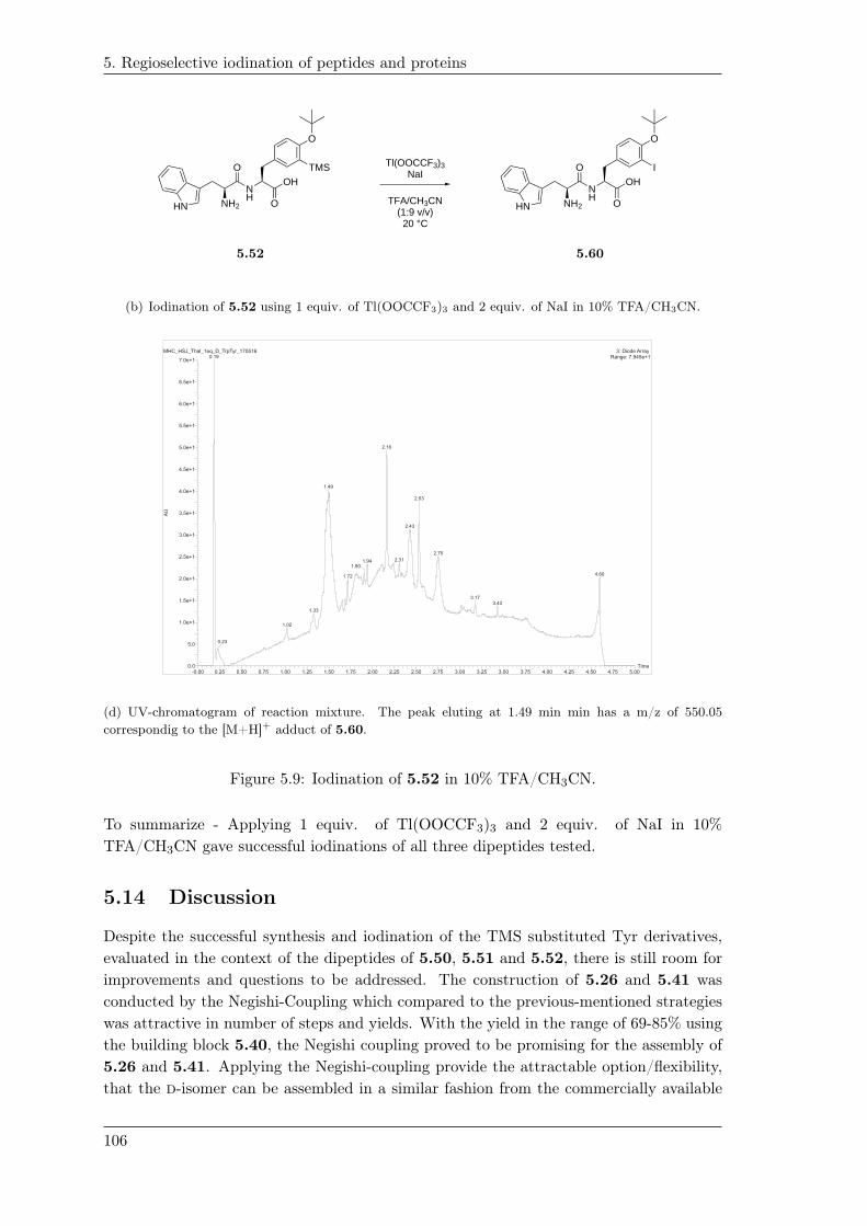
5. Regioselective iodination of peptides and proteins
Tl(OOCCF3)3NaI
TFA/CH3CN(1:9 v/v)20 °C
O
NH2HN ONH
O
OHTMS O
NH2HN ONH
O
OHI
5.52
5.53
5.60
(b) Iodination of 5.52 using 1 equiv. of Tl(OOCCF3)3 and 2 equiv. of NaI in 10% TFA/CH3CN.
Time-0.00 0.25 0.50 0.75 1.00 1.25 1.50 1.75 2.00 2.25 2.50 2.75 3.00 3.25 3.50 3.75 4.00 4.25 4.50 4.75 5.00
AU
0.0
5.0
1.0e+1
1.5e+1
2.0e+1
2.5e+1
3.0e+1
3.5e+1
4.0e+1
4.5e+1
5.0e+1
5.5e+1
6.0e+1
6.5e+1
7.0e+1
MHC_HSJ_Thal_1eq_D_TrpTyr_170516 3: Diode Array Range: 7.945e+10.19
2.16
1.49
1.33
1.02
0.23
1.941.90
1.72
2.53
2.43
2.31
2.75
4.60
3.173.43
(d) UV-chromatogram of reaction mixture. The peak eluting at 1.49 min min has a m/z of 550.05correspondig to the [M+H]+ adduct of 5.60.
Figure 5.9: Iodination of 5.52 in 10% TFA/CH3CN.
To summarize - Applying 1 equiv. of Tl(OOCCF3)3 and 2 equiv. of NaI in 10%
TFA/CH3CN gave successful iodinations of all three dipeptides tested.
5.14 Discussion
Despite the successful synthesis and iodination of the TMS substituted Tyr derivatives,
evaluated in the context of the dipeptides of 5.50, 5.51 and 5.52, there is still room for
improvements and questions to be addressed. The construction of 5.26 and 5.41 was
conducted by the Negishi-Coupling which compared to the previous-mentioned strategies
was attractive in number of steps and yields. With the yield in the range of 69-85% using
the building block 5.40, the Negishi coupling proved to be promising for the assembly of
5.26 and 5.41. Applying the Negishi-coupling provide the attractable option/flexibility,
that the d-isomer can be assembled in a similar fashion from the commercially available
106

5. Regioselective iodination of peptides and proteins
d-isomer of 5.31 or 5.33, respectively. This could be attractive as the d-isomer when
entering into a peptide sequence, could enhance the in vivo-stability as reported [110].
The Negishi-coupling has likewise been reported using Fmoc-β-iodo-Ala-OMe [163]. The
N,N -Boc-methyl β-iodo-alanine methyl ester 5.61 is likewise hypothesized to be reachable
within a few number of steps (Scheme 5.15). This provides another building block for the
Negishi-coupling, which will give the methyl substituted α-amino group which likewise
could enhance the stability against proteolysis. These examples demonstrate and state
the chemical flexibility in the Negishi-coupling allowing different protecting groups and
starting materials in the synthesis of synthetic amino acids.
Substitution/Iodination
COOMeBocN
I
COOMeBocN
HO
COOHBocHN
HOMethylation
Commercial available
5.61
Scheme 5.15: Retrosynthetic analysis for the synthesis of the methylated Negishi building
block 5.61.
Despite the formation of the diastereomer of Marfey’s reagent of 5.43, the stereochem-
ically outcome of the peptide bond formation of 5.50 and 5.51 seemed not conclusive
as 1H-NMR indicated mixtures of diastereomers. Further experiments and analysis are
needed of the linear peptides of 5.50 and 5.51 to confirm the stereochemistry, optionally
supported by the synthesis of the corresponding d-isomer.
In regard to the synthesis of the dipeptides – though that the dipeptides were designed
in order to evaluate the proof of selectivity in the iodination reaction, and since the
dipeptides are most likely not going to find use elsewhere - the significant formation of
the α-N -alkylation as well as the cyclized diketopiperazines during the hydrogenolysis
reaction imply that another approach would have been preferable. Modifications to the
applied strategy, could be the use of orthogonal protecting groups of the α-amino group
and C-terminus, as well as carrying out the deprotections step-wise. One orthogonal com-
bination that could have been advantageous, would be to mask the α-amino group as the
Boc carbamate while the C-terminus remains as the benzyl ester. Hydrogenation of the
benzyl ester followed by Boc deprotection using TMSOTf and 2,6-lutidine will potentially
lead to the linear dipeptide with less probability of forming the diketopiperazine. Another
possibility that potentially could suppress the formation of diketopiperazine would be to
synthesize the tri-peptide with a Gly or Ala residue inbetween, 5.43 and the His, Phe,
Tyr, or Trp, respectively. Cyclization of the corresponding tri-peptides is less likely be-
cause this occurs by formation of a 9-membered ring compared to the 6-membered of the
diketopiperazine.
The iodination reactions by thallation seemed promising, though optimization could have
been interesting in order to improve the selectivity. In order to improve the selectivity of
iodination and the difficulty of the iodination of the Trp-dipeptide 5.52, Boc-protection
of the indole could potentially result in deactivation of the indole, by which the selectivity
of the iodination could be improved towards the ipso-substitution of the TMS. In regard
107

5. Regioselective iodination of peptides and proteins
to the difficulties in the purification of 5.49 and the loss during hydrogenolysis, the strat-
egy of using Z-His-OH without any protecting groups of the imidazole was a risky choice.
Using His, with the τ -N protected as the Trt or Boc could likewise lower the nucleophilic-
ity of the imidazole ring, which in terms of the synthesis and the iodination would have
become beneficial. The failure of the hydrogenation of the His-dipeptide 5.49 remains an
open question. The hydrogenolysis was reported successfully for Z-His-Phe(4-TMS)-OBn
in the previous study in the Mads Clausen group.
Despite that the iodination of the hydrogenated 5.49 was not tested in this study, (4-
TMS)Phe was reported to displayed clear selectivity over the imidazole of His in the
iodination of 5.16. Based on this result, it was envisioned that the iodination of 5.53
would exhibit selectivity towards the ipso-substitution of the TMS-group of Tyr. This
assumption will still have to be tested and analyzed.
5.15 Future perspectives
Short term future perspectives of this study will first of all be to evaluate the selectivity
in the iodination reaction of 5.53. Likewise, the iodination reaction on a Boc-protected
indole of Trp has to be tested. This will conclusively demonstrate the scope of using TMS
substituted Tyr for iodination.
Moreover, the compatibility with SPPS will have to be addressed. Questions like: Would
it be possible to iodinate on the resin? It is possible to cleave the desired (protected)
peptide from an acid-labile resin without proto-desilylation? Will iodination by thallation
be feasible subsequent to cleavage of the peptide in its protected form? Finally testing the
iodination in a system like the labeling of Leu-enkephalin or somatostatin 5.2 would state
the application of using the TMS-group as a placeholder to direct iodinations reactions of
peptides to be regioselective.
Long-term future perspectives; having established the use of the TMS as directing-group
with the synthesis of the TMS-derivatives of Phe and Tyr, it would be interesting to de-
velop the TMS derivatives of His and Trp. Installing the TMS group in the 2-position of
the indole of Trp could potentially provide selectivity in electrophilic aromatic iodinations
according to the β-cationic effect, also observed in vinylic-TMS systems. ipso-Iodination
of a TMS positioned in the 2-position of indole-derivatives have been reported [164, 165].
Employing the TMS group at the indole is hypothesized to favor iodination without oxi-
dation of the indole to the oxindole and subsequent cleavage of the peptide linkage.
5.16 Conclusion
TMS-substituted Tyr derivatives 5.26 and 5.41 were successfully synthesized in good
yields via the corresponding Negishi cross-coupling of 5.40 and the methyl Boc-β-
iodoalanine esters 5.33 or 5.31, respectively. The Boc-group was chemoselectively
removed using TMSOTf and 2,6-lutidine which allowed the transformation of 5.42 to 5.45
which is useful in the Fmoc-based strategy. The dipeptides of 5.46, 5.47, 5.48 and 5.49
108

5. Regioselective iodination of peptides and proteins
were likewise constructed from 5.43 and the Z-protected amino acids. Hydrogenolysis
of the Cbz-protected α-amino group and the benzyl ester caused serious difficulties
as reductive N -alkylation and diketopiperazine formation were observed, depending on
the solvent used. The stereochemistry of the Phe-dipeptide 5.50 and Tyr-dipeptide
5.51 seemed questionable as, 1H-NMR indicated what was believed to be mixtures
of diastereomers. Based on these observations, the stereochemical control has to be
substantiated by further analysis.
Iodination via thallation of the Phe and Tyr dipeptides of 5.50 and 5.51 displayed clear
selectivity for the ipso-substitution, while iodination of Trp-dipeptide 5.52 occurred but
with the formation of several side-products.
Proof-of-concept for the use of TMS substituted Tyr to direct the iodination was
demonstrated for 5.50, 5.51 and 5.52. Further optimization will be needed in the
iodination of 5.52.
109


Experimental 66’-O-TBDPS-sucrose (1.10). Sucrose 1.3
O11
44
55HO
HOHO
22
66
333'3'
5'5'1'1'
4'4'
O
6'6'
O
OH
OH
OH
O 2'2'
OH
TBDPS
(10 g, 29.2 mmol) was suspended in pyridine
(100 mL) and DMAP (0.36 g, 2.92 mmol) was
added. The reaction mixture was left stirred
for 1 h. TBDPSCl (4.65 mL, 17.5 mmol) was
added and the reaction mixture was heated to
70 ◦C for 3 h. TBDPSCl (4.65 mL, 17.5 mmol) was again added to the reaction mixture at
20 ◦C and the reaction mixture was left stirred overnight. Pyridine was removed in vacuo
and the residue was purified by dry column vacuum chromatography (EtOH in EtOAc1
with 2% increments) affording 1.10 as an amorphous solid.
yield = (6.8 g, 40%)
m.p.: 174◦C.
[α]20D
= +38◦ (c 1.00, MeOH)
1H-NMR (400 MHz, MeOD-d4): δ = 7.77-7.67 (m, 4H, o-HAr), 7.46-7.35 (m, 6H, m-
HAr+p-HAr), 5.44 (d, J 1→2 = 3.9 Hz, 1H, H1), 4.13-4.01 (m, 2H, H3′+H4′), 3.99-3.84 (m,
3H, H5′+H6′), 3.79-3.72 (m, 1H, H5), 3.68-3.50 (m, 5H, H3+H6+H1′), 3.35-3.25 (m, 2H,
H2+H4), 1.04 (s, 9H, (CH3)3C-Si).
13C-NMR (100 MHz, MeOD-d4): δ = 136.8 (2C, o-CAr), 136.8 (2C, o-CAr), 134.6
(i -CAr), 134.5 (i -CAr), 130.8 (2C, p-CAr), 128.8 (2C, m-CAr), 128.8 (2C, m-CAr), 105.6
(C2′), 93.0 (C1), 83.7 (C5′), 79.0 (C3′), 76.2 (C4′), 74.9 (C3), 73.9 (C5), 73.2 (C2), 71.4
(C4), 66.5 (C6′), 64.3 (C1′), 62.4 (C6), 27.3 (3C, (CH3)3C-Si), 20.0 ((CH3)3C-Si).
HR-MS. Calculated adducts: [M+H2O]+=598.2440, [M+Na]+=603.2232, [M+K]+= 619.1971.
Observed adducts: [M+H2O]+=598.2675, [M+Na]+=603.2226, [M+K]+=619.1952.
ATR-FTIR (cm−1): 3285.79, 2930.17, 2857.74, 1470.26, 1428.36, 1390.34, 1111.82, 507.18,
1067.96, 1043.53, 994.46, 930.50, 701.83, 805.57.
1FiXme Fatal: Dette er en fejl, Tjek TLC når du er tilbage
111

6. Experimental
Hepta-isobutyric-6’-O-TBDPS-sucrose (1.24).
O11
44
55O
OO
22
66
333'3'
5'5'1'1'
4'4'
O
6'6'
O
O
O
O
O 2'2'
O
TBDPS
O
O
O
O
O
O
O
1.10 (2.00 g, 3.44 mmol) was dissolved in pyri-
dine (40 mL) and DMAP (0.114 g, 0.924 mmol)
was added. To the reaction mixture was added
isobutyric anhydride (11.4 mL, 66.7 mmol) and
the reaction mixture was left stirred at 20 ◦C
overnight. TLC analysis was used to follow
the the reaction. Upon reaction completion,
the reaction mixture was concentrated in vacuo
and Et2O (200 mL) was added followed by ex-
traction with saturated aq. NaHCO3 solution
(5x200 mL). The organic phase was subsequently collected and concentrated in vacuo.
The crude residue was purified by dry column vacuum chromatography (EtOAc in hep-
tane with 2% increments) yielding 1.24 as a transparent oily texture.
yield = (2.97 g, 80%).
[α]20D
= +37◦ (c 1.00, EtOH)
1H-NMR (400 MHz, DMSO-d6): δ= 7.64-7.56 (m, 4H, o-HAr), 7.50-7.37 (m, 6H, m-
HAr+p-HAr), 5.62 (d, J 1→2 = 3.7 Hz, 1H, H1), 5.49-5.46 (m, 2H, H3′+H4′), 5.35 (t, J 3→2
= J 3→4 = 10.0 Hz, 1H, H3), 5.04 (t, J 4→3 = J 4→5 = 10.0 Hz, 1H, H4), 4.86 (dd, J 2→3
= 10.6 Hz, J 2→1 = 3.6 Hz, 1H, H2), 4.24 (ddd,J 5→4 = 10.3 Hz, J 5→6a = 4.4 Hz, J 5→6b
= 1.8 Hz, 1H, H5), 4.20-4.02 (m, 4H, H1′+H5′+H6a), 3.90 (dd, J = 13 Hz6b→6a, J =
1.8 Hz6b→5, 1H, H6b), 3.86-3.75 (m, 2H, H6′), 2.62-2.52 (m, 3H, (CH3)2CH-), 2.48-2.32
(m, 4H, (CH3)2CH-), 1.16-1.08 (m, 12H, (CH3)2CH-), 1.07-0.94 (m, 39H, (CH3)2CH- +
(CH3)3C-Si).
13C-NMR (100 MHz, DMSO-d6): δ = 175.6 ((CH3)2CH-C(O)-O-), 175.3 ((CH3)2CH-
C(O)-O-), 175.2 ((CH3)2CH-C(O)-O-), 175.0 (3C, (CH3)2CH-C(O)-O-), 174.6 ((CH3)2CH-
C(O)-O-), 135.0 (4C, o-CAr), 132.4 (i -CAr), 132.3 (i -CAr), 130.0 (2C, p-CAr), 127.9 (4C,
m-CAr), 102.4 (C2′), 89.1 (C1), 80.0 (C5′), 75.3 (C4′), 73.5 (C3′), 69.2 (C3), 69.1 (C2),
68.1 (C5), 67.1 (C4), 63.8 (C6′), 63.3 (C1′), 61.2 (C6), 33.3-32.9 (7C, (CH3)2CH-), 30.7
((CH3)3C-Si), 26.4 (3C,(CH3)3C-Si), 19.0-18.0 (14C, (CH3)2CH-)).
HR-MS. Calculated adducts: [M+H2O]+=1088.5376, [M+Na]+=1093.5163, [M+K]+=1109.4902.
Observed adducts, [M+H2O]+=1088.5609, [M+Na]+=1093.5175, [M+K]+=1109.4892.
ATR-FTIR (cm−1): 3285.44, 2973.83, 2934.80, 2877.85, 1741.41, 1470.12, 1388.47, 1247.91,
1186.65, 1139.69, 1111.82, 1071.63, 1030.51, 823.65, 742.56, 504.26, 702.23.
112

6. Experimental
Hepta-2,3,4,6,1’,3’,4’-O-isobutyryl-sucrose
O11
44
55O
OO
22
66
333'3'
5'5'1'1'
4'4'
O
6'6'
OH
O
O
O
O 2'2'
OO
O
O
O
O
O
O
(1.25). Hepta-isobutyryl-6’-O-TBDPS-sucrose
1.24 (1.032 g, 0.95 mmol) was solubilized un-
der inert atmosphere in dry THF (20 mL). To
the stirred reaction mixture was added acetic
acid (0.082 mL, 1.43 mmol) followed by the
drop-wise addition of a 1.0 M TBAF solution
in THF (1.2 mL, 1.2 mmol). The reaction mix-
ture was left stirred at 20 ◦C overnight. The
reaction was followed by TLC. Upon reaction
completion, the reaction mixture was directly purified by dry column vacuum chromatog-
raphy (EtOAc in heptane with 4% increments). This afforded the deprotected product
1.25 as a transparent oily texture.
yield = (0.768 g, 97%).
[α]20D
= +47◦ (c 1.00, EtOH)
1H-NMR (400 MHz, DMSO-d6): δ= 5.67 (d, J 1→2 = 3.7 Hz, 1H, H1), 5.47 (d, J 3′→4′
= 7.5 Hz, 1H, H3′), 5.40 (t, J 3→2 = J 3→4 = 9.9 Hz, 1H, H3), 5.34 (t, J 4′→3′ = J 4′→5′
= 7.5 Hz, 1H, H4′), 5.11 (t, J 4→3 = J 4→5 = 9.9 Hz, 1H, H4), 4.96 (t, J = 5.5 Hz, 1H,
-CH2-O-H), 4.91 (dd, J 2→3 = 10.4 Hz, J 2→1 = 3.6 Hz, 1H, H2), 4.34 (ddd, J 5→4 = 10.4
Hz, J 5→6a = 4.4 Hz, J 5→6b = 1.8 Hz, 1H, H5), 4.21 (dd, J 6a→6b = 13.0 Hz, J 6a→5 =
4.4 Hz, 1H, H6a), 4.12-4.00 (m, 4H, H1′+H5′+H6b), 3.60 (m, 2H, H6′), 2.66-2.34 (m, 7H
(DMSO overlap), (CH3)2CH-), 1.21-0.95 (m, 42 H, CH3)2CH-).
13C-NMR (100 MHz, DMSO-d6): δ= 175.8 ((CH3)2CH-C(O)-O-), 175.4 ((CH3)2CH-
C(O)-O-), 175.3 ((CH3)2CH-C(O)-O-), 175.0 (3C, (CH3)2CH-C(O)-O-), 174.6 ((CH3)2CH-
C(O)-O-), 102.2 (C2′), 89.1 (C1), 80.8 (C5′), 75.2 (C3′), 73.7 (C4′), 69.3 (C3), 69.2 (C2),
68.0 (C5), 67.3 (C4), 63.3 (C1′), 61.5 (C6′), 61.4 (C6), 33.3-32.9 (7C, (CH3)2CH-), 19.0-
18.0 (14C, (CH3)2CH-).
HR-MS. Calculated adducts: [M+H2O]+=850.4193, [M+Na]+=855.3985, [M+K]+=871.3724.
Observed adducts: [M+H2O]+=850.4449, [M+Na]+=855.3999, [M+K]+=871.3738.
ATR-FTIR (cm−1): 3531.46, 2974.80, 2939.02, 2878.48, 1739.30, 1470.09, 1388.51, 1344.51,
1247.10, 1186.51, 1142.42, 1077.10, 1048.73, 1005.66, 748.36.
113
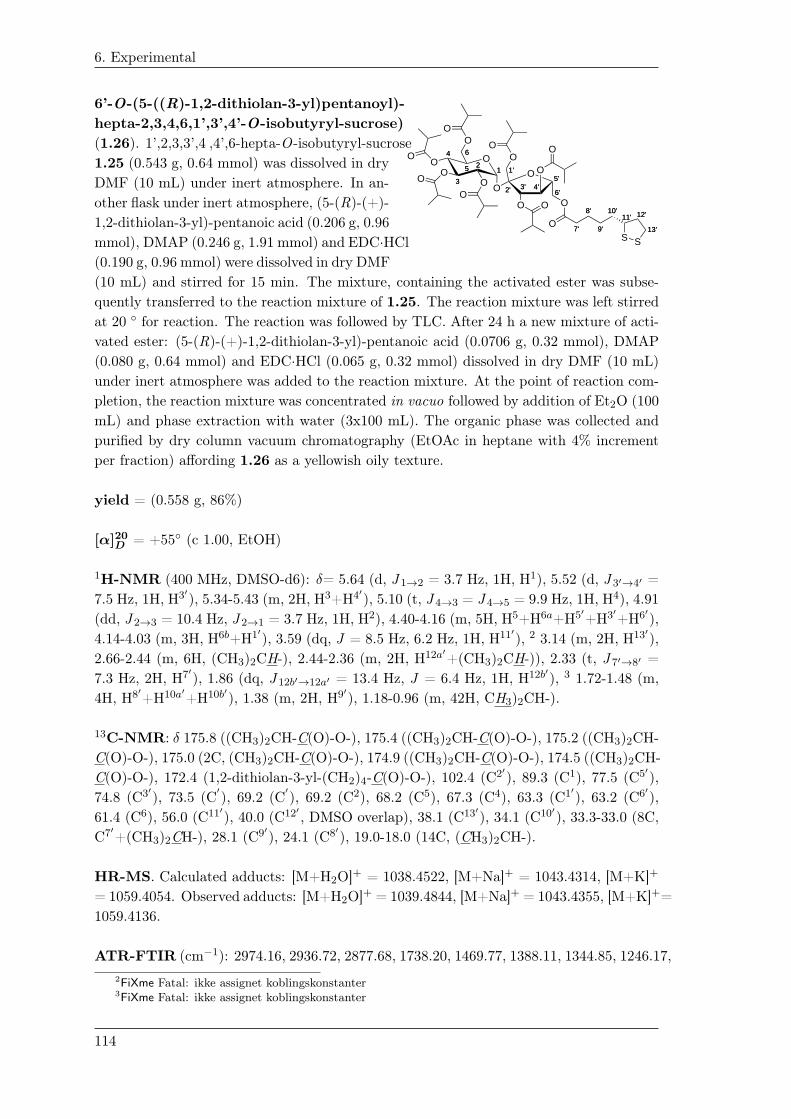
6. Experimental
6’-O-(5-((R)-1,2-dithiolan-3-yl)pentanoyl)-
O11
44
55O
OO
22
66
333'3'
5'5'1'1'
4'4'
O
6'6'
O
O
O
O
O 2'2'
OO
O
O
O
O
O
O
7'7'O
8'8'
9'9'
10'10'11'11'
S
12'12'
S13'13'
hepta-2,3,4,6,1’,3’,4’-O-isobutyryl-sucrose)
(1.26). 1’,2,3,3’,4 ,4’,6-hepta-O-isobutyryl-sucrose
1.25 (0.543 g, 0.64 mmol) was dissolved in dry
DMF (10 mL) under inert atmosphere. In an-
other flask under inert atmosphere, (5-(R)-(+)-
1,2-dithiolan-3-yl)-pentanoic acid (0.206 g, 0.96
mmol), DMAP (0.246 g, 1.91 mmol) and EDC·HCl
(0.190 g, 0.96 mmol) were dissolved in dry DMF
(10 mL) and stirred for 15 min. The mixture, containing the activated ester was subse-
quently transferred to the reaction mixture of 1.25. The reaction mixture was left stirred
at 20 ◦ for reaction. The reaction was followed by TLC. After 24 h a new mixture of acti-
vated ester: (5-(R)-(+)-1,2-dithiolan-3-yl)-pentanoic acid (0.0706 g, 0.32 mmol), DMAP
(0.080 g, 0.64 mmol) and EDC·HCl (0.065 g, 0.32 mmol) dissolved in dry DMF (10 mL)
under inert atmosphere was added to the reaction mixture. At the point of reaction com-
pletion, the reaction mixture was concentrated in vacuo followed by addition of Et2O (100
mL) and phase extraction with water (3x100 mL). The organic phase was collected and
purified by dry column vacuum chromatography (EtOAc in heptane with 4% increment
per fraction) affording 1.26 as a yellowish oily texture.
yield = (0.558 g, 86%)
[α]20D
= +55◦ (c 1.00, EtOH)
1H-NMR (400 MHz, DMSO-d6): δ= 5.64 (d, J 1→2 = 3.7 Hz, 1H, H1), 5.52 (d, J 3′→4′ =
7.5 Hz, 1H, H3′), 5.34-5.43 (m, 2H, H3+H4′), 5.10 (t, J 4→3 = J 4→5 = 9.9 Hz, 1H, H4), 4.91
(dd, J 2→3 = 10.4 Hz, J 2→1 = 3.7 Hz, 1H, H2), 4.40-4.16 (m, 5H, H5+H6a+H5′+H3′+H6′),
4.14-4.03 (m, 3H, H6b+H1′), 3.59 (dq, J = 8.5 Hz, 6.2 Hz, 1H, H11′), 2 3.14 (m, 2H, H13′),
2.66-2.44 (m, 6H, (CH3)2CH-), 2.44-2.36 (m, 2H, H12a′+(CH3)2CH-)), 2.33 (t, J 7′→8′ =
7.3 Hz, 2H, H7′), 1.86 (dq, J 12b′→12a′ = 13.4 Hz, J = 6.4 Hz, 1H, H12b′), 3 1.72-1.48 (m,
4H, H8′+H10a′+H10b′), 1.38 (m, 2H, H9′), 1.18-0.96 (m, 42H, CH3)2CH-).
13C-NMR: δ 175.8 ((CH3)2CH-C(O)-O-), 175.4 ((CH3)2CH-C(O)-O-), 175.2 ((CH3)2CH-
C(O)-O-), 175.0 (2C, (CH3)2CH-C(O)-O-), 174.9 ((CH3)2CH-C(O)-O-), 174.5 ((CH3)2CH-
C(O)-O-), 172.4 (1,2-dithiolan-3-yl-(CH2)4-C(O)-O-), 102.4 (C2′), 89.3 (C1), 77.5 (C5′),
74.8 (C3′), 73.5 (C′
), 69.2 (C′
), 69.2 (C2), 68.2 (C5), 67.3 (C4), 63.3 (C1′), 63.2 (C6′),
61.4 (C6), 56.0 (C11′), 40.0 (C12′ , DMSO overlap), 38.1 (C13′), 34.1 (C10′), 33.3-33.0 (8C,
C7′+(CH3)2CH-), 28.1 (C9′), 24.1 (C8′), 19.0-18.0 (14C, (CH3)2CH-).
HR-MS. Calculated adducts: [M+H2O]+ = 1038.4522, [M+Na]+ = 1043.4314, [M+K]+
= 1059.4054. Observed adducts: [M+H2O]+ = 1039.4844, [M+Na]+ = 1043.4355, [M+K]+=
1059.4136.
ATR-FTIR (cm−1): 2974.16, 2936.72, 2877.68, 1738.20, 1469.77, 1388.11, 1344.85, 1246.17,
2FiXme Fatal: ikke assignet koblingskonstanter
3FiXme Fatal: ikke assignet koblingskonstanter
114

6. Experimental
1185.60, 1140.82, 1074.25, 1028.24, 963.11, 916.58, 849.06, 748.51, 476.19.
115

6. Experimental
p-TMS-phenol (2.4). p-Bromophenol 2.3 (2.37 g, 13.311
2233
44
5566
OH
TMS
mmol) was solubilized in dry THF (150 mL) under an argon
atmosphere and cooled to -78 ◦C. n-BuLi (1.6 M, 25 mL,
40.0 mmol) was added drop-wise to the reaction mixture at
-78 ◦C. 15 min upon addition, the reaction mixture was removed from the cooling bath
and allowed to reach 20 ◦C for 2 h leading to a white cloudy suspension. The reaction
mixture was re-cooled to -78 ◦C and TMSCl (5.12 mL, 40.0 mmol) was then added to the
reaction mixture which was left in the cooling bath and allowed to reach 20 ◦C overnight.
The reaction was quenched by addition of sat. NH4Cl (40 mL). H2O (50 mL) was added
and the mixture was then extracted using EtOAc (3x100 mL). The organic phases were
combined, dried (Na2SO4) and concentrated in vacuo followed by purification by dry col-
umn vacuum chromatography (1% EtOAc/heptane linear increments). The product 2.4
was further refined by sublimation at 50 ◦C.
yield = (1.74 g, 78%).
m.p.: 74.3 ◦C.
1H-NMR (400 MHz, CDCl3) δ: 7.41 (d, Jm→o = 8.5 Hz, 2H, m-HAr), 6.84 (d, J o→m =
8.5 Hz, 2H, o-HAr), 4.85 (s, 1H, p-TMS-Ph-O-H), 0.24 (s, 9H, (CH3)3-Si).
13C-NMR (100 MHz, CDCl3) δ: 156.3 (C1), 135.1 (2C, C3+C5), 131.8 (C4), 115.0 (2C,
C2+C6), -0.8 (3C, (CH3)3-Si).
LC-MS: Calculated adducts [M-H]−: 165.07. Observed adducts: [M-H]−: 165.07.
ATR-FTIR (cm−1): 3174.58, 2952.11, 2774.57, 2695.51, 2636.61, 1892.45, 1597.19, 1582.07,
1501.04, 1419.11, 1358.65, 1249.24, 1231.76, 1179.69, 1105.29, 837.99, 813.08, 752.72,
709.83, 649.42, 514.15.
All observed spectral shifts are in agreement with literature [] [29].
116
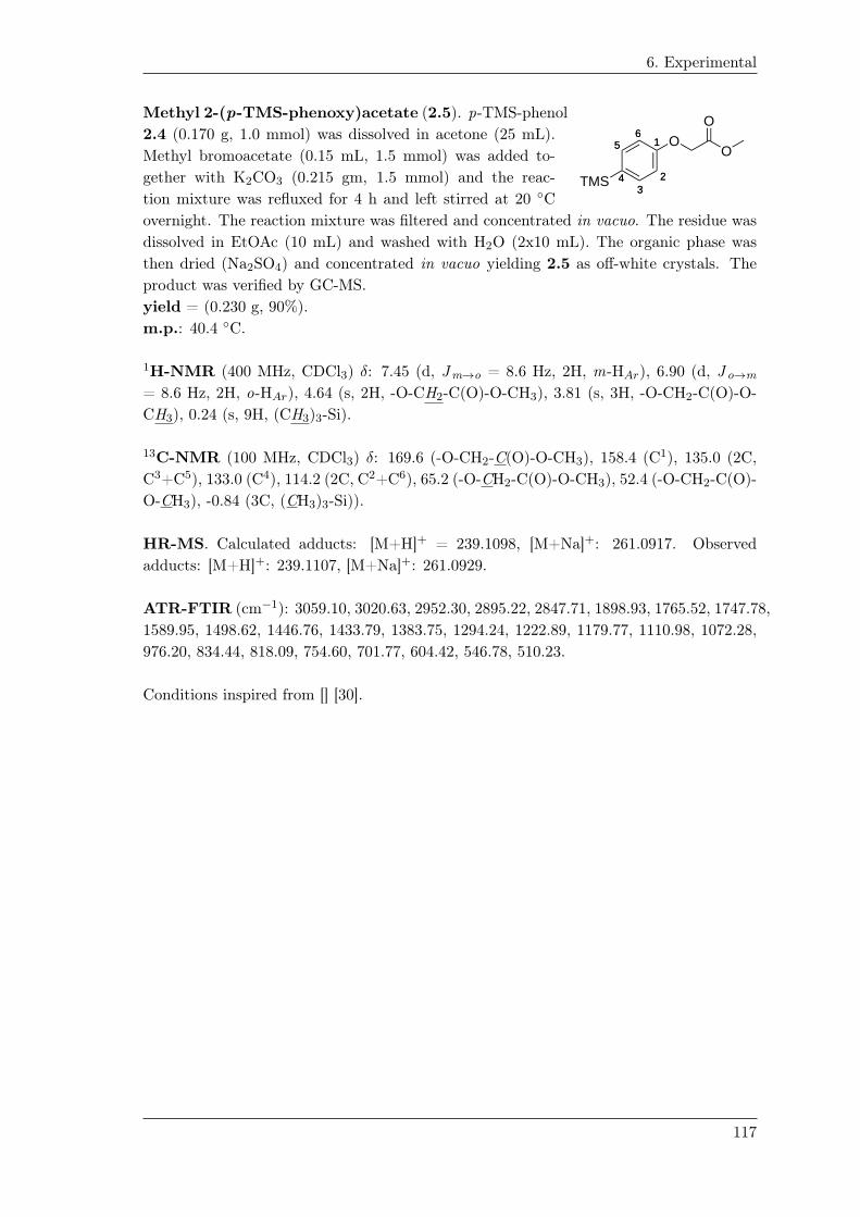
6. Experimental
Methyl 2-(p-TMS-phenoxy)acetate (2.5). p-TMS-phenol
11
2233
44
5566
O
TMS
O
O2.4 (0.170 g, 1.0 mmol) was dissolved in acetone (25 mL).
Methyl bromoacetate (0.15 mL, 1.5 mmol) was added to-
gether with K2CO3 (0.215 gm, 1.5 mmol) and the reac-
tion mixture was refluxed for 4 h and left stirred at 20 ◦C
overnight. The reaction mixture was filtered and concentrated in vacuo. The residue was
dissolved in EtOAc (10 mL) and washed with H2O (2x10 mL). The organic phase was
then dried (Na2SO4) and concentrated in vacuo yielding 2.5 as off-white crystals. The
product was verified by GC-MS.
yield = (0.230 g, 90%).
m.p.: 40.4 ◦C.
1H-NMR (400 MHz, CDCl3) δ: 7.45 (d, Jm→o = 8.6 Hz, 2H, m-HAr), 6.90 (d, J o→m
= 8.6 Hz, 2H, o-HAr), 4.64 (s, 2H, -O-CH2-C(O)-O-CH3), 3.81 (s, 3H, -O-CH2-C(O)-O-
CH3), 0.24 (s, 9H, (CH3)3-Si).
13C-NMR (100 MHz, CDCl3) δ: 169.6 (-O-CH2-C(O)-O-CH3), 158.4 (C1), 135.0 (2C,
C3+C5), 133.0 (C4), 114.2 (2C, C2+C6), 65.2 (-O-CH2-C(O)-O-CH3), 52.4 (-O-CH2-C(O)-
O-CH3), -0.84 (3C, (CH3)3-Si)).
HR-MS. Calculated adducts: [M+H]+ = 239.1098, [M+Na]+: 261.0917. Observed
adducts: [M+H]+: 239.1107, [M+Na]+: 261.0929.
ATR-FTIR (cm−1): 3059.10, 3020.63, 2952.30, 2895.22, 2847.71, 1898.93, 1765.52, 1747.78,
1589.95, 1498.62, 1446.76, 1433.79, 1383.75, 1294.24, 1222.89, 1179.77, 1110.98, 1072.28,
976.20, 834.44, 818.09, 754.60, 701.77, 604.42, 546.78, 510.23.
Conditions inspired from [] [30].
117
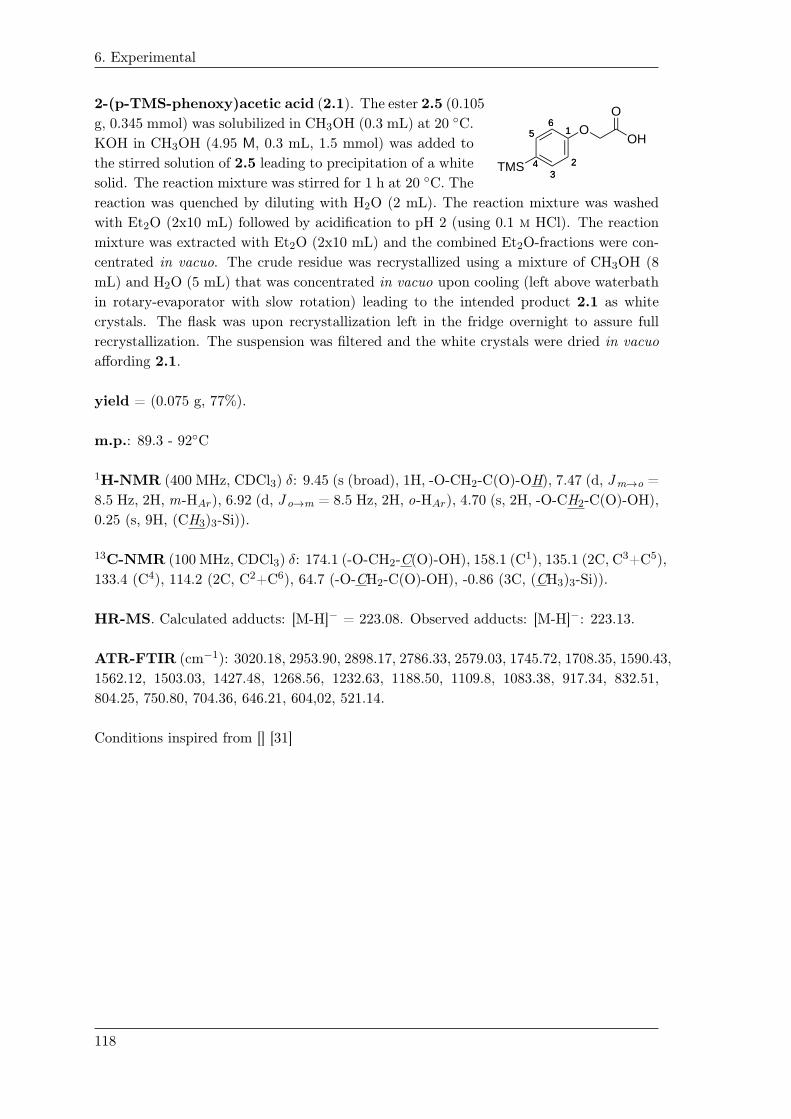
6. Experimental
2-(p-TMS-phenoxy)acetic acid (2.1). The ester 2.5 (0.105
11
2233
44
5566
O
TMS
OH
Og, 0.345 mmol) was solubilized in CH3OH (0.3 mL) at 20 ◦C.
KOH in CH3OH (4.95 M, 0.3 mL, 1.5 mmol) was added to
the stirred solution of 2.5 leading to precipitation of a white
solid. The reaction mixture was stirred for 1 h at 20 ◦C. The
reaction was quenched by diluting with H2O (2 mL). The reaction mixture was washed
with Et2O (2x10 mL) followed by acidification to pH 2 (using 0.1 m HCl). The reaction
mixture was extracted with Et2O (2x10 mL) and the combined Et2O-fractions were con-
centrated in vacuo. The crude residue was recrystallized using a mixture of CH3OH (8
mL) and H2O (5 mL) that was concentrated in vacuo upon cooling (left above waterbath
in rotary-evaporator with slow rotation) leading to the intended product 2.1 as white
crystals. The flask was upon recrystallization left in the fridge overnight to assure full
recrystallization. The suspension was filtered and the white crystals were dried in vacuo
affording 2.1.
yield = (0.075 g, 77%).
m.p.: 89.3 - 92◦C
1H-NMR (400 MHz, CDCl3) δ: 9.45 (s (broad), 1H, -O-CH2-C(O)-OH), 7.47 (d, Jm→o =
8.5 Hz, 2H, m-HAr), 6.92 (d, J o→m = 8.5 Hz, 2H, o-HAr), 4.70 (s, 2H, -O-CH2-C(O)-OH),
0.25 (s, 9H, (CH3)3-Si)).
13C-NMR (100 MHz, CDCl3) δ: 174.1 (-O-CH2-C(O)-OH), 158.1 (C1), 135.1 (2C, C3+C5),
133.4 (C4), 114.2 (2C, C2+C6), 64.7 (-O-CH2-C(O)-OH), -0.86 (3C, (CH3)3-Si)).
HR-MS. Calculated adducts: [M-H]− = 223.08. Observed adducts: [M-H]−: 223.13.
ATR-FTIR (cm−1): 3020.18, 2953.90, 2898.17, 2786.33, 2579.03, 1745.72, 1708.35, 1590.43,
1562.12, 1503.03, 1427.48, 1268.56, 1232.63, 1188.50, 1109.8, 1083.38, 917.34, 832.51,
804.25, 750.80, 704.36, 646.21, 604,02, 521.14.
Conditions inspired from [] [31]
118
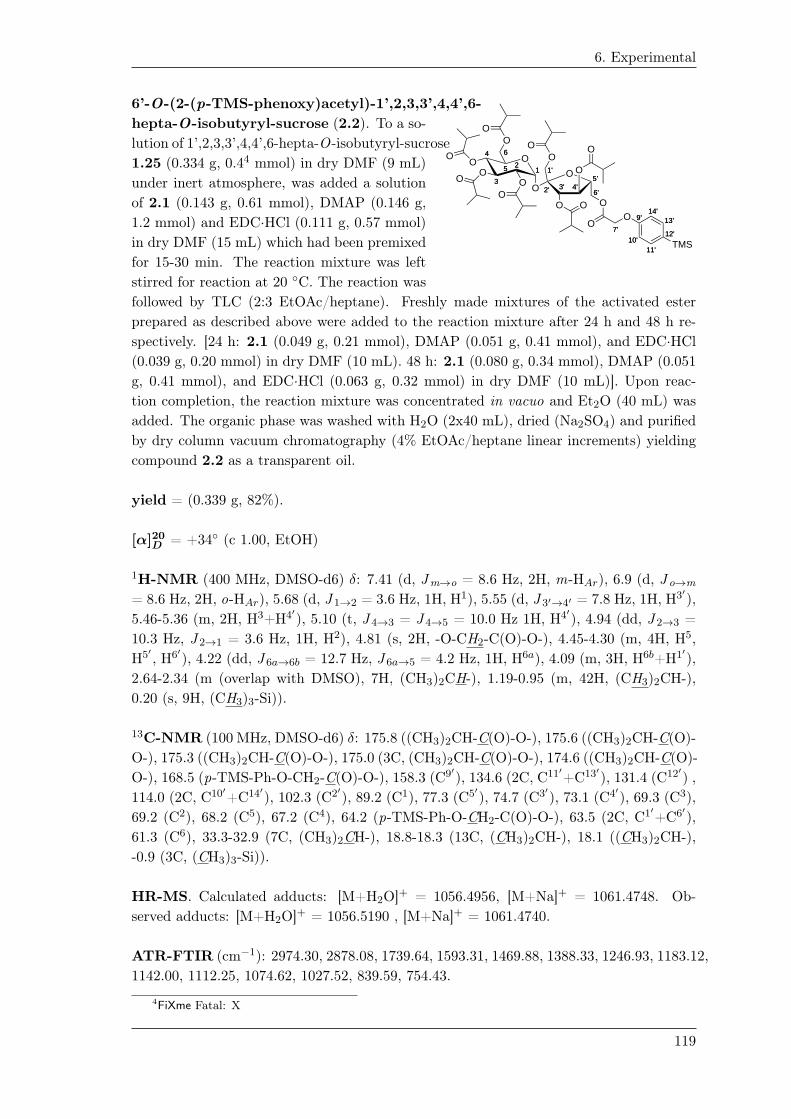
6. Experimental
6’-O-(2-(p-TMS-phenoxy)acetyl)-1’,2,3,3’,4,4’,6-
O11
44
55O
OO
22
66
333'3'
5'5'1'1'
4'4'
O
6'6'
O
O
O
O
O 2'2'
OO
O
O
O
O
O
O
7'7'O
O 9'9'
10'10'11'11'
12'12'
13'13'14'14'
TMS
hepta-O-isobutyryl-sucrose (2.2). To a so-
lution of 1’,2,3,3’,4,4’,6-hepta-O-isobutyryl-sucrose
1.25 (0.334 g, 0.44 mmol) in dry DMF (9 mL)
under inert atmosphere, was added a solution
of 2.1 (0.143 g, 0.61 mmol), DMAP (0.146 g,
1.2 mmol) and EDC·HCl (0.111 g, 0.57 mmol)
in dry DMF (15 mL) which had been premixed
for 15-30 min. The reaction mixture was left
stirred for reaction at 20 ◦C. The reaction was
followed by TLC (2:3 EtOAc/heptane). Freshly made mixtures of the activated ester
prepared as described above were added to the reaction mixture after 24 h and 48 h re-
spectively. [24 h: 2.1 (0.049 g, 0.21 mmol), DMAP (0.051 g, 0.41 mmol), and EDC·HCl
(0.039 g, 0.20 mmol) in dry DMF (10 mL). 48 h: 2.1 (0.080 g, 0.34 mmol), DMAP (0.051
g, 0.41 mmol), and EDC·HCl (0.063 g, 0.32 mmol) in dry DMF (10 mL)]. Upon reac-
tion completion, the reaction mixture was concentrated in vacuo and Et2O (40 mL) was
added. The organic phase was washed with H2O (2x40 mL), dried (Na2SO4) and purified
by dry column vacuum chromatography (4% EtOAc/heptane linear increments) yielding
compound 2.2 as a transparent oil.
yield = (0.339 g, 82%).
[α]20D
= +34◦ (c 1.00, EtOH)
1H-NMR (400 MHz, DMSO-d6) δ: 7.41 (d, Jm→o = 8.6 Hz, 2H, m-HAr), 6.9 (d, J o→m
= 8.6 Hz, 2H, o-HAr), 5.68 (d, J 1→2 = 3.6 Hz, 1H, H1), 5.55 (d, J 3′→4′ = 7.8 Hz, 1H, H3′),
5.46-5.36 (m, 2H, H3+H4′), 5.10 (t, J 4→3 = J 4→5 = 10.0 Hz 1H, H4′), 4.94 (dd, J 2→3 =
10.3 Hz, J 2→1 = 3.6 Hz, 1H, H2), 4.81 (s, 2H, -O-CH2-C(O)-O-), 4.45-4.30 (m, 4H, H5,
H5′ , H6′), 4.22 (dd, J 6a→6b = 12.7 Hz, J 6a→5 = 4.2 Hz, 1H, H6a), 4.09 (m, 3H, H6b+H1′),
2.64-2.34 (m (overlap with DMSO), 7H, (CH3)2CH-), 1.19-0.95 (m, 42H, (CH3)2CH-),
0.20 (s, 9H, (CH3)3-Si)).
13C-NMR (100 MHz, DMSO-d6) δ: 175.8 ((CH3)2CH-C(O)-O-), 175.6 ((CH3)2CH-C(O)-
O-), 175.3 ((CH3)2CH-C(O)-O-), 175.0 (3C, (CH3)2CH-C(O)-O-), 174.6 ((CH3)2CH-C(O)-
O-), 168.5 (p-TMS-Ph-O-CH2-C(O)-O-), 158.3 (C9′), 134.6 (2C, C11′+C13′), 131.4 (C12′) ,
114.0 (2C, C10′+C14′), 102.3 (C2′), 89.2 (C1), 77.3 (C5′), 74.7 (C3′), 73.1 (C4′), 69.3 (C3),
69.2 (C2), 68.2 (C5), 67.2 (C4), 64.2 (p-TMS-Ph-O-CH2-C(O)-O-), 63.5 (2C, C1′+C6′),
61.3 (C6), 33.3-32.9 (7C, (CH3)2CH-), 18.8-18.3 (13C, (CH3)2CH-), 18.1 ((CH3)2CH-),
-0.9 (3C, (CH3)3-Si)).
HR-MS. Calculated adducts: [M+H2O]+ = 1056.4956, [M+Na]+ = 1061.4748. Ob-
served adducts: [M+H2O]+ = 1056.5190 , [M+Na]+ = 1061.4740.
ATR-FTIR (cm−1): 2974.30, 2878.08, 1739.64, 1593.31, 1469.88, 1388.33, 1246.93, 1183.12,
1142.00, 1112.25, 1074.62, 1027.52, 839.59, 754.43.
4FiXme Fatal: X
119

6. Experimental
Methyl 2-(p-iodo-phenoxy)acetate (2.6).
11
2233
44
5566
O
I
O
O
Synthesis of 2.6 using Tl(OOCCF3)3 and NaI.
The ester of 2.5 (0.107 g, 0.45 mmol) was dissolved in TFA/CH3CN (7 mL, 3:7 v/v). A
stock solution of Tl(OOCCF3)3 in TFA/CH3CN (184 mM, 3:7 v/v), was prepared from
which an aliquot of Tl(OOCCF3)3 (4.4 mL, 0.81 mmol) was added to the reaction mix-
ture. The reaction mixture turned slightly red and was left stirred at 20 ◦C. After 40 min,
NaI (0.237 g, 1.58 mmol) was added to the reaction mixture resulting in a dark black/red
solution. After 5 min, the reaction was quenched using aq. sat. Na2CO3 (20 mL). The so-
lution turned slightly orange with a precipitate. The solution was extracted using EtOAc
(3x10 mL) and the organic phases were combined, washed with Na2S2O3 (10 mL, 10%
w/w), dried (Na2SO4) and concentrated in vacuo. The crude product was re-suspended
in CDCl3 and filtered. The filtrate was analyzed by 1H-NMR and corresponded to the
intended product 2.6. Purity by 1H-NMR, 90%.
Synthesis of 2.6 using ICl.
The ester of 2.5 (0.057 g, 0.24 mmol) was dissolved in dry CH2Cl2 (1 mL) and cooled
to 0 ◦C. A stock solution of ICl in dry CH2Cl2 (1.54 M, 0.25 g·mL dry CH2Cl2) was
prepared from which ICl (0.022 mL, 0.034 mmol) was added to the reaction mixture at
0 ◦C. The reaction mixture was left stirred for 3 h at 0 ◦C. After 3 h, the reaction was
quenched using aq. Na2S2O3 (2 mL, 10% w/w). The organic phase was collected and
the H2O phase was further washed using EtOAc (3x3 mL). The organic phases were com-
bined, dried (Na2SO4) and concentrated in vacuo. The residue was then purified by dry
column vacuum chromatography (4% EtOAc/heptane increments) yielding methyl 2-(p-
iodo-phenoxy)acetate 2.6. yield = (0.048 g, 77%). Conditions inspired from [32]. 5
Synthesis of 2.6 using NIS and TMSOTf.
The ester of 2.5 (0.063 g, 0.27 mmol) and NIS (0.077 g, 0.35 mmol) were suspended in
dry CH2Cl2 (1 mL). TMSOTf (0.01 mL, 0.055 mmol) was added and the reaction mix-
ture was left stirred at 20 ◦C. After approx. 20 h, the reaction mixture had turned dark
red and more homogenous. The reaction was followed by TLC analysis (EtOAc/heptane,
2:3 v/v). Upon reaction completion, the reaction was quenched using aq. Na2S2O3 (2
mL, 10% w/w) and extracted using EtOAc (3x3 mL). The organic phases were combined,
dried (Na2SO4) and purified by dry column vacuum chromatography (4% EtOAc/heptane
linear increments) yielding 2.6. yield = (0.057 g, 82%). 6
5FiXme Fatal: MHC-HSJ-49 040314
6FiXme Fatal: MHC-HSJ-48
120

6. Experimental
Synthesis of 2.6 using CAT and NaI.
The ester of 2.5 (0.101 g, 0.42 mmol), CAT (0.240 g, 0.85 mmol) and NaI (0.174 g, 1.16
mmol) were suspended in DMF (2 mL) and stirred for 1 h min at 20 ◦C (solution turned
orange). After 1 h, the reaction mixture was diluted with H2O and dilute HCl (5% w/w)
was added. The mixture was extracted using EtOAc (2x10 mL) and the H2O phase was
further washed with EtOAc (2x10 mL). The organic phases were combined and washed
with aq. Na2S2O3 (2 x 15 mL, 10% w/w), H2O (2x15 mL) and sat. brine (15 mL). The
organic phase was dried (Na2SO2) and the residue was purified by dry column vacuum
chromatography (4% EtOAc/heptane linear increments). The purification afforded methyl
2-(p-iodo-phenoxy)-acetate 2.6. yield = (0.084 g, 70%). MHC-HSJ-39. Conditions in-
spired from [] 7 [33].
Analysis data of 2.6.
m.p.: 67.8 - 70.2◦C1H-NMR (400 MHz, CDCl3) δ: 7.57 (d, Jm→o = 9.0 Hz, 2H, m-HAr), 6.67 (d, J o→m =
9.0 Hz, 2H, o-HAr), 4.60 (s, 2H, -O-CH2-C(O)-O-CH3), 3.80 (s, 3H, -O-CH2-C(O)-O-CH3).
13C-NMR (100 MHz, CDCl3) δ: 169.1 (-O-CH2-C(O)-O-CH3), 157.8 (C1), 138.5 (2C,
C3+C5), 117.1 (2C C2+C6), 84.3 (C4), 65.4 (-O-CH2-C(O)-O-CH3), 52.5 (-O-CH2-C(O)-
O-CH3).
HR-MS. Calculated adducts. [M+H]+: 292.9669, [M+Na]+: 314.9489. Observed adducts:
[M+H]+: 292.9670, [M+Na]+: 314.9494.
ATR-FTIR (cm−1): 3504.51, 3081.41, 3062.84, 3034.40, 3006.62, 2947.07, 2923.58, 2853.45,
1757.27, 1583.21, 1567.55, 1484.10, 1435.82, 1394.37, 1282.07, 1241.99, 1209.64, 1173.24,
1082.27, 1060.00, 1000.55, 971.95, 914.30, 834.80, 810.24, 713.94, 597.46, 510.84.
7FiXme Fatal: kilde skal indsættes
121

6. Experimental
6’-O-(2-(p-iodo-phenoxy)acetyl)-1’,2,3,3’,4,4’,6-hepta-O-isobutyryl-sucrose, (2.7).
O11
44
55O
OO
22
66
333'3'
5'5'1'1'
4'4'
O
6'6'
O
O
O
O
O 2'2'
OO
O
O
O
O
O
O
7'7'O
O 9'9'
10'10'11'11'
12'12'
13'13'14'14'
I
Synthesis of 2.7 using Tl(OOCCF3)3 and NaI.
The TMS precursor 2.2 (0.030 g, 0.029 mmol) was dissolved in TFA/CH3CN (0.46 mL, 3:7
v/v) at 20 ◦C. Tl(OOCCF3)3 (0.289 mL, 0.062 mmol) was added from a Tl(OOCCF3)3 in
TFA/CH3CN stock solution (215 mM, 3:7 v/v) and the reaction mixture was left stirred
at 20 ◦C for reaction for 30 min. NaI (0.022 g, 0.147 mmol) was added after 30 min by
which the reaction mixture turned deep red/black. After another 5 min, the reaction was
quenched by addition of aq. sat. Na2CO3 (3 mL). The suspension was extracted using
EtOAc (3x3 mL). TLC (2:3 EtOAc/heptane) of the EtOAc phase indicated full conversion.
The product 2.7 was isolated as a transparent oil by dry column vacuum chromatography
(4% EtOAc/heptane linear increments). Yield = (0.029 g, 97%).
Conditions inspired from [34] 8
Synthesis of 2.7 using ICl.
The TMS precursor 2.2 (0.029 g, 0.028 mmol) was dissolved in dry CH2Cl2 (1 mL).
ICl (0.025 g, 0.15 mmol) was dissolved in dry CH2Cl2 (0.25 mL) and then added to the
reaction mixture of 2.2 at 0 ◦C. TLC analysis (EtOAc/heptane 2:3 v/v) indicated full
completion after 2 h at 0 ◦C. The reaction was quenched by addition of aq. Na2S2O3 (2
mL, 10% w/w). The mixture was extracted using EtOAc (3x3 mL). The organic phases
were combined, dried (Na2SO4) and concentrated in vacuo yielding the product 2.7 as a
transparent oil. Yield = (0.028 g, 93%).
Conditions inspired from [32]. 9
Synthesis of 2.7 using NIS and TMSOTf.
The TMS precursor 2.2 (0.028 g, 0.027 mmol) and NIS (0.010 g, 0.045 mmol) were
dissolved in dry CH2Cl2 (1 mL). A stock solution of TMSOTf in CH2Cl2 (691 mM,
TMSOTf/CH2Cl2 - 1:7 v/v) was prepared, from which TMSOTf (0.015 mL, 0.010 mmol)
was added to the reaction mixture. The reaction was left stirred at 20 ◦C. TLC analysis
(EtOAc/heptane 2:3 v/v) conducted after 2 h indicated full completion. The reaction was
quenched by addition of aq. Na2S2O3 (2 mL, 10% w/w). The product was extracted using
EtOAc (3x3 mL). The organic phases were combined, dried (Na2SO4) and concentrated
8FiXme Fatal: MHC-HSJ-52.
9FiXme Fatal: MHC-HSJ-51
122

6. Experimental
in vacuo yielding the product 2.7 as a transparent oil. yield = (0.024 g, 81% 10).11
Analysis data of 2.7.
[α]20D
= 12◦ 13
1H-NMR (400 MHz, DMSO-d6) δ: 7.58 (d, Jm→o = 9.0 Hz, 2H, m-HAr), 6.78 (d,
J o→m = 9.0 Hz, 2H, o-HAr), 5.67 (d, J 1→2 = 3.6 Hz, 1H, H1), 5.54 (d, J 3′→4′ = 7.5 Hz,
1H, H3′), 5.43-5.35 (m, 2H, H3+H4′), 5.10 (t, J 4→3 = J 4→5 = 10.2 Hz, 1H, H4), 4.95
(dd, J 2→3 = 10.2 Hz, J 2→1 = 3.6 Hz, 1H, H2), 4.80 (s, 2H, -O-CH2-C(O)-O-), 4.44-4.28
(m, 4H, H5, H5′ , H6′), 4.22 (dd, J 6a→6b = 12.7 Hz, J 6a→5 = 4.2 Hz, 1H, H6a), 4.09 (m,
3H, H6b+H1′), 2.65-2.35 (m, 7H (overlap with DMSO), (CH3)2CH-), 1.18 - 0.95 (m, 42H,
(CH3)2CH-).
13C-NMR (100 MHz, DMSO-d6) δ: 175.8 ((CH3)2CH-C(O)-O-), 175.6 ((CH3)2CH-C(O)-
O-), 175.2 ((CH3)2CH-C(O)-O-), 175.0 (2C, (CH3)2CH-C(O)-O-), 174.9 ((CH3)2CH-C(O)-
O-), 174.6 ((CH3)2CH-C(O)-O-), 168.3 (p-I-Ph-O-CH2-C(O)-O-), 157.6 (C9′), 138.0 (2C,
C11′+C13′), 117.3 (2C, C10′+C14′), 102.3 (C2′), 89.2 (C1), 84.0 (C12′), 77.3 (C5′), 74.7
(C3′), 73.2 (C4′), 69.3 (C3), 69.1 (C2), 68.2 (C5), 67.2 (C4), 64.5 (p-I-Ph-O-CH2-C(O)-O-),
63.8 (C6′), 63.4 (C1′), 61.3 (C6), 33.3-33.0 (7C, (CH3)2CH-), 18.8-18.3 (13C, (CH3)2CH-),
18.1 ((CH3)2CH-).
HR-MS. Calculated adducts: [M+H2O]+= 1110.3527, [M+Na]+ = 1115.3319. Observed
adducts: [M+H2O]+ = 1110.3776, [M+Na]+ = 1115.3333.
ATR-FTIR (cm−1): 3470.02, 2974.30, 2937.67, 2877.84, 1860.25, 1740.03, 1699.63, 1592.28,
1486.69, 1456.79, 1388.22, 1289.06, 1238.30, 1184.04, 1145.05, 1072.93, 1027.80, 1001.50,
917.32, 816.79, 762.93, 694.72, 666.78. 14
10FiXme Fatal: Dobbelttjek!
11FiXme Fatal: MHC-HSJ-50.
12FiXme Fatal: XX
13FiXme Fatal: (c , )
14FiXme Fatal: SAMME IR-DATA fra SAIB-I ICl.
123

6. Experimental
2,4-diiodo-6-TMS-phenol (2.26). 2,4,6-triiodo-phenol 1.37
11
22
3344
5566 OH
I TMS
I(2 g, 4.11 mmol) was dissolved in dry THF (7 mL). Et3N
(0.75 mL, 6.06 mmol) was added together with TMSCl (0.83
mL, 6.24 mmol) to the stirred reaction mixture of 1.37 at 20◦C. A precipitate was observed. After 2 h, hexane (25 mL)
was added and the suspension was filtered through celite. The filtrate was washed with
H2O (25 mL), dried (Na2SO4) and concentrated in vacuo. The residue was re-dissolved in
dry THF (7 mL) and cooled to -78 ◦C. s-BuLi (1.4 M, 5.5 mL, 7.7 mmol) was added drop-
wise to the reaction mixture of the TMS protected phenol 1.37. The reaction mixture
was left stirred in the dry ice bath and allowed to reach 20 ◦C overnight. The reaction
was quenched using aq. sat. NH4Cl (5 mL) and H2O (50 mL) was added. The organic
phase was separated, dried (Na2SO4) and purified by dry column vacuum chromatography
(using 1% EtOAc in heptane linear increments) affording 2.26 as a white solid.
yield = (1.21 g, 72%).
m.p.: 95 - 96◦C
1H-NMR (400 MHz, CDCl3) δ: 7.91 (d, J 5→3 = 2.0 Hz, 1H, H5), 7.51 (d, J 3→5 =
2.0 Hz, 1H, H3), 5.44 (s, 4,6-diiodo-2-TMS-Ph-O-H), 0.28 (s, 9H, (CH3)3-Si)).
13C-NMR (100 MHz, CDCl3) δ: 158.5 (C1), 146.1 (C5), 144.1 (C3), 129.8 (C2), 88.1
(C6), 83.8 (C4), -1.21 (3C, (CH3)3-Si).
LC-MS. Calculated adducts: [M+H]+ = 417.8747, [M+Na]+ = 440.8639. Observed
adducts: [M+H]+ = XX15, [M+Na]+ = XX16.
ATR-FTIR (cm−1): 3488.97, 2948.15, 2893.94, 1553.39, 1536.40, 1421.83, 1369.93, 1306.31,
1225.85, 1158.03, 1063.34, 839.51, 761.26, 692.83, 638.75, 620.06, 536.46.
15FiXme Fatal: Mangler
16FiXme Fatal: Mangler
124

6. Experimental
Methyl 2-(2,4-diiodo-6-TMS-phenoxy)acetate (2.27).
11
22
3344
5566 O
I TMS
I
7788 O
99
OThe phenol 2.26 (0.4 g, 0.96 mmol) was dissolved in acetone.
K2CO3 (0.2 g, 1.44 mmol) was added to the reaction mix-
ture followed by methyl bromoacetate (0.15 mL, 1.53 mmol).
The reaction mixture was then refluxed for 3.5 h after which
it was filtered. The filtrate was concentrated in vacuo and the residue was re-dissolved
in EtOAc (30 mL). The EtOAc phase was washed with H2O and sat. brine. The EtOAc
phase was dried (Na2SO4) and concentrated in vacuo affording 2.27 as a white solid.
yield = (0.428 g, 96%).
m.p.: 87 - 89◦C
1H-NMR (400 MHz, CDCl3) δ: 8.11 (d, 1H, J 5→3 = 2.1 Hz, H5), 7.63 (d, 1H, J 3→5
= 2.1 Hz, H3), 4.56 (s, 2H, -O-CH2-C(O)-O-CH3), 3.58 (s, 3H, -O-CH2-C(O)-O-CH3),
0.28 (s, 9H, (CH3)3-Si)).
13C-NMR (100 MHz, CDCl3) δ: 168.4 (C8), 161.8 (C1), 148.9 (C5), 144.5 (C3), 138.4
(C2), 92.1 (C6), 90.5 (C4), 70.1 (C7), 52.4 (C9), -0.38 (3C, (CH3)3-Si).
HR-MS. Calculated adducts: [M+H]+ = 490.9031, [M+Na]+ = 512.8850. Observed
adducts: [M+H]+ = 490.9028, [M+Na]+ = 512.8850.
ATR-FTIR (cm−1): 2953.00, 2918.89, 2902.46, 1760.24, 1523.54, 1434.36, 1404.47, 1245.74,
1197.71, 1061.86, 838.73, 772.49, 694.79, 641.86, 623.05.
125
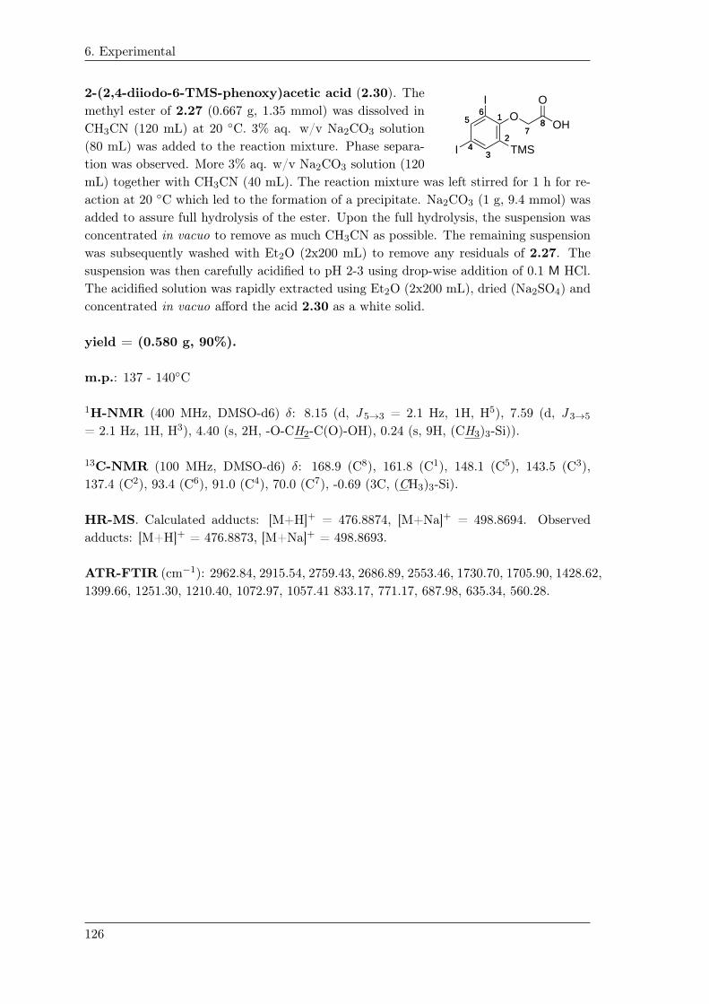
6. Experimental
2-(2,4-diiodo-6-TMS-phenoxy)acetic acid (2.30). The
11
22
3344
5566 O
I TMS
I
7788 OH
Omethyl ester of 2.27 (0.667 g, 1.35 mmol) was dissolved in
CH3CN (120 mL) at 20 ◦C. 3% aq. w/v Na2CO3 solution
(80 mL) was added to the reaction mixture. Phase separa-
tion was observed. More 3% aq. w/v Na2CO3 solution (120
mL) together with CH3CN (40 mL). The reaction mixture was left stirred for 1 h for re-
action at 20 ◦C which led to the formation of a precipitate. Na2CO3 (1 g, 9.4 mmol) was
added to assure full hydrolysis of the ester. Upon the full hydrolysis, the suspension was
concentrated in vacuo to remove as much CH3CN as possible. The remaining suspension
was subsequently washed with Et2O (2x200 mL) to remove any residuals of 2.27. The
suspension was then carefully acidified to pH 2-3 using drop-wise addition of 0.1 M HCl.
The acidified solution was rapidly extracted using Et2O (2x200 mL), dried (Na2SO4) and
concentrated in vacuo afford the acid 2.30 as a white solid.
yield = (0.580 g, 90%).
m.p.: 137 - 140◦C
1H-NMR (400 MHz, DMSO-d6) δ: 8.15 (d, J 5→3 = 2.1 Hz, 1H, H5), 7.59 (d, J 3→5
= 2.1 Hz, 1H, H3), 4.40 (s, 2H, -O-CH2-C(O)-OH), 0.24 (s, 9H, (CH3)3-Si)).
13C-NMR (100 MHz, DMSO-d6) δ: 168.9 (C8), 161.8 (C1), 148.1 (C5), 143.5 (C3),
137.4 (C2), 93.4 (C6), 91.0 (C4), 70.0 (C7), -0.69 (3C, (CH3)3-Si).
HR-MS. Calculated adducts: [M+H]+ = 476.8874, [M+Na]+ = 498.8694. Observed
adducts: [M+H]+ = 476.8873, [M+Na]+ = 498.8693.
ATR-FTIR (cm−1): 2962.84, 2915.54, 2759.43, 2686.89, 2553.46, 1730.70, 1705.90, 1428.62,
1399.66, 1251.30, 1210.40, 1072.97, 1057.41 833.17, 771.17, 687.98, 635.34, 560.28.
126

6. Experimental
6,6’-di-O-(2-(2,4-diiodo-6-TMS-phenoxy)acetyl)-
O11
44
55O
OO
22
66
333'3'
5'5'1'1'
4'4'
O
6'6'
O
O
O
O
O 2'2'
O
77
O
O
O
O
O
O
O
O
7'7'O
O 9'9'
10'10'11'11'
12'12'
13'13'14'14'
I
991010
1111
12121313
1414
I
I
TMS
I
TMS
1’,2,3,3’,4,4’,-hexa-O-isobutyryl-sucrose (2.31).
The diol of 1.34 was dissolved in dry CH2Cl2(1.5 mL). The acid 2.30 (0.376 g, 0.79 mmol),
EDC·HCl (0.159 g, 0.79 mmol) and DMAP (0.136
g, 1.1 mmol) were mixed in dry CH2Cl2 (3.2
mL) and added immediately to the solution of
the diol (final concentration 68 mm). The re-
action mixture was left stirred at 20 ◦C for re-
action and followed by LC-MS analysis. Upon
reaction completion (approx. 2-3 h), the reac-
tion mixture was directly purified by dry column vacuum chromatography (using 1% linear
increment of EtOAc/heptane) affording 2.31 as a white solid.
yield = (0.454 g, 83%).
m.p.: 65 - 68◦C
[α]20D
= +24◦ (c 1.00, EtOH)
1H-NMR (400 MHz, CDCL3) δ: 8.09 (d, J 11→13 = J 11′→13′ = 2.1 Hz, 2H, H11+H11′)
7.617 (d, J 13→11 = J 13′→11′ = 2.1 Hz, 1H H13 or H13′) 7.614 (d, J 13→11 = J 13′→11′ =
2.1 Hz, 1H H13 or H13′) 5.62 (d, J 1→2 = 3.6 Hz, 1H, H1), 5.57 (d, J 3′→4′ = 7.6 Hz, 1H,
H3′) 5.48 (t, J 3→2 = J 3→4 = 9.9 Hz, 1H, H3) 5.39 (t, J 4′→3′ = J 4′→5′ = 7.6 Hz, 1H,
H4′) 5.14 (t, J 4→3 = J 4→5 = 9.9 Hz, 1H, H4) 4.96 (dd, J 2→3 = 10.3 Hz, J 2→1 = 3.6 Hz,
1H, H2) 4.65 (s, 2H, -O-CH2-C(O)-O-) 4.58 (s, 2H, -O-CH2-C(O)-O-) 4.54 (dd, J 6a′→6b′
= 12.0 Hz, J 6a′→5′ = 3.9 Hz, 1H, H6a′) 4.50-4.31 (m, 4H, H5+H6b′+H6) 4.25 (dt, J 5′→4′
= 7.7 Hz, J 5′→6a′ = J 5′→6b′ = 3.9 Hz, 1H, H5′) 4.04 (d, J 1a′→1b′ = 11.9 Hz, 1H, H1′)
3.98 (d, J 1b′→1a′ = 11.9 Hz, 1H, H1′) 2.72-2.37 (m, 6H, (CH3)2CH-) 1.31-1.03 (m, 36H,
(CH3)2CH-) 0.30 (9H, (CH3)3-Si) 0.28 (9H, (CH3)3-Si).
13C-NMR (100 MHz, CDCL3) δ: 176.2 ((CH3)2CH-C(O)-O), 176.0 ((CH3)2CH-C(O)-O),
176.0 ((CH3)2CH-C(O)-O), 175.8 ((CH3)2CH-C(O)-O), 175.8 ((CH3)2CH-C(O)-O), 175.2
((CH3)2CH-C(O)-O), 167.7 (2,4-diiodo-6-TMS-Ph-O-CH2-C(O)-O-), 167.5 (2,4-diiodo-6-
TMS-Ph-O-CH2-C(O)-O-), 161.9 (C9/C9′), 161.8 (C9/C9′), 148.9 (C11/C11′), 148.8 (C11/C11′),
144.4 (C13/C13′), 144.3 (C13/C13′), 138.5 (C14/C14′), 138.4 (C14/C14′), 103.2 (C1′), 92.1
(C10/C10′), 91.9 (C10/C10′), 90.5 (C12/C12′), 90.4 (C12/C12′), 89.7 (C1), 78.4 (C5′), 74.8
(C3′), 74.0 (C4′), 69.9 (2C, 2,4-diiodo-6-TMS-Ph-O-CH2-C(O)-O-)), 69.8 (C2), 69.4 (C3),
68.8 (C5), 67.6 (C4), 65.1 (C6′), 63.4 (C1′), 62.5 (C6), 34.1-33.8 (6C, (CH3)2CH-), 19.3-
18.8 (12C, (CH3)2CH-), -0.3 (3C, (CH3)3-Si), -0.3 (3C, (CH3)3-Si).
HR-MS. Calculated adducts: [M+H2O]+ = 1696.1171, [M+Na]+ = 1701.0958. Ob-
served adducts: [M+H2O]+ = 1696.1417, [M+Na]+ = 1701.0958.
ATR-FTIR (cm−1): 2972.69, 2876.53, 1741.03, 1469.02, 1407.33, 1388.03, 1247.70, 1184.43,
1142.48, 1070.97, 839.48, 750.97.
127


Concluding remarks 7This thesis has described the development of five diagnostic tools that are applicable within
different fields of diagnostics and theranostics. In the Nanoguide project, a nanoparticle
based fiducial marker for image-guided radiotherapy was developed based on AuNPs and
the gelation matrix of SAIB (SAIB/EtOH/PLA (75:20:5 vol%)). The fiducial marker was
injectable through 25 G hypodermic needles. The scientific challenge was the surface-
engineering of the AuNPs to ensure full dispersion (homogenous distribution) within the
hydrophobic environment of the SAIB gel. Polymeric coatings like PEG5000-SH and
PNIPAM3500-SH was tested together with a dithiolane-functionalized SAIB derivative
1.26 that was synthesized in four steps from sucrose. The choice of coating was decisive for
the homogeneity of the SAIB-formulation. Coating of AuNPs using 1.26 was unsuccess-
ful. Highest loading and best achievable homogeneity was obtained when PNIPAM3500-SH
coated AuNPs was applied (30 mg PNIPAM-coated AuNPs mL−1) in the formulation.
The SAIB formulation containing PNIPAM coated AuNPs was tested in vivo as a fidu-
cial marker in mice and a canine dog, displaying contrast in the range of 1200 HU which
decreased to 800 HU over 8 weeks indicating potentially aggregation of AuNPs within the
SAIB formulation. The observed inhomogeneity of the formulation led to the development
of the hexa-iodinated derivative of SAIB 1.33.
A tissue marker for surgical guidance of non-palpable tumors in breast cancer was also de-
veloped based on the fiducial marker of 1.33 and the synthesis of [125I]-2.7. An aryl-TMS
precursor of SAIB 2.2 was synthesized in 4 steps from sucrose that underwent radioio-
dination under carrier free conditions using Tl(OOCCF3)3 and NaI. The radiochemical
yield was 83% with a radiochemical purity of 95%. The formulation of 1.33 and [125I]-2.7
was tested in vivo as a potential tissue marker in mice and was dosimetric evaluated for
application in humans. A [125I]-biodistribution study indicated an limited uptake in vivo
in the thyroid gland of 3.5% ID·g−1 after seven days in mice. The dosimetric analysis
calculated for human patients indicated that vital organs and tissues would not be con-
siderably affected by the radiation dose from the gel deposit nor from uptake as indicated
in the biodistribution.
A more suitable aryl-TMS precursor 2.31 was synthesized that upon the radio-iodination
would be identical to 1.33 that was approved by the Danish Health Authority for use as
medical device in patients. The radioiodination and in vivo study of 2.31 are currently
on-going.
A small library of iodide-based contrast agents for remote loading in liposomes was syn-
thesized for blood pool imaging. The amino functionalized contrast agents of 3.10 and
3.15 were remote loaded into liposomes where only 3.15 was successfully loaded with an
encapsulation efficiency of 85%. An in vivo study indicated that the amount loaded of
129

7. Concluding remarks
3.15 did not correspond to sufficient contrast to be visible in blood pool imaging. It was
concluded that the gradient established across bilayer membrane was not sufficient to load
the amount needed to achieve a proper contrast.
During the stay in the group of David Thompson, the CD/pluronic-based PRs of 4.13
and 4.12 was synthesized. The presence of the DOTA moiety was questionable according
to NMR analysis. Further structural characterization is undertaken before the chelation
of 64Cu and in vivo study. Lastly, a TMS substituted tyrosine derivatives of 5.26 and
5.41 was developed for the site-specific iodination of peptides and proteins. These deriva-
tives were synthesized in yields of 69% and 85% respectively from the Negishi coupling
of the corresponding building blocks of 5.40 and 5.33 or 5.31 respectively. Compound
5.41 was transformed into the corresponding dipeptides of 5.50, 5.51 and 5.52 that were
used to evaluate the selectivity in the iodination reaction. Iodination was performed by
Tl(OOCCF3) and NaI which showed ipso-iodination of the TMS moiety in the dipeptides
of 5.50 and 5.51. The iodination of 5.52 also indicated the ipso-iodination of the TMS-
group, though further optimization is needed in order to perform the iodination in a more
clean fashion. The first steps towards the regioselective iodination of peptide and proteins
using a TMS-group as placeholder has been demonstrated from this study.
130

Bibliography
[1] R. Baskar, K. A. Lee, R. Yeo, and K.-W. Yeoh, “Cancer and radiation therapy:
current advances and future directions,” Int J Med Sci, vol. 9, no. 3, pp. 193–199,
2012.
[2] D. Verellen, M. De Ridder, N. Linthout, K. Tournel, G. Soete, and G. Storme,
“Innovations in image-guided radiotherapy,” Nature Reviews Cancer, vol. 7, no. 12,
pp. 949–960, 2007.
[3] D. J. Brenner, L. R. Hlatky, P. J. Hahnfeldt, Y. Huang, and R. K. Sachs, “The
linear-quadratic model and most other common radiobiological models result in
similar predictions of time-dose relationships,” Radiation Research, vol. 150, no. 1,
pp. 83–91, 1998. [Online]. Available: http://www.jstor.org/stable/3579648
[4] L. A. Dawson and M. B. Sharpe, “Image-guided radiotherapy: rationale, benefits,
and limitations,” The Lancet Oncology, vol. 7, no. 10, pp. 848 – 858, 2006. [Online].
Available: http://www.sciencedirect.com/science/article/pii/S1470204506709044
[5] D. A. Jaffray, “Image-guided radiotherapy: from current concept to future
perspectives,” Nature Reviews Clinical Oncology, vol. 9, no. 12, pp. 688–699, 2012.
[6] R. I. Jølck, T. Binderup, A. E. Hansen, J. B. Scherman, P. Munch af Rosenschold,
A. Kjær, and T. L. Andresen, “Injectable colloidal gold in a sucrose acetate
isobutyrate gelating matrix with potential use in radiation therapy,” Advanced
Healthcare Materials, vol. 3, no. 10, pp. 1680–1687, 2014. [Online]. Available:
http://dx.doi.org/10.1002/adhm.201300668
[7] M. Imura, K. Yamazaki, H. Shirato, R. Onimaru, M. Fujino, S. Shimizu,
T. Harada, S. Ogura, H. Dosaka-Akita, K. Miyasaka, and M. Nishimura, “Insertion
and fixation of fiducial markers for setup and tracking of lung tumors in
radiotherapy,” International Journal of Radiation Oncology*Biology*Physics,
vol. 63, no. 5, pp. 1442 – 1447, 2005. [Online]. Available:
http://www.sciencedirect.com/science/article/pii/S0360301605007133
[8] D. J. Moseley, E. A. White, K. L. Wiltshire, T. Rosewall, M. B. Sharpe, J. H.
Siewerdsen, J.-P. Bissonnette, M. Gospodarowicz, P. Warde, C. N. Catton, and
D. A. Jaffray, “Comparison of localization performance with implanted fiducial
markers and cone-beam computed tomography for on-line image-guided
radiotherapy of the prostate,” International Journal of Radiation
Oncology*Biology*Physics, vol. 67, no. 3, pp. 942 – 953, 2007. [Online]. Available:
http://www.sciencedirect.com/science/article/pii/S0360301606033633
[9] R. I. Jølck, J. S. Rydhög, A. N. Christensen, A. E. Hansen, L. M. Bruun,
H. Schaarup-Jensen, A. S. von Wenck, B. Børresen, A. T. Kristensen, M. H.
Clausen, A. Kjær, K. Conradsen, R. Larsen, P. M. af Rosenschöld, and T. L.
131

BIBLIOGRAPHY
Andresen, “Injectable colloidal gold for use in intrafractional 2d image-guided
radiation therapy,” Advanced Healthcare Materials, vol. 4, no. 6, pp. 856–863,
2015. [Online]. Available: http://dx.doi.org/10.1002/adhm.201400651
[10] S. S. Korreman, T. Juhler-Nøttrup, and A. L. Boyer, “Respiratory gated beam
delivery cannot facilitate margin reduction, unless combined with respiratory
correlated image guidance,” Radiotherapy and Oncology, vol. 86, no. 1, pp. 61 –
68, 2008. [Online]. Available:
http://www.sciencedirect.com/science/article/pii/S0167814007005488
[11] S. Yousefi, B. T. Collins, C. A. Reichner, E. D. Anderson, C. Jamis-Dow,
G. Gagnon, S. Malik, B. Marshall, T. Chang, and F. Banovac, “Complications of
thoracic computed tomography–guided fiducial placement for the purpose of
stereotactic body radiation therapy,” Clinical Lung Cancer, vol. 8, no. 4, pp. 252 –
256, 2007. [Online]. Available:
http://www.sciencedirect.com/science/article/pii/S1525730411705150
[12] N. Kothary, J. J. Heit, J. D. Louie, W. T. Kuo, B. W. L. Jr, A. Koong, D. T.
Chang, D. Hovsepian, D. Y. Sze, and L. V. Hofmann, “Safety and efficacy of
percutaneous fiducial marker implantation for image-guided radiation therapy,”
Journal of Vascular and Interventional Radiology, vol. 20, no. 2, pp. 235 – 239,
2009. [Online]. Available:
http://www.sciencedirect.com/science/article/pii/S105104430800883X
[13] N. Bhagat, N. Fidelman, J. C. Durack, J. Collins, R. L. Gordon, J. M. LaBerge,
and R. K. Kerlan, “Complications associated with the percutaneous insertion of
fiducial markers in the thorax,” CardioVascular and Interventional Radiology,
vol. 33, no. 6, pp. 1186–1191, 2010. [Online]. Available:
http://dx.doi.org/10.1007/s00270-010-9949-0
[14] J. Hong, N. Eclov, Y. Yu, A. Rao, S. Dieterich, Q.-T. Le, M. Diehn, D. Sze,
J. Billy Loo, N. Kothary, and P. Maxim, “Migration of implanted markers for
image-guided lung tumor stereotactic ablative radiotherapy,” Journal of Applied
Clinical Medical Physics, vol. 14, no. 2, 2013.
[15] X.-D. Zhang, M.-L. Guo, H.-Y. Wu, Y.-M. Sun, Y.-Q. Ding, X. Feng, and L.-A.
Zhang, “Irradiation stability and cytotoxicity of gold nanoparticles for
radiotherapy,” Int J Nanomedicine, vol. 4, pp. 165–73, 2009.
[16] W. Eck, A. I. Nicholson, H. Zentgraf, W. Semmler, and S. Bartling,
“Anti-cd4-targeted gold nanoparticles induce specific contrast enhancement of
peripheral lymph nodes in x-ray computed tomography of live mice,” Nano
Letters, vol. 10, no. 7, pp. 2318–2322, 2010, pMID: 20496900. [Online]. Available:
http://dx.doi.org/10.1021/nl101019s
[17] J. F. Hainfeld, D. N. Slatkin, T. M. Focella, and H. M. Smilowitz, “Gold
nanoparticles: a new x-ray contrast agent,” The British Journal of Radiology,
vol. 79, no. 939, pp. 248–253, 2006, pMID: 16498039. [Online]. Available:
http://dx.doi.org/10.1259/bjr/13169882
132

BIBLIOGRAPHY
[18] D. Xi, S. Dong, X. Meng, Q. Lu, L. Meng, and J. Ye, “Gold nanoparticles as
computerized tomography (ct) contrast agents,” RSC Adv., vol. 2, pp.
12 515–12 524, 2012. [Online]. Available: http://dx.doi.org/10.1039/C2RA21263C
[19] D. Kim, S. Park, J. H. Lee, Y. Y. Jeong, and S. Jon, “Antibiofouling
polymer-coated gold nanoparticles as a contrast agent for in vivo x-ray computed
tomography imaging,” Journal of the American Chemical Society, vol. 129, no. 24,
pp. 7661–7665, 2007, pMID: 17530850. [Online]. Available:
http://dx.doi.org/10.1021/ja071471p
[20] Y. Lu, Y. Yu, and X. Tang, “Sucrose acetate isobutyrate as an in situ forming
system for sustained risperidone release,” Journal of Pharmaceutical Sciences,
vol. 96, no. 12, pp. 3252–3262, 2007. [Online]. Available:
http://dx.doi.org/10.1002/jps.21091
[21] J. Siepmann and N. A. Peppas, “Higuchi equation: Derivation, applications, use
and misuse,” International Journal of Pharmaceutics, vol. 418, no. 1, pp. 6 – 12,
2011, mathematical modeling of drug delivery systems:Fifty years after Takeru
Higuchi’s models. [Online]. Available:
http://www.sciencedirect.com/science/article/pii/S0378517311002687
[22] F. Tasselli, Non-solvent Induced Phase Separation Process (NIPS) for Membrane
Preparation. Berlin, Heidelberg: Springer Berlin Heidelberg, 2015, pp. 1–3.
[23] J. Turkevich, P. C. Stevenson, and J. Hillier, “A study of the nucleation and
growth processes in the synthesis of colloidal gold,” Discuss. Faraday Soc., vol. 11,
pp. 55–75, 1951. [Online]. Available: http://dx.doi.org/10.1039/DF9511100055
[24] G. Frens, “Controlled nucleation for the regulation of the particle size in
monodisperse gold suspensions,” Nature, vol. 241, no. 105, pp. 20–22, 1973.
[25] S. Jarosz and M. Mach, “Regio- and stereoselective transformations of sucrose at
the terminal positions,” European Journal of Organic Chemistry, vol. 2002, no. 5,
pp. 769–780, 2002. [Online]. Available:
http://dx.doi.org/10.1002/1099-0690(200203)2002:5<769::AID-EJOC769>3.0.CO;2-F
[26] Y. Queneau, S. Jarosz, B. Lewandowski, and J. Fitremann, “Sucrose chemistry and
applications of sucrochemicals,” ser. Advances in Carbohydrate Chemistry and
Biochemistry, D. Horton, Ed. Academic Press, 2007, vol. 61, pp. 217 – 292.
[Online]. Available:
http://www.sciencedirect.com/science/article/pii/S0065231807610051
[27] H. Karl, C. K. Lee, and R. Khan, “Synthesis and reactions of
tert-butyldiphenylsilyl ethers of sucrose,” Carbohydrate Research, vol. 101, no. 1,
pp. 31 – 38, 1982. [Online]. Available:
http://www.sciencedirect.com/science/article/pii/S0008621500807922
[28] I. R. Vlahov, P. I. Vlahova, and R. J. Linhardt, “Regioselective synthesis of sucrose
monoesters as surfactants,” Journal of carbohydrate chemistry, vol. 16, no. 1, pp.
1–10, 1997.
133
BIBLIOGRAPHY
[29] ——, “Regioselective synthesis of sucrose monoesters as surfactants,” Journal of
Carbohydrate Chemistry, vol. 16, no. 1, pp. 1–10, 1997. [Online]. Available:
http://www.tandfonline.com/doi/abs/10.1080/07328309708006506
[30] A. Sofian, C. K. Lee, and A. Linden, “Regioselective sulfonylation of
6,1?,6?-tri-o-tritylsucrose through dibutylstannylation: synthesis of 4?-o-sulfonyl
derivatives of sucrose,” Carbohydrate Research, vol. 337, no. 24, pp. 2377 – 2381,
2002. [Online]. Available:
http://www.sciencedirect.com/science/article/pii/S0008621502003476
[31] B. C. Mei, E. Oh, K. Susumu, D. Farrell, T. J. Mountziaris, and H. Mattoussi,
“Effects of ligand coordination number and surface curvature on the stability of
gold nanoparticles in aqueous solutions,” Langmuir, vol. 25, no. 18, pp.
10 604–10 611, 2009, pMID: 19588955. [Online]. Available:
http://dx.doi.org/10.1021/la901423z
[32] A. A. Volkert, V. Subramaniam, M. R. Ivanov, A. M. Goodman, and A. J. Haes,
“Salt-mediated self-assembly of thioctic acid on gold nanoparticles,” ACS Nano,
vol. 5, no. 6, pp. 4570–4580, 2011, pMID: 21524135. [Online]. Available:
http://dx.doi.org/10.1021/nn200276a
[33] M. Lista, D. Z. Liu, and P. Mulvaney, “Phase transfer of noble metal nanoparticles
to organic solvents,” Langmuir, vol. 30, no. 8, pp. 1932–1938, 2014, pMID:
24479856. [Online]. Available: http://dx.doi.org/10.1021/la404569h
[34] S. J. Hurst, A. K. R. Lytton-Jean, and C. A. Mirkin, “Maximizing dna loading on
a range of gold nanoparticle sizes,” Analytical Chemistry, vol. 78, no. 24, pp.
8313–8318, 2006, pMID: 17165821. [Online]. Available:
http://dx.doi.org/10.1021/ac0613582
[35] H. D. Hill, J. E. Millstone, M. J. Banholzer, and C. A. Mirkin, “The role radius of
curvature plays in thiolated oligonucleotide loading on gold nanoparticles,” ACS
Nano, vol. 3, no. 2, pp. 418–424, 2009, pMID: 19236080. [Online]. Available:
http://dx.doi.org/10.1021/nn800726e
[36] K. B. Cederquist and C. D. Keating, “Curvature effects in dna:au nanoparticle
conjugates,” ACS Nano, vol. 3, no. 2, pp. 256–260, 2009, pMID: 19236058.
[Online]. Available: http://dx.doi.org/10.1021/nn9000726
[37] C. Xu, G. A. Tung, and S. Sun, “Size and concentration effect of gold nanoparticles
on x-ray attenuation as measured on computed tomography,” Chemistry of
Materials, vol. 20, no. 13, pp. 4167–4169, 2008, pMID: 19079760. [Online].
Available: http://dx.doi.org/10.1021/cm8008418
[38] Z. Wang, L. Wu, and W. Cai, “Size-tunable synthesis of monodisperse
water-soluble gold nanoparticles with high x-ray attenuation,” Chemistry – A
European Journal, vol. 16, no. 5, pp. 1459–1463, 2010. [Online]. Available:
http://dx.doi.org/10.1002/chem.200902463
[39] T. L. Andresen, R. I. Jølck, and M. Albrechtsen, “Gel formulations for guiding
radiotherapy,” Patent WO2 014 187 962 (A1), 11 27, 2014.
134
BIBLIOGRAPHY
[40] J. Scherman Rydhög, R. Irming Jølck, T. L. Andresen, and P. Munck af
Rosenschöld, “Quantification and comparison of visibility and image artifacts of a
new liquid fiducial marker in a lung phantom for image-guided radiation therapy,”
Medical Physics, vol. 42, no. 6, pp. 2818–2826, 2015. [Online]. Available:
http://scitation.aip.org/content/aapm/journal/medphys/42/6/10.1118/1.4919616
[41] J. S. Rydhög, S. R. Mortensen, K. R. Larsen, P. Clementsen, R. I. Jølck,
M. Josipovic, M. C. Aznar, L. Specht, T. L. Andresen, P. M. af Rosenschöld, and
G. F. Persson, “Liquid fiducial marker performance during radiotherapy of locally
advanced non small cell lung cancer,” Radiotherapy and Oncology, vol. 121, no. 1,
pp. 64 – 69, 2016. [Online]. Available:
http://www.sciencedirect.com/science/article/pii/S0167814016311690
[42] N. N. Y. Janssen, J. Nijkamp, T. Alderliesten, C. E. Loo, E. J. T. Rutgers, J.-J.
Sonke, and M. T. F. D. Vrancken Peeters, “Radioactive seed localization in breast
cancer treatment,” British Journal of Surgery, vol. 103, no. 1, pp. 70–80, 2016.
[43] M. van der Noordaa, K. Pengel, E. Groen, E. van Werkhoven, E. Rutgers, C. Loo,
W. Vogel, and M. V. Peeters, “The use of radioactive iodine-125 seed localization
in patients with non-palpable breast cancer: A comparison with the radioguided
occult lesion localization with 99m technetium,” European Journal of Surgical
Oncology (EJSO), vol. 41, no. 4, pp. 553 – 558, 2015.
[44] E. L. Postma, A. J. Witkamp, M. A. van den Bosch, H. M. Verkooijen, and R. van
Hillegersberg, “Localization of nonpalpable breast lesions,” Expert Review of
Anticancer Therapy, vol. 11, no. 8, pp. 1295–1302, 2011.
[45] L. Langhans, T. Klausen, T. Tvedskov, M.-L. Talman, P. Oturai, I. Vejborg,
N. Kroman, and B. Hesse, “Preparation and administration of i-125 labeled seeds
for localization of nonpalpable breast lesions,” Current Radiopharmaceuticals,
vol. 9, no. 2, pp. 143–149, 2016. [Online]. Available:
http://www.eurekaselect.com/node/133756/article
[46] J. Rarick, B. F. Kimler, and O. Tawfik, “Comparison of margin status and lesional
size between radioactive seed localized vs conventional wire localized breast
lumpectomy specimens,” Annals of Diagnostic Pathology, vol. 21, pp. 47–52, 2016.
[47] M. W. Barentsz, M. A. A. J. van den Bosch, W. B. Veldhuis, P. J. van Diest,
R. M. Pijnappel, A. J. Witkamp, and H. M. Verkooijen, “Radioactive seed
localization for non-palpable breast cancer,” British Journal of Surgery, vol. 100,
no. 5, pp. 582–588, 2013. [Online]. Available: http://dx.doi.org/10.1002/bjs.9068
[48] B. Pouw, I. van der Ploeg, S. Muller, R. V. Olmos, L. Janssen-Pinkse,
H. Oldenburg, and M. V. Peeters, “Simultaneous use of an 125i-seed to guide
tumour excision and 99mtc-nanocolloid for sentinel node biopsy in non-palpable
breast-conserving surgery,” European Journal of Surgical Oncology (EJSO),
vol. 41, no. 1, pp. 71 – 78, 2015. [Online]. Available:
http://www.sciencedirect.com/science/article/pii/S0748798314011834
135

BIBLIOGRAPHY
[49] G. D. Dodd, K. Fry, and W. Delany, “Pre-operative localization of occult
carcinoma of the breast,” Management of the Patient with Cancer, pp. 88–113,
1965. [Online]. Available: www.scopus.com
[50] M. Ahmed, I. T. Rubio, J. M. Klaase, and M. Douek, “Surgical treatment of
nonpalpable primary invasive and in situ breast cancer,” Nat Rev Clin Oncol,
vol. 12, no. 11, pp. 645–663, Nov 2015, review. [Online]. Available:
http://dx.doi.org/10.1038/nrclinonc.2015.161
[51] A. Luini, S. Zurrida, V. Galimberti, and G. Paganelli, “Radioguided surgery of
occult breast lesions,” European Journal of Cancer, vol. 34, no. 1, pp. 204 – 205,
1998, correspondence.
[52] A. Luini, S. Zurrida, G. Paganelli, V. Galimberti, V. Sacchini, S. Monti,
P. Veronesi, G. Viale, and U. Veronesi, “Comparison of radioguided excision with
wire localization of occult breast lesions,” British Journal of Surgery, vol. 86, no. 4,
pp. 522–525, 1999. [Online]. Available:
http://dx.doi.org/10.1046/j.1365-2168.1999.01078.x
[53] G. Mariani, L. Moresco, G. Viale, G. Villa, M. Bagnasco, G. Canavese,
J. Buscombe, H. W. Strauss, and G. Paganelli, “Radioguided sentinel lymph node
biopsy in breast cancer surgery,” Journal of Nuclear Medicine, vol. 42, no. 8, pp.
1198–1215, 2001. [Online]. Available:
http://jnm.snmjournals.org/content/42/8/1198.abstract
[54] C. Thind, S. Tan, S. Desmond, O. Harris, H. Ramesh, L. Chagla, A. Ray, and
R. Audisio, “Snoll. sentinel node and occult (impalpable) lesion localization in
breast cancer,” Clinical Radiology, vol. 66, no. 9, pp. 833 – 839, 2011. [Online].
Available: http://www.sciencedirect.com/science/article/pii/S0009926011001565
[55] G. Mariani, P. Erba, G. Villa, M. Gipponi, G. Manca, G. Boni, F. Buffoni,
F. Castagnola, G. Paganelli, and H. W. Strauss, “Lymphoscintigraphic and
intraoperative detection of the sentinel lymph node in breast cancer patients: The
nuclear medicine perspective,” Journal of Surgical Oncology, vol. 85, no. 3, pp.
112–122, 2004. [Online]. Available: http://dx.doi.org/10.1002/jso.20023
[56] J. W. Jakub, R. J. Gray, A. C. Degnim, J. C. Boughey, M. Gardner, and C. E.
Cox, “Current status of radioactive seed for localization of non palpable breast
lesions,” The American Journal of Surgery, vol. 199, no. 4, pp. 522 – 528, 2010.
[Online]. Available:
http://www.sciencedirect.com/science/article/pii/S0002961009005212
[57] R. J. Gray, C. Salud, K. Nguyen, E. Dauway, J. Friedland, C. Berman, E. Peltz,
G. Whitehead, and C. E. Cox, “Randomized prospective evaluation of a novel
technique for biopsy or lumpectomy of nonpalpable breast lesions: Radioactive
seed versus wire localization,” Annals of Surgical Oncology, vol. 8, no. 9, pp.
711–715, 2001. [Online]. Available: http://dx.doi.org/10.1007/s10434-001-0711-3
[58] J. W. Jakub, R. J. Gray, A. C. Degnim, J. C. Boughey, M. Gardner, and C. E.
Cox, “Current status of radioactive seed for localization of non palpable breast
136

BIBLIOGRAPHY
lesions,” The American Journal of Surgery, vol. 199, no. 4, pp. 522 – 528, 2010.
[Online]. Available:
http://www.sciencedirect.com/science/article/pii/S0002961009005212
[59] S. H. Goudreau, J. P. Joseph, and S. J. Seiler, “Preoperative radioactive seed
localization for nonpalpable breast lesions: Technique, pitfalls, and solutions,”
RadioGraphics, vol. 35, no. 5, pp. 1319–1334, 2015, pMID: 26274097. [Online].
Available: http://dx.doi.org/10.1148/rg.2015140293
[60] E. V. Bloomquist, N. Ajkay, S. Patil, A. E. Collett, T. G. Frazier, and A. V.
Barrio, “A randomized prospective comparison of patient-assessed satisfaction and
clinical outcomes with radioactive seed localization versus wire localization,” The
Breast Journal, vol. 22, no. 2, pp. 151–157, 2016. [Online]. Available:
http://dx.doi.org/10.1111/tbj.12564
[61] J. S. Sung, V. King, C. M. Thornton, J. D. Brooks, C. W. Fry, M. El-Tamer, L. T.
Dauer, E. Brogi, J. M. S. Germain, and E. A. Morris, “Safety and efficacy of
radioactive seed localization with i-125 prior to lumpectomy and/or excisional
biopsy,” European Journal of Radiology, vol. 82, no. 9, pp. 1453 – 1457, 2013.
[Online]. Available:
http://www.sciencedirect.com/science/article/pii/S0720048X13002039
[62] M. H. Rønnest, F. Nissen, P. J. Pedersen, T. O. Larsen, W. Mier, and M. H.
Clausen, “A mild method for regioselective labeling of aromatics with radioactive
iodine,” European Journal of Organic Chemistry, vol. 2013, no. 19, pp. 3970–3973,
2013. [Online]. Available: http://dx.doi.org/10.1002/ejoc.201300419
[63] T. H. Chan and I. Fleming, “Electrophilic substitution of organosilicon
compounds- applications to organic synthesis,” Synthesis, vol. 1979, no. 10, pp.
761–786, 1979, 761.
[64] J. Clayden, N. Greeves, S. Warren, and P. Wothers, Organic chemistry, ser. 1.
Edition. Oxford university press, 2004, (ISBN) 019850346 6.
[65] A. McKillop, J. D. Hunt, M. J. Zelesko, J. S. Fowler, E. C. Taylor, G. McGillivray,
and F. Kienzle, “Thallium in organic synthesis. xxii. electrophilic aromatic
thallation using thallium(iii) trifluoroacetate. simple synthesis of aromatic iodides,”
Journal of the American Chemical Society, vol. 93, no. 19, pp. 4841–4844, 1971.
[Online]. Available: http://dx.doi.org/10.1021/ja00748a029
[66] D. S. Wilbur, W. E. Stone, and K. W. Anderson, “Regiospecific incorporation of
bromine and iodine into phenols using (trimethylsilyl)phenol derivatives,” The
Journal of Organic Chemistry, vol. 48, no. 9, pp. 1542–1544, 1983. [Online].
Available: http://dx.doi.org/10.1021/jo00157a035
[67] J. Clayden, Organolithiums: selectivity for synthesis. Elsevier, 2002, vol. 23.
[68] M. A. Reed, M. T. Chang, and V. Snieckus, “Anionic o -> alpha - and beta-vinyl
carbamoyl translocation of 2-(o-carbamoyl) stilbenes,” Organic Letters, vol. 6,
no. 14, pp. 2297–2300, 2004, pMID: 15228263. [Online]. Available:
http://dx.doi.org/10.1021/ol049740t
137
BIBLIOGRAPHY
[69] M. P. Sibi and V. Snieckus, “The directed ortho lithiation of o-aryl carbamates. an
anionic equivalent of the fries rearrangement,” The Journal of Organic Chemistry,
vol. 48, no. 11, pp. 1935–1937, 1983. [Online]. Available:
http://dx.doi.org/10.1021/jo00159a040
[70] J. Morin, Y. Zhao, and V. Snieckus, “Reductive cleavage of aryl o-carbamates to
phenols by the schwartz reagent. expedient link to the directed ortho metalation
strategy,” Organic Letters, vol. 15, no. 16, pp. 4102–4105, 2013, pMID: 23919281.
[Online]. Available: http://dx.doi.org/10.1021/ol401547d
[71] K. W. Quasdorf, M. Riener, K. V. Petrova, and N. K. Garg, “Suzuki?miyaura
coupling of aryl carbamates, carbonates, and sulfamates,” Journal of the American
Chemical Society, vol. 131, no. 49, pp. 17 748–17 749, 2009, pMID: 19928764.
[Online]. Available: http://dx.doi.org/10.1021/ja906477r
[72] M. Kauch and D. Hoppe, “Synthesis of substituted phenols by directed
ortho-lithiation of in situ n-silyl-protected o-aryl n-monoalkylcarbamates,”
Canadian Journal of Chemistry, vol. 79, no. 11, pp. 1736–1746, 2001. [Online].
Available: http://www.nrcresearchpress.com/doi/abs/10.1139/v01-146
[73] ——, “Synthesis of halogenated phenols by directed ortho-lithiation and
ipso-iododesilylation reactions of o-aryl n-isopropylcarbamates,” Synthesis, vol.
2006, no. 10, pp. 1578–1589, 2006.
[74] R. Li-Yuan Bao, R. Zhao, and L. Shi, “Progress and developments in the turbo
grignard reagent i-prmgcl[middle dot]licl: a ten-year journey,” Chem. Commun.,
vol. 51, pp. 6884–6900, 2015. [Online]. Available:
http://dx.doi.org/10.1039/C4CC10194D
[75] E. Shirakawa, T. Kurahashi, H. Yoshida, and T. Hiyama,
“Diphenylphosphinophenolate: a ligand for the palladium-catalysed silylation of
aryl halides activating simultaneously both palladium and silicon,” Chem.
Commun., pp. 1895–1896, 2000. [Online]. Available:
http://dx.doi.org/10.1039/B006165O
[76] B. Wang, H.-X. Sun, and Z.-H. Sun, “Lioac-catalyzed chemoselective deprotection
of aryl silyl ethers under mild conditions,” The Journal of Organic Chemistry,
vol. 74, no. 4, pp. 1781–1784, 2009, pMID: 19146386. [Online]. Available:
http://dx.doi.org/10.1021/jo802472s
[77] C.-E. Yeom, H. W. Kim, S. Y. Lee, and B. M. Kim, “Dbu-mediated mild and
chemoselective deprotection of aryl silyl ethers and tandem biaryl ether
formation,” Synlett, vol. 2007, no. 01, pp. 0146–0150, 2007.
[78] T. Ikawa, S. Masuda, T. Nishiyama, A. Takagi, and S. Akai, “Synthesis of
fluorinated aromatic compounds by one-pot benzyne generation and nucleophilic
fluorination,” Australian Journal of Chemistry, vol. 67, no. 3, pp. 475–480, 2014.
[79] J. G. Adamson, M. A. Blaskovich, H. Groenevelt, and G. A. Lajoie, “Simple and
convenient synthesis of tert-butyl ethers of fmoc-serine, fmoc-threonine, and
138
BIBLIOGRAPHY
fmoc-tyrosine,” The Journal of Organic Chemistry, vol. 56, no. 10, pp. 3447–3449,
1991. [Online]. Available: http://dx.doi.org/10.1021/jo00010a050
[80] F. Hallouard, N. Anton, P. Choquet, A. Constantinesco, and T. Vandamme,
“Iodinated blood pool contrast media for preclinical x-ray imaging applications – a
review,” Biomaterials, vol. 31, no. 24, pp. 6249–6268, 2010.
[81] C.-Y. Kao, E. A. Hoffman, K. C. Beck, R. V. Bellamkonda, and A. V.
Annapragada, “Long-residence-time nano-scale liposomal iohexol for x-ray–based
blood pool imaging,” Academic Radiology, vol. 10, no. 5, pp. 475–483, 2003.
[82] M. Bettmann and T. Morris, “Recent advances in contrast agents,” Radiologic
clinics of North America, vol. 24, no. 3, p. 347—357, September 1986. [Online].
Available: http://europepmc.org/abstract/MED/3529226
[83] M. Bourin, P. Jolliet, and F. Ballereau, “An overview of the clinical
pharmacokinetics of x-ray contrast media,” Clinical Pharmacokinetics, vol. 32,
no. 3, pp. 180–193, 1997.
[84] W. He, K. Ai, and L. Lu, “Nanoparticulate x-ray ct contrast agents,” Science
China Chemistry, vol. 58, no. 5, pp. 753–760, 2015.
[85] J. Gubernator, “Active methods of drug loading into liposomes: recent strategies
for stable drug entrapment and increased in vivo activity,” Expert Opinion on
Drug Delivery, vol. 8, no. 5, pp. 565–580, 2011, pMID: 21492058.
[86] G. Storm and D. J. Crommelin, “Liposomes: quo vadis?” Pharmaceutical Science
and Technology Today, vol. 1, no. 1, pp. 19–31, 1998.
[87] M. R. Zalutsky, M. A. Noska, and S. E. Seltzer, “Characterization of liposomes
containing iodine-125-labeled radiographic contrast agents.” Investigative
Radiology, vol. 22, no. 2, pp. 141–147, 1987.
[88] W. Krause, J. Leike, A. Sachse, and G. Schuhmann-Giampieri, “Characterization of
iopromide liposomes.” Investigative Radiology, vol. 28, no. 11, pp. 1028–1032, 1993.
[89] S. E. Seltzer, “Contrast-carrying liposomes current status.” Investigative
Radiology, vol. 23, pp. S122–S125, 1988.
[90] S. E. Seltzer, M. A. Davis, D. F. Adams, P. M. Shulkin, W. J. Landis, and
A. Havron, “Liposomes carrying diatrizoate: Characterization of biophysical
properties and imaging applications.” Investigative Radiology, vol. 19, no. 2, pp.
142–151, 1984.
[91] P. J. Ryan, M. A. Davis, and D. L. Melchior, “The preparation and characterization
of liposomes containing x-ray contrast agents,” Biochimica et Biophysica Acta
(BBA) - General Subjects, vol. 756, no. 1, pp. 106–110, 1983. [Online]. Available:
http://www.sciencedirect.com/science/article/pii/0304416583900296
[92] J. U. Leike, A. Sachse, and K. Rupp, “Characterization of continuously extruded
iopromide-carrying liposomes for computed tomography blood pool imaging,”
Investigative Radiology, vol. 36, no. 6, pp. 303–308, 2001.
139

BIBLIOGRAPHY
[93] A. Sachse, J. U. Leike, T. Schneider, S. E. Wagner, G. L. Röling, W. Krause, and
M. Brandl, “Biodistribution and computed tomography blood-pool imaging
properties of polyethylene glycol-coated iopromide-carrying liposomes,”
Investigative Radiology, vol. 32, no. 1, pp. 44–50, 1997.
[94] S. E. Seltzer, M. Blau, L. W. Herman, R. L. Hooshmand, L. A. Herman, D. F.
Adams, S. R. Minchey, and A. S. Janoff, “Contrast material-carrying liposomes:
biodistribution, clearance, and imaging characteristics.” Radiology, vol. 194, no. 3,
pp. 775–781, 1995, pMID: 7862978.
[95] J. W. Nichols and D. W. Deamer, “Net proton-hydroxyl permeability of large
unilamellar liposomes measured by an acid-base titration technique,” Proceedings
of the National Academy of Sciences, vol. 77, no. 4, pp. 2038–2042, 1980.
[96] D. Zucker, D. Marcus, Y. Barenholz, and A. Goldblum, “Liposome drugs’ loading
efficiency: A working model based on loading conditions and drug’s
physicochemical properties,” Journal of Controlled Release, vol. 139, no. 1, pp.
73–80, 2009. [Online]. Available:
http://www.sciencedirect.com/science/article/pii/S0168365909003873
[97] I. V. Zhigaltsev, G. Winters, M. Srinivasulu, J. Crawford, M. Wong, L. Amankwa,
D. Waterhouse, D. Masin, M. Webb, N. Harasym, L. Heller, M. B. Bally, M. A.
Ciufolini, P. R. Cullis, and N. Maurer, “Development of a weak-base docetaxel
derivative that can be loaded into lipid nanoparticles,” Journal of Controlled
Release, vol. 144, no. 3, pp. 332–340, 2010.
[98] R. Parkesh, W. Gowin, T. C. Lee, and T. Gunnlaugsson, “Synthesis and evaluation
of potential ct (computer tomography) contrast agents for bone structure and
microdamage analysis,” Org. Biomol. Chem., vol. 4, pp. 3611–3617, 2006.
[99] D. B. Elrod, R. Partha, D. Danila, S. W. Casscells, and J. L. Conyers, “An
iodinated liposomal computed tomographic contrast agent prepared from a
diiodophosphatidylcholine lipid,” Nanomedicine: Nanotechnology, Biology and
Medicine, vol. 5, no. 1, pp. 42–45, 2009.
[100] R. Takeda, S. Y. Ryu, J. H. Park, and K. Nakanishi, “Additivity in cd amplitudes
of p-phenylbenzyl ethers and p-phenylbenzoates of 2-aminosugars,” Tetrahedron,
vol. 46, no. 16, pp. 5533–5542, 1990. [Online]. Available:
http://www.sciencedirect.com/science/article/pii/S0040402001877516
[101] A. F. Abdel-Magid, K. G. Carson, B. D. Harris, C. A. Maryanoff, and R. D. Shah,
“Reductive amination of aldehydes and ketones with sodium
triacetoxyborohydride. studies on direct and indirect reductive amination
procedures,” The Journal of Organic Chemistry, vol. 61, no. 11, pp. 3849–3862,
1996, pMID: 11667239. [Online]. Available: http://dx.doi.org/10.1021/jo960057x
[102] J. Henriksen and A. T.L., “"personal communications".”
[103] V. Wintgens, T. T. Nielsen, K. L. Larsen, and C. Amiel, “Size-controlled
nanoassemblies based on cyclodextrin-modified dextrans,” Macromolecular
Bioscience, vol. 11, no. 9, pp. 1254–1263, 2011.
140

BIBLIOGRAPHY
[104] J. Szejtli, “Past, present and futute of cyclodextrin research,” Pure and Applied
Chemistry, vol. 76, no. 10, pp. 1825–1845, 2004.
[105] S. Loethen, J. Kim, and D. H. Thompson, “Biomedical applications of cyclodextrin
based polyrotaxanes,” Polymer Reviews, vol. 47, no. 3, pp. 383–418, 2007.
[106] Y. A. Mondjinou, L. A. McCauliff, A. Kulkarni, L. Paul, S.-H. Hyun, Z. Zhang,
Z. Wu, M. Wirth, J. Storch, and D. H. Thompson, “Synthesis of
2-hydroxypropyl-?-cyclodextrin/pluronic-based polyrotaxanes via heterogeneous
reaction as potential niemann-pick type c therapeutics,” Biomacromolecules,
vol. 14, no. 12, pp. 4189–4197, 2013.
[107] C. J. Collins, L. A. McCauliff, S.-H. Hyun, Z. Zhang, L. N. Paul, A. Kulkarni,
K. Zick, M. Wirth, J. Storch, and D. H. Thompson, “Synthesis, characterization,
and evaluation of pluronic-based ?-cyclodextrin polyrotaxanes for mobilization of
accumulated cholesterol from niemann-pick type c fibroblasts,” Biochemistry,
vol. 52, no. 19, pp. 3242–3253, 2013, pMID: 23560535.
[108] C. J. Collins, Y. Mondjinou, B. Loren, S. Torregrosa-Allen, C. J. Simmons, B. D.
Elzey, N. Ayat, Z.-R. Lu, and D. Thompson, “Influence of molecular structure on
the in vivo performance of flexible rod polyrotaxanes,” Biomacromolecules, vol. 17,
no. 9, pp. 2777–2786, 2016, pMID: 27387820.
[109] T. T. Nielsen, V. Wintgens, C. Amiel, R. Wimmer, and K. L. Larsen, “Facile
synthesis of ?-cyclodextrin-dextran polymers by “click” chemistry,”
Biomacromolecules, vol. 11, no. 7, pp. 1710–1715, 2010, pMID: 20553039.
[110] S. Okarvi, “Peptide-based radiopharmaceuticals: Future tools for diagnostic
imaging of cancers and other diseases,” Medicinal Research Reviews, vol. 24, no. 3,
pp. 357–397, 2004.
[111] A. J. Fischman, J. W. Babich, and H. W. Strauss, “A ticket to ride: Peptide
radiopharmaceuticals,” Journal of Nuclear Medicine, vol. 34, no. 12, pp.
2253–2263, 1993.
[112] D. Blok, R. I. J. Feitsma, P. Vermeij, and E. J. K. Pauwels, “Peptide
radiopharmaceuticals in nuclear medicine,” European Journal of Nuclear Medicine,
vol. 26, no. 11, pp. 1511–1519, 1999.
[113] M. Schottelius and H.-J. Wester, “Molecular imaging targeting peptide receptors,”
Methods, vol. 48, no. 2, pp. 161 – 177, 2009, molecular Imaging.
[114] S. J. Goldsmith, “Radioimmunoassay: Review of basic principles,” Seminars in
Nuclear Medicine, vol. 5, no. 2, pp. 125–152, 2017/02/05 1975. [Online]. Available:
http://dx.doi.org/10.1016/S0001-2998(75)80028-6
[115] S. D. Sharma, G. Toth, and V. J. Hruby, “A simple general method for
(radio)iodination of a phenylalanine residue in peptides: preparation of
[cyclic][d-pen2,4’-125i-phe4,d-pen5]enkephalin, a peptide with extraordinary
selectivity for .delta.-opioid receptors,” The Journal of Organic Chemistry, vol. 56,
no. 16, pp. 4981–4983, 1991.
141
BIBLIOGRAPHY
[116] A. Dirksen and P. E. Dawson, “Rapid oxime and hydrazone ligations with aromatic
aldehydes for biomolecular labeling,” Bioconjugate Chemistry, vol. 19, no. 12, pp.
2543–2548, 2008, pMID: 19053314.
[117] M. Glaser, M. Morrison, M. Solbakken, J. Arukwe, H. Karlsen, U. Wiggen,
S. Champion, G. M. Kindberg, and A. Cuthbertson, “Radiosynthesis and
biodistribution of cyclic rgd peptides conjugated with novel [18f]fluorinated
aldehyde-containing prosthetic groups,” Bioconjugate Chemistry, vol. 19, no. 4, pp.
951–957, 2008, pMID: 18341272.
[118] J. Darius and D. R. RICHARDSON, “William hunter and radioiodination:
revolutions in the labelling of proteins with radionuclides of iodine.”
[119] N. M. Alexander, “Oxidative cleavage of tryptophanyl peptide bonds during
chemical- and peroxidase-catalyzed iodinations,” Journal of Biological Chemistry,
vol. 249, no. 6, pp. 1946–1952, 1974.
[120] A. S. McFARLANE, “Efficient trace-labelling of proteins with iodine,” Nature, vol.
182, no. 4627, pp. 53–53, Jul 1958.
[121] R. H. Seevers and R. E. Counsell, “Radioiodination techniques for small organic
molecules,” Chemical Reviews, vol. 82, no. 6, pp. 575–590, 1982.
[122] R. S. YALOW and S. A. BERSON, “Immunoassay of endogenous plasma insulin in
man,” Obesity Research, vol. 4, no. 6, pp. 583–600, 1996.
[123] W. M. HUNTER and F. C. GREENWOOD, “Preparation of iodine-131 labelled
human growth hormone of high specific activity,” Nature, vol. 194, no. 4827, pp.
495–496, May 1962.
[124] F. C. Greenwood, W. M. Hunter, and J. S. Glover, “The preparation of
(131)i-labelled human growth hormone of high specific radioactivity,” Biochem J,
vol. 89, no. 1, pp. 114–123, Oct 1963, 14097352[pmid].
[125] P. R. Salacinski, C. McLean, J. E. Sykes, V. V. Clement-Jones, and P. J. Lowry,
“Iodination of proteins, glycoproteins, and peptides using a solid-phase oxidizing
agent, 1,3,4,6-tetrachloro-3?,6?-diphenyl glycoluril (iodogen),” Analytical
Biochemistry, vol. 117, no. 1, pp. 136 – 146, 1981.
[126] P. J. Fraker and J. C. Speck, “Protein and cell membrane iodinations with a
sparingly soluble chloroamide, 1,3,4,6-tetrachloro-3a,6a-diphenylglycoluril,”
Biochemical and Biophysical Research Communications, vol. 80, no. 4, pp. 849 –
857, 1978.
[127] W. Wood, C. Wachter, and P. Scriba, “Experiences using chloramine-t and 1, 3, 4,
6-tetrachloro-3α, 6α-diphenylglycoluril (lodogen®) for radioiodination of
materials for radioimmunoassay,” Clinical Chemistry and Laboratory Medicine,
vol. 19, no. 10, pp. 1051–1056, 1981.
[128] A. E. Bolton and W. M. Hunter, “The labelling of proteins to high specific
radioactivities by conjugation to a 125i-containing acylating agent. application to
the radioimmunoassay,” Biochemical Journal, vol. 133, no. 3, pp. 529–538, 1973.
142

BIBLIOGRAPHY
[129] G. Vaidyanathan and M. R. Zalutsky, “Radioiodination of antibodies via
n-succinimidyl 2,4-dimethoxy-3-(trialkylstannyl)benzoates,” Bioconjugate
Chemistry, vol. 1, no. 6, pp. 387–393, 1990.
[130] ——, “Protein radiohalogenation: observations on the design of n-succinimidyl
ester acylation agents,” Bioconjugate Chemistry, vol. 1, no. 4, pp. 269–273, 1990.
[131] S. Garg, P. K. Garg, and M. R. Zalutsky, “N-succinimidyl
5-(trialkylstannyl)-3-pyridinecarboxylates: a new class of reagents for protein
radioiodination,” Bioconjugate Chemistry, vol. 2, no. 1, pp. 50–56, 1991.
[132] G. Vaidyanathan, D. J. Affleck, and M. R. Zalutsky, “Radioiodination of proteins
using n-succinimidyl 4-hydroxy-3-iodobenzoate,” Bioconjugate Chemistry, vol. 4,
no. 1, pp. 78–84, 1993, pMID: 8431515.
[133] G. Vaidyanathan and M. R. Zalutsky, “Preparation of n-succinimidyl
3-[[ast]i]iodobenzoate: an agent for the indirect radioiodination of proteins,” Nat.
Protocols, vol. 1, no. 2, pp. 707–713, Jul 2006.
[134] M. Pruszynski, E. Koumarianou, G. Vaidyanathan, S. Chitneni, and M. R.
Zalutsky, “D-amino acid peptide residualizing agents bearing
n-hydroxysuccinimido- and maleimido-functional groups and their application for
trastuzumab radioiodination,” Nuclear Medicine and Biology, vol. 42, no. 1, pp. 19
– 27, 2015.
[135] S. W. Hadley and D. S. Wilbur, “Evaluation of iodovinyl antibody conjugates:
comparison with a p-iodobenzoyl conjugate and direct radioiodination,”
Bioconjugate Chemistry, vol. 1, no. 2, pp. 154–161, 1990. [Online]. Available:
http://dx.doi.org/10.1021/bc00002a011
[136] P. Dumas, B. Maziere, N. Autissier, and R. Michel, “Specificite de l’iodotyrosine
desiodase des microsomes thyroïdiens et hepatiques,” Biochimica et Biophysica
Acta (BBA) - Enzymology, vol. 293, no. 1, pp. 36 – 47, 1973. [Online]. Available:
http://www.sciencedirect.com/science/article/pii/0005274473903732
[137] E. Escher, “A new method of iodine labelling of peptide hormones,” Journal of
Receptor Research, vol. 4, no. 1-6, pp. 331–336, 1984. [Online]. Available:
http://dx.doi.org/10.3109/10799898409042559
[138] S. R. Wilson and L. A. Jacob, “Iodination of aryltrimethylsilanes. a mild approach
to (iodophenyl)alanine,” The Journal of Organic Chemistry, vol. 51, no. 25, pp.
4833–4836, 1986.
[139] D. S. Wilbur, D. K. Hamlin, R. R. Srivastava, and H. D. Burns, “Synthesis and
radioiodination of n-boc-p-(tri-n-butylstannyl)-l-phenylalanine tetrafluorophenyl
ester: Preparation of a radiolabeled phenylalanine derivative for peptide synthesis,”
Bioconjugate Chemistry, vol. 4, no. 6, pp. 574–580, 1993, pMID: 8305529.
[140] P. J. Pedersen and M. H. Clausen, “Modified approach to labeling of phenylalanine
in peptides with 125i,” Unpublished Results (Draft).
143

BIBLIOGRAPHY
[141] J. M. Padrón, G. Kokotos, T. Mart??n, T. Markidis, W. A. Gibbons, and V. S.
Mart??n, “Enantiospecific synthesis of ?-amino acid semialdehydes: a key step for
the synthesis of unnatural unsaturated and saturated ?-amino acids,” Tetrahedron:
Asymmetry, vol. 9, no. 19, pp. 3381 – 3394, 1998.
[142] J. M. Chalker, C. S. C. Wood, and B. G. Davis, “A convenient catalyst for aqueous
and protein suzuki?miyaura cross-coupling,” Journal of the American Chemical
Society, vol. 131, no. 45, pp. 16 346–16 347, 2009, pMID: 19852502.
[143] R. F. W. Jackson, N. Wishart, A. Wood, K. James, and M. J. Wythes,
“Preparation of enantiomerically pure protected 4-oxo .alpha.-amino acids and
3-aryl .alpha.-amino acids from serine,” The Journal of Organic Chemistry, vol. 57,
no. 12, pp. 3397–3404, 1992.
[144] C. L. Oswald, T. Carrillo-Márquez, L. Caggiano, and R. F. Jackson, “Negishi
cross-coupling reactions of ?-amino acid-derived organozinc reagents and aromatic
bromides,” Tetrahedron, vol. 64, no. 4, pp. 681 – 687, 2008.
[145] A. J. Ross, H. L. Lang, and R. F. W. Jackson, “Much improved conditions for the
negishi cross-coupling of iodoalanine derived zinc reagents with aryl halides,” The
Journal of Organic Chemistry, vol. 75, no. 1, pp. 245–248, 2010, pMID: 19938812.
[146] R. Bollhagen, M. Schmiedberger, K. Barlos, and E. Grell, “A new reagent for the
cleavage of fully protected peptides synthesised on 2-chlorotrityl chloride resin,” J.
Chem. Soc., Chem. Commun., pp. 2559–2560, 1994. [Online]. Available:
http://dx.doi.org/10.1039/C39940002559
[147] Y. Han, S. L. Bontems, P. Hegyes, M. C. Munson, C. A. Minor, S. A. Kates,
F. Albericio, and G. Barany, “Preparation and applications of xanthenylamide
(xal) handles for solid-phase synthesis of c-terminal peptide amides under
particularly mild conditions1-3,” The Journal of Organic Chemistry, vol. 61,
no. 18, pp. 6326–6339, 1996, pMID: 11667474. [Online]. Available:
http://dx.doi.org/10.1021/jo960312d
[148] M. Mergler, J. Gosteli, P. Grogg, R. Nyfeler, and R. Tanner, “Sasrin a versatile tool
in peptide synthesis and solid-phase organic chemistry,” CHIMIA International
Journal for Chemistry, vol. 53, no. 1-2, pp. 29–34, 1999. [Online]. Available:
http://www.ingentaconnect.com/content/scs/chimia/1999/00000053/F0020001/art00007
[149] E. Atherton, C. J. Logan, and R. C. Sheppard, “Peptide synthesis. part 2.
procedures for solid-phase synthesis using n[small
alpha]-fluorenylmethoxycarbonylamino-acids on polyamide supports. synthesis of
substance p and of acyl carrier protein 65-74 decapeptide,” J. Chem. Soc., Perkin
Trans. 1, pp. 538–546, 1981. [Online]. Available:
http://dx.doi.org/10.1039/P19810000538
[150] J. Hansen, F. Diness, and M. Meldal, “C-terminally modified peptides via cleavage
of the hmba linker by o-, n- or s-nucleophiles,” Org. Biomol. Chem., vol. 14, pp.
3238–3245, 2016. [Online]. Available: http://dx.doi.org/10.1039/C6OB00213G
144

BIBLIOGRAPHY
[151] A. Klapars and S. L. Buchwald, “Copper-catalyzed halogen exchange in aryl
halides:? an aromatic finkelstein reaction,” Journal of the American Chemical
Society, vol. 124, no. 50, pp. 14 844–14 845, 2002, pMID: 12475315.
[152] H. M. M. Bastiaans, J. L. van der Baan, and H. C. J. Ottenheijm, “Flexible and
convergent total synthesis of cyclotheonamide b,” The Journal of Organic
Chemistry, vol. 62, no. 12, pp. 3880–3889, 1997.
[153] D. S. Bose and V. Lakshminarayana, “Lewis acid-mediated selective removal of
n-tert-butoxycarbonyl protective group (t-boc),” Synthesis, vol. 1999, no. 01, pp.
66–68, 1999, 66.
[154] J. G. Adamson, M. A. Blaskovich, H. Groenevelt, and G. A. Lajoie, “Simple and
convenient synthesis of tert-butyl ethers of fmoc-serine, fmoc-threonine, and
fmoc-tyrosine,” The Journal of Organic Chemistry, vol. 56, no. 10, pp. 3447–3449,
1991.
[155] T. Maegawa, Y. Fujiwara, T. Ikawa, H. Hisashi, Y. Monguchi, and H. Sajiki,
“Novel deprotection method of fmoc group under neutral hydrogenation
conditions,” Amino Acids, vol. 36, no. 3, pp. 493–499, 2009. [Online]. Available:
http://dx.doi.org/10.1007/s00726-008-0109-7
[156] P. Marfey, “Determination ofd-amino acids. ii. use of a bifunctional reagent,
1,5-difluoro-2,4-dinitrobenzene,” Carlsberg Research Communications, vol. 49,
no. 6, p. 591, 1984.
[157] R. Bhushan and H. Brückner, “Marfey’s reagent for chiral amino acid analysis: A
review,” Amino Acids, vol. 27, no. 3, pp. 231–247, 2004.
[158] J. Coste and P. Jouin, (1H-Benzotriazol-1-yloxy)tripyrrolidino-phosphonium
Hexafluorophosphate. John Wiley & Sons, Ltd, 2001.
[159] J. Coste, D. Le-Nguyen, and B. Castro, “Pybop: A new peptide coupling reagent
devoid of toxic by-product,” Tetrahedron Letters, vol. 31, no. 2, pp. 205 – 208,
1990.
[160] M. B. Andrus, N-Ethylmorpholine. John Wiley & Sons, Ltd, 2001.
[161] S. Lin, Z.-Q. Yang, B. H. B. Kwok, M. Koldobskiy, C. M. Crews, and S. J.
Danishefsky, “Total synthesis of tmc-95a and -b via a new reaction leading to
z-enamides. some preliminary findings as to sar,” Journal of the American
Chemical Society, vol. 126, no. 20, pp. 6347–6355, 2004, pMID: 15149232.
[162] H. Sajiki, K. Hattori, and K. Hirota, “The formation of a novel
pd/c?ethylenediamine complex catalyst:? chemoselective hydrogenation without
deprotection of the o-benzyl and n-cbz groups,” The Journal of Organic
Chemistry, vol. 63, no. 22, pp. 7990–7992, 1998.
[163] H. Shao, J. W. Lockman, and J. R. Parquette, “Coupled conformational equilibria
in ?-sheet peptide?dendron conjugates,” Journal of the American Chemical
Society, vol. 129, no. 7, pp. 1884–1885, 2007, pMID: 17256863.
145

BIBLIOGRAPHY
[164] M. A. Campo and R. C. Larock, “Synthesis of fluoren-9-ones by the
palladium-catalyzed cyclocarbonylation of o-halobiaryls,” The Journal of Organic
Chemistry, vol. 67, no. 16, pp. 5616–5620, 2002, pMID: 12153259.
[165] A. Fînaru, A. Berthault, T. Besson, G. Guillaumet, and S. Berteina-Raboin,
“Microwave-assisted solid-phase synthesis of 5-carboxamido-n-acetyltryptamine
derivatives,” Organic Letters, vol. 4, no. 16, pp. 2613–2615, 2002, pMID: 12153191.
146
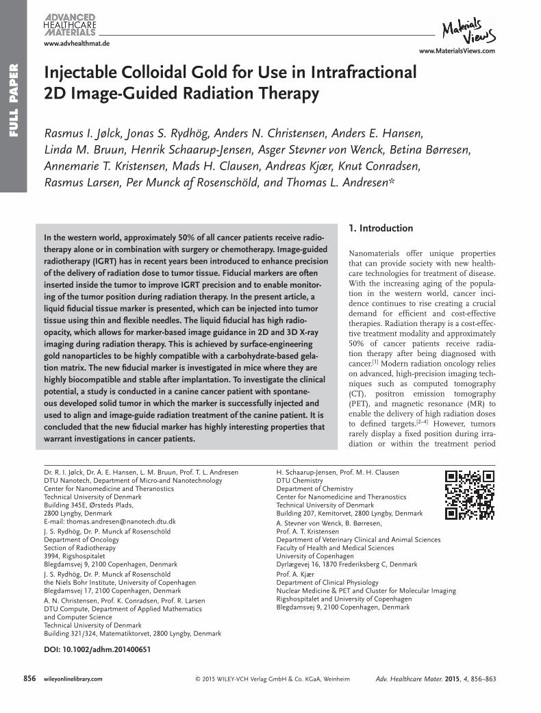
FULL
PAPER
© 2015 WILEY-VCH Verlag GmbH & Co. KGaA, Weinheim856 wileyonlinelibrary.com
Injectable Colloidal Gold for Use in Intrafractional 2D Image-Guided Radiation Therapy
Rasmus I. Jølck , Jonas S. Rydhög , Anders N. Christensen , Anders E. Hansen , Linda M. Bruun , Henrik Schaarup-Jensen , Asger Stevner von Wenck , Betina Børresen , Annemarie T. Kristensen , Mads H. Clausen , Andreas Kjær , Knut Conradsen , Rasmus Larsen , Per Munck af Rosenschöld , and Thomas L. Andresen*
DOI: 10.1002/adhm.201400651
In the western world, approximately 50% of all cancer patients receive radio-therapy alone or in combination with surgery or chemotherapy. Image-guided radiotherapy (IGRT) has in recent years been introduced to enhance precision of the delivery of radiation dose to tumor tissue. Fiducial markers are often inserted inside the tumor to improve IGRT precision and to enable monitor-ing of the tumor position during radiation therapy. In the present article, a liquid fi ducial tissue marker is presented, which can be injected into tumor tissue using thin and fl exible needles. The liquid fi ducial has high radio-opacity, which allows for marker-based image guidance in 2D and 3D X-ray imaging during radiation therapy. This is achieved by surface-engineering gold nanoparticles to be highly compatible with a carbohydrate-based gela-tion matrix. The new fi ducial marker is investigated in mice where they are highly biocompatible and stable after implantation. To investigate the clinical potential, a study is conducted in a canine cancer patient with spontane-ous developed solid tumor in which the marker is successfully injected and used to align and image-guide radiation treatment of the canine patient. It is concluded that the new fi ducial marker has highly interesting properties that warrant investigations in cancer patients.
1. Introduction
Nanomaterials offer unique properties that can provide society with new health-care technologies for treatment of disease. With the increasing aging of the popula-tion in the western world, cancer inci-dence continues to rise creating a crucial demand for effi cient and cost-effective therapies. Radiation therapy is a cost-effec-tive treatment modality and approximately 50% of cancer patients receive radia-tion therapy after being diagnosed with cancer. [ 1 ] Modern radiation oncology relies on advanced, high-precision imaging tech-niques such as computed tomography (CT), positron emission tomography (PET), and magnetic resonance (MR) to enable the delivery of high radiation doses to defi ned targets. [ 2–4 ] However, tumors rarely display a fi xed position during irra-diation or within the treatment period
Dr. R. I. Jølck, Dr. A. E. Hansen, L. M. Bruun, Prof. T. L. Andresen DTU Nanotech, Department of Micro-and Nanotechnology Center for Nanomedicine and Theranostics Technical University of Denmark Building 345E, Ørsteds Plads, 2800 Lyngby , Denmark E-mail: [email protected] J. S. Rydhög, Dr. P. Munck af Rosenschöld Department of Oncology Section of Radiotherapy 3994, Rigshospitalet Blegdamsvej 9, 2100 Copenhagen , Denmark J. S. Rydhög, Dr. P. Munck af Rosenschöld the Niels Bohr Institute, University of Copenhagen Blegdamsvej 17, 2100 Copenhagen , Denmark A. N. Christensen, Prof. K. Conradsen, Prof. R. Larsen DTU Compute, Department of Applied Mathematics and Computer Science Technical University of DenmarkBuilding 321/324, Matematiktorvet, 2800 Lyngby , Denmark
H. Schaarup-Jensen, Prof. M. H. Clausen DTU Chemistry Department of Chemistry Center for Nanomedicine and Theranostics Technical University of Denmark Building 207 , Kemitorvet, 2800 Lyngby , Denmark A. Stevner von Wenck, B. Børresen, Prof. A. T. Kristensen Department of Veterinary Clinical and Animal Sciences Faculty of Health and Medical Sciences University of Copenhagen Dyrlægevej 16, 1870 Frederiksberg C , Denmark Prof. A. Kjær Department of Clinical Physiology Nuclear Medicine & PET and Cluster for Molecular Imaging Rigshospitalet and University of Copenhagen Blegdamsvej 9, 2100 Copenhagen , Denmark
Adv. Healthcare Mater. 2015, 4, 856–863
www.advhealthmat.dewww.MaterialsViews.com

FULL P
APER
857wileyonlinelibrary.com© 2015 WILEY-VCH Verlag GmbH & Co. KGaA, Weinheim
due to breathing motion, changes in organ fi lling, and tumor size. [ 5–7 ] To compensate for variations in tumor position fre-quent imaging (kilovoltage radiographs or fl uoroscopy—also referred to as 2D X-ray imaging) during the course of treatment or image-guided radiation therapy (IGRT) is now being used in the clinic to improve radiation precision and accuracy, thereby reducing the treatment toxicity and optimizing therapeutic effi -cacy. [ 2,6,8 ] When the tumor position cannot be correlated with internal reference points such as the skeleton, tumor localiza-tion can be enhanced by alignment to radiographic markers implanted in or near the tumor. [ 9–11 ] Such markers can improve treatment through optimized planning, tumor positioning accuracy, and tumor tracking resulting in more precise dose delivery and enable treatment of cancers, which are presently not eligible for radiotherapy. [ 12,13 ] Tissue markers currently used in the clinic are metal-based solid implants with large physical dimensions, which require complicated insertion procedures and therefore the risk of complications. Lung tumor inser-tion is especially problematic, with pneumothorax (reported in 33%–68% of patients) as well as bleeding being reported as primary complications. [ 14–16 ] In order to increase the usability and patient compliance of radiographic markers, we recently described the concept of a liquid injectable fi ducial marker by exploiting polyethylene glycol (PEG)-coated gold nanoparticles (AuNPs) in a nonsolvent-induced phase separation (NIPS) gelation matrix. [ 17 ] This system was found to be biocompatible and clearly visible on 3D X-ray imaging, However, due to the chemical properties of the encapsulated AuNPs only 10 mg AuNPs mL −1 providing approximately 200 Hounsfi eld units (HU) of contrast could be included, which is insuffi cient for 2D X-ray visualization. For gating-based radiation procedures, often exploited in for example lung cancer patients, 2D X-ray visualization is required for intrafractional real-time imaging of moving tumors. [ 6,18–20 ] Additionally, fi ducial marker visualiza-tion using 2D X-ray techniques lowers the X-ray exposure level to the patients and may reduce treatment time compared to 3D X-ray-based procedures. [ 2 ] Furthermore, with the increasing focus on stereotactic radiation procedures and the clinical intro-duction of proton therapy, visualization of tumor position and motion becomes crucial to optimize irradiation of cancerous tissue. [ 21,22 ] Therefore, radiopaque fi ducial markers continue to be in high demand and development of new marker systems is of paramount importance.
In the present work, we have investigated a novel liquid fi ducial marker (nanogel) system based on sucrose acetate isobutyrate (SAIB) and uniform poly( N -isopropyl acrylamide) (PNIPAM)-coated AuNPs with radio-opacity adequate for 2D X-ray visualization. SAIB, a fully acylated derivative of sucrose, is a biodegradable nonpolymeric compound, which has been approved for oral consumption and is generally regarded as safe (GRAS) by the FDA. SAIB mixtures with ethanol (EtOH) behaves as Newtonian liquids and have a low viscosity (50–200 mPa s) suited for injection using thin hypodermic needles. [ 23–25 ] Upon hydration, an amorphous (gel-like) solid is formed, which effi -ciently encapsulate high concentrations (>100 mg Au mL −1 ) of PNIPAM-coated AuNPs providing contrast levels suffi cient for visualization in 2D X-ray imaging techniques such as fl uoroscopy and ExacTrac imaging (Brainlab AG, Feldkirchen, Germany). The developed nanogel has been characterized in
vitro and the in vivo stability and visibility was evaluated in immunocompetent mice over a period of 8 weeks. Proof-of-concept in a clinical canine cancer patient with a spontaneous growing tumor was additionally established and the developed nanogel with PNIPAM-coated AuNPs was applied in IGRT in a canine cancer patient where marker visibility, stability, and marker migration were evaluated over time during the course of radiotherapy. The developed nanogel fulfi lls the require-ments for use in guidance of radiation therapy as it is easy to inject, remains stable over the full time course of radiation treatment (6 weeks), shows no sign of migration, and is the fi rst liquid fi ducial marker to enable visualization using both 2D and 3D X-ray-based imaging techniques.
2. Results and Discussion
Visualization of fi ducial markers in 2D X-ray imaging requires high electron density within the nanogel to possess effi -cient X-ray attenuation/radio-opacity, which can be achieved by doping the injectable gel with colloid AuNPs. We recently exploited the use of PEG-coated AuNPs but due to the high hydrophilicity of the PEG-coated AuNPs only 10 mg AuNPs mL −1 could be included in the liquid marker, which was insuf-fi cient for 2D X-ray visibility. Higher AuNP-concentrations resulted in a signifi cant burst release due to the higher prefer-ence of the hydrophilic PEG-coated AuNPs for the surrounding aqueous phase. [ 17 ]
2.1. Engineering AuNPs for Enhanced Nanogel Compatibility
In order to increase the concentration of AuNPs and enhance the compatibility of the AuNPs with the hydrophobic SAIB-based gelation matrix, two different strategies were investi-gated: i) Masking the AuNPs by chemisorption of a dithiolane SAIB derivate ((SH) 2 -SAIB) ( 5 ) (see Section 2.1.1, and Sup-porting Information) and ii) using commercially available thiol-functionalized PNIPAM with a molecular weight of 3.500 Da (SH-PNIPAM 3.500 ). Both strategies resulted in hydrophobic AuNPs, which was anticipated to reduce the previously observed AuNP burst release from the nanogels. Hence, we hypothesized that realization of one or both of these strategies should enable formulation of nanogels with higher concentra-tions of AuNPs resulting in higher X-ray contrast levels.
2.1.1. Synthesis of SAIB-Based Capping Agent for AuNP Functionalization
The dithiolane-functionalized SAIB derivate 5 was prepared as outlined in Scheme S1 (Supporting Information) starting from sucrose ( 1 ). Mono silyl protection of the 6′-OH position of the fructofuranose unit was achieved using tert -butyl(chloro)diphe-nylsilane (TBDPS-Cl) and 4-dimethylaminopyridine (DMAP) yielding 6′-O-TBDPS-sucrose ( 2 ). [ 26 ] The remaining hydroxyl groups were isobutyrylated using isobutyric anhydride and DMAP resulting hepta-isobutyryl-6′-O-TBDPS-sucrose ( 3 ). Initial attempts of deprotection of the TBDPS group using
Adv. Healthcare Mater. 2015, 4, 856–863
www.advhealthmat.dewww.MaterialsViews.com
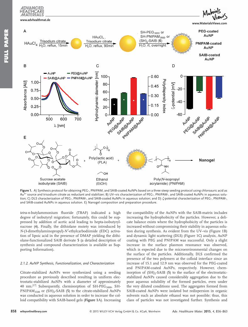
FULL
PAPER
858 wileyonlinelibrary.com © 2015 WILEY-VCH Verlag GmbH & Co. KGaA, Weinheim
tetra - n -butylammonium fl uoride (TBAF) indicated a high degree of isobutyryl migration; fortunately, this could be sup-pressed by addition of acetic acid leading to hepta-isobutyryl-sucrose ( 4 ). Finally, the dithiolane moiety was introduced by N -(3-dimethylaminopropyl)- N′ -ethylcarbodiimide (EDC) activa-tion of lipoic acid in the presence of DMAP yielding the dithi-olane-functionalized SAIB derivate 5 (a detailed description of synthesis and compound characterization is available as Sup-porting Information).
2.1.2. AuNP Synthesis, Functionalization, and Characterization
Citrate-stabilized AuNPs were synthesized using a seeding procedure as previously described resulting in uniform elec-trostatic-stabilized AuNPs with a diameter of approximately 40 nm. [ 17 ] Subsequently, chemisorption of SH-PEG 5000 , SH-PNIPAM 3500 or (SH) 2 -SAIB ( 5 ) to the citrate-stabilized AuNPs was conducted in aqueous solution in order to increase the col-loid compatibility with SAIB-based gels ( Figure 1 A). Increasing
the compatibility of the AuNPs with the SAIB-matrix includes increasing the hydrophobicity of the particles. However, a deli-cate balance exists where the hydrophobicity of the particles is increased without compromising their stability in aqueous solu-tion during synthesis. As evident from the UV–vis (Figure 1 B) and dynamic light scattering (DLS) (Figure 1 C) analysis, AuNP coating with PEG and PNIPAM was successful. Only a slight increase in the surface plasmon resonance was observed, which is expected due to the microenvironmental changes on the surface of the particles. Additionally, DLS confi rmed the presence of the two polymers at the colloid interface since an increase of 15.1 and 12.9 nm was observed for the PEG-coated and PNIPAM-coated AuNPs, respectively. However, chem-isorption of (SH) 2 -SAIB ( 5 ) to the surface of the electrostatic-stabilized AuNPs caused considerably aggregation due to the poor aqueous solubility of the formed particles, even under the very diluted conditions used. The aggregates formed from SAIB-coated AuNPs were isolated but redispersion in organic solvents such as absolute ethanol was not possible; thus, this class of particles was not investigated further. Synthesis and
Figure 1. A) Synthesis protocol for obtaining PEG-, PNIPAM, and SAIB-coated AuNPs based on a three steep seeding protocol using chloroauric acid as Au 3+ source and trisodium citrate as reductant and stabilizer; B) UV–vis characterization of PEG-, PNIPAM-, and SAIB-coated AuNPs in aqueous solu-tion; C) DLS characterization of PEG-, PNIPAM-, and SAIB-coated AuNPs in aqueous solution; and D) ζ-potential characterization of PEG-, PNIPAM-, and SAIB-coated AuNPs in aqueous solution. E) Nanogel composition and preparation procedure.
Adv. Healthcare Mater. 2015, 4, 856–863
www.advhealthmat.dewww.MaterialsViews.com

FULL P
APER
859wileyonlinelibrary.com© 2015 WILEY-VCH Verlag GmbH & Co. KGaA, Weinheim
subsequently coating of AuNPs with (SH) 2 -SAIB ( 5 ) in pure organic solvent using the Burst–Shiffi n method may be fea-sible, but this was not attempted due to the small size of particles formed by this method (<10 nm) and the fact that non-biocompatible solvents such as toluene and toxic phase-transfer agents have to be used, which could limit their use in biological settings. [ 27 ]
Additionally, handling of PNIPAM-coated AuNPs was found to be superior compared to PEG-coated AuNPs as these could be lyophilized and stored for prolonged time as an air stabile nanopowder, which readily dispersed into MQ-H 2 O and a wide range of protic- and aprotic organic solvents including anhy-drous ethanol without causing any changes to particle mor-phology (Figure S1, Supporting Information). This enabled easy up-concentration, storage, and nanogel formulation com-pared to the previous described method where AuNPs were transferred into anhydrous EtOH by centrifugation prior to use.
2.2. Contrast Evaluation for 2D X-Ray Visualization of Nanogel in Humans
In order to evaluate the required concentration of AuNPs within the nanogel for visibility on 2D X-rays images in humans, a simulation of 2D digitally reconstructed radiographs (DRRs) was performed based on clinical 3D CT-data for a patient diag-nosed with lung cancer. A lung tumor model was chosen as this region is considered as a worst-case scenario due to the high concentration of bone structure in the thoracic region. A simulated nanogel (500 µL) was digitally placed inside the lung tumor of the patient and the specifi ed HU contrast level was varied from 500 to 2000 HU with increments steps of 500 HU on the 3D CT-data. 2D DRRs were created for all angles entering perpendicular around the patient in steps of 5° using a ray-tracing algorithm [ 28,29 ] resulting in 72 simulated projec-tions for each contrast level ( Figure 2 ). The center position of the DRRs was chosen to be in the center of the simulated nanogel placed centrally within the lung tumor of the patient. On the resulting DRRs, the visibility for each contrast level was evaluated by a contrast-to-noise ratio (CNR) approach, where a region of interest (ROI) for one projection and contrast level was contoured to defi ne the nanogel and then transferred to all other projections. The mean intensity ( M m ) and standard devia-tion ( σ m ) of ROIs were calculated and compared to the mean
( M b ) and standard deviation ( σ b ) from a ROI on a DRR with no simulated nanogel present according to a CNR evaluation (Equation ( 1) ).
M M
σ σ=
−−
CNR m b
m2
b2
(1)
The CNR level can be considered a quantitative measurement of the visibility of the object measured compared to the back-ground contrast from the surroundings. Generally, high CNR translate to increased visibility of the marker. We considered the simulated nanogel visible in the 2D DRR if CNR > 2. This value corresponds to a signifi cance level of 0.05 obtained from a two-tailed t -test with a large number of degrees-of-freedom. [ 30 ] As evident from Figure 2 , some angles requires a higher X-ray contrast level of the nanogel in order to obtain a CNR > 2 as these penetrates bone structure prior to reaching the nanogel. Other angles are more easily accessible and thereby result in lower background contrast, thus making the nanogel more vis-ible on the 2D DRRs. Based on the lung cancer patient projec-tion simulation on the DRRs, it was evident that the contrast level required for CNR > 2 in 95% of all perpendicular angles was approximately 1000 HU (spherical nanogel with a total volume of 500 µL) corresponding to a nanogel with approxi-mately 30 mg AuNPs mL −1 . A comparison of the 2D X-ray contrast level from PNIPAM-coated AuNPs (10, 30, 60, and 90 mg mL −1 ) and clinically used solid fi ducial markers is provided in Figure S2, Supporting Information.
2.3. In Vitro Evaluation of Nanogel Performance
In vitro release kinetics of encapsulated PEG-coated and PNIPAM-coated AuNPs in SAIB nanogels doped with polylactic acid (PLA) with the composition SAIB/EtOH/PLA (75:20:5) + 30 mg AuNPs mL −1 were examined by placing small droplets (200 µL) of nanogels into MQ-H 2 O at physiological tempera-ture. Small aliquots of MQ-H 2 O were removed at specifi c time intervals and replaced with fresh MQ-H 2 O to mimic the sink effect in vivo. The amount of released AuNPs was determined by UV–vis spectroscopy using a standard curve of individual synthesized and coated AuNPs with a known concentration (Figure S3, Supporting Information). Injection of nanogel into aqueous solution causes an effl ux of EtOH from the nanogel into the aqueous compartment. Effl ux of EtOH results in rapid increase in gel viscosity, [ 24 ] but may also induce a burst release of encapsulated AuNPs. [ 24 ] Indeed, a rapid burst release of PEG-coated AuNPs from nanogels composed of SAIB/EtOH/PLA (75:20:5) + 30 mg PEG-coated AuNPs mL −1 was observed. In contrast, the use of PNIPAM-coated AuNPs resulted in highly stable nanogels with no (<1%) AuNP release over time in vitro despite the high AuNP concentration in the gel (Figure S3, Supporting Information). Concentrations as high as 100 mg PNIPAM-coated AuNPs mL −1 were evaluated without induced burst release of encapsulated PNIPAM-coated AuNPs (data not shown). The enhanced compatibility of the PNIPAM-coated AuNPs with the SAIB matrix compared to the PEG-coated counterpart can be explained by the enhanced hydropho-bicity and potential hydrogen bonding interactions between
Adv. Healthcare Mater. 2015, 4, 856–863
www.advhealthmat.dewww.MaterialsViews.com
Figure 2. Contrast to noise (CNR) level versus angle of entrance for a simulated nanogel (500 µL) with various contrast levels (500–2000 HU) placed digitally inside a lung tumor.

FULL
PAPER
860 wileyonlinelibrary.com © 2015 WILEY-VCH Verlag GmbH & Co. KGaA, Weinheim
the PNIPAM-coating polymers and the polyester backbone of SAIB. The positive in vitro stability results and known target AuNP concentration for 2D X-ray visibility encouraged us to further evaluate the nanogels composed of SAIB/EtOH/PLA (75:20:5) + 30 mg PNIPAM-coated AuNPs mL −1 in vivo using immunocompetent mice.
2.4. In Vivo Evaluation of Nanogel Performance
2.4.1. Evaluation of Nanogel in Immunocompetent Healthy NMRI-Mice
Nanogels (200 µL) composed of SAIB/EtOH/PLA (75:20:5) + 30 mg PNIPAM-coated AuNPs mL −1 were injected subcutane-ously in immunocompetent NMRI-mice ( n = 5) at the upper left fl ank using a 25G needle under general anesthesia. The nanogel was visualized using micro-CT imaging over a period of 8 weeks (images acquired directly after injection and again after 24, 48 h, 1, 2, 4, and 8 weeks) using a dedicated small-animal scanner ( Figure 3 ). Based on the recorded micro-CT images, the nanogel X-ray contrast level, degradation rate, and homogeneity were eval-uated as a function of time ( Figure 4 ). No signifi cant changes in the nanogel volume were observed after the initial EtOH effl ux phase, which caused approximately 20% reduction of nanogel volume in good correlation with the known EtOH content of the nanogel prior to injection. The nanogel doped with 30 mg PNIPAM-coated AuNPs mL −1 provided a mean X-ray contrast level in the range of 1200 HU, which is suffi cient for 2D X-ray visualization
as previously described. A reduction of the mean contrast level of the nanogel was observed over time possible due to some AuNP leakage or aggregation of particles within the nanogel. Figure 4 B clearly displays the increased maximum contrast level within the nanogel during the fi rst week following intra-gel AuNP aggregation resulting in very high contrast levels (>15.000 HU) (Figure S4, Supporting Information). The homogeneity of the nanogel in terms of PNIPAM-coated AuNP distribution within the gel matrix was analyzed further by investigating each voxel of the micro-CT images as illustrated in Figure 4 C. As evident from the cumulative histograms for a representative mouse, the PNIPAM-coated AuNPs were found to gradually aggregate within the nanogel as previously observed for PEG-coated AuNPs within the same gel matrix. [ 17 ] In fact, no signifi cant difference was found in the variance of the PEG-coated and PNIPAM-coated AuNPs within the nanogels as analyzed by the Wilcoxon rank-sum test ( p = 0.0734). [ 31 ] The inhomogeneous PNIPAM-coated AuNPs distribution within the nanogel could be visualized in the mice due to the high resolution and small slice thickness (0.092 × 0.092 × 0.092 mm 3 ) of the micro-CT scanner. The aggregation phenomenon of the PNIPAM-coated AuNPs within the nanogel has no clinical impact as human clinical scanners operate with signifi cant larger slice thickness (2–3 mm) and lower resolution (1 × 1 mm 2 ) causing the AuNP microstructure aggregation to be below the resolution of recorded images.
2.4.2. Evaluation of Nanogel in a Canine Cancer Patient with Spontaneously Developed Solid Tumor
In order to fully validate the concept of liquid injectable fi du-cial markers for IGRT, the nanogel was evaluated in a canine cancer patient suffering from spontaneously developed malig-nant solid tumor (mast cell tumor). Solid tumors often show an increased interstitial fl uid pressure (IFP) possibly due to vessel abnormalities, fi brosis, and contraction of the intersti-tial matrix mediated by stromal fi broblasts. [ 32,33 ] Increased IFP may serve as an obstacle for intratumoral injection of the devel-oped nanogel as the available intracellular space for marker assembly post-injection is limited and the increased IFP may repel the nanogel into adjacent areas. Additionally, migration of the nanogel over time may compromise the accuracy of fi ducial
Adv. Healthcare Mater. 2015, 4, 856–863
www.advhealthmat.dewww.MaterialsViews.com
Figure 3. Representative 3D reconstruction from micro-CT images of mouse. Nanogel composed of SAIB/EtOH/PLA (75:20:5) + 30 mg PNIPAM-coated AuNPs mL −1 administered subcutaneously to immunocompetent mice at the upper left fl ank. Nanogel visualized from different angles.
Figure 4. In vivo characteristics of nanogel composed of SAIB/EtOH/PLA (75:20:5) + 30 mg PNIPAM-coated AuNPs mL −1 administered subcutane-ously to immunocompetent mice at the upper left fl ank. A) Nanogel degradation over a period of 8 weeks (mean ± SEM, n = 5); B) Mean (left axis) and maximum (right axis) X-ray contrast level of nanogel over a period of 8 weeks (mean ± SEM, n = 5) and C) Accumulated histogram of nanogel contrast level within each pixel from a representative mouse over a period of 30 d.

FULL P
APER
861wileyonlinelibrary.com© 2015 WILEY-VCH Verlag GmbH & Co. KGaA, Weinheim
marker-based IGRT and potentially result in misguidance of the external radiation beam, which, in turn, leads to poor tumor coverage and increased radiation to healthy adjacent tissue. [ 16,34 ] Such obstacles are not encountered for subcutaneous injections of the nanogel as evaluated in immunocompetent NMRI-mice, hence the canine cancer patient with a spontaneously devel-oped solid tumor (mast cell tumor) served as a clinical animal model with high translational value for evaluating the clinical potential of the developed nanogel.
A companion dog (Male, Rhodesian Ridgeback, 10 years, body weight 38 kg) diagnosed with a subcutaneous mast cell tumor (maximum distance ( x × y × z ); 1.82 × 5.81 × 5.32 cm 3 , CT volume; 31.64 cm 3 ) adherent to the underlying soft tissue located over the dorsal aspect of the left shoulder blade was given an intratumoral injection of nanogel (300 µL) composed of SAIB/EtOH/PLA (75:20:5) + 30 mg PNIPAM-coated AuNPs mL −1 under general anesthesia ( Figure 5 ) prior to radiation therapy. Approximately 24 h after the injection of the nanogel, a diagnostic planning CT-scan of the tumor/nanogel region was performed and the therapeutic regimen consisting of four frac-tions of 6 Gy radiation therapy planned accordingly. Radiation therapy was delivered over 16 d on a clinical linear accelerator equipped with on-board imaging (OBI) capabilities (3D cone beam CT (CBCT) and 2D X-ray imaging).
Prior to each fraction of radiotherapy, CBCT ( Figure 6 ) images and 2D X-ray imaging ( Figure S5, Supporting Informa-tion) were acquired and used for positioning based on nanogel and tumor. Radiation therapy was delivered according to the nanogel position. The size ( Table 1 ) and stability of the nanogel in terms of migration and deformation were evaluated based on
the recorded CBCT images (Figure 6 ) by manually contouring the nanogel using 300 HU as a lower threshold. The nanogel size remained constant over the time of treatment with no change in overall volume was observed. The small variations observed in Table 1 are a consequence of motion artifacts from respiratory movement on the recorded CBCT images. Addition-ally, image analysis revealed that the nanogel retained its 3D shape and was found to be immobilized/associated with the tumor as no migration of the nanogel was observed during the course of radiation therapy despite the canine patient exer-cising and resting on the tumor inducing mechanical stress on the nanogel. Finally, exposure to four fractions of 6 Gy each was not found to alter the nanogel morphology as evaluated based on the CBCT images. The nanogel was well tolerated and no clinical side effects were observed throughout the study.
3. Conclusion
A novel nanogel composition based on PNIPAM-coated AuNPs formulated in a mixture of SAIB/EtOH/PLA (70:20:5) has been developed. PNIPAM-coated AuNPs were found to be highly compatible with the hydrophobic SAIB-based gel matrix facili-tating encapsulation of high concentration of colloidal gold, which enables 2D X-ray visualization of the nanogel. This was confi rmed by a simulation of 2D DRRs of a patient suffering from lung cancer. The developed nanogel is the fi rst inject-able fi ducial marker that can be visualized in 2D X-ray imaging thereby enabling intra- and interfractional IGRT by referencing the nanogel position. The nanogel performance was evaluated
Adv. Healthcare Mater. 2015, 4, 856–863
www.advhealthmat.dewww.MaterialsViews.com
Figure 5. 3D reconstruction based on CT images of canine patient with injected nanogel. Nanogel composed of SAIB/EtOH/PLA (75:20:5) + 30 mg PNIPAM-coated AuNPs mL −1 administered intratumorally into a canine suffering from an intermediate-grade subcutaneous mast cell tumor (maximum distance ( x × y × z ); 1.82 × 5.81 × 5.32 cm 3 , CT volume; 31.64 cm 3 ) adherent to the underlying soft tissue located over the dorsal aspect of the left shoulder blade. A) Full-body scan of the canine. Area of interest indicated with a red box. B–D) Nanogel visualized from different angles.
Figure 6. Nanogel composed of SAIB/EtOH/PLA (75:20:5) + 30 mg PNIPAM-coated AuNPs mL −1 administered intratumorally into a canine mast cell tumor visualized by CBCT imaging. A) Transversal plane (day 0); B) frontal plane (day 0); C) sagittal plane (day 0). Contours in A–C represent the nanogel on day 6 (green), day 9 (yellow), day 13 (blue), and day 16 (red). The light blue contour represents the gross tumor volume. D) 3D renderings of all the four surfaces represent the nanogel from each day of treatment.
FULL
PAPER
862 wileyonlinelibrary.com © 2015 WILEY-VCH Verlag GmbH & Co. KGaA, Weinheim
in vivo in immunocompetent mice by subcutaneous injection. Excellent visibility of the nanogel by micro-CT imaging was observed with a mean contrast level of 1200 HU after injec-tion. Over time, the PNIPAM-coated AuNPs were found to form microaggregates within the gel matrix as confi rmed by image analysis of the high-resolution micro-CT images. This phenomenon was found to have no clinical impact due to the larger slice thickness of human clinical scanners and lower res-olution compared to small animal micro-CT scanners.
The concept of an injectable fi ducial marker for use in IGRT was fi nally validated in a canine cancer patient with a large spon-taneous solid tumor. The canine patient provides translational understanding and allowed us to evaluate the clinical potential of the nanogel. The nanogel displayed high image contrast on both conventional CT and CBCT images and could addition-ally be identifi ed by 2D X-ray imaging, thus providing suffi cient radiographic properties for visualization with all the imaging modalities associated with modern linear accelerators. Further-more, the nanogel retained its size and 3D structure within the tumor microenvironment and no migration of the nanogel was observed within the time period for the radiotherapy treat-ment procedure. The developed nanogel was utilized to align the canine patient prior to external beam radiation therapy, was found to be unaffected by the deposited radiation dose within the tumor, and no side effects were observed in neither the murine nor the clinically relevant canine model. However, it should be noted that AuNPs are currently not approved for human use and the knowledge about the potential short- and long-term toxico-logical effects after AuNP exposure is still limited. [ 36 ]
The developed nanogel presented in this article is the fi rst liquid fi ducial marker, which can be inserted using thin and fl exible needles and with a high enough contrast to make it vis-ible in 2D X-ray imaging, which is an important feature for use in IGRT. Furthermore, active pharmaceutical ingredients may be included in the nanogel formulation to improve the thera-peutic benefi t for the patient, and the AuNPs may enhance the effect of the radiation therapy if the gel is injected directly into tumors. [ 37,38 ] In conclusion, the nanogel may provide both human and veterinary radiation oncologists with a new tool for improving planning, alignment, and tumor tracking in radio-therapy and could improve treatment outcome by providing a more precise delivery of the radiation dose to tumors.
4. Experimental Section Materials : Tetrachloroauric acid trihydrate was purchased from Wako
Chemicals GmbH (Neuss, Germany) and MeO-PEG 5000 -SH was purchased
from Rapp Polymere (Tuebingen, Germany). SH-PNIPAM ( M w = 3500Da, PDI = 1.24) was purchased from Polymer Source Inc. (Dorval, Canada). All other chemicals including SAIB, PLA ( M w = 10–18 kDa), trisodium citrate dihydrate, Au-standard for inductively coupled plasma atomic emission spectroscopy (ICP-AES) (1000 mg L −1 ), hydrochloride acid and nitric acid were purchased from Sigma–Aldrich (Schnelldorf, Germany). All chemicals were used as received from the manufacturer.
Synthesis of (SH) 2 -SAIB ( 5 ) : (SH) 2 -SAIB ( 5 ) was synthesized over four steps starting from sucrose ( 1 ) in an overall yield of 27%. Full synthetic procedure including chemical characterization ( 1 H NMR, 13 C NMR, HRMS, and FTIR) is available in the Supporting Information.
AuNP Synthesis and Characterization : AuNPs were synthesized as previously described using a two-step seeding procedure resulting in citrate-stabilized AuNPs with an average diameter of 40 nm. [ 17 ] The citrate-stabilized AuNPs were subsequently coated with either SH-PEG 5000 , SH-PNIPAM 3500, or (SH) 2 -SAIB ( 5 ) using eight molecules per nm 2 surface area. Chemisorption of coating materials was in all cases conducted overnight at RT after which the volume was reduced to approximately 15 mL by centrifugation (9.500 RPM, 15 min). The PEG-coated AuNPs, PNIPAM-coated AuNPs, and SAIB-coated AuNPs colloid solutions were washed with MQ-H 2 O (3 × 15 mL) and the fi nally up-concentrated to approximately 75 mg Au mL −1 as determined by ICP-MS using an ICP-MS Au standard (1000 mg L −1 ) as reference. Colloid solutions (citrate-stabilized AuNPs, PEG-, PNIPAM-, and SAIB-coated AuNPs) were furthermore characterized by UV–vis spectrometry, DLS, and ζ-potential measured. PNIPAM-coated AuNPs were fi nally characterized by TEM (Figure S2, Supporting Information).
Nanogel Formulation : SAIB (1.75 g, 90 w/w% in EtOH) and PLA (105 mg) were mixed and homogenized by a ball homogenizer (frequency 30 s −1 , 30 min) to give a clear homogenous solution. Lyophilized PNIPAM-coated AuNPs (63 mg) was weighed off and redispersed into anhydrous EtOH (310 µL) by extensive vortexing and added to the SAIB/PLA mixture to give a fi nal nanogel forming matrix composed of SAIB/EtOH/PLA (75:20:5) with 3.0 w/w% (30 mg mL −1 ) PNIPAM-coated AuNPs. Formed nanogels were stored at RT and used within 4 h from preparation with vortexing prior to use. Nanogels with PEG-coated AuNPs were prepared as described elsewhere. [ 17 ]
In Vitro Release of PNIPAM - and PEG -Coated AuNPs from Nanogel : Nanogels composed of SAIB/EtOH/PLA (75:20:5) with 3.0 w/w% (30.0 mg mL −1 ) of either PEG- or PNIPAM-coated AuNPs were prepared as described above. Nanogels (200 µL) were added to glass vials containing MQ-H 2 O (10.0 mL, 37 °C) and aliquots (1.0 mL) were removed as a function of time and replaced with an equal amount of MQ-H 2 O. The release profi le from both nanogels was measured by correlating the UV–vis absorbance of the individual AuNPs with a standard curve based on Au-standards of known concentration (5.0–50.0 µg mL −1 ).
Simulation of CNR in Lung Cancer Patient : 2D DRRs were performed based on clinical 3D CT-data for a patient diagnosed with lung cancer. 2D DRRs were created using the drrCode ray-tracing algorithm available online (https://code.google.com/p/drrsuite/downloads/list). 2D DRRs were created using a source to detector distance of 150 cm and a detector size of 768 × 1024 pixels (identical to a clinical settings). For conversion from HU to attenuation coeffi cient, a μρ for water was used assuming monochromatic photons of 120 kV. ROI contouring and CNR calculations for the simulated projections were performed in MATLAB (The MathWorks Inc., Natick, MA, USA) using an in-house-developed simulation and analysis tool.
Animal Models (Mice) : Immunocompetent 7-week-old female NMRI mice ( n = 5) were given a subcutaneous injection (200 µL) of SAIB/EtOH/PLA (75:20:5) + 30 mg mL −1 PNIPAM-coated AuNPs using a 25 G needle in the upper left fl ank of the anesthetized mice. The animals were given free access to food and water and micro-CT scans were recorded over a period of 8 weeks (24, 48 h, 1, 2, 4, and 8 weeks) using a dedicated small-animal MicroCAT II system (Siemens Medical Solutions, Malvern, PA, USA). Micro-CT images were recorded using the following settings; tube voltage of 67 kVp, tube current of 500 µA, 360 rotation steps, an exposure time of 400 ms, and a voxel size of 0.092 × 0.092 × 0.092 mm 3 . The animal experiments were approved by the Animal Research Committee of the Danish Ministry of Justice.
Adv. Healthcare Mater. 2015, 4, 856–863
www.advhealthmat.dewww.MaterialsViews.com
Table 1. Nanogel volume following intratumoral injection in a canine model.
Day # Nanogel volume [cm 3 ]
0 0.20
6 0.18
9 0.21
13 0.22
16 0.19

FULL P
APER
863wileyonlinelibrary.com© 2015 WILEY-VCH Verlag GmbH & Co. KGaA, Weinheim
Canine : A privately owned companion dog (Male, Rhodesian Ridgeback, 10 years, bodyweight 38 kg) suffering from a spontaneously developed subcutaneous mast cell tumor (maximum distance ( x × y × z ); 1.82 × 5.81 × 5.32 cm 3 , CT volume; 31.64 cm 3 ) adherent to the underlying soft tissue located over the dorsal aspect of the left shoulder blade was used as a clinical relevant model. In conjunction with the standard diagnostic procedures, nanogel (300 µL) was manually injected centrally in the tumor using a 23 G needle and 1 mL syringe. Approximately 24 h after the injection of the nanogel, a diagnostic CT scan (Biograph 40, Siemens Medical Solutions, Erlangen, Germany) of the tumor region was performed with the dog under general anesthesia using the following acquisition parameters; 120 kV, 150 mAs, pixel size 0.977 × 0.977 × 1.00 mm 3 ). As part of the therapeutic procedure radiation therapy was planned according to the tumor histology and imaging characteristic, and a therapeutic regimen consisting of four fractions of 6 Gy radiation treatments was chosen. Radiation therapy was delivered by a linear accelerator (Novalis TX, Varian Medical Systems, Palo Alto, CA, USA) with on-board imaging capabilities (Cone Beam CT and 2D X-ray imaging) and orthogonal ExacTrac radiography (Brainlab AG, Feldkirchen, Germany). No side effects were observed throughout the study. The canine cancer patient study was approved by the Ethics and Administrative Committee at the Department of Veterinary Clinical and Animal Sciences, Faculty of Health and Medical Sciences, University of Copenhagen, Denmark.
Analysis of Nanogel Homogeneity : The nanogels were delineated by hand using ITK-SNAP. [ 35 ] All data analyses were done in MATLAB (The MathWorks Inc., Natick, MA, USA) using in-house- developed software. Based on the voxel values in the gels mean and max values, as well as cumulative histograms were calculated. The cumulative histograms were used for homogeneity evaluation, as changes in the AuNP distribution are refl ected in the voxels values and thus the histograms.
Supporting Information Supporting Information is available from the Wiley Online Library or from the author.
Acknowledgements This work has been funded by the Technical University of Denmark (DTU), the Niels Bohr Institute, the Danish Council for Strategic Research (Nanoguide , application no. 0603-00442B), and the Novo Foundation.
Received: October 17, 2014 Revised: December 3, 2014
Published online: January 21, 2015
[1] R. Baskar , K. A. Lee , R. Yeo , K. Yeoh , Int. J. Med. Sci. 2012 , 9 , 193 . [2] L. A. Dawson , M. B. Sharpe , Lancet Oncol. 2006 , 7 , 848 . [3] J. Bussink , J. H. A. M. Kaanders , W. T. A. van der Graaf ,
W. J. G. Oyen , Nat. Rev. Clin. Oncol. 2011 , 8 , 233 . [4] D. Verellen , R. M. De , N. Linthout , K. Tournel , G. Soete , G. Storme ,
Nat. Rev. Cancer 2007 , 7 , 949 . [5] K. M. Langen , D. T. Jones , Int. J. Radiat. Oncol. Biol. Phys. 2001 , 50 , 265 . [6] H. Shirato , Y. Seppenwoolde , K. Kitamura , R. Onimura , S. Shimizu ,
Semin. Radiat. Oncol. 2004 , 14 , 10 . [7] P. Giraud , E. Yorke , S. Jiang , L. Simon , K. Rosenzweig , G. Mageras ,
Cancer Radiother. 2006 , 10 , 269 . [8] M. Falk , P. M. af Rosenschold , P. Keall , H. Cattell , B. C. Cho ,
P. Poulsen , S. Povzner , A. Sawant , J. Zimmerman , S. Korreman , Radiother. Oncol. 2010 , 94 , 218 .
[9] D. P. Harley , W. S. Krimsky , S. Sarkar , D. Highfi eld , C. Aygun , B. Gurses , Ann. Thorac. Surg. 2010 , 89 , 368 .
[10] M. Imura , K. Yamazaki , H. Shirato , R. Onimaru , M. Fujino , S. Shimizu , T. Harada , S. Ogura , H. Dosaka-Akita , K. Miyasaka , M. Nishimura , Int. J. Radiat. Oncol. Biol. Phys. 2005 , 63 , 1442 .
[11] D. J. Moseley , E. A. White , K. L. Wiltshire , T. Rosewall , M. B. Sharpe , J. H. Siewerdsen , J. Bissonnette , M. Gospodarowicz , P. Warde , C. N. Catton , D. A. Jaffray , Int. J. Radiat. Oncol. Biol. Phys. 2007 , 67 , 942 .
[12] I. Gauthier , J. Carrier , D. Beliveau-Nadeau , B. Fortin , D. Taussky , Int. J. Radiat. Oncol. Biol. Phys. 2009 , 74 , 1128 .
[13] R. Khosa , S. Nangia , K. S. Chufal , D. Ghosh , R. Kaul , L. Sharma , J. Cancer Res. Ther. 2010 , 6 , 172 .
[14] S. Yousefi , B. T. Collins , C. A. Reichner , E. D. Anderson , C. Jamis-Dow , G. Gagnon , S. Malik , B. Marshall , T. Chang , F. Banovac , Clin. Lung Cancer 2007 , 8 , 252 .
[15] N. Kothary , J. J. Heit , J. D. Louie , W. T. Kuo , B. W. J. Loo , A. Koong , D. T. Chang , D. Hovsepian , D. Y. Sze , L. V. Hofmann , J. Vasc. Interv. Radiol. 2009 , 20 , 235 .
[16] N. Bhagat , N. Fidelman , J. C. Durack , J. Collins , R. L. Gordon , J. M. LaBerge , R. K. J. Kerlan , Cardiovasc. Interv. Radiol. 2010 , 33 , 1186 .
[17] R. I. Jølck , T. Binderup , A. E. Hansen , J. B. Scherman , P. M. af Rosenschold , A. Kjær , T. L. Andresen , Adv. Healthcare Mater. 2014 , 3 , 1680 .
[18] H. Shirato , K. Suzuki , G. C. Sharp , K. Fujita , R. Onimaru , M. Fujino , N. Kato , Y. Osaka , R. Kinoshita , H. Taguchi , S. Onodera , K. Miyasaka , Int. J. Radiat. Oncol. Biol. Phys. 2006 , 64 , 1229 .
[19] T. Juhler-Nottrup , S. S. Korreman , A. N. Pedersen , G. F. Persson , L. R. Aarup , H. Nystrom , M. Olsen , N. Tarnavski , L. Specht , Acta Oncol. 2008 , 47 , 1406 .
[20] S. S. Korreman , T. Juhler-Nottrup , G. F. Persson , P. A. Navrsted , M. Enmark , H. Nystrom , L. Specht , Acta Oncol. 2008 , 47 , 1390 .
[21] M. L. Taylor , T. Kron , R. D. Franich , Acta Oncol. 2011 , 50 , 483 . [22] C. Rubio , O. Hernando , R. Morera , T. Leroy , S. E. Lartigau , Rep.
Pract. Oncol. Radiother. 2013 , 18 , 387 . [23] Y. Lu , H. He , Y. Cui , X. Tang , Yao Xue Xue Bao. 2007 , 42 , 445 . [24] Y. Lu , Y. Yu , X. Tang , J. Pharm. Sci. 2007 , 96 , 3252 . [25] X. Lin , S. Yang , J. Gou , M. Zhao , Y. Zhang , N. Qi , H. He , C. Cai ,
X. Tang , P. Guo , J. Mater. Sci.: Mater. Med. 2012 , 23 , 443 . [26] M. M. Andrade , M. T. Barros , Tetrahedron 2004 , 60 , 9235 . [27] M. Brust , M. Walker , D. Bethell , D. J. Schiffrin , R. Whyman , Chem.
Commun. 1994 , 7 , 801 . [28] F. Jacobs , E. Sundermann , B. De Sutter , M. Christiaens ,
I. Lemahieu , J .Comput. Inf. Technol. 1998 , 6 , 89 . [29] R. L. Siddon , Phys. Med. Biol. 1985 , 30 , 817 . [30] Y. Chen , J. J. O’Connell , C. J. Ko , R. R. Mayer , A. Belard ,
J. E. McDonough , Phys. Med. Biol. 2012 , 57 , 155 . [31] F. Wilcoxon , J. Econ. Entomol. 1946 , 39 , 269 . [32] C. Heldin , K. Rubin , K. Pietras , A. Oestman , Nat. Rev. Cancer 2004 ,
4 , 806 . [33] P. Vaupel , Semin. Radiat. Oncol. 2004 , 14 , 198 . [34] J. C. Hong , N. C. W. Eclov , Y. Yu , A. K. Rao , S. Dieterich , Q. Le ,
M. Diehn , D. Y. Sze , B. W. Loo Jr ., N. Kothary , P. G. Maxim , J. Appl. Clin. Med. Phys. 2013 , 14 , 4046 .
[35] P. A. Yushkevich , J. Piven , H. C. Hazlett , R. G. Smith , S. Ho , J. C. Gee , G. Gerig , Neuroimage 2006 , 31 , 1116 .
[36] A. Gerber , M. Bundschuh , D. Klingelhofer , D. A. Groneberg , J. Occup. Med. Toxicol. 2013 , 8 , 32 .
[37] X. Zhang , D. Wu , X. Shen , J. Chen , Y. Sun , P. Liu , X. Liang , Biomate-rials 2012 , 33 , 6408 .
[38] X. Zhang , J. Chen , Z. Luo , D. Wu , X. Shen , S. Song , Y. Sun , P. Liu , J. Zhao , S. Huo , S. Fan , F. Fan , X. Liang , J. Xie , Adv. Healthcare Mater. 2014 , 3 , 133 .
Adv. Healthcare Mater. 2015, 4, 856–863
www.advhealthmat.dewww.MaterialsViews.com
Copyright WILEY-VCH Verlag GmbH & Co. KGaA, 69469 Weinheim, Germany, 2015.
Supporting Information
for Adv. Healthcare Mater., DOI: 10.1002/adhm.201400651
Injectable Colloidal Gold for Use in Intrafractional 2D Image-Guided Radiation Therapy
Rasmus I. Jølck, Jonas S. Rydhög, Anders N. Christensen,Anders E. Hansen, Linda M. Bruun, Henrik Schaarup-Jensen,Asger Stevner von Wenck, Betina Børresen, Annemarie T.Kristensen, Mads H. Clausen, Andreas Kjær, Knut Conradsen,Rasmus Larsen, Per Munck af Rosenschöld, and Thomas L.Andresen*

1
Copyright WILEY-VCH Verlag GmbH & Co. KGaA, 69469 Weinheim, Germany, 2014.
Supporting Information
Injectable Colloidal Gold for use in Intrafractional 2D Image Guided Radiation
Therapy
Rasmus I. Jølck, Jonas S. Rydhög, Anders N. Christensen, Anders E. Hansen, Linda M. Bruun,
Henrik Schaarup-Jensen, Asger Stevner von Wenck, Betina Børresen, Annemarie T.
Kristensen, Mads H. Clausen, Andreas Kjær, Knut Conradsen, Rasmus Larsen, Per Munck af
Rosenschöld and Thomas L. Andresen*
Dr. R. I. Jølck, Dr. A. E. Hansen, L. M. Bruun and Prof. T. L. Andresen.
DTU Nanotech, Department of Micro-and Nanotechnology, Center for Nanomedicine and
Theranostics, Technical University of Denmark, Building 345E, Ørsteds Plads, 2800 Lyngby,
Denmark.
E-mail: [email protected]
J. S. Rydhög, Dr. P. Munck af Rosenschöld
Department of Oncology, Section of Radiotherapy, 3994, Rigshospitalet, Blegdamsvej 9,
2100 Copenhagen, Denmark and the Niels Bohr Institute, University of Copenhagen,
Blegdamsvej 17, 2100 Copenhagen, Denmark.
A. N. Christensen, Prof. K. Conradsen and Prof. R. Larsen.
DTU Compute, Department of Applied Mathematics and Computer Science, Technical
University of Denmark, Building 321/324, Matematiktorvet, 2800 Lyngby, Denmark.
H. Schaarup-Jensen and Prof. M. H. Clausen.
DTU Chemistry, Department of Chemistry, Center for Nanomedicine and Theranostics,
Technical University of Denmark, Building 207, Kemitorvet, 2800 Lyngby, Denmark.
Prof. A. Kjær
Department of Clinical Physiology, Nuclear Medicine & PET and Cluster for Molecular
Imaging, Rigshospitalet and University of Copenhagen, Blegdamsvej 9, 2100 Copenhagen,
Denmark.
Prof. A. T. Kristensen, A. Stevner von Wenck and B. Børresen
Department of Veterinary Clinical and Animal Sciences, Faculty of Health and Medical
Sciences, University of Copenhagen, Dyrlægevej 16, 1870 Frederiksberg C, Denmark.

2
1. Synthetic Procedure to (SH)2-SAIB and Product Characterization
Scheme S1. Synthesis scheme for obtaining (SH)2-SAIB (5) over four synthetic steps starting from sucrose (1)
resulting in (SH)2-SAIB (5) in an overall yield of 27%.
General Information
Nuclear Magnetic Resonance (NMR) was conducted on a Bruker AscendTM
400 MHz -
operating at 401.3 MHz for 1H and 100.62 MHz for
13C - with a 5 mm H – Broadband Dual
Channel z-gradient Prodigy cryoprobe instrumentation. All spectrums were measured at 298
K and the spectrums recorded were calibrated according to the solvent residues. Thin Layer
Chromatography (TLC) analysis was conducted by use of TLC Silica gel 60 F254 aluminum
sheets, Merck KGaA, 64271 Darmstadt, Germany. As developing agent a mixture (Cemol-
dip) consisting of 9 g cerium sulfate, 47 g ammonium molybdate, 849 mL H2O and 94 mL of
sulfuric acid was applied on the TLC plate followed by heat treatment. For dry column
purification, a silica gel 60 (0.015-0.040 mm) from Merck KGaA, 64271 Darmstadt, Germany
was used. High Resolution Mass spectroscopy (HR-MS) was conducted on a Bruker
Daltronics maXis 3G QTOF-MS fitted with a Dionex Ultimate 3000 UHPLC instrumentation.
Attenuated Total Reflection Fourier Transformation Inferred radiation (ATR-FTIR) analysis
was conducted on a Bruker Alpha platinum ATR instrumentation with a single reflection
diamond ATR module. Melting point was measured with a Stuart SMP 30 instrumentation.
Sucrose (99.5%, 57-50-1), 4-(dimethylamino)pyridine (DMAP, puriss, 1122-58-3), isobutyric
anhydride (97%, 97-72-3), (R)-(+)-1,2-dithiolane-pentanoic acid (97%, 1200-22-2), N-(3-
dimethylaminopropyl)-N’-ethylcarbodiimide hydrochloride (EDAC·HCl, 98%, 25952-53-8)
and tetra-n-butylammoniumfluoride (TBAF, 1.0 M, 429-41-4) in THF were purchased from
Sigma Aldrich (Steinheim, Germany) and used as received. tert-Butyl(chloro)diphenylsilane
(TBDPS-Cl, 98%, 58479-61-1) was purchased from ABCR (Karlsruhe, Germany).
(2R,3R,4S,5S,6R)-2-(((2S,3S,4S,5R)-5-(((tert-Butyldiphenylsilyl)oxy)methyl)-3,4-dihydroxy-2-
(hydroxymethyl)tetrahydrofuran-2-yl)oxy)-6-(hydroxymethyl)tetrahydro-2H-pyran-3,4,5-triol
(2). To a suspension of sucrose (1) (10 g, 29.2 mmol) in pyridine (100 mL) was added DMAP
(0.36 g, 2.92 mmol). The reaction mixture was stirred at room temperature for 1 h followed
by addition of TBDPS-Cl (4.65 mL, 17.5 mmol) and heated to 70 ⁰C for 3 h. Another portion
of TBDPS-Cl (4.65 mL, 17.5 mmol) was added to the reaction mixture at room temperature
and then left stirred overnight. The reaction was followed by TLC. Pyridine was then
removed in vacuo and the crude compound was purified by dry column vacuum
chromatography (EtOH in EtOAc with 2% increments). Yield: 6.8 g amorphous solid; 40%.

3
Mp: 174 °C. 1H-NMR (400 MHz, MeOD): δ = 1.04 (s, 9H), 3.25-3.35 (m, 2H), 3.50-3.68 (m,
5H), 3.72-3.79 (m, 1H), 3.84-3.99 (m, 3H), 4.01-4.13 (m, 2H), 5.44 (d, J=3.9 Hz, 1H), 7.35-
7.46 (m, 6H), 7.67-7.77 (m, 4H). 13
C-NMR (100 MHz, MeOD): δ = 20.0, 27.3 (3C), 62.4,
64.3, 66.5, 71.4, 73.2, 73.9, 74.9, 76.2, 79.0, 83.7, 93.0, 105.6, 128.8 (2C), 128.8 (2C), 130.8
(2C), 134.5, 134.6, 136.8 (2C), 136.8 (2C); HR-MS. Calculated adducts: [M+H+] =
581.2413, [M+Na+] = 603.2232, [M+K
+] = 619.1971. Observed adducts, [M+H2O] =
598.2675, [M+Na+] = 603.2226, [M+K
+] = 619.1952. ATR-FTIR (cm
-1): 507.18, 701.83,
805.57, 930.50, 994.46, 1043.53, 1067.96, 1111.82, 1390.34, 1428.36, 1470.26, 2857.74,
2930.17, 3285.79. (2R,3R,4S,5R,6R)-2-(((2S,3S,4R,5R)-5-(((tert-Butyldiphenylsilyl)oxy)methyl)-3,4-bis(iso-
butyryloxy)-2-((isobutyryloxy)methyl)tetrahydrofuran-2-yl)oxy)-6-((isobutyryloxy)methyl-
)tetrahydro-2H-pyran-3,4,5-triyl tris(2-methylpropanoate) (3). 6’-O-TBDPS-sucrose (2) (2.00
g, 3.44 mmol) was dissolved into pyridine (40 mL) together with DMAP (0.114 g, 0.924
mmol). Isobutyric anhydride (11.4 mL, 66.7 mmol) was added and the reaction mixture was
stirred at room temperature overnight. The reaction was followed by TLC. At the point of
reaction completion, the reaction mixture was concentrated in vacuo and diethyl ether (200
mL) was added followed by extraction with saturated aqueous sodium bicarbonate solution
(5×200 mL). The organic phase was subsequently collected and concentrated in vacuo. The
crude product was purified by dry column purification (EtOAc in heptane with 2%
increments). Yield = 2.97 g transparent oily texture; 80%.1H-NMR (400 MHz, DMSO): δ =
0.94-1.07 (m, 39H), 1.08-1.16 (m, 12H), 2.32-2.48 (m, 4H), 2.52-2.62 (m, 3H), 3.75-3.86 (m,
2H), 3.90 (dd, J = 13 Hz, 2 Hz, 1H), 4.02-4.20 (m, 4H), 4.24 (ddd, J = 10.3 Hz, 4.4 Hz, 1.8
Hz, 1H), 4.86 (dd, J = 10.6 Hz, 3.6 Hz, 1H), 5.04 (t, J = 10.0 Hz, 1H), 5.35 (t, J = 10.0 Hz,
1H); 5.46-5.49 (m, 2H); 5.62 (d, J = 3.7 Hz, 1H), 7.37-7.50 (m, 6H), 7.56-7.64 (m, 4H).13
C-
NMR (100 MHz, DMSO): δ = 18.0-19.0 (14C), 26.4 (3C), 30.7, 32.9-33.3 (7C), 61.2, 63.3,
63.8, 67.1, 68.1, 69.1, 69.2, 73.5, 75.3, 80.0, 89.1, 102.4, 127.9 (4C), 130.0 (2C), 132.3, 132.4,
135.0 (4C), 174.6, 175.0 (3C), 175.2, 175.3, 175.6,. HR-MS. Calculated adducts: [M+H+] =
1071.5343, [M+Na+] = 1093.5163, [M+K
+] = 1109.4902. Observed adducts, [M+H2O] =
1088.5609, [M+Na+] = 1093.5175, [M+K
+] = 1109.4892. ATR-FTIR (cm
-1): 504.26, 702.23,
742.56, 823.65, 1030.51, 1071.63, 1111.82, 1139.69, 1186.65, 1247.91, 1388.47, 1470.12,
1741.41, 2877.85, 2934.80, 2973.83, 3285.44.
(2R,3R,4S,5R,6R)-2-(((2S,3S,4R,5R)-5-(hydroxymethyl)-3,4-bis(isobutyryloxy)-2-(iso-
butyryloxy)methyl)tetrahydrofuran-2-yl)oxy)-6-((isobutyryloxy)methyl)tetrahydro-2H-pyran-
3,4,5-triyl tris(2-methylpropanoate) (4).
Hepta-isobutyryl-6’-O-TBDPS-sucrose (3) (1.032 g, 0.95 mmol) was dissolved in dry THF
(20 mL) under inert atmosphere. Acetic acid (0.082 mL, 1.43 mmol) was added to the stirred
mixture followed by the addition of a 1.0 M TBAF solution in THF (1.2 mL, 1.2 mmol). The
reaction mixture was stirred at room temperature overnight. The reaction was followed by
TLC. The reaction mixture was directly purified by dry column purification (EtOAc in
heptane with 4% increments). Yield: 0.768 g transparent oily texture; 97%. 1H-NMR (400
MHz, DMSO): δ = 0.95-1.21 (m, 42 H), 2.34-2.66 (m, 7H (DMSO overlap)), 3.60 (m, 2H),
4.00-4.12 (m, 4H), 4.21 (dd, J = 13.0 Hz, 4.4 Hz, 1H), 4.34 (ddd, J = 10.4 Hz, 4.5 Hz, 1.8 Hz,
1H), 4.91 (dd, J = 10.4, 3.6 Hz, 1H), 4.96 (t, J = 5.5 Hz, 1H), 5.11 (t, J = 9.9 Hz, 1H), 5.34 (t,
J = 7.5 Hz, 1H), 5.40 (t, J = 9.9 Hz, 1H), 5.47 (d, J = 7.5 Hz, 1H), 5.67 (d, J = 3.5 Hz, 1H). 13
C-NMR (100 MHz, DMSO): δ = 18.0-19.0 (14C), 32.9-33.3 (7C), 61.4, 61.5, 63.3, 67.3,
68.0, 69.2, 69.3, 73.7, 75.2, 80.8, 89.1, 102.2, 174.6, 175.0 (3C), 175.3, 175.4, 175.8. HR-MS.
Calculated adducts: [M+H+] = 833.4165, [M+Na
+] = 855.3985, [M+K
+] = 871.3724.
Observed adducts: [M+H2O] = 850.4449, [M+Na+] = 855.3999, [M+K
+] = 871.3738. ATR-
4
FTIR (cm-1
): 748.36, 1005.66, 1048.73, 1077.10, 1142.42, 1186.51, 1247.10, 1344.51,
1388.51, 1470.09, 1739.30, 2878.48, 2939.02, 2974.80, 3531.46.
(2R,3R,4S,5R,6R)-2-(((2S,3S,4R,5R)-5-(((5-((R)-1,2-dithiolan-3-yl)pentanoyl)oxy)methyl)-
3,4-bis(isobutyryloxy)-2-((isobutyryloxy)methyl)tetrahydrofuran-2-yl)oxy)-6-((isobutyryloxy)-
methyl)tetrahydro-2H-pyran-3,4,5-triyl tris(2-methylpropanoate) (5).
Hepta-isobutyryl-sucrose (4) (0.543 g, 0.64 mmol) was dissolved in dry DMF (10 mL) under
inert atmosphere. In another flask under inert atmosphere, (R)-(+)-1,2-Dithiolane-pentanoic
acid (0.206 g, 0.96 mmol), DMAP (0.246 g, 1.91 mmol) and EDAC·HCl (0.190 g, 0.96
mmol) were dissolved in dry DMF (10 mL) and stirred for 15 min. The mixture, containing
the activated ester was subsequently transferred to the reaction mixture of hepta-isobutyryl-
sucrose (4). The reaction mixture was left stirred at room temperature for reaction. The
reaction was followed by TLC. After 24 h a new mixture of activated ester: (R)-(+)-1,2-
Dithiolane-pentanoic acid (0.0706 g, 0.32 mmol), DMAP (0.080 g, 0.64 mmol) and
EDAC·HCl (0.065 g, 0.32 mmol) dissolved in dry DMF (10 mL) under inert atmosphere was
added to the reaction mixture. At the point of reaction completion, the reaction mixture was
concentrated in vacuo followed by addition of diethyl ether (100 mL) and phase extraction
with water (3×100 mL). The organic phase was collected and finally purified by dry column
purification (EtOAc in heptane with 4% increment per fraction). Yield: 0.558 g yellowish oily
texture; 86%. 1H-NMR (400 MHz, DMSO): δ = 0.96-1.18 (m, 42H), 1.38 (m, 2H), 1.48-1.72
(m, 4H), 1.86 (dq, J = 13.4 Hz, 6.4 Hz, 1H), 2.33 (t, J = 7.3 Hz, 2H), 2.36-2.44 (m, 2H), 2.44-
2.66 (m, 6H), 3.14 (m, 2H), 3.59 (dq, J = 8.5 Hz, 6.2 Hz, 1H), 4.03-4.14 (m, 3H), 4.16-4.40
(m, 5H), 4.91 (dd, J = 10.4 Hz, 3.6 Hz, 1H), 5.10 (t, J = 9.9 Hz, 1H), 5.34-5.43 (m, 2H), 5.52
(d, J = 7.5 Hz, 1H); 5.64 (d, J = 3.5 Hz, 1H). 13
C-NMR (100 MHz, DMSO): δ = 18.0-19.0
(14C), 24.1, 28.1, 33.0-33.3 (8C), 34.1, 38.1, 40.0 (DMSO overlap), 56.0, 61.4, 63.2, 63.3,
67.3, 68.2, 69.2, 69.2, 73.5, 74.8, 77.5, 89.3, 102.4, 172.4, 174.5, 174.9, 175.0 (2C), 175.2,
175.4, 175.8. HR-MS. Calculated adducts: [M+H+] = 1021.4495, [M+Na
+] = 1043.4314,
[M+K+] = 1059.4054. Observed adducts, [M+H2O] = 1039.4844, [M+Na
+] = 1043.4355,
[M+K+] = 1059.4136. ATR-FTIR (cm
-1): 476.19, 748.51, 849.06, 916.58, 963.11, 1028.24,
1074.25, 1140.82, 1185.60, 1246.17, 1344.85, 1388.11, 1469.77, 1738.20, 2877.68, 2936.72,
2974.16.
2. AuNP Characterization and Handling
General considerations
The synthesized AuNPs were diluted with MQ-H2O (Au-seeds ×4), citrate stabilized AuNPs
(×4) and PNIPAM-coated AuNPs (×4000) prior to UV-vis, DLS and ζ-potential
measurements.
UV-vis Measurements
The UV-vis absorption spectra of the synthesized AuNPs were measured using a Unicam
Helios α Uni 9423 UV-vis spectrophotometer by scanning the UV-range from 450-900 nm.
UV-vis absorption spectra of hydrosols were measured using disposable PS cuvettes whereas
organosols were measured in quarts cuvettes of identical dimensions (1.0 cm × 1.0 cm). The
apparent solvent was used in the reference cell for the individual measurements and blanks
were run prior to AuNP analysis.
Dynamic Light Scattering Measurements
The hydrodynamic diameter and polydispersity of the synthesized AuNPs were determined
using a Zeta PALS Analyzer (Brookhaven Instruments, NY, USA). Data was fitted using
built-in software to calculate the hydrodynamic size of the AuNP-colloids.
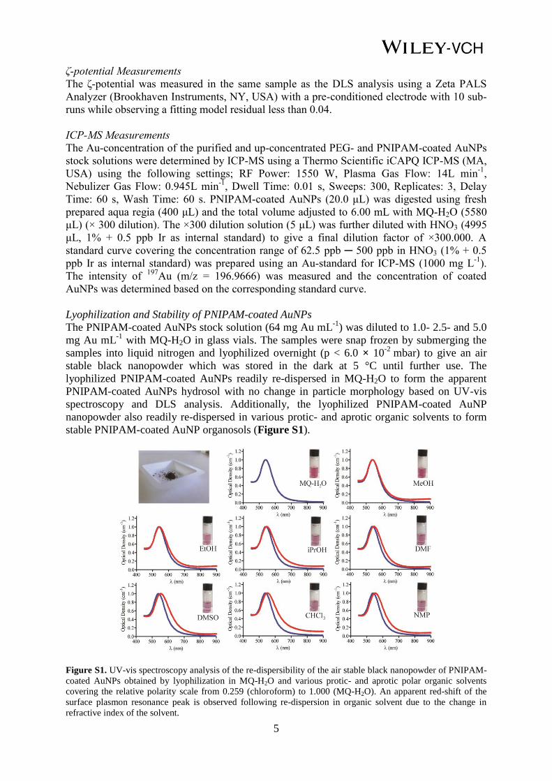
5
ζ-potential Measurements
The ζ-potential was measured in the same sample as the DLS analysis using a Zeta PALS
Analyzer (Brookhaven Instruments, NY, USA) with a pre-conditioned electrode with 10 sub-
runs while observing a fitting model residual less than 0.04.
ICP-MS Measurements
The Au-concentration of the purified and up-concentrated PEG- and PNIPAM-coated AuNPs
stock solutions were determined by ICP-MS using a Thermo Scientific iCAPQ ICP-MS (MA,
USA) using the following settings; RF Power: 1550 W, Plasma Gas Flow: 14L min-1
,
Nebulizer Gas Flow: 0.945L min-1
, Dwell Time: 0.01 s, Sweeps: 300, Replicates: 3, Delay
Time: 60 s, Wash Time: 60 s. PNIPAM-coated AuNPs (20.0 μL) was digested using fresh
prepared aqua regia (400 μL) and the total volume adjusted to 6.00 mL with MQ-H2O (5580
μL) (× 300 dilution). The ×300 dilution solution (5 μL) was further diluted with HNO3 (4995
μL, 1% + 0.5 ppb Ir as internal standard) to give a final dilution factor of ×300.000. A
standard curve covering the concentration range of 62.5 ppb ─ 500 ppb in HNO3 (1% + 0.5
ppb Ir as internal standard) was prepared using an Au-standard for ICP-MS (1000 mg L-1
).
The intensity of 197
Au (m/z = 196.9666) was measured and the concentration of coated
AuNPs was determined based on the corresponding standard curve.
Lyophilization and Stability of PNIPAM-coated AuNPs
The PNIPAM-coated AuNPs stock solution (64 mg Au mL-1
) was diluted to 1.0- 2.5- and 5.0
mg Au mL-1
with MQ-H2O in glass vials. The samples were snap frozen by submerging the
samples into liquid nitrogen and lyophilized overnight (p < 6.0 × 10⁻²
mbar) to give an air
stable black nanopowder which was stored in the dark at 5 °C until further use. The
lyophilized PNIPAM-coated AuNPs readily re-dispersed in MQ-H2O to form the apparent
PNIPAM-coated AuNPs hydrosol with no change in particle morphology based on UV-vis
spectroscopy and DLS analysis. Additionally, the lyophilized PNIPAM-coated AuNP
nanopowder also readily re-dispersed in various protic- and aprotic organic solvents to form
stable PNIPAM-coated AuNP organosols (Figure S1).
Figure S1. UV-vis spectroscopy analysis of the re-dispersibility of the air stable black nanopowder of PNIPAM-
coated AuNPs obtained by lyophilization in MQ-H2O and various protic- and aprotic polar organic solvents
covering the relative polarity scale from 0.259 (chloroform) to 1.000 (MQ-H2O). An apparent red-shift of the
surface plasmon resonance peak is observed following re-dispersion in organic solvent due to the change in
refractive index of the solvent.

6
Figure S2. A) Characterization of PNIPAM-coated AuNP by TEM; B) Comparison of the obtained contrast
level in 2D x-ray imaging of AuNPs containing nanogels (10- 30-, 60- and 90 mg AuNP mL-1
) and solid fiducial
markers used in the clinic. Markers visualized in a 10 cm solid water phantom using the following settings; 80
kVp, 80 mA, 80 ms.
Figure S3. A) Standard curve of UV-absorbance at λ = 537 nm as a function of PNIPAM-coated AuNP
concentration in MQ-H2O (r2 = 1.00); B) In vitro release kinetics from SAIB/EtOH/PLA (75:20:5) with either 30
mg mL-1
PEG-coated AuNPs or PNIPAM-coated AuNPs in MQ-H2O at 37 °C. Each point represents the mean ±
SEM (n = 3). C) Representative image of nanogels containing either PEG-coated or PNIPAM-coated AuNPs
after 24 h incubation in MQ-H2O.
3. X-ray Visualization of Nanogel in Mice/Canine
Figure S4. Visualization of nanogel composed of SAIB/EtOH/PLA (75:20:5) + 30 mg PNIPAM-coated AuNPs
mL-1
administered subcutanously injection in immunocompetent mice. Aggregation of PNIPAM-coated AuNPs
within the gel matrix visualized by reduction of the contrast level on the micro-CT image.

7
Figure S5. Nanogel composed of SAIB/EtOH/PLA (75:20:5) + 30mg PNIPAM-coated AuNPs mL
-1
administered intratumorally into canine suffering from a solid subcutaneous mast cell tumor (maximum distance
(x × y × z); 1.82 cm × 5.81 cm × 5.32 cm, CT volume; 31.64 cm3) adherent to the underlying soft tissue located
over the dorsal aspect of the left shoulder blade. A) OBI 2D x-ray imaging of nanogel and B) ExacTrac imaging
of nanogel. Both images recorded prior to the first fraction of radiation therapy 6 days after injection.