synthesis and characterization of lipid-polymer hybrid
TRANSCRIPT

Synthesis and Characterization of Lipid-Polymer Hybrid Nanoparticles for Combinatorial Drug Delivery
By
Kathy Stavropoulos
Submitted to the graduate degree program in Pharmaceutical Chemistry and the Graduate Faculty of the University of Kansas in partial fulfillment of the requirements for the degree of
Master of Sciences.
________________________________
Chairperson Christian Schöneich
________________________________
John Stobaugh
________________________________
Santosh Aryal
Date Defended: June 29, 2011

ii
The Thesis Committee for Kathy Stavropoulos
certifies that this is the approved version of the following thesis:
Synthesis and Characterization of Lipid-Polymer Hybrid Nanoparticles for Combinatorial Drug Delivery
________________________________
Chairperson Christian Schöneich
Date approved: June 29, 2011

iii
Abstract
Overcoming obstacles like multidrug resistance, short circulation half-life, and
nonspecific systemic distribution is an ongoing challenge in cancer therapy. One
application to address these concerns is to engineer a drug delivery vehicle that has
versatile functionality, good serum stability, circulates in the body long enough to reach
the targeting tissues, and is biocompatible. A promising formulation platform that
embodies these features is the lipid-polymer hybrid nanoparticles. The surface
characteristics of these nanoparticles such as charge, lipid density, and targeting ligands
can be modified to allow for specific cellular uptake, controlled drug releases kinetics,
and enhanced pharmacokinetics. In this work, it was found that the hybrid nanoparticles
could easily be fabricated with negatively and positively charged lipids in order to change
the overall surface charge. The particle size remained in the desirable range and the
distribution was narrow. The lipid-polymer hybrid nanoparticle by design has the
capacity to co-encapsulate hydrophobic and lipophilic drugs. To investigate,
camptothecin and a cisplatin derivative were dually loaded within the hybrid nanoparticle
system. This combination formulation was characterized by dynamic light scattering for
particle size, zeta potential, and polydispersity index as well as in vitro drug release and
cytotoxicity. The particle size was below 100 nm and the distribution was narrow. The
release studies showed that the addition of the two drugs within the lipid-polymer hybrid
nanoparticle system did not affect the release profiles of the individual drugs. The ability
for co-encapsulation and the similar overall drug release profiles for camptothecin and
cisplatin derivative in the combination compared to single drug loaded controls valuates
this already useful drug delivery platform.

iv
Acknowledgements
I would like to thank the Pharmaceutical Chemistry faculty members and staff for
all their guidance and support. I am especially grateful to the Zhang research group at
UCSD for adopting me and allowing me to collaborate with them on their exciting
research projects. I would also like to thank my committee members for taking the time
out of their busy schedules for me. Last and certainly not least, I would like to thank my
friends and family for all their encouragement during this endeavor.
v
Table of Contents
Chapter 1: Literature Review 1
1.1 Review of Nanomedicine 2
1.2 Nanoparticle Drug Delivery 5
1.3 Lipid-Polymer Hybrid Nanoparticles 14
1.4 References 18
Chapter 2: Synthesis and Characterization of Hybrid Nanoparticles 24
2.1 Introduction 25
2.2 Materials and Methods 26
2.2.1 Hybrid Nanoparticle Synthesis 26
2.3 Characterization 29
2.3.1 Particle Size and Polydispersity Index (PDI) 29
2.3.2 Zeta Potential 29
2.3.3 Scanning Electron Microscopic (SEM) Analysis 29
2.3.4 Transmission Electron Microscopic (TEM) Analysis 30
2.4 Results and Discussion 30
2.5 Conclusions and Outlook 42
2.6 References 44
Chapter 3: Combinatorial Drug Delivery with Lipid-Polymer Hybrid
Nanoparticles 45
3.1 Introduction 46
3.2 Materials and Methods 49
3.2.1 Preparation of bis(2-stearoylhydraziny)platinum (II) chloride (BSPC, Pt-lipid) 49
vi
3.2.2 Lipid-Polymer Hybrid Nanoparticle Synthesis 52
3.3 Characterization 52
3.3.1 Pt-lipid 53
3.3.2 Lipid-Polymer Hybrid Nanoparticles 54
3.4 Drug Loading 54
3.4.1 UV-Vis Spectroscopy 55
3.4.2 Inductively Coupled Plasma Optical Emission Spectroscopy (ICP-OES) 55
3.4.3 Reversed-Phase High-Performance Liquid Chromatography (RP-HPLC) 55
3.5 In Vitro Drug Release 55
3.6 In Vitro Cytotoxicity 56
3.7 Results and Discussion 57
3.8 Conclusions and Outlook 70
3.9 References 71

vii
Index of Figures
Figure 1.1 This diagram shows a schematic representation 7
of endocytotic processes.
Figure 1.2 Schematic representation displaying the cells and 10
blood vessels involved with the enhanced
permeability and retention (EPR) effect.
Figure 1.3 This diagram shows a representation of the 15
anatomy of a lipid-polymer hybrid nanoparticle.
Figure 2.1 Effect of DOTAP concentration on nanoparticle 33
size and polydispersity index (PDI)
Figure 2.2 Effect of Egg PA concentration on nanoparticle 34
size and polydispersity index (PDI)
Figure 2.3 Effect of Egg PC concentration on nanoparticle 35
size and polydispersity index (PDI)
Figure 2.4 The SEM image shows particle morphology of 36
hybrid nanoparticles with Egg PC as the lipid.
Figure 2.5 The TEM micrograph shows the internal structure 37
of the hybrid nanoparticles with Egg PC as the lipid.

viii
Figure 2.6 Zeta potential of control lipid vesicle solutions show 38
that each lipid has a different surface charge.
Figure 2.7 Effect of DOTAP concentration on zeta potential. 39
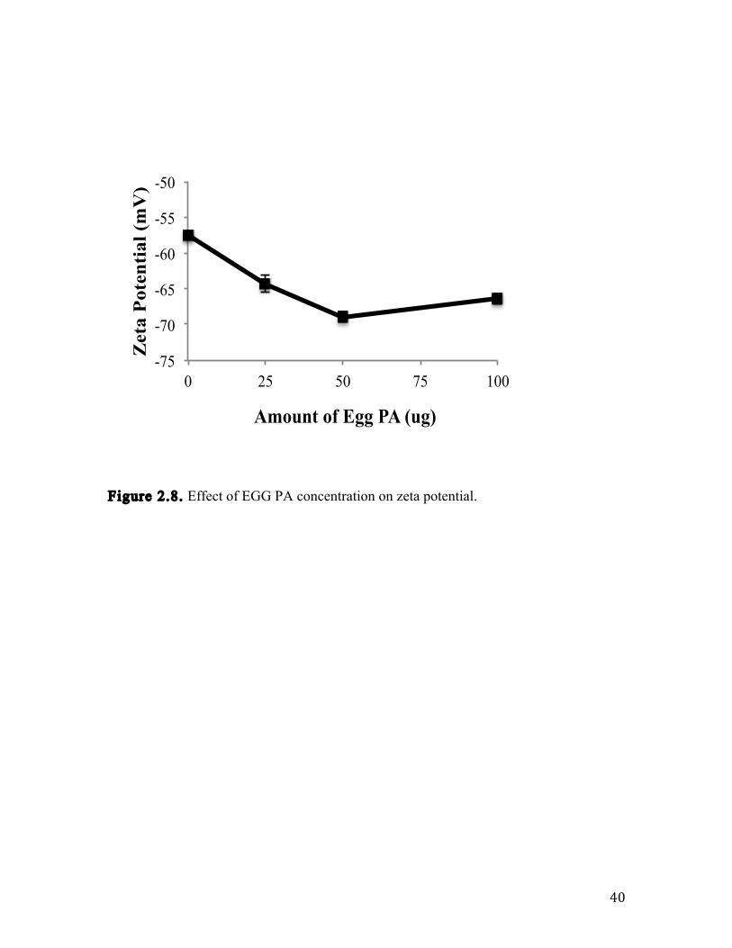
Figure 2.8 Effect of Egg PA concentration on zeta potential. 40
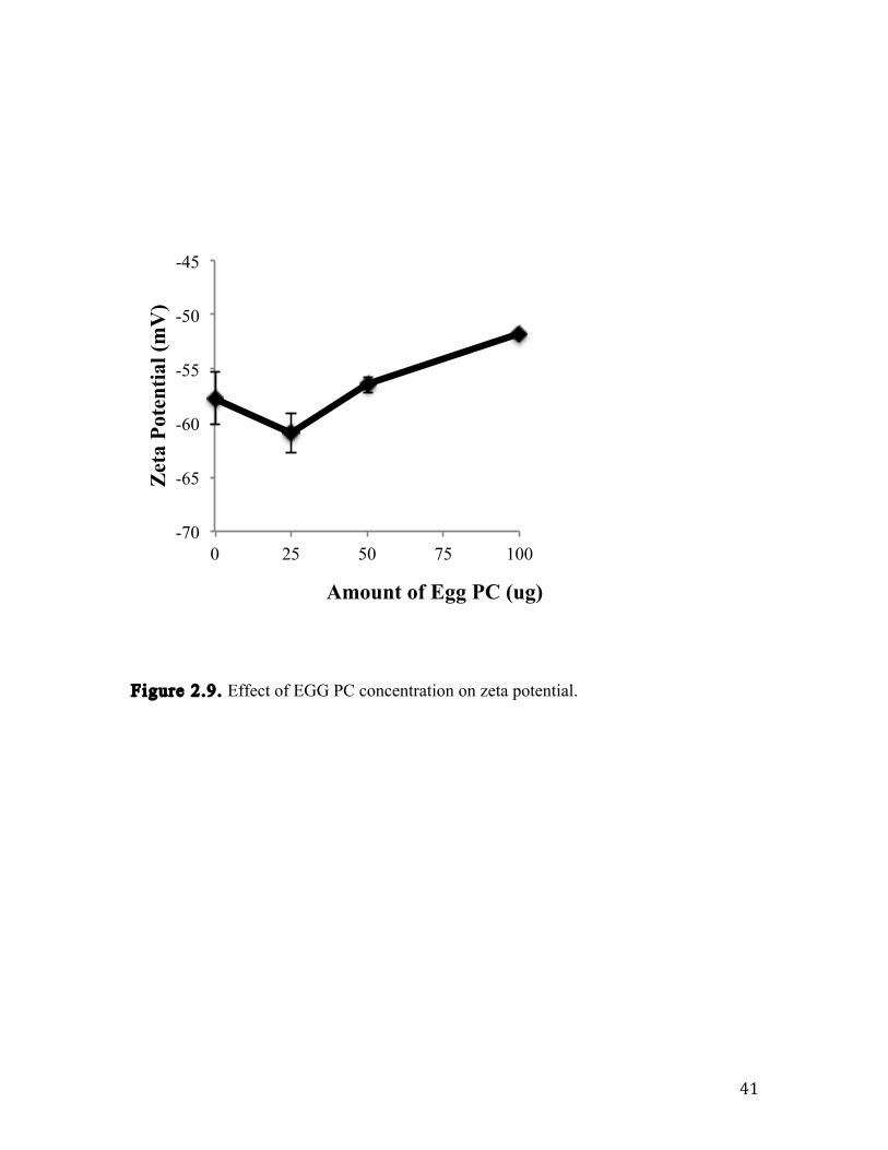
Figure 2.9 Effect of Egg PC concentration on zeta potential. 41
Figure 3.1 The synthetic scheme for bis(2-stearoylhydraziny) 51
platinum (II) chloride (Pt-lipid).
Figure 3.2 195Pt NMR spectra show the chemical shift for the 60
prepared Pt-lipid compound is different from that of
the platinum standard, which indicates that a new
chemical entity was synthesized.
Figure 3.3 Electrospray ionization mass spectrometry shows the 61
mass to charge ratios of the Pt-lipid complex.
Figure 3.4 Dynamic light scattering measurements of the drug 62
nanoparticles show similar particle size for the three
types of hybrid formulations.

ix
Figure 3.5 This is a representative SEM image of the combinatorial 64
nanoparticles.
Figure 3.6 This graph shows the drug loading results for both 65
camptothecin and Platinum separately as control
nanoparticles and dually loaded in combination nanoparticles.
Figure 3.7 This calibration plot by HPLC was used to determine 66
drug loading for CPT in the cytotoxicity samples because
the drug levels were too low to be detected by UV-Vis
spectroscopy.
Figure 3.8 UV-Vis spectroscopy was used to determine CPT 67
content during the in vitro release studies.
Figure 3.9 The in vitro release profile compares the drug 68
release of CPT and of Platinum from the
combination hybrid nanoparticles in which the CPT
releases faster after 4 hours. It also shows that the
addition of another drug to the hybrid nanoparticle
system does not affect overall drug release as compared
x
with control single drug loaded formulations.
Figure 3.10 The cytotoxicity results show that the combination 69
formulation is capable of reducing cell viability in the
ovarian cancer cells.

xi
Index of Tables
Table 2.1 Hybrid nanoparticle formulation compositions. 28
Table 3.1 Table shows the average particle size, zeta 63
potentials, and PDI for each of the nanoparticle
formulations.

xii

Chapter 1: Literature Review

2
1.1 Review of Nanomedicine
In recent years, the application of nanotechnology has been translated to medicine.
Nanotechnology encompasses the design, synthesis, and characterization of materials and
or devices, which are functionally organized in at least one dimension on the nanoscale
[1]. The use of these nanoscale or nanostructured materials in medicine, termed as
nanomedicine, has unique medical properties and effects owing to the small size (1 –
1000 nm) and structure [2]. The ability to engineer and control materials in this size rage
results in new medical efforts, innovative chemistry techniques, and novel manufacturing
approaches [2]. Nanomedicine has the capacity to change the landscape of healthcare and
drug delivery by enhancing the developability of biologically active drug candidates with
poor pharmaceutical properties such as solubility and circulation half-life [3].
Nanomedinces and nanomaterials are engineered to have specific functions, which utilize
the physical properties and characteristics for diagnosis and treatment of disease [4].
These materials are able to be used as carriers to cross membranes, mediate molecular
interactions, and detect molecular changes [4]. Nanomaterials have a high surface to
volume ratio. This increased surface area can be coated or tagged with other molecules,
which results in the formation of multifunctional nanomaterials [4]. Nanomaterials can be
engineered to have different shapes, sizes, surface chemistry, particle density, and
chemical compositions [4]. Because of their design, nanomedicines have applications in
drug delivery, in vivo and in vitro diagnostics, biomaterials, active implants, in vivo
imaging, biosensing, cell labeling, and tissue engineering [1-3, 5,6].
In vivo imaging employs the use of magnetic nanoparticles, quantum dots,
fluorophores, and carbon nanotubes [2,3,5,6]. An example is Gastromark (ferumoxsil®),

3
which is a marketed product composed of superparamagnetic iron oxide nanoparticles
used as a contrast agent for magnetic resonance imaging [2]. Fluorescent quantum dots
are nanocrystals that have higher extinction coefficients than traditional fluorophores,
which makes this technology useful for imaging [6]. Carbon nanotubes can act as
biocompatible supportive substrates that can incorporate fluorophores and other
molecules [7]. Using nanomaterials for in vivo imaging is a faster, less invasive, and a
more accurate way to diagnose diseases and to monitor disease states and progression [3].
In the future, these types of imaging probes may be able to assist surgeons in locating
tumors within the body and to identify adjacent structures [3].
In vitro diagnostics is another application for nanomedicine, which uses
nanoparticles, nanowires, nanotubes, nanoarrays, and cantilevers [2,3]. Lateral flow
assays are marketed products that utilize colloidal gold to test ovulation, HIV infection,
and pregnancy [2]. In this case, an antibody for a specific analyte is conjugated to the
nanoparticle surface. Gold nanoparticles are widely used because they have good stability,
which avoids the chance of false positive readings [2]. With the use of these materials,
disease detection can be quick, high throughput, and more accurate by using biomarkers
with higher sensitivity [3]. In the future, novel analytes could be measured such as
Alzheimer’s plaques [2]. Using nanomaterials for in vitro diagnostics is advantageous
because they can improve sensitivity, reduce cost, and consume less of the sample [2,3].
Biomaterials have mechanical properties than can be used as medical implants,
dental restoratives, and bone substitutes [3]. One example of a biomaterial is the
nanoparticle composite found in the dental restorative Filtek Supreme®, which is a
marketed product produced by 3M [2]. Vitoss® is a marketed nano-hydroxyapatite based

4
product that is used in the repair of bone defects [2]. Another example of biomaterials in
the market is Anticoat®, which is a silver nanoparticle based wound dressing [2].
Nanomedicines have been especially successful as drug delivery vehicles. This
may be due to the fact that diseases originate at the molecular level, which is on the
nanoscale and can be caused by gene mutations, misfolded proteins, viral and bacterial
infections, cell misfunction, and cell miscommunications [4]. In order to address these
modes of disease, nanocarrier delivery systems were developed. Nanotechnology
formulation platforms include liposomes, nanoparticles, polymeric micelles, dendrimers,
nanocantilevers, carbon nanotubes, aptamers, quantum dots, and polymer conjugates [8].
Liposomes consist of a phospholipid bilayer and an aqueous core for drug encapsulation
of water-soluble molecules. Marketed liposomal products include Doxil and Myocet
(liposomal doxorubicin), Ambisome (liposomal Amphotericin B), DaunoXome
(liposomal daunorubicin) and Depocyt (liposomal cytarabine) [2]. There are also several
examples of marketed products that are polymer conjugates. Polyethylene glycol (PEG)
is conjugated to a molecule in order to increase circulation time [9]. Pegasys (PEG-α-
interferon-2a) and PEG-Intron (PEG-α-interferon-2b) are both therapies for hepatitis C in
which PEG is conjugated to a protein [2]. These marketed products are considered first
generation nanosystems because the drug is contained within a system used for passive
targeting [3].
Nanomedicine can be a solution for cancer therapy where the current treatments
have some problems that include non-specific systemic distribution of the drug,
inadequate drug concentration reaching the target site, normal tissue toxicity, and drug
resistance [8,10]. Nanomedicines can be used to overcome these obstacles that

5
conventional medicines cannot address. Because of their size and surface properties,
nanomedicines can accumulate in tumor sites due to the enhanced permeability and
retention (EPR) effect [4]. Nanomedicines have the capacity to encapsulate multiple
drugs in order to yield combinatorial delivery, increase circulation time, and exhibit
controlled drug release kinetics [11]. This allows for improved dose scheduling, which
leads to patient compliance.
The impact of nanotechnology for drug delivery is that the characteristics of the
vehicles such as size, charge, surface hydrophobicity, ligand type, and density of ligands
on the surface can enhance pharmacokinetic properties such as circulation half-life and
biodistribution while also improving pharmaceutical properties such as drug solubility
[12]. Because of this, nanotechnology is beneficial for the pharmaceutical industry since
it can provide life-style extensions for drugs after patents have expired, new classes of
drug therapeutics can be developed, and the biologically active molecules that have poor
pharmaceutical properties can be re-investigated [3,12].
1.2 Nanoparticle Drug Delivery
Within the body there are multiple biological barriers in which drug delivery
vehicles need to be engineered to overcome. There are different mechanisms for delivery
vehicles to target tissues and be sequestered by specific cells. One particular biological
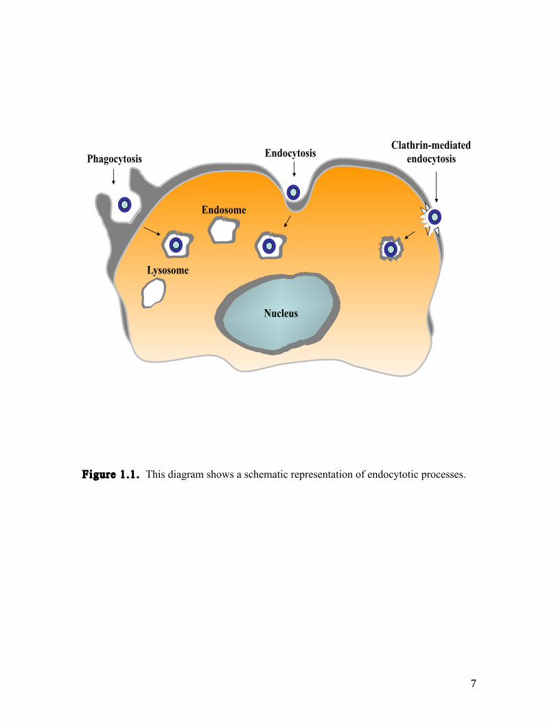
process that is important for nanomedicine delivery is endocytosis (Figure 1.1). For
endocytosis to occur, a molecule or material must be recognized in the bloodstream via
specialized absorptive proteins [13]. The recognized material undergoes adhesion onto
the cellular membrane. After adhesion, the material becomes engulfed into the cell. There

6
are specific mechanisms of endocytosis and it has been reported in the literature that
poly(lactic-co-glycolic acid) (PLGA) nanoparticles enter cells through clathrin-mediated
endocytosis as well as caveolae-independent pathways, depending on the cell type [13].
Bare PLGA nanoparticles have negative surface charge and since cell membranes are
negatively charged, these nanoparticles would internalize at a slow rate as a result of
charge repulsion. Clathrin-mediated endocytosis occurs when a pit in the cell membrane
is formed by the polymerization of clathrin-1 and other assembly proteins [13]. Ligands
that bind to receptors get engulfed in this pit. An enzyme pinches off the assembled
vesicle, and then the clathrin coating is removed. The vesicle fuses with endosomes and
gets sorted by the cell [13]. It is also reported that positively charged nanomaterials enter
cells via clathrin-mediated endocytosis at a faster rate than negatively charged
nanoparticles [13]. Nanoparticle surface charge can be designed according to the desired
route of cellular transport.
7
Figure 1.1. This diagram shows a schematic representation of endocytotic processes.
Lysosome
Endosome
Endocytosis Phagocytosis
Clathrin-mediated
endocytosis
Nucleus

8
The blood brain barrier (BBB) is another biologically barrier that is challenging
for conventional therapeutics to cross in order to treat central nervous system (CNS)
diseases. The blood brain barrier is comprised of endothelial cells, pericytes, astrocytes,
and a basal membrane [14]. The brain capillaries are covered by pinocytes and
microvessel endothelial cells which makes for a compact structure with tight junctions
[14]. Molecules are able to cross the BBB by diffusion mechanisms as long as the
molecules are lipophilic, not ionizable at physiological pH, and have a size below 400 kD
[14]. Due to their small size and design, nanoparticles have the capacity to penetrate
through the tight junctions in the BBB and protect the drug from enzymatic degradation
[14]. These advantages make the nanoparticle drug delivery platform an attractive option
for treatment of CNS diseases including cancer.
In the treatment of cancer, the biological barrier for chemoagents to overcome is
permeation through the cancer cell networks. Cancer cells that make up tumors have
unique biology and anatomy that differs form healthy cells [11]. A way to take advantage
of this unique biology and anatomy is via the enhance permeability and retention (EPR)
effect (Figure 1.2). Tumors and cancer cells proliferate quite rapidly and don’t have
enough oxygen and nutrients to sustain such a quick growth rate, so they grow blood
vessels in disorganized heterogeneous networks that lead to a large vascular density [15].
This biology is shown in Figure 1.2. This disorganization provides gaps in the
endothelium cell-cell junctions. Tumor blood vessels also have larger pores, which
increases the permeability and hydraulic conductivity causing the ERP effect [11]. In
addition to the defective vascular arrangement and extensive blood vessel growth, cancer
cells also have impaired lymphatic drainage [15]. Nanoparticle drug delivery vehicles can

9
reach these cancer cells and tumor cells by passive or active targeting and take advantage
of the EPR effect. Passive targeting is when the drug and or drug carrier accumulates at
the desired site owing to physical and chemical properties of the formulation or
pharmacological factors [8]. Formulation factors include particle size distribution while
the pharmacological factors are the leaky tumor vasculature, the EPR effect, and the
tumor microenvironment, which is acidic due to glycolysis [8]. The other approach is
active targeting, which involves attaching a targeting ligand to the nanocarrier. This
targeted nanoparticle internalizes within the cancer cell through receptor-mediated
endocytosis [8,13].

10
Figure 1.2. Schematic representation displaying the cells and blood vessels involved
with the enhanced permeability and retention (EPR) effect.
Endothelial cells
Tumor
Blood vessel
Leaky vasculature
Tumor blood vessels
Nanoparticle

11
To date, the most common cancer treatments are chemotherapy, radiation, and
surgery. The challenging aspects involved with cancer therapy include nonspecific
systemic distribution, low drug levels reaching the tumor site, cytotoxicity, poor stability,
and multidrug resistant tumor cells [8,10,16]. Multidrug resistance (MDR) is a major
hurdle in cancer therapy because it decreases the efficacy of drugs through multiple
mechanisms [17]. This phenomenon involves an active efflux of a large range of
cytotoxic drugs out of the cytoplasm by membrane-bound transporters [16]. One example
is the P-glycoprotein (P-gp), which is an active membrane-bound efflux pump [18].
Over-expression of P-gp and other membrane transporters can lead to MDR [19]. Other
cellular mechanisms that contribute to MDR are drug molecule reactions with
intracellular nucleophiles like glutathione, repair of drug-induced damage to the DNA,
altered proteins that affect apoptotic pathways, and an altered drug target [16,17]. Non-
cellular events that can lead to MDR include high interstitial pressures at the tumor site,
which decreases drug permeability, lower pH, hypoxia (drugs generating free radicals),
and the extracellular matrix effect [16]. Therapeutic materials can also be removed from
the systemic circulation by the mononuclear phagocyte system that comprises of kupffer
cells in the liver and macrophages in the spleen and bone marrow [20]. Nanoparticle drug
delivery vehicles can be designed to address these challenges associated with current
cancer treatment.
Nanosystems are distinct from other cancer therapeutics because the nanocarrier
itself can also have therapeutic effects along with the actual drug [8]. Nanoparticles can
be designed to carry large payloads, have attached targeting ligands, encapsulate multiple
drugs for combinatorial therapy, and have the ability to bypass drug resistance

12
mechanisms [8]. Material selection is an important consideration for nanomedicines.
Biodegradable, biocompatible, and physiological lipids are chosen for formulation
development in an attempt to reduce immunogenicity and minimize toxicity [21].
Colloidal drug carriers such as micelles, nanoemulsions, nanosuspensions, polymeric
nanoparticles, and liposomes are formulation platforms that are used to address drug
solubility and stability issues [21]. One example of a nanoparticle delivery vehicle is a
dendrimer, which is a biodegradable branch-like structure that consists of a core (two or
more reactive groups) with repeated units covalently bound to the core and peripheral
functional groups [22]. Drugs can be encapsulated or conjugated to the dendrimer and
delivered to tumor sites through the EPR effect or by using targeting ligands like peptides
and antibodies [22]. Another example is quantum dots, which are nanocrystals that have
improved fluorescent properties over traditional fluorophores and can be used as drug
carriers or as tags for other drug carriers [23]. Liposomes have a lipid bilayer in which
the surface characteristics can be modified by lipid type and lipid charge. Cationic
liposomes are established in the literature to have antimicrobial action due to the
adsorption of the positively charged lipid bilayer onto the bacterial cell membrane, which
changes the membrane surface charge from negative to positive and induces apoptotic
cell death [24]. With liposomes and polymeric nanoparticles, multiple drugs can be co-
encapsulated in a single system for combination delivery.
Combination delivery has been used in malaria, HIV/AIDS, and cancer [25]. This
approach is employed in cancer therapy to minimize multidrug resistance and reduce
cytotoxicity. Combining chemoagents hits different targets and displays different toxicity
profiles, which can improve efficacy or have comparable efficacy and decreased toxicity

13
[25]. An example of biochemical synergy for the treatment of nonlymphocytic leukemia
is the combination of anthracycline daunorubicin (a DNA intercalator) with ara-C (a
DNA polymerase inhibitor), which interferes with DNA repair and DNA synthesis [25].
For colorectal cancer, administering leucovorin prior to 5-fluorouracil enhanced that
ability to bind and block the action of thymidilate synthetase [25]. From a patient
compliance view, it would be elegant to contain multiple drugs within one delivery
vehicle. This could lead to a more convenient dose-scheduling regimen and would
improve patient quality of life. Having this in mind, nanodelivery platforms have to be
simple, scalable, broad-based, and meet Food and Drug Administration (FDA)
requirements [26]. Formulation platforms that are successful should be engineered with
these specific properties: biocompatibility, biodegradation, encapsulation efficiency,
colloidal stability, improved pharmacokinetics, and controlled drug release kinetics [26].
Lipid-polymer hybrid nanoparticles as a drug delivery platform is one that embodies the
lipid shell characteristics of a liposome and the hydrophobic core of a polymeric
nanoparticle. The fabrication process is reproducible and encapsulation efficiency is
sufficient for camptothecin (CPT) and a cisplatin derivative, which both show
cytotoxicity in A2780 human ovarian carcinoma cells. Hydrophobic drugs can be
encapsulated in the polymeric core while targeting ligands can be tagged to the lipid shell.
The robustness and versatility makes this formulation platform practical for cancer
treatment and it can be loaded with multiple drug agents for combinatorial delivery.

14
1.3 Lipid-Polymer Hybrid Nanoparticles
Nanomedicine drug delivery systems for cancer therapy are designed to protect the
drug from inactivation due to the biological environment, protect non-pathological tissues
from non-specific toxic actions of the drug, and to change or control drug
pharmacokinetics [9]. Lipid-polymer hybrid nanoparticles are a nanomedicine
formulation platform that can be used for cancer treatment. The anatomy of a hybrid
nanoparticle consists of a hydrophobic poly(lactic-co-glycolic acid) (PLGA) polymer
core, a lipid monolayer surrounding the core, and a lipid-PEG (for example: 1,2-
Distearoyl-sn-glycero-3-phosphoethanolamine-N-carboxy(poly(ethylene glycol)) 2000
(DSPE-PEG-COOH)), which is distributed within the lipid monolayer to form a
polyethylene glycol (PEG) corona [27]. The polymeric core affects drug encapsulation
and release. Drug release from the nanoparticles begins with diffusion processes,
followed by erosion, then swelling of the matrix [28]. The polymer degrades due to
hydrolysis and the degradation rate depends on the polymer composition and molecular
weight [28]. The lipid shell serves the purpose as a biocompatible shield, a template for
surface modifications, and a barrier for preventing water-soluble drugs from leaking out
of the core [29]. The corona affects biodistribution and circulation half-life. The PEG
corona provides electrostatic and steric stabilization as well as a protective layer from
adsorptive recognition proteins in the bloodstream [9, 27]. There are multiple fabrication
methods for preparing lipid-hybrid nanoparticles found in the literature which include,
but are not limited to solvent – evaporation, emulsion method, nanoprecipitation followed
by self – assembly, and a sonication method [30-32].

15
Figure 1.3. This diagram shows a representation of the anatomy of a lipid-polymer
hybrid nanoparticle.
PLGA core
Lipid
Lipid-PEG

16
The versatility of this lipid-polymer hybrid nanoparticle platform allows for
surface chemistry modifications. Li et. al., used cationic lipids in their hybrid
nanoparticles in order to form a DNA complex for gene delivery [33]. The end group on
the PEG that makes up the corona can be changed from a carboxyl group to an amine or a
methoxy group in order to change the surface zeta potential [34]. In a study done by
Salvador-Morales et. al., it was shown that the surface chemistry of the hybrid
nanoparticles affects human plasma and serum absorption patterns by inducing different
levels of compliment activation [34]. They also performed coagulation studies that
showed no hybrid nanoparticle formulation with the modified surface groups activated
the coagulation cascade [34]. These studies exhibited the potential for the lipid-polymer
hybrid nanoparticles to be a viable immunocompatible delivery option. Another type of
surface chemistry modification is the addition of targeting ligands, which are used to
increase cellular uptake and accumulation in the tumor sites. Different types of ligands
are used to target hybrid nanoparticles to cancers cells such as antibodies, antibody
fragments, proteins, small molecules, aptamers, and peptides [35]. The ligands should
induce receptor-mediated endocytosis and have the correct conformation to maintain
affinity for its corresponding receptors [35]. Also the ligand must not disturb the steric
and electrostatic stabilization that comes from the PEG corona. If the ligand
concentration is too high then it mitigates the stealth action of the protective PEG layer
[35]. An example from the literature using targeting ligands comes from Hu et. al., in
which they conjugated a half-antibody to lipid-polymer hybrid nanoparticles for use in
pancreatic cancer treatment [36]. Another example from the literature is the use of wheat
germ agglutinin (WGA) on the surface of PLGA nanoparticles for targeted intracellular

17
delivery of paclitaxel [37]. WGA binds to the N-actetylglucosamine and the sialic acid
residues on the cell membrane, which leads to cellular internalization by receptor-
mediated endocytosis [37].
With all the functionality that is available to lipid-polymer hybrid nanoparticles,
another opportunity can be used with this system for combinatorial delivery. In this
system a hydrophobic drug can be encapsulated in the PLGA core while a lipophilic drug
can be incorporated within the lipid shell. Co-formulation of multiple drugs in a single
system has the advantage of delivering the correct drug ratio to the target of interest as
well as synergistic therapeutic effects, suppressed drug resistance, and a timed drug
exposure control [38]. One proof of concept example by Kolishetti et. al., is the
encapsulation of docetaxel with a cisplatin prodrug conjugated to the polymer to treat
prostate cancer cells [38]. Similarly, Aryal et. al., have demonstrated the combinatorial
drug delivery system in which paclitaxel (a hydrophobic drug) and gemcitabine (a
hydrophilic drug) were conjugated by a hydrolysable linker, followed by the
encapsulation of the drug conjugate into a hybrid nanoparticle [39].
Nanoparticle formulation platforms have several advantages in delivering cancer
therapeutics. They provide a system that improves drug solubility, increases half-life
circulation due to evasion of the mononuclear phagocytic system, enhances the drug
accumulation in target cells, provides a stable drug release, and reduces efflux pump-
mediated drug resistance [40]. In this work, the next steps were taken with the
nanoparticle platform to optimize surface charge in order to take advantage of cellular
uptake mechanisms and to encapsulate multiple drugs within a single system in order to
improve the cytotoxicity and drug release kinetics.

18
1.4 References
[1] G. Caruso, M. Caffo, C. Alafaci, G. Raudino, D. Cafarella, S. Lucerna, F.M.
Salpietro, F. Tomasello, Could nanoparticle systems have a role in the treatment of
cerebral gliomas? Nanomedicine: NBM (2011) 1-9.
[2] V. Wagner, A. Dullart, A-K. Black, A. Zweck, The emerging nanomedicine
landscape. Nature Biotechnology 24 (10) (2006) 1211-1217.
[3] J. Shi, A.R. Votruba, O.C. Farokhzad, R. Langer, Nanotechnology in drug
delivery and tissue engineering: from discovery to applications. Nano Lett. (10) (2010)
3223-3230.
[4] B.Y.S. Kim, J.T. Rutka, W.C.W. Chan, Nanomedicine. N ENGL J MED 363 (25)
(2010) 2434-2443.
[5] B.S. Sekhon, S.R. Kamboj, Inorganic nanomedicine – part 1. Nanomedicine:
NBM (6) (2010) 516-522.
[6] B.S. Sekhon, S.R. Kamboj, Inorganic nanomedicine – part 2. Nanomedicine:
NBM (6) (2010) 612-618.
[7] S. Beg, M. Rizwan, A.M. Sheikh, M.S. Hasnain, K. Anwer, K. Kohli,
Advancements in carbon nanotubes: basics, biomedical applications and toxicity. JPP
(63) (2011) 141-163.
[8] R. Misra, S. Archaya, S.K. Sahoo, Cancer nanotechnology: application of
nanotechnology in cancer therapy. Drug Discovery Today 15 (19/20) (2010) 842-850.

19
[9] T. Musacchio, V.P. Torchilin, Recent advances in lipid-based pharmaceutical
nanocarriers. Frontiers in Bioscience (16) (2011) 1388-1412.
[10] A. Shapira, Y.D. Livney, H.J. Broxterman, Y.G. Assaraf, Nanomedicine for
targeted cancer therapy: towards the overcoming of drug resistance. Drug. Resist. Updat.
(2011)
[11] R.K. Jain, T. Stylianopoulos, Delivering nanomedicine to solid tumors. Nature
Clinical Oncology Reviews (7) (2010) 653-664.
[12] O.C. Farokhzad, R. Langer, Impact of nanotechnology on drug delivery. ACS
Nano 3 (1) (2009) 16-20.
[13] G. Sahay, D.Y. Alakhova, A.V. Kabanov, Endocytosis of nanomedicines, Journal
of Controlled Release (145) (2010) 182-195.
[14] J.K. Sahni, S. Doggui, J. Ali, S. Baboota, L. Dao, C. Ramassamy,
Neurotherapeutic applications of nanoparticles in Alzheimer’s disease. Journal of
Controlled Release (2011)
[15] S. Archaya, S.K. Sahoo, PLGA nanoparticles containing various anticancer agents
and tumor delivery by EPR effect. Advanced Drug Delivery Review (63) (2010) 170-183.
[16] H.L. Wong, R. Bendayan, A.M. Rauth, Y. Li, X.Y. Wu, Chemotherapy with
anticancer drugs encapsulated in solid lipid nanoparticles. Advanced Drug Delivery
Review (59) (2007) 491-504.
[17] A.J. Shuhendler, R.Y. Cheung, J. Manais, A. Connor, A.M. Rauth, X.Y. Wu, A
novel doxorubicin – mitomycin C co-encapsulated nanoparticle formulation exhibits anti-

20
cancer synergy in multidrug resistant human breast cancer cells. Breast Cancer Res Treat
(119) (2010) 255-269.
[18] F. Wang, Y-C. Wang, S. Dou, M-H. Xiong, T-M. Sun, J. Wang, Doxorubicin-
tethered responsive gold nanoparticles facilitate intracellular drug delivery for
overcoming multidrug resistance in cancer cells. ACS Nano (2011)
[19] H.L. Wong, A.M. Rauth, R. Bendayan, J.L. Manais, M. Ramaswamy, Z. Liu, S.Z.
Erhan, X.Y. Wu, A new polymer-lipid hybrid nanoparticle system increases cytotoxicity
of doxorubicin against multidrug-resistant human breast cancer cells. Pharmaceutical
Research 23 (7) (2006) 1574-1585.
[20] N.T. Huynh, C. Passirani, P. Saulnier, J.P. Benoit, Lipid nanocapsules: a new
platform for nanomedicine. International Journal of Pharmaceutics (379) (2009) 201-209.
[21] S. Das, A. Chaudhury, Recent advances in lipid nanoparticle formulations with
solid matrix for drug delivery. AAPS Pharm Sci Tech 12 (1) (2010) 62-76.
[22] Y. Cheng, L. Zhao, Y. Li, T. Xi, Design of biocompatible dendrimers for cancer
diagnosis and therapy: current status and future perspectives. Chem. Soc. Rev. (40)
(2010) 2673-2703.
[23] Y. Wang, L. Chen, Quantum dots, lighting up the research and development of
nanomedicine. Nanomedicine: NBM (2011) 1-18.
[24] L.D. Melo, E.M. Mamizuka, A.M. Carmona-Ribeiro, Antimicrobial particles
from cationic lipid and polyelectrolytes. Langmuir 26 (14) (2010) 12300-12306.

21
[25] F. Greco, M.J. Vincent, Combination therapy: opportunities and challenges for
polymer-drug conjugates as anticancer medicines. Advanced Drug Delivery Reviews (61)
(2010) 1203-1213.
[26] J.H. Adair, M.P. Parette, E.I. Altinoglu, M. Kester, Nanoparticles for drug
delivery. ACS Nano 4 (9) (2010) 4967-4970.
[27] J.M. Chan, L. Zhang, K.P. Yuet, G. Liao, J-W. Rhee, R. Langer, O.C. Farokhzad,
PLGA-lecithin-PEG core-shell nanoparticles for controlled drug delivery. Biomaterials
(30) (2009) 1627-1634.
[28] N. Kanthamneni, A. Chaudhary, J. Wang, S. Prabhu, Nanoparticle delivery of
novel drug combination regimens for the chemoprevention of colon cancer. International
Journal of Oncology (37) (2010) 177-185.
[29] W.S. Cheow, K. Hadinoto, Factors affecting drug encapsulation and stability of
lipid-polymer hybrid nanoparticles. Colloids and Surfaces B: Biointerfaces (85) (2011)
214-220.
[30] R.H. Fang, S. Aryal, C-M.J. Hu, L. Zhang, Quick synthesis of lipid-polymer
hybrid nanoparticles with low polydispersity using a single-step sonication method.
Langmuir (26) (2010) 16958-16962.
[31] L. Zhang, J.M. Chan, F.X. Gu, J-W. Rhee, A.Z. Wang, A.F. Radovic-Moreno, F.
Alexis, R. Langer, O.C. Farokhzad, Self-assembled lipid-polymer hybrid nanoparticles: a
robust drug delivery platform. ACS Nano 2 (8) (2008) 1696-1702.

22
[32] J.S. Hong, S.M. Stavis, S.H. DePaoli Lacerda, L.E. Locascio, S.R. Raghavan, M.
Gaitan, Microfluidic directed self-assembly of liposome – hydrogel hybrid nanoparticles.
Langmuir 26 (13) (2010) 11581-11588.
[33] J. Li, Y-Z. He, W. Li, Y-Z. Shen, Y-R. Li, Y-F. Wang, A novel polymer-lipid
hybrid nanoparticle for efficient nonviral gene delivery. Acta Pharmacologica Sinica (31)
(2010) 509-514.
[34] C. Salvador-Morales, L. Zhang, R. Langer, O.C. Farokhzad,
Immunocompatibility properties of lipid-polymer hybrid nanoparticles with
heterogeneous surface functional groups. Biomaterials (30) (2009) 2231-2240.
[35] M. Wang, M. Thanou, Targeting nanoparticles to cancer. Pharmacological
Research (16) (2010) 90-99.
[36] C-M.J. Hu, S. Kaushal, H.S.T. Cao, S. Aryal, M. Sartor, S. Esener, M. Bouvet, L.
Zhang, Half-antibody functionalized lipid-polymer hybrid nanoparticles for targeted drug
delivery to carcinoembryonic antigen presenting pancreatic cancer cells. Molecular
Pharmaceutics 7 (3) (2010) 914-920.
[37] C. Wang, P.C. Ho, L.Y. Lim, Wheat germ agglutinin – conjugated PLGA
nanoparticles for enhanced intracellular delivery of paclitaxel to colon cancer cells.
International Journal of Pharmaceutics (400) (2010) 201-210.
[38] N. Kolishetti, S. Dhar, P.M. Valencia, L.Q. Lin, R. Karnik, S.J. Lippard, R.
Langer, O.C. Farokhzad, Engineering of self-assembled nanoparticle platform for
precisely controlled combination drug therapy. PNAS 107 (42) (2010) 17939-17944.

23
[39] S. Aryal, C-M. Hu, L. Zhang, Combinatorial drug conjugation enabled
nanoparticle dual drug delivery. Small (6) (2010) 1442-1448.
[40] Z. Chen, Small-molecule delivery by nanoparticles for anticancer therapy. Trends
in Molecular Medicine 16 (12) (2010) 594-602.

Chapter 2: Synthesis and Characterization of Hybrid Nanoparticles

25
2.1 Introduction
Nanoparticles have been used in medicine for diagnostic therapy. Two major
classes of nanocarriers used for delivering therapeutic drugs in disease therapy are
biodegradable polymeric nanoparticles and liposomes [1]. The advantageous size range
(10 – 200 nm) inherent to nanoparticles is favorable for endocytotic intercellular uptake
[2]. This capability to permeate through cell walls makes the polymeric nanoparticle
platform practical as a nanomedicine for cancer therapy [3]. These vehicles can
accumulate in the tumor site through leaky tumor vascular structures, which is useful for
prolonged drug exposure to the tumor site [4].
Polymeric nanoparticles are practical drug delivery vehicles because they can be
used to encapsulate hydrophobic drugs, which would otherwise have too low aqueous
solubility for other drug delivery systems [1]. Circulation time in the body can be
increased with polyethylene glycol (PEG) as part of the corona of the particle, which
enables the particle to avoid phagocytosis mechanisms and reach the targeted tissues to
release the drug [5]. Surface modifications can be engineered to the hybrid nanoparticle
platform in order to target specific tissues and improve cellular uptake. For instance, the
effect of particle charge has on impact of the mode of action of cellular uptake. If the
nanoparticles are positively charged, they would enter the cell by means of clathrin-
mediated endocytosis [6]. Negatively charged nanoparticles would internalize at a slower
rate since the cellular wall itself is negatively charged [6].
In this work, the hybrid nanoparticle platform that was developed by Fang et. al.
was used [1]. These hybrid nanoparticles were fabricated using the sonication method in

26
order to modify surface characteristics of the particles. By doing this, the drug can be
targeted for delivery to specific tissues and have improved cytotoxicity as well as an
enhanced pharmacokinetic profile, which could lead to more efficacious therapy and
patient compliance [7].
2.2 Materials and Methods
Ester-terminated poly(DL-lactic-co-glycolic acid) (PLGA) (inherent viscosity =
0.82 dL/g) was obtained from LACTEL Absorbable Polymers (Pelham, AL). 1,2-
Distearoyl-sn-glycero-3-phosphoethanolamine-N-carboxy(poly(ethylene glycol)) 2000
(DSPE-PEG-COOH), L-α-phosphatidylcholine (Egg Chicken, EGG PC), L-α-
phosphatic acid (Egg, Chicken) (EGG PA), and 1,2-dioleoyl-3-trimethylammonium-
propane (chloride salt) (DOTAP) were obtained from Avanti Polar Lipids (Alabaster,
AL). Acetonitrile and all other solvents were purchased from Sigma-Aldrich (St. Louis,
MO) and used without further purification.
2.2.1 Hybrid Nanoparticle Synthesis
Hybrid nanoparticles were prepared using the sonication method as described by
Fang et. al [1]. The surface zeta potential of these nanoparticles was tuned by varying the
nature of the lipids. Prior to the synthesis, the stock solutions of all the materials were
prepared as shown in Table 1 and kept under 4 0C for further use. These stock solutions
were used only for the period of three weeks and there after the solutions were replaced
by freshly prepared solutions. In a typical hybrid nanoparticle synthesis, 25 µg of EGG-
PC and 250 µg of DSPE-PEG dissolved in 275 µL of 4 % EtOH were diluted to 3.3 mL
with water. To this solution, 1 mg of PLGA dissolved in 400 µL of acetonitrile (ACN)

27
was added under sonication. The calculated amount of deionized water was added to
adjust the final volume to 4 mL and the volume ratio of aqueous to organic solution was
9:1. The mixture was sonicated in a capped glass vial for 5 min using a Fisher Scientific
(FS30D) bath sonicator at a frequency of 42 kHz and power of 100 W. The solutions
were washed 3 times with deionized water using a Millipore (Amicon Ultra) centrifuge
filter with a molecular weight cutoff of 10 kDa. The samples were concentrated down to
1 mg of PLGA polymer to 1 mL of particle solution. All other formulations were
prepared similarly as shown in Table 1 by changing the lipid accordingly to obtain hybrid
nanoparticles with various surface zeta-potential. Control lipid vesicle solutions were
prepared to confirm the surface charge of each lipid. Briefly, 100 µg of lipid was added
to 900 µL of water and was vortexed. The resulting solutions were measured for zeta
potential values as described in the following section.

28
Formulation Amount lipid
(1 mg/mL)
Amount DSPE-
PEG-COOH
(1 mg/mL)
Amount PLGA
(2.5 mg/mL)
Amount Water
1 0 µL 250 µL 400 µL 3350 µL
2 25 µL 250 µL 400 µL 3300 µL
3 50 µL 250 µL 400 µL 3250 µL
4 100 µL 250 µL 400 µL 3150 µL
Table 1. Hybrid nanoparticle formulation compositions.

29
2.3 Characterization
After successful synthesis of various lipid coated hybrid nanoparticles, the
particles were characterized using different state-of-art analytical technique including
dynamic light scattering (DLS), scanning electron microscope (SEM), and transmission
electron microscopy (TEM).
2.3.1 Particle Size and Polydispersity Index (PDI)
Particle size measurements were performed by using dynamic light scattering
(DLS) technique (Malvern Zetasizer, ZEN 3600). Three subruns were carried out per
measurement, and the average values were taken.
2.3.2 Zeta Potential
Zeta potential measurements were taken using the Malvern Zetasizer (ZEN 3600)
in which the electrophoretic mobility on the surface of the nanostructures was measured.
The measurements were carried out at room temperature with the backscatter angle of
173°. Three subruns were carried out per measurement and the average values were taken.
2.3.3 Scanning Electron Microscopic (SEM) Analysis
Scanning electron microscopy is the technique used to look at morphology and
surface structure of the materials. Samples for SEM were prepared by dropping 5 µL of a
nanoparticle solution onto a polished silicon wafer. After drying the droplet at room
temperature overnight, the sample was coated with chromium and then imaged under
Phillips XL 30 ESEM.

30
2.3.4 Transmission Electron Microscopic (TEM) Analysis
Transmission electron microscopy is the technique used to look at the internal
structure of the materials. In order to understand the internal structure of hybrid
nanoparticles, a drop of the nanoparticle solution at a concentration of 4 µg/mL was
deposited onto a glow-discharged carbon-coated grid. Five minutes after the sample was
deposited the grid was rinsed with ten drops of distilled water. A drop of 1% uranyl
acetate stain was added to the grid. The grid was subsequently dried and visualized using
a FEI 200KV Sphera microscope.
2.4 Results and Discussion
Several synthetic polymers approved by the US FDA, such as poly(lactic co-
glycolic acid) (PLGA) and polycaprolactone (PCL), have been used in biomedical
applications including drug delivery systems and tissue engineering [8]. In drug delivery,
the hydrophobic and hydrophilic block copolymers that self-assemble into nanostructures
have an immediate application. In addition, the nanoparticles can be sealed with
biomolecules such as lipids, which can enhance the surface property of these
nanoparticles. The lipid-polymer hybrid nanoparticles are capable of having a sustained
drug-release profile, and higher loading capacity for poorly water-soluble drugs.
The hybrid nanoparticle platform has the versatility to be engineered for specific
needs because its ease of tuning the lipid constructs on the surface. Because of these
unique characteristics, hybrid nanoparticles have attracted interest from academia and
industry, even though they are still in a relatively early stage of development [8]. In the
current formulation, various lipids depending on their cationic, anionic, and neutral

31
charge have been employed in order to synthesize nanoparticles that show promise as
drug delivery vehicles.
As shown in Figures 2.1, 2.2, and 2.3, the hydrodynamic size of these hybrid
nanoparticles exhibit an average size of ~100 nm. All the nanoparticles prepared herein
are uniform and unimodel in size distribution with a narrow polydispersity index (PDI).
The formation of uniform nanoparticles was further characterized using electron
microscopy. Both surface and internal structures suggested the formation of well-defined
spherical nanoparticles. The SEM image (Figure 2.4) shows that the hybrid nanoparticles
have a spherical morphology. The shape of the particle will play a key role in
pharmacokinetics, drug release, and cell uptake. It also confirms that there is a narrow
particle size distribution within the formulation with particles having ~100 nm size. On
the other hand, the TEM micrographs further confirm the formation of lipid coated
polymeric nanoparticles. The TEM micrograph (Figure 2.5) showed the spherical units
that were sealed with thin lipid monolayer. The negative staining clearly indicates the
higher contrast on the circumference of the nanospheres that confirms the presence of
lipid monolayer. It is evident from TEM image that during the nanoprecipitation process
the hydrophobic PLGA polymer amassed to contribute the core of the nanoparticles
whereas lipid are assembled onto the surface of the nanoparticles.
As shown in Figure 2.7, the surface potential of the hybrid nanoparticles that are
shielded with cationic lipid i.e., DOTAP shows the decrease in negative zeta-potential
whereas that of the anionic lipid EGG PA (Figure 2.8) shows an increase in negative
zeta-potential. Although the overall charge of hybrid nanoparticles was negative due to
the presence of –COOH group in DSPE-PEG-COOH, the tuning the amount of the

32
second lipid component tunes the over all charge. As shown in Figure 2.9, EGG PC,
which is a neutral lipid didn’t contribute significantly to tune the surface zeta-potential
due to DSPE-PEG-COOH. This further confirms the capacity to modify the zeta-potential
by changing the nature and the concentration of the lipids.

33
Figure 2.1. Effect of DOTAP concentration on nanoparticle size and polydispersity
index (PDI)
0.0!
0.2!
0.4!
0.6!
0.8!
1.0!
0!
20!
40!
60!
80!
100!
120!
0! 20! 40! 60! 80! 100!
PD
I!
Z-A
ver
ag
e S
ize
(nm
)!
Amount DOTAP (ug)!
Particle
Size!
PDI!

34
Figure 2.2. Effect of EGG PA concentration on nanoparticle size and polydispersity
index (PDI)
0.0
0.2
0.4
0.6
0.8
1.0
0
30
60
90
120
0 20 40 60 80 100
PD
I
Z-A
ver
age
Siz
e (n
m)
Amount of Egg PA (ug)
Particle Size
PDI

35
Figure 2.3. Effect of EGG PC concentration on nanoparticle size and polydispersity
index (PDI)
0.0
0.2
0.4
0.6
0.8
1.0
0
30
60
90
120
0 20 40 60 80 100
PD
I
Z-A
ver
age
Siz
e (n
m)
Amount of Egg PC (ug)
Particle Size
PDI

36
Figure 2.4. The SEM image shows particle morphology of hybrid nanoparticles with
Egg PC as the lipid.

37
Figure 2.5. The TEM micrograph shows the internal structure of the hybrid
nanoparticles with Egg PC as the lipid.

38
Figure 2.6. Zeta potential of control lipid vesicle solutions show that each lipid has a
different surface charge.

39
Figure 2.7. Effect of DOTAP concentration on zeta potential.
-60
-50
-40
-30
-20
0 25 50 75 100
Zet
a P
ote
nti
al
(mV
)
Amount of DOTAP (ug)
40
Figure 2.8. Effect of EGG PA concentration on zeta potential.
-75
-70
-65
-60
-55
-50
0 25 50 75 100
Zeta
Pote
nti
al
(mV
)
Amount of Egg PA (ug)
41
Figure 2.9. Effect of EGG PC concentration on zeta potential.
-70
-65
-60
-55
-50
-45
0 25 50 75 100
Zet
a P
ote
nti
al
(mV
)
Amount of Egg PC (ug)

42
2.5 Conclusions and Outlook
The hybrid nanoparticle system is a robust platform for drug delivery because the
particle size can be maintained at ~100 nm, and the surface charge can be modified with
lipid concentration in an addition step of the fabrication process. Without lipids, the
nanoparticle has a highly negative surface charge due to the carboxy group of the DSPE-
PEG. Surface charge can be easily tuned by choosing the appropriate lipid type and by
changing the lipid concentration. Depending on where the drug needs to be delivered,
nanoparticles with a positive surface charge can enter cells through clathrin-mediated
endocytosis, which is relatively quick while negatively charged nanoparticles internalize
slower due to the negatively charged cell membranes. The particle size is an important
component because nanoformulations can provide more improved drug release profiles
and pharmacokinetic properties. With the sonication method of nanoparticle fabrication,
the particles have a low polydispersity index, which is indicative of a narrow size
distribution. The particles produced from this method have a spherical morphology
according to the SEM data. This particle shape may have an impact on release kinetics as
well as biodistribution. These results show that the hybrid nanoparticle platform can be
tuned to have different surface charge. The versatility and the ease to apply surface
charge modifications for this drug delivery system can be useful for targeting specific
tissues and cells in different disease states. The fabrication process is reliable and
produces particles with a polymer core and a lipid shell, which was confirmed by the
TEM results. Hydrophobic drugs can be encapsulated in the polymeric core while
lipophilic drugs can be encapsulated in the lipid shell. This hybrid nanoparticle system

43
proves useful as a way to deliver multiple drugs for combination therapy, which can
reduce drug resistance with chemotherapeutic agents for example. These results show
that the hybrid nanoparticle platform can be tuned to have different surface charge. The
versatility and the ease to apply surface charge modifications for this drug delivery
system can be useful for targeting specific tissues and cells in different disease states.

44
2.6 References
[1] R.H. Fang, S. Aryal, C-M.J. Hu, L. Zhang, Quick synthesis of lipid-polymer
hybrid nanoparticles with low polydispersity using a single-step sonication method.
Langmuir (26) (2010) 16958-16962.
[2] K.H. Bae, H.J. Chung, T.G. Park, Nanomaterials for cancer therapy and imaging.
Mol. Cells 31 (2011) 295-302.
[3] B.Y.S. Kim, J.T. Rutka, W.C.W. Chan, Nanomedicine. N. Engl. J. Med. 363 (25)
(2010) 2434-2443.
[4] A. Shapira, Y.D. Livney, H.J. Broxterman, Y.G. Assaraf, Nanomedicine for
targeted cancer therapy: towards the overcoming of drug resistance. Drug. Resist. Updat.
(2011)
[5] L. Zhang, L. Zhang, Lipid-polymer hybrid nanoparticles: synthesis,
characterization and applications. Nano LIFE (1) (2010) 163-173.
[6] G. Sahay, D.Y. Alakhova, A.V. Kabanov, Endocytosis of nanomedicines. J.
Control. Release (145) (2010) 182-195.
[7] H.L Wong, A.M Rauth, R. Bendayan, J.L. Manais, M. Ramaswarmy, Z. Lui, S.Z.
Erhan, X.Y. Wu, A new polymer-lipid hybrid nanoparticle system increases cytotoxicity
of doxorubicin against multidrug-resistant human breast cancer cells. Pharmaceutical
Research 23 (7) (2006) 1574-1585.
[8] C-M.J. Hu, S. Aryal, L. Zhang, Nanoparticles assisted combination therapies for
effective cancer treatment. Therapeutic Delivery (1) (2010) 323-334.

Chapter 3: Combinatorial Drug Delivery with Lipid-Polymer Hybrid
Nanoparticles
46
3.1 Introduction
Cisplatin, cisdiamine dichloroplatinum (II), is a square planar complex comprised
of two amine groups and two chloride ions in a cis configuration around the metal center
[1]. This compound is known for its antibacterial and cytotoxic capacity. Cisplatin works
by targeting the DNA. The mode of action is that the molecule covalently binds to the
DNA and in the process it distorts the double helix structure, which leads to cell death by
apoptosis [1]. However, this mode of action is not a selective one and that limits the
amount of cisplatin that can be dosed because of cytotoxicity to the patient. The dose-
limiting toxicity as well as nephrotoxicity and neurotoxicity are side effects associated
with the molecule [2]. Cisplatin has chemical properties that make it challenging for drug
formulation. The solubility is low in water and it is insoluble in organic solvents. During
the fabrication process, this lack of solubility would prevent effective drug encapsulation
in a polymeric nanoparticle that contains a hydrophobic core. Low drug loading would
lead to low blood levels and an inadequate therapeutic effect. The strategy is to improve
the hydrophobicity and the organic solubility in order to effectively conjugate or
encapsulate into a drug delivery system. In the literature, Cai et. al. developed a
hyaluronan – cisplatin conjugate [3]. Another approach is to synthesize cisplatin with
stearic hydrazide groups in the amine positions of the molecule in order to increase the
lipophilicity. Chemically modifying cisplatin into bis(2-stearoylhydrazinyl)platinum(II)
chloride (Pt-lipid) allows the compound to be loaded into a hybrid nanoparticle. It also
has solubility in organic solvents, such as THF.
Camptothecin (CPT) is another widely used anticancer chemotherapeutic agent. It
comes from the wood, bark, and fruit of the tree Camptotheca acuminate [4]. The mode

47
of action for CPT is that it selectively inhibits mammalian topoisomerase I, a DNA
replication enzyme that is overly expressed in different tumor types including colon,
ovarian, and esophageal carcinomas [4]. Topoisomerases are the enzymes that unwind
the DNA. CPT confines the topoisomerase-I with DNA in a cleavage complex. This
inhibition delays DNA replication, S-phase stops, apoptosis is initiated and leads to tumor
cell death. In findings by Shao et. al. and Xia et. al., camptothecin can up-regulate pro-
apoptotic proteins such as Fas, Fas ligand, Bax, and p21 [5,6]. CPT is efficacious in the
lactone form, but when it is circulating in the body and exposed to physiological pH the
lactone ring undergoes reversible hydrolysis leading to the more water-soluble and less
active carboxylate form [7]. Human serum albumin has a high binding affinity for the
carboxylate form, shifting the equilibrium in favor of the carboxylate form [6]. CPT is a
S-phase specific drug that requires prolonged exposure to tumor sites in order to be
effective. The drug has poor water solubility, poor in vivo stability of the active form, and
toxicity [7]. In order to overcome these challenges, formulation strategies such as
polymeric nanoparticles and liposomes have been employed.
Multidrug resistance is a common problem with cancer therapy and is an issue
because of the multiple mechanisms that are accessible within the body. Multidrug
resistance can be caused by a decrease in cytotoxicity of the drug to the cancer cells due
to increased metabolism, reactions with increased levels of intracellular nucleophiles like
glutathione, repair of drug-induced damage to DNA, and over expression of membrane-
bound transporters, such as P-glycoprotein, that lower intracellular levels of the drug
[8,9]. Combination therapy is a way to address some of these issues. Combination can
mean co-administering multiple drugs in different delivery vehicles or multiple drugs

48
contained within a single delivery vehicle. Combination therapy has been used to treat
malaria, HIV/AIDS, and cancer [10]. For cancer treatment, the combination of chemo-
agents affects different targets and displays different toxicity profiles, which can improve
drug efficacy or have comparable drug efficacy and reduced toxicity. Such examples of
combination chemotherapy include the use of anthracycline daunorubicin, a DNA
intercalator, with ara-C, a DNA polymerase inhibitor, for acute nonlymphocytic leukemia
[10]. This combination of drugs interferes with DNA repair and DNA synthesis. Another
example is for the treatment of colorectal cancer. Leucovorin is administered prior to 5-
fluorouracil in order to enhance the binding and the ability to block the action of
thymidilate synthetase in order to prevent DNA synthesis and repair.
In this work, the approach of chemical modification was used in order to improve
the pharmaceutical properties of cisplatin, so that it would be advantageous for the drug
delivery system. The hybrid nanoparticle fabrication process can allow for loading of
multiple drugs within the single delivery vehicle. A cisplatin derivative and CPT were
both loaded in lipid-polymer hybrid nanoparticles, where CPT is encapsulated in the
polymeric core and the cisplatin derivative makes up the lipid shell, in order to deliver
both drugs as a combination therapy. With both drugs co-delivered from the same
nanoparticle system, an increase in potency is seen in the cytotoxicity results, the particle
size is smaller, there is a decreased in burst release in vitro, and there is increased drug
loading capacity. This combinatorial drug delivery in lipid-polymer hybrid nanoparticles
can provide a functional way to deliver chemotherapeutic agents while avoiding the
problem of multidrug resistance.

49
3.2 Materials and Methods
Hydrogenated L-α-phosphatidylcholine (Egg-PC) and 1,2-distearoyl-sn-glycero-
3-phosphoethanolamine-N-[carboxy(polyethylene glycol)-2000] (ammonium salt)
(DSPE-PEG) were purchased from Avanti Polar Lipids, Inc. (Alabaster, AL). Ester-
terminated poly(DL-lactic-co-glycolic acid) (PLGA) (inherent viscosity = 0.82 dL/g) was
obtained from LACTEL Absorbable Polymers (Pelham, AL). (S)-(+)-Camptothecin
(CPT) was purchased from Tokyo Chemical Industry Co., Ltd. (Tokyo, Japan.). Stearic
hydrazide was purchased from Tokyo Kasei Kogyo Co. Ltd. and used directly. Potassium
tetrachloroplatinate (II), acetonitrile (ACN), tetrahydrofuran (THF), and all other
chemicals used herein were purchased from Sigma-Aldrich Co. and used without further
purification.
3.2.1 Preparation of bis(2-stearoylhydraziny)platinum (II) chloride (BSPC,
Pt-lipid)
Bis(2-stearoylhydrazinyl)platinum(II) chloride (Pt-lipid) was prepared according
to an earlier unpublished procedure from Aryal et al. Pt-lipid was synthesized in a
biphasic solvent at room temperature. In a typical experiment, 10 mg (0.024 mmol) of
potassium tetrachloroplatinate (II) was dissolved in 2 mL 0.05 M HCl and was reacted
with 14.38 mg (0.048 mmol) of stearic hydrazide dissolved in 2mL of methylene chloride
under vigorous stirring. After three days of reaction under vigorous agitation, the red
color of the aqueous layer disappeared. Subsequently the organic phase becomes yellow
colored. The organic phase was collected and precipitated in ether. Finally, the product
was purified by column chromatography (5% methanol in chloroform) and thin layer

50
chromatography (3 % methanol in chloroform) product Rf= 0.44. 195Pt NMR spectra
were recorded in CDCl3 using a Varian Mercury 400 MHz spectrometer. For 195Pt NMR
measurement, the shift in Pt-lipid resonance was measured with respect to the standard
saturated solution of potassiumtetrachloro palatinate (II) in 0.05 M HCl containing 10%
of D2O. Samples were measured at the spectral width of 21615.8 Hz with spectral
frequency of 107.22 MHz within a 200 ppm offset. Electrospray ionization mass
spectrometry (ESI-MS, Thermo LCQdeca spectrometer) was used to determine the mass
of the compound. ESI-MS (negative): m/z: 861.08 [M-H]-, 896.94 [M+Cl]-, 825.33 [M-
HCl-H]-. 195Pt NMR δ ppm; -1578.0 (standard), -1377.2 (product).

51
Figure 3.1. The synthetic scheme for bis(2-stearoylhydraziny)platinum (II) chloride
(Pt-lipid).

52
3.2.2 Lipid-Polymer Hybrid Nanoparticle Synthesis
In a typical preparation, 200 µg of a platinum-lipid (Pt-lipid) solution in
tetrahydrofuran (THF) was placed in a glass vial and the THF was evaporated using
nitrogen gas. After the solvent has been dried off, 260 µL of a 1 mg/mL DSPE-PEG
solution in 4% ethanol was added to the vial and the volume was adjusted to 2 mL with
4% ethanol. The sample was mixed while stirring at 80°C. In a separate glass vial, 100
µg of CPT in 100 uL of THF and 1 mg of PLGA in ACN was mixed and diluted to 1 mL
with ACN. For the nanoprecipitation process, this 1 mg/mL polymer solution with 100
µg CPT was added dropwise to the Pt-lipid, DSPE-PEG sample on the heat plate. After
that, 1 mL of water was added dropwise to the sample. The sample was then removed
from the heat and placed on a stir plate at room temperature to stir for two hours in order
to evaporate any leftover organic solvent. The solutions were washed 3 times with
deionized water using a Millipore (Amicon Ultra) centrifuge filter with a molecular
weight cutoff of 10 kDa. The samples were concentrated down to 1 mg of PLGA polymer
to 1 mL of particle solution. Control samples were also prepared according to the
procedure using EGG PC instead of Pt-lipid.
3.3 Characterization
Pt-lipid was characterized by 195Pt-NMR to determine chemical identity. Thin
layer chromatography (TLC) was used to purify the product and electrospray ionization
mass spectrometry (ESI-MS) was used to confirm the molecular weight of the product.
Particle size, polydispersity index (PDI), and zeta potential were measured to characterize
the effect of dual drug encapsulation of camptothecin (CPT) and Pt-lipid on lipid-

53
polymer hybrid nanoparticle. Scanning electron microscopy (SEM) was employed to
determine particle morphology and surface structure.
3.3.1 Pt-lipid
3.3.1.1 195Pt-NMR
195Pt NMR spectra were recorded in deuterated chloroform (CDCl3) using a
Varian Mercury 400 MHz spectrometer. For 195Pt NMR measurement, the shift in Pt-lipid
resonance was measured with respect to the standard saturated solution of
potassiumtetrachloro palatinate (II) in 0.05 M HCl containing 10% of D2O. Samples were
measured at the spectral width of 21615.8 Hz with spectral frequency of 107.22 MHz
within a 200 ppm offset. 195Pt NMR δ ppm; -1578.0 (standard), -1377.2 (product).
3.3.1.2 TLC (Rf)
The product was purified by column chromatography (5% methanol in
chloroform) and by thin layer chromatography (3 % methanol in chloroform) with the
product Rf= 0.44.
3.3.1.3 ESI-MS
Electrospray ionization mass spectrometry (ESI-MS, Thermo LCQdeca
spectrometer) was used to determine the mass of the compound. ESI-MS (negative): m/z:
861.08 [M-H]-, 896.94 [M+Cl]-, 825.33 [M-HCl-H]-.

54
3.3.2 Lipid-Polymer Hybrid Nanoparticles
3.3.2.1 Particle Size and Polydispersity Index (PDI)
Particle size measurements were performed by using dynamic light scattering
(DLS) technique (Malvern Zetasizer, ZEN 3600). Three subruns were carried out per
measurement and the average values were taken.
3.3.2.2 Zeta Potential
Zeta potential measurements were taken using the Malvern Zetasizer (ZEN 3600)
in which the electrophoretic mobility on the surface of the nanostructures was measured.
The measurements were carried out at room temperature with the backscatter angle of
173°. Three subruns were carried out per measurement, and the average values were
taken.
3.3.2.3 Microscopic Analysis by SEM
Scanning electron microscopy was the technique used to look at morphology and
surface structure of the hybrid nanoparticles. Samples for SEM were prepared by
dropping 5 mL of a nanoparticle solution onto a polished silicon wafer. After drying the
droplet at room temperature overnight, the sample was coated with chromium and then
imaged.
3.4 Drug Loading
The initial amounts of both drugs contained within the lipid-polymer hybrid
nanoparticles were assessed in order to determine encapsulation efficiency and a starting
point for in vitro release studies. The initial camptothecin content was analyzed by UV-

55
Vis spectroscopy while the initial Pt-lipid content was measured by inductively coupled
plasma analysis.
3.4.1 UV-Vis Spectroscopy
Lipid-polymer hybrid nanoparticle samples containing CPT were lyophilized and
the remaining solids were dissolved in tetrahydrofuran (THF). Samples were analyzed
with the UV-Vis spectrophotometer (TECAN, Infinite M200) using an absorbance
wavelength of 362 nm.
3.4.2 Inductively Coupled Plasma Optical Emission Spectroscopy (ICP-
OES)
Samples with Pt-lipid were measured using the ICP-OES (Perkin Elmer, Optima
3000XL converted to Dual View) technique. Ytrium was used as an internal standard.
3.4.3 Reversed-Phase High-Performance Liquid Chromatography (RP-
HPLC)
Drug loading for the samples used for the cytotoxicity study was determined
using a C18 column with an Agilent Series 1100 system. The mobile phase was 60:40
(v/v) acetonitrile:water and the column temperature was set to 40°C.
3.5 In Vitro Drug Release
To measure the release profile of CPT from the lipid-polymer hybrid
nanoparticles, the dialysis technique was used (10 kDa molecular weight cut off).
Samples were dialyzed against 2L of pH 7.4 phosphate buffered saline (PBS) at 37°C. At

56
each time point, samples from three mini dialysis units were collected separately for drug
quantitation by UV-Vis spectroscopy.
To measure the release profile of Pt-lipid from the lipid-polymer hybrid
nanoparticles, the dialysis technique was used (12-14 kDa molecular weight cut off).
Each formulation was dialyzed against 25 mL of pH 7.4 PBS at 37°C. At each timepoint,
3 mL of dialysis media was removed and collected, and 3 mL of fresh PBS was added.
The samples were analyzed by ICP-OES to determine platinum metal content.
3.6 In Vitro Cytotoxicity
Cell viability was performed according to previous procedures published by Aryal
et. al [11]. Cytotoxicity of the lipid-polymer hybrid nanoparticles was assessed against
A2780 human ovarian carcinoma cell line using the (3-(4,5-Dimethylthiazol-2-yl)-2,5-
diphenyltetrazolium bromide (MTT) assay. First, A2780 human ovarian carcinoma cells
were seeded (2×10⁴) in 96-well plates and incubated for 24 hours. Next, the medium was
replaced with 150 µL of fresh medium and incubated with 50 µL of lipid-hybrid
nanoparticle formulations for four hours. Then the excess nanoparticles were removed,
and cells were washed three times with fresh buffer followed by the addition of fresh
medium. The plates were then incubated for 72 hours and measured by MTT reagent
following a protocol provided by the manufacturer. Fresh cell media and nanoparticles
prepared with EGG PC were used as negative controls. Free drug at various
concentrations was used as positive controls.

57
3.7 Results and Discussion
In previous unpublished works done by Aryal et. al., the synthetic approach was
taken due to the ease of the coordination reaction with the potassium salt of the platinum
chloride, potassium tetrachloroplatinate (II). In a biphasic solvent, Pt (II) is attached to
the acyl chains and extracted into the organic solvent. During this extraction step, the
inorganic salt of Pt (II) is converted to the organo platinum (II) complex (bis(2-
stearoylhydraziny)platinum (II) chloride) (Pt-lipid). This formed Pt-lipid complex has
decent solubility in organic solvent with a quantitative yield, ~ 75%. For the Pt-lipid,
characterization was done by nuclear magnetic resonance to determine the chemical
identity of the molecule after synthesis. Figure 3.2 shows a single signal for the standard
at δ -1578.0 ppm. When coordinated with stearic hydrazide the chemical shift moves
upfield at δ -1377.2 ppm. The cis configuration was confirmed by using the Kurnakov’s
test. The sample was treated with thiourea, which resulted in the formation of a yellow
precipitate followed by crystallization. Yellow colored crystals shaped like needles were
formed. The Pt-lipid complex was further confirmed by ESI-MS by determining the mass
of the compound. The mass spectral data, shown in Figure 3.3, are in agreement with the
calculated values and display the proper isotopic mass distribution patterns.
Camptothecin and cisplatin are S-phase chemoagents that disrupt DNA synthesis,
which ultimately leads to cell apoptosis [1,4]. A convenient and elegant way to combine
both modes of action from each drug is to formulate them together in a single lipid-
polymer hybrid nanoparticle delivery vehicle. By delivering them together an increase in
the therapeutic effect could be translated to further reduce cancer cell viability. The lipid-
polymer hybrid nanoparticle platform is robust system in which the hydrophobic CPT can
58
be encapsulated inside the PLGA polymeric core and the lipophilic Pt-lipid complex can
comprise the lipid shell with the Platinum as the head group. Three sets of formulations
were fabricated: control nanoparticles containing only CPT, control nanoparticles
containing only the Pt-lipid complex, and a combination nanoparticle formulation that
contains both drugs. Physical characterization was done on these nanoparticles to
determine particle size, zeta potential, polydispersity index, and morphology. As seen in
Figure 3.4 and Table 3.1, particle size for the CPT formulation was 65 nm and the
Platinum loaded one was 80 nm. When the two drugs are dually loaded in the system, the
particle size was 61 nm. The combination particle size reflects closely with the CPT
control nanoparticles. The polydispersity index values are indicative of homogeneous
distribution of particles. Table 3.1 shows the surface charge values for each of the
formulations, with the CPT control nanoparticles having the most negative charge (- 72
mV). The combination nanoparticles have a similar surface charge to that of the Pt-lipid
control nanoparticles, both having a zeta potential ~ - 60 mV. According to Figure 3.5,
the morphology for the combination hybrid nanoparticles was found to be spherical.
Drug loading percentage was determined for each drug from the combination
nanoparticles. As seen in Figure 3.6, the control CPT nanoparticles had a 1.7% percent
loading and control Pt-lipid nanoparticles had 0.6% Platinum loading, while the
combination particles had 1.2% CPT and 0.9% Platinum drug loading. The combination
formulation has a decrease in the CPT loaded content, but shows an increase in the
Platinum loading. One possible explanation could be that the lipid portion of the Platinum
complex is also incorporated into the polymeric core, which would decrease available
space for CPT to reside in the core.

59
In vitro release studies were conducted and it is shown in Figure 3.9 that the CPT
releases from the combination hybrid nanoparticle faster than the Platinum. The results
show that there is minimal difference on release profiles when comparing the system
loaded with one drug as opposed to dually loaded. Adding another drug does not
significantly affect the release profile. This information suggests that the core does not
affect hydrolysis of the shell. Within 72 hours, 100% of the CPT was released while only
55% of the Platinum was released. This could be an indication that Platinum release may
last longer from the hybrid nanoparticles than 72 hours, but future studies would need to
be conducted to confirm that. In vitro cytotoxicity was also examined with the lipid-
polymer hybrid combination formulations. In Figure 3.10, the combination formulation
shows that it can reduce ovarian cancer cell viability. Future studies need to be conducted
where the relative cell viability is tested and compared against the free drug, a mixture of
both free drugs, the single drug loaded hybrid nanoparticles, a cocktail mixture of the
single drug loaded hybrid nanoparticles, and the combination nanoparticle formulation in
order to determine if there is synergism in the dual loaded system.
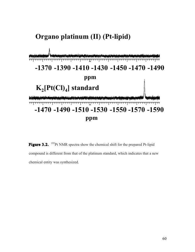
60
Figure 3.2. 195Pt NMR spectra show the chemical shift for the prepared Pt-lipid
compound is different from that of the platinum standard, which indicates that a new
chemical entity was synthesized.

61
Figure 3.3. Electrospray ionization mass spectrometry shows the mass to charge ratios
of the Pt-lipid complex.
810 830 850 870 890 910 m/z
0
20
40
60
Rela
tive A
bu
nd
an
ce
825.33
896.94
861.08
[M-H]-
[M+Cl]-
[M-HCl-H]-
80
100

62
Figure 3.4. Dynamic light scattering measurements of the drug nanoparticles show
similar particle size for the three types of hybrid formulations.
0
2
4
6
8
10
12
14
1 10 100 1000
% In
tens
ity
Particle Size (nm)
Pt-lipid NPs
CPT NPs
Combination Pt-lipid CPT NPs

63
Formulation Particle Size
(nm)
Zeta Potential
(mV)
PDI
Pt-lipid
nanoparticles 80.1 ± 0.8 -59.8 ± 0.5 0.219 ± 0.008
CPT nanoparticles
64.9 ± 0.3 -72.1 ± 2.0 0.188 ± 0.008
Pt-lipid CPT
combination
nanoparticles
61.1 ± 0.1 -61.6 ± 0.4 0.255 ± 0.002
Table 3.1. Table shows the average particle size, zeta potentials, and PDI for each of
the nanoparticle formulations.

64
Figure 3.5. This is a representative SEM image of the combinatorial nanoparticles.
200 nm

65
Figure 3.6. This graph shows the drug loading results for both camptothecin and
Platinum separately as control nanoparticles and dually loaded in combination
nanoparticles.
0.0
0.2
0.4
0.6
0.8
1.0
1.2
1.4
1.6
1.8
2.0
CPT Loaded Hybrid
Nanoparticles (CPT
content)
Dual Loaded Hybrid
Nanoparticles (CPT
Content)
Platinum Loaded Hybrid
Nanoparticles (Platinum
Content)
Dual Loaded Hybrid
Nanoparticles (Platinum
Content)
Dru
g L
oa
din
g P
erce
nta
ge
(%)
CPT and Platinum Drug Loading

66
Figure 3.7. This calibration plot by HPLC was used to determine drug loading for CPT
in the cytotoxicity samples because the drug levels were too low to be detected by UV-
Vis spectroscopy.
y = 460.85x - 2.9496 R² = 0.99939
0
500
1000
1500
2000
2500
0 1 2 3 4 5 6 Rel
ativ
e In
tens
ity (m
Au)
CPT concentration (ug/mL)
CPT- Calibration using HPLC

67
Figure 3.8. UV-Vis spectroscopy was used to determine CPT content during the in
vitro release studies.
y = 0.0205x + 0.0148 R² = 0.99562
0
0.2
0.4
0.6
0.8
1
1.2
0 10 20 30 40 50 60
Abs
orba
nce
CPT concentration (ug/mL)
CPT Calibration Plot

68
Figure 3.9. The in vitro release profile compares the drug release of CPT and of
Platinum from the combination hybrid nanoparticles in which the CPT releases faster
after 4 hours. It also shows that the addition of another drug to the hybrid nanoparticle
system does not affect overall drug release profile as compared with control single drug
loaded formulations.
0
10
20
30
40
50
60
70
80
90
100
0 5 10 15 20 25 30 35 40 45 50 55 60 65 70 75 80 Dru
g R
ele
ase
Percen
tage (
%)
Time (Hours)
CPT and Platinum In Vitro Release
Platinum release from
Combination Hybrid
NPs
CPT Release from
Combination Hybrid
NPs
CPT Release from
Hybrid NPs
Platinum release from
Hybrid NPs

69
Figure 3.10. The cytotoxicity results show that the combination formulation is capable
of reducing cell viability in ovarian cancer cells.
0.0!
0.2!
0.4!
0.6!
0.8!
1.0!
1.2!
0.1! 1! 10! 100! 1000!
Rel
ati
ve
Cel
l V
iab
ilit
y!
NP Concentration (ug/mL)!
In Vitro Cytotoxicity!

70
3.8 Conclusions and Outlook
The current course of cancer treatment falls short because of problems associated
with nonspecific systemic distribution leading to cytotoxicity of healthy cells, poor
circulation half-life, instability in the bloodstream causing decreases in efficacy,
insufficient drug concentrations at the desired sites, and multidrug resistance. This work
provides an approach to address the aforementioned concerns. By combining multiple
drugs into a single delivery system, which can be functionalized to target specific tissues
and cells, the drugs could have the opportunity to reach the targeted sites with the correct
mass ratios and the possibility to induce therapeutic synergism.
71
3.9 References
[1] C. Sanchez-Cano, M.J. Hannon, Novel and emerging approaches for the delivery
of metallo-drugs. Dalton Transactions (48) (2009) 10702-10711.
[2] S. Dhar, N. Kolishetti, S.J. Lippard, O.C. Farokhzad, Targeted delivery of a
cisplatin prodrug for safer and more effective prostate cancer therapy in vivo. PNAS 108
(5) (2011) 1850-1855.
[3] S. Cai, Y. Xie, N.M. Davies, M.S. Cohen, M.L. Forrest, Carrier-based
intralymphatic cisplatin chemotherapy for the treatment of metastatic squamous cell
carcinoma of the head and neck. Ther. Deliv. 1 (2) (2010) 237-245.
[4] P.A. McCarron, W.M. Marouf, D.J. Quinn, F. Fay, R.E. Burden, S.A. Olwill, C.J.
Scott, Antibody targeting of camptothecin-loaded PLGA nanoparticles to tumor cells.
Bioconjugate Chem 19 (8) (2008) 1561-1569.
[5] R.G. Shao, C.X. Cao, W. Nieves-Neira, M.T. Diamanche-Boitrel, E. Solary, Y.
Pommier. Activation of the Fas pathway independently of Fas ligand during apoptosis
induced by camptothecin in p53 mutant human carcinoma cells. Oncogene (20) (2001)
1852-1859.
[6] S. Xia, E.M. Rosen, J. Laterra, Sensitization of glioma cells to Fas-dependent
apoptosis by chemotherapy-induced oxidative stress. Cancer Res (65) (2005) 5248-5255.
[7] S. Svenson, M. Wolfgans, J. Hwang, J. Ryan, S. Eliasaf, Preclinical to clinical
development of the novel camptothecin nanopharmaceutical CRLX101. J. Control.
Release (2011)
72
[8] A.J. Shuhendler, R.Y. Cheung, J. Manais, A. Connor, A.M. Rauth, X.Y. Wu, A
novel doxorubicin – mitomycin C co-encapsulated nanoparticle formulation exhibits anti-
cancer synergy in multidrug resistant human breast cancer cells. Breast Cancer Res Treat
(119) (2010) 255-269.
[9] H.L. Wong, A.M Rauth, R. Bendayan, J.L. Manais, M. Ramaswamy, Z. Liu, S.Z.
Erhan, X.Y. Wu, A new polymer-lipid hybrid nanoparticle system increases cytotoxicity
of doxorubicin against multidrug-resistant human breast cancer cells. Pharmaceutical
Research 23 (7) (2006) 1574-1585.
[10] F. Greco, M.J. Vincent, Combination therapy: opportunities and challenges for
polymer-drug conjugates as anticancer nanomedicines. Advanced Drug Delivery
Reviews (61) (2009) 1203-1213.
[11] S. Aryal, C-M. Hu, L. Zhang, Polymer-cisplatin conjugate nanoparticles for acid-
responsive drug delivery. ACS Nano (4) (2010) 251-258.

73