synthesis and characterization of four alkyl 2-cyanoacylate
TRANSCRIPT

1
Thermal Development of Latent Fingermarks on Porous Surfaces – Further
Observations and Refinements
Di Fei Song a, Daniel Sommerville a, Adam G. Brown b, Ronald G. Shimmon a, Brian J.
Reedy a,*, Mark Tahtouh a.
a Centre for Forensic Science, University of Technology, Sydney, PO Box 123, Broadway
NSW 2007 Australia.
b Forensic Services Group, New South Wales Police Service, 1 Charles Street Parramatta
NSW 2150 Australia.
* Corresponding author. Tel.: +61 2 9514 1709; fax: +61 2 9514 1460. Email address:
[email protected] (Brian J. Reedy)
The invention described in this paper is the subject of Patent Application number
PCT/AU2008/001771. Those wishing to obtain further details should email their inquiry to

2
Abstract
In a further study of the thermal development of fingermarks on paper and similar surfaces, it
is demonstrated that direct contact heating of the substrate using coated or ceramic surfaces at
temperatures in excess of 230 C produces results superior to those obtained using hot air.
Fingermarks can also be developed in this way on other cellulose-based substrates such as
wood and cotton fabric, though ridge detail is difficult to obtain in the latter case.
Fluorescence spectroscopy indicates that the phenomena observed during the thermal
development of fingermarks can be reproduced simply by heating untreated white copy paper
or filter paper, or these papers treated with solutions of sodium chloride or alanine. There is
no evidence to suggest that the observed fluorescence of fingermarks heated on paper is due
to a reaction of fingermark constituents on or with the paper. Instead, we maintain that the
ridge contrast observed first as fluorescence, and later as brown charring, is simply an
acceleration of the thermal degradation of the paper. Thermal degradation of cellulose, a
major constituent of paper and wood, is known to give rise to a fluorescent product if
sufficient oxygen is available.[1-5] However, the absence of atmospheric oxygen has only a
slight effect on the thermal development of fingermarks, indicating that there is sufficient
oxygen already present in paper to allow the formation of the fluorescent and charred
products. In a depletion study comparing thermal development of fingermarks on paper with
development using ninhydrin, the thermal technique was found to be as sensitive as ninhydrin
for six out of seven donors. When thermal development was used in sequence with ninhydrin
and DFO, it was found that only fingermarks that had been developed to the fluorescent stage
(a few seconds of heating) could subsequently be developed with the other reagents. In the
reverse sequence, no useful further development was noted for fingermarks that were treated
thermally after having been developed with ninhydrin or DFO. Aged fingermarks, including

3
marks from one-year-old university examination papers were successfully developed using
the thermal technique.
Keywords: forensic science, fingermarks, fingermarks, thermal development, fluorescence,
charring.

4
1. Introduction
As a general phenomenon, the thermal development of latent fingermarks on paper has been
known for several decades at least. Scott’s Fingerprint Mechanics,[6] as revised by Olsen in
1978, cites earlier publications from the 1940s in which paper is heated by an iron. At that
time, it was concluded that this was not a practical method for fingermark development,
although fire scenes might yield useful, though inadvertently-developed marks. Bleay et al
[7] have also noted this possibility and show an example of a mark developed (by an
unknown mechanism) on a glossy card.
The application of heat to develop latent marks on paper was revisited by Almog and Marmur
in 1981.[8] They conducted a series of “baking” experiments on paper with latent
fingermarks using an electric furnace. They achieved fingermark “charring” in the
temperature range of 260 to 275 °C, with baking times of 20 to 30 seconds. However,
background coloration appeared in all of the samples and marks older than four days
generally showed up as “unresolved stains”. The technique was again concluded to be
inferior to ninhydrin.
Recently, we reported that very rapid heating in dry air in the vicinity of 300 C will very
successfully develop latent fingermarks on paper in two stages: (i) a fluorescent (but
otherwise invisible) fingermark is developed after very short heating times (several seconds)
and (ii) a visible fingermark with excellent ridge detail and contrast is developed after further
heating.[9] We also observed that the latter (visible) stage could yield improved contrast
when observed under ultraviolet rather than visible illumination. At first, it seemed
remarkable that stage (i) had not previously been reported, and that stage (ii) had not
previously been optimized to give results that could compete with those yielded by chemical
reagents. Upon reflection, it seems that the windows of time and temperature required to

5
give good fingermark development (without complete charring or combustion of paper) were
too narrow for easy discovery, though these phenomena have probably been observed
previously without being attributed much significance.
In 2009, Dominick et al[10,11] also reported the thermal development of fingermarks to the
fluorescent stage. Eccrine fingermarks on filter paper exposed to temperatures between 130
and 180 C for 20 minutes were observed to fluoresce under violet-blue, blue and green light.
Contrast was diminished above 170 C for this heating time. These workers noted that amino
acids, sodium chloride and urea all caused fluorescence above that of the background paper,
although the sodium chloride-induced fluorescence was not observed at temperatures below
190 C. They speculated that amino acid decomposition products could contribute to the
fluorescence.
In our experience (at temperatures over 200 C), the overall effect seems to arise from an
accelerated charring or scorching of the paper under the fingermark ridges, rather than by a
different chemical reaction of the fingermark constituent with the paper. This is borne out by
the observation that clean paper, when heated, goes through the same stages of fluorescence
followed by visible browning, but more slowly than in the presence of fingermark
residues.[9] However, to understand and perhaps better control the thermal development of
fingermarks on paper, it may be instructive to consider the thermal degradation of cellulose (a
major component of paper), which is known to result in chemiluminescence,[1-5] as well as
whether similar thermal development phenomena can be observed with other cellulose-based
materials.
Another conclusion of our previous article was that direct contact heating (such as with hot
metal plates) did not yield good contrast, because the background coloration (essentially

6
scorching) appeared to develop as rapidly as the fingermark ridges, and so contrast was poor.
Instead, we recommended the application of hot air, with the rationale that the heating
medium needed to be a poor conductor of heat.
In the current work, we report new refinements to the thermal development technique that
make it even more rapid and convenient, and should allow it to be automated more readily.
New surfaces, such as wood and cotton fabric are investigated for their ability to yield
developed fingermarks with heating. We also describe experiments aimed at determining the
role of oxygen in the thermal development process, and we compare thermal development
results with those obtained using conventional reagents such as ninhydrin. In addition, we
have investigated the mechanism by which thermal development occurs in order to confirm
that the effect is a result of accelerated decomposition of the substrate and not due to a
reaction between chemicals in the fingermark residue and/or the paper.

7
2. Materials and Methods
2.1. Sample preparation and treatment
Latent fingermarks were prepared on porous surfaces according to the following general
method. The donor’s hands were not washed prior to the deposition of a fingermark deposit.
Before the next deposit, the donor’s fingers were allowed to recharge either by waiting for a
moment and/or rubbing their fingers across an oily region of the face. Eccrine-rich samples
were prepared by allowing the donors’ clean hands to become sweaty from exercise. For the
depletion series of samples collected in the comparison study, the donors were not allowed to
recharge between each deposit.
The surfaces used during this study include the following:
• Australian 80% recycled 80 gsm white copy paper
• Reflex 80 gsm white copy paper
• white envelopes and brown paper
• unpolished wood
• cotton-based fabric
Sixteen donors (seven males and nine females) aged between 13 – 30 years were used during
this study. Each donor was arbitrarily assigned a number for ease of reference. In all cases,
unless otherwise specified, samples were treated within 24 hours of deposition.
Paper samples were treated using one of the following heating devices or methods: a wire-
embedded element furnace (B & L Tetlow Pty Ltd, Victoria, Australia); a Remington
Wet2Straight 230 °C hair straightener; a Sunbeam sandwich toaster with Teflon non-stick
surface; a set up of glass Petri dishes heated on hot plates; and thin aluminium plates and
thicker aluminium blocks heated by the furnace. In the current work, the hair straightener

8
provided the best results and therefore all images in the Results and Discussion section below
are of samples treated via this technique unless otherwise indicated.
Results were imaged by the Rofin Poliview IV set up with Retiga 2000R CCTV camera,
Rofin Polilight PL 500 and filters. Images of visible fingermarks were taken under white
light or UV light (350 nm). Luminescent images were captured with 505 nm excitation and
observation through a 555 nm high pass filter. Images of ninhydrin-treated samples were
taken under white light with a yellow band pass filter.
2.2. Effect of oxygen concentration
Nine fingermark samples were collected from each of the two donors, and each sample was
split in half to produce a total of thirty-six samples. These samples were thermally treated in
a nitrogen dry box (PLAS Labs, Lansing MI USA). The left halves were developed in the
dry box filled with air, while the right halves were developed in the dry box after it had been
flushed thirteen times with nitrogen to produce an atmosphere of approximately 100%
nitrogen.
2.3. Spot tests
To better understand the cause of charring in the thermal development of fingermarks, several
compounds were chosen to perform spot tests. Hexane solutions of linseed oil and long
carbon chain (C20) compounds (1% and 10 %v/v for each) were selected to model the oils and
long chain alkanes present in sebaceous secretions, while aqueous solutions of alanine,
serine, glycine and sodium chloride (NaCl) (0.1, 0.01, 0.001 M for each) were prepared to
mimic the amino acids and salts in eccrine secretions. Spots of the prepared solutions were
deposited in volumes of 2, 5 and 10 μL on Australian brand white copy paper, then treated
using a hair straightener for 2 seconds before checking for any fluorescent development
9
(Polyview system described above), then a further 2 minutes before checking for signs of
visible charring.
In a second set of tests, on both white copy paper, and on Whatman 41 filter papers, dried
spots of both sodium chloride (10% w/v) and alanine (1% w/v) solutions (in pairs) were
heated with the hair straightener at a nominal temperature of 230 C for various time periods
(10, 60, 120, 300 and 600 s). These samples were then examined using the Polyview system
and in fluorescence spectroscopy tests (Section 2.7).
2.4. Aged samples
Fingermarks were collected from three donors and split into halves. The left halves were
treated with the thermal technique on the same day, and the right halves were treated after a
period of ageing. The ageing periods for these samples were: 2, 5, 7, 16, 36, 61, and 84 days.
Further aged samples were taken from university examination papers that were in excess of
one year old. Twenty front pages were cut into a third of their original A4 size (for
convenience purposes), and thermally treated to the fluorescent stage.
2.5. Comparisons with ninhydrin
A depletion series of twelve samples was collected from seven donors and split into halves,
producing twenty-four samples for each donor. The left halves were treated with the thermal
technique, while the right halves were treated with ninhydrin on the same day. The samples
were then photographed and pieced together for comparison.
Ninhydrin solution was prepared as follows: 0.5% w/v ninhydrin; 0.5% v/v acetic acid; 5.5%
v/v ethanol and 0.5% v/v ethyl acetate in 93.5% v/v HFE 7100. All ninhydrin treatments

10
were conducted at the NSW Police Force Laboratory at Pemulwuy, Sydney in line with
standard operating procedures.
2.6. Sequencing
The thermal technique was studied in sequence before and after DFO and ninhydrin
treatments. The ninhydrin formulation used was the same as described in the previous
section. The following DFO formulation was used:
• Stock solution: 0.4 g DFO; 15 mL dichloromethane; 32 mL methanol; 3 mL acetic
acid
• Working solution: 9 mL DFO stock solution in 100 mL HFC4310
2.7. Fluorescence spectroscopy
Excitation (400-550 nm) and emission (500-700 nm) fluorescence spectra were collected
using a Varian Eclipse spectrometer, equipped with a solid sampling accessory. Spectra were
recorded for each heating time from the (chemically) untreated paper, and from each of the
alanine- and the sodium chloride-treated regions.
3. Results and discussion
3.1. Method development
Previous work by Brown et al concluded that introducing the sample into a hot air chamber,
such as an oven, was the optimum technique for the thermal development of latent
fingermarks on paper.[9] This technique was reproduced in the current work with similar
results. Fingermarks were developed over a temperature range of 240 to 315 °C.
Development times ranged from five seconds to two minutes, depending on the temperature
of the oven and the desired stage of development. Increasing the temperature led to shorter

11
development times. Better results were achieved in the lower temperature range of 240 to
280 °C. Within this range, fluorescent marks could be developed within 10 to 15 seconds,
after which time (with continued heating) the marks became visible. In the higher
temperature range of 280 to 315 °C, the development became harder to control due to the
shorter time frame; ordinary copy paper would become scorched and began burning after
roughly 20 seconds.
There were many impractical aspects of this method, particularly that the introduction of the
sample necessarily induced temperature fluctuations within the chamber that greatly
influenced the degree of development of the sample. In addition to this, the nature of the
ovens or furnaces used dictated that the sample could not be monitored during treatment,
making the technique difficult to execute optimally.
The re-investigation of direct contact heating of the sample was undertaken using a variety of
heating surfaces. A different approach to that employed by Brown et al [9] was trialled.
Instead of heating the sample by direct contact with metal surfaces, heat was applied using
less thermally-conductive surfaces. The apparatus/materials used to carry out these
experiments were a commercial hair straightener (with “ceramic”-coated surfaces), a non-
stick surface sandwich toaster and heated glass. At the conclusion of testing it was
determined that the hair straightener was superior to the other devices. This was due
primarily to the high control of temperature afforded by the hair straightener, the high
portability of the device and the speed with which samples could be treated.
These advantages displayed by the hair straightener also made it more favourable when
compared with the oven/furnace. Another advantage of the hair straightener is the ability of
the operator to easily monitor and control the progress of development. This reduces the risk
of destroying the evidence due to overheating of the sample. In addition to these advantages,

12
the quality of the fingermarks developed using the hair straightener was at least equal to that
for fingermarks developed using the oven/furnace.
3.2. Stages of fingermark development
We have previously reported that the thermal development of fingermarks on paper follows
three stages as follows:
i) fluorescent (but otherwise invisible) marks are developed after rapid heating - this
fluorescence is best observed at around 550 nm, with illumination at around 500 nm;
ii) browning of marks becomes visible with longer heating times;
iii) fingermarks lose contrast as paper turns dark brown with further heating.[9]
Fingermarks developed to fluorescent (i) and visible (ii) stages were achieved with both hot
air and the direct contact heating methods described above. As noted in previous work,[9]
the use of UV illumination on thermally developed (visible stage) fingermarks greatly
improves the contrast between the fingermark ridges and the paper background. This is
because the charring of the paper under the fingermark ridges removes or blocks the natural
UV-stimulated fluorescence of the paper, which persists in the background, provided the
heating time has not been execessive. When the quality of fingermarks developed to the
visible stage is poor or barely noticeable under white light, the ridges can appear quite
strongly under UV (see Fig. 1).

13
Fig. 1. Fingermarks (from donor 3, left and donor 8, right) photographed under white light
and UV.
In addition to the stages of development, the quality and contrast of fingermark ridges after
thermal development has been investigated. It has been observed that samples developed to
the fluorescent stage can generally achieve ridge quality equal to that of the (UV-enhanced)
visible stage. However, the contrast of visible charred marks under white light differed from
sample to sample, and is generally observed to be inferior to the quality obtained at the
fluorescent stage and under UV. As an example, the fingermark in Fig. 2 was developed to
the fluorescent stage and photographed, then further developed to the visible stage and
photographed under white light and UV.

14
Fig. 2. A fingermark (donor 4) developed to, and photographed in the fluorescent stage (a),
then developed to the visible stage and photographed under white light (b) and UV (c).
In addition to the fluorescent and visible stages of development previously identified, another
stage of development has been observed, in which the developed fingermark is both
fluorescent and visible. When heat is applied for short time periods, the fingermark becomes
fluorescent. As heating continues, the fingermark ridges become visible under UV, though
remain mostly invisible under white light. The visibility of charred ridges increases as the
heating times lengthen, but at the same time the ridges remain fluorescent, allowing the
fingermark to be observed with similar quality by either fluorescence or under UV. As the
heating time further increases, the visible charring intensifies and the contrast of the
fluorescent ridges diminishes, either because the fluorescence of the ridges decreases or
because that of the background increases (or both). The various stages of development are
shown in Fig. 3.

15
Fig. 3. Fluorescent (left), white light (middle) and UV (right) visualisation of fingermarks
(donor 4) at different stages of development with hair straightener.
3.3. Effects of fingermark constituents on the thermal degradation of paper

16
Spot tests with the C20 alkane compound on white copy paper did not lead to fluorescence or
cause visible charring greater than that of the background paper. For linseed oil, fluorescence
and charring did not occur at 1% v/v concentration, but were both achieved at 10% v/v
concentration. Spot tests with all three amino acids and NaCl yielded fluorescence and
visible charring, and so more detailed tests were performed to compare the behaviour of the
heat–induced fluorescence of paper with and without treatment with solutions of an amino
acid (alanine) and sodium chloride (NaCl).
For untreated white copy paper heated with the hair straightener at 230 C, the 505/>555 nm
fluorescence grew steadily with heating time for the first five minutes, then did not change
with further heating (up to seven minutes), although the browning of the paper continued.
Where NaCl was applied to this paper, the 505/>555 nm fluorescence (as observed on the
Polyview system) was much brighter than that of the (heated/chemically-untreated) paper and
peaked earlier, after which the fluorescence of the (heated/chemically-untreated) paper
caught up to the NaCl spot, which continued to darken under white light. Upon the first
examination of these samples, the NaCl-treated spots did not lose fluorescence with further
heating, but after three days, the spots that had been heated longer than one minute no longer
fluoresced more brightly than the paper. This behaviour and the variation in results observed
for different brands of white copy paper led us to follow the suggestion of Dominick et al
[11] to use filter paper in further tests. However, it is worth noting that alanine spots heated
at 230 C on white copy paper fluoresced much more brightly than the NaCl spots; this
fluorescence peaked very early (at around 10 s) and was almost entirely diminished by 120 s,
although the paper continued to darken (brown char) over this time.
In similar experiments on white filter paper, the alanine spots behaved exactly as described
above for copy paper, while the NaCl spots peaked in fluorescence (505/>555 nm) at about

17
60 s heating time. In both cases, fluorescence diminished as browning continued. The
chemically-untreated paper behaved in the same manner, but on a much slower time scale
(still fluorescent after 600 s heating). The filter paper samples were further examined using
fluorescence spectroscopy (Section 3.9).
It was concluded from the observations with C20 alkanes and the linseed oil that sebaceous
secretions alone are unlikely to cause the fluorescence and charring in the thermal
development of fingermarks. Paper samples with fingermarks from a consistently good
donor and a poor donor for the thermal technique were immersed in water for five to ten
minutes to remove aqueous constituents in the fingermarks, and then dried at room
temperature. Subsequent attempts to develop these samples to fluorescence and the visible
stage were not successful. This result suggests that water-insoluble components of
fingermark residue play little role in the development of fingermarks using the thermal
technique.
A comparison of depletion series of eccrine-rich and sebaceous-rich fingermarks from a poor
donor (in terms of the thermal technique) was undertaken. Although as previously noted, true
sebaceous secretions would be unlikely to cause thermal development, it must be recognized
that in reality, sebaceous secretions will contains eccrine components due to the ubiquitous
nature of eccrine glands. As found by Dominick et al, [11] the eccrine fingermarks generally
displayed a higher contrast than those of a sebaceous nature, due to the higher concentrations
of amino acids and salts in the eccrine secretions. The evidence from this experiment is in
conflict with previous work which regarded charring as a universal developer for the organic
constituents of perspiration.[8]
3.4. Effect of varying oxygen concentration

18
It has been suggested that oxygen accelerates the decomposition of cellulose which
contributes to char formation.[3,5,12] Therefore, it was initially predicted that if some or all
of the atmospheric oxygen were replaced with nitrogen, the process of thermal development
might be slowed. Samples developed in air and ~100% nitrogen atmospheres were both
developed in the glove box under similar conditions on the same day to eliminate other
possible parameters that could influence the development outcome. A large beaker of water
was placed within the glove box and allowed to equilibrate for two hours in both atmospheres
to generate similar humidity levels. Samples developed in ~100% nitrogen atmosphere were
placed in the glove box and flushed with nitrogen thirteen times to ensure that most, if not all,
of the adsorbed oxygen was removed from the paper. After development (hair straightener),
the left and right halves of each fingermark sample were pieced together to be photographed
under the same conditions. This method of photography was only used in this experiment
and not in the comparisons (see Section 3.7) and aged fingermark studies (see Section 3.5),
where the left and right halves were photographed separately under individual optimum
lighting conditions.
Viewed under UV and white light, the eighteen samples from two donors at different
development stages did show a slight difference in the charring of fingermark ridges or the
background coloration between the air- and nitrogen-developed halves. Fig. 4 displays three
samples from one donor developed for 60 seconds, 90 seconds and 4 minutes.
19
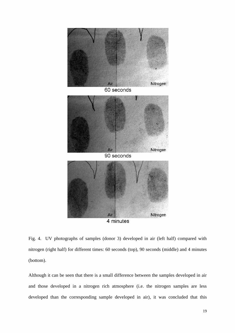
Fig. 4. UV photographs of samples (donor 3) developed in air (left half) compared with
nitrogen (right half) for different times: 60 seconds (top), 90 seconds (middle) and 4 minutes
(bottom).
Although it can be seen that there is a small difference between the samples developed in air
and those developed in a nitrogen rich atmosphere (i.e. the nitrogen samples are less
developed than the corresponding sample developed in air), it was concluded that this

20
difference was not exploitable and did not give the technique any particular advantage over
the traditional development in air.
For shorter heating times, no differences in fluorescence were observed between the samples
developed in air and nitrogen (Fig. 5). However, a small distinction in fluorescence was
observed at the longer heating times. As shown in Fig. 6, at the heating times of three and
four minutes, the fluorescence has diminished from its earlier peak, but appears slightly
stronger in the nitrogen-developed half. It therefore appears that, as might be expected, the
presence of oxygen may accelerate the fluorescent-to-visible (charred) transition in the
thermal development of fingermarks. As indicated above, the use of a nitrogen-rich
atmosphere does not provide any significant advantages over heating the sample in a regular
atmosphere as the slowing of the fingermark development is insignificant.

21
Fig. 5. Photographs of fluorescent samples from two donors (top: donor 4; bottom: donor 3)
both developed for 2 seconds in air (left half) and nitrogen (right half).

22
Fig. 6. Photographs of fluorescent samples (donor 4) developed in air (left half) and nitrogen
(right half) at development times of 3 (top) and 4 minutes (bottom).
3.5. Aged sample / exam papers
Fig. 7 shows the oldest fingermarks that were developed to the fluorescent stage from three
different donors. Fingermarks older than this could be developed, but with reduced ridge
detail.

23
Four comparisons of samples developed to the visible stage (and photographed under UV)
from each of two donors (only) are shown as examples in Fig. 8 and Fig. 9. In these
photographs, the left halves were developed on the day of preparation, while the right halves
were developed after aging. As shown, samples aged up to 12 weeks were developed with
ridge detail for donor 1, and up to 9 weeks for donor 4. In Fig. 8, it can be seen that the 12
week sample displays less contrast and ridge clarity than in the fresh control sample, however
this effect is not seen in any of the other samples from any of the donors. The results
demonstrate that it is possible to reveal aged fingermarks, but a more exhaustive study on the
effects of ageing, using a higher number of donors, should be the subject of future research.
Fig. 7. Aged fingermarks from three donors (from left to right: donor 6, 4 and 1) developed
to the fluorescent stage. Aged: 5 weeks (left), 9 weeks (middle) and 12 weeks (right).

24
Fig. 8. Comparison (donor 4) of developed fresh samples (left) with aged samples (right)
from one donor.

25
Fig. 9. Comparison (donor 1) of developed fresh samples (left) with aged samples (right)
from one donor.
Twenty front pages from examination papers more than one year old were treated with the
thermal technique to the fluorescent stage. The A4 pages were cut into thirds because it was
a convenient size for the hair straightener, and only one third from each page was treated.
One or more marks were developed on ten out of the twenty samples, giving a total of 26
marks. Fig. 10 shows an example of a fingermark and a partial palm-mark developed on the
examination papers. This indicates that fingermarks of more than one year old could be
developed with the thermal technique, but further aging studies under various environmental
storage conditions are required.
Fig. 10. Photographs of fingermarks developed with the thermal technique on old
examination pages
3.6. Alternative substrate treatment

26
Fingermarks were developed to the fluorescent and visible stages on a thin piece of
unpolished wood (Fig. 11) that fitted between the plates of the hair straightener. The
fingermarks became fluorescent (505/555 nm) almost immediately upon heating.
Fig. 11. Fingermarks (donor 4) developed on unpolished wood to fluorescent stage (top), and
the visible stage, observed under white light (middle) and UV illumination (bottom).

27
Attempts were also made to develop fingermarks to the fluorescent and visible stages on two
types of masking tape. None of the samples could be developed to the fluorescent stage,
while samples developed to the visible stage showed high background coloration, with
minimal ridge detail under visible light. UV illumination was able to improve the quality of
these samples, revealing good ridge quality (Fig. 12). In general, masking tape was a
difficult surface, and the process of heating released a strong odor. The thermal treatment of
adhesive tape should be conducted in a fume hood or well-ventilated area with appropriate
safety equipment.
Fig. 12. Fingermarks (donor 4) developed on masking tape, and photographed under UV.
Attempts were made to develop fingermarks on cotton-based fabrics to the fluorescent and
visible stages. No ridge detail was developed, but the presence of fingermarks could be
identified (Fig. 13). The texture of the substrate seems to limit the application of the thermal

28
development technique to fabrics, although further testing with more finely-woven substrates
may give more favorable results.
Fig. 13. Fingermarks (donor 4) developed on fabric to the fluorescent stage (top) and visible
stage (bottom).
The stages of fingermark development on a white envelope were found to be similar to that of
white copy paper; fluorescent marks were developed with shorter heating times and visible
marks with longer heating times. Fingermarks deposited on the sticky side of the glue strip

29
could be developed to the visible stage (Fig. 14). On brown paper, fingermarks were
developed to the fluorescent stage (Fig. 15). Fingermark developed to the visible stage on
brown paper were difficult to observe due to the brown background.
Fig. 14. Photographed under UV illumination: a fingermark (donor 5) developed on the non-
sticky side (left) and the sticky-side (right) of the glue stripe on a white envelope.
Fig. 15. Fingermark (donor 5) developed to the fluorescent stage on brown paper.
Results were obtained from fingermarks deposited over various inks and toners imprinted on
white copy paper. Fluorescent fingermarks could not be developed on top of inks that did not
fluoresce under the lighting conditions used to observe thermal development fluorescence.
Fig. 16 shows an example of fingermarks developed to the fluorescent stage over black

30
laser-printed text. It can be seen that the fluorescent ridges are interrupted by the black toner.
Further heating to develop these samples to the visible stage was still unable to develop ridge
detail over the toner.
Fig. 16. Fingermarks (donor 4) deposited over laser-printed text developed to the fluorescent
stage.
In an interesting example, fingermarks deposited over a red ink which was fluorescent under
the lighting parameters used (505 nm illumination; 555 nm high pass filter), were observed to
fluoresce over the ink upon thermal treatment (Fig. 17). Longer heating was observed to
quench the fluorescence of the ink.

31
Fig. 17. Fingermarks (donor 4) deposited over red ink and pencil developed to the
fluorescent stage (left) then visible stage (right).
3.7. Comparision with ninhydrin treatment
Ninhydrin is the most commonly used technique for the development of fingermarks on
porous surfaces and has been generally used as the standard method of reference for
comparison with other techniques. Therefore, ninhydrin was the technique used as a
comparison with the thermal technique on depletion series of fingermarks from seven donors.
The selected donors included people who had previously been identified as good and poor
donors for the thermal technique.

32
It is difficult to make an objective assessment of the relative fingermark ridge quality
achieved using these two techniques but little difference was observed between the thermal
and ninhydrin techniques for most of the donors.
Using the results of this comparison study, an objective assessment of the relative sensitivity
of the two techniques can be made since the amount of fingermark residue present decreases
with each successive fingermark deposit within the depletion series. Therefore, the more
exploitable fingermarks that are developed within a series for a particular donor, the more
sensitive the technique. An assessment of relative sensitivity was made by identifying the
last fingermark within the depletion series that showed exploitable ridge detail for each
technique and for each donor (see Table 1).
Number of fingermarks developed (with exploitable ridge detail)
Donor Thermal technique Ninhydrin
1 6 6
2 6 6
3 3 3
4 7 7
5 4 11
6 1 1
7 2 2
Table 1. Number of fingermarks developed (with exploitable ridge detail) within depletion
series for thermal technique and ninhydrin.
These results indicate that the thermal technique was as sensitive as ninhydrin for six out of
the seven donors tested.

33
3.8. Use of thermal development within a fingermark sequence
In the sequencing studies carried out, it was determined that the thermal technique should be
used in a sequence before DFO and ninhydrin, and that thermal development must be to the
fluorescent stage only, with the shortest heating time. Successful ninhydrin-based
development after thermal development was achieved in thermal samples developed strictly
to the fluorescent stage, with a heating time no longer than a few seconds. Samples
developed to the stage where the sample was both fluorescent and visible could not
subsequently be developed with ninhydrin. The use of ninhydrin after thermal development
produced purple ridges as usually observed during ninhydrin treatment (see Fig. 18). This
treatment did not cause the sample to lose the fluorescence produced by thermal
development. For the four donors used for this sequencing study, no additional ridge
characteristics were detected with the application of ninhydrin after thermal treatment.
Fig. 18. Fingermark developed with heat (left) followed by ninhydrin (right).

34
The use of thermal development after ninhydrin treatment was found to be of no value. The
background quickly turned brown upon heating, and as the heating time increased, the purple
ridges turned to dark purple and then brown, which resulted in reduced contrast.
The use of DFO after thermal development gave similar results to ninhydrin; only samples
exposed to the shortest heating times were able subsequently to be developed with DFO.
Samples developed with DFO after thermal treatment showed an increase in fluorescence,
and in some cases increased ridge details (Fig. 19).
Fig. 19. Fingermark developed with heat (left) followed by DFO (right). Both images were
captured with 505 nm excitation and observation through a 555 nm high pass filter.
3.9. Fluorescence spectra of heated paper

35
Fluorescence spectra were obtained to compare the fluorescent properties of unheated white
filter paper with filter paper that had been heated various lengths of time, with and without
the prior application of solutions of sodium chloride and alanine. The examination of these
samples using filtered light was described above in Section 3.3. In the absence of
microfluorometric information from thermally developed fingermark ridges, the aim was to
determine spectroscopically whether paper undergoes fluorescent changes upon heating that
are consistent with the observed behavior of thermally developed fingermark ridges, as
suggested by other results reported above. Excitation spectra were used to select an
appropriate excitation wavelength for the collection of emission spectra.
Unheated white paper fluoresces “naturally” across the visible range when illuminated with
UV or blue light. Our observations reported above indicate that this fluorescence decreases
in intensity with heating of the paper (hence the better contrast of charred fingermarks when
viewed under UV light). The fluorescence that arises from the heating of paper, observed
above 500 nm, therefore overlaps with decreasing “natural” fluorescence, making the
interpretation of spectra in this region somewhat complicated if spectral subtraction (of
untreated paper, say) is attempted. Thus, the spectra described and shown here are
unsubtracted, and therefore contain contributions from both types of fluorescence. Emission
spectra (480 nm excitation) from the heating of alanine-treated filter paper and NaCl-treated
filter paper are shown in Fig. 20 and Fig. 21, respectively. Fig. 22 shows a comparison of
alanine-treated and NaCl-treated spectra of similar intensities, and Fig. 23 shows the
excitation spectra (for emission at 550 nm) for heated (230 C) filter paper after no chemical
treatment, alanine treatment and NaCl treatment.

36
Fig. 20. Fluorescence emission spectra (480 nm excitation) of heated, alanine-treated paper.
Fig. 21. Fluorescence emission spectra (480 nm excitation) of heated, NaCl-treated paper.

37
Fig. 22. Comparison of fluorescence emission spectra (480 nm excitation) of alanine- and
NaCl-treated paper (heating times chosen to give similar intensities).
Fig. 23. Fluorescence excitation spectra (550 nm emission) of heated paper after various
treatments.
The most important general observations to be made regarding these results are (i) that the
fluorescence spectra (excitation and emission) obtained for heated filter paper are virtually
identical (save for intensity differences) for alanine-treated, NaCl-treated and chemically-

38
untreated paper; (ii) that the spectra obtained are very similar to those obtained by Dominick
et al [10,11] for heated filter paper. This strongly suggests for the treatments tested, the
fluorescent species produced are the same in every case, and these must arise from the
heating of paper alone, rather than the reaction of alanine or sodium on or with the paper.
Along with other evidence presented here, this tends to support our earlier proposition that
the thermal development of fingermarks on paper occurs predominantly through the
accelerated heating of paper under the fingermark ridges, rather than through any reaction of
fingermark residues on or with the paper. Since cellulose is known to fluoresce upon heating
(to temperatures similar to those used in this study),[3-5] our observations that fingermarks
can be thermally developed on other cellulose-containing substrates such as wood and cotton
also support this proposition.
The presence of alanine accelerates the thermal degradation of paper more than does sodium
chloride, which is consistent with the conclusion by Dominick et al [11] that amino acids are
chiefly responsible for the thermally-induced fluorescence of fingermarks. Differences in the
ability of different fingermark constituents to enhance paper degradation are presumably
related to differing thermal (rather than chemical) properties of these constituents, such as
their relative effects on the thermal conductivity of the paper. These effects could be related
to the different affinities that these constituents have for cellulose/paper. This would explain
the apparent requirement for fingermark development that the paper be heated in/by a poorly
conductive medium such as air or ceramics; direct contact heating using metal surfaces
results in poor contrast because the heat is transferred to both the ridges and the background
at similar rates.

39
4. Conclusion
The thermal development of fingermarks on paper and other surfaces has great potential as a
simple, low-cost, chemical-free method for fingermark detection and visualization,
particularly in situations where development might not otherwise be attempted, for reasons of
time and expense. The development of an automated, high-throughput device for developing
fingermarks on bulk quantities of documents is strongly suggested. In a study of the
sensitivity of the thermal technique relative to that of ninhydrin, we have found that for six
out of the seven donors used, the thermal technique was at least as sensitive as ninhydrin. In
a study of aged marks, it was found that marks aged at least one year could be s uccessfully
developed using the thermal technique.
The chief potential disadvantage of the thermal technique is that it is inherently destructive,
but if fingermarks are developed only to the fluorescent stage, they can still be developed by
ninhydrin in a sequence, implying that amino acids can survive short heating periods. It is
probable that any DNA present in a latent fingermark would be destroyed by heating, but
further work is required to establish the heating conditions beyond which no useful DNA
would be recovered. It should be noted that the actual temperatures attained by the samples
(over a period of a few seconds of heating to the fluorescent stage) are probably lower in
most cases than the nominal treatment temperatures reported (ie the temperature of heating
media or devices). Since the destructive nature of the technique would not always be an issue
(especially in situations where fingermarks would not otherwise be developed for reasons of
time or expense), and since many other fingermark development techniques are at least
partially destructive, this disadvantage need not preclude the use of this technique in
appropriate casework situations. In casework, where an unknown paper type or other
substrate may be encountered, the recommended procedure would be to first test temperature
and heating time requirements on a similar substrate, then, with those results in mind, adopt a

40
slow, stop-start approach with frequent monitoring of the sample for fluorescence. The
design of devices that utilize the thermal development phenomenon should incorporate
mechanisms for monitoring and controlling the progress of development to prevent sample
damage. Another potential disadvantage of the technique is that the heating of certain
substrates, such as coated papers, can generate fumes that may be unsafe for the operator, and
so the heating of such samples in a properly-ventilated enclosure is recommended.
The results of the current study show that although hot air at 250-300 C can be used for the
thermal development of fingermarks on paper, direct contact heating with non-metallic
surfaces at temperatures above 230 C is more convenient and can give better results, as it is
easier to control. Such surfaces include coated heating elements, and heated glass and
ceramics. We have also shown that the application of heat can be used to develop
fingermarks on wood and other cellulose-based materials such as cotton, although in the latter
case, the weave of a fabric will usually obscure the fingermark ridge pattern. The oxidation
of cellulose with molecular oxygen is known to produce a fluorescent product[3-5] that may
explain the early fluorescence of fingermarks during the heating process. However, the
current work has shown the thermal development of fingermarks can still be carried out in an
atmosphere of pure nitrogen, with only a marginal decrease in the speed of development by
direct contact heating at 230 C. This implies that if the fluorescence and later charring of
paper is a reaction with oxygen, there is already sufficient oxygen in the constituents of the
paper, or adsorbed strongly to the paper, to facilitate the oxidation process.
Finally, we have shown that the thermally-induced fluorescence of paper has the same origin,
whether the paper has been treated first with an amino acid (alanine), sodium chloride or real
fingermark residues, or has simply been heated without chemical treatment. In all of these
cases, the optimal wavelengths for observation of the fluorescence were identical, as were the

41
fluorescence excitation and emission spectra. However, the process (including the eventual
decrease in fluorescence and the browning of the paper) is greatly accelerated by the presence
of the amino acid, and to a lesser extent, by sodium chloride. This seems to confirm that the
thermal development of fingermarks is due to faster heating of the substrate paper under the
fingermark ridges, and not due to any specific reaction of fingermark residue constituents on
or with the paper.
Acknowledgments
The authors would like to thank the Forensic Services Group, New South Wales Police
Service, for assistance with the ninhydrin/DFO/thermal comparison studies.
References
[1] M. Pletenikova, L. Matisova-Rychla, J. Rychly, and I. Lacik, Chemiluminescence
related to degradation of thermally oxidized pullulans. Comparison with cellulose and
dextran. Carbohydr. Polym. 69 (2007) 50-64.
[2] L. Matisova-Rychla, J. Rychly, A. Ebringerova, K. Csomorova, and A. Malovikova,
Chemiluminescence accompanying the oxidation of hemicelluloses. Polym. Degrad.
Stab. 93 (2008) 1674-1680.
[3] J. Rychly, L. Matisova-Rychla, M. Lazar, I. Janigova, M. Strlic, D. Kocar, J. Hanus,
J. Minarikova, and S. Katuscak, Thermal oxidation of cellulose investigated by
chemiluminescence. The effect of magnesium and calcium carbonates and of different
pHs. C. R. Chimie 9 (2006) 1425-1432.
[4] J. Rychly, M. Strlic, L. Matisova-Rychla, and J. Kolar, Chemiluminescence from
paper. I. Kinetic analysis of thermal oxidation of cellulose. Polym. Degrad. Stab. 78
(2002) 357-367.
[5] M. Strlic, J. Kolar, B. Pihlar, J. Rychly, and L. Matisova-Rychla, Chemiluminescence
during thermal and thermo-oxidative degradation of cellulose. Eur. Polym. J. 36
(2000) 2351-2358.
[6] R.D. Olsen, Scott's fingerprint mechanics, Charles C Thomas, Springfield, Illinois,
1978.
[7] S.M. Bleay, G. Bradshaw, and J.E. Moore, Fingerprint development and imaging
newsletter: Special edition. Home Office Scientific Development Branch April (2006)
1-32.
[8] J. Almog, and A. Marmur, Chemical reagents for the development of latent
fingerprints. IV: The charring process. J. Forensic Sci. 26 (1981) 393-397.
[9] A.G. Brown, D. Sommerville, B.J. Reedy, R.G. Shimmon, and M. Tahtouh,
Revisiting the thermal development of latent fingerprints on porous surfaces: New
aspects and refinements. J. Forensic Sci. 54 (2009) 114-121.

42
[10] A.J. Dominick, N.N. Daeid, S.M. Bleay, and V. Sears, Recoverability of Fingerprints
on Paper Exposed to Elevated Temperatures--Part 1: Comparison of Enhancement
Techniques. J. Forensic Ident. 59 (2009) 325-339.
[11] A.J. Dominick, N.N. Daeid, S.M. Bleay, and V. Sears, Recoverability of Fingerprints
on Paper Exposed to Elevated Temperatures--Part 2: Natural Fluorescence. J.
Forensic Ident. 59 (2009) 340-355.
[12] S. Soares, G. Camino, and S. Levchik, Comparative study of the thermal
decomposition of pure cellulose and pulp paper. Polym. Degrad. Stab. 49 (1995) 275-
283.