synthesis and cell adhesion studies of linear and
TRANSCRIPT
1
Synthesis and cell adhesion studies of
linear and hyperbranched poly(butyl
methacrylate) and poly(t-butyl acrylate)
Kayleigh J Cox-Nowak
Submitted for the degree of Doctor of Philosophy
Department of Chemistry
October 2013
2
Contents Acknowledgements .................................................................................................................. 6
Abbreviations ........................................................................................................................... 7 Abstract ..................................................................................................................................... 8 1 - Introduction ........................................................................................................................ 9 Context ...................................................................................................................................... 9 1.1 Emulsion polymerisation ................................................................................................. 10
1.2 RAFT polymerisation ...................................................................................................... 15 1.3 Ozonolysis ......................................................................................................................... 18 1.4 Hyperbranched polymers ................................................................................................ 20 1.5 Interpenetrating polymer networks ............................................................................... 23
A note on terminology ................................................................................................... 23
1.6 Stimuli responsive interpenetrating polymer networks ............................................... 24
1.7 Cell adhesion and biocompatibility ................................................................................ 26 2 - Synthesis of oligo(n-butyl methacrylate) with acid or amine end groups ................... 30
2.1 Introduction ...................................................................................................................... 30 2.2 Results and Discussion ..................................................................................................... 32
2.2.1 Monomer starve-fed emulsion polymerisation of butyl methacrylate and
butadiene ............................................................................................................................. 32
2.2.2 Formation of oligo(butyl methacrylate) and end group functionalization .......... 38
2.2.3 Characterisation of functionalised oligomers ......................................................... 41
2.2.4 Cell contact studies ................................................................................................... 45
Human Dermal Fibroblasts .............................................................................................. 45
Human Renal Epithelial Cells........................................................................................... 50
2.3 Conclusions ....................................................................................................................... 54 2.4 Experimental .................................................................................................................... 56
Instrumentation.................................................................................................................. 56
2.4.1 Monomer starve-fed emulsion polymerisation of butyl methacrylate and
butadiene ............................................................................................................................. 56
Materials ......................................................................................................................... 56
Equipment ...................................................................................................................... 57
Method ............................................................................................................................ 59
2.4.2 Ozonolysis of Poly (Butyl methacrylate-co-Butadiene) and generation of acid
end groups........................................................................................................................... 60
Materials ......................................................................................................................... 60
Equipment ...................................................................................................................... 60
3
Method ............................................................................................................................ 60
Purification of acidic poly(BMA-co-BD) ..................................................................... 61
2.4.3 Diamine Addition to Oligo(BMA-co-BD) with acid end groups ........................... 62
Materials ......................................................................................................................... 62
Method ............................................................................................................................ 62
Purification of amidated oligomers .............................................................................. 63
2.4.4 Culture of fibroblast and epithelial cells ................................................................. 63
Human dermal fibroblasts ................................................................................................ 63
Materials ......................................................................................................................... 63
Equipment ...................................................................................................................... 63
Culture ............................................................................................................................ 63
Passage ............................................................................................................................ 64
Human renal epithelial cells .............................................................................................. 64
Materials ......................................................................................................................... 64
Preparation of media ..................................................................................................... 64
Culture ............................................................................................................................ 65
2.4.5 Culture of cells in direct contact with oligomers .................................................... 65
Materials ......................................................................................................................... 65
Equipment ...................................................................................................................... 65
Preparation of polymer films ........................................................................................ 65
Cell counting and seeding .............................................................................................. 66
Alamarblue® Assay ....................................................................................................... 67
Cell visualisation ............................................................................................................ 68
Giemsa ............................................................................................................................. 68
Hematoxylin and Eosin Y .............................................................................................. 68
3 - Hyperbranched poly(n-butyl methacrylate) and linear analogues with acid or amine
end groups: synthesis and cytocompatibility ....................................................................... 70 3.1 Introduction ...................................................................................................................... 70
3.2 Results and Discussion ..................................................................................................... 73
3.2.1 Hyperbranched polymer synthesis and functionalisation ..................................... 73
3.2.2. Characterisation of hyperbranched polymers with acid end groups .................. 81
4
3.2.3 Linear polymer characterisation ............................................................................. 83
3.2.4 Cell contact studies ................................................................................................... 87
Human Dermal Fibroblasts .......................................................................................... 87
Human Renal Epithelial Cells....................................................................................... 98
3.3 Conclusions ..................................................................................................................... 110
3.4 Experimental .................................................................................................................. 111
3.4.1 Synthesis of RAFT chain transfer agent: 4-vinylbenzyl-pyrrolecarbodithioate
............................................................................................................................................ 111
Materials ....................................................................................................................... 111
Method .......................................................................................................................... 111
3.4.2 RAFT polymerisation of n-butyl methacrylate using 4-vinylbenzyl pyrrole
carbodithioate chain transfer agent ............................................................................... 112
Materials ....................................................................................................................... 112
Equipment .................................................................................................................... 113
Method .......................................................................................................................... 113
3.4.3 Reaction of hyperbranched butyl methacrylate polymers with excess ACVA . 114
Materials ....................................................................................................................... 114
Method .......................................................................................................................... 114
3.4.4 Coupling of linear butyl methacrylate copolymers to diamines ......................... 115
Materials ....................................................................................................................... 115
Method .......................................................................................................................... 115
Purification of amidated polymers ............................................................................. 116
3.4.5 Culture of cells in direct contact with polymers .................................................. 116
Materials ....................................................................................................................... 116
Equipment .................................................................................................................... 116
Preparation of polymer films and cell seeding .......................................................... 116
4 - Hyperbranched Poly(t-butyl acrylate) .......................................................................... 118
4.1 Introduction .................................................................................................................... 118 4.2 Results and discussion ................................................................................................... 121
4.2.1 Polymer synthesis and characterisation................................................................ 121
4.2.2 Characterisation of acid functional polymers and embedding into semi-
interpenetrating networks ............................................................................................... 127
5
4.2.3 Cell contact studies ................................................................................................. 130
Human Dermal Fibroblasts ........................................................................................ 130
Human Renal Epithelial Cells..................................................................................... 136
4.3 Conclusions ..................................................................................................................... 141
4.4 Experimental .................................................................................................................. 143
4.4.1 Synthesis of RAFT chain transfer agent: 4-vinylbenzyldithiobenzoate............. 143
Materials ....................................................................................................................... 143
Method .......................................................................................................................... 143
4.4.2 RAFT polymerisation of t-butyl acrylate using 4-vinylbenzyl dithiobenzoate
chain transfer agent ......................................................................................................... 144
Materials ....................................................................................................................... 144
Equipment .................................................................................................................... 144
Method .......................................................................................................................... 145
4.4.3 Reaction of hyperbranched t-butylacrylate polymers with excess ACVA ........ 146
Materials ....................................................................................................................... 146
Method .......................................................................................................................... 146
4.4.4 Embedding of hyperbranched polymers into semi-interpenetrating networks 146
Materials ....................................................................................................................... 146
Preparation of semi-IPNs ............................................................................................ 147
4.4.5 Culture of cells in direct contact with polymers .................................................. 148
Materials ....................................................................................................................... 148
Equipment .................................................................................................................... 148
Preparation of polymer films and cell seeding .......................................................... 148
5 - Overall conclusions and future work ............................................................................ 150
Future work ...................................................................................................................... 152
6 - References........................................................................................................................ 154

6
Acknowledgements
I would like to thank Professor Steve Rimmer who gave me the opportunity to work on this
project and who has provided continued assistance and support.
My thanks also go to Melanie Hannah, Keith Owen and Jennifer Louth for their excellent
technical support. Dr. Kathryn Swindells, Dr. Richard England & Dr. Paul Bonner provided
support with polymer theory and Katherine Brown provided emulsion polymerisation
training. Dr Steve Carter and Dr. Prodip Saker gave assistance with the synthesis of chain
transfer agents. I would also like to extend my gratitude to Dr. Lian Hutchings who kindly
allowed the use of his triple detection GPC system and Sarah Canning who provided polymer
samples (linear PolyBMA-co-4VBA).
Deserving of special mention are Annika Clifton and Andrew McKenzie who gave me
valuable advice on various aspects of cell culture. I credit all of the members of the Polymer
Centre who provided me with a working environment that was friendly and stimulating.
Last but not least I would like to thank my mum, Franciska, who encouraged my studies from
a young age and Dave for his support and unending patience when my work was anti-social.
This work was sponsored by the BBSRC.
7
Abbreviations
(semi-)IPN (Semi-)Interpenetrating polymer network
ACVA 4,4'-azobiscyanovaleric acid
BMA Butyl methacrylate
CAM Cell adhesion molecule
CTA Chain transfer agent
DMEM Dulbecco's modified Eagle's medium
DMSO Dimethyl sulfoxide
EDC 1-Ethyl-3-(3-dimethylaminopropyl)carbodiimide
EDTA Ethylenediaminetetraacetic acid
FBS Feotal bovine serum
FT-IR Fourier transform infra-red spectroscopy
GPC Gel permeation chromatography
HBP Hyperbranched polymer
HDF Human dermal fibroblast
IR Infra-red
NMR Nuclear magnetic resonance
OD Optical density
PBS Phosphate buffered saline PEG-PEI-
PBLG Poly(ethylene glycol)–polyethylenimine–poly(γ-benzyl l-
glutamate)
PLGA Poly(lactide-co-glycolic acid)
pnipam Poly(n-isopropylacrylamide)
RAFT Radical addition-fragmentation chain transfer polymerisation
REpC Human renal epithelial cells
SEC Size exclusion chromatography
tBuAc Tertiary-butyl acrylate
UV Ultraviolet
VBA 4-vinyl benzoic acid
8
Abstract
A library of polymers/oligomers with three different architectures was synthesised. Short
chain, linear oligomers were produced by performing oxidative cleavage on a poly(butyl
methacrylate-co-butadiene) polymer. Although butadiene is a gaseous monomer, it was found
that careful control over the reaction conditions led to successful copolymerisation in an
unpressurised reactor. Hyperbranched polymers of n-butyl methacrylate and t-butyl acrylate
were synthesised by RAFT polymerisation with 4-vinylbenzyl-pyrrolecarbodithioate (CTA1)
and 4-vinylbenzyl dithiobenzoate (CTA2). A variety of analytical techniques, such as
elemental analysis and NMR, were used to characterise the polymers and confirm the
hyperbranched structure. Some variation in monomer conversion and CTA uptake was seen
under different polymerisation conditions.
After synthesis and characterisation, it was found that the polymer end groups could be
modified through work up with diamine or 4.4‘-azobiscyanovaleric acid. Linear oligomers of
butyl methacrylate were functionalised with amines whilst hyperbranched polymers were
given acid functional end groups. FT-IR and elemental analysis were used to monitor the
success of the end group reactions.
As the polymers could be applied as films, they were assessed as cell culture substrates using
Human dermal fibroblasts (HDF) and Human renal epithelial cells (HREp). A linear butyl
methacrylate-co-4-vinyl benzoic acid copolymer was also assessed in comparison to the
hyperbranched structures. It was observed that the two cell types had different responses to
each of the polymers. Fibroblast cells showed better rates of adhesion and proliferation on
acid-functionalised polymers, whilst epithelial cells performed best on the amine-
functionalised moieties.
This work provides useful information for the synthesis and preparation of new biomaterials.
It has been found that polymer functionality must be considered when compatibility with a
specific cell type is desired, and polymers with the potential to be incorporated into future
biomaterials are highlighted.
9
1 - Introduction
Context
Today, teams of polymer chemists and cell biologists are working together in an effort to
create evermore practical and functional materials for use in tissue engineering, cell re-
growth and therapy and agent delivery. The purpose of this introduction is to review and
correlate the work carried out in the areas of controlled polymer architecture and
biocompatibility. Many potential bio-interactive polymers have been identified, but rarely are
cell culture experiments performed which would provide adherence and toxicity information.
Any commercial material must be able to fit within a narrow specification range. HBPs are
attractive biomaterials as they can be synthesised with high control and within narrow
polydispersities[1]. The use of HBPs in biomaterials ranges from carriers to degradable
materials. A HBP with tertiary amino groups was synthesised by Park et al.[2] that was able
to efficiently transfect DNA with low toxicity. In addition, protein immobilisation has been
demonstrated by Shen[3], [4] and Cosulich[5] which opens the possibility of ‗enzyme-based
bioobjects‘. Lin and Zhang et al. created a biodegradable blend material with enhanced
properties by combining HBPoly(ester amide) with polylactide[6], and Chen et al. reported
the successful synthesis of an inherently biodegradable cationic HBP of PEG-PEI-PBLG[7].
These polymers all show positive bio-interactions and indicate that the amine functionality
may be a positive inclusion for increasing cell adherence and culture.
Polymers with carboxylic acid functionality are also considered to have a degree of
biocompatibility[8]. Observations by MacNeil et al. have indicated that a polymer containing
ca. 3% acid promotes the attachment of keratinocytes and osteoblast-like cells[9], [10].
The MacNeil group have also utilised the technique of electrospinning to form polymers with
acid functionality, that can be used for tissue regeneration and drug release [11], [12].
This project investigated the emulsion polymerisation of P(BMA-BD) and the
biocompatibility of oligomers after being cleaved with ozone and functionalised with amine
or acid. Also explored was the synthesis of and biocompatibility of HBP(BMA) and
HBP(tBuAc). Some polymers were reacted with 4,4‘-azobis4-cyanovaleric acid (ACVA) or
diamines in order to observe the effect of end group functionality on cell adhesion.
To our knowledge this is the first known use of RAFT to form hyperbranched polymers using
t-butyl acrylate and butyl methacrylate monomers. It was investigated what effect varying the
amount of chain transfer agent (CTA) had on the polymerisation and also on the final
product. The original aim was to synthesise a range of hyperbranched polymers, and then use
10
these to build a library of semi-interpenetrating polymer networks. However, the HBPs did
not have high enough molecular weights to stay entrapped in the networks.
In order to investigate the polymers‘ biocompatibility, they were cast into films so that cells
could be grown in direct contact, where it was found that introducing acid functionality was
advantageous. Amino groups have been considered to be more cell interactive than
carboxylic acid and OH functionality [13], [14]. The evidence provided by this work
indicates that the cell-interactivity of a functional group is also dependent on the cell type
tested against. With carefully planned experiments, it is possible to observe the effect of
molecular weight, branching and end group modifications on HBPs. The observations
described within indicate that non-mesenchymal cells show a preference for amine and
dithioate functional polymers over acids. This knowledge is essential for the creation of
future materials that possess both the necessary physical and chemical properties for
continued healthy cell adhesion and proliferation in vivo. This introduction will discuss
current methods and trends in RAFT and emulsion polymerisation, followed by an overview
of synthetic polymers (specifically hyperbranched or crosslinked) and their uses in modern
biomaterials.
1.1 Emulsion polymerisation
Emulsion polymerisation is used in a variety of applications ranging from adhesives, rubbers,
drug delivery and paper additives[15]–[22]. The product obtained from emulsion
polymerisation is a polymer dispersed in water and colloidally stabilised by surfactant –
called a latex or polymer dispersion. Typically the reaction is free radical and occurs in a
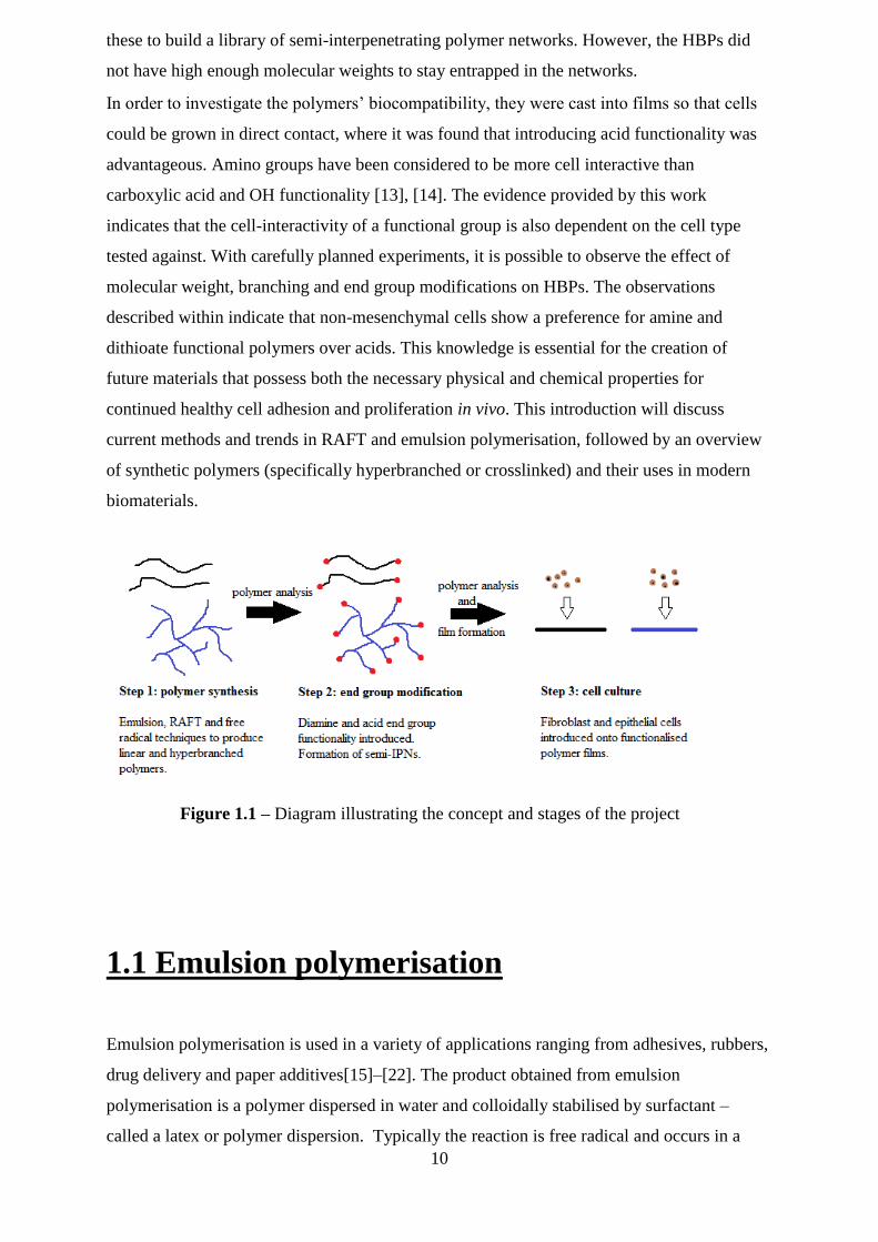
Figure 1.1 – Diagram illustrating the concept and stages of the project
11
heterophase system, where an immiscible liquid (the monomer) is held in the dispersion
medium (water) by a surfactant.
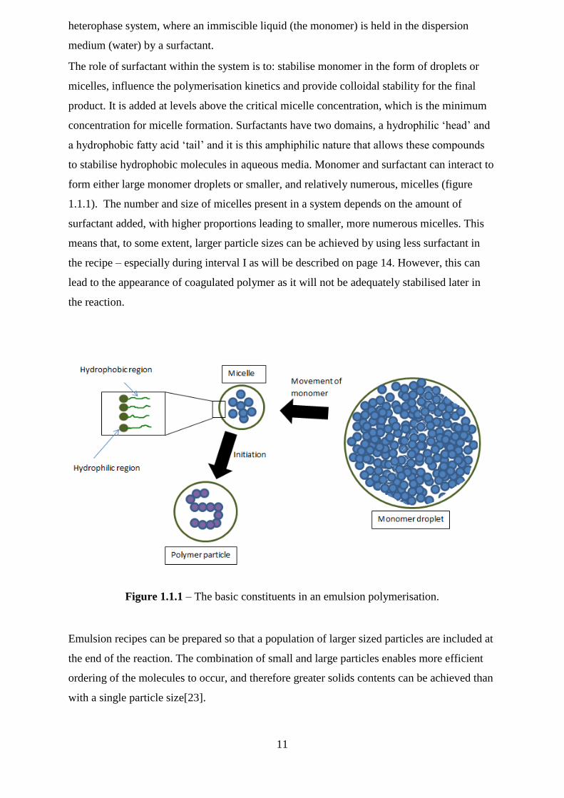
The role of surfactant within the system is to: stabilise monomer in the form of droplets or
micelles, influence the polymerisation kinetics and provide colloidal stability for the final
product. It is added at levels above the critical micelle concentration, which is the minimum
concentration for micelle formation. Surfactants have two domains, a hydrophilic ‗head‘ and
a hydrophobic fatty acid ‗tail‘ and it is this amphiphilic nature that allows these compounds
to stabilise hydrophobic molecules in aqueous media. Monomer and surfactant can interact to
form either large monomer droplets or smaller, and relatively numerous, micelles (figure
1.1.1). The number and size of micelles present in a system depends on the amount of
surfactant added, with higher proportions leading to smaller, more numerous micelles. This
means that, to some extent, larger particle sizes can be achieved by using less surfactant in
the recipe – especially during interval I as will be described on page 14. However, this can
lead to the appearance of coagulated polymer as it will not be adequately stabilised later in
the reaction.
Figure 1.1.1 – The basic constituents in an emulsion polymerisation.
Emulsion recipes can be prepared so that a population of larger sized particles are included at
the end of the reaction. The combination of small and large particles enables more efficient
ordering of the molecules to occur, and therefore greater solids contents can be achieved than
with a single particle size[23].
12
Many different types of surfactant, or emulsifier, are available to assist the dispersion of
monomers in emulsion polymerisation. The most commonly used are those with an anionic
head group, such as sulfates, sulfonates, carboxylates and phosphates. Available emulsifiers
in this group include sodium dodecyl sulfate and dioctyl sodium sulfosuccinate. There also
exist cationic surfactants, these are normally amines or contain a quaternary ammonium
cation, and non-ionic stabilisers such as long chain alcohols. However, it is often found that
the strongest amphiphilic behaviour exists with molecules up to a chain length of C22, beyond
which the hydrophobic domain dominates and prevents solubility in water. A dispersant is a
surface-active molecule that is added to an emulsion to aid the dispersal of additives such as
pigments. In this case, the dispersant adsorbs directly onto the surface of the pigment
molecules in order to prevent clumping and flocculation.
The key property of any monomer for polymerisation is that it must be unsaturated and allow
addition across the bond. For a monomer to be suitable for emulsion polymerisation it must
only be sparingly soluble in the dispersal medium – usually water. The solubility of some
commonly used monomers that are used in emulsion polymerisation is listed in table 1.1.2.
Monomer Solubility in water g/l-1
Styrene 0.07
Butadiene 0.8
Vinyl Chloride 7
Methyl methacrylate 16
Vinyl acetate 25
Butyl acrylate 2
Butyl methacrylate 3
Table 1.1.2 – Solubility of some common monomers used in emulsion polymerisation.
A typical commercial emulsion contains 30-50% of monomer[24], [25], with the end use of
the product determining how much is required. The majority of monomer within the reaction
system exists within the large monomer droplets which are stabilised by surfactant molecules.
The rest is either present within surfactant micelles, or free within the dispersion medium.
To provide a source of radicals, either a redox coupling system or a thermal initiator is
employed, although universally the initiator will be water soluble and oil-insoluble. This
means that propagation cannot occur on the monomer droplets as previously discussed. The
benefit of a redox coupling system is that it can be used to produce radicals at low
13
temperatures. This means reactions can be performed at 6°C instead of common industrial
processes that occur at temperatures of 75°C and above.
Unlike other techniques, emulsion polymerisation is able to attain high molecular weights
with no detriment to reaction rates. This is because propagating polymer chains are kept
separated by the dispersal medium, reducing the chance of termination by coupling.
Employing a chain transfer agent such as n-dodecyl mercaptan can keep molecular weight
lowered. Chain transfer is the effect that is responsible for lower observed polymer molecular
weights than predicted. Chain transfer occurs through the termination of a propagating chain
through abstraction of a hydrogen atom, or other species present in the reaction system (for
instance monomer, solvent and initiator). If the rate of chain transfer is greater than the rate of
propagation, then very short chain polymers are formed. In addition to the termination of a
growing chain, chain transfer also releases a free radical which is able to reinitiate
polymerisation. If the rate of reinitiation is rapid, then no effect is observed on the rate of
polymerisation. However, a slow rate of reinitiation will lead to a decrease in overall rate of
polymerisation. The effect of chain transfer on the degree of polymerisation can be calculated
using the general form of the Mayo equation (equation 1.1.3)
( )
Equation 1.1.3 – General form of the Mayo equation, where Xn= number average degree of
polymerisation, Rp = Rate of polymerisation, Ri = rate of initiation, C = chain transfer
constant, M = monomer, S = chain transfer agent and I = initiator.
The theory for the mechanism of emulsion polymerisation was first proposed by Harkins, and
further developed by Smith and Ewart, in the 1940‘s based on studies of poly(butadiene) and
styrene respectively[26]–[28]. The mechanisms proposed were insightful for the time but it is
now known that the migration of the (electronegative) initiator into the micelles was
overlooked.
Smith-Ewart-Harkins theory splits the polymerisation into three intervals. The theory states
that interval I involves the dispersal of monomer and surfactant into large droplets and
smaller micelles. Small amounts of monomer diffuse though the dispersion medium into the
micelles, and a water-soluble initiator is added. The initiator forms free radicals that in the
first instance begin the polymerisation with monomers present in the aqueous phase. As
monomer molecules are added to the growing oligomers, their hydrophobicity increases until
they reach a critical length. At this length the oligomers begin to leave the aqueous phase and
enter the monomer-swollen micelles. Once inside the micelles, there is plenty of monomer
14
present to continue the propagation of oligomers and the micelle then becomes a particle
nuclei. The embryonic particles continue to grow, and their colloidal stability is maintained
by the donation of surfactant molecules from shrinking monomer micelles[29].
The depletion of the monomer micelles marks the end of interval I. An average of about 102-3
micelles are successfully converted into latex particles - this is much greater than the number
of monomer droplets, which generally only act as monomer reservoirs and are not converted
into particles. The concentration of surfactant contributes strongly to particle nucleation; this
is predicted by Smith-Ewart theory that states the number of nucleated particles per unit
volume of water is proportional to [surfactant]0.6
. Particle nucleation continues until about 10-
20% conversion of monomer, and it controls the particles size and particle size distribution of
latexes. A seed latex, which bypasses interval I, is commonly used commercially to ensure
batch-to-batch repeatability and generally a smaller particle size can be achieved by
increasing the amount of surfactant.
The initiator produces two radicals, which react with the monomer in the surfactant micelles
to form z-mer oligomeric radicals. The z-mer is the oligomer chain length that is no longer
soluble in the continuous phase.
Persulfate molecules initiate polymerisation by undergoing homolysis in the aqueous phase
and then adding across the unsaturated bond in the monomer. The radical active site is then
regenerated and propagation occurs as this radical adds to further monomer molecules. This
is known as interval II. As monomer is added into the polymer chain, more molecules
disperse into the micelles from the large droplets of monomer. The radical active site is also
constantly regenerated after the addition of each discrete molecule of monomer allowing
polymer growth to continue. Interval III is considered to occur when all of the free monomer
is present in the polymer particles, and the rate of polymerisation will begin to steadily to
steadily decrease. Termination can occur by combination where two growing chains come
together to form one dead polymer or by disproportionation, where a growing chain donates a
proton to another. This can lead to branched polymers as a result of chain transfer.
Smith-Ewart theory does not account for the homogenous nucleation that occurs when using
slightly more water soluble monomers, such as vinyl acetate or methyl methacrylate. In this
case because of the higher presence of free monomer in the dispersal medium, it is possible to
get z-mer formation in that phase. Therefore, the polymerisation of these monomers can be
performed without the use of surfactant.
15
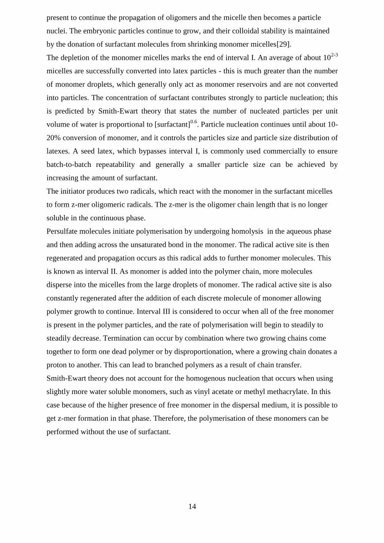
1.2 RAFT polymerisation
Reversible addition-fragmentation transfer (RAFT) polymerisation is a highly versatile
controlled radical polymerisation (CRP) technique that was first reported by Rizzardo, Moad
and Thang in 1998[30]. It is intrinsically tolerant to functionality and allows for
polymerisation of a wide range of monomers such as acrylic acid, hydroxyethyl methacrylate
and dimethylaminoethyl methacrylate[31]–[36]. A variety of polymer architectures have been
synthesised using RAFT polymerisation including brushes, stars, hyperbranched and
dendrimers[37]. A good review of the versatility of RAFT with regards to polymer
architecture has been provided by Chong et al.[38] and also by Perrier and
Takolpuckdee[39]. RAFT can also be used to synthesise polymers with controlled molecular
weights and narrow polydispersities, as evidenced by Moad et al.[40], Pelet and Putnam[41]
and others [36], [42].
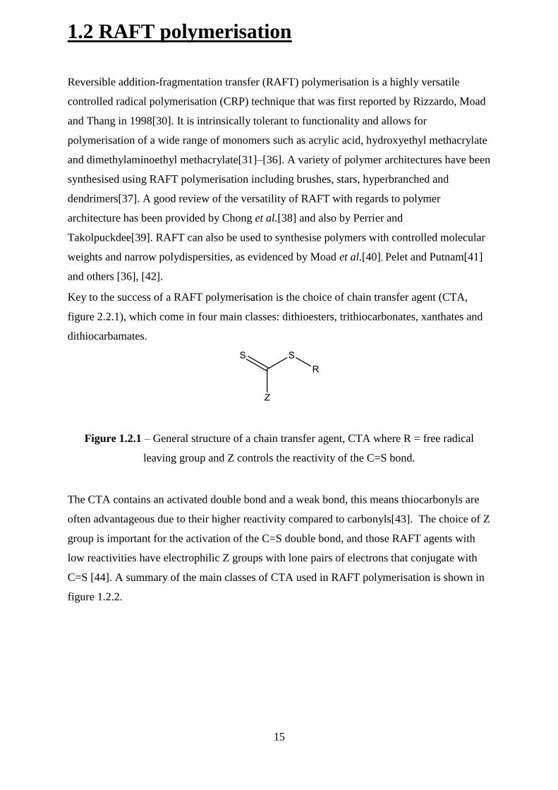
Key to the success of a RAFT polymerisation is the choice of chain transfer agent (CTA,
figure 2.2.1), which come in four main classes: dithioesters, trithiocarbonates, xanthates and
dithiocarbamates.
Figure 1.2.1 – General structure of a chain transfer agent, CTA where R = free radical
leaving group and Z controls the reactivity of the C=S bond.
The CTA contains an activated double bond and a weak bond, this means thiocarbonyls are
often advantageous due to their higher reactivity compared to carbonyls[43]. The choice of Z
group is important for the activation of the C=S double bond, and those RAFT agents with
low reactivities have electrophilic Z groups with lone pairs of electrons that conjugate with
C=S [44]. A summary of the main classes of CTA used in RAFT polymerisation is shown in
figure 1.2.2.
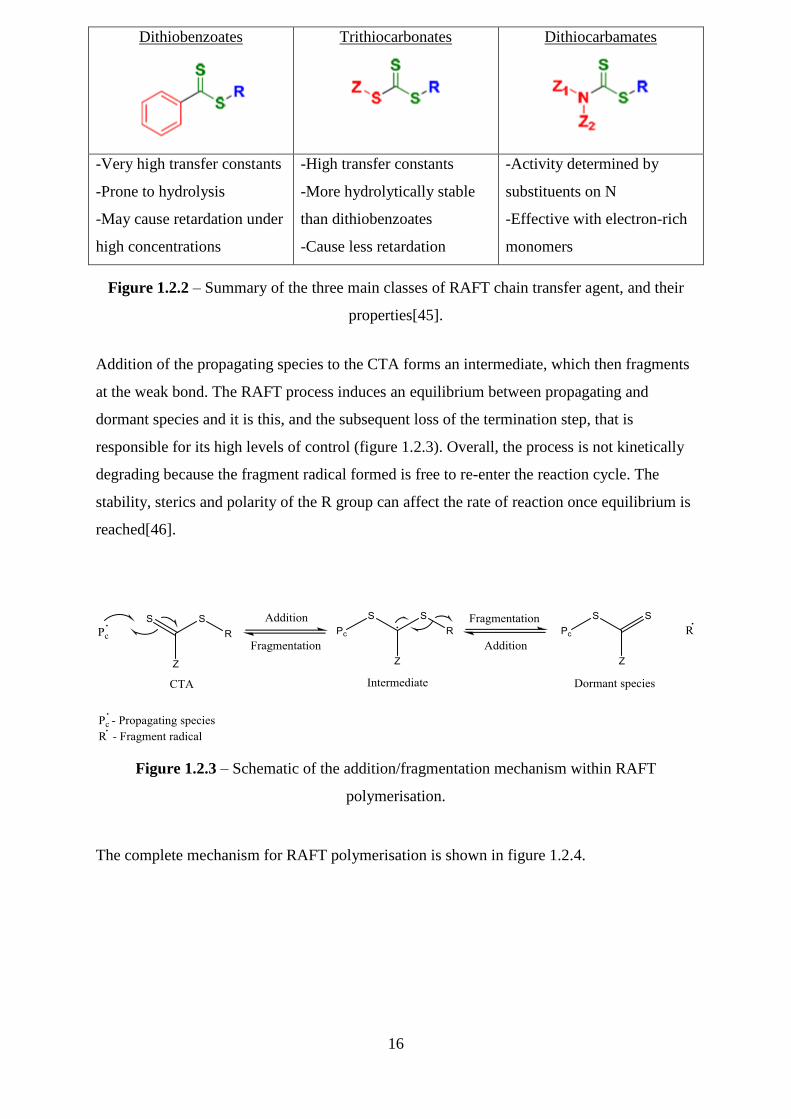
16
Dithiobenzoates
Trithiocarbonates
Dithiocarbamates
-Very high transfer constants
-Prone to hydrolysis
-May cause retardation under
high concentrations
-High transfer constants
-More hydrolytically stable
than dithiobenzoates
-Cause less retardation
-Activity determined by
substituents on N
-Effective with electron-rich
monomers
Figure 1.2.2 – Summary of the three main classes of RAFT chain transfer agent, and their
properties[45].
Addition of the propagating species to the CTA forms an intermediate, which then fragments
at the weak bond. The RAFT process induces an equilibrium between propagating and
dormant species and it is this, and the subsequent loss of the termination step, that is
responsible for its high levels of control (figure 1.2.3). Overall, the process is not kinetically
degrading because the fragment radical formed is free to re-enter the reaction cycle. The
stability, sterics and polarity of the R group can affect the rate of reaction once equilibrium is
reached[46].
Figure 1.2.3 – Schematic of the addition/fragmentation mechanism within RAFT
polymerisation.
The complete mechanism for RAFT polymerisation is shown in figure 1.2.4.

17
Figure 1.2.4 – Full scheme of RAFT polymerisation[31].
Both initiation and termination are identical to normal radical polymerisations. However,
shortly after initiation the propagating chain will add to the CTA forming an intermediate
radical, which fragments into a new radical and a polymeric dithiocarbonate. The newly
formed radical proceeds to react with monomer to form a new propagating chain which is
then subject to the same addition/fragmentation process. As equilibrium is reached between
Pn*, Pm* and the dormant species, there is equal opportunity for all chains to grow and
therefore low polydispersity is achievable with this technique. Some polymers will retain the
CTA end group as a result of RAFT, allowing functional end groups to be applied post-
polymerisation.
The main disadvantage of the RAFT procedure is the synthesis of the CTA itself, which often
requires an inert atmosphere and the use of hazardous materials, such as CS2. However,
modern syntheses are being published using softer reagents[47] and also report high yields
without the need for strict anaerobic conditions[48].
In the presence of a cross-linker RAFT polymerisation enables branching whilst quenching
cross-linking[49] and hyperbranched polymers with polydispersities as low as 1.3 have been
produced using this system[50]. The amount of CTA present in the system has been seen to
control levels of branching, however too high quantities can decrease monomer conversion.
18
This is a known effect, although if the mechanism is primarily an increase in side reactions,
or a decrease in the fragmentation reaction is still the subject of some controversy[51]–[53].
A variety of hyperbranched polymers have thus been synthesised using RAFT
polymerisation including poly(N-isopropyl acrylamide) (PNIPAM) [54] and poly(methyl
methacrylate)[55], with the latter case indicating that monomer concentration plays a role in
determining the microstructure of the final product. It has been demonstrated by Sherrington
that using a CTA to reduce the primary chain length can lead to a corresponding reduction of
gel formation[56]–[61].
Hyperbranched polymers offer the opportunity to functionalise the numerous chain ends
imparting, for example, protein binding capabilities[5], [62], [63] and the tolerance of RAFT
permits the introduction of biodegradable bonds within a hyperbranched structure[64], [65].
These advantages offer great possibilities for increasing bioconjugation using this technique.
The labile C-S bond that is retained in polymers during RAFT allows the introduction of
functionality through reaction with amines[30]. Moad et al. further expand this by
demonstrating the preparation of carboxy- and primary amino functional RAFT
polymers[66], after showing that the sulfur-containing end groups of RAFT polymerization
can be removed[67].
The CTA can also be used as a source for introducing carboxy functionality, although
hydrolytic instability can be a problem[68]–[70].
RAFT and other living polymerisation techniques offer the special ability to grow polymers
directly from the surface of a protein. This has been achieved by the conjugation of initiator
on cysteine thiol groups that are present on the surface of the protein[71]. RAFT
polymerisation also allows the synthesis of block copolymers[72]–[74] and this is an
important feature for conjugate polymers, as when one block collapses the other block can
remain soluble and prevent phase separation. The introduction of two different end groups on
one polymer via RAFT has been used by Kulkarni to improve polymer-protein
conjugation[75]. Polymers are attractive protein conjugates as they can be designed across a
wide range of compositions, structures and sizes[76]–[78] and potential uses range from
markers to enzyme inhibitors.
1.3 Ozonolysis
Ozonolysis is the breakdown of unsaturated organic compounds by the use of ozone.
Cleavage of the carbon-carbon bond generates an ozonide and its subsequent work up leads
to the formation of two carbonyl compounds. Carl Harries was the son-in-law of the inventor
19
Werner Siemens who designed some of the earliest ozone generators. Harries first
investigated the interaction of ozone with a range of olefins, from ethylene to
cycloalkanes[79]–[82] where he found that a 1:1 addition occurs between alkene and ozone
to form the ozonide species. By choosing the appropriate work up (for instance, hydrogen
peroxide) products with aldehyde, ketone or carboxylic acid functionality were produced.
Harries concluded that oxidative cleavage by ozone is an undemanding process with a good
degree of manipulation.
Ozone can be generated in the lab by passing oxygen through a high-voltage electrical
current[83] and two molecules of ozone are formed for every three of oxygen that enter the
system.
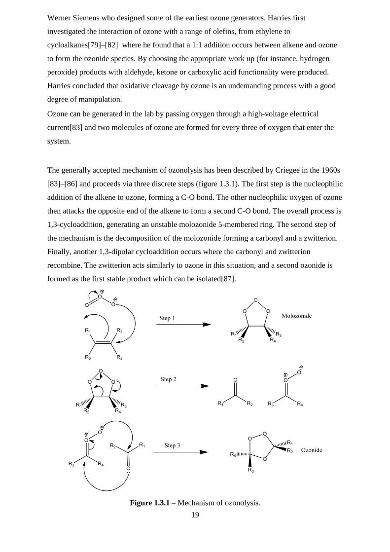
The generally accepted mechanism of ozonolysis has been described by Criegee in the 1960s
[83]–[86] and proceeds via three discrete steps (figure 1.3.1). The first step is the nucleophilic
addition of the alkene to ozone, forming a C-O bond. The other nucleophilic oxygen of ozone
then attacks the opposite end of the alkene to form a second C-O bond. The overall process is
1,3-cycloaddition, generating an unstable molozonide 5-membered ring. The second step of
the mechanism is the decomposition of the molozonide forming a carbonyl and a zwitterion.
Finally, another 1,3-dipolar cycloaddition occurs where the carbonyl and zwitterion
recombine. The zwitterion acts similarly to ozone in this situation, and a second ozonide is
formed as the first stable product which can be isolated[87].
Figure 1.3.1 – Mechanism of ozonolysis.
20
The final ozonide, whilst stable, can be explosive when heated at concentrations in excess of
20 wt% and is usually decomposed in situ. This can be achieved using a gentle or strong
reduction, forming two aldehyde products, or oxidatively to produce carboxylic acids. When
ozonolysis is performed under aqueous conditions, the zwitterion formed in step 2 is
stabilised and can yield hydroperoxides through reaction with water[88].
In a commercial setting, ozone must compete with methods such as through the use of
permanganese and chromic acid that are used to oxidate alkene bonds[89]. However, ozone is
often preferred for use in the cosmetic and pharmaceutical industries, where impurities from
heavy metals can necessitate complex purification procedures.
1.4 Hyperbranched polymers
The history of hyperbranched polymers can be traced back to the end of 19th, when it was
observed that a resin was formed from the combination of tartaric acid (A2B2 monomer) and
glycerol, a B3 monomer[90]. In 1901 the results of combining an A2 monomer (phthalic
anhydride or phthalic acid) with the B3 monomer glycerol. This reaction was studied by
Callahan, Arsem and Kienle et al.[91], [92] to reach conclusions about viscosity that are still
valid today. The first commercialised plastics, which were phenolic polymers, were
introduced in 1909 by Baekeland via the Bakelite Company[93]. The polymerisation between
an A2 monomer (formaldehyde) and a B3 monomer (phenol) produces a hyperbranched
structure just before gelation occurs. Statistical mechanics was used in the 1940s by
Flory[94]–[97] in order to determine the molecular weight distribution of hyperbranched
polymers. Flory also developed the concepts of the degree of branching and highly branched
species. Flory further developed his theory in 1952 with the synthesis of highly branched
polymers without gelation. This could be achieved through the polycondensation of ABn
monomers (where n>=2)[98]. In 1982 highly branched polymers were reported by
Kricheldorf as the products of an AB + AB2 polymerisation[99]. The term ‗Hyperbranched
polymer‘ was first used by Kim and Webster[100] in 1988 after the successful synthesis of
HBpolyphenylene. Since that time hyperbranched polymers have seen a boon as an
alternative to the more intricate dendrimers; wth applications in coatings, drug delivery,
rheology modifiers and biomaterials. Hyperbranched polymers are being researched for many
potential uses including host-guest encapsulation[101].
21
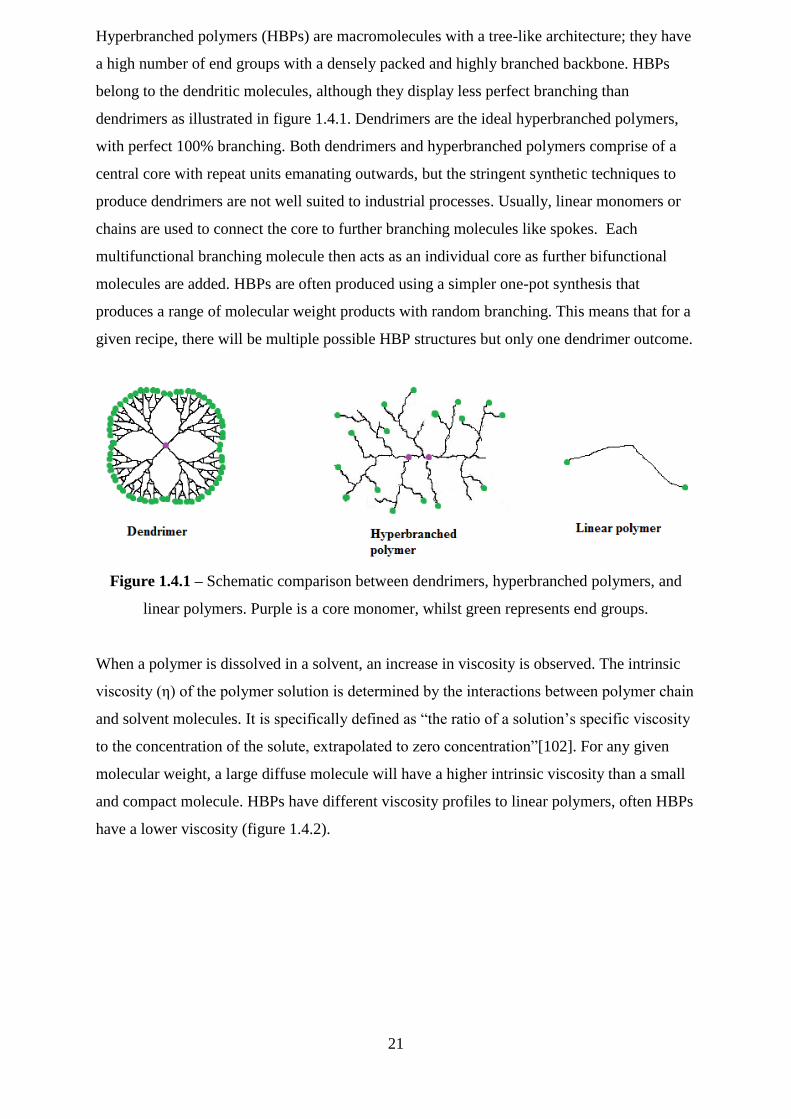
Hyperbranched polymers (HBPs) are macromolecules with a tree-like architecture; they have
a high number of end groups with a densely packed and highly branched backbone. HBPs
belong to the dendritic molecules, although they display less perfect branching than
dendrimers as illustrated in figure 1.4.1. Dendrimers are the ideal hyperbranched polymers,
with perfect 100% branching. Both dendrimers and hyperbranched polymers comprise of a
central core with repeat units emanating outwards, but the stringent synthetic techniques to
produce dendrimers are not well suited to industrial processes. Usually, linear monomers or
chains are used to connect the core to further branching molecules like spokes. Each
multifunctional branching molecule then acts as an individual core as further bifunctional
molecules are added. HBPs are often produced using a simpler one-pot synthesis that
produces a range of molecular weight products with random branching. This means that for a
given recipe, there will be multiple possible HBP structures but only one dendrimer outcome.
Figure 1.4.1 – Schematic comparison between dendrimers, hyperbranched polymers, and
linear polymers. Purple is a core monomer, whilst green represents end groups.
When a polymer is dissolved in a solvent, an increase in viscosity is observed. The intrinsic
viscosity (η) of the polymer solution is determined by the interactions between polymer chain
and solvent molecules. It is specifically defined as ―the ratio of a solution‘s specific viscosity
to the concentration of the solute, extrapolated to zero concentration‖[102]. For any given
molecular weight, a large diffuse molecule will have a higher intrinsic viscosity than a small
and compact molecule. HBPs have different viscosity profiles to linear polymers, often HBPs
have a lower viscosity (figure 1.4.2).
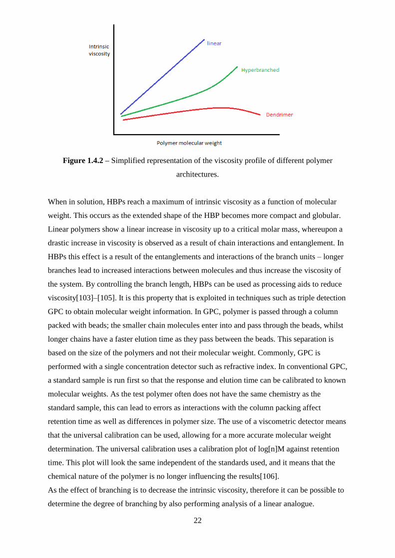
22
Figure 1.4.2 – Simplified representation of the viscosity profile of different polymer
architectures.
When in solution, HBPs reach a maximum of intrinsic viscosity as a function of molecular
weight. This occurs as the extended shape of the HBP becomes more compact and globular.
Linear polymers show a linear increase in viscosity up to a critical molar mass, whereupon a
drastic increase in viscosity is observed as a result of chain interactions and entanglement. In
HBPs this effect is a result of the entanglements and interactions of the branch units – longer
branches lead to increased interactions between molecules and thus increase the viscosity of
the system. By controlling the branch length, HBPs can be used as processing aids to reduce
viscosity[103]–[105]. It is this property that is exploited in techniques such as triple detection
GPC to obtain molecular weight information. In GPC, polymer is passed through a column
packed with beads; the smaller chain molecules enter into and pass through the beads, whilst
longer chains have a faster elution time as they pass between the beads. This separation is
based on the size of the polymers and not their molecular weight. Commonly, GPC is
performed with a single concentration detector such as refractive index. In conventional GPC,
a standard sample is run first so that the response and elution time can be calibrated to known
molecular weights. As the test polymer often does not have the same chemistry as the
standard sample, this can lead to errors as interactions with the column packing affect
retention time as well as differences in polymer size. The use of a viscometric detector means
that the universal calibration can be used, allowing for a more accurate molecular weight
determination. The universal calibration uses a calibration plot of log[n]M against retention
time. This plot will look the same independent of the standards used, and it means that the
chemical nature of the polymer is no longer influencing the results[106].
As the effect of branching is to decrease the intrinsic viscosity, therefore it can be possible to
determine the degree of branching by also performing analysis of a linear analogue.

23
Rheology studies have been successful in characterising the average branching of
polyolefins[107], [108] and 1H and 13C melt-state NMR have also been used in the
characterisation of starch[109], polyethylene[110] and polyacrylates[111]. HBPs have much
greater chemical reactivity than their linear analogues, due to the high proportion of end
groups[112]–[114]. The percentage functionality can also be controlled by control over the
degree of branching / number of end groups.
1.5 Interpenetrating polymer networks
A note on terminology
Occasionally within the literature the term ‗hydrogel interpenetrating network‘ (hydrogel
IPN) is shortened only to hydrogel. The term ‗double network hydrogel‘ is also used to
describe a sub-class of interpenetrating polymer network (IPN). Whilst hydrogels and IPNs
can share many characteristics it is important to define them as two separate species. A
hydrogel is a single, sometimes copolymer, network that is highly swollen in water. Whilst it
can, in certain situations, be applicable to call an IPN a hydrogel the reverse does not apply
and for clarity this report shall not use the two terms interchangeably.
Interpenetrating polymer networks (IPNs) consist of two or more polymers, in network form,
which whilst being heavily intermingled are not chemically bound to each other (figure
1.5.1)[115]. This is not to be confused with a polymer blend or alloy, which is a mixture of
non-crosslinked polymers. IPNs are generally considered to be composite materials, and are
heterogeneous systems with defined phase boundaries between their component parts. Semi-
IPNs refer to a system where one of the polymers is not crosslinked.
Figure 1.5.1 – Diagram illustrating full and semi-IPNs.
Full IPN Semi-IPN
24
At present, the state-of-the-art mainly describes semi-IPNS in which the non-crosslinked
component is linear and there are very few reports of instances where this component is
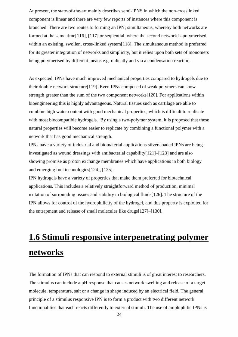
branched. There are two routes to forming an IPN; simultaneous, whereby both networks are
formed at the same time[116], [117] or sequential, where the second network is polymerised
within an existing, swollen, cross-linked system[118]. The simultaneous method is preferred
for its greater integration of networks and simplicity, but it relies upon both sets of monomers
being polymerised by different means e.g. radically and via a condensation reaction.
As expected, IPNs have much improved mechanical properties compared to hydrogels due to
their double network structure[119]. Even IPNs composed of weak polymers can show
strength greater than the sum of the two component networks[120]. For applications within
bioengineering this is highly advantageous. Natural tissues such as cartilage are able to
combine high water content with good mechanical properties, which is difficult to replicate
with most biocompatible hydrogels. By using a two-polymer system, it is proposed that these
natural properties will become easier to replicate by combining a functional polymer with a
network that has good mechanical strength.
IPNs have a variety of industrial and biomaterial applications silver-loaded IPNs are being
investigated as wound dressings with antibacterial capability[121]–[123] and are also
showing promise as proton exchange membranes which have applications in both biology
and emerging fuel technologies[124], [125].
IPN hydrogels have a variety of properties that make them preferred for biotechnical
applications. This includes a relatively straightforward method of production, minimal
irritation of surrounding tissues and stability in biological fluids[126]. The structure of the
IPN allows for control of the hydrophilicity of the hydrogel, and this property is exploited for
the entrapment and release of small molecules like drugs[127]–[130].
1.6 Stimuli responsive interpenetrating polymer
networks
The formation of IPNs that can respond to external stimuli is of great interest to researchers.
The stimulus can include a pH response that causes network swelling and release of a target
molecule, temperature, salt or a change in shape induced by an electrical field. The general
principle of a stimulus responsive IPN is to form a product with two different network
functionalities that each reacts differently to external stimuli. The use of amphiphilic IPNs is
25
already showing promise for controlled and targeted drug release[131], where the
hydrophobic network acts to quell swelling of its hydrophilic counterpart leading to
aggregation around the target molecule.
An example of this is an IPN containing networks of Konjac glucomannan (KGM) and
poly(acrylic acid) (PAA). KGM is a fibre derived from the Amorphophallus konjac
plant[132] , and has been explored as a means to target drug delivery to the colon.
Interpenetrating polymer networks formed from KGM and PAA have been loaded with
vitamin B12 and then subjected to a model digestive tract system. These IPNs showed
significant vitamin release when exposed to colonic conditions, due to the combined
enzymatic attack upon the KGM and the pH response of the PAA[133]. In this case, the
KGM network acts to prevent degradation in the harsh digestive conditions prior to reaching
the bowel, whilst the PAA pH response assists molecule release from the network.
However, homo-IPNs, where both networks are chemically identical, have also been
investigated. Poly(N-isopropylacrylamide) (PNIPAM) is a well-documented temperature
responsive polymer[134] and it is intuitive that this response could be conveyed to an IPN.
PNIPAM homo-IPNs show an enhanced trapping of bovine serum albumin (BSA) above the
LCST, as well as greater mechanical properties when compared to a single network PNIPAM
hydrogel[129]. PNIPAM‘s temperature response has also been exploited to create an
injectable semi-IPN that hardens in vivo, to reinforce scleral tissue that has been degenerated
by myopia[135]. IPNs that exhibit a bending response when exposed to an electrical field
have been synthesised. These IPN systems typically couple a strong electrolyte network, such
as PNIPAM[136] and Poly(vinyl sulfonic acid) (PVSA)[137], [138] with a second network
that imparts other desirable properties such as pH responsiveness or biodegradability. As the
IPN is exposed to an electrical field, the positive ions in the electrolytic network migrate
towards the cathode with the result being partial shielding of the sulfonate and carboxylate
groups and, ultimately, a reduction in hydration. These IPNs could provide breakthroughs
relating to biological switches and sensors.
There is a wide scope for introducing functionality and stimuli response to IPNs, and the
examples given above are by no means exhaustive but have been chosen to demonstrate the
range of applications and responses that are possible.
26
1.7 Cell adhesion and biocompatibility
The success of any biomaterial clearly rests upon its biocompatibility. Hydrophilic polymers
such as PAA and PNIPAM, and natural polymers such as KGM are considered
biocompatible[139] and as a result there is a bias towards these compounds when developing
biomaterials.
There are many examples of cell growth upon a hydrogel or IPN scaffold. Neural tissue has
been grown upon networks of a tri-block copolymer of ethylene glycol, glycolic acid and
lactic acid[140], dextran/gelatin IPNs have exhibited endothelial cell adhesion[141], as have
IPNs with a collagen/glycopolymer composition[142].
Antifouling materials also require careful control of biocompatibility to prevent
environmental pollution and current work is focussing towards protein resistant
networks[143], [144]. Hyperbranched fluoropolymers have been explored as antifouling
materials[145], [146] but their unknown toxicity and bioaccumulation data has caused a shift
towards networks of poly(isoprene) and poly(N-vinylpyrrolidinone)[147] alongside further
investigation of the more traditional poly(ethylene glycol) based materials[148], [149].
Surface chemistry, including surface tension[150], hydrophilicity[151], [152] and
zwitterionic nature[153], is known to be of considerable importance in antifouling. Polymer
networks appear to be a logical solution to the problem of fouling.
When formulating a new biomaterial, there are tactics available to the researcher to improve
the chances of cell adhesion, growth and biocompatibility. As previously mentioned there are
well-documented polymers that are known to be non-toxic and biocompatible. Early workers
considered that controlling the amphilicity of the biomaterials surface would be sufficient to
ensure cell adhesion and proliferation. However, although there are clear effects of
amphilicity on cell adhesion these strategies have rarely proved effective enough for the
clinic. A much more effective technique is to place peptide sequences within the structure;
the RGD motif in particular is known to confer cell adhesion[154] (where R = arginine, G =
glycine and D = aspartic acid). The modification of materials with specific peptide sequences
is an expensive and time-consuming process and an emerging technique is to apply alkyl
amines to a material[155]. These act to mimic the lysine functionalities that are modified by
extracellular enzymes during cell proliferation[155], [156]. Finally, careful thought must be
given to the final structure of any network designed to sustain tissue regeneration, as too
narrow a mesh size will inhibit any cell growth. Material architecture is also expressly
important in the emerging field of stem cell differentiation[157].
27
There are four primary cell assays that are undertaken to assess biocompatibility of a new
material. These are: cell contact toxicity, cell extract toxicity, cell seeding and cell
proliferation. Together these assays build a picture that informs if a material itself is toxic to
cells, whether it leaches toxic chemicals, and finally if cells are able to adhere to and
proliferate upon the material‘s surface.
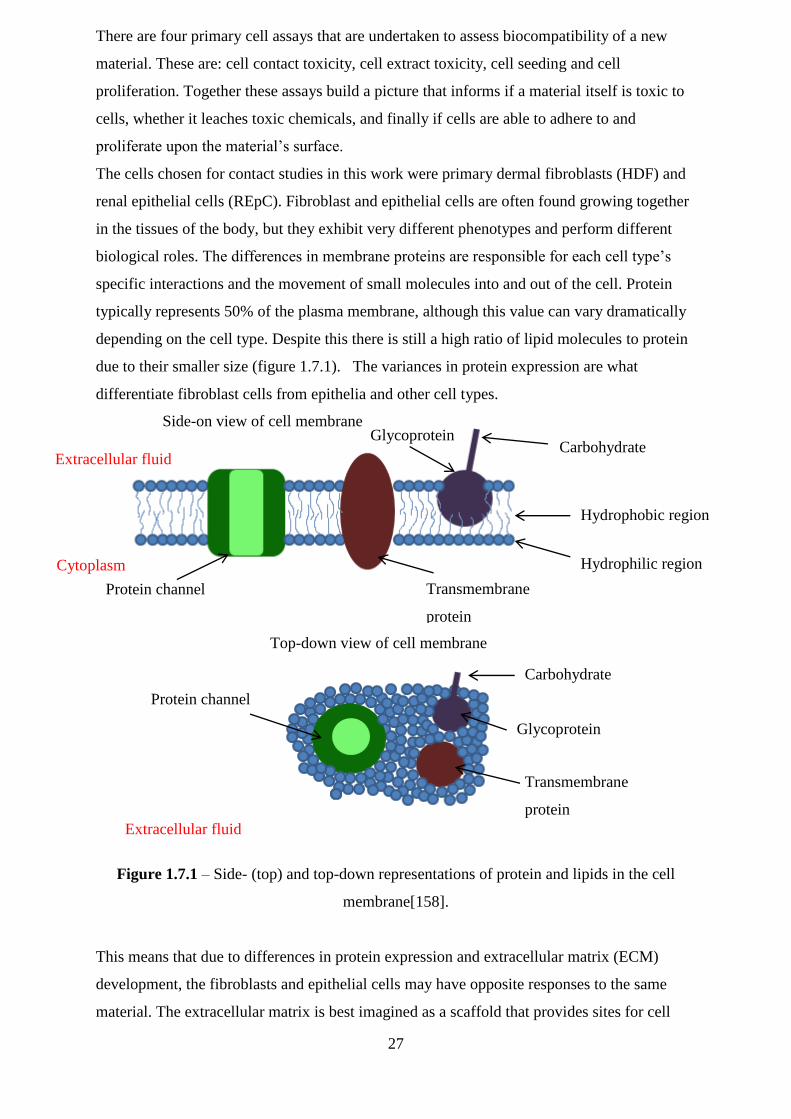
The cells chosen for contact studies in this work were primary dermal fibroblasts (HDF) and
renal epithelial cells (REpC). Fibroblast and epithelial cells are often found growing together
in the tissues of the body, but they exhibit very different phenotypes and perform different
biological roles. The differences in membrane proteins are responsible for each cell type‘s
specific interactions and the movement of small molecules into and out of the cell. Protein
typically represents 50% of the plasma membrane, although this value can vary dramatically
depending on the cell type. Despite this there is still a high ratio of lipid molecules to protein
due to their smaller size (figure 1.7.1). The variances in protein expression are what
differentiate fibroblast cells from epithelia and other cell types.
Figure 1.7.1 – Side- (top) and top-down representations of protein and lipids in the cell
membrane[158].
This means that due to differences in protein expression and extracellular matrix (ECM)
development, the fibroblasts and epithelial cells may have opposite responses to the same
material. The extracellular matrix is best imagined as a scaffold that provides sites for cell
Transmembrane
protein
Protein channel
Hydrophilic region
Hydrophobic region
Carbohydrate Glycoprotein
Extracellular fluid
Cytoplasm
Carbohydrate
Transmembrane
protein
Glycoprotein
Protein channel
Extracellular fluid
Side-on view of cell membrane
Top-down view of cell membrane
28
attachment and structural support. In addition, it also acts as a store for small signalling
molecules that regulate inflammation, cell movement and angiogenesis[159].
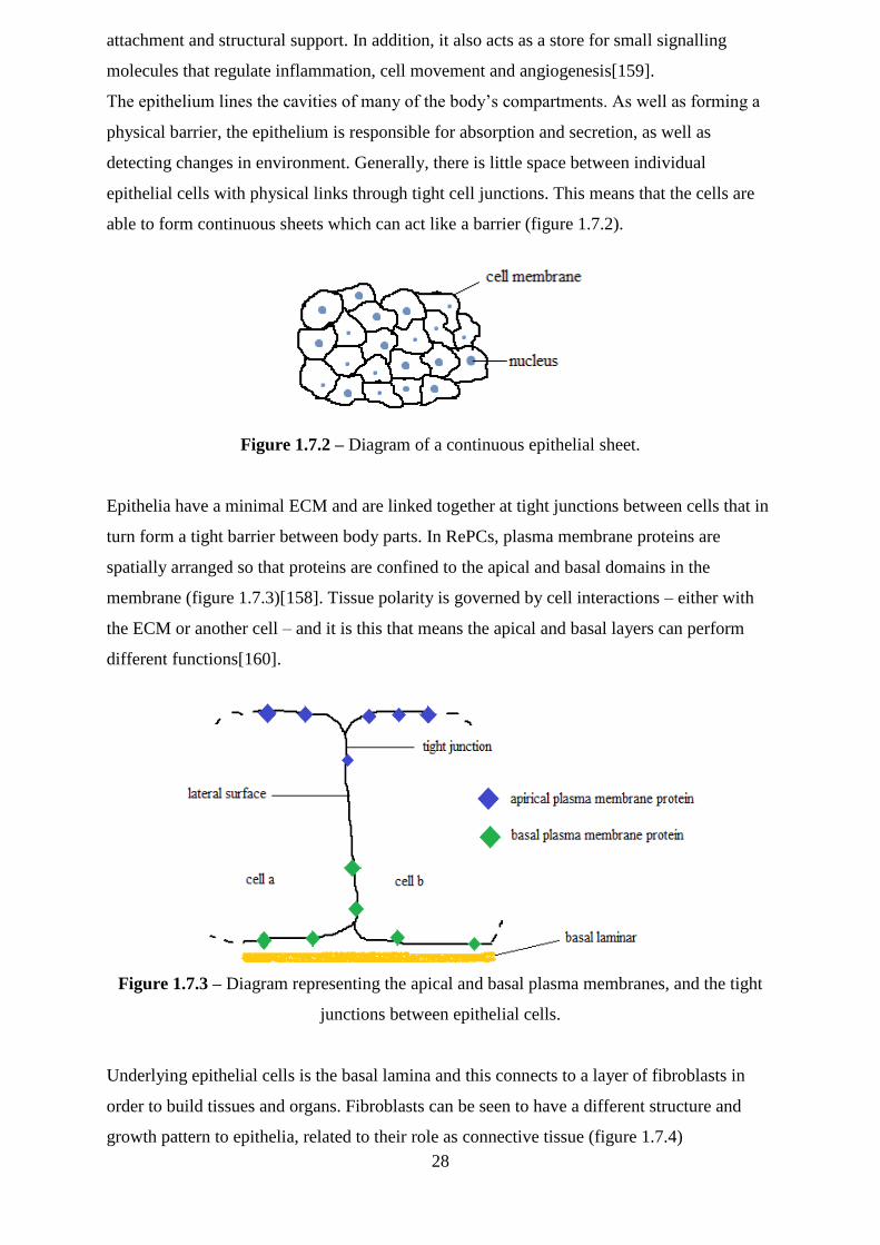
The epithelium lines the cavities of many of the body‘s compartments. As well as forming a
physical barrier, the epithelium is responsible for absorption and secretion, as well as
detecting changes in environment. Generally, there is little space between individual
epithelial cells with physical links through tight cell junctions. This means that the cells are
able to form continuous sheets which can act like a barrier (figure 1.7.2).
Figure 1.7.2 – Diagram of a continuous epithelial sheet.
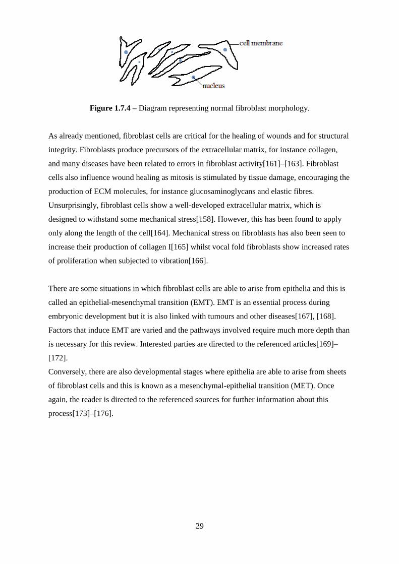
Epithelia have a minimal ECM and are linked together at tight junctions between cells that in
turn form a tight barrier between body parts. In RePCs, plasma membrane proteins are
spatially arranged so that proteins are confined to the apical and basal domains in the
membrane (figure 1.7.3)[158]. Tissue polarity is governed by cell interactions – either with
the ECM or another cell – and it is this that means the apical and basal layers can perform
different functions[160].
Figure 1.7.3 – Diagram representing the apical and basal plasma membranes, and the tight
junctions between epithelial cells.
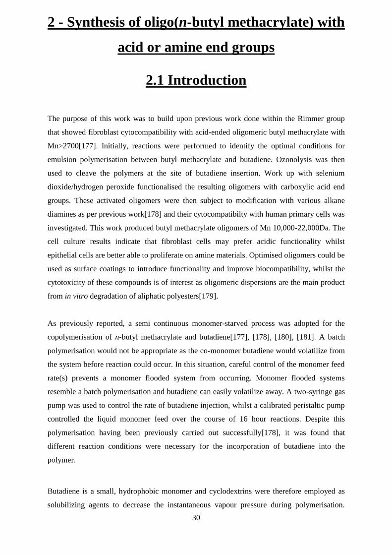
Underlying epithelial cells is the basal lamina and this connects to a layer of fibroblasts in
order to build tissues and organs. Fibroblasts can be seen to have a different structure and
growth pattern to epithelia, related to their role as connective tissue (figure 1.7.4)
29
Figure 1.7.4 – Diagram representing normal fibroblast morphology.
As already mentioned, fibroblast cells are critical for the healing of wounds and for structural
integrity. Fibroblasts produce precursors of the extracellular matrix, for instance collagen,
and many diseases have been related to errors in fibroblast activity[161]–[163]. Fibroblast
cells also influence wound healing as mitosis is stimulated by tissue damage, encouraging the
production of ECM molecules, for instance glucosaminoglycans and elastic fibres.
Unsurprisingly, fibroblast cells show a well-developed extracellular matrix, which is
designed to withstand some mechanical stress[158]. However, this has been found to apply
only along the length of the cell[164]. Mechanical stress on fibroblasts has also been seen to
increase their production of collagen I[165] whilst vocal fold fibroblasts show increased rates
of proliferation when subjected to vibration[166].
There are some situations in which fibroblast cells are able to arise from epithelia and this is
called an epithelial-mesenchymal transition (EMT). EMT is an essential process during
embryonic development but it is also linked with tumours and other diseases[167], [168].
Factors that induce EMT are varied and the pathways involved require much more depth than
is necessary for this review. Interested parties are directed to the referenced articles[169]–
[172].
Conversely, there are also developmental stages where epithelia are able to arise from sheets
of fibroblast cells and this is known as a mesenchymal-epithelial transition (MET). Once
again, the reader is directed to the referenced sources for further information about this
process[173]–[176].
30
2 - Synthesis of oligo(n-butyl methacrylate) with
acid or amine end groups
2.1 Introduction
The purpose of this work was to build upon previous work done within the Rimmer group
that showed fibroblast cytocompatibility with acid-ended oligomeric butyl methacrylate with
Mn>2700[177]. Initially, reactions were performed to identify the optimal conditions for
emulsion polymerisation between butyl methacrylate and butadiene. Ozonolysis was then
used to cleave the polymers at the site of butadiene insertion. Work up with selenium
dioxide/hydrogen peroxide functionalised the resulting oligomers with carboxylic acid end
groups. These activated oligomers were then subject to modification with various alkane
diamines as per previous work[178] and their cytocompatibilty with human primary cells was
investigated. This work produced butyl methacrylate oligomers of Mn 10,000-22,000Da. The
cell culture results indicate that fibroblast cells may prefer acidic functionality whilst
epithelial cells are better able to proliferate on amine materials. Optimised oligomers could be
used as surface coatings to introduce functionality and improve biocompatibility, whilst the
cytotoxicity of these compounds is of interest as oligomeric dispersions are the main product
from in vitro degradation of aliphatic polyesters[179].
As previously reported, a semi continuous monomer-starved process was adopted for the
copolymerisation of n-butyl methacrylate and butadiene[177], [178], [180], [181]. A batch
polymerisation would not be appropriate as the co-monomer butadiene would volatilize from
the system before reaction could occur. In this situation, careful control of the monomer feed
rate(s) prevents a monomer flooded system from occurring. Monomer flooded systems
resemble a batch polymerisation and butadiene can easily volatilize away. A two-syringe gas
pump was used to control the rate of butadiene injection, whilst a calibrated peristaltic pump
controlled the liquid monomer feed over the course of 16 hour reactions. Despite this
polymerisation having been previously carried out successfully[178], it was found that
different reaction conditions were necessary for the incorporation of butadiene into the
polymer.
Butadiene is a small, hydrophobic monomer and cyclodextrins were therefore employed as
solubilizing agents to decrease the instantaneous vapour pressure during polymerisation.

31
Cyclodextrins are cyclic oligosaccharides, composed of glucopyranose units seen in figure
2.1.1. These structures adopt a toroidal shape, with a central pore that is less hydrophilic than
the surrounding aqueous environment[182] and enable the formation of inclusion complexes
with small hydrophobic monomers.
Figure 2.1.1– From left to right, structure of α, β and γ cyclodextrins. Internal diameter (nm)
of each cyclodextrin is shown.
Cyclodextrins allow the aqueous phase initiation and Z-mer formation of molecules that are
not normally able to undergo emulsion polymerisation[183], [184] and Ritter has notably
reported the formation of CD-monomer complexes that have then been polymerised[185]–
[189] using such hydrophobic monomers such as styrene and isobornyl acrylate. The use of
cyclodextrins in biomaterials is not disadvantageous and several reviews into the
pharmaceutical uses of cyclodextrins have been provided by Loftsson[190]–[192]. Studies
have also indicated that the cytotoxicity of cyclodextrins follows γ < α < β, where γ is least
toxic[193], [194] and cyclodextrins administered orally are practically non-toxic[195].
Oxidative cleavage is a non-selective technique whereby the two alkene carbons of a C=C
bond are converted to carbonyl functionality. When polymer molecules are exposed to ozone,
both 1,2- and 1,4- orientated alkene bonds are cleaved by the mechanism described in chapter
1.3. For the purposes of the project non-selectivity is not a disadvantage, as any cleavage that
does not occur in the polymer backbone will simply give rise to acid functional pendant
groups.
Cell studies were performed using primary human dermal fibroblasts (HDF) and human renal
epithelial cells (REpC) in direct contact with oligomers over 72 hours. Cell viability and
microscopy studies were performed after 24 and 72 hours to assess cell adhesion, growth and
health. It was expected that the two cell types might show a preference for oppositely
functional materials, due to the differences in their plasma membrane phenotype as
previously discussed in chapter 1.
32
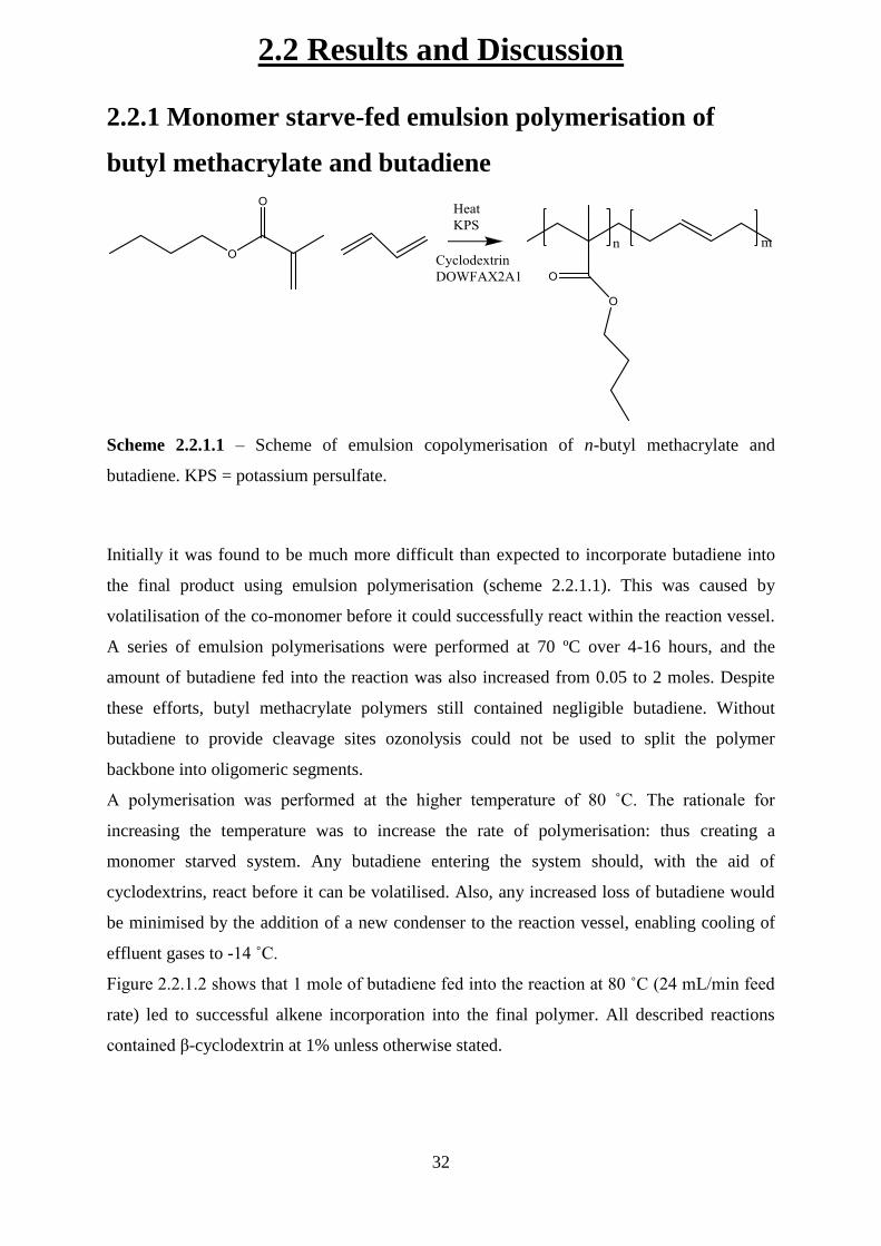
2.2 Results and Discussion
2.2.1 Monomer starve-fed emulsion polymerisation of
butyl methacrylate and butadiene
Scheme 2.2.1.1 – Scheme of emulsion copolymerisation of n-butyl methacrylate and
butadiene. KPS = potassium persulfate.
Initially it was found to be much more difficult than expected to incorporate butadiene into
the final product using emulsion polymerisation (scheme 2.2.1.1). This was caused by
volatilisation of the co-monomer before it could successfully react within the reaction vessel.
A series of emulsion polymerisations were performed at 70 ºC over 4-16 hours, and the
amount of butadiene fed into the reaction was also increased from 0.05 to 2 moles. Despite
these efforts, butyl methacrylate polymers still contained negligible butadiene. Without
butadiene to provide cleavage sites ozonolysis could not be used to split the polymer
backbone into oligomeric segments.
A polymerisation was performed at the higher temperature of 80 ˚C. The rationale for
increasing the temperature was to increase the rate of polymerisation: thus creating a
monomer starved system. Any butadiene entering the system should, with the aid of
cyclodextrins, react before it can be volatilised. Also, any increased loss of butadiene would
be minimised by the addition of a new condenser to the reaction vessel, enabling cooling of
effluent gases to -14 ˚C.
Figure 2.2.1.2 shows that 1 mole of butadiene fed into the reaction at 80 ˚C (24 mL/min feed
rate) led to successful alkene incorporation into the final polymer. All described reactions
contained β-cyclodextrin at 1% unless otherwise stated.

33
Figure 2.2.1.2 – 1H NMR spectrum of poly(butyl methacrylate-co-butadiene) polymerised at
80˚C.
The resonances at 5.57ppm and 6.12ppm indicate the predominant 1,4-insertion of butadiene
in the cis and trans configuration. The peak at 5.05ppm is due to 1,2-terminal olefins whilst
the signal at 5.45ppm is due to non-terminal 1,2- inserted butadiene[196]. The data obtained
here shows some variation from literature values, and this is thought to be due the electron
withdrawing effects of the butyl methacrylate co-monomer. The 13
C NMR spectrum provides
further evidence that the majority of butadiene insertion occurred in a 1,4-configuration.
(figure 2.2.1.3). The 1,2-insertion of butadiene would result in two sp2 carbon environments,
and therefore two opposing peaks would be observed on the 13
C spectrum. The single peak
shown here correlates well with cis-polybutadiene that has been previously
characterised[197].
G
A
D
F E
C B
34
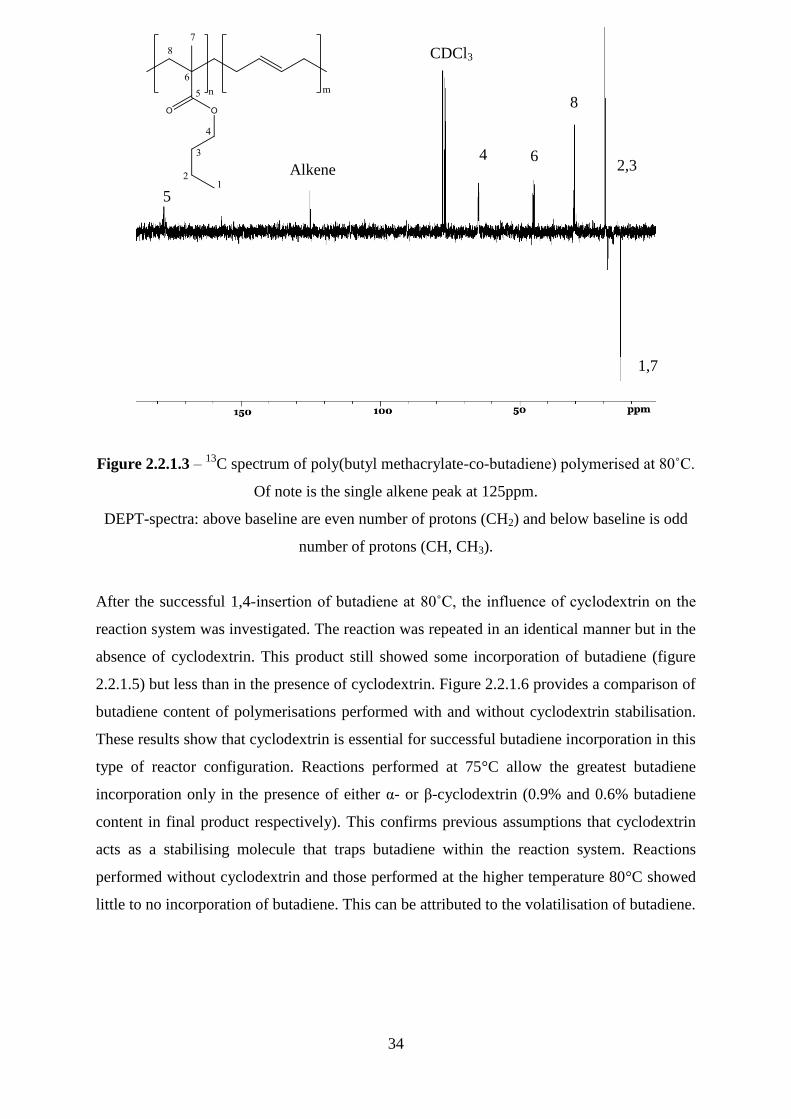
Figure 2.2.1.3 – 13
C spectrum of poly(butyl methacrylate-co-butadiene) polymerised at 80˚C.
Of note is the single alkene peak at 125ppm.
DEPT-spectra: above baseline are even number of protons (CH2) and below baseline is odd
number of protons (CH, CH3).
After the successful 1,4-insertion of butadiene at 80˚C, the influence of cyclodextrin on the
reaction system was investigated. The reaction was repeated in an identical manner but in the
absence of cyclodextrin. This product still showed some incorporation of butadiene (figure
2.2.1.5) but less than in the presence of cyclodextrin. Figure 2.2.1.6 provides a comparison of
butadiene content of polymerisations performed with and without cyclodextrin stabilisation.
These results show that cyclodextrin is essential for successful butadiene incorporation in this
type of reactor configuration. Reactions performed at 75°C allow the greatest butadiene
incorporation only in the presence of either α- or β-cyclodextrin (0.9% and 0.6% butadiene
content in final product respectively). This confirms previous assumptions that cyclodextrin
acts as a stabilising molecule that traps butadiene within the reaction system. Reactions
performed without cyclodextrin and those performed at the higher temperature 80°C showed
little to no incorporation of butadiene. This can be attributed to the volatilisation of butadiene.
1,7
2,3
8
6 4 Alkene
5
CDCl3
35
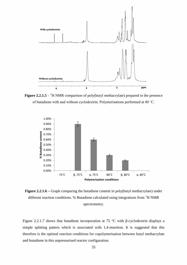
Figure 2.2.1.5 – 1H NMR comparison of poly(butyl methacrylate) prepared in the presence
of butadiene with and without cyclodextrin. Polymerisations performed at 80 ˚C.
Figure 2.2.1.6 – Graph comparing the butadiene content in poly(butyl methacrylate) under
different reaction conditions. % Butadiene calculated using integrations from 1H NMR
spectrometry.
Figure 2.2.1.7 shows that butadiene incorporation at 75 °C with β-cyclodextrin displays a
simple splitting pattern which is associated with 1,4-insertion. It is suggested that this
therefore is the optimal reaction conditions for copolymerisation between butyl methacrylate
and butadiene in this unpressurised reactor configuration.
0.00%
0.10%
0.20%
0.30%
0.40%
0.50%
0.60%
0.70%
0.80%
0.90%
1.00%
75°C β, 75°C α, 75°C 80°C β, 80°C α, 80°C
% B
uta
die
ne
co
nte
nt
Polymerisation conditions

36
Figure 2.2.1.7 - 1H NMR comparison of poly(butyl methacrylate) prepared in the presence of
butadiene with and without cyclodextrin. Polymerisations performed at 75˚C. Alkene proton
resonances seen at 5.5 and 6.1ppm.
All polymers were subjected to SEC GPC using tetrahydrofuran as the solvent. Table 2.2.1.8
provides a summary of this analysis. It is possible to see here that β-cyclodextrin produces
polymers of slightly lower Mn than α-cyclodextrin. This is because as higher amounts of
butadiene are incorporated, the average propagation rate constant ( ) decreases. The of
n-butyl methacrylate at 60 ºC is 976[198], whilst it is only 0.057x10⁻³
for butadiene at 30
ºC[93].
Reaction Conditions % Butadiene PDI
80°C, β-CDX, 1mole BD, 0.63moles BMA 0.2% 16300 44700 89250 2.7
80°C, 1mole BD, 0.63moles BMA 0.3% 40850 124000 252550 3.0
75°C, β-CDX, 1mole BD, 0.63moles BMA 0.9% 18450 57550 129100 3.1
75°C, 1mole BD, 0.63moles BMA 0 35650 80300 183550 2.3
80°C, α-CDX, 1mole BD, 0.63moles BMA 0 22500 57800 112350 2.6
75°C, α-CDX, 1mole BD, 0.63moles BMA 0.6% 40800 88200 153580 2.2
Table 2.2.1.8 – GPC analysis of butyl methacrylate polymers. Where CDX is β-cyclodextrin.
and given in Daltons.
37
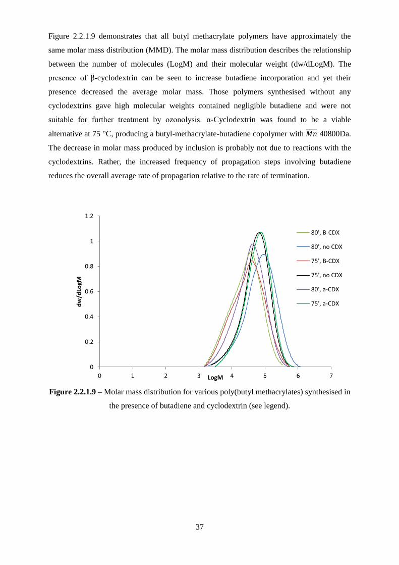
Figure 2.2.1.9 demonstrates that all butyl methacrylate polymers have approximately the
same molar mass distribution (MMD). The molar mass distribution describes the relationship
between the number of molecules (LogM) and their molecular weight (dw/dLogM). The
presence of β-cyclodextrin can be seen to increase butadiene incorporation and yet their
presence decreased the average molar mass. Those polymers synthesised without any
cyclodextrins gave high molecular weights contained negligible butadiene and were not
suitable for further treatment by ozonolysis. α-Cyclodextrin was found to be a viable
alternative at 75 °C, producing a butyl-methacrylate-butadiene copolymer with 40800Da.
The decrease in molar mass produced by inclusion is probably not due to reactions with the
cyclodextrins. Rather, the increased frequency of propagation steps involving butadiene
reduces the overall average rate of propagation relative to the rate of termination.
Figure 2.2.1.9 – Molar mass distribution for various poly(butyl methacrylates) synthesised in
the presence of butadiene and cyclodextrin (see legend).
0
0.2
0.4
0.6
0.8
1
1.2
0 1 2 3 4 5 6 7
dw
/dLo
gM
LogM
80', B-CDX
80', no CDX
75', B-CDX
75', no CDX
80', a-CDX
75', a-CDX
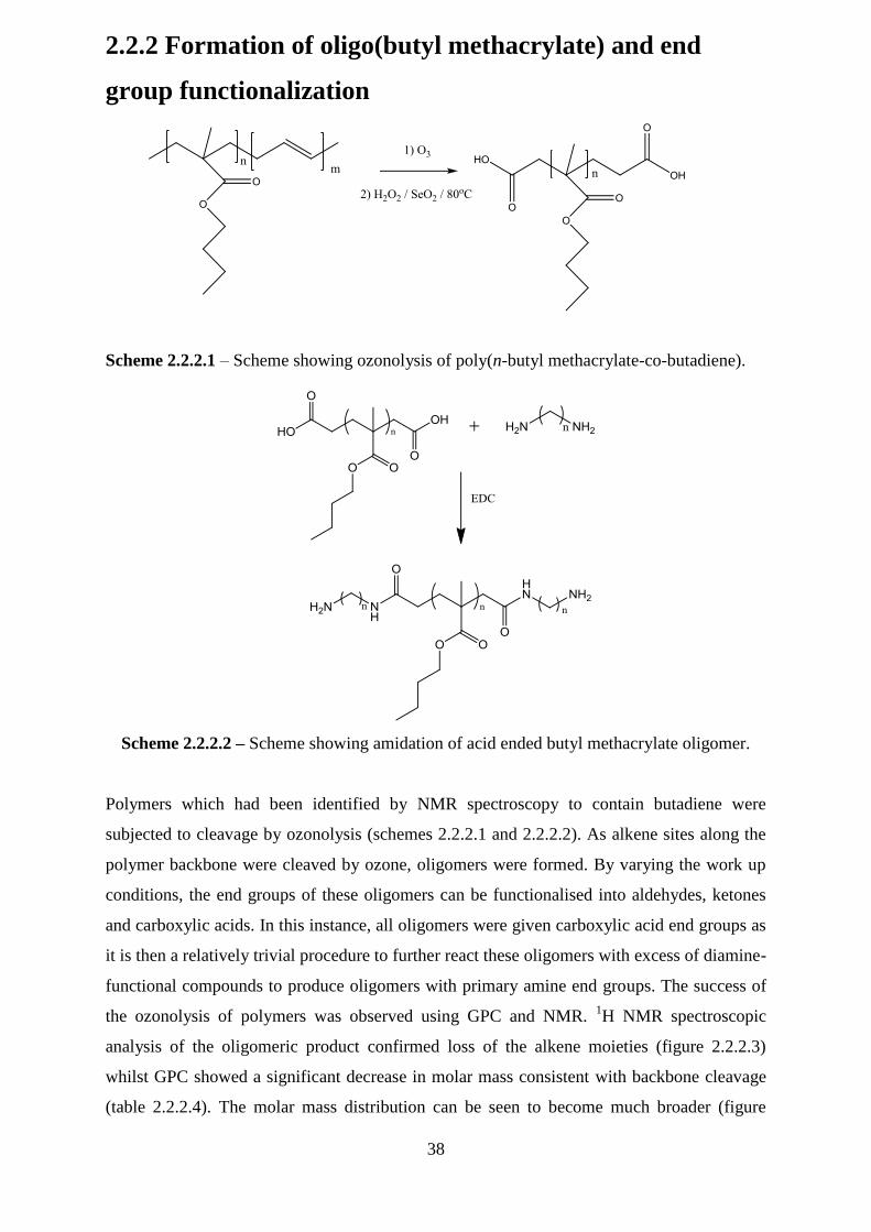
38
2.2.2 Formation of oligo(butyl methacrylate) and end
group functionalization
Scheme 2.2.2.1 – Scheme showing ozonolysis of poly(n-butyl methacrylate-co-butadiene).
Scheme 2.2.2.2 – Scheme showing amidation of acid ended butyl methacrylate oligomer.
Polymers which had been identified by NMR spectroscopy to contain butadiene were
subjected to cleavage by ozonolysis (schemes 2.2.2.1 and 2.2.2.2). As alkene sites along the
polymer backbone were cleaved by ozone, oligomers were formed. By varying the work up
conditions, the end groups of these oligomers can be functionalised into aldehydes, ketones
and carboxylic acids. In this instance, all oligomers were given carboxylic acid end groups as
it is then a relatively trivial procedure to further react these oligomers with excess of diamine-
functional compounds to produce oligomers with primary amine end groups. The success of
the ozonolysis of polymers was observed using GPC and NMR. 1H NMR spectroscopic
analysis of the oligomeric product confirmed loss of the alkene moieties (figure 2.2.2.3)
whilst GPC showed a significant decrease in molar mass consistent with backbone cleavage
(table 2.2.2.4). The molar mass distribution can be seen to become much broader (figure
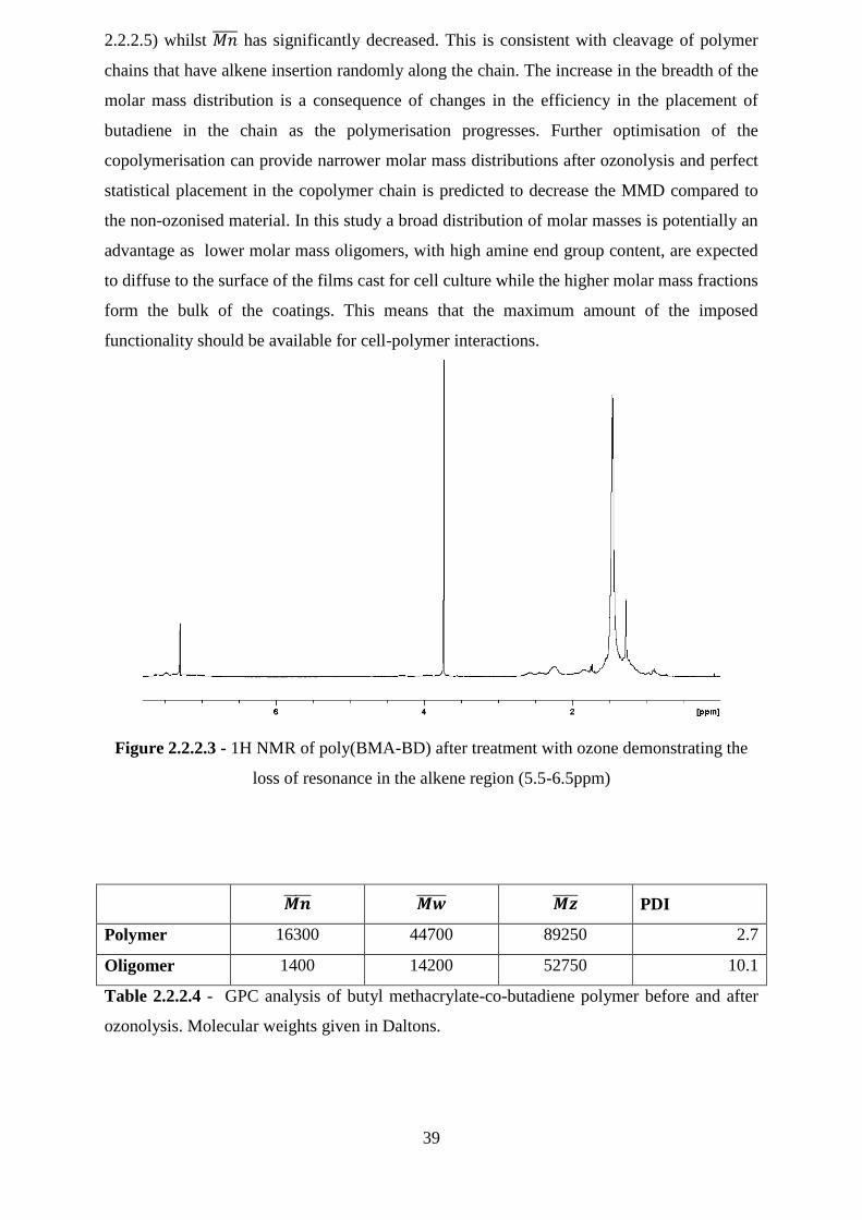
39
2.2.2.5) whilst has significantly decreased. This is consistent with cleavage of polymer
chains that have alkene insertion randomly along the chain. The increase in the breadth of the
molar mass distribution is a consequence of changes in the efficiency in the placement of
butadiene in the chain as the polymerisation progresses. Further optimisation of the
copolymerisation can provide narrower molar mass distributions after ozonolysis and perfect
statistical placement in the copolymer chain is predicted to decrease the MMD compared to
the non-ozonised material. In this study a broad distribution of molar masses is potentially an
advantage as lower molar mass oligomers, with high amine end group content, are expected
to diffuse to the surface of the films cast for cell culture while the higher molar mass fractions
form the bulk of the coatings. This means that the maximum amount of the imposed
functionality should be available for cell-polymer interactions.
Figure 2.2.2.3 - 1H NMR of poly(BMA-BD) after treatment with ozone demonstrating the
loss of resonance in the alkene region (5.5-6.5ppm)
PDI
Polymer 16300 44700 89250 2.7
Oligomer 1400 14200 52750 10.1
Table 2.2.2.4 - GPC analysis of butyl methacrylate-co-butadiene polymer before and after
ozonolysis. Molecular weights given in Daltons.
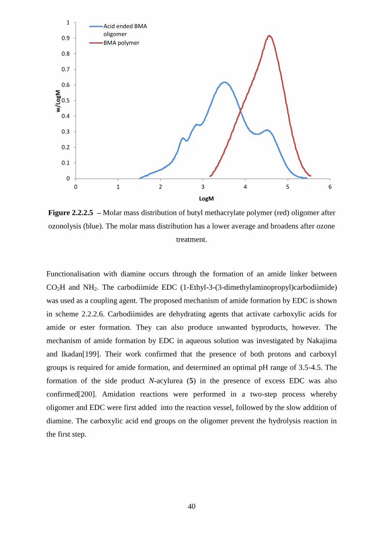
40
Figure 2.2.2.5 – Molar mass distribution of butyl methacrylate polymer (red) oligomer after
ozonolysis (blue). The molar mass distribution has a lower average and broadens after ozone
treatment.
Functionalisation with diamine occurs through the formation of an amide linker between
CO2H and NH2. The carbodiimide EDC (1-Ethyl-3-(3-dimethylaminopropyl)carbodiimide)
was used as a coupling agent. The proposed mechanism of amide formation by EDC is shown
in scheme 2.2.2.6. Carbodiimides are dehydrating agents that activate carboxylic acids for
amide or ester formation. They can also produce unwanted byproducts, however. The
mechanism of amide formation by EDC in aqueous solution was investigated by Nakajima
and Ikadan[199]. Their work confirmed that the presence of both protons and carboxyl
groups is required for amide formation, and determined an optimal pH range of 3.5-4.5. The
formation of the side product N-acylurea (5) in the presence of excess EDC was also
confirmed[200]. Amidation reactions were performed in a two-step process whereby
oligomer and EDC were first added into the reaction vessel, followed by the slow addition of
diamine. The carboxylic acid end groups on the oligomer prevent the hydrolysis reaction in
the first step.
0
0.1
0.2
0.3
0.4
0.5
0.6
0.7
0.8
0.9
1
0 1 2 3 4 5 6
w/L
ogM
LogM
Acid ended BMAoligomer
BMA polymer

41
Scheme 2.2.2.6 – Reaction scheme showing the formation of amide (7) and by-products
using EDC coupling agent. Where 1 = Carbocation derivative of carbodiimide, 2 = urea
derivative, 3 = O-acylisourea, 4 = carbocation derivative of O-acylisorea, 5 = N-acylurea, 6 =
carboxylate, 7 = amide and 8 = carboxylic anhydride.
2.2.3 Characterisation of functionalised oligomers
After end group modification oligomers were subject to various characterisation techniques.
FT-IR studies confirmed that amines and amide linkers were absent from the acidic oligomer
but present in those that had been treated with amine. Figure 2.2.3.1 shows that the acidic
oligomer has the signature broad absorbance band at ~3400 cm-1
due to the OH stretch. The
acidic oligomer also displays peaks at the frequencies 1700 cm-1
and 1200cm-1
which could
be caused by C=O and O-C stretches in the BMA pendant groups and/or in the acid end
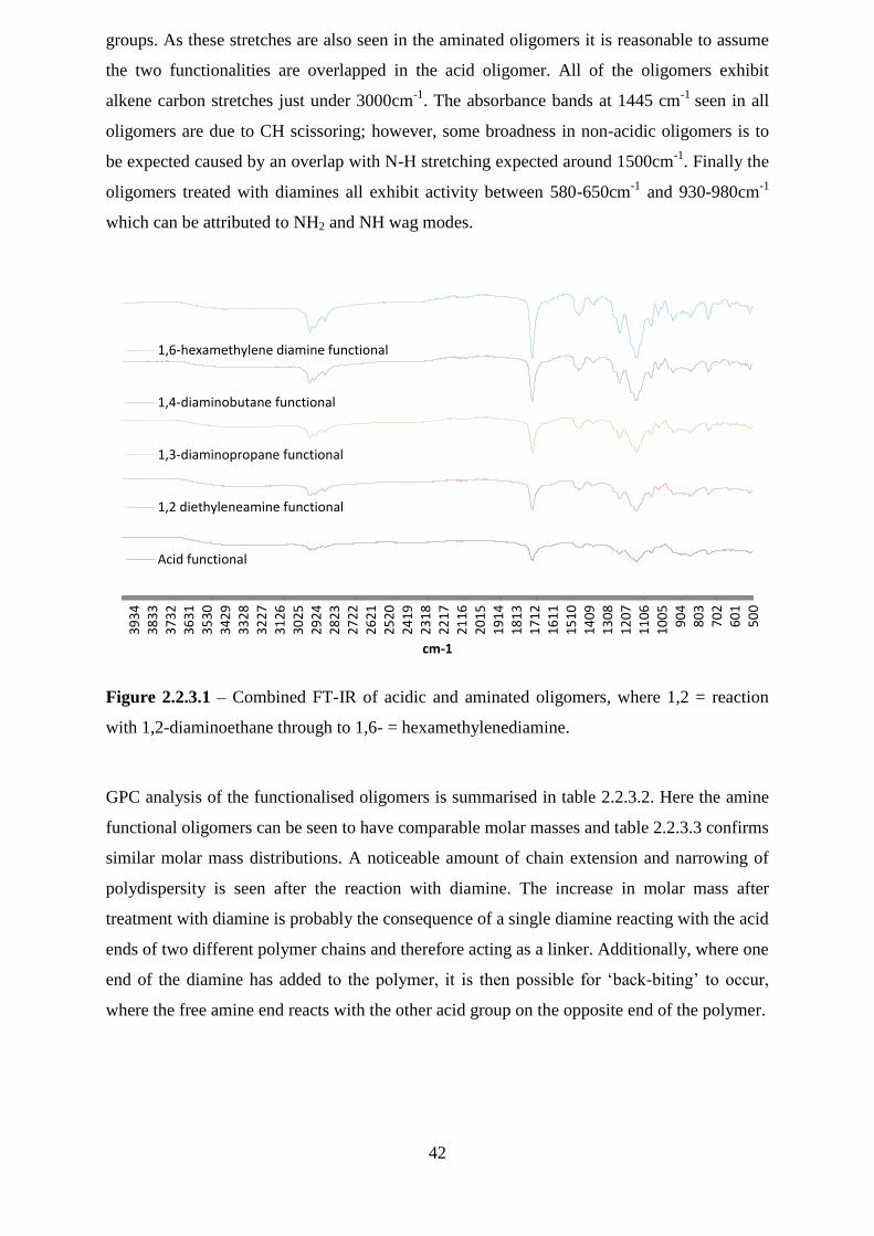
42
groups. As these stretches are also seen in the aminated oligomers it is reasonable to assume
the two functionalities are overlapped in the acid oligomer. All of the oligomers exhibit
alkene carbon stretches just under 3000cm-1
. The absorbance bands at 1445 cm-1
seen in all
oligomers are due to CH scissoring; however, some broadness in non-acidic oligomers is to
be expected caused by an overlap with N-H stretching expected around 1500cm-1
. Finally the
oligomers treated with diamines all exhibit activity between 580-650cm-1
and 930-980cm-1
which can be attributed to NH2 and NH wag modes.
Figure 2.2.3.1 – Combined FT-IR of acidic and aminated oligomers, where 1,2 = reaction
with 1,2-diaminoethane through to 1,6- = hexamethylenediamine.
GPC analysis of the functionalised oligomers is summarised in table 2.2.3.2. Here the amine
functional oligomers can be seen to have comparable molar masses and table 2.2.3.3 confirms
similar molar mass distributions. A noticeable amount of chain extension and narrowing of
polydispersity is seen after the reaction with diamine. The increase in molar mass after
treatment with diamine is probably the consequence of a single diamine reacting with the acid
ends of two different polymer chains and therefore acting as a linker. Additionally, where one
end of the diamine has added to the polymer, it is then possible for ‗back-biting‘ to occur,
where the free amine end reacts with the other acid group on the opposite end of the polymer.
50
06
01
70
28
03
90
41
00
51
10
61
20
71
30
81
40
91
51
01
61
11
71
21
81
31
91
42
01
52
11
62
21
72
31
82
41
92
52
02
62
12
72
22
82
32
92
43
02
53
12
63
22
73
32
83
42
93
53
03
63
13
73
23
83
33
93
4
cm-1
1,6-hexamethylene diamine functional
1,4-diaminobutane functional
1,3-diaminopropane functional
1,2 diethyleneamine functional
Acid functional
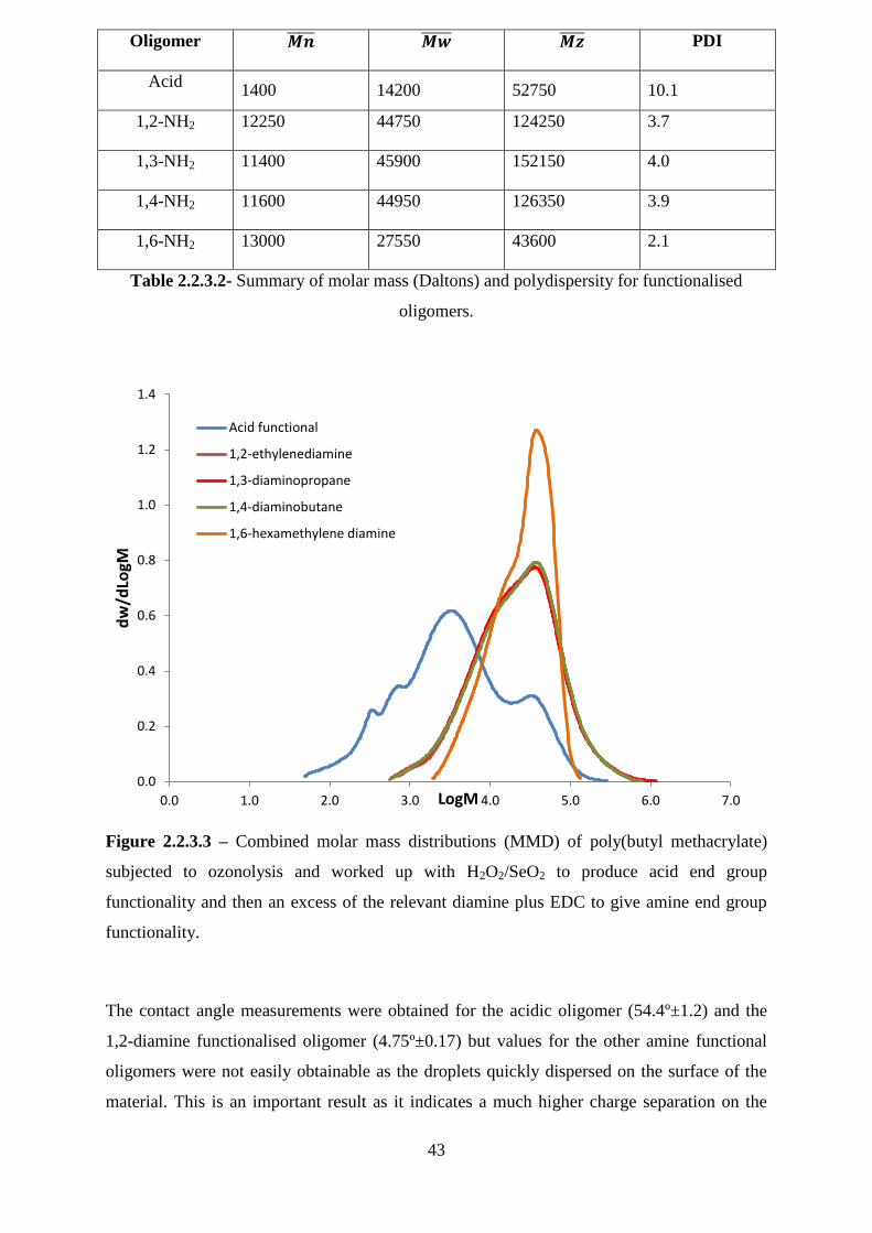
43
Oligomer PDI
Acid 1400 14200 52750 10.1
1,2-NH2 12250 44750 124250 3.7
1,3-NH2 11400 45900 152150 4.0
1,4-NH2 11600 44950 126350 3.9
1,6-NH2 13000 27550 43600 2.1
Table 2.2.3.2- Summary of molar mass (Daltons) and polydispersity for functionalised
oligomers.
Figure 2.2.3.3 – Combined molar mass distributions (MMD) of poly(butyl methacrylate)
subjected to ozonolysis and worked up with H2O2/SeO2 to produce acid end group
functionality and then an excess of the relevant diamine plus EDC to give amine end group
functionality.
The contact angle measurements were obtained for the acidic oligomer (54.4º±1.2) and the
1,2-diamine functionalised oligomer (4.75º±0.17) but values for the other amine functional
oligomers were not easily obtainable as the droplets quickly dispersed on the surface of the
material. This is an important result as it indicates a much higher charge separation on the
0.0
0.2
0.4
0.6
0.8
1.0
1.2
1.4
0.0 1.0 2.0 3.0 4.0 5.0 6.0 7.0
dw
/dLo
gM
LogM
Acid functional
1,2-ethylenediamine
1,3-diaminopropane
1,4-diaminobutane
1,6-hexamethylene diamine

44
amidated oligomers. This again confirms the successful coupling reaction with diamine, and
the difference in surface properties between acid and amine functional oligomers. These
properties are vitally important when developing new biomaterials[201], [202] and it is
expected that cell interactions will differ between acid and amine functional oligomers.
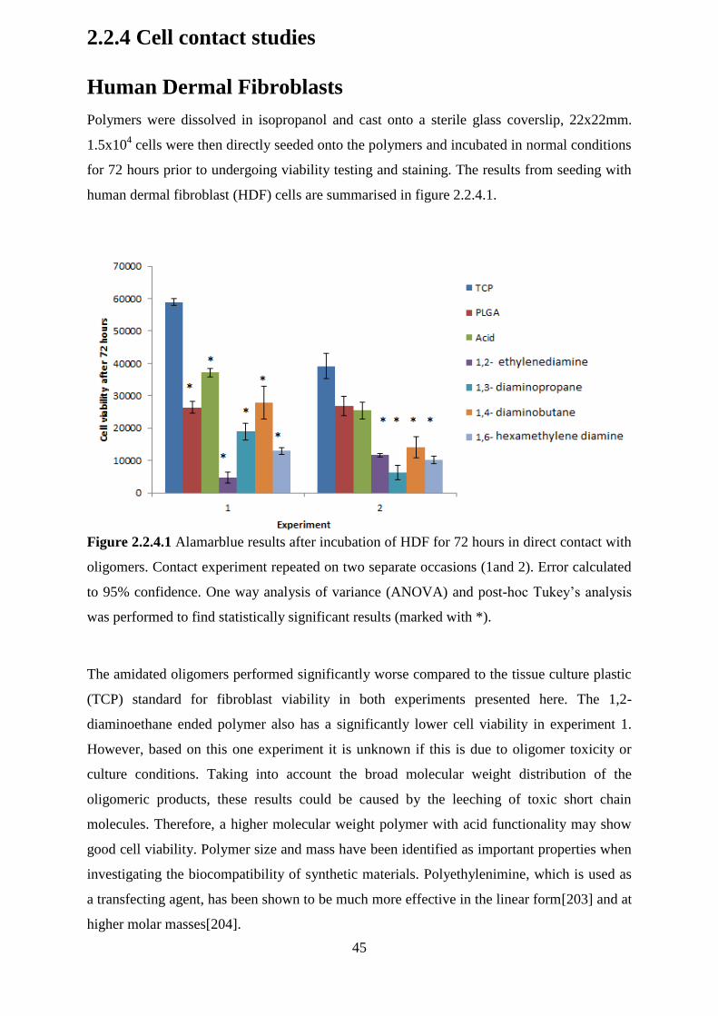
45
2.2.4 Cell contact studies
Human Dermal Fibroblasts
Polymers were dissolved in isopropanol and cast onto a sterile glass coverslip, 22x22mm.
1.5x10⁴ cells were then directly seeded onto the polymers and incubated in normal conditions
for 72 hours prior to undergoing viability testing and staining. The results from seeding with
human dermal fibroblast (HDF) cells are summarised in figure 2.2.4.1.
Figure 2.2.4.1 Alamarblue results after incubation of HDF for 72 hours in direct contact with
oligomers. Contact experiment repeated on two separate occasions (1and 2). Error calculated
to 95% confidence. One way analysis of variance (ANOVA) and post-hoc Tukey‘s analysis
was performed to find statistically significant results (marked with *).
The amidated oligomers performed significantly worse compared to the tissue culture plastic
(TCP) standard for fibroblast viability in both experiments presented here. The 1,2-
diaminoethane ended polymer also has a significantly lower cell viability in experiment 1.
However, based on this one experiment it is unknown if this is due to oligomer toxicity or
culture conditions. Taking into account the broad molecular weight distribution of the
oligomeric products, these results could be caused by the leeching of toxic short chain
molecules. Therefore, a higher molecular weight polymer with acid functionality may show
good cell viability. Polymer size and mass have been identified as important properties when
investigating the biocompatibility of synthetic materials. Polyethylenimine, which is used as
a transfecting agent, has been shown to be much more effective in the linear form[203] and at
higher molar masses[204].
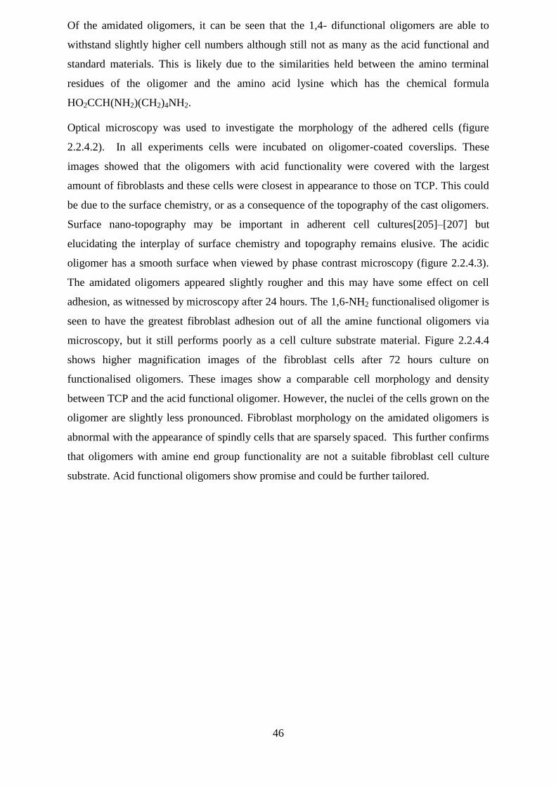
46
Of the amidated oligomers, it can be seen that the 1,4- difunctional oligomers are able to
withstand slightly higher cell numbers although still not as many as the acid functional and
standard materials. This is likely due to the similarities held between the amino terminal
residues of the oligomer and the amino acid lysine which has the chemical formula
HO2CCH(NH2)(CH2)4NH2.
Optical microscopy was used to investigate the morphology of the adhered cells (figure
2.2.4.2). In all experiments cells were incubated on oligomer-coated coverslips. These
images showed that the oligomers with acid functionality were covered with the largest
amount of fibroblasts and these cells were closest in appearance to those on TCP. This could
be due to the surface chemistry, or as a consequence of the topography of the cast oligomers.
Surface nano-topography may be important in adherent cell cultures[205]–[207] but
elucidating the interplay of surface chemistry and topography remains elusive. The acidic
oligomer has a smooth surface when viewed by phase contrast microscopy (figure 2.2.4.3).
The amidated oligomers appeared slightly rougher and this may have some effect on cell
adhesion, as witnessed by microscopy after 24 hours. The 1,6-NH2 functionalised oligomer is
seen to have the greatest fibroblast adhesion out of all the amine functional oligomers via
microscopy, but it still performs poorly as a cell culture substrate material. Figure 2.2.4.4
shows higher magnification images of the fibroblast cells after 72 hours culture on
functionalised oligomers. These images show a comparable cell morphology and density
between TCP and the acid functional oligomer. However, the nuclei of the cells grown on the
oligomer are slightly less pronounced. Fibroblast morphology on the amidated oligomers is
abnormal with the appearance of spindly cells that are sparsely spaced. This further confirms
that oligomers with amine end group functionality are not a suitable fibroblast cell culture
substrate. Acid functional oligomers show promise and could be further tailored.
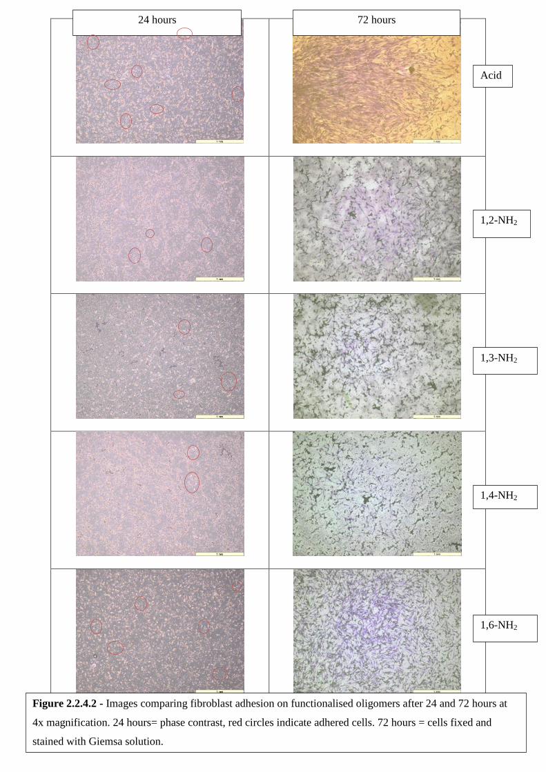
47
24 hours 72 hours
Acid
1,2-NH2
1,3-NH2
1,4-NH2
1,6-NH2
Figure 2.2.4.2 - Images comparing fibroblast adhesion on functionalised oligomers after 24 and 72 hours at
4x magnification. 24 hours= phase contrast, red circles indicate adhered cells. 72 hours = cells fixed and
stained with Giemsa solution.
48
Figure 2.2.4.3 – Phase contrast images of oligomer films after drying. Where A = acid
functional end groups and B= 1,NH2, C=1,3-NH2 ,D=1,4-NH2 amine functional end groups.
A B
C D
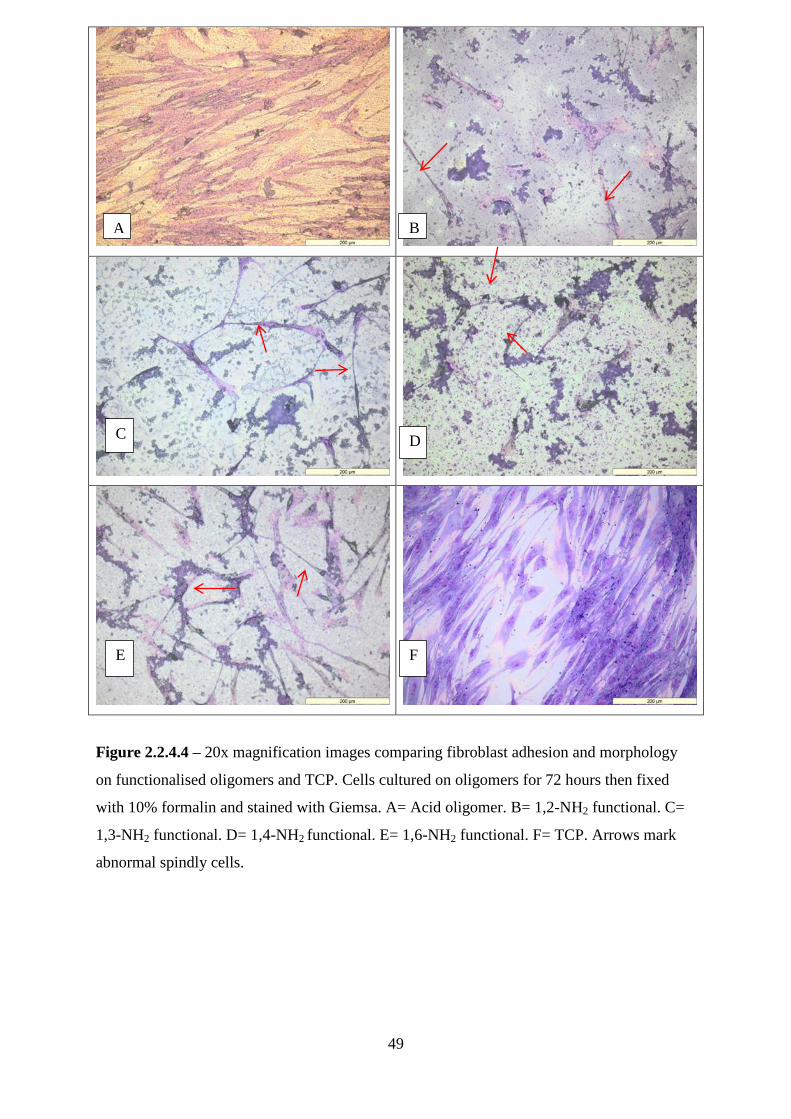
49
Figure 2.2.4.4 – 20x magnification images comparing fibroblast adhesion and morphology
on functionalised oligomers and TCP. Cells cultured on oligomers for 72 hours then fixed
with 10% formalin and stained with Giemsa. A= Acid oligomer. B= 1,2-NH2 functional. C=
1,3-NH2 functional. D= 1,4-NH2 functional. E= 1,6-NH2 functional. F= TCP. Arrows mark
abnormal spindly cells.
A B
D C
E F
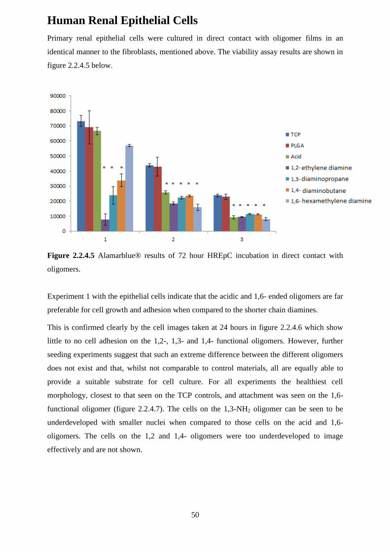
50
Human Renal Epithelial Cells
Primary renal epithelial cells were cultured in direct contact with oligomer films in an
identical manner to the fibroblasts, mentioned above. The viability assay results are shown in
figure 2.2.4.5 below.
Figure 2.2.4.5 Alamarblue® results of 72 hour HREpC incubation in direct contact with
oligomers.
Experiment 1 with the epithelial cells indicate that the acidic and 1,6- ended oligomers are far
preferable for cell growth and adhesion when compared to the shorter chain diamines.
This is confirmed clearly by the cell images taken at 24 hours in figure 2.2.4.6 which show
little to no cell adhesion on the 1,2-, 1,3- and 1,4- functional oligomers. However, further
seeding experiments suggest that such an extreme difference between the different oligomers
does not exist and that, whilst not comparable to control materials, all are equally able to
provide a suitable substrate for cell culture. For all experiments the healthiest cell
morphology, closest to that seen on the TCP controls, and attachment was seen on the 1,6-
functional oligomer (figure 2.2.4.7). The cells on the 1,3-NH2 oligomer can be seen to be
underdeveloped with smaller nuclei when compared to those cells on the acid and 1,6-
oligomers. The cells on the 1,2 and 1,4- oligomers were too underdeveloped to image
effectively and are not shown.
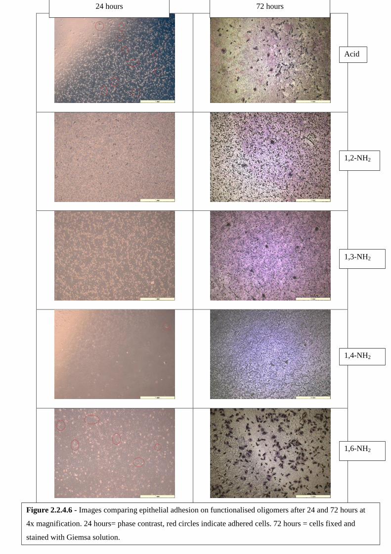
51
24 hours 72 hours
Acid
1,2-NH2
1,3-NH2
1,4-NH2
1,6-NH2
Figure 2.2.4.6 - Images comparing epithelial adhesion on functionalised oligomers after 24 and 72 hours at
4x magnification. 24 hours= phase contrast, red circles indicate adhered cells. 72 hours = cells fixed and
stained with Giemsa solution.
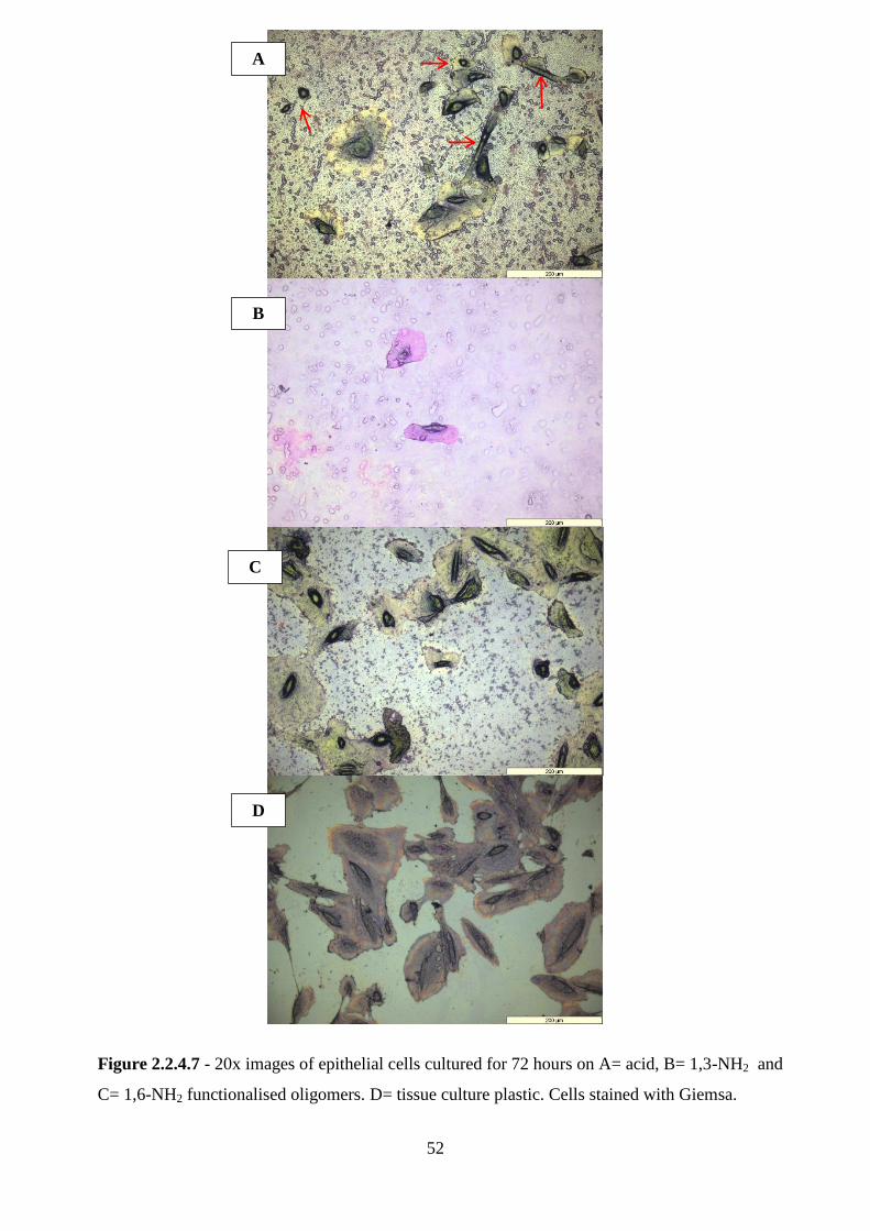
52
A
B
C
D
Figure 2.2.4.7 - 20x images of epithelial cells cultured for 72 hours on A= acid, B= 1,3-NH2 and
C= 1,6-NH2 functionalised oligomers. D= tissue culture plastic. Cells stained with Giemsa.

53
The cell morphology on the acid oligomer is erratic. Some cells display good adhesion and
prominent nuclei comparable with the TCP control whilst others appear rounded or
enlongated (marked with arrows on figure 2.2.4.7 A). This indicates that whilst epithelial
cells are able to adhere and proliferate on the acid functional material, this growth is not
entirely healthy and normal. Epithelial cells express transglutaminases (Tgase), calcium-
dependent enzymes that are able to cross link free amine groups to the gamma-carboxamide
group of bound glutamine[208]–[210] Tgases can be extracellular or reside within the
cytoplasm and almost since their discovery have been linked to cell morphology and
adhesion[211], [212]. They are of interest especially as they have been previously shown to
aid the adhesion and mobility of epithelial cells[213]. It is possible Tgases are acting to
stabilise the epithelial cells and assist the formation of cell-polymer linkages.
54
2.3 Conclusions
Through the course of this work it has been identified that cyclodextrins are essential for the
successful inclusion of butadiene into an n-butyl methacrylate backbone, as is a reaction
temperature of 75 °C. However, despite this polymers still showed relatively low levels of
butadiene incorporation when compared to the amount of monomer added to the reaction.
Copolymers of butyl methacrylate-butadiene were successfully cleaved using ozone and then
successfully worked up to contain either acid or amine end group functionality. The
successful end group functionalization of the butyl methacrylate oligomer was shown using
FT-IR and elemental analysis.
Contact angle measurements were not possible on many of the films formed by the amidated
oligomers. This indicates a surface with a high charge separation and provides reassurance
that much of the amine functionality was present on the surface of the films.
Fibroblast and epithelial cell culture on the functionalised oligomers showed that overall, no
material was a competitor for the standards tissue culture plastic and poly(lactide-glycolic
acid). Dermal fibroblast cells showed greatest cell adherence and proliferation on the acid
functional oligomer. Optical microscopy showed that these cells were comparable to those
cultured on TCP control material. Growth on the amine functional oligomers was sparse and
the cells displayed an abnormal spindly morphology. Cell viability results taken in hand with
size exclusion chromatography suggest that low molecular weight molecules could leach
from the oligomers which could potentially be toxic to cells.
The results of the renal epithelial cell culture in direct contact with functionalised oligomers
showed that the 1,6-diamine functional oligomer was the preferred. Some abnormal epithelial
growth was observed on the acid functional oligomer. This is in direct contrast to the
fibroblast cell culture results, and is probably due to the expression of transglutaminase by
epithelial cells. Transglutaminase (tgase) may be able to catalyse the cross-linking of 1,6-
diamine groups present in the oligomer film with glutamate residues in extracellular
biomolecules.
Fibroblasts, being structural cells, are believed to rely on a different adhesion mechanism. It
is proposed that the acid functional polymers are mimicking the negatively charged
proteoglycans that reside within the extracellular matrix. The presence of negatively charged
species ensures that cells stay hydrated, through the attracted of water and they may also
allow the storage of growth factors.
In conclusion, these results suggest the potential for new cell culture substrates using
oligo(BMA) functionalised with acid (fibroblast cells) or 1,6-diamine end groups (epithelial

55
cells). However, both systems require further optimisation through oligomer compositon and
their purification. Additionally, longer term culture studies are required and further cell
assays: for instance the DNA stain pico green and the fluorescent live/dead staining system
can both be used to assess cell health.
56
2.4 Experimental
Instrumentation
NMR spectra were obtained using either a Bruker AC-250 (operating frequency 1H- 250
MHz, 13
C- 62.5 MHz) or a Bruker AMX2-400 (operating frequency 1H- 400 MHz,
13C- 100
MHz) spectrometer depending on the required resolution. Analysis was performed using
TopSpin™.
THF Gel permeation chromatography was performed using a Polymer Laboratories LC1150
pump with dual model 481 Lambda-Max LC spectrophotometer UV detector obtained from
ERMA and Viscotek RI detector. The stationary phase comprised 2 x 40 cm Polymer
Laboratories PL gel mixed-E columns (5 mm particle size, Effective MW range 103 – 0.2 x
106 g mole-1) + guard columns. The flow rate of the mobile phase was 1.0 ml/min.
Automatic sampling was performed by a Gilson auto injector and analysis was performed
using Cirrus™ GPC software.
Triple detection GPC was performed using a Viscotek TDA 302 complete with a GPCmax
autosampler with THF as the mobile phase (flow rate 1.0 ml/min). The stationary phase
comprised of 2 x 300 mm Polymer Laboratories PL gel mixed-C columns (effective range
200 – 2 x 10⁶ gmol-1
). Analysis was performed using OmniSEC™ software.
Ultrafiltration was performed using a stirred 400 cm3 Millipore ultrafiltration cell with a
regenerated cellulose filter under 4-bar nitrogen pressure.
Colorimetric studies were performed using end point analysis on a Dynex Technologies
MRXII plate reader with a 570nm filter and a 600nm reference filter.
Infrared spectra were obtained using a Perkin Elmer Spectrum 100 FT-IR spectrometer with
diamond tip ATR.
2.4.1 Monomer starve-fed emulsion polymerisation of
butyl methacrylate and butadiene
Materials
Butadiene (99+%, Aldrich), Potassium persulfate (99+%, Aldrich), Potassium hydrogen
phosphate (98+%, Aldrich), DOWFAX2A1 surfactant solution (alkyldiphenyloxide
disulfonate, Dow Chemical Company USA), sodium chloride (Fisher, Lab reagent grade),
Methanol (Fisher, HPLC grade) Beta-cyclodextrin hydrate (Alfa Aesar) and Alpha-
cyclodextrin hydrate (Alfa Aesar) were used as received.
57
Butyl methacrylate (99%, Aldrich) was purified before use to remove any inhibitor present. It
was twice washed with equal portions of aqueous 2% sodium hydroxide solution, followed
by two equal portions of distilled water. This washed butyl methacrylate was then distilled
under reduced pressure, prior to use and stored at –4.5 ˚C.
Distilled water was used throughout.
Equipment
Polymerisations were performed in a 1L double-jacketed glass reactor (Radleys, UK)
equipped with an overhead mechanical glass stirrer, a nitrogen inlet, a reflux condenser, a
temperature probe and an inlet for gaseous and liquid monomer addition. Liquid monomers
were added using a peristaltic pump (Watson Marlow 505S) and gaseous butadiene was
added using an infusion pump (Precidor Type 5003, Infors HT). The peristaltic pump was
calibrated after every three uses, and tubing was cleaned using successive flows of distilled
water and acetone. Monomer addition was controlled so that addition of both liquid and gas
was completed at the same time and the butadiene molar feed rate was adjusted to coincide
with the liquid monomer. The equipment set up can be seen in figure 2.4.1.1.
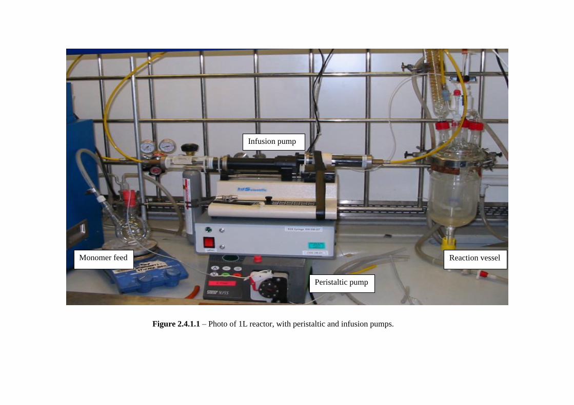
58
Infusion pump
Peristaltic pump
Reaction vessel Monomer feed
Figure 2.4.1.1 – Photo of 1L reactor, with peristaltic and infusion pumps.
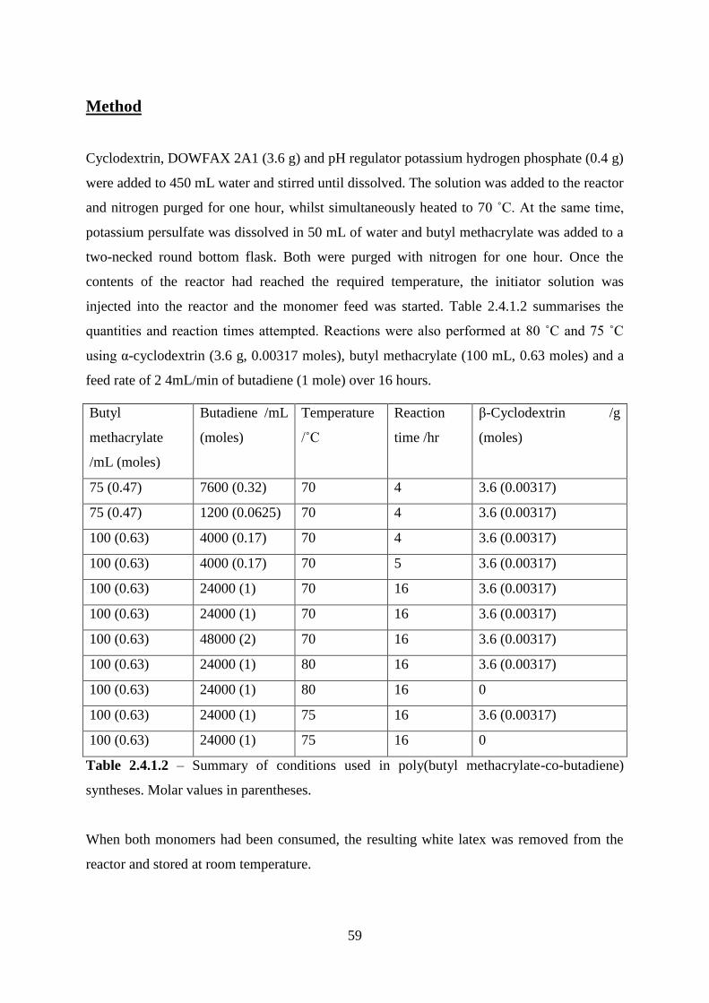
59
Method
Cyclodextrin, DOWFAX 2A1 (3.6 g) and pH regulator potassium hydrogen phosphate (0.4 g)
were added to 450 mL water and stirred until dissolved. The solution was added to the reactor
and nitrogen purged for one hour, whilst simultaneously heated to 70 ˚C. At the same time,
potassium persulfate was dissolved in 50 mL of water and butyl methacrylate was added to a
two-necked round bottom flask. Both were purged with nitrogen for one hour. Once the
contents of the reactor had reached the required temperature, the initiator solution was
injected into the reactor and the monomer feed was started. Table 2.4.1.2 summarises the
quantities and reaction times attempted. Reactions were also performed at 80 ˚C and 75 ˚C
using α-cyclodextrin (3.6 g, 0.00317 moles), butyl methacrylate (100 mL, 0.63 moles) and a
feed rate of 2 4mL/min of butadiene (1 mole) over 16 hours.
Butyl
methacrylate
/mL (moles)
Butadiene /mL
(moles)
Temperature
/˚C
Reaction
time /hr
β-Cyclodextrin /g
(moles)
75 (0.47) 7600 (0.32) 70 4 3.6 (0.00317)
75 (0.47) 1200 (0.0625) 70 4 3.6 (0.00317)
100 (0.63) 4000 (0.17) 70 4 3.6 (0.00317)
100 (0.63) 4000 (0.17) 70 5 3.6 (0.00317)
100 (0.63) 24000 (1) 70 16 3.6 (0.00317)
100 (0.63) 24000 (1) 70 16 3.6 (0.00317)
100 (0.63) 48000 (2) 70 16 3.6 (0.00317)
100 (0.63) 24000 (1) 80 16 3.6 (0.00317)
100 (0.63) 24000 (1) 80 16 0
100 (0.63) 24000 (1) 75 16 3.6 (0.00317)
100 (0.63) 24000 (1) 75 16 0
Table 2.4.1.2 – Summary of conditions used in poly(butyl methacrylate-co-butadiene)
syntheses. Molar values in parentheses.
When both monomers had been consumed, the resulting white latex was removed from the
reactor and stored at room temperature.
60
Solid polymers were retrieved from the latex by using sodium chloride to induce coagulation.
The white solid was filtered and washed thoroughly with water and then methanol before
being dried in a vacuum oven at 50 ˚C. GPC analysis was performed using tetrahydrofuran
(GPC grade, Fisher) eluent.
Results of NMR spectroscopy:
(1H, 400 MHz, CDCl3) δppm 0.97 (A), 0.98 (E), 1.42 (B), 1.64 (C), 1.96 (F,I), 4.17 (D), 5.57
(G), 6.12 (G)
(13
C, 100 MHz, CDCl3) δppm 13.7 (1,7), 19.3 (2,3), 30.2 (8), 44.9 (6), 64.6 (4), 76.5
(CDCl3), 77.2 (CDCl3), 125.1 (Alkene), 180.7-174.4 (5).
2.4.2 Ozonolysis of Poly (Butyl methacrylate-co-
Butadiene) and generation of acid end groups
Materials
Hydrogen peroxide (35% v/v, Alfa Aesar), selenium dioxide (98%, Aldrich), toluene (Fisher)
and Amberlite IRA 400(Cl) (Aldrich) used as received.
Equipment
Ozone was generated by passing oxygen through an electrical discharge generator (Type BA,
Wallace & Tiernan, UK) at a rate of 1.74ghour-1
.
Method
The poly(BMA-co-BD) latex (200 mL) was added to a 1L 3-neck round bottom flask,
equipped with a magnetic stirrer and dropping funnel. An equal volume of distilled water
(200 mL) was added with stirring. Toluene (50 mL) was added dropwise to the diluted latex
over 4 hours at room temperature. The swollen latex was then stirred for a further 24hours.
After this time a reflux condenser was added to the flask and it was placed in an ice bath. A
glass inlet was used to introduce ozone at a rate of 1.74 ghour-1
for 6-8 hours, with constant
stirring of the latex throughout. The ice bath was topped up as required during this time. On
removal of the ozone inlet, a nitrogen inlet was introduced for an hour to remove any residual
ozone from within the flask.
61
Selenium dioxide (4 g, 0.03 moles) and hydrogen peroxide solution (35 % v/v) (100 mL, 1
mole) were then added to the colloidally stable latex with stirring. This was heated to 80oC
and refluxed for 24 hours to generate acid end group functionality.
Purification of acidic poly(BMA-co-BD)
Amberlite IRA 400(Cl) (20 g) was placed into a conical flask with 100mL of latex. The flask
was placed onto an orbital shaker with a very low setting and gently shaken. After about 5
hours the latex was decanted into a second conical flask containing fresh Amberlite IRA 400
(Cl) and was shaken gently overnight. The latex was then removed and placed into dialysis
visking tubing and placed into distilled water. Twice daily water changes occurred over a
period of one week. The latexes were then removed from the dialysis tubing and any toluene
present was removed by reduced pressure. The water was then azeotropically removed by
addition of ethanol and then removing by reduced pressure.
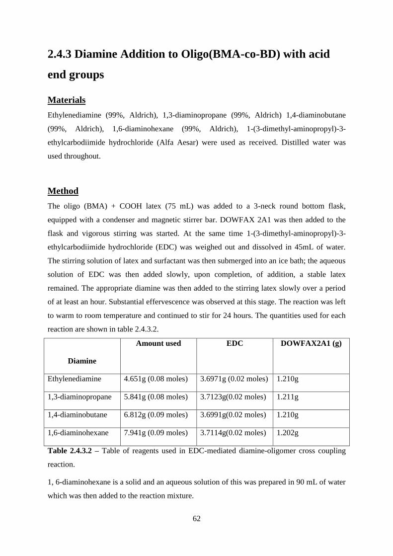
62
2.4.3 Diamine Addition to Oligo(BMA-co-BD) with acid
end groups
Materials
Ethylenediamine (99%, Aldrich), 1,3-diaminopropane (99%, Aldrich) 1,4-diaminobutane
(99%, Aldrich), 1,6-diaminohexane (99%, Aldrich), 1-(3-dimethyl-aminopropyl)-3-
ethylcarbodiimide hydrochloride (Alfa Aesar) were used as received. Distilled water was
used throughout.
Method
The oligo (BMA) + COOH latex (75 mL) was added to a 3-neck round bottom flask,
equipped with a condenser and magnetic stirrer bar. DOWFAX 2A1 was then added to the
flask and vigorous stirring was started. At the same time 1-(3-dimethyl-aminopropyl)-3-
ethylcarbodiimide hydrochloride (EDC) was weighed out and dissolved in 45mL of water.
The stirring solution of latex and surfactant was then submerged into an ice bath; the aqueous
solution of EDC was then added slowly, upon completion, of addition, a stable latex
remained. The appropriate diamine was then added to the stirring latex slowly over a period
of at least an hour. Substantial effervescence was observed at this stage. The reaction was left
to warm to room temperature and continued to stir for 24 hours. The quantities used for each
reaction are shown in table 2.4.3.2.
Diamine
Amount used EDC DOWFAX2A1 (g)
Ethylenediamine 4.651g (0.08 moles) 3.6971g (0.02 moles) 1.210g
1,3-diaminopropane 5.841g (0.08 moles) 3.7123g(0.02 moles) 1.211g
1,4-diaminobutane 6.812g (0.09 moles) 3.6991g(0.02 moles) 1.210g
1,6-diaminohexane 7.941g (0.09 moles) 3.7114g(0.02 moles) 1.202g
Table 2.4.3.2 – Table of reagents used in EDC-mediated diamine-oligomer cross coupling
reaction.
1, 6-diaminohexane is a solid and an aqueous solution of this was prepared in 90 mL of water
which was then added to the reaction mixture.
63
Purification of amidated oligomers
The stable emulsions were removed placed into dialysis visking tubing and placed into
distilled water for 2 weeks, with daily changes of water taking place.
2.4.4 Culture of fibroblast and epithelial cells
All described protocols performed under sterile conditions in a dedicated laminar flow hood,
using 70% alcohol (ethanol or isopropanol) and Virkon® solutions as cleaning agents.
Human dermal fibroblasts
Materials
Primary normal dermal fibroblasts (ATCC), Dulbecco‘s modified Eagle‘s medium (HEPES
buffered, high glucose, + pyruvate, Life Technologies), fetal bovine serum (FBS; sterile
filtered, non-USAorigin, Sigma), penicillin-streptomycin (pen-strep; 10000µ, Life
Technologies), trypsin-EDTA (0.25%, Life Technologies), trypan blue stain (Life
Technologies).
Equipment
Purecell NU-5100 CO2 Direct Heat Air Jacketed Incubator (Nuaire).
Culture
60 mL of DMEM was removed from the 500 mL bottle, and replaced with 50 mL FBS and
10 mL penicillin-streptomycin. The bottle was agitated before 9 mL of complete medium was
removed and placed in a T75 cell culture flask. This aliquot of medium was warmed in the
incubator for 30 minutes, whilst the remaining media was stored in the fridge for up to six
weeks. After 30 minutes the cryopreserved cells were gently agitated in a water bath at 37 °C
for 1 minute, until the frozen ‗plug‘ broke inside the vial. The cells were then quickly
transferred to the pre-prepared T75 and gently swirled to thaw completely. The cells were
kept in the incubator at 5% CO2, 37 °C for 24 hours, in order to adhere, before a complete
media change was performed. Complete media changes were then performed every 2-3 days,
with daily checks of cell health using phase contrast microscopy.
64
Passage
When cells were observed to be 80-90% confluent, passage was performed to ensure the
continued rate of mitosis. The media was aspirated from the cells, and they were washed with
10 mL of PBS. Each T75 was then treated with 3 mL of trypsin-EDTA, tapped and agitated
until the cells were observed to be detached from the flask. The trypsin was then neutralised
by the addition of an equal amount (3 mL) of complete fmedia. The cell suspension was then
transferred to a centrifuge tube and spun at 1400rpm for 5 minutes, until a cell pellet could be
observed on the bottom of the tube. The supernatant was carefully aspirated from the pellet,
which was then resuspended in a known amount of complete fmedia. A cell count (described
in section 2.4.5) was performed at this stage, and cells were then reseeded into T75 flasks at a
density of 2x10⁶ cells. 10 mL of
fmedia was added to each flask, and cells were then
subjected to ordinary incubation and culture.
Human renal epithelial cells
Materials
Primary human renal epithelial cells (ATCC), DMEM/F-12 (no phenol red, HEPES buffered,
+ L-glutamine, Life Technologies), hydrocortisone (Bioreagent, Sigma) , epidermal growth
factor (EGF; recombinant, expressed in E. coli, lyophilized powder, Sigma) , penicillin-
streptomycin (pen-strep; 10000 µ, Life Technologies), fetal bovine serum (FBS; sterile
filtered, non-USAorigin, Sigma), phenol red (Bioreagent, Sigma), insulin-transferrin-
selenium supplement A (ITS-A; Life Technologies), GlutaMAX™ (Life Technologies).
100 µg of EGFwas dissolved in 400 µL DMEM/F-12 and stored in the freezer -20 ºC.
18 µg of hydrocortisone was dissolved in 20ul of ethanol, which was then made up to 20 mL
with DMEM/F-12. 1 mL aliquots were stored in the freezer at -20 ºC.
FBS and pen-strep were stored in 25 mL and 10 mL aliquots at -20 ºC, respectively.
Preparation of media
40 mL of DMEM/F-12 was removed from each 500mL bottle. To the remaining media was
added 5 mL ITS-A, 5 mL pen-strep, 5 mL GlutaMAX™, 5mL pen-strep, 25 mL FBS, 20 µl
EGF solution, 20 µl hydrocortisone solution and 5 mg phenol red. The media was gently
agitated and stored in the fridge for up to six weeks.

65
Culture
The culture of HREp cells followed the same protocols as described for fibroblast cells in
section 2.4.4, with the use of HREp medium as prepared above used throughout.
Due to the lower amount of FBS present in complete HREp medium, a ratio of 2 mL trypsin :
4 mL neutralising media was used during the passage procedure of epithelial cells.
2.4.5 Culture of cells in direct contact with oligomers
Materials
Functionalised oligomers as synthesised in sections 2.4.2 and 2.4.3, isopropanol (absolute,
Fisher), complete media (prepared as described above), trypsin-EDTA (Life Technologies),
PLGA (50:50, Polysciences), DMSO (anhydrous, Sigma), Alamarblue® (Life Technologies),
phosphate buffered saline (Bioreagent, Sigma), hematoxylin solution according to Weigert
(parts A and B, Aldrich), eosin y (99%, Sigma Aldrich), 10% neutral buffered formalin
solution (Sigma), giemsa stain modified solution (Sigma), ethanol (absolute, Fisher), glacial
acetic acid (99%, Fisher).
Distilled water was used throughout unless otherwise specified.
Equipment
IR spot lamp 75W (Exo Terra).
Preparation of polymer films
Polymers were dissolved in isopropanol at a concentration of 5 mg in 1 mL solvent, whilst
the PLGA was dissolved in DMSO at the same concentration. The polymer solutions were
well agitated and sonicated for 30 seconds if required. 100 mL of polymer solution was then
pipetted onto each sterile glass coverslip. An IR lamp was used to heat treat the polymer
films until the solvent appeared visibly removed. At this point, each coverslip-with-film was
placed into its own well of a six well plate and washed with sterile PBS to remove any
residual solvent.
Fibroblasts or epithelial cells were treated with trypsin as previously described, but instead of
re-seeding into T-75 flasks, they were seeded directly onto the films at a density 1x10⁴
cells/well. 2 mL of complete media added was to each well and the cells were incubated for
24 hours at 37 ºC, 5%CO2. After 24 hours phase contrast imaging and a full media change
was performed. After 72 hours cells were assayed and fixed for staining.
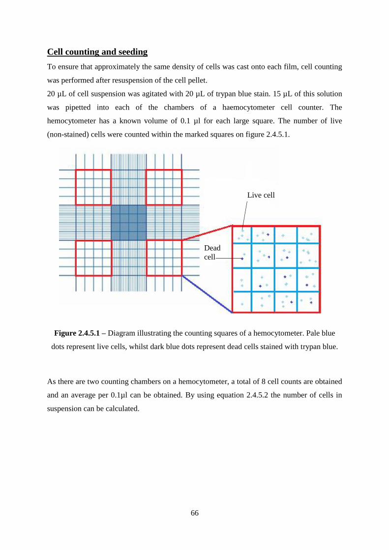
66
Cell counting and seeding
To ensure that approximately the same density of cells was cast onto each film, cell counting
was performed after resuspension of the cell pellet.
20 µL of cell suspension was agitated with 20 µL of trypan blue stain. 15 µL of this solution
was pipetted into each of the chambers of a haemocytometer cell counter. The
hemocytometer has a known volume of 0.1 µl for each large square. The number of live
(non-stained) cells were counted within the marked squares on figure 2.4.5.1.
Figure 2.4.5.1 – Diagram illustrating the counting squares of a hemocytometer. Pale blue
dots represent live cells, whilst dark blue dots represent dead cells stained with trypan blue.
As there are two counting chambers on a hemocytometer, a total of 8 cell counts are obtained
and an average per 0.1µl can be obtained. By using equation 2.4.5.2 the number of cells in
suspension can be calculated.
Live cell
Dead
cell

67
Equation 2.4.5.2 - Equation used to calculate total number of cells in m mLs of solution
after obtaining average cell count (n) from hemocytometer. It is necessary to multiply by 2
due to the half dilution with trypan blue in the counting chamber.
After obtaining a total cell count it was then possible to seed the cells at the required density.
Alamarblue® Assay
The Alamarblue® assay was performed as per the manufacturer‘s instructions[214]. As
colorimetric analysis was being used to assess the colour change of rezasurin to resorufin, an
incubation period of 16 hours with 10% Alamarblue® was found to be optimal for the cell
types used. A typical standard curve obtained for the Alamarblue® assay is shown in figure
2.4.5.3.
Figure 2.4.5.3 - Typical Alamarblue® standard curve.
The number of cells present in each well was then calculated using the following equation:
y = 6E-07x + 0.1225 R² = 0.9425
0
0.02
0.04
0.06
0.08
0.1
0.12
0.14
0.16
0 5000 10000 15000 20000 25000 30000 35000 40000
OD
57
0n
m
Number of cells
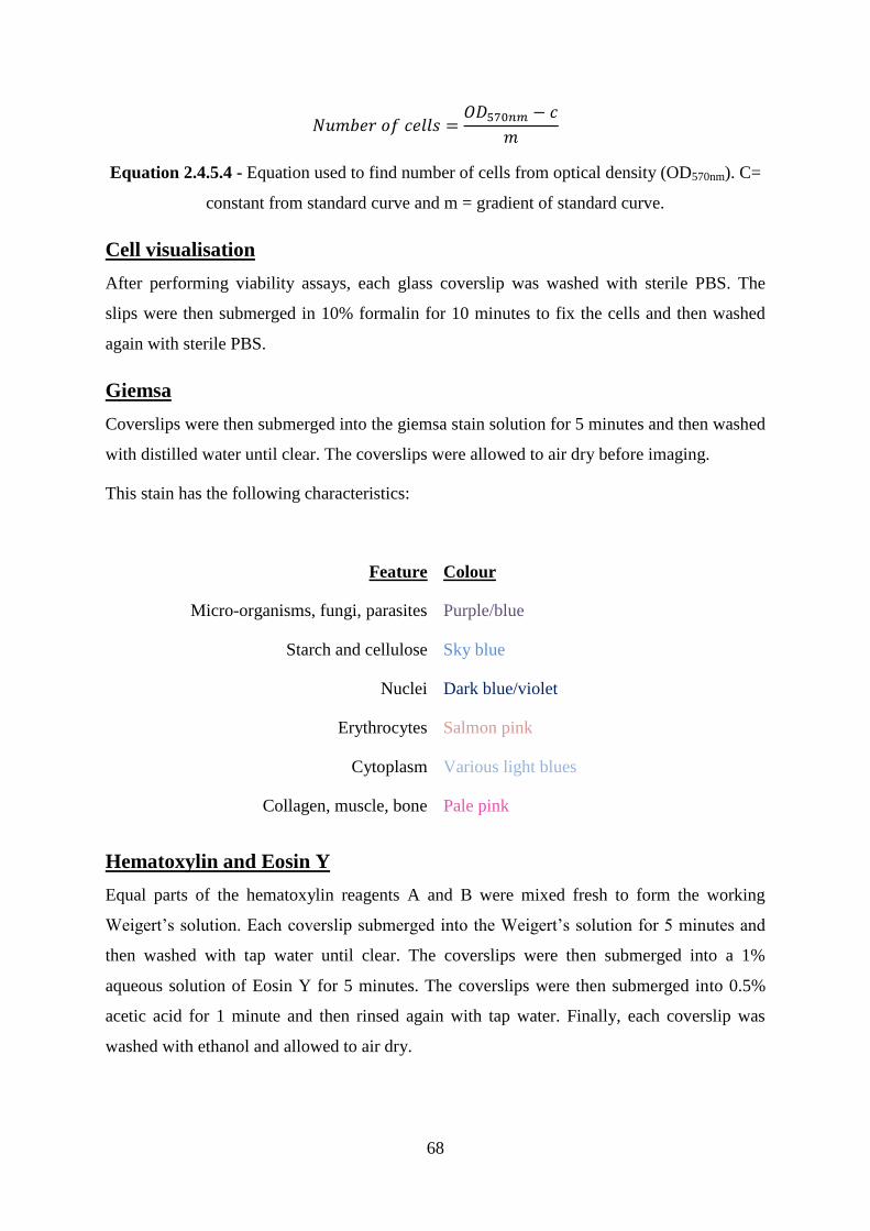
68
Equation 2.4.5.4 - Equation used to find number of cells from optical density (OD570nm). C=
constant from standard curve and m = gradient of standard curve.
Cell visualisation
After performing viability assays, each glass coverslip was washed with sterile PBS. The
slips were then submerged in 10% formalin for 10 minutes to fix the cells and then washed
again with sterile PBS.
Giemsa
Coverslips were then submerged into the giemsa stain solution for 5 minutes and then washed
with distilled water until clear. The coverslips were allowed to air dry before imaging.
This stain has the following characteristics:
Feature Colour
Micro-organisms, fungi, parasites Purple/blue
Starch and cellulose Sky blue
Nuclei Dark blue/violet
Erythrocytes Salmon pink
Cytoplasm Various light blues
Collagen, muscle, bone Pale pink
Hematoxylin and Eosin Y
Equal parts of the hematoxylin reagents A and B were mixed fresh to form the working
Weigert‘s solution. Each coverslip submerged into the Weigert‘s solution for 5 minutes and
then washed with tap water until clear. The coverslips were then submerged into a 1%
aqueous solution of Eosin Y for 5 minutes. The coverslips were then submerged into 0.5%
acetic acid for 1 minute and then rinsed again with tap water. Finally, each coverslip was
washed with ethanol and allowed to air dry.

69
This stain has the following characteristics:
Feature Colour
Nuclei Blue (hematoxylin)
Erythrocytes Very bright pink ( eosin Y)
Muscle, cytoplasm Deep pink (eosin Y)
Collagen Pale pink (eosin Y)
70
3 - Hyperbranched poly(n-butyl methacrylate)
and linear analogues with acid or amine end
groups: synthesis and cytocompatibility
3.1 Introduction
The purpose of this part of the work was to synthesise novel hyperbranched (HB) butyl
methacrylate (BMA) polymers using RAFT polymerisation and to observe cell-polymer
interactions on coatings derived from these polymers. To our knowledge, the synthesis of
hyperbranched poyl(BMA) using RAFT has not been reported to date. Experiments were
performed to ascertain the optimal ratio of monomer:chain transfer agent to produce
hyperbranched polymers with end groups capable of being modified. These materials, if they
exhibit satisfactory properties, might be suitable as cell-adhesive coatings for tissue implants.
The addition of acid monomers in tissue scaffolds has been shown to enhance wound healing
and promote angiogenesis[215], [216]. This is due to the differences in hydrophilicity and
hydrophobicity between comonomers creating the right amphiphilic environment for cell
growth. Also important for cell adhesion are the proteins and molecules that are present on
the cell surface, known as cell adhesion molecules (CAMs). Ruoslahti has published popular
reviews on both integrins and proteoglycans in cell adhesion[217], [218]. A review of the
role of peptides has been been provided by LeBaron et al.[219] and these have been utilised
by members of the Rimmer group to improve cell adhesion[220]–[222]. CAMs are also
discussed in chapter 1 section 7.
One objective was to test the hypothesis that a hyperbranched polymer with multiple acid
end groups would be a good substrate for cell adhesion and proliferation. This would then
allow future projects to optimise these materials for use as tissue scaffold coatings. This
would involve using a polymer coating to introduce cell-adhesive functionality onto an inert
structural material. In order to assess the performance of the hyperbranched polymers, they
were compared with linear BMA polymers that had been copolymerised with 4-vinyl benzoic
acid. These acid functional polymers were then reacted with diamines to investigate if this
would provide enhanced cell adhesion. Diamines were chosen to functionalize the polymers
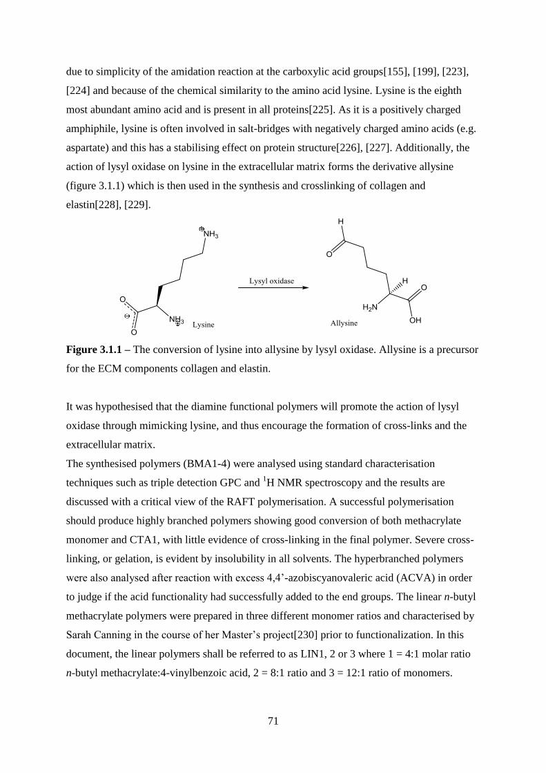
71
due to simplicity of the amidation reaction at the carboxylic acid groups[155], [199], [223],
[224] and because of the chemical similarity to the amino acid lysine. Lysine is the eighth
most abundant amino acid and is present in all proteins[225]. As it is a positively charged
amphiphile, lysine is often involved in salt-bridges with negatively charged amino acids (e.g.
aspartate) and this has a stabilising effect on protein structure[226], [227]. Additionally, the
action of lysyl oxidase on lysine in the extracellular matrix forms the derivative allysine
(figure 3.1.1) which is then used in the synthesis and crosslinking of collagen and
elastin[228], [229].
Figure 3.1.1 – The conversion of lysine into allysine by lysyl oxidase. Allysine is a precursor
for the ECM components collagen and elastin.
It was hypothesised that the diamine functional polymers will promote the action of lysyl
oxidase through mimicking lysine, and thus encourage the formation of cross-links and the
extracellular matrix.
The synthesised polymers (BMA1-4) were analysed using standard characterisation
techniques such as triple detection GPC and 1H NMR spectroscopy and the results are
discussed with a critical view of the RAFT polymerisation. A successful polymerisation
should produce highly branched polymers showing good conversion of both methacrylate
monomer and CTA1, with little evidence of cross-linking in the final polymer. Severe cross-
linking, or gelation, is evident by insolubility in all solvents. The hyperbranched polymers
were also analysed after reaction with excess 4,4‘-azobiscyanovaleric acid (ACVA) in order
to judge if the acid functionality had successfully added to the end groups. The linear n-butyl
methacrylate polymers were prepared in three different monomer ratios and characterised by
Sarah Canning in the course of her Master‘s project[230] prior to functionalization. In this
document, the linear polymers shall be referred to as LIN1, 2 or 3 where 1 = 4:1 molar ratio
n-butyl methacrylate:4-vinylbenzoic acid, 2 = 8:1 ratio and 3 = 12:1 ratio of monomers.
72
These polymers were assessed for their suitability as analogues to the BMA library and also
the outcomes of diamine coupling reactions were studied.
As these polymers are intended for use as surface coatings, they were cast upon glass
coverslips before cell seeding was carried out as described in chapter 2. Primary dermal
fibroblast and renal epithelial cells were used for direct contact studies, at a density of
1.5x10⁴cells per film. Cells were grown on each polymer in triplicate in each experiment and
each experiment was repeated on three separate occasions using identical methods and
conditions (experiments 1, 2 and 3). The cell viability on the polymers after 72 hours was
assessed using the Alamarblue® assay and optical microscopy.

73
3.2 Results and Discussion
3.2.1 Hyperbranched polymer synthesis and
functionalisation
The first step of the RAFT polymerisation was the synthesis of the RAFT agent 4-
vinylbenzyl pyrrole carbodithioate chain transfer agent (CTA1). This is shown in scheme
3.2.1.1 and described in detail in chapter 3.4.1.
Scheme 3.2.1.1 – Scheme of 4-vinylbenzyl-pyrrolecarbodithioate synthesis. 4-VBC = 4-
Vinylbenzyl chloride.
Hyperbranched polymers of butyl methacrylate were produced in four molar ratios with
pyrrole carbodithioate (CTA 1) and 4-4‘azobis cyanovaleric acid was used as a free radical
initiator in RAFT polymerisation (scheme 3.2.1.2 and table 3.2.1.5). The synthesis was
performed using a freeze-pump-thaw technique with an aprotic solvent. Polymers with higher
quantities of n-butyl methacrylate were sensitive to gelling in the ampoule, probably caused
by cross-links through bimolecular termination. It was found that this effect could be lessened
by minimising the amount of initiator present in the system, thus decreasing the total number
of radicals present.
Scheme 3.2.1.2 – RAFT polymerisation of n-butyl methacrylate using ACVA initiator.
NaH
DMF
CS2
4-VBC
DMF
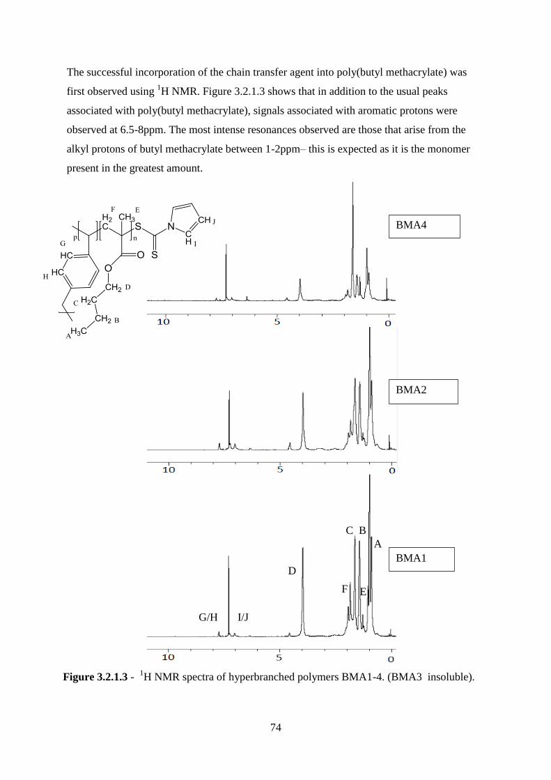
74
The successful incorporation of the chain transfer agent into poly(butyl methacrylate) was
first observed using 1H NMR. Figure 3.2.1.3 shows that in addition to the usual peaks
associated with poly(butyl methacrylate), signals associated with aromatic protons were
observed at 6.5-8ppm. The most intense resonances observed are those that arise from the
alkyl protons of butyl methacrylate between 1-2ppm– this is expected as it is the monomer
present in the greatest amount.
A
C B
F
D
E
G/H
BMA1
BMA2
BMA4
Figure 3.2.1.3 - 1H NMR spectra of hyperbranched polymers BMA1-4. (BMA3 insoluble).
I/J
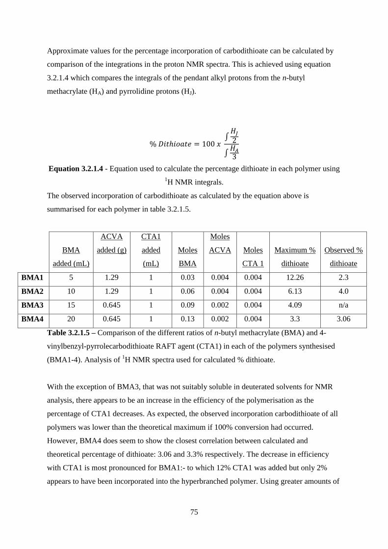
75
Approximate values for the percentage incorporation of carbodithioate can be calculated by
comparison of the integrations in the proton NMR spectra. This is achieved using equation
3.2.1.4 which compares the integrals of the pendant alkyl protons from the n-butyl
methacrylate (HA) and pyrrolidine protons (HJ).
∫
∫
Equation 3.2.1.4 - Equation used to calculate the percentage dithioate in each polymer using
1H NMR integrals.
The observed incorporation of carbodithioate as calculated by the equation above is
summarised for each polymer in table 3.2.1.5.
BMA
added (mL)
ACVA
added (g)
CTA1
added
(mL)
Moles
BMA
Moles
ACVA Moles
CTA 1
Maximum %
dithioate
Observed %
dithioate
BMA1 5 1.29 1 0.03 0.004 0.004 12.26 2.3
BMA2 10 1.29 1 0.06 0.004 0.004 6.13 4.0
BMA3 15 0.645 1 0.09 0.002 0.004 4.09 n/a
BMA4 20 0.645 1 0.13 0.002 0.004 3.3 3.06
Table 3.2.1.5 – Comparison of the different ratios of n-butyl methacrylate (BMA) and 4-
vinylbenzyl-pyrrolecarbodithioate RAFT agent (CTA1) in each of the polymers synthesised
(BMA1-4). Analysis of 1H NMR spectra used for calculated % dithioate.
With the exception of BMA3, that was not suitably soluble in deuterated solvents for NMR
analysis, there appears to be an increase in the efficiency of the polymerisation as the
percentage of CTA1 decreases. As expected, the observed incorporation carbodithioate of all
polymers was lower than the theoretical maximum if 100% conversion had occurred.
However, BMA4 does seem to show the closest correlation between calculated and
theoretical percentage of dithioate: 3.06 and 3.3% respectively. The decrease in efficiency
with CTA1 is most pronounced for BMA1:- to which 12% CTA1 was added but only 2%
appears to have been incorporated into the hyperbranched polymer. Using greater amounts of
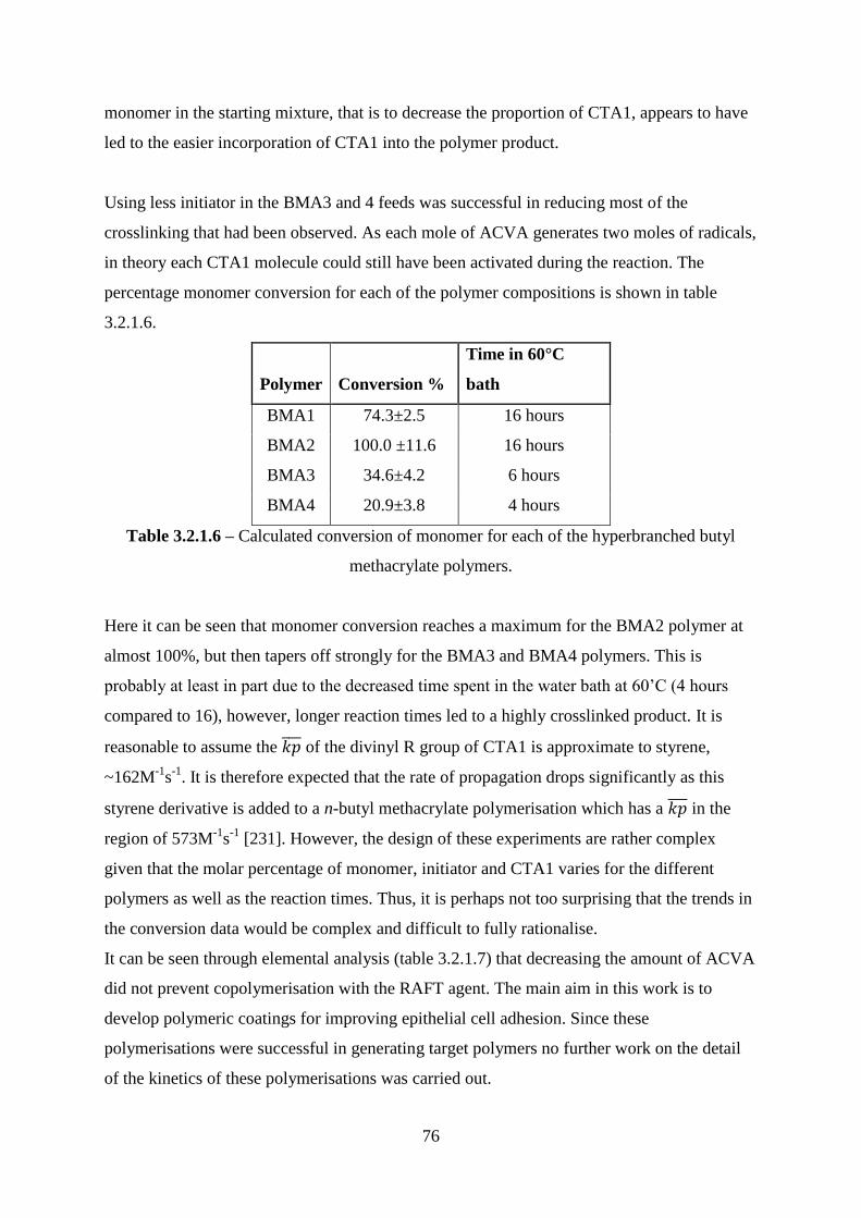
76
monomer in the starting mixture, that is to decrease the proportion of CTA1, appears to have
led to the easier incorporation of CTA1 into the polymer product.
Using less initiator in the BMA3 and 4 feeds was successful in reducing most of the
crosslinking that had been observed. As each mole of ACVA generates two moles of radicals,
in theory each CTA1 molecule could still have been activated during the reaction. The
percentage monomer conversion for each of the polymer compositions is shown in table
3.2.1.6.
Polymer Conversion %
Time in 60°C
bath
BMA1 74.3±2.5 16 hours
BMA2 100.0 ±11.6 16 hours
BMA3 34.6±4.2 6 hours
BMA4 20.9±3.8 4 hours
Table 3.2.1.6 – Calculated conversion of monomer for each of the hyperbranched butyl
methacrylate polymers.
Here it can be seen that monomer conversion reaches a maximum for the BMA2 polymer at
almost 100%, but then tapers off strongly for the BMA3 and BMA4 polymers. This is
probably at least in part due to the decreased time spent in the water bath at 60‘C (4 hours
compared to 16), however, longer reaction times led to a highly crosslinked product. It is
reasonable to assume the of the divinyl R group of CTA1 is approximate to styrene,
~162M-1
s-1
. It is therefore expected that the rate of propagation drops significantly as this
styrene derivative is added to a n-butyl methacrylate polymerisation which has a in the
region of 573M-1
s-1
[231]. However, the design of these experiments are rather complex
given that the molar percentage of monomer, initiator and CTA1 varies for the different
polymers as well as the reaction times. Thus, it is perhaps not too surprising that the trends in
the conversion data would be complex and difficult to fully rationalise.
It can be seen through elemental analysis (table 3.2.1.7) that decreasing the amount of ACVA
did not prevent copolymerisation with the RAFT agent. The main aim in this work is to
develop polymeric coatings for improving epithelial cell adhesion. Since these
polymerisations were successful in generating target polymers no further work on the detail
of the kinetics of these polymerisations was carried out.
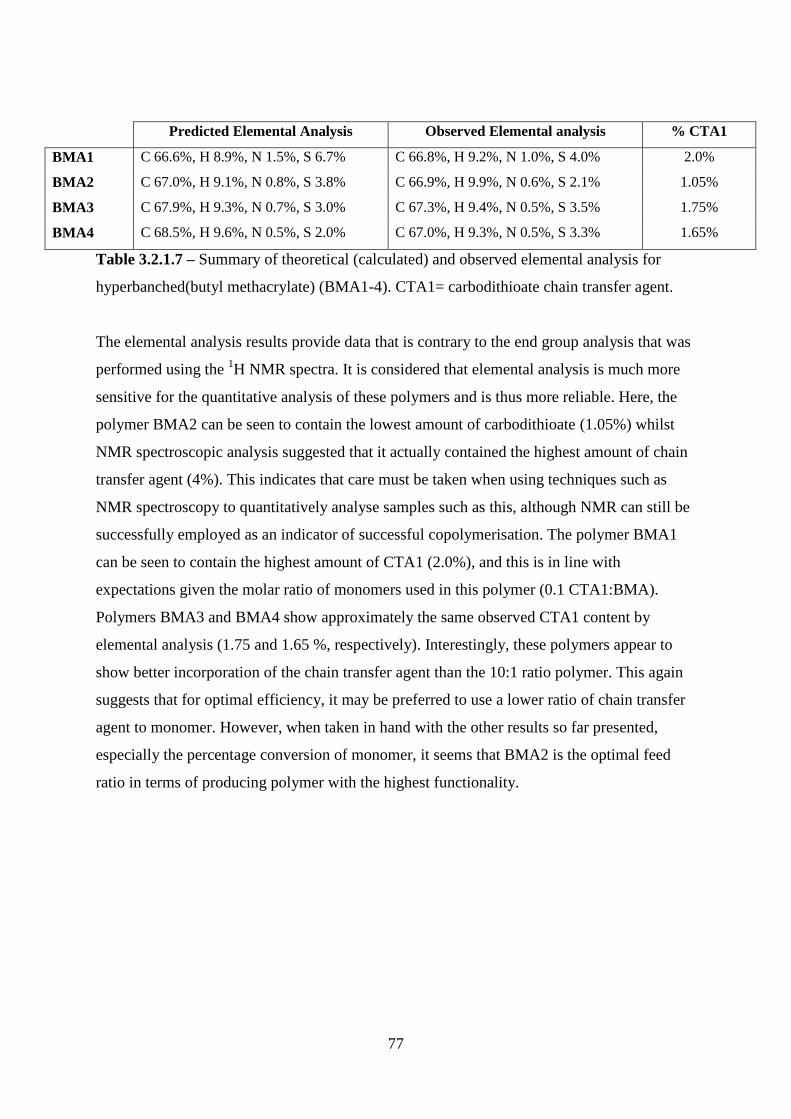
77
Predicted Elemental Analysis Observed Elemental analysis % CTA1
BMA1 C 66.6%, H 8.9%, N 1.5%, S 6.7% C 66.8%, H 9.2%, N 1.0%, S 4.0% 2.0%
BMA2 C 67.0%, H 9.1%, N 0.8%, S 3.8% C 66.9%, H 9.9%, N 0.6%, S 2.1% 1.05%
BMA3 C 67.9%, H 9.3%, N 0.7%, S 3.0% C 67.3%, H 9.4%, N 0.5%, S 3.5% 1.75%
BMA4 C 68.5%, H 9.6%, N 0.5%, S 2.0% C 67.0%, H 9.3%, N 0.5%, S 3.3% 1.65%
Table 3.2.1.7 – Summary of theoretical (calculated) and observed elemental analysis for
hyperbanched(butyl methacrylate) (BMA1-4). CTA1= carbodithioate chain transfer agent.
The elemental analysis results provide data that is contrary to the end group analysis that was
performed using the 1H NMR spectra. It is considered that elemental analysis is much more
sensitive for the quantitative analysis of these polymers and is thus more reliable. Here, the
polymer BMA2 can be seen to contain the lowest amount of carbodithioate (1.05%) whilst
NMR spectroscopic analysis suggested that it actually contained the highest amount of chain
transfer agent (4%). This indicates that care must be taken when using techniques such as
NMR spectroscopy to quantitatively analyse samples such as this, although NMR can still be
successfully employed as an indicator of successful copolymerisation. The polymer BMA1
can be seen to contain the highest amount of CTA1 (2.0%), and this is in line with
expectations given the molar ratio of monomers used in this polymer (0.1 CTA1:BMA).
Polymers BMA3 and BMA4 show approximately the same observed CTA1 content by
elemental analysis (1.75 and 1.65 %, respectively). Interestingly, these polymers appear to
show better incorporation of the chain transfer agent than the 10:1 ratio polymer. This again
suggests that for optimal efficiency, it may be preferred to use a lower ratio of chain transfer
agent to monomer. However, when taken in hand with the other results so far presented,
especially the percentage conversion of monomer, it seems that BMA2 is the optimal feed
ratio in terms of producing polymer with the highest functionality.
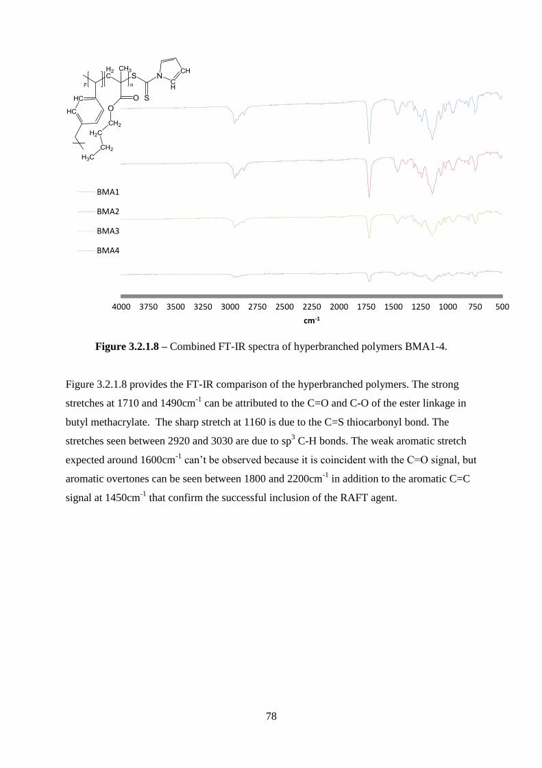
78
Figure 3.2.1.8 – Combined FT-IR spectra of hyperbranched polymers BMA1-4.
Figure 3.2.1.8 provides the FT-IR comparison of the hyperbranched polymers. The strong
stretches at 1710 and 1490cm-1
can be attributed to the C=O and C-O of the ester linkage in
butyl methacrylate. The sharp stretch at 1160 is due to the C=S thiocarbonyl bond. The
stretches seen between 2920 and 3030 are due to sp3 C-H bonds. The weak aromatic stretch
expected around 1600cm-1
can‘t be observed because it is coincident with the C=O signal, but
aromatic overtones can be seen between 1800 and 2200cm-1
in addition to the aromatic C=C
signal at 1450cm-1
that confirm the successful inclusion of the RAFT agent.
5007501000125015001750200022502500275030003250350037504000
cm-1
BMA1
BMA2
BMA3
BMA4
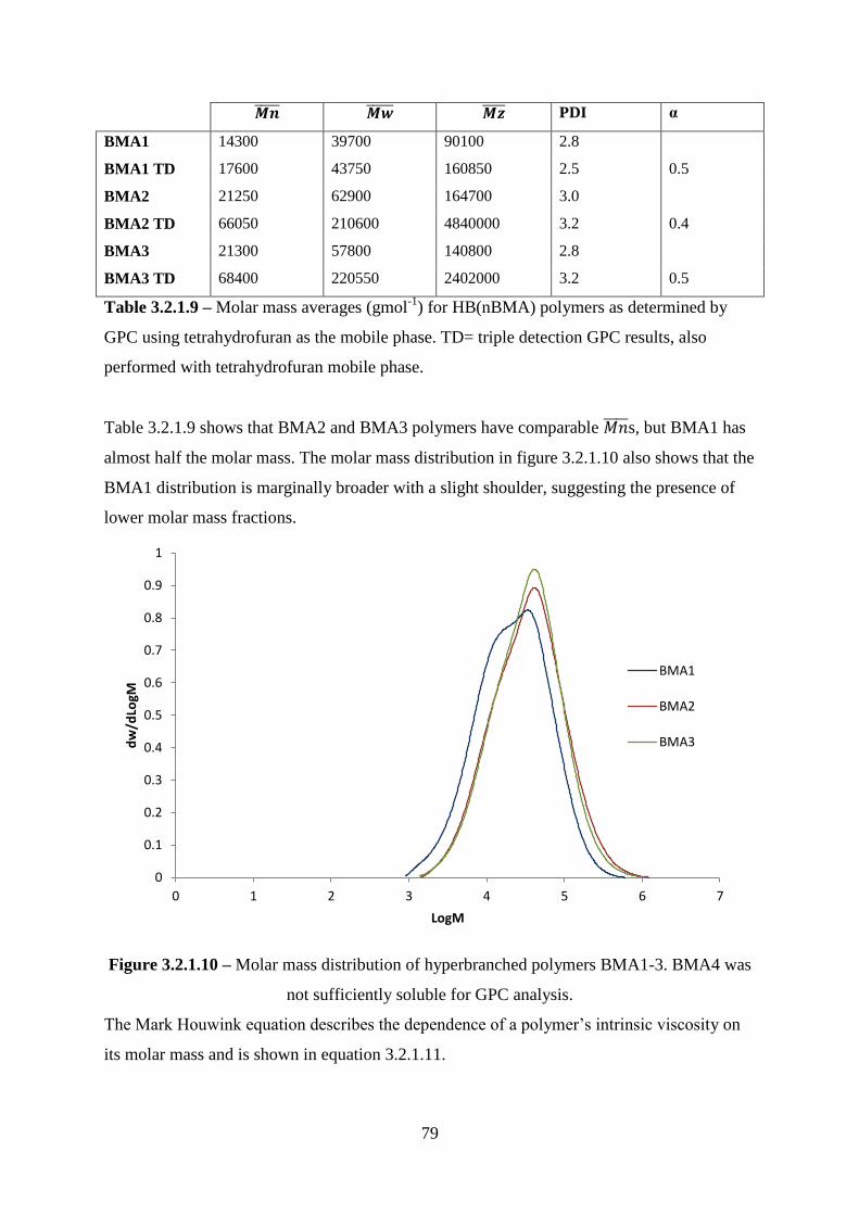
79
PDI α
BMA1 14300 39700 90100 2.8
BMA1 TD 17600 43750 160850 2.5 0.5
BMA2 21250 62900 164700 3.0
BMA2 TD 66050 210600 4840000 3.2 0.4
BMA3 21300 57800 140800 2.8
BMA3 TD 68400 220550 2402000 3.2 0.5
Table 3.2.1.9 – Molar mass averages (gmol-1
) for HB(nBMA) polymers as determined by
GPC using tetrahydrofuran as the mobile phase. TD= triple detection GPC results, also
performed with tetrahydrofuran mobile phase.
Table 3.2.1.9 shows that BMA2 and BMA3 polymers have comparable s, but BMA1 has
almost half the molar mass. The molar mass distribution in figure 3.2.1.10 also shows that the
BMA1 distribution is marginally broader with a slight shoulder, suggesting the presence of
lower molar mass fractions.
Figure 3.2.1.10 – Molar mass distribution of hyperbranched polymers BMA1-3. BMA4 was
not sufficiently soluble for GPC analysis.
The Mark Houwink equation describes the dependence of a polymer‘s intrinsic viscosity on
its molar mass and is shown in equation 3.2.1.11.
0
0.1
0.2
0.3
0.4
0.5
0.6
0.7
0.8
0.9
1
0 1 2 3 4 5 6 7
dw
/dLo
gM
LogM
BMA1
BMA2
BMA3
80
Equation 3.2.1.11 – Mark-Houwink equation, where [ŋ] is intrinsic viscosity, K and α are
polymer-dependent constants (α being the Mark-Houwink exponent) and Mr is the viscosity
molar mass average.
The value for α can vary between 0 for solid spheres and 2 for rod shaped structures, and an α
of 0.4 to 0.5 indicates a random coil structure[232]. By using triple detection GPC, which
incorporates the measuring of viscosity, it was possible to calculate α for the HB poly(butyl
methacrylate)s (table 3.2.1.9). Table 3.2.1.9 shows correlates relatively well between the
two GPC techniques, in that the trend in molar mass averages is the same. As expected, the
absolute values of molar mass differ between the two GPC techniques with almost three-fold
higher reported for polymers BMA2 and 3 using the triple detector set up. This effect is a
clear indication of the branched nature of the polymers, as increased branching decreases the
hydrodynamic volume of the coil at a constant molar mass, and it is this parameter that
controls the retention time in a GPC experiment. Calculations suggested that ~74% monomer
was converted in polymerisation BMA1 (table 3.2.1.6), but the low molar mass results
suggest much of this was converted into more numerous polymers with lower chain length.
This is to be compared with polymers BMA2 and 3 which exhibited monomer conversions of
100 and 35%, respectively, and much higher molar masses. It is clear, as expected, that as the
amount of CTA1 and initiator increased in the reaction the rate of chain growth decreased
providing lower molar mass averages and lower monomer conversion. In order to maintain
relative ratios of CTA1 to initiator, as the CTA1 was increased the initiator concentration was
also increased: the latter increases the rate of conversion, whilst increasing the former
decreases conversion.
The α values quoted above indicate that the synthesised polymers all have a tight random coil
structure. Our observed values for α correspond well with the α values previously reported
for hyperbranched polystyrene[233], polyesteramide[234] and polymethyl methacrylate[235].
However, the values of α are higher than the previously reported values when a similar CTA
was copolymerised with N-isopropyl acrylamide. This suggests that the branching self-
condensation process is less effective with this class of chain transfer agent when
polymerising methacrylates, such as BMA. Despite this, the α values still suggest branched
structures. It is also important to note that the system with the highest conversion (BMA2)
had the lowest α, which is an indication of increased branching.
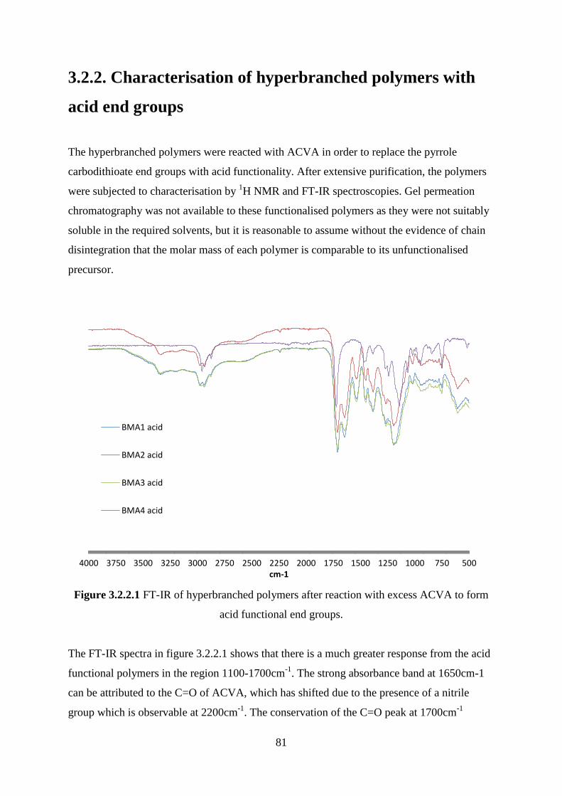
81
3.2.2. Characterisation of hyperbranched polymers with
acid end groups
The hyperbranched polymers were reacted with ACVA in order to replace the pyrrole
carbodithioate end groups with acid functionality. After extensive purification, the polymers
were subjected to characterisation by 1H NMR and FT-IR spectroscopies. Gel permeation
chromatography was not available to these functionalised polymers as they were not suitably
soluble in the required solvents, but it is reasonable to assume without the evidence of chain
disintegration that the molar mass of each polymer is comparable to its unfunctionalised
precursor.
Figure 3.2.2.1 FT-IR of hyperbranched polymers after reaction with excess ACVA to form
acid functional end groups.
The FT-IR spectra in figure 3.2.2.1 shows that there is a much greater response from the acid
functional polymers in the region 1100-1700cm-1
. The strong absorbance band at 1650cm-1
can be attributed to the C=O of ACVA, which has shifted due to the presence of a nitrile
group which is observable at 2200cm-1
. The conservation of the C=O peak at 1700cm-1
5007501000125015001750200022502500275030003250350037504000cm-1
BMA1 acid
BMA2 acid
BMA3 acid
BMA4 acid

82
indicates that the butyl methacrylate polymer backbone remains intact. This is a good
indicator of the successful reaction with ACVA, replacing the carbodithioate groups with
acid functionality. Further evidence for this can be seen in the absorbance band at 2220cm-1
which arises from the C≡N in the ACVA molecule, the broad stretch at 3200-3400cm-1
due to
hydrogen bonding between O-H groups and at 1390cm-1
from the C-O-H bend, as well as the
new N-H stretch at 1520cm-1
. It is worth noting that BMA4 does not appear to be
successfully functionalised by this reaction, with none of these signals present. This could be
due to inaccessibility of the carbodithioate groups, or crosslinking, as further reactions with
ACVA were fruitless. If polymer BMA4 has been crosslinked, this would also explain its
insolubility in analytical solvents such as THF and CDCl3.
Figure 3.2.2.2 – Representative 1H NMR of hyperbranched butyl methacrylate polymer after
treatment with excess ACVA.
Figure 3.2.2.2 shows a representative 1H NMR spectra of polymers BMA1-3 after the
reaction with excess ACVA. Polymer BMA4 was too insoluble for NMR analysis, again
indicating that it had crosslinked during the course of the reaction. The success of this
reaction is noted by a new peak at 8ppm that is due to OH of the carboxylic acid. Two further
A,F
C B
E
J G
D
N/L/M H
83
peaks are evident at 2.8 and 3ppm, which can be assigned to the CH2 of ACVA and indicates
the successful introduction of ACVA onto the polymer chain ends. The signals seen between
1-3ppm are a result of the alkyl protons present within the P(BMA).
3.2.3 Linear polymer characterisation
Linear polymers, analogous to the HB polymers, were supplied by Masters student Sarah
Canning. These were copolymers of butyl methacrylate and 4-vinyl benzoic acid in various
ratios (LIN1, 2 and 3 being 4:1, 8:1, and 12:1 molar ratios, respectively) in order to mimic the
hyperbranched functionality after reaction with ACVA. These polymers were fully
characterised and reacted with an excess of diamines, before being used as substrates in direct
culture with epithelial and fibroblast cells.
The reaction with 1,6-diaminohexane produced much more effervescence than the reaction
with other diamines, and the amount of recovered polymer was not sufficient to perform cell
culture and all analysis techniques. Due to limited amounts of the supplied acid-functional
polymers, instead of repeating the reaction greater amounts of the other amidated polymers
were produced so that cell culture and analysis could be comfortably performed.
In figure 3.2.3.1 the sharp signal at 1740cm-1
exhibited by the acid polymer is the C=O
stretch. This stretch is notable by its shift to 1677cm-1
in the amidated polymers, as is usual
for an amide bond. This shows that the reactions with excess amine and EDC were successful
in adding diamines to a copolymer of butyl methacrylate and 4-vinyl benzoic acid.

84
Figure 3.2.3.1 –Combined FT-IR of Poly(BMA-co-4-vinylbenzoic acid) and amidated
derivatives which has been subsequently reacted with an excess of various diamines as
previously described.
90
95
100
105
110
115
120
125
50
06
10
72
08
30
94
01
05
01
16
01
27
01
38
01
49
01
60
01
71
01
82
01
93
02
04
02
15
02
26
02
37
02
48
02
59
02
70
02
81
02
92
03
03
03
14
03
25
03
36
03
47
03
58
03
69
03
80
03
91
0
%T
cm-1
Acid
1,2- functional
1,3- functional
1,4- functional
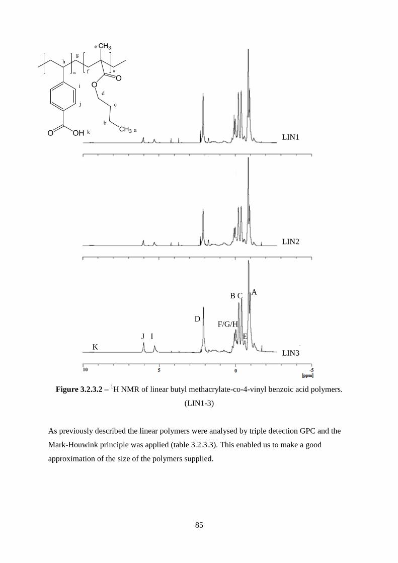
85
Figure 3.2.3.2 – 1H NMR of linear butyl methacrylate-co-4-vinyl benzoic acid polymers.
(LIN1-3)
As previously described the linear polymers were analysed by triple detection GPC and the
Mark-Houwink principle was applied (table 3.2.3.3). This enabled us to make a good
approximation of the size of the polymers supplied.
LIN3
LIN2
LIN1
A
E
J I
K
D
B C
F/G/H
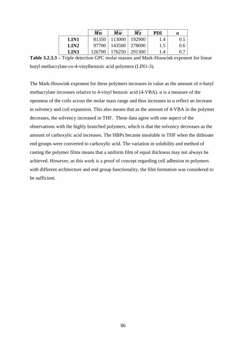
86
PDI α
LIN1 81350 113000 192900 1.4 0.5
LIN2 97700 143500 278000 1.5 0.6
LIN3 126700 176250 291300 1.4 0.7
Table 3.2.3.3 – Triple detection GPC molar masses and Mark-Houwink exponent for linear
butyl methacrylate-co-4-vinylbenzoic acid polymers (LIN1-3).
The Mark-Houwink exponent for these polymers increases in value as the amount of n-butyl
methacrylate increases relative to 4-vinyl benzoic acid (4-VBA). α is a measure of the
openness of the coils across the molar mass range and thus increases in α reflect an increase
in solvency and coil expansion. This also means that as the amount of 4-VBA in the polymer
decreases, the solvency increased in THF. These data agree with one aspect of the
observations with the highly branched polymers, which is that the solvency decreases as the
amount of carboxylic acid increases. The HBPs became insoluble in THF when the dithioate
end groups were converted to carboxylic acid. The variation in solubility and method of
casting the polymer films means that a uniform film of equal thickness may not always be
achieved. However, as this work is a proof of concept regarding cell adhesion to polymers
with different architecture and end group functionality, the film formation was considered to
be sufficient.
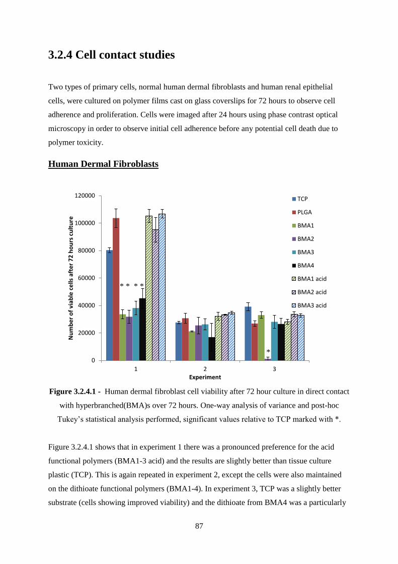
87
3.2.4 Cell contact studies
Two types of primary cells, normal human dermal fibroblasts and human renal epithelial
cells, were cultured on polymer films cast on glass coverslips for 72 hours to observe cell
adherence and proliferation. Cells were imaged after 24 hours using phase contrast optical
microscopy in order to observe initial cell adherence before any potential cell death due to
polymer toxicity.
Human Dermal Fibroblasts
Figure 3.2.4.1 - Human dermal fibroblast cell viability after 72 hour culture in direct contact
with hyperbranched(BMA)s over 72 hours. One-way analysis of variance and post-hoc
Tukey‘s statistical analysis performed, significant values relative to TCP marked with *.
Figure 3.2.4.1 shows that in experiment 1 there was a pronounced preference for the acid
functional polymers (BMA1-3 acid) and the results are slightly better than tissue culture
plastic (TCP). This is again repeated in experiment 2, except the cells were also maintained
on the dithioate functional polymers (BMA1-4). In experiment 3, TCP was a slightly better
substrate (cells showing improved viability) and the dithioate from BMA4 was a particularly
0
20000
40000
60000
80000
100000
120000
1 2 3
Nu
mb
er
of
viab
le c
ells
aft
er
72
ho
urs
cu
ltu
re
Experiment
TCP
PLGA
BMA1
BMA2
BMA3
BMA4
BMA1 acid
BMA2 acid
BMA3 acid
* * * *
*
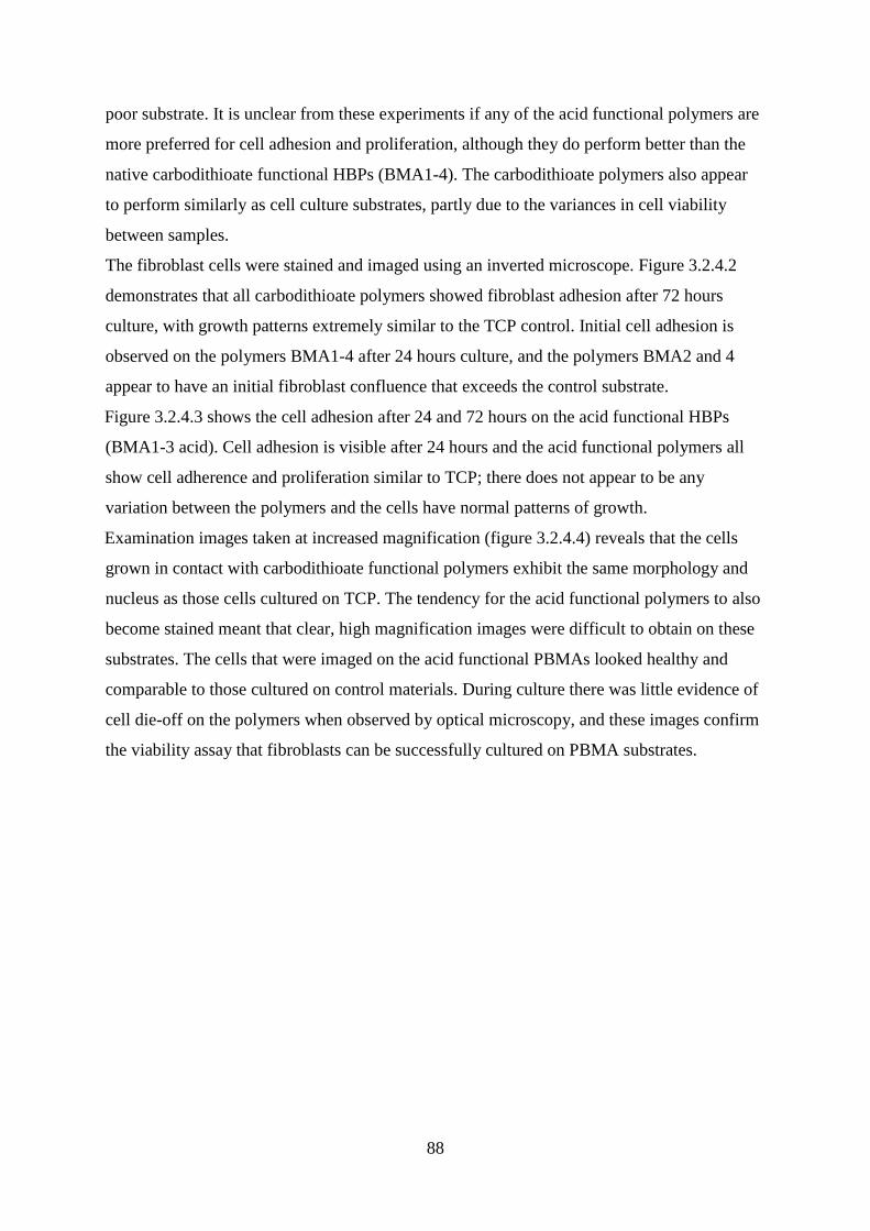
88
poor substrate. It is unclear from these experiments if any of the acid functional polymers are
more preferred for cell adhesion and proliferation, although they do perform better than the
native carbodithioate functional HBPs (BMA1-4). The carbodithioate polymers also appear
to perform similarly as cell culture substrates, partly due to the variances in cell viability
between samples.
The fibroblast cells were stained and imaged using an inverted microscope. Figure 3.2.4.2
demonstrates that all carbodithioate polymers showed fibroblast adhesion after 72 hours
culture, with growth patterns extremely similar to the TCP control. Initial cell adhesion is
observed on the polymers BMA1-4 after 24 hours culture, and the polymers BMA2 and 4
appear to have an initial fibroblast confluence that exceeds the control substrate.
Figure 3.2.4.3 shows the cell adhesion after 24 and 72 hours on the acid functional HBPs
(BMA1-3 acid). Cell adhesion is visible after 24 hours and the acid functional polymers all
show cell adherence and proliferation similar to TCP; there does not appear to be any
variation between the polymers and the cells have normal patterns of growth.
Examination images taken at increased magnification (figure 3.2.4.4) reveals that the cells
grown in contact with carbodithioate functional polymers exhibit the same morphology and
nucleus as those cells cultured on TCP. The tendency for the acid functional polymers to also
become stained meant that clear, high magnification images were difficult to obtain on these
substrates. The cells that were imaged on the acid functional PBMAs looked healthy and
comparable to those cultured on control materials. During culture there was little evidence of
cell die-off on the polymers when observed by optical microscopy, and these images confirm
the viability assay that fibroblasts can be successfully cultured on PBMA substrates.
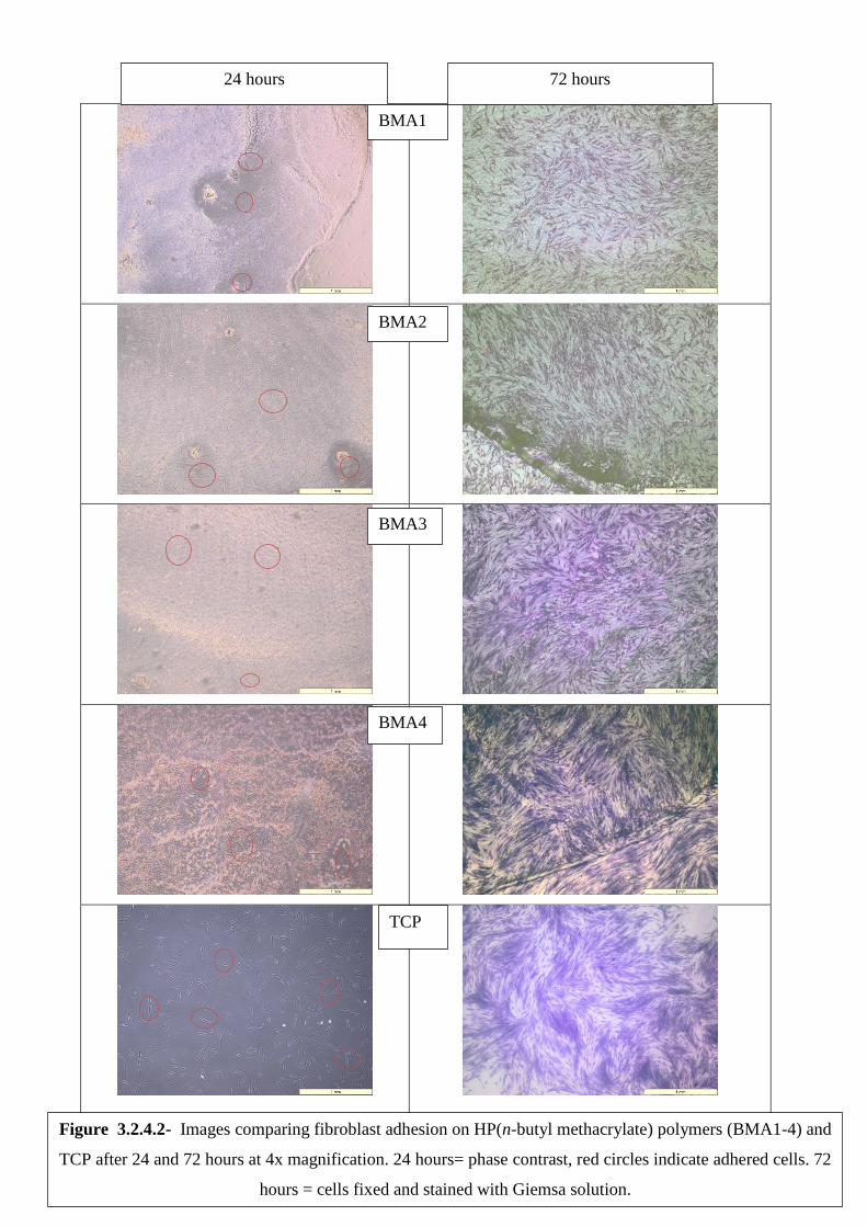
89
24 hours 72 hours
BMA1
BMA2
BMA3
BMA4
TCP
Figure 3.2.4.2- Images comparing fibroblast adhesion on HP(n-butyl methacrylate) polymers (BMA1-4) and
TCP after 24 and 72 hours at 4x magnification. 24 hours= phase contrast, red circles indicate adhered cells. 72
hours = cells fixed and stained with Giemsa solution.
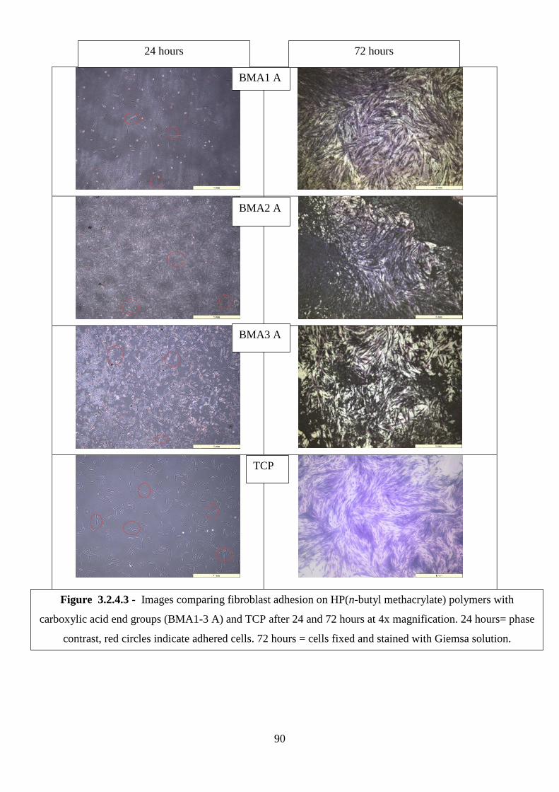
90
24 hours 72 hours
BMA1 A
BMA2 A
BMA3 A
TCP
Figure 3.2.4.3 - Images comparing fibroblast adhesion on HP(n-butyl methacrylate) polymers with
carboxylic acid end groups (BMA1-3 A) and TCP after 24 and 72 hours at 4x magnification. 24 hours= phase
contrast, red circles indicate adhered cells. 72 hours = cells fixed and stained with Giemsa solution.
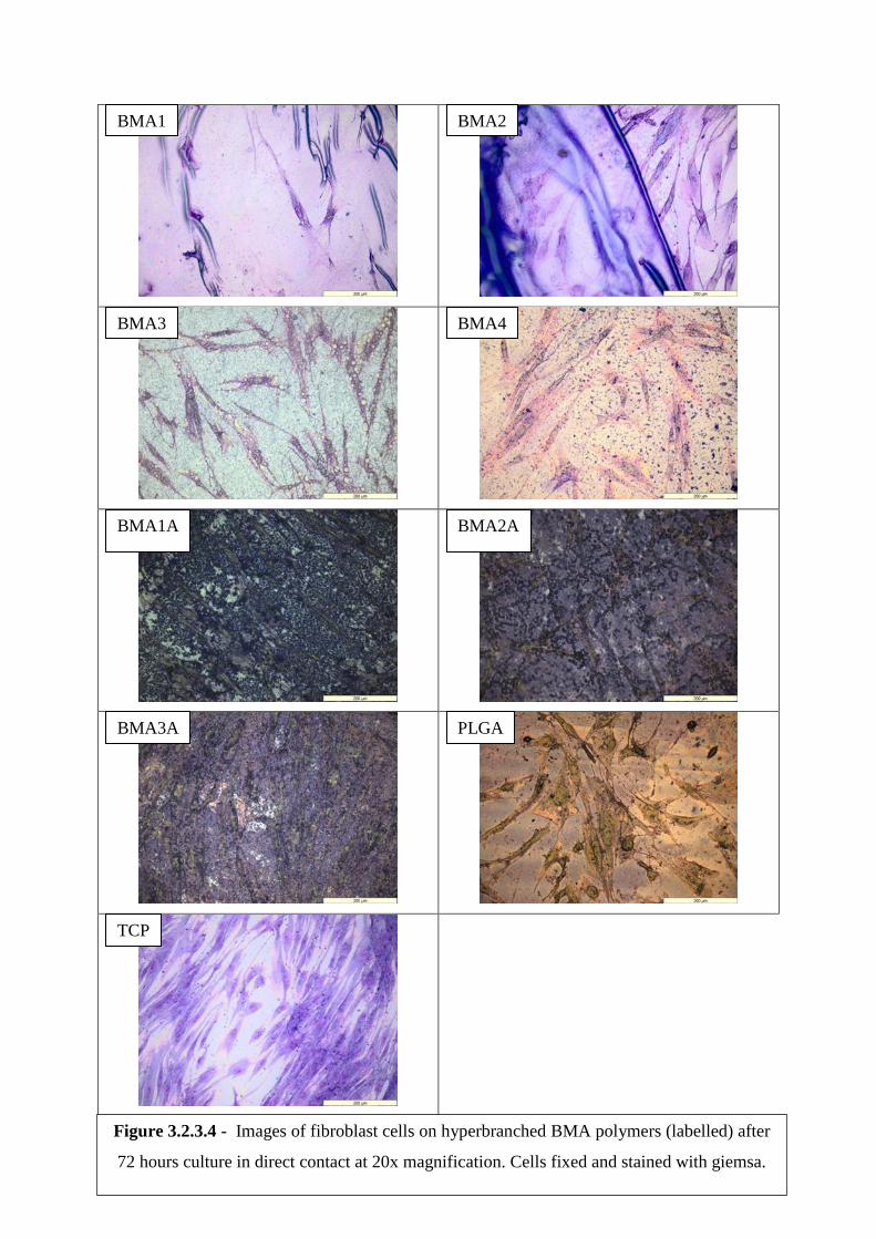
91
Figure 3.2.3.4 - Images of fibroblast cells on hyperbranched BMA polymers (labelled) after
72 hours culture in direct contact at 20x magnification. Cells fixed and stained with giemsa.
BMA3 BMA4
BMA1A BMA2A
BMA3A PLGA
TCP
BMA1 BMA2
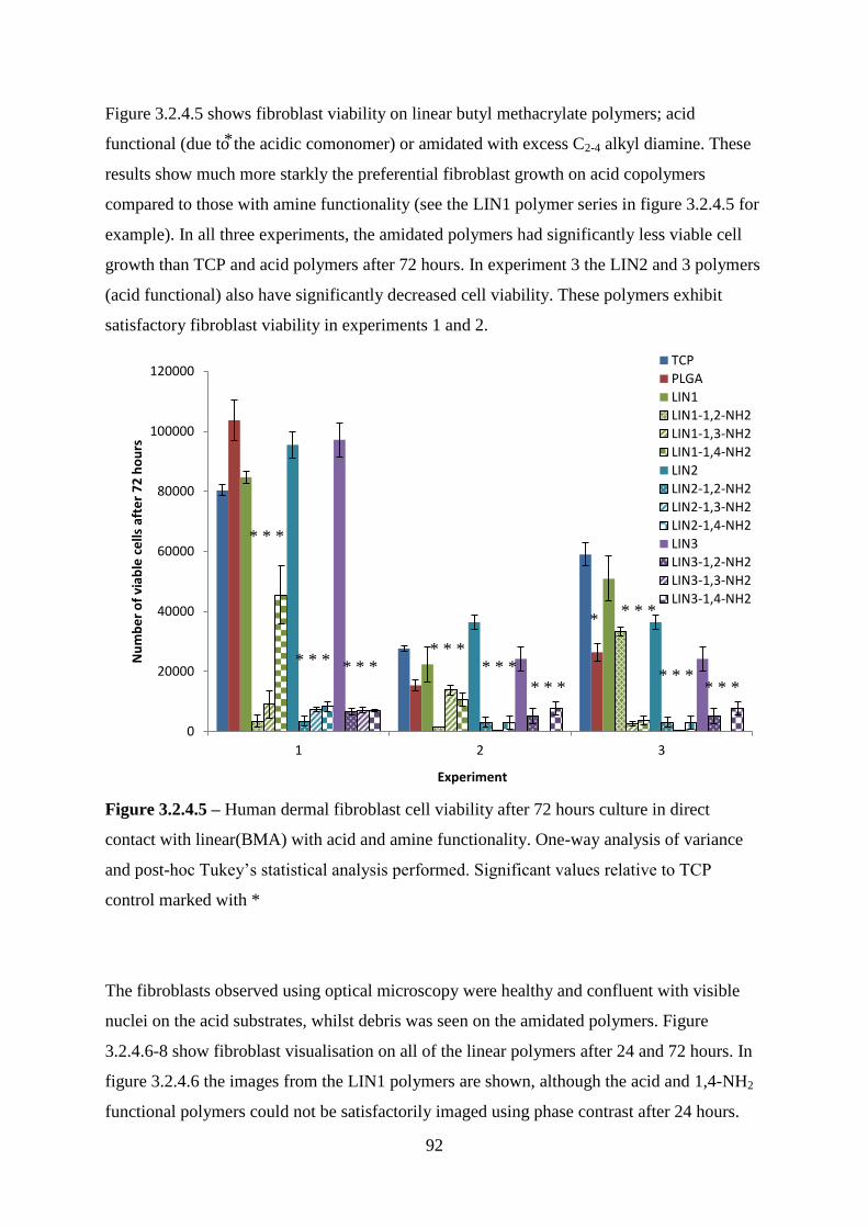
92
Figure 3.2.4.5 shows fibroblast viability on linear butyl methacrylate polymers; acid
functional (due to the acidic comonomer) or amidated with excess C2-4 alkyl diamine. These
results show much more starkly the preferential fibroblast growth on acid copolymers
compared to those with amine functionality (see the LIN1 polymer series in figure 3.2.4.5 for
example). In all three experiments, the amidated polymers had significantly less viable cell
growth than TCP and acid polymers after 72 hours. In experiment 3 the LIN2 and 3 polymers
(acid functional) also have significantly decreased cell viability. These polymers exhibit
satisfactory fibroblast viability in experiments 1 and 2.
Figure 3.2.4.5 – Human dermal fibroblast cell viability after 72 hours culture in direct
contact with linear(BMA) with acid and amine functionality. One-way analysis of variance
and post-hoc Tukey‘s statistical analysis performed. Significant values relative to TCP
control marked with *
The fibroblasts observed using optical microscopy were healthy and confluent with visible
nuclei on the acid substrates, whilst debris was seen on the amidated polymers. Figure
3.2.4.6-8 show fibroblast visualisation on all of the linear polymers after 24 and 72 hours. In
figure 3.2.4.6 the images from the LIN1 polymers are shown, although the acid and 1,4-NH2
functional polymers could not be satisfactorily imaged using phase contrast after 24 hours.
0
20000
40000
60000
80000
100000
120000
1 2 3
Nu
mb
er
of
viab
le c
ells
aft
er
72
ho
urs
Experiment
TCP
PLGA
LIN1
LIN1-1,2-NH2
LIN1-1,3-NH2
LIN1-1,4-NH2
LIN2
LIN2-1,2-NH2
LIN2-1,3-NH2
LIN2-1,4-NH2
LIN3
LIN3-1,2-NH2
LIN3-1,3-NH2
LIN3-1,4-NH2
* * *
*
* * *
*
* * *
*
* * *
*
* * *
* * * *
*
* * *
*
* * *
* * * *
*
*
*
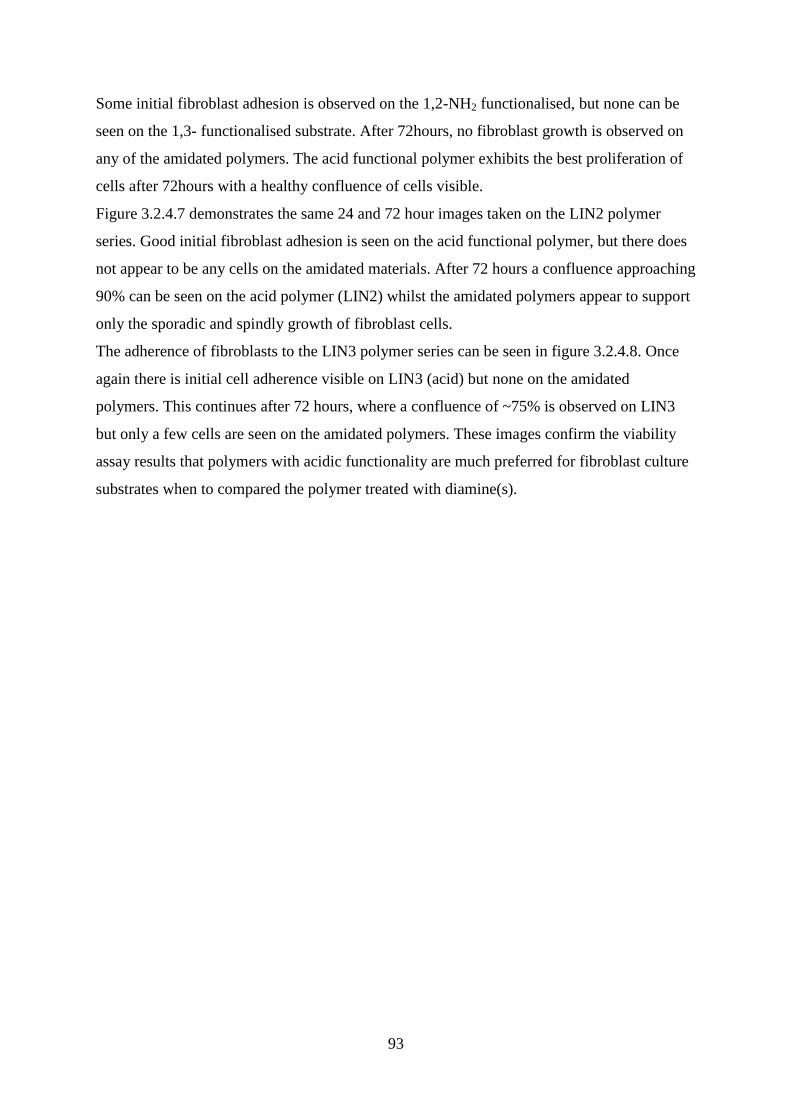
93
Some initial fibroblast adhesion is observed on the 1,2-NH2 functionalised, but none can be
seen on the 1,3- functionalised substrate. After 72hours, no fibroblast growth is observed on
any of the amidated polymers. The acid functional polymer exhibits the best proliferation of
cells after 72hours with a healthy confluence of cells visible.
Figure 3.2.4.7 demonstrates the same 24 and 72 hour images taken on the LIN2 polymer
series. Good initial fibroblast adhesion is seen on the acid functional polymer, but there does
not appear to be any cells on the amidated materials. After 72 hours a confluence approaching
90% can be seen on the acid polymer (LIN2) whilst the amidated polymers appear to support
only the sporadic and spindly growth of fibroblast cells.
The adherence of fibroblasts to the LIN3 polymer series can be seen in figure 3.2.4.8. Once
again there is initial cell adherence visible on LIN3 (acid) but none on the amidated
polymers. This continues after 72 hours, where a confluence of ~75% is observed on LIN3
but only a few cells are seen on the amidated polymers. These images confirm the viability
assay results that polymers with acidic functionality are much preferred for fibroblast culture
substrates when to compared the polymer treated with diamine(s).
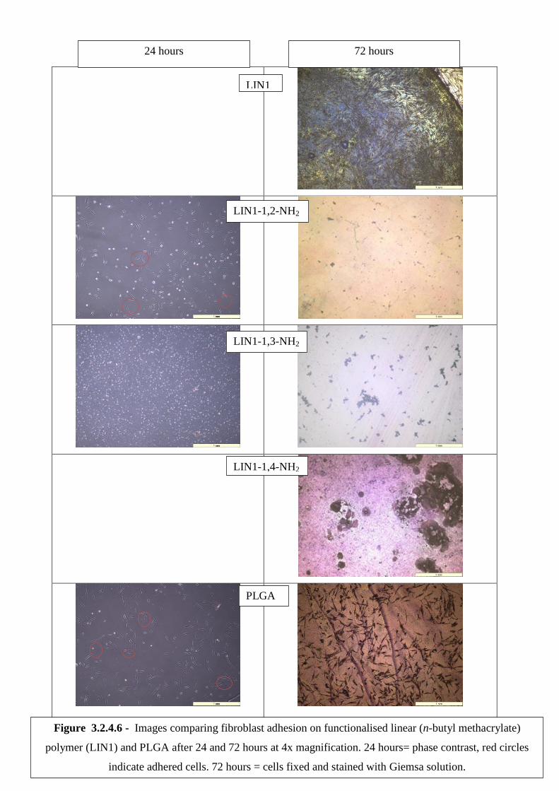
94
24 hours 72 hours
LIN1
LIN1-1,2-NH2
LIN1-1,3-NH2
LIN1-1,4-NH2
PLGA
Figure 3.2.4.6 - Images comparing fibroblast adhesion on functionalised linear (n-butyl methacrylate)
polymer (LIN1) and PLGA after 24 and 72 hours at 4x magnification. 24 hours= phase contrast, red circles
indicate adhered cells. 72 hours = cells fixed and stained with Giemsa solution.
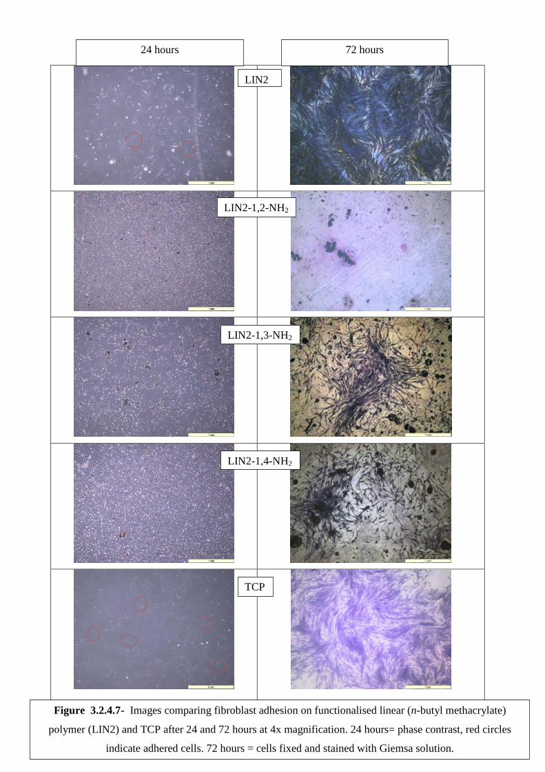
95
24 hours 72 hours
LIN2
LIN2-1,2-NH2
LIN2-1,3-NH2
LIN2-1,4-NH2
TCP
Figure 3.2.4.7- Images comparing fibroblast adhesion on functionalised linear (n-butyl methacrylate)
polymer (LIN2) and TCP after 24 and 72 hours at 4x magnification. 24 hours= phase contrast, red circles
indicate adhered cells. 72 hours = cells fixed and stained with Giemsa solution.
96
24 hours 72 hours
LIN3
LIN3-1,2-NH2
LIN3-1,3-NH2
LIN3-1,4-NH2
TCP
Figure 3.2.4.8 - Images comparing fibroblast adhesion on functionalised linear (n-butyl methacrylate)
polymer (LIN3) and PLGA after 24 and 72 hours at 4x magnification. 24 hours= phase contrast, red circles
indicate adhered cells. 72 hours = cells fixed and stained with Giemsa solution.
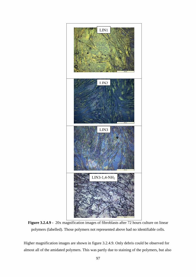
97
Figure 3.2.4.9 - 20x magnification images of fibroblasts after 72 hours culture on linear
polymers (labelled). Those polymers not represented above had no identifiable cells.
Higher magnification images are shown in figure 3.2.4.9. Only debris could be observed for
almost all of the amidated polymers. This was partly due to staining of the polymers, but also
LIN1
LIN2
LIN3
LIN3-1,4-NH2
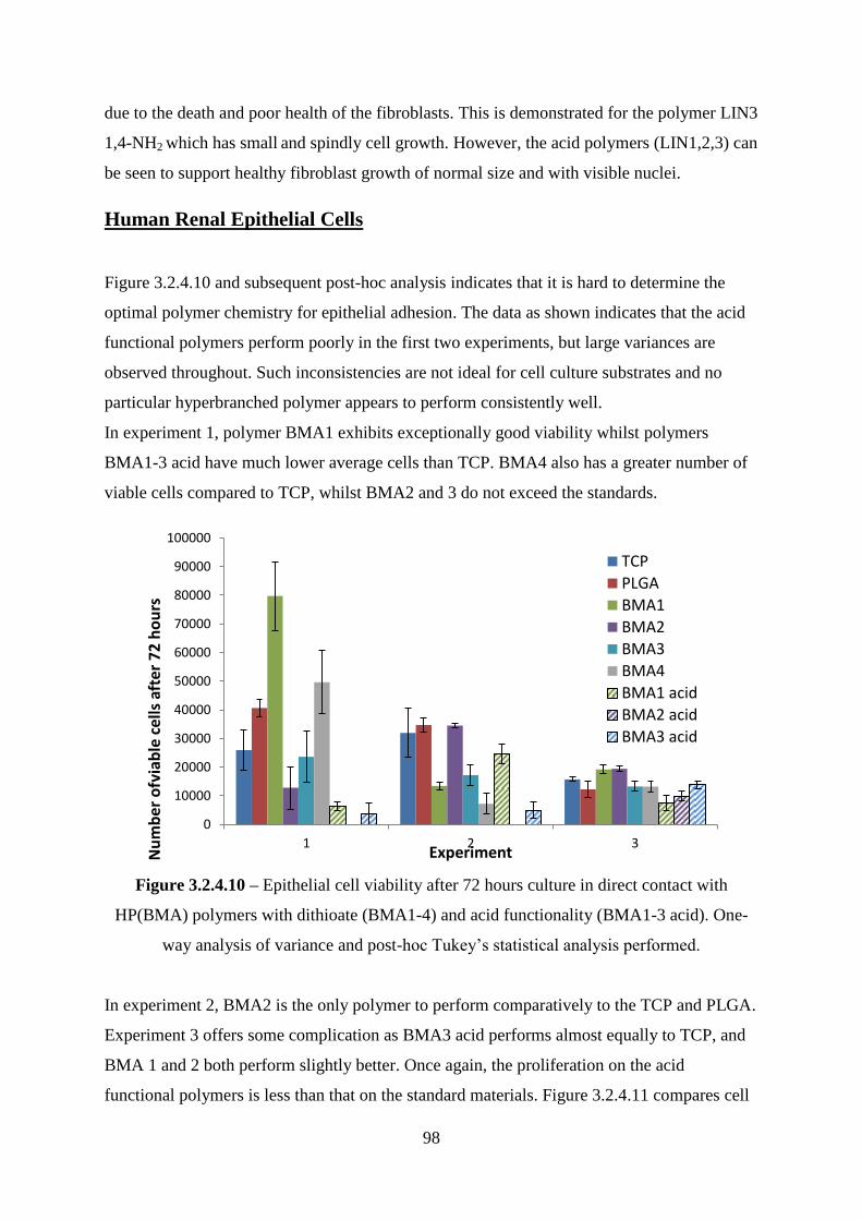
98
due to the death and poor health of the fibroblasts. This is demonstrated for the polymer LIN3
1,4-NH2 which has small and spindly cell growth. However, the acid polymers (LIN1,2,3) can
be seen to support healthy fibroblast growth of normal size and with visible nuclei.
Human Renal Epithelial Cells
Figure 3.2.4.10 and subsequent post-hoc analysis indicates that it is hard to determine the
optimal polymer chemistry for epithelial adhesion. The data as shown indicates that the acid
functional polymers perform poorly in the first two experiments, but large variances are
observed throughout. Such inconsistencies are not ideal for cell culture substrates and no
particular hyperbranched polymer appears to perform consistently well.
In experiment 1, polymer BMA1 exhibits exceptionally good viability whilst polymers
BMA1-3 acid have much lower average cells than TCP. BMA4 also has a greater number of
viable cells compared to TCP, whilst BMA2 and 3 do not exceed the standards.
Figure 3.2.4.10 – Epithelial cell viability after 72 hours culture in direct contact with
HP(BMA) polymers with dithioate (BMA1-4) and acid functionality (BMA1-3 acid). One-
way analysis of variance and post-hoc Tukey‘s statistical analysis performed.
In experiment 2, BMA2 is the only polymer to perform comparatively to the TCP and PLGA.
Experiment 3 offers some complication as BMA3 acid performs almost equally to TCP, and
BMA 1 and 2 both perform slightly better. Once again, the proliferation on the acid
functional polymers is less than that on the standard materials. Figure 3.2.4.11 compares cell
0
10000
20000
30000
40000
50000
60000
70000
80000
90000
100000
1 2 3
Nu
mb
er o
fvia
ble
ce
lls a
fter
72
ho
urs
Experiment
TCP
PLGA
BMA1
BMA2
BMA3
BMA4
BMA1 acid
BMA2 acid
BMA3 acid
99
images taken after 24 and 72 hours culture on the different polymer substrates. The TCP
images indicate that there is a good initial adhesion of cells after 24 hours, which continue to
proliferate for the remaining time of the experiment. This is a pattern mimicked by the BMA1
polymer, but to a lesser extent. BMA2 does not appear to show many cells adhered after
24hours, but after 72 hours this substrate has a confluence of about 50% - a number which is
comparable with the TCP. BMA3 appears to show relatively numerous adhered cells after 24
hours but only a few cells are visible after 72 hours, suggesting that the cells are becoming
unadhered or are suffering a toxic effect from this polymer. The polymer itself interfered with
the imaging of the cells adhered to BMA4, however after 72 hours there appears to be some
adhered cells, but fewer than can be seen on the TCP.
The optical microscopy images of epithelial adhesion on the acid functional HBPs is shown
in figure 3.2.4.12. There is very poor initial adhesion visible on all three of the acid substrates
after 24 hours. BMA2 acid shows the greatest number of adhered cells after 24 hours but it
still performs poorly when compared to TCP. After 72 hours, the TCP can be seen to have
approximately 70% confluence whilst only a few sparse cells are observed on the acid
functional polymers.
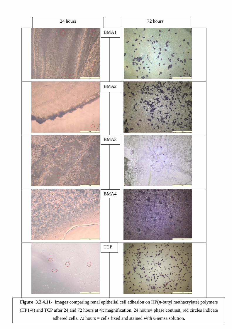
100
24 hours 72 hours
BMA1
BMA2
BMA3
BMA4
TCP
Figure 3.2.4.11- Images comparing renal epithelial cell adhesion on HP(n-butyl methacrylate) polymers
(HP1-4) and TCP after 24 and 72 hours at 4x magnification. 24 hours= phase contrast, red circles indicate
adhered cells. 72 hours = cells fixed and stained with Giemsa solution.
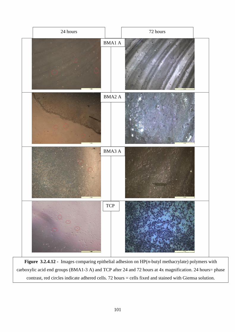
101
24 hours 72 hours
BMA1 A
BMA2 A
BMA3 A
TCP
Figure 3.2.4.12 - Images comparing epithelial adhesion on HP(n-butyl methacrylate) polymers with
carboxylic acid end groups (BMA1-3 A) and TCP after 24 and 72 hours at 4x magnification. 24 hours= phase
contrast, red circles indicate adhered cells. 72 hours = cells fixed and stained with Giemsa solution.

102
Figure 3.2.4.13 compares the cell morphology of the dithioate and acid functional polymers
at 20x magnification. From the TCP and PLGA standards it can be seen that epithelial cells
generally grow in close proximity, and that approximately 1/3rd
of the cell area is accounted
for by the nucleus. The cells cultured on HBpolymers BMA1 and 2 have healthy cell
morphologies comparable with the standards. There are only a few sparse cells on BMA3 and
once again the polymer interfered with the quality of images for BMA4. The cells on the acid
functional materials appear to have shrunken and malformed morphologies and do not appear
healthy.
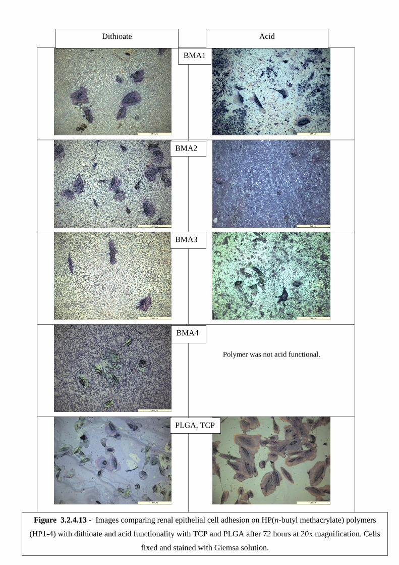
103
Polymer was not acid functional.
Dithioate Acid
BMA1
BMA2
BMA3
BMA4
PLGA, TCP
Figure 3.2.4.13 - Images comparing renal epithelial cell adhesion on HP(n-butyl methacrylate) polymers
(HP1-4) with dithioate and acid functionality with TCP and PLGA after 72 hours at 20x magnification. Cells
fixed and stained with Giemsa solution.
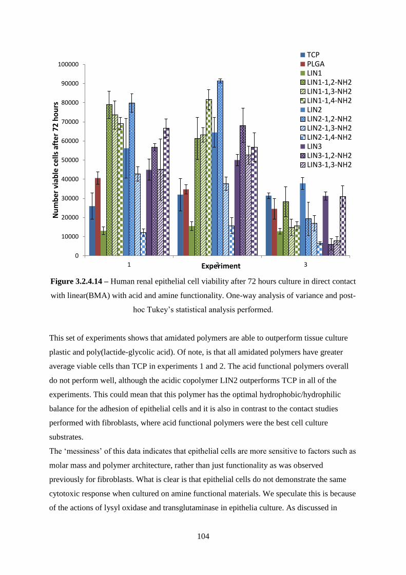
104
Figure 3.2.4.14 – Human renal epithelial cell viability after 72 hours culture in direct contact
with linear(BMA) with acid and amine functionality. One-way analysis of variance and post-
hoc Tukey‘s statistical analysis performed.
This set of experiments shows that amidated polymers are able to outperform tissue culture
plastic and poly(lactide-glycolic acid). Of note, is that all amidated polymers have greater
average viable cells than TCP in experiments 1 and 2. The acid functional polymers overall
do not perform well, although the acidic copolymer LIN2 outperforms TCP in all of the
experiments. This could mean that this polymer has the optimal hydrophobic/hydrophilic
balance for the adhesion of epithelial cells and it is also in contrast to the contact studies
performed with fibroblasts, where acid functional polymers were the best cell culture
substrates.
The ‗messiness‘ of this data indicates that epithelial cells are more sensitive to factors such as
molar mass and polymer architecture, rather than just functionality as was observed
previously for fibroblasts. What is clear is that epithelial cells do not demonstrate the same
cytotoxic response when cultured on amine functional materials. We speculate this is because
of the actions of lysyl oxidase and transglutaminase in epithelia culture. As discussed in
0
10000
20000
30000
40000
50000
60000
70000
80000
90000
100000
1 2 3
Nu
mb
er v
iab
le c
ells
aft
er 7
2 h
ou
rs
Experiment
TCPPLGALIN1LIN1-1,2-NH2LIN1-1,3-NH2LIN1-1,4-NH2LIN2LIN2-1,2-NH2LIN2-1,3-NH2LIN2-1,4-NH2LIN3LIN3-1,2-NH2LIN3-1,3-NH2
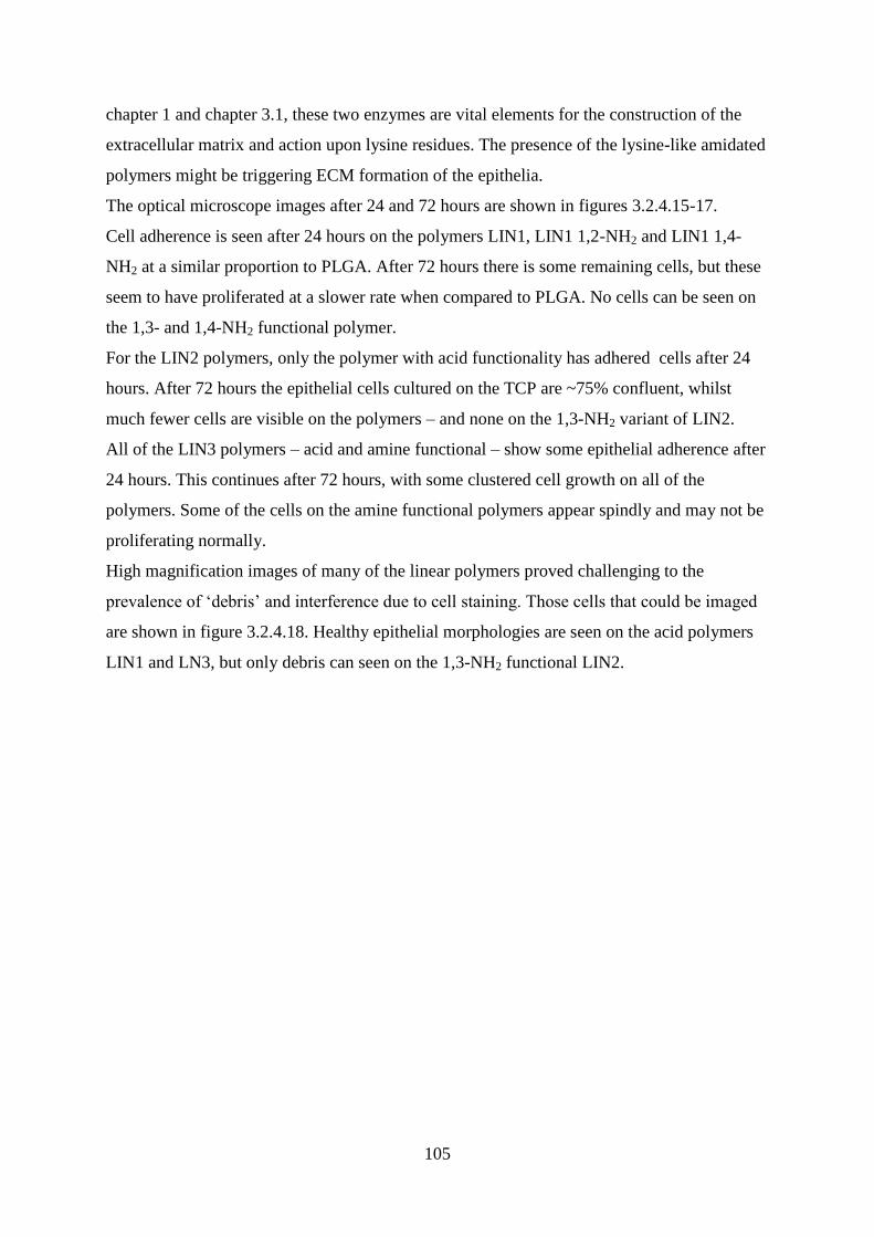
105
chapter 1 and chapter 3.1, these two enzymes are vital elements for the construction of the
extracellular matrix and action upon lysine residues. The presence of the lysine-like amidated
polymers might be triggering ECM formation of the epithelia.
The optical microscope images after 24 and 72 hours are shown in figures 3.2.4.15-17.
Cell adherence is seen after 24 hours on the polymers LIN1, LIN1 1,2-NH2 and LIN1 1,4-
NH2 at a similar proportion to PLGA. After 72 hours there is some remaining cells, but these
seem to have proliferated at a slower rate when compared to PLGA. No cells can be seen on
the 1,3- and 1,4-NH2 functional polymer.
For the LIN2 polymers, only the polymer with acid functionality has adhered cells after 24
hours. After 72 hours the epithelial cells cultured on the TCP are ~75% confluent, whilst
much fewer cells are visible on the polymers – and none on the 1,3-NH2 variant of LIN2.
All of the LIN3 polymers – acid and amine functional – show some epithelial adherence after
24 hours. This continues after 72 hours, with some clustered cell growth on all of the
polymers. Some of the cells on the amine functional polymers appear spindly and may not be
proliferating normally.
High magnification images of many of the linear polymers proved challenging to the
prevalence of ‗debris‘ and interference due to cell staining. Those cells that could be imaged
are shown in figure 3.2.4.18. Healthy epithelial morphologies are seen on the acid polymers
LIN1 and LN3, but only debris can seen on the 1,3-NH2 functional LIN2.

106
24 hours 72 hours
LIN1
LIN1 1,2-NH2
LIN1 1,3-NH2
LIN1 1,4-NH2
PLGA
Figure 3.2.4.15- Images comparing epithelial adhesion on functionalised linear (n-butyl methacrylate)
polymer (LIN1) and PLGA after 24 and 72 hours at 4x magnification. 24 hours= phase contrast, red circles
indicate adhered cells. 72 hours = cells fixed and stained with Giemsa solution.
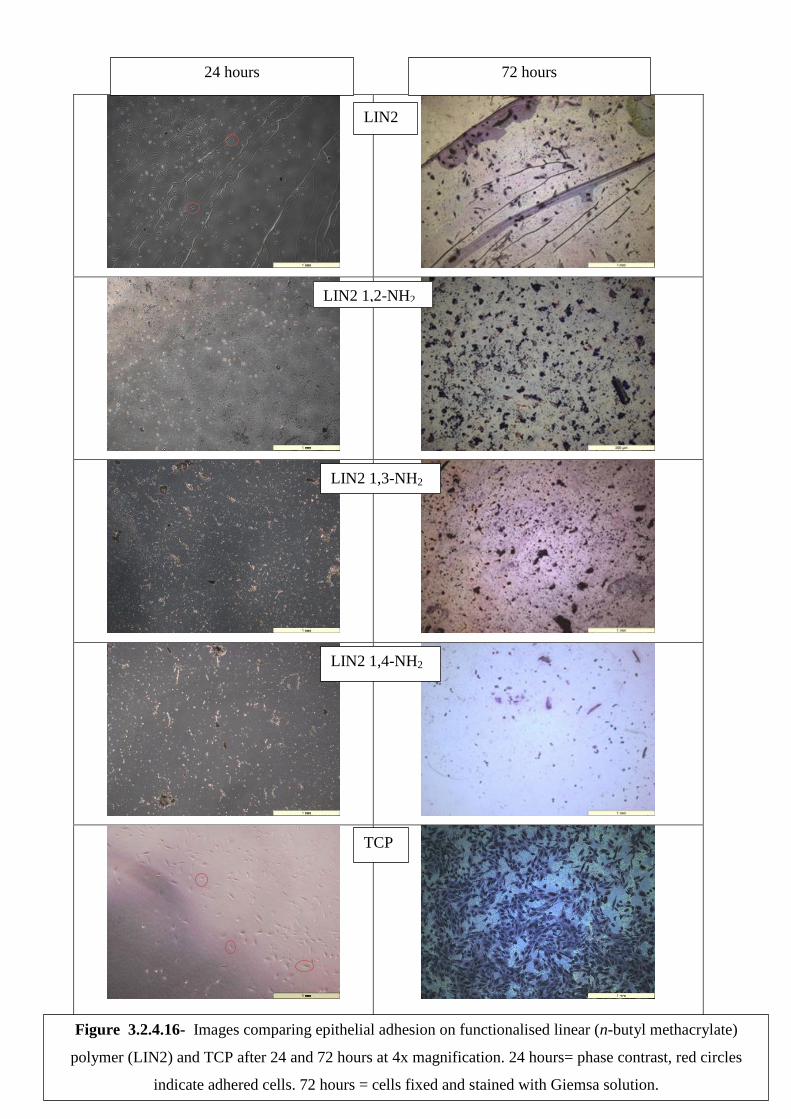
107
24 hours 72 hours
LIN2
LIN2 1,2-NH2
LIN2 1,3-NH2
LIN2 1,4-NH2
TCP
Figure 3.2.4.16- Images comparing epithelial adhesion on functionalised linear (n-butyl methacrylate)
polymer (LIN2) and TCP after 24 and 72 hours at 4x magnification. 24 hours= phase contrast, red circles
indicate adhered cells. 72 hours = cells fixed and stained with Giemsa solution.
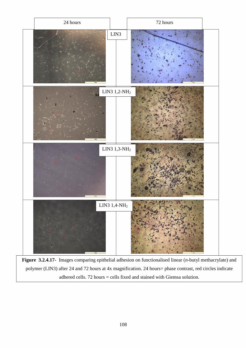
108
24 hours 72 hours
LIN3
LIN3 1,2-NH2
LIN3 1,3-NH2
LIN3 1,4-NH2
Figure 3.2.4.17- Images comparing epithelial adhesion on functionalised linear (n-butyl methacrylate) and
polymer (LIN3) after 24 and 72 hours at 4x magnification. 24 hours= phase contrast, red circles indicate
adhered cells. 72 hours = cells fixed and stained with Giemsa solution.
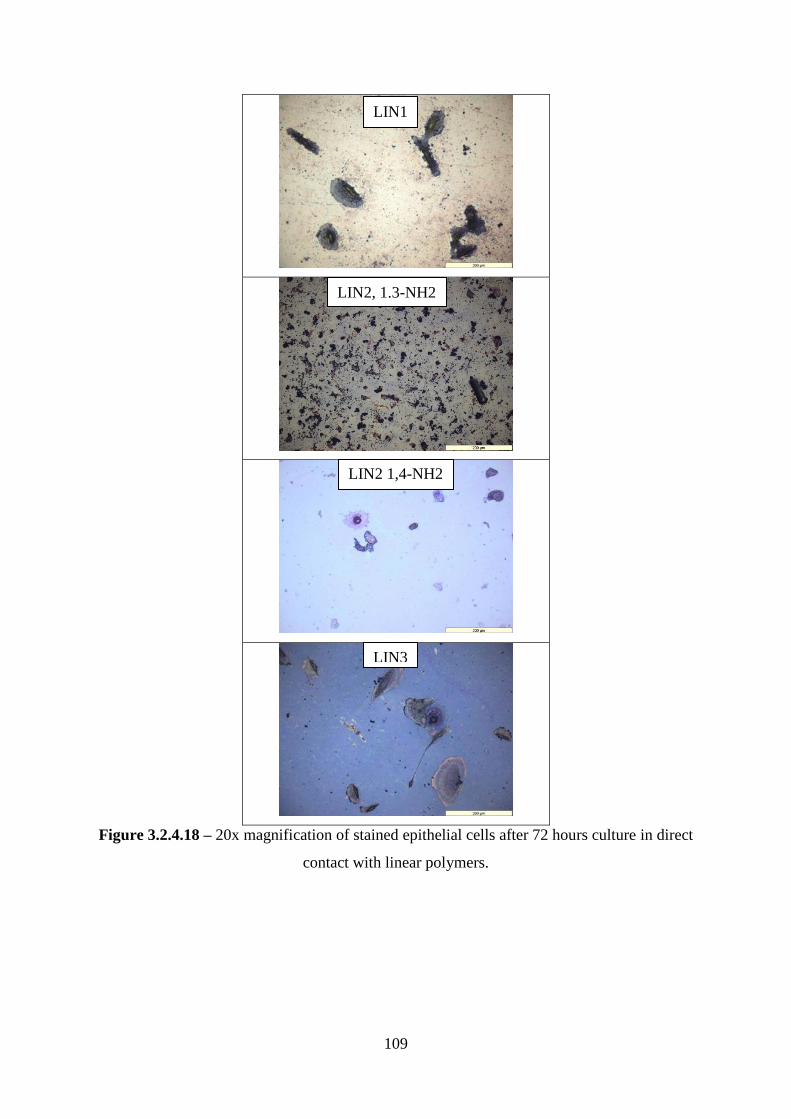
109
Figure 3.2.4.18 – 20x magnification of stained epithelial cells after 72 hours culture in direct
contact with linear polymers.
LIN1
LIN2, 1.3-NH2
LIN2 1,4-NH2
LIN3
110
3.3 Conclusions
Through elemental and chromatographic analysis, we found that 6% of CTA1 in the RAFT
polymerisation with n-butyl methacrylate was optimal for conversion of monomer and
branching of the polymer. Those polymerisations that contained 4 and 3% of CTA1 showed
poor conversion as the system was deficient in initiator and flooded with methacrylate
monomer. This was also possibly the cause for the suspected crosslinking of BMA4, which
was the only polymer that did not successfully react with an excess of ACVA.
The linear polymers supplied for comparison were found to have similar α values to the
synthesised hyperbranched polymers, and these were also successfully reacted with a small
library of diamines to create a range of functional linear polymers for cell culture studies.
Through the incubation of cells in direct contact with polymers over 72 hours, it was found
that HDFs grew equally well on all of the synthesised BMA polymers. The cells cultured on
both acid and dithioate end groups show healthy morphologies as on the control materials.
There is some evidence, such as greater average cell viabilities, to suggest that dermal
fibroblast cells are best cultured in the presence of acid functional materials.
The preference for acid functional polymers is most striking on the linear materials, where
very little HDF growth is observed on the amine functional polymers. Some fibroblast growth
was seen on the polymer LIN3 1,4-NH2, but the cells did not appear to be healthy.
Further experiments might also expose further nuances between the acid content of polymer
and cell proliferation.
Conversely, the epithelial cells were found to show no real preference for any of the dithioate
PBMAs. The hyperbranched polymers BMA1 and 2 seem to be relatively better epithelial
culture substrates than the 3 and 4 varients, and in some experiments had greater viability
than TCP. The epithelial cells showed a strong preference for adherence and proliferation on
the amine functional linear polymers. The acid functional polymers – both hyperbranched
and linear – showed poor proliferation and cell growth was sparse with unusual
morphologies.
These experiments have shown that acid functional linear polymers and hyperbranched
poly(BMA) are suitable dermal fibroblast culture substrates. For the culture of renal epithelial
cells, the preferred functionality is amine – possibly due to its chemical similarity to lysine
which can be utilised by ECM forming enzymes such as lysyl oxidase.
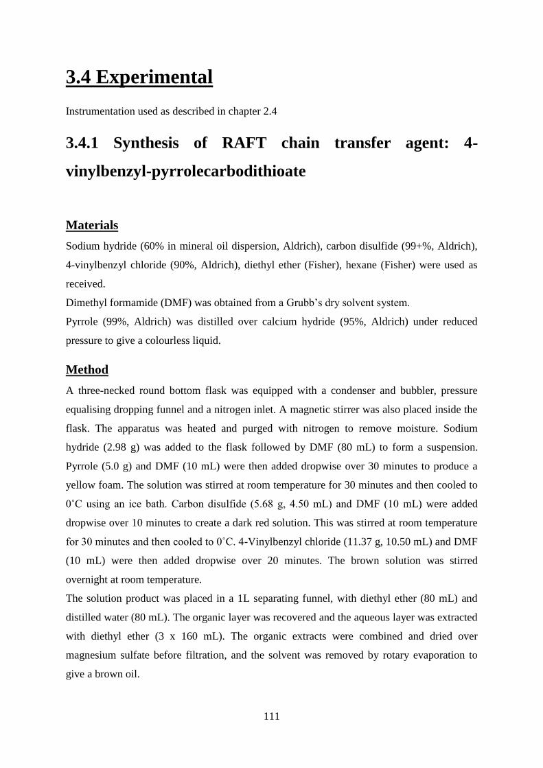
111
3.4 Experimental
Instrumentation used as described in chapter 2.4
3.4.1 Synthesis of RAFT chain transfer agent: 4-
vinylbenzyl-pyrrolecarbodithioate
Materials
Sodium hydride (60% in mineral oil dispersion, Aldrich), carbon disulfide (99+%, Aldrich),
4-vinylbenzyl chloride (90%, Aldrich), diethyl ether (Fisher), hexane (Fisher) were used as
received.
Dimethyl formamide (DMF) was obtained from a Grubb‘s dry solvent system.
Pyrrole (99%, Aldrich) was distilled over calcium hydride (95%, Aldrich) under reduced
pressure to give a colourless liquid.
Method
A three-necked round bottom flask was equipped with a condenser and bubbler, pressure
equalising dropping funnel and a nitrogen inlet. A magnetic stirrer was also placed inside the
flask. The apparatus was heated and purged with nitrogen to remove moisture. Sodium
hydride (2.98 g) was added to the flask followed by DMF (80 mL) to form a suspension.
Pyrrole (5.0 g) and DMF (10 mL) were then added dropwise over 30 minutes to produce a
yellow foam. The solution was stirred at room temperature for 30 minutes and then cooled to
0˚C using an ice bath. Carbon disulfide (5.68 g, 4.50 mL) and DMF (10 mL) were added
dropwise over 10 minutes to create a dark red solution. This was stirred at room temperature
for 30 minutes and then cooled to 0˚C. 4-Vinylbenzyl chloride (11.37 g, 10.50 mL) and DMF
(10 mL) were then added dropwise over 20 minutes. The brown solution was stirred
overnight at room temperature.
The solution product was placed in a 1L separating funnel, with diethyl ether (80 mL) and
distilled water (80 mL). The organic layer was recovered and the aqueous layer was extracted
with diethyl ether (3 x 160 mL). The organic extracts were combined and dried over
magnesium sulfate before filtration, and the solvent was removed by rotary evaporation to
give a brown oil.
112
The oil was purified by flash chromatography, with a column of 6cm diameter, using 100%
hexane. The solvent was removed by rotary evaporation to give 5.6 g of a bright yellow oil.
The air sensitive product was stored under nitrogen at -18˚C.
Mpt: ~25˚C. Elemental analysis: C: 64.7%, H: 5.2%, N: 5.4%, S:24.7%.
Figure 3.4.1.2 - 1H NMR of 4-vinylbenzyl-pyrrolecarbodithioate (250 MHz, CDCl3) δppm
4.62 (s, HA), 5.29 (d, J = 10.81 Hz, HC), 5.78 (d, J = 17.57 Hz, HB), 6.35 (HJ, HK), 6.73 (dd,
J = 17.60, 10.90 Hz, HD), 7.20 (HI, HL), 7.28 (solvent), 7.45-7.31 (HH, HF), 7.73 (HE, HG).
3.4.2 RAFT polymerisation of n-butyl methacrylate using
4-vinylbenzyl pyrrole carbodithioate chain transfer agent
Materials
4-Vinylbenzyl pyrrole carbodithioate (produced as previously described), n-butyl
methacrylate (98%, Aldrich), 4,4‘-azobis 4-cyanovaleric acid (ACVA, ≥98%, Aldrich), 1,4-
dioxane (anhydrous, 99.8%, Sigma-Aldrich), methanol (Fisher) were used as received.
113
Equipment
Ampoules were degassed on a high vacuum line equipped with a Pirani gauge.
Method
4-vinylbenzyl pyrrole carbodithioate, n-butyl methacrylate, dioxane (25 mL) and ACVA
were mixed together until the solid initiator had dissolved. The resulting solution was
pipetted into a 50 mL ampoule and placed onto the vacuum line but was not exposed to
vacuum. The solution was frozen using liquid nitrogen and then opened to the vacuum until
the gauge dropped to a steady output. The ampoule‘s exposure to vacuum was then ceased as
its contents were left to thaw. Once thawing was completed, the full process was repeated
until a negligible rise in pressure was observed when the ampoule was opened to vacuum.
The end pressure in the ampoule was approximately 3x10⁻³
mBar. The ampoule was sealed
using a gas/oxygen blowtorch and placed in a water bath at 60 ˚C for up to 24 hrs to undergo
polymerisation.
Table 3.4.2.1 summarises the quantities of reagents used.
n-butyl methacrylate /mL Chain transfer agent /mL ACVA /g
5 (0.03) 1 (0.004) 1.29 (0.004)
10 (0.06) 1 (0.004) 1.29 (0.004)
15 (0.09) 1 (0.004) 1.29 (0.004)
20 (0.12) 1 (0.004) 1.29 (0.004)
15 (0.09) 1 (0.004) 0.65 (0.002)
20 (0.12) 1 (0.004) 0.65 (0.002)
Table 3.4.2.1 – summary of reagent quantities used in RAFT polymerisation of n-butyl
methacrylate. Molar values in parenthesis.
Products were precipitated by transferring into 4x by volume of methanol and leaving to
stand, followed by filtering and drying in a vacuum oven producing a yellow solid. Dried
products were stored in closed sample tubes at room temperature.
Polymers were analysed by 1H and
13C NMR, size exclusion chromatography, FT-IR and
elemental analysis.
NMR spectroscopy results:
1H (400 MHz, CDCl3) δppm 0.97 (A), 1.12 (E), 1.42 (B), 1.48 (C), 1.85(F) 4.02 (D), 6.46
(J/I), 7.83 (G/H)
114
3.4.3 Reaction of hyperbranched butyl methacrylate
polymers with excess ACVA
Materials
Acetone (HPLC grade, Fisher), ethanol (absolute, Fisher), methanol (HPLC grade, Fisher),
ultrapure water (18.8Ω, MilliQ systems), HBP(n-butyl methacrylate) synthesised as described
in section 3.3.2 and 4,4‘-azobis 4-cyanovaleric acid (ACVA, ≥98%, Aldrich) were used as
received. Dimethyl formamide (DMF) was obtained from a Grubb‘s dry solvent system.
Method
A three necked round bottom flask was equipped with a magnetic stirrer, nitrogen inlet and a
condenser. The apparatus was nitrogen purged and heated to 60°C. A known amount of
polymer was dissolved in dry DMF and then injected into the warm, dry flask which was kept
under a nitrogen atmosphere. 60molar equivalents of ACVA were then dissolved in 10-15mL
DMF (up to 1 minute of sonication was used to assist dissolution) and this was then injected
into in the flask. The solution was stirred for 16hours at 60°C before the addition of another
60molar equivalents of ACVA. This process was repeated for four additions.
The polymer was purified from ACVA by first removing the DMF under reduced pressure.
The solid polymer was then dissolved in ultrapure water (MilliQ) and waste ACVA was
removed via vacuum filtration. The polymer was washed with methanol and then further
purified by ultrafiltration through a 3kDa cellulose membrane using a solvent system of 9:1
acetone:ethanol.
The reaction was deemed successful when elemental analysis showed that no sulfur was
present in the final product, indicating the complete removal of the dithionate protecting
groups.
Polymers were analysed by 1H and
13C NMR, size exclusion chromatography, FT-IR and
elemental analysis (results shown within this chapter).
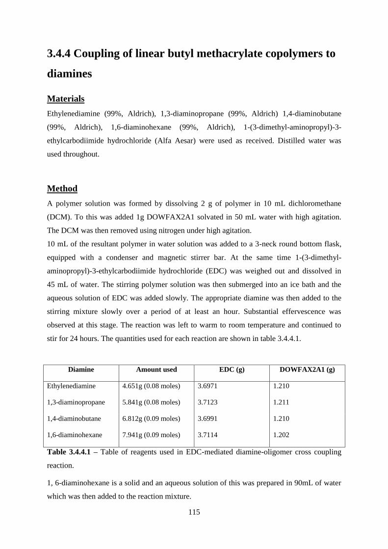
115
3.4.4 Coupling of linear butyl methacrylate copolymers to
diamines
Materials
Ethylenediamine (99%, Aldrich), 1,3-diaminopropane (99%, Aldrich) 1,4-diaminobutane
(99%, Aldrich), 1,6-diaminohexane (99%, Aldrich), 1-(3-dimethyl-aminopropyl)-3-
ethylcarbodiimide hydrochloride (Alfa Aesar) were used as received. Distilled water was
used throughout.
Method
A polymer solution was formed by dissolving 2 g of polymer in 10 mL dichloromethane
(DCM). To this was added 1g DOWFAX2A1 solvated in 50 mL water with high agitation.
The DCM was then removed using nitrogen under high agitation.
10 mL of the resultant polymer in water solution was added to a 3-neck round bottom flask,
equipped with a condenser and magnetic stirrer bar. At the same time 1-(3-dimethyl-
aminopropyl)-3-ethylcarbodiimide hydrochloride (EDC) was weighed out and dissolved in
45 mL of water. The stirring polymer solution was then submerged into an ice bath and the
aqueous solution of EDC was added slowly. The appropriate diamine was then added to the
stirring mixture slowly over a period of at least an hour. Substantial effervescence was
observed at this stage. The reaction was left to warm to room temperature and continued to
stir for 24 hours. The quantities used for each reaction are shown in table 3.4.4.1.
Diamine Amount used EDC (g) DOWFAX2A1 (g)
Ethylenediamine 4.651g (0.08 moles) 3.6971 1.210
1,3-diaminopropane 5.841g (0.08 moles) 3.7123 1.211
1,4-diaminobutane 6.812g (0.09 moles) 3.6991 1.210
1,6-diaminohexane 7.941g (0.09 moles) 3.7114 1.202
Table 3.4.4.1 – Table of reagents used in EDC-mediated diamine-oligomer cross coupling
reaction.
1, 6-diaminohexane is a solid and an aqueous solution of this was prepared in 90mL of water
which was then added to the reaction mixture.
116
Purification of amidated polymers
The stable emulsions were removed placed into dialysis visking tubing and placed into
distilled water for 2 weeks, with daily changes of water taking place.
The modified polymers were analysed by FT-IR and elemental analysis (results shown within
this chapter).
3.4.5 Culture of cells in direct contact with polymers
Materials
Human dermal fibroblasts and human renal epithelial were cultured and prepared as
described in chapter 2.
Hyperbranched butyl methacrylate (BMA1-4) as synthesised in section 3.4.2, and with acid
end groups as described in section 3.4.3 (BMA1-4 acid). Linear copolymers were used as
received from Sarah Canning[230] and also amidated (section 3.4.4), isopropanol (absolute,
Fisher), complete media (prepared as described in chapter 2), trypsin-EDTA (Life
Technologies), PLGA (50:50, Polysciences), DMSO (Anhydrous, Sigma), Alamarblue®
(Life Technologies), phosphate buffered saline (Bioreagent, Sigma), hematoxylin solution
according to Weigert (parts A and B, Aldrich), eosin y (99%, Sigma Aldrich), 10% neutral
buffered formalin solution (Sigma), giemsa stain modified solution (Sigma), ethanol
(absolute, Fisher), glacial acetic acid (99%, Fisher) were all used as received.
Distilled water was used throughout unless otherwise specified.
Equipment
IR spot lamp 75W (Exo Terra), glass coverslip (22x22mm, Menzel).
Glass coverslips were sterilised by autoclave.
Preparation of polymer films and cell seeding
Polymers were dissolved in the solvents listed in table 3.4.5.1 at a concentration of 5mg/mL.
The polymer solutions were well agitated and sonicated for up to 30 seconds if required.
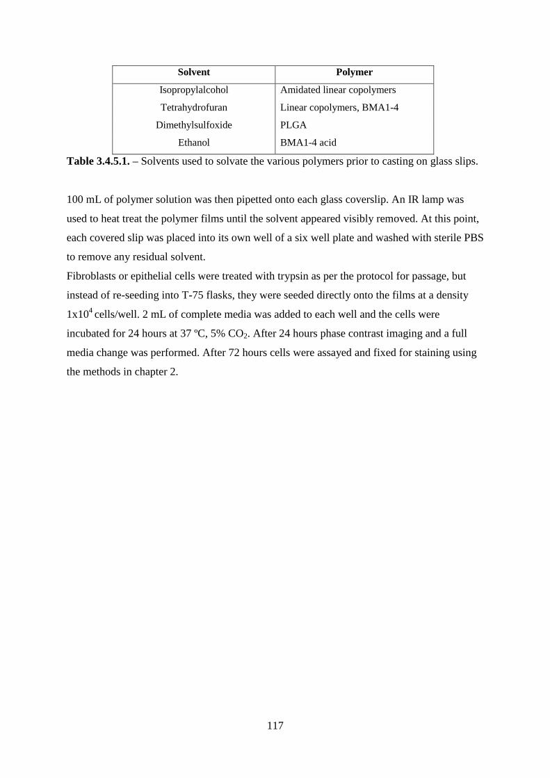
117
Solvent Polymer
Isopropylalcohol Amidated linear copolymers
Tetrahydrofuran Linear copolymers, BMA1-4
Dimethylsulfoxide PLGA
Ethanol BMA1-4 acid
Table 3.4.5.1. – Solvents used to solvate the various polymers prior to casting on glass slips.
100 mL of polymer solution was then pipetted onto each glass coverslip. An IR lamp was
used to heat treat the polymer films until the solvent appeared visibly removed. At this point,
each covered slip was placed into its own well of a six well plate and washed with sterile PBS
to remove any residual solvent.
Fibroblasts or epithelial cells were treated with trypsin as per the protocol for passage, but
instead of re-seeding into T-75 flasks, they were seeded directly onto the films at a density
1x10⁴
cells/well. 2 mL of complete media was added to each well and the cells were
incubated for 24 hours at 37 ºC, 5% CO2. After 24 hours phase contrast imaging and a full
media change was performed. After 72 hours cells were assayed and fixed for staining using
the methods in chapter 2.
118
4 - Hyperbranched Poly(t-butyl acrylate)
4.1 Introduction
The purpose of this work was to produce hyperbranched t-butyl acrylate using the RAFT
procedure already described for n-butyl methacrylate. The hypothesis was that having
observed the cellular response to acidic polymers, it would be possible to create a polymeric
coating that would show good cell adhesion by trapping a branched polymer within a
crosslinked coating. The choice of t-butyl acrylate monomer was governed by the desire to
form a hyperbranched polymer that, along with having functionalised end groups, could also
be subjected to further hydrolysis and chemical modifcations to produce either acid or amine
functionality at the t-butyl sites.
T-butyl acrylate has a glass transition temperature, Tg, of 43 °C compared to -54 °C for n-
butyl acrylate and 20 °C for n-butyl methacrylate. When a polymer resides at temperatures
below its Tg, it appears crystalline and ‗glassy‘. At temperatures above the Tg, individual
chains have enough energy to overcome the weak intermolecular forces (usually London
dispersion forces and hydrogen bonds) to allow flow to occur. This means that the inclusion
of t-butyl acrylate can help raise the Tg and improve the mechanical properties of the
polymer. A strong polymer with a reasonably high Tg is required to prevent wear, especially
for the application of a cell-adhesive coating in replacement joints. This has also been
exploited in order to control the memory response of polymeric stents. The thermoresponse
was induced by warming the polymer stent to above its Tg, allowing it to be compressed for
storage. The polymer could then decompressed by rewarming and softening. A stent was
synthesised usng t-buyl acrylate that would remain compressed at room temperature but
reformed its original shape at body temperature[236]. Control over the Tg is important not
only for issues of storage and application, but also for the form of the polymer at body
temperature. Polyurethanes have been extensively studied as so-called shape memory
polymers (SMPs) due to excellent biocompatibility and a wide range of available Tgs[237],
[238].
The ease with which it is possible to hydrolyse t-BuAc and subsequently subject it to
chemical modification makes it an attractive comonomer for polymeric biomaterials[239]–
[243].

119
T-butyl acrylate has previously been reported as a precursor in the formation of poly(acrylic
acid). The graft copolymers prepared by Kriegel et al. were synthesised using atom-transfer
radical polymerisation and ring-opening metathesis techniques[244], which allows for good
control over the architecture of the final polymer. Zhou, however, produced hyperbranched
polymers using a stepwise graft procedure[245]. The prepared films from both of these
reactions showed a high density of carboxylic acid functionality. By employing a weak acid,
it has been demonstrated that macrophages and endothelial cells can be corralled into the
hydrophobic valleys present on a patterned surface[205]. This finding has implications for
controlled 3-dimensional cell culture, allowing cells to grow beyond single layer sheets in
vitro. The results presented previously in this work indicate that dermal fibroblast and renal
epithelial cells would also be suitable for corralling using acid- and amine- function
HBP(butyl methacrylate).
The results of cell viability on HBP(t-butyl acrylate) also suggests that functionalised
Walters and Hirt have previously reported the synthesis of amine functional polymers from t-
butyl acrylate [224] . Although this work demonstrates the tunability of t-butyl acrylate
polymers, no cell compatibility work was performed[246]. This is a pattern repeated
throughout the current literature and this work hopes to shine some light on the
biocompatibility of functionalised t-butyl acrylate polymers.
The RAFT polymerisation was carried out as previously described, using four different ratios
of monomer to carbodithioate chain transfer agent (4-vinylbenzyldithiobenzoate, CTA 2) in
the reaction mix.
After characterisation the polymers were reacted with excess ACVA, as described in chapter
3, to remove the CTA end groups and replace them with acid functionality. The polymers,
both in native form and after reaction with excess ACVA, were subjected to the same cell
testing as the hyperbranched n-butyl methacrylate polymers, so that a direct comparison
could be made between the two hyperbranched materials. These polymers are also envisaged
to act as a precursor for hyperbranched poly(acrylic acid) by subsequent removal of the tert-
butyl group. By reaction with stoichiometric amounts of diamine this could provide carefully
tailored amphiphilic biomaterials as previously demonstrated.
The intricacies of IPNs have already been discussed in Chapter 1 section 9. Hydrogels often
have limited mechanical properties, and (semi-)IPNs are an emerging alternative. Semi-IPNs
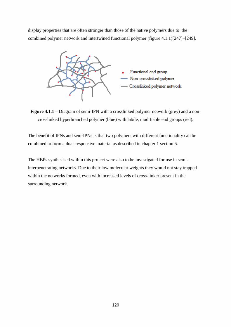
120
display properties that are often stronger than those of the native polymers due to the
combined polymer network and intertwined functional polymer (figure 4.1.1)[247]–[249].
Figure 4.1.1 – Diagram of semi-IPN with a crosslinked polymer network (grey) and a non-
crosslinked hyperbranched polymer (blue) with labile, modifiable end groups (red).
The benefit of IPNs and sem-IPNs is that two polymers with different functionality can be
combined to form a dual-responsive material as described in chapter 1 section 6.
The HBPs synthesised within this project were also to be investigated for use in semi-
interpenetrating networks. Due to their low molecular weights they would not stay trapped
within the networks formed, even with increased levels of cross-linker present in the
surrounding network.
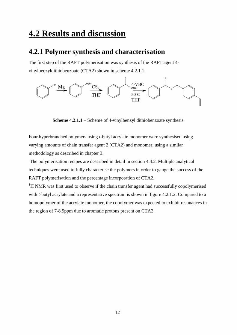
121
4.2 Results and discussion
4.2.1 Polymer synthesis and characterisation
The first step of the RAFT polymerisation was synthesis of the RAFT agent 4-
vinylbenzyldithiobenzoate (CTA2) shown in scheme 4.2.1.1.
Scheme 4.2.1.1 – Scheme of 4-vinylbenzyl dithiobenzoate synthesis.
Four hyperbranched polymers using t-butyl acrylate monomer were synthesised using
varying amounts of chain transfer agent 2 (CTA2) and monomer, using a similar
methodology as described in chapter 3.
The polymerisation recipes are described in detail in section 4.4.2. Multiple analytical
techniques were used to fully characterise the polymers in order to gauge the success of the
RAFT polymerisation and the percentage incorporation of CTA2.
1H NMR was first used to observe if the chain transfer agent had successfully copolymerised
with t-butyl acrylate and a representative spectrum is shown in figure 4.2.1.2. Compared to a
homopolymer of the acrylate monomer, the copolymer was expected to exhibit resonances in
the region of 7-8.5ppm due to aromatic protons present on CTA2.
Mg CS2
THF
4-VBC
50ºC
THF

122
Figure 4.2.1.2 - Representative
1H NMR spectrum of hyperbranched(t-butyl acrylate)
polymers. 400 MHz, CDCl3
The spectra showed that the copolymerisation between t-butyl acrylate and the branching
chain transfer agent was successful and that aromatic protons are present in the polymer. The
only source of the signals seen at 6.5-8ppm is the CTA2 comonomer. The most intense
resonance at 1.5ppm can be associated with the sp3 protons present on the pendant methyl
groups of t-butyl acrylate. As this is the monomer present in greatest quantities, it is
predictable that these protons will be most numerous. The resonance at 2.1ppm can be
assigned to the ArR2CH that occurs on the insertion of CTA2 into the polymer backbone,
whilst the resonance at 4.9ppm occurs because of the R2(CO)CH protons contributed by t-
butyl acrylate. End group analysis was performed using equation 4.2.1.3 in order to
quantitatively compare the proton spectra of the polymers and estimate the percentage
incorporation of CTA2. This showed that tBuAc4 had an estimated incorporation of 4%
CTA2, whilst tBuAC3 and 4 had 7% and 6%, respectively: almost double their predicted
maximum amount of CTA2 (table 4.2.1.4).
A
C
B F/G/C/D/E
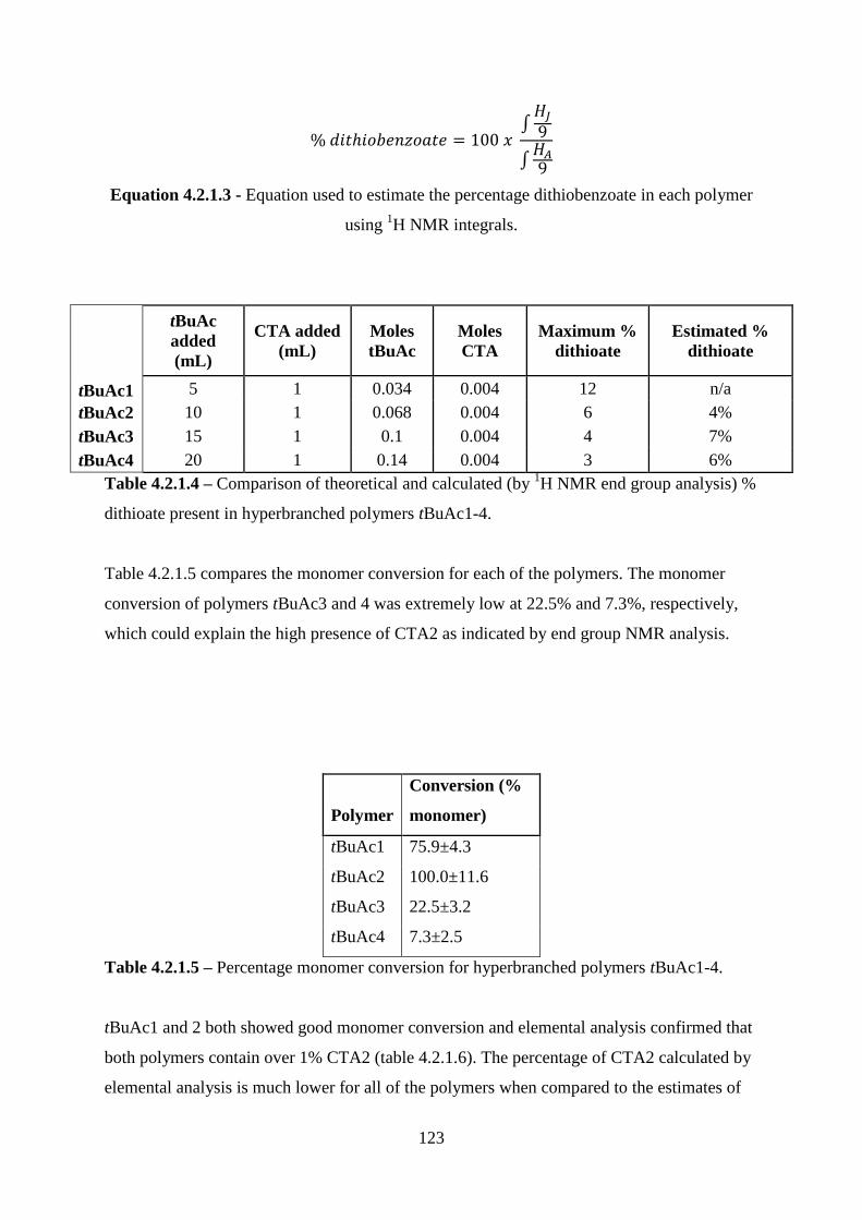
123
∫
∫
Equation 4.2.1.3 - Equation used to estimate the percentage dithiobenzoate in each polymer
using 1H NMR integrals.
tBuAc1
tBuAc
added
(mL)
CTA added
(mL)
Moles
tBuAc
Moles
CTA
Maximum %
dithioate
Estimated %
dithioate
5 1 0.034 0.004 12 n/a
tBuAc2 10 1 0.068 0.004 6 4%
tBuAc3 15 1 0.1 0.004 4 7%
tBuAc4 20 1 0.14 0.004 3 6%
Table 4.2.1.4 – Comparison of theoretical and calculated (by 1H NMR end group analysis) %
dithioate present in hyperbranched polymers tBuAc1-4.
Table 4.2.1.5 compares the monomer conversion for each of the polymers. The monomer
conversion of polymers tBuAc3 and 4 was extremely low at 22.5% and 7.3%, respectively,
which could explain the high presence of CTA2 as indicated by end group NMR analysis.
Polymer
Conversion (%
monomer)
tBuAc1 75.9±4.3
tBuAc2 100.0±11.6
tBuAc3 22.5±3.2
tBuAc4 7.3±2.5
Table 4.2.1.5 – Percentage monomer conversion for hyperbranched polymers tBuAc1-4.
tBuAc1 and 2 both showed good monomer conversion and elemental analysis confirmed that
both polymers contain over 1% CTA2 (table 4.2.1.6). The percentage of CTA2 calculated by
elemental analysis is much lower for all of the polymers when compared to the estimates of
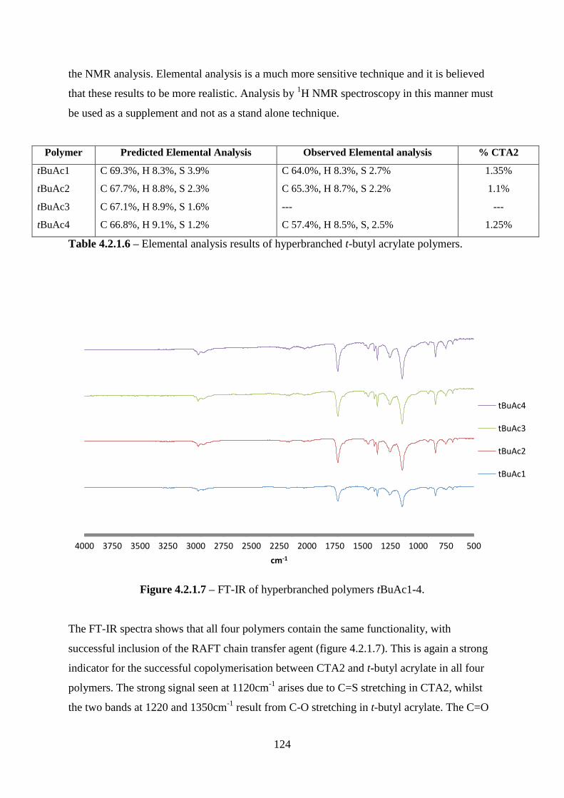
124
the NMR analysis. Elemental analysis is a much more sensitive technique and it is believed
that these results to be more realistic. Analysis by 1H NMR spectroscopy in this manner must
be used as a supplement and not as a stand alone technique.
Polymer Predicted Elemental Analysis Observed Elemental analysis % CTA2
tBuAc1 C 69.3%, H 8.3%, S 3.9% C 64.0%, H 8.3%, S 2.7% 1.35%
tBuAc2 C 67.7%, H 8.8%, S 2.3% C 65.3%, H 8.7%, S 2.2% 1.1%
tBuAc3 C 67.1%, H 8.9%, S 1.6% --- ---
tBuAc4 C 66.8%, H 9.1%, S 1.2% C 57.4%, H 8.5%, S, 2.5% 1.25%
Table 4.2.1.6 – Elemental analysis results of hyperbranched t-butyl acrylate polymers.
Figure 4.2.1.7 – FT-IR of hyperbranched polymers tBuAc1-4.
The FT-IR spectra shows that all four polymers contain the same functionality, with
successful inclusion of the RAFT chain transfer agent (figure 4.2.1.7). This is again a strong
indicator for the successful copolymerisation between CTA2 and t-butyl acrylate in all four
polymers. The strong signal seen at 1120cm-1
arises due to C=S stretching in CTA2, whilst
the two bands at 1220 and 1350cm-1
result from C-O stretching in t-butyl acrylate. The C=O
5007501000125015001750200022502500275030003250350037504000
cm-1
tBuAc4
tBuAc3
tBuAc2
tBuAc1

125
of t-butyl acrylate is observed as a strong signal at 1700cm-1
. Aromatic overtones are
observed at 1900-2100cm-1
whilst sp3 and sp
2 C-H stretches are visible at 2980 cm
-1.
PDI
tBuAc1 8250 18530 35330 2.24
tBuAc2 11030 54230 199420 4.9
tBuAc3 3859 8532 18613 2.21
tBuAc4 24750 61080 120750 2.47
Table 4.2.1.8 - Molecular weight data (daltons) for HP(tBuAc) polymers as determined by
size exclusion GPC using tetrahydrofuran as the mobile phase.
Figure 4.2.1.9 - Molar mass distribution of polymers tBuAc1-4.
Table 4.2.1.8 provides a summary of the polymers‘ molar masses whilst figure 4.2.1.9 shows
the MMD of tBuAc1-3. It can be seen that relatively low molar mass averages are obtained
with this branching RAFT agent, and this is possibly because higher rate of retardation is
experienced when using an aromatic carbodithioester with an acrylate monomer[250].
However, it is unknown if this retardation is caused directly by a low rate of fragmentation or
by making side reactions more likely to occur. Ting et al. have shown experimentally that the
0
0.2
0.4
0.6
0.8
1
1.2
6.1 5.7 5.4 5.1 4.7 4.4 4.1 3.8 3.5 3.2 2.9 2.6 2.3 2.0
dw
/dLo
gM
LogM
tBuAc1
tBuAc2
tBuAc3
tBuAc4
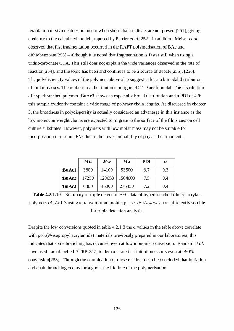
126
retardation of styrene does not occur when short chain radicals are not present[251], giving
credence to the calculated model proposed by Perrier et al.[252]. In addition, Meiser et al.
observed that fast fragmentation occurred in the RAFT polymerisation of BAc and
dithiobenzoate[253] – although it is noted that fragmentation is faster still when using a
trithiocarbonate CTA. This still does not explain the wide variances observed in the rate of
reaction[254], and the topic has been and continues to be a source of debate[255], [256].
The polydispersity values of the polymers above also suggest at least a bimodal distribution
of molar masses. The molar mass distributions in figure 4.2.1.9 are bimodal. The distribution
of hyperbranched polymer tBuAc3 shows an especially broad distribution and a PDI of 4.9;
this sample evidently contains a wide range of polymer chain lengths. As discussed in chapter
3, the broadness in polydispersity is actually considered an advantage in this instance as the
low molecular weight chains are expected to migrate to the surface of the films cast on cell
culture substrates. However, polymers with low molar mass may not be suitable for
incorporation into semi-IPNs due to the lower probability of physical entrapment.
PDI α
tBuAc1 3800 14100 53500 3.7 0.3
tBuAc2 17250 129050 1504000 7.5 0.4
tBuAc3 6300 45000 276450 7.2 0.4
Table 4.2.1.10 – Summary of triple detection SEC data of hyperbranched t-butyl acrylate
polymers tBuAc1-3 using tetrahydrofuran mobile phase. tBuAc4 was not sufficiently soluble
for triple detection analysis.
Despite the low conversions quoted in table 4.2.1.8 the α values in the table above correlate
with poly(N-isopropyl acrylamide) materials previously prepared in our laboratories; this
indicates that some branching has occurred even at low monomer conversion. Rannard et al.
have used radiolabelled ATRP[257] to demonstrate that initiation occurs even at >90%
conversion[258]. Through the combination of these results, it can be concluded that initiation
and chain branching occurs throughout the lifetime of the polymerisation.
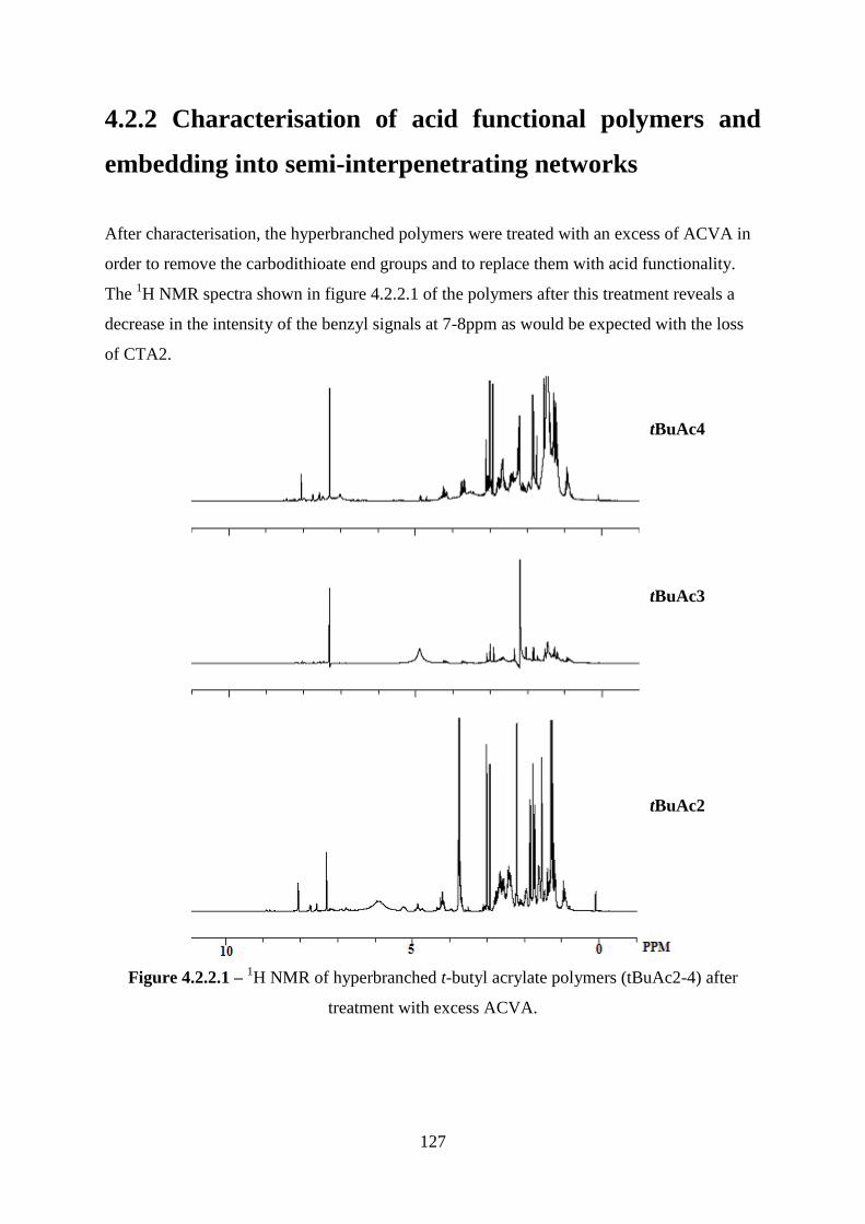
127
4.2.2 Characterisation of acid functional polymers and
embedding into semi-interpenetrating networks
After characterisation, the hyperbranched polymers were treated with an excess of ACVA in
order to remove the carbodithioate end groups and to replace them with acid functionality.
The 1H NMR spectra shown in figure 4.2.2.1 of the polymers after this treatment reveals a
decrease in the intensity of the benzyl signals at 7-8ppm as would be expected with the loss
of CTA2.
Figure 4.2.2.1 – 1H NMR of hyperbranched t-butyl acrylate polymers (tBuAc2-4) after
treatment with excess ACVA.
tBuAc4
tBuAc3
tBuAc2
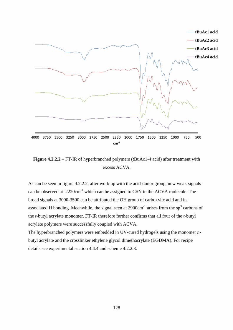
128
Figure 4.2.2.2 – FT-IR of hyperbranched polymers (tBuAc1-4 acid) after treatment with
excess ACVA.
As can be seen in figure 4.2.2.2, after work up with the acid-donor group, new weak signals
can be observed at 2220cm-1
which can be assigned to C≡N in the ACVA molecule. The
broad signals at 3000-3500 can be attributed the OH group of carboxylic acid and its
associated H bonding. Meanwhile, the signal seen at 2900cm-1
arises from the sp3 carbons of
the t-butyl acrylate monomer. FT-IR therefore further confirms that all four of the t-butyl
acrylate polymers were successfully coupled with ACVA.
The hyperbranched polymers were embedded in UV-cured hydrogels using the monomer n-
butyl acrylate and the crosslinker ethylene glycol dimethacrylate (EGDMA). For recipe
details see experimental section 4.4.4 and scheme 4.2.2.3.
5007501000125015001750200022502500275030003250350037504000
cm-1
tBuAc1 acid
tBuAc2 acid
tBuAc3 acid
tBuAc4 acid

129
The aim was to use a process between the sequential and simultaneous methods to form semi-
IPNs by dissolving the hyperbranched polymers in the polymerisation solvent and then
forming the crosslinked networks around them. A summary of the networks formed and their
properties is shown in table 4.2.2.4.
nBuAc
/g
DVB
/g
EGDMA
/g
IPA
/mL HBP /g
HMPP
/mg Properties
3.2 0.3 0 1 0.7 40
Slightly yellow gel, very brittle and
crumbled.
3.2 0.3 0 1 0 20 Clear, brittle gel that curled and broke.
3.2 0.15 0 1 0 20 Clear, less brittle gel, sticky.
3.2 0 0.5 1 0 20 Clear, less brittle again, some breakage.
3.2 0 0.25 1 0 20 Clear, not brittle but sticky.
3.2 0.15 0 1 0.5 20 Yellow gel. Slightly brittle, not sticky
3.2 0 0.25 1 0.5 20 Yellow gel. Not brittle or sticky.
Table 4.2.2.4 – Composition and properties of synthesised networks and sem-IPNs.
It was found that the inclusion of HB(tBuAc) enhanced the mechanical properties over single
network gels. Semi-IPNs were found to increase the rigidity and decrease the ‗stickiness‘
when compared to the single network analogue. This is due to a hardening of the gels, as the
HBP is incorporated into the network and reinforces the structure through non-covalent
interactions.
The cured gels were washed in isopropyl alcohol overnight in order to remove any residual
monomer. However, figure 4.2.2.5 illustrates that the ‗embedded‘ polymer leached from the
network over 24 hours. This can be observed as a colour change in the solvent, as the
coloured HBP leaves the clear network.
Scheme 4.2.2.3 – Scheme showing the formation of poly(n-butyl acrylate) hydrogel
network with ethyleneglycol dimethacrylate cross-linker.
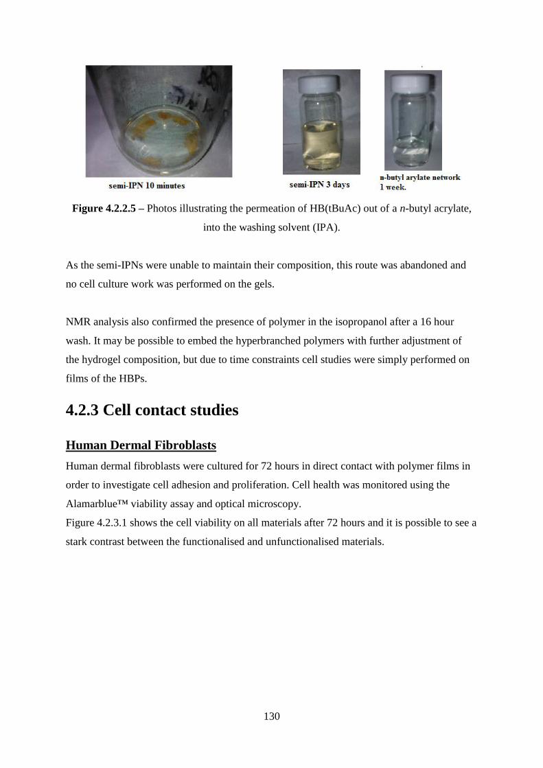
130
Figure 4.2.2.5 – Photos illustrating the permeation of HB(tBuAc) out of a n-butyl acrylate,
into the washing solvent (IPA).
As the semi-IPNs were unable to maintain their composition, this route was abandoned and
no cell culture work was performed on the gels.
NMR analysis also confirmed the presence of polymer in the isopropanol after a 16 hour
wash. It may be possible to embed the hyperbranched polymers with further adjustment of
the hydrogel composition, but due to time constraints cell studies were simply performed on
films of the HBPs.
4.2.3 Cell contact studies
Human Dermal Fibroblasts
Human dermal fibroblasts were cultured for 72 hours in direct contact with polymer films in
order to investigate cell adhesion and proliferation. Cell health was monitored using the
Alamarblue™ viability assay and optical microscopy.
Figure 4.2.3.1 shows the cell viability on all materials after 72 hours and it is possible to see a
stark contrast between the functionalised and unfunctionalised materials.
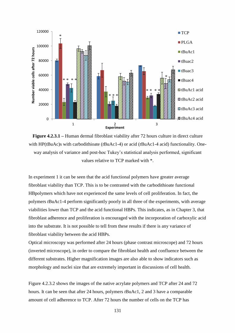
131
Figure 4.2.3.1 – Human dermal fibroblast viability after 72 hours culture in direct culture
with HP(tBuAc)s with carbodithioate (tBuAc1-4) or acid (tBuAc1-4 acid) functionality. One-
way analysis of variance and post-hoc Tukey‘s statistical analysis performed, significant
values relative to TCP marked with *.
In experiment 1 it can be seen that the acid functional polymers have greater average
fibroblast viability than TCP. This is to be contrasted with the carbodithioate functional
HBpolymers which have not experienced the same levels of cell proliferation. In fact, the
polymers tBuAc1-4 perform significantly poorly in all three of the experiments, with average
viabilities lower than TCP and the acid functional HBPs. This indicates, as in Chapter 3, that
fibroblast adherence and proliferation is encouraged with the incorporation of carboxylic acid
into the substrate. It is not possible to tell from these results if there is any variance of
fibroblast viability between the acid HBPs.
Optical microscopy was performed after 24 hours (phase contrast microscope) and 72 hours
(inverted microscope), in order to compare the fibroblast health and confluence between the
different substrates. Higher magnification images are also able to show indicators such as
morphology and nuclei size that are extremely important in discussions of cell health.
Figure 4.2.3.2 shows the images of the native acrylate polymers and TCP after 24 and 72
hours. It can be seen that after 24 hours, polymers tBuAc1, 2 and 3 have a comparable
amount of cell adherence to TCP. After 72 hours the number of cells on the TCP has
0
20000
40000
60000
80000
100000
120000
1 2 3
Nu
mb
er
viab
le c
ells
aft
er
72
ho
urs
Experiment
TCP
PLGA
tBuAc1
tBuac2
tBuac3
tBuac4
tBuAc1 acid
tBuAc2 acid
tBuAc3 acid
tBuAc4 acid
* * * *
* * * *
*
*
* * *
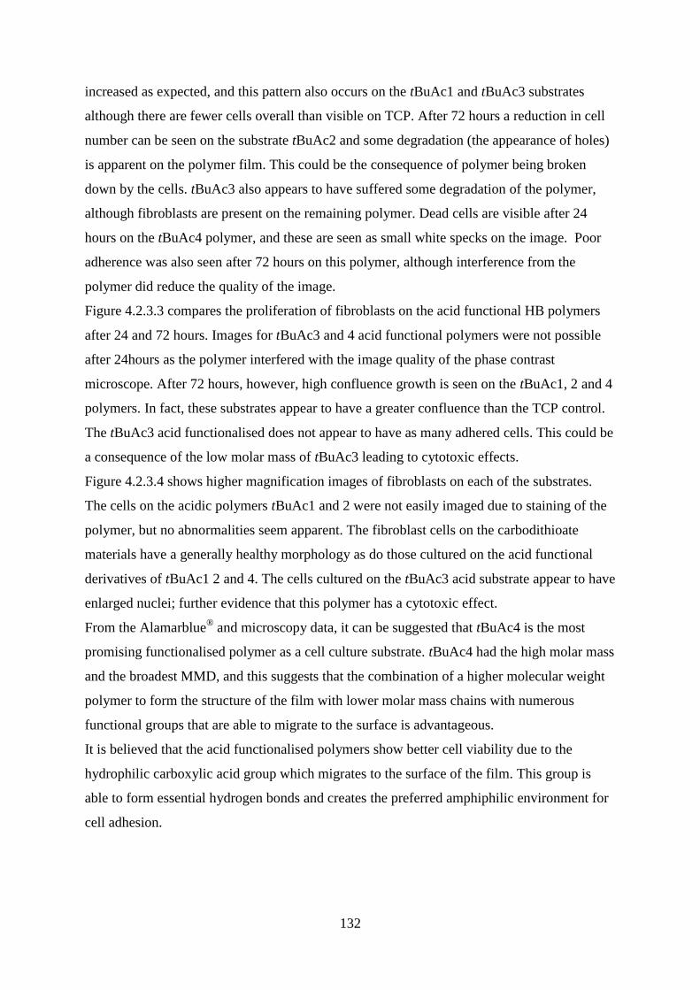
132
increased as expected, and this pattern also occurs on the tBuAc1 and tBuAc3 substrates
although there are fewer cells overall than visible on TCP. After 72 hours a reduction in cell
number can be seen on the substrate tBuAc2 and some degradation (the appearance of holes)
is apparent on the polymer film. This could be the consequence of polymer being broken
down by the cells. tBuAc3 also appears to have suffered some degradation of the polymer,
although fibroblasts are present on the remaining polymer. Dead cells are visible after 24
hours on the tBuAc4 polymer, and these are seen as small white specks on the image. Poor
adherence was also seen after 72 hours on this polymer, although interference from the
polymer did reduce the quality of the image.
Figure 4.2.3.3 compares the proliferation of fibroblasts on the acid functional HB polymers
after 24 and 72 hours. Images for tBuAc3 and 4 acid functional polymers were not possible
after 24hours as the polymer interfered with the image quality of the phase contrast
microscope. After 72 hours, however, high confluence growth is seen on the tBuAc1, 2 and 4
polymers. In fact, these substrates appear to have a greater confluence than the TCP control.
The tBuAc3 acid functionalised does not appear to have as many adhered cells. This could be
a consequence of the low molar mass of tBuAc3 leading to cytotoxic effects.
Figure 4.2.3.4 shows higher magnification images of fibroblasts on each of the substrates.
The cells on the acidic polymers tBuAc1 and 2 were not easily imaged due to staining of the
polymer, but no abnormalities seem apparent. The fibroblast cells on the carbodithioate
materials have a generally healthy morphology as do those cultured on the acid functional
derivatives of tBuAc1 2 and 4. The cells cultured on the tBuAc3 acid substrate appear to have
enlarged nuclei; further evidence that this polymer has a cytotoxic effect.
From the Alamarblue® and microscopy data, it can be suggested that tBuAc4 is the most
promising functionalised polymer as a cell culture substrate. tBuAc4 had the high molar mass
and the broadest MMD, and this suggests that the combination of a higher molecular weight
polymer to form the structure of the film with lower molar mass chains with numerous
functional groups that are able to migrate to the surface is advantageous.
It is believed that the acid functionalised polymers show better cell viability due to the
hydrophilic carboxylic acid group which migrates to the surface of the film. This group is
able to form essential hydrogen bonds and creates the preferred amphiphilic environment for
cell adhesion.
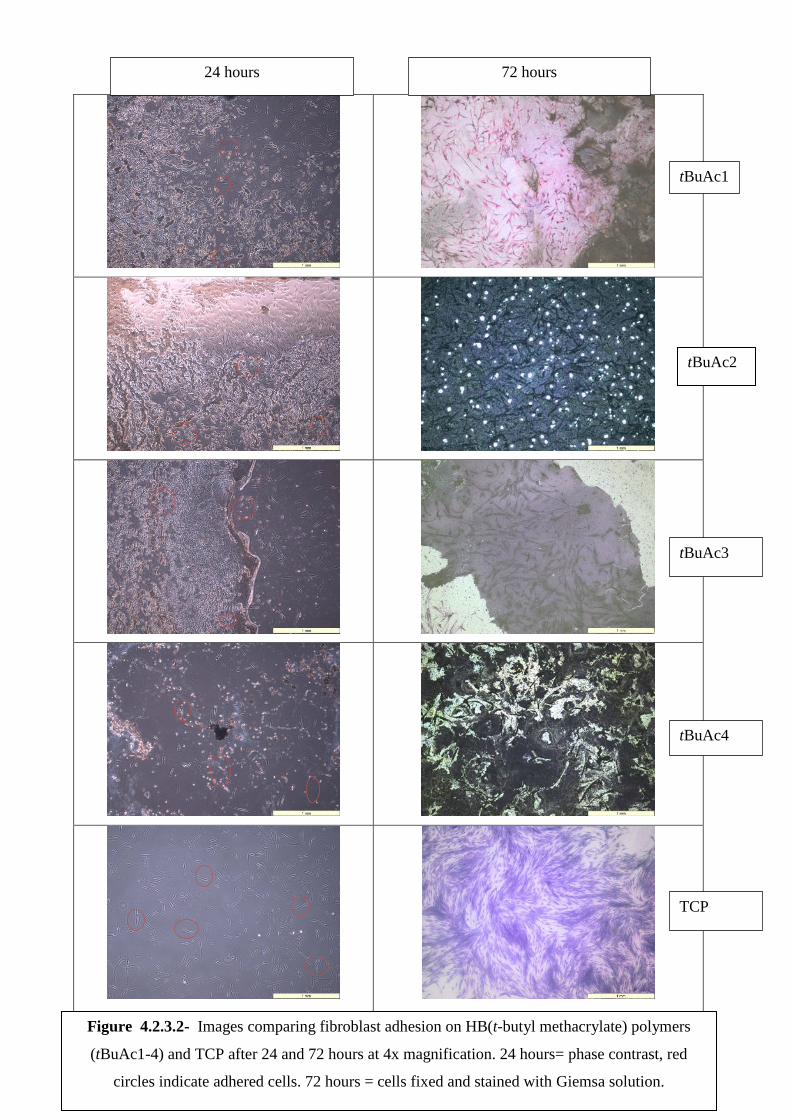
133
24 hours 72 hours
tBuAc1
tBuAc3
tBuAc4
TCP
tBuAc2
Figure 4.2.3.2- Images comparing fibroblast adhesion on HB(t-butyl methacrylate) polymers
(tBuAc1-4) and TCP after 24 and 72 hours at 4x magnification. 24 hours= phase contrast, red
circles indicate adhered cells. 72 hours = cells fixed and stained with Giemsa solution.
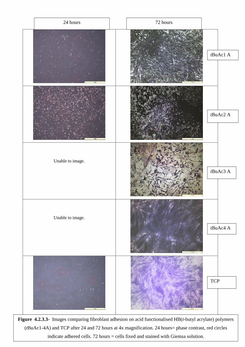
134
Unable to image.
Unable to image.
24 hours 72 hours
tBuAc1 A
tBuAc2 A
tBuAc3 A
tBuAc4 A
TCP
Figure 4.2.3.3- Images comparing fibroblast adhesion on acid functionalised HB(t-butyl acrylate) polymers
(tBuAc1-4A) and TCP after 24 and 72 hours at 4x magnification. 24 hours= phase contrast, red circles
indicate adhered cells. 72 hours = cells fixed and stained with Giemsa solution.

135
Figure 4.2.3.4- Images comparing fibroblast adhesion on HB(t-butyl acrylate) polymers (tBuAc1-4) with
carbodithioate or acid end groupds, TCP and PLGA after 72 hours at 20x magnification. Cells fixed and
stained with Giemsa solution.
Carbodithionate Acid
tBuAc1
tBuAc2
tBuAc3
tBuAc4
Figure 4.2.3.4- Images comparing fibroblast adhesion on HB(t-butyl acrylate) polymers (tBuAc1-4) with
carbodithioate or acid end groupds, TCP and PLGA after 72 hours at 20x magnification. Cells fixed and
stained with Giemsa solution.
PLGA, TCP
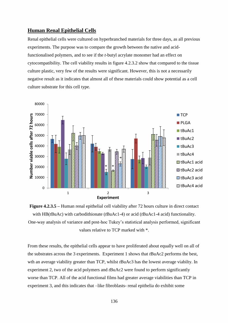
136
Human Renal Epithelial Cells
Renal epithelial cells were cultured on hyperbranched materials for three days, as all previous
experiments. The purpose was to compare the growth between the native and acid-
functionalised polymers, and to see if the t-butyl acrylate monomer had an effect on
cytocompatibility. The cell viability results in figure 4.2.3.2 show that compared to the tissue
culture plastic, very few of the results were significant. However, this is not a necessarily
negative result as it indicates that almost all of these materials could show potential as a cell
culture substrate for this cell type.
Figure 4.2.3.5 – Human renal epithelial cell viability after 72 hours culture in direct contact
with HB(tBuAc) with carbodithionate (tBuAc1-4) or acid (tBuAc1-4 acid) functionality.
One-way analysis of variance and post-hoc Tukey‘s statistical analysis performed, significant
values relative to TCP marked with *.
From these results, the epithelial cells appear to have proliferated about equally well on all of
the substrates across the 3 experiments. Experiment 1 shows that tBuAc2 performs the best,
wth an average viability greater than TCP, whilst tBuAc3 has the lowest average viabilty. In
experiment 2, two of the acid polymers and tBuAc2 were found to perform significantly
worse than TCP. All of the acid functional films had greater average viabilities than TCP in
experiment 3, and this indicates that –like fibroblasts- renal epithelia do exhibit some
0
10000
20000
30000
40000
50000
60000
70000
80000
1 2 3
Nu
mb
er v
iab
le c
ells
aft
er 7
2 h
ou
rs
Experiment
TCP
PLGA
tBuAc1
tBuAc2
tBuAc3
tBuAc4
tBuAc1 acid
tBuAc2 acid
tBuAc3 acid
tBuAc4 acid
* *
*
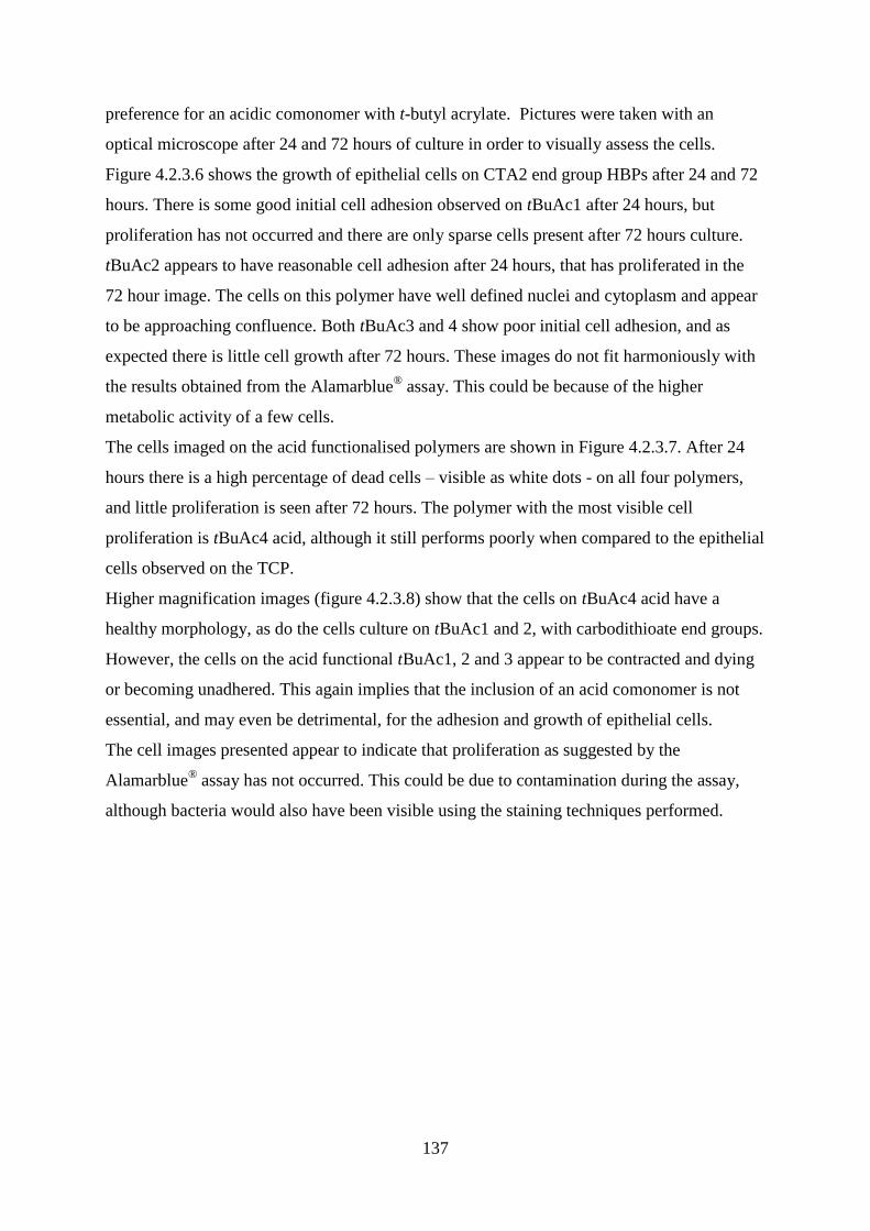
137
preference for an acidic comonomer with t-butyl acrylate. Pictures were taken with an
optical microscope after 24 and 72 hours of culture in order to visually assess the cells.
Figure 4.2.3.6 shows the growth of epithelial cells on CTA2 end group HBPs after 24 and 72
hours. There is some good initial cell adhesion observed on tBuAc1 after 24 hours, but
proliferation has not occurred and there are only sparse cells present after 72 hours culture.
tBuAc2 appears to have reasonable cell adhesion after 24 hours, that has proliferated in the
72 hour image. The cells on this polymer have well defined nuclei and cytoplasm and appear
to be approaching confluence. Both tBuAc3 and 4 show poor initial cell adhesion, and as
expected there is little cell growth after 72 hours. These images do not fit harmoniously with
the results obtained from the Alamarblue® assay. This could be because of the higher
metabolic activity of a few cells.
The cells imaged on the acid functionalised polymers are shown in Figure 4.2.3.7. After 24
hours there is a high percentage of dead cells – visible as white dots - on all four polymers,
and little proliferation is seen after 72 hours. The polymer with the most visible cell
proliferation is tBuAc4 acid, although it still performs poorly when compared to the epithelial
cells observed on the TCP.
Higher magnification images (figure 4.2.3.8) show that the cells on tBuAc4 acid have a
healthy morphology, as do the cells culture on tBuAc1 and 2, with carbodithioate end groups.
However, the cells on the acid functional tBuAc1, 2 and 3 appear to be contracted and dying
or becoming unadhered. This again implies that the inclusion of an acid comonomer is not
essential, and may even be detrimental, for the adhesion and growth of epithelial cells.
The cell images presented appear to indicate that proliferation as suggested by the
Alamarblue® assay has not occurred. This could be due to contamination during the assay,
although bacteria would also have been visible using the staining techniques performed.
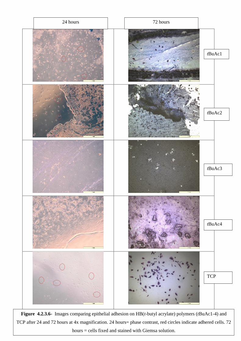
138
24 hours 72 hours
tBuAc1
tBuAc2
tBuAc3
tBuAc4
TCP
Figure 4.2.3.6- Images comparing epithelial adhesion on HB(t-butyl acrylate) polymers (tBuAc1-4) and
TCP after 24 and 72 hours at 4x magnification. 24 hours= phase contrast, red circles indicate adhered cells. 72
hours = cells fixed and stained with Giemsa solution.
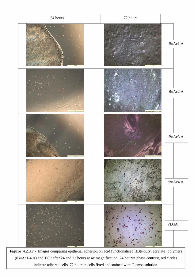
139
24 hours 72 hours
tBuAc1 A
tBuAc2 A
tBuAc3 A
tBuAc4 A
PLGA
Figure 4.2.3.7 - Images comparing epithelial adhesion on acid functionalised HB(t-butyl acrylate) polymers
(tBuAc1-4 A) and TCP after 24 and 72 hours at 4x magnification. 24 hours= phase contrast, red circles
indicate adhered cells. 72 hours = cells fixed and stained with Giemsa solution.
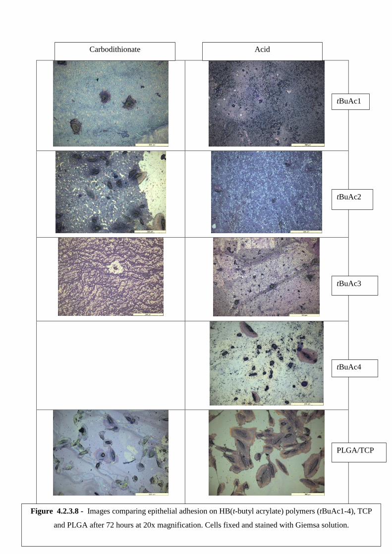
140
Carbodithionate Acid
tBuAc1
tBuAc2
tBuAc3
tBuAc4
PLGA/TCP
Figure 4.2.3.8 - Images comparing epithelial adhesion on HB(t-butyl acrylate) polymers (tBuAc1-4), TCP
and PLGA after 72 hours at 20x magnification. Cells fixed and stained with Giemsa solution.

141
4.3 Conclusions
The main aim of this work was to show the successful RAFT polymerisation of t-butyl
acrylate, and to examine the cytocompatibility before and after hydrolysis of the monomer.
The chain transfer agent, CTA2, was especially chosen so that it could also act as a branching
agent in the formation of hyperbranched polymers. Four polymers were synthesised with
different ratios of monomer:CTA2. The polymer tBuac2 showed almost complete conversion
of the monomer, whilst tBuAc3 and 4 had very low monomer conversion. Elemental analysis
of tBuAc2 show that it has the lowest level of CTA2 than any of the other polymers, and it
also has the broadest molar mass distribution. It is difficult to pinpoint the exact cause of this,
as each polymer was made with different levels of monomer, initiator and CTA2.
Despite the lower level of monomer conversion of the polymers tBuAc1 and 3, there was still
evidence of branching by looking at the Mark-Houwink equation and through the
incorporation of CTA2. This is contrary to what is reported in the literature, where it is said
that polymers tend to stay linear until higher conversion levels and then begin to branch later
in the reaction[257], [258].
All of the synthesised polymers were successfully reacted with ACVA in order to produce
HBPs with carboxylic acid end groups. The low molecular weight of the HBPs may
subsequently lead to less hindrance and easier access to the polymer end groups.
The incorporation of HBPs into semi-IPNs was not successful. The un-crosslinked polymer
could be seen to leech out of the gels over 3 days. However, despite this proof-of-concept
was provided as the gels with incorporated HBPs were seen to have beneficial qualities over
the single network. This was observed as the gels being less ‗sticky‘ and more rigid.
The cell proliferation assay indicated that fibroblasts have a strong preference for acid
functionalised polymers. Fibroblast cells were also seen to adhere and proliferate to all of the
polymers tested – both with dithiobenzoate and carboxylic acid end groups. The acid
functional derivative of tBuAc4 showed the healthiest and most prolific cell growth and it
also had the greatest molecular weight as determined by GPC. As previously discussed, low
molecular weight polymers can be toxic in their own right[259]. From these results, the
synthesised acid functional polymers could be further optimised as fibroblast cell culture
substrates.
In contrast, however, renal epithelial cell culture showed a stronger preference for the
dithiobenzoate functional polymers. The epithelial cells were seen to both adhere and
142
proliferate on the dithiobenzoate in a comparable manner to TCP. Epithelials cultured on the
acid functional HBPs showed very little to no adherence and proliferation over 72 hours.
High magnification images were hampered by polymer staining but the dithiobenzoate
polymer tBuAc2 did display healthy epithelial growth.
This work has shown that t-butyl acrylate can be successfully polymerised using RAFT and
dithiobenzoate CTA. The HBPs were all successfully reacted with ACVA to remove the
dithiobenzoate group remaining after the polymerisation and replace it with carboxylic acid
functionality. Culture with fibroblast cells indicates that these acid functional polymers could
be used as cell culture substrates, but none of the polymers were a good substrate for
epithelial cells.
143
4.4 Experimental
Instrumentation used as described in chapter 2.4.
4.4.1 Synthesis of RAFT chain transfer agent: 4-
vinylbenzyldithiobenzoate
Materials
Bromobenzene (99%, Aldrich), magnesium turnings (99.95%, Aldrich), carbon disulfide
(99+%, Aldrich), 4-vinylbenzyl chloride (90%, Aldrich), diethyl ether (Fischer), hexane
(Fischer), ethyl acetate (Fischer) were used as recieved.
THF was obtained from a Grubb‘s dry solvent system.
Method
A three-necked round bottom flask was equipped with a condenser and bubbler, pressure
equalising dropping funnel and a nitrogen inlet. Magnesium turnings (1.7 g) and a magnetic
stirrer bar were added to the flask, which was then gently heated and stirred under a nitrogen
environment for 40 minutes. The flask was left to cool to room temperature, before the
dropwise addition of bromobenzene (10 g) in THF (40 mL) over 30 minutes. Gentle heat was
provided to instigate the exothermic reaction, and cooling via an ice bath was provided as
required. A green solution was formed, which was left to stir for 30 minutes at room
temperature.
Carbon disulfide (5 g) and THF (30 mL) were then added dropwise over 10minutes, and an
orange to red colour change of the reaction solution was observed. The flask contents were
cooled to 0 ˚C using an ice bath and stirred for 30 minutes, before being heated to 50 ˚C and
stirred for a further 30 minutes. At this higher temperature, 4-vinylbenzyl chloride (10 g) in
THF (30 mL) was added dropwise over 10 minutes.
The solution was left to cool to room temperature and stirred overnight. The red liquid
product was placed in a 1 L separating funnel, with diethyl ether (80 mL) and distilled water
(80 mL). The organic layer was recovered and the aqueous layer was extracted with diethyl
ether (3 x 100 mL). The organic extracts were combined and dried over magnesium sulfate
before filtration and the solvent was removed by rotary evaporation to give a red oil.
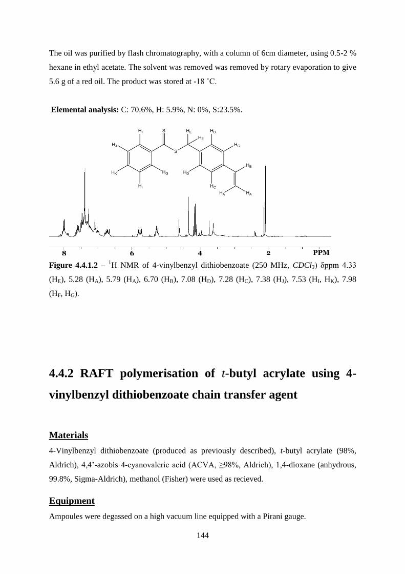
144
The oil was purified by flash chromatography, with a column of 6cm diameter, using 0.5-2 %
hexane in ethyl acetate. The solvent was removed was removed by rotary evaporation to give
5.6 g of a red oil. The product was stored at -18 ˚C.
Elemental analysis: C: 70.6%, H: 5.9%, N: 0%, S:23.5%.
Figure 4.4.1.2 – 1H NMR of 4-vinylbenzyl dithiobenzoate (250 MHz, CDCl3) δppm 4.33
(HE), 5.28 (HA), 5.79 (HA), 6.70 (HB), 7.08 (HD), 7.28 (HC), 7.38 (HJ), 7.53 (HI, HK), 7.98
(HF, HG).
4.4.2 RAFT polymerisation of t-butyl acrylate using 4-
vinylbenzyl dithiobenzoate chain transfer agent
Materials
4-Vinylbenzyl dithiobenzoate (produced as previously described), t-butyl acrylate (98%,
Aldrich), 4,4‘-azobis 4-cyanovaleric acid (ACVA, ≥98%, Aldrich), 1,4-dioxane (anhydrous,
99.8%, Sigma-Aldrich), methanol (Fisher) were used as recieved.
Equipment
Ampoules were degassed on a high vacuum line equipped with a Pirani gauge.

145
Method
4-Vinylbenzyl dithiobenzoate, t-butyl acrylate, dioxane (25 mL) and ACVA were mixed
together until the solid initiator had dissolved. The resulting solution was pipetted into a 50
mL ampoule and placed onto the vacuum line but was not exposed to vacuum. The solution
was frozen using liquid nitrogen and then opened to the vacuum until the gauge dropped to a
steady output. The ampoule‘s exposure to vacuum was then ceased as its contents were left to
thaw. Once thawing was completed, the full process was repeated until a negligible rise in
pressure was observed when the ampoule was opened to vacuum. The end pressure in the
ampoule was approximately 3x10⁻³
mBar. The ampoule was sealed using a gas/oxygen
blowtorch and placed in a water bath at 60˚C for up to 24 hrs to undergo polymerisation.
Table 4.4.2.1 summarises the quantities of reagents used.
t-Butyl acrylate /mL Chain transfer agent /mL ACVA /g
5 (0.034) 1 (4.0x10⁻³
) 0.025 (8.9x10⁻⁵
)
10 (0.068) 1 (4.0x10⁻³
) 0.025 (8.9x10⁻⁵
)
15 (0.1) 1 (4.0x10⁻³
) 0.025 (8.9x10⁻⁵
)
20 (0.14) 1 (4.0x10⁻³
) 0.025 (8.9x10⁻⁵
)
5 (0.034) 1 (4.0x10⁻³
) 1.29 (4.6x10⁻³
)
10 (0.068) 1 (4.0x10⁻³
) 1.29 (4.6x10⁻³
)
15 (0.1) 1 (4.0x10⁻³
) 1.29 (4.6x10⁻³
)
20 (0.14) 1 (4.0x10⁻³
) 1.29 (4.6x10⁻³
)
15 (0.1) 1 (4.0x10⁻³
) 0.65 (2.3x10⁻³
)
20 (0.14) 1 (4.0x10⁻³
) 0.65 (2.3x10⁻³
)
Table 4.4.2.1 – summary of reagent quantities used in RAFT polymerisation of t-Butyl
acrylate. Moles shown in parenthesis.
Products were precipitated by transferring into 4x by volume of methanol and leaving to
stand, followed by filtering and drying in a vacuum oven producing an orange solid. Dried
products were stored in closed sample tubes at room temperature.
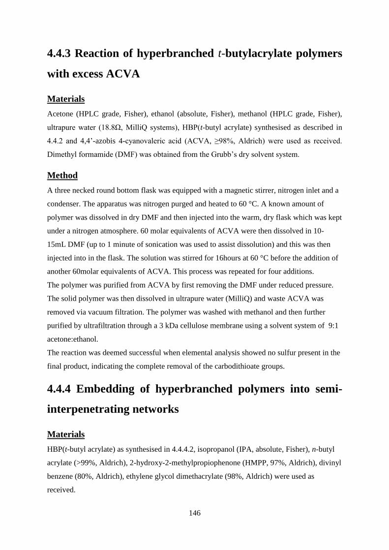
146
4.4.3 Reaction of hyperbranched t-butylacrylate polymers
with excess ACVA
Materials
Acetone (HPLC grade, Fisher), ethanol (absolute, Fisher), methanol (HPLC grade, Fisher),
ultrapure water (18.8Ω, MilliQ systems), HBP(t-butyl acrylate) synthesised as described in
4.4.2 and 4,4‘-azobis 4-cyanovaleric acid (ACVA, ≥98%, Aldrich) were used as received.
Dimethyl formamide (DMF) was obtained from the Grubb‘s dry solvent system.
Method
A three necked round bottom flask was equipped with a magnetic stirrer, nitrogen inlet and a
condenser. The apparatus was nitrogen purged and heated to 60 °C. A known amount of
polymer was dissolved in dry DMF and then injected into the warm, dry flask which was kept
under a nitrogen atmosphere. 60 molar equivalents of ACVA were then dissolved in 10-
15mL DMF (up to 1 minute of sonication was used to assist dissolution) and this was then
injected into in the flask. The solution was stirred for 16hours at 60 °C before the addition of
another 60molar equivalents of ACVA. This process was repeated for four additions.
The polymer was purified from ACVA by first removing the DMF under reduced pressure.
The solid polymer was then dissolved in ultrapure water (MilliQ) and waste ACVA was
removed via vacuum filtration. The polymer was washed with methanol and then further
purified by ultrafiltration through a 3 kDa cellulose membrane using a solvent system of 9:1
acetone:ethanol.
The reaction was deemed successful when elemental analysis showed no sulfur present in the
final product, indicating the complete removal of the carbodithioate groups.
4.4.4 Embedding of hyperbranched polymers into semi-
interpenetrating networks
Materials
HBP(t-butyl acrylate) as synthesised in 4.4.4.2, isopropanol (IPA, absolute, Fisher), n-butyl
acrylate (>99%, Aldrich), 2-hydroxy-2-methylpropiophenone (HMPP, 97%, Aldrich), divinyl
benzene (80%, Aldrich), ethylene glycol dimethacrylate (98%, Aldrich) were used as
received.
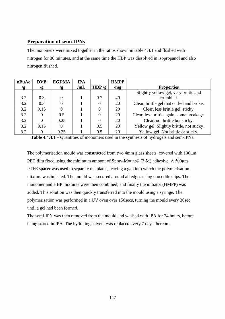
147
Preparation of semi-IPNs
The monomers were mixed together in the ratios shown in table 4.4.1 and flushed with
nitrogen for 30 minutes, and at the same time the HBP was dissolved in isopropanol and also
nitrogen flushed.
nBuAc
/g
DVB
/g
EGDMA
/g
IPA
/mL HBP /g
HMPP
/mg Properties
3.2 0.3 0 1 0.7 40
Slightly yellow gel, very brittle and
crumbled.
3.2 0.3 0 1 0 20 Clear, brittle gel that curled and broke.
3.2 0.15 0 1 0 20 Clear, less brittle gel, sticky.
3.2 0 0.5 1 0 20 Clear, less brittle again, some breakage.
3.2 0 0.25 1 0 20 Clear, not brittle but sticky.
3.2 0.15 0 1 0.5 20 Yellow gel. Slightly brittle, not sticky
3.2 0 0.25 1 0.5 20 Yellow gel. Not brittle or sticky.
Table 4.4.4.1 – Quantities of monomers used in the synthesis of hydrogels and sem-IPNs.
The polymerisation mould was constructed from two 4mm glass sheets, covered with 100μm
PET film fixed using the minimum amount of Spray-Mount® (3-M) adhesive. A 500μm
PTFE spacer was used to separate the plates, leaving a gap into which the polymerisation
mixture was injected. The mould was secured around all edges using crocodile clips. The
monomer and HBP mixtures were then combined, and finally the initiator (HMPP) was
added. This solution was then quickly transferred into the mould using a syringe. The
polymerisation was performed in a UV oven over 150secs, turning the mould every 30sec
until a gel had been formed.
The semi-IPN was then removed from the mould and washed with IPA for 24 hours, before
being stored in IPA. The hydrating solvent was replaced every 7 days thereon.
148
4.4.5 Culture of cells in direct contact with polymers
Materials
Human dermal fibroblasts and human renal epithelial were cultured and prepared as
described in chapter 2.
Hyperbranched tbutyl acrylate (tBuAc1-4) as synthesised in section 4.4.2, and with acid end
groups as described in section 4.4.3 (tBuAc1-4 acid). isopropanol (absolute, Fisher),
complete media (prepared as described in chapter 2), trypsin-EDTA (Life Technologies),
PLGA (50:50, Polysciences), DMSO (Anhydrous, Sigma), Alamarblue® (Life
Technologies), phosphate buffered saline (Bioreagent, Sigma), hematoxylin solution
according to Weigert (parts A and B, Aldrich), eosin y (99%, Sigma Aldrich), 10% neutral
buffered formalin solution (Sigma), giemsa stain modified solution (Sigma), ethanol
(absolute, Fisher), glacial acetic acid (99%, Fisher) were used as received.
Distilled water was used throughout unless otherwise specified.
Equipment
IR spot lamp 75W (Exo Terra), glass coverslip (22x22mm, Menzel).
Glass coverslips were sterilised by autoclave.
Preparation of polymer films and cell seeding
Polymers were dissolved in the solvents listed in table 4.4.5.1 at a concentration of 5 mg/mL.
The polymer solutions were well agitated and sonicated for up to 30 seconds if required.
Solvent Polymer
Tetrahydrofuran tBuAc1-4
Dimethylsulfoxide PLGA
Ethanol tBuAc1-3 acid
Table 4.4.5.1. – Solvents used to solvate the various polymers prior to casting on glass slips.
100mL of polymer solution was then pipetted onto each glass coverslip. An IR lamp was
used to heat treat the polymer films until the solvent appeared visibly removed. At this point,
each covered slip was placed into its own well of a six well plate and washed with sterile PBS
to remove any residual solvent.

149
Fibroblasts or epithelial cells were treated with trypsin as per the protocol for passage, but
instead of re-seeding into T-75 flasks, they were seeded directly onto the films at a density
1x10⁴cells/well. 2 mL of complete media was added to each well and the cells were incubated
for 24 hours at 37ºC, 5% CO2. After 24 hours phase contrast imaging and a full media change
was performed. After 72 hours cells were assayed and fixed for staining using the methods in
chapter 2.
150
5 - Overall conclusions and future work
The aim of this thesis was to develop new synthetic procedures for hyperbranched polymers,
and also further our understanding into how imposing end group functionality can impact
upon biocompatibility. Other properties that were assessed were polymer mass and degree of
branching.
The greatest challenge in the synthesis of oligo(BMA) laid with the initial copolymerisation
with butadiene. A series of experiements were performed in an unpressurised reactor,
allowing the determination of optimal reaction conditions. It was found that the use of α- or
β-cyclodextrin and a carefully controlled reaction temperature of 75-80 °C was essential for
copolymerisation to occur. The amount of cyclodextrin included in the reaction was not
found to have any significant effect above 0.003 moles; this is because cyclodextrin acts as a
carrier molecule for butadiene and is not used up during the course of the polymerisation. The
presence of cyclodextrin contaminant in the final product is not a barrier to cell based studies
as these molecules do possess some cytocompatibility.
The copolymer was successfully cleaved at the sites of 1,4-butadiene insertion using the well-
established technique of ozonolysis and oxidative cleavage. The creation of oligomers in this
way allowed further reaction of the end groups to carboxylic acid and diamine functionality.
GPC confirmed the successful polymer cleavage as the oligomers had a decrease in
molecular weight and FT-IR and elemental analysis was used to confirm the end group
functionality.
Cell adhesion studies of oligomers with fibroblast and epithelial cells showed that different
functionality is preferred by different cell types. When cultured on the synthesised materials,
fibroblast cells showed the greatest proliferation on the acid functionalised oligomer whilst
epithelial cells performed best on the 1,6-amidated oligomer. This is most likely due to
differences in protein expression which are observed as variances in adhesion mechanism and
ECM formation. None of the oligomers showed as great cell adhesion as the control materials
TCP and PLGA – indicating that further work is required to produce fully biocompatible
oligomers.
Hyperbranched polymers of n-butyl methacrylate and t-butyl acrylate were produced using
RAFT polymerisation via two different chain transfer agents (CTA1 and 2).

151
Again a series of polymerisations were performed and the optimal amount of CTA1 in the
polymerisation with BMA was found to be 4%, which gave almost total monomer
conversion. CTA2, a benzyl carbodithioate, was employed with the tBuAc monomer. These
polymerisations had relatively low conversion and it is believed this is due to a retardation
effect that is experienced when copolymerising acrylates with a benzyl CTA. During the
polymerisation of mixes with higher levels of monomer, gelation indicated the occurance of
cross-linking and therefore the amount of initiator in the feed was decreased.
Triple detection GPC was successfully used to substantiate the claimed hyperbranched
structure of the polymer. Using excess ACVA, the end groups of each polymer were replaced
with carboxylic acid functionality, which was confirmed by elemental analysis and FT-IR.
The linear n-butyl methacrylate copolymers supplied by Sarah Canning were characterised
and found to be sufficient for comparison with HB(BMA). These linear polymers already
possessed acid functionality through the incorporation of the comonomer 4-divinyl benzoic
acid. Linear polymers were amidated at the 4-vinyl benzoic acid groups through work up
with excess diamines. All of the functionalised polymers were assessed for their
biocompatibility, where it was found that fibroblast cells show a significant preference for
acid functional polymers and not the amidated varients. This echoes the response observed on
the shorter chain oligo(BMA)s - fibroblast cells grown on the amine functional materials
either did not grow, or displayed unhealthy morphologies.
When compared together, it can be seen that there is no significant differences in the
fibroblast viabilities between linear, oligomeric and hyperbranched P(BMA) – except in the
cases where amine functionality were present. Amine functional linear polymers and
unmodified HP polymers show a significant decrease in cell adhesion compared to standard
materials such as tissue culture plastic (TCP) and poly(lactide-glycolic acid) (PLGA). When
the functionality was preferable fibroblast cells were able to proliferate equally as well on
linear and HB polymers, showing that HBPs are a viable substrate for new biomaterials.
The strong preference of fibroblasts for acid functional polymers was not observed for renal
epithelial cells. The epithelial cells also showed higher average viabilities on linear(BMA)
when compared to hyperbranched and oligomeric analogues – although it should be
cautioned that lower viabilities were also seen on the control materials. There was also an
overall increase in proliferation seen on the HB(tBuAc). It is very likely that the percentage
functionality and hydrophilicity, which are related to the structure, of the polymer contribute
to the ability of cells to adhere.
152
It is not surprising that the two cell types displayed different reactions when exposed to the
same material, due to their differences in phenotype and gene expression. As discussed
previously, the differences in cell genotype can be seen as differences in the localised
extracellular matrix and its formation.
An attempt to produce semi-IPNs using n-butyl acrylate network with HBP(tBuAc) was
made. Although these semi-IPNs showed enhanced properties over the single networks, the
HBPs leached into the storage solvent over a few days.
This evidence shows that the formation of a new biomaterial is highly complex, and one
functionality is unlikely to be harmonious with all cell types, indicating that the thoughtful
functionalisation of polymers can increase their cytocompatibilty. When developing new
materials for use in cell culture and tissue regeneration, it is important to consider the cell
types to make sure that optimal polymer architecture and functionality is used. For instance,
one would pick an amine functionalised polymer for use with epithelial cells, and acid
functionality for fibroblast cells.
Future work
It is thought that some low Mw toxicity was observed during cell culture with oligo(BMA). A
better method for the synthesis and purification of these molecules is required if further cell
adhesion experiements are to be performed. Fine tuning of the polymerisation may produce a
more regular distributon of butadiene so that the cleaved oligomers are of similar chain
length. Meanwhile, purification techniques such as filtration and dialysis could be used to
further remove very short chain oligomers.
Functional polymers can be subjected to titration, in order to assess the amount of acid
functionality, which can then be used to find the optimal %acid required for fibroblast
adherence.
By work up of the acid functional HBPs with diamines, it would be possible to form amine
functional HBPs. These may show an increase in cell proliferation when used as a substrate
for epithelial cells.
The RAFT polymerisation of t-BuAc could be repeated with a more compatible CTA – this
would produce higher molecular weight HBPs that may be better suited for both cell culture
and inclusion in semi-IPNs.
153
With increased time it would be beneficial to repeat the cell culture experiments with more
sophisticated assays and imaging. For instance, fluorescent dyes are available that stain
specific proteins and methods such as ELISA can be used to observe protein expression.
Assaying for the presence of small molecules such as ATP can also provide valuable insight
into cells‘ metabolic rate.
HB(BMA) polymers may not show the same leaching effects as HB(tBuAc) when
incorporated into semi-IPNs. These polymers showed some agreeable cell adherence and
proliferaton, and it would be interesting to see how this is affected on inclusion in a non-toxic
n-butyl acrylate network. Once the optimal polymer/IPN composition had been found, this
would be suitable for use as a structured cell culture substrate in the laboratory, or as a
biomaterial with applications in wound healing and tissue regeneration.

154
6 - References
[1] H. Frey and R. Haag, ―Dendritic polyglycerol: a new versatile biocompatible
material,‖ Rev. Mol. Biotechnol., vol. 90, no. 3–4, pp. 257–267, May 2002.
[2] Y. Lim, S.-M. Kim, Y. Lee, W. Lee, T. Yang, M. Lee, H. Suh, and J. Park, ―Cationic
Hyperbranched Poly(amino ester): A Novel Class of DNA Condensing Molecule with
Cationic Surface, Biodegradable Three-Dimensional Structure, and Tertiary Amine
Groups in the Interior,‖ J. Am. Chem. Soc., vol. 123, no. 10, pp. 2460–2461, Feb.
2001.
[3] G. Shen, C. Cai, K. Wang, and J. Lu, ―Improvement of antibody immobilization using
hyperbranched polymer and protein A,‖ Anal. Biochem., vol. 409, no. 1, pp. 22–27,
Feb. 2011.
[4] G. Shen, M. Liu, X. Cai, and J. Lu, ―A novel piezoelectric quartz crystal
immnuosensor based on hyperbranched polymer films for the detection of α-
Fetoprotein,‖ Anal. Chim. Acta, vol. 630, no. 1, pp. 75–81, Dec. 2008.
[5] M. E. Cosulich, S. Russo, S. Pasquale, and A. Mariani, ―Performance evaluation of
hyperbranched aramids as potential supports for protein immobilization,‖ Polymer
(Guildf)., vol. 41, no. 13, pp. 4951–4956, Jun. 2000.
[6] Y. Lin, K.-Y. Zhang, Z.-M. Dong, L.-S. Dong, and Y.-S. Li, ―Study of Hydrogen-
Bonded Blend of Polylactide with Biodegradable Hyperbranched Poly(ester amide),‖
Macromolecules, vol. 40, no. 17, pp. 6257–6267, Aug. 2007.
[7] H. Tian, C. Deng, H. Lin, J. Sun, M. Deng, X. Chen, and X. Jing, ―Biodegradable
cationic PEG–PEI–PBLG hyperbranched block copolymer: synthesis and micelle
characterization,‖ Biomaterials, vol. 26, no. 20, pp. 4209–4217, Jul. 2005.
[8] S. Kumar, N. Prakash, and D. Datta, ―Biopolymers Based on Carboxylic Acids
Derived from Renewable Resources,‖ in in Biopolymers, John Wiley & Sons, Inc.,
2011, pp. 167–182.
[9] R. M. France, R. D. Short, R. A. Dawson, and S. Macneil, ―Attachment of human
keratinocytes to plasma co-polymers of acrylic acid/octa-1,7-diene and allyl
amine/octa-1,7-diene,‖ J. Mater. Chem., vol. 8, no. 1, pp. 37–42, 1998.
[10] R. Daw, S. Candan, A. J. Beck, A. J. Devlin, I. M. Brook, S. MacNeil, R. A. Dawson,
and R. D. Short, ―Plasma copolymer surfaces of acrylic acid/1,7 octadiene: Surface
characterisation and the attachment of ROS 17/2.8 osteoblast-like cells,‖ Biomaterials,
vol. 19, no. 19, pp. 1717–1725, Oct. 1998.
[11] P. Deshpande, R. McKean, K. A. Blackwood, R. A. Senior, A. Ogunbanjo, A. J. Ryan,
and S. MacNeil, ―Using poly(lactide-co-glycolide) electrospun scaffolds to deliver
cultured epithelial cells to the cornea,‖ Regen. Med., vol. 5, no. 3, pp. 395–401, May
2010.
155
[12] I. Cantón, R. Mckean, M. Charnley, K. A. Blackwood, C. Fiorica, A. J. Ryan, and S.
MacNeil, ―Development of an Ibuprofen-releasing biodegradable PLA/PGA
electrospun scaffold for tissue regeneration,‖ Biotechnol. Bioeng., vol. 105, no. 2, pp.
396–408, 2010.
[13] M. Jaiswal, V. Koul, A. K. Dinda, S. Mohanty, and K. G. Jain, ―Cell adhesion and
proliferation studies on semi-interpenetrating polymeric networks (semi-IPNs) of
polyacrylamide and gelatin,‖ J. Biomed. Mater. Res. Part B Appl. Biomater., vol. 98B,
no. 2, pp. 342–350, 2011.
[14] K. Y. Lee and D. J. Mooney, ―Hydrogels for Tissue Engineering,‖ Chem. Rev., vol.
101, no. 7, pp. 1869–1880, Jul. 2001.
[15] O. Sadanao and O. Teruaki, ―Emulsion ink for stencil printing,‖ US Pat. 5,779,777,
1998.
[16] H. Warson and C. A. Finch, Applications of Synthetic Resin Latices: Fundamental
Chemistry of Latices and Applications in Adhesives. Volume 1. Chichester: John Wiley
& Sons, Ltd., 2001, p. 730.
[17] P. P. Constantinides, A. Tustian, and D. R. Kessler, ―Tocol emulsions for drug
solubilization and parenteral delivery,‖ Adv. Drug Deliv. Rev., vol. 56, no. 9, pp.
1243–1255, May 2004.
[18] L. C. Collins-Gold, R. T. Lyons, and L. C. Bartholow, ―Parenteral emulsions for drug
delivery,‖ Adv. Drug Deliv. Rev., vol. 5, no. 3, pp. 189–208, Sep. 1990.
[19] N. C. Lehman, S. W. Rhein, and J. T. Anderson, ―One part woodworking adhesive
compostion,‖ 68722782005.
[20] T. Tsutsumi, M. Sawada, and Y. Nakano, ―Polymer Emulsion-Based Ink Jet Colorant
and Ink,‖ in International Conference on Digital Printing Technologies, 1999, pp.
133–136.
[21] R. Wang, P.-M. Wang, and X.-G. Li, ―Physical and mechanical properties of styrene-
butadiene rubber emulsion modified cement mortars,‖ Cem. Concr. Res., vol. 35, no.
5, pp. 900–906, 2005.
[22] R. Jovanović and M. A. Dubé, ―Emulsion-Based Pressure-Sensitive Adhesives: A
Review,‖ J. Macromol. Sci. - Polym. Rev., vol. 44, no. 1, pp. 1–51, 2004.
[23] F. Chu and a. Guyot, ―High solids content latexes with low viscosity,‖ Colloid Polym.
Sci., vol. 279, no. 4, pp. 361–367, Apr. 2001.
[24] H. Y. Erbil, Vinyl Acetate Emulsion Polymerization and Copolymerization With
Acrylic Monomers. Boca Raton: CRC Press LLC, 2000, p. 324.
[25] H. Warson and C. A. Finch, Applications of Synthetic Resin Lattices Volume 2:
Lattices in Surface Coatings; Emulsion Paints. Chichester: John Wiley & Sons, Ltd.,
2001.

156
[26] W. D. Harkins, ―A General Theory of the Mechanism of Emulsion Polymerization 1,‖
J. Am. Chem. Soc., vol. 69, no. 6, pp. 1428–1444, 1947.
[27] W. D. Harkins, ―A General Theory Of The Reaction Loci In Emulsion Polymerization.
2,‖ J. Chem. Phys., vol. 14, no. 1, pp. 47–48, 1946.
[28] W. V. Smith and R. H. Ewart, ―Kinetics of Emulsion Polymerization,‖ J. Chem. Phys.,
vol. 16, no. 6, p. 592, 1948.
[29] C. S. Chern, ―Emulsion polymerization mechanisms and kinetics,‖ Prog. Polym. Sci.,
vol. 31, no. 5, pp. 443–486, May 2006.
[30] J. Chiefari, Y. K. B. Chong, F. Ercole, J. Krstina, J. Jeffery, T. P. T. Le, R. T. A.
Mayadunne, G. F. Meijs, C. L. Moad, G. Moad, E. Rizzardo, S. H. Thang, and C.
South, ―Living Free-Radical Polymerization by Reversible Addition-Fragmentation
Chain Transfer : The RAFT Process,‖ Macromolecules, vol. 31, pp. 5559–5562, 1998.
[31] G. Moad, E. Rizzardo, and S. H. Thang, ―Living radical polymerization by the RAFT
process - A first update,‖ Aust. J. Chem., vol. 59, no. 10, pp. 669–692, 2006.
[32] G. Moad, E. Rizzardo, and S. H. Thang, ―Living Radical Polymerization by the RAFT
Process – A Second Update,‖ Australian Journal of Chemistry, vol. 62 , no. 11. pp.
1402–1472, 2009.
[33] G. Moad, E. Rizzardo, and S. H. Thang, ―Toward Living Radical Polymerization,‖
Acc. Chem. Res., vol. 41, no. 9, pp. 1133–1142, Aug. 2008.
[34] G. Moad, J. Chiefari, B. Y. K. Chong, J. Krstina, R. T. A. Mayadunne, A. Postma, E.
Rizzardo, and S. H. Thang, ―Living free radical polymerization with reversible
addition – fragmentation chain transfer ( the life of RAFT ),‖ Polym. Int., vol. 1001,
no. January, pp. 993–1001, 2000.
[35] G. Moad, E. Rizzardo, and S. H. Thang, ―Radical addition-fragmentation chemistry in
polymer synthesis,‖ Polymer (Guildf)., vol. 49, no. 5, pp. 1079–1131, 2008.
[36] C. Barner-Kowollik, Handbook of RAFT polymeristion. Weinheim, Germany:
WILEY-VCH Verlag, 2009.
[37] A. Gregory and M. H. Stenzel, ―Complex polymer architectures via RAFT
polymerization: From fundamental process to extending the scope using click
chemistry and nature‘s building blocks,‖ Prog. Polym. Sci., vol. 37, no. 1, pp. 38–105,
Jan. 2012.
[38] Y. K. Chong, T. P. T. Le, G. Moad, E. Rizzardo, and S. H. Thang, ―A More Versatile
Route to Block Copolymers and Other Polymers of Complex Architecture by Living
Radical Polymerization: The RAFT Process,‖ Macromolecules, vol. 32, no. 6, pp.
2071–2074, Feb. 1999.

157
[39] P. Sébastien and T. Pittaya, ―Macromolecular design via reversible addition-
fragmentation chain transfer (RAFT)/xanthates (MADIX) polymerization,‖ J. Polym.
Sci. Part A Polym. Chem., vol. 43, no. 22, pp. 5347–5393, 2005.
[40] G. Moad, E. Rizzardo, and S. H. Thang, ―Living Radical Polymerization by the RAFT
Process,‖ Aust. J. Chem., vol. 58, no. 6, p. 379, 2005.
[41] J. M. Pelet and D. Putnam, ―High Molecular Weight Poly(methacrylic acid) with
Narrow Polydispersity by RAFT Polymerization,‖ Macromolecules, vol. 42, no. 5, pp.
1494–1499, Feb. 2009.
[42] D. B. Thomas, A. J. Convertine, L. J. Myrick, C. W. Scales, A. E. Smith, A. B. Lowe,
Y. A. Vasilieva, N. Ayres, and C. L. McCormick, ―Kinetics and Molecular Weight
Control of the Polymerization of Acrylamide via RAFT†,‖ Macromolecules, vol. 37,
no. 24, pp. 8941–8950, Nov. 2004.
[43] J. Clayden Nick Greeves, Stuart Warren and Peter Wothers, Organic Chemistry.
Oxford: Oxford University Press, 2001, p. 1264.
[44] J. Chiefari, R. T. A. Mayadunne, C. L. Moad, G. Moad, E. Rizzardo, A. Postma, M. A.
Skidmore, and S. H. Thang, ―Thiocarbonylthio Compounds (SdC(Z)S-R) in Free
Radical Polymerization with Reversible Addition-Fragmentation Chain Transfer
(RAFT Polymerization). Effect of the Activating Group Z,‖ Macromolecules, vol. 36,
pp. 2273–2283, 2003.
[45] Sigma-Aldrich, ―RAFT Polymerisation,‖ 2012. [Online]. Available:
http://www.sigmaaldrich.com/materials-science/polymer-science/raft-
polymerization.html. [Accessed: 10-Dec-2012].
[46] B. Y. K. Chong, J. Krstina, T. P. T. Le, G. Moad, A. Postma, E. Rizzardo, and S. H.
Thang, ―Thiocarbonylthio Compounds [ SdC ( Ph ) S-R ] in Free Radical
Polymerization with Reversible Addition-Fragmentation Chain Transfer ( RAFT
Polymerization ). Role of the Free-Radical Leaving Group ( R ),‖ vol. 36, pp. 2256–
2272, 2003.
[47] M. R. Wood, D. J. Duncalf, S. P. Rannard, and Sã©. Perrier, ―Selective One-Pot
Synthesis of Trithiocarbonates, Xanthates, and Dithiocarbamates for Use in
RAFT/MADIX Living Radical Polymerizations,‖ Org. Lett., vol. 8, no. 4, pp. 553–
556, 2006.
[48] J. Skey and R. K. O‘Reilly, ―Facile one pot synthesis of a range of reversible addition-
fragmentation chain transfer (RAFT) agents,‖ Chem. Commun., no. 35, pp. 4183–
4185, 2008.
[49] B. Liu, A. Kazlauciunas, J. T. Guthrie, and S. Perrier, ―One-Pot Hyperbranched
Polymer Synthesis Mediated by Reversible Addition Fragmentation Chain Transfer
(RAFT) Polymerization,‖ Macromolecules, vol. 38, no. 6, pp. 2131–2136, 2005.
[50] B. Liu, A. Kazlauciunas, J. T. Guthrie, and S. Perrier, ―Influence of reaction
parameters on the synthesis of hyperbranched polymers via reversible addition
158
fragmentation chain transfer (RAFT) polymerization,‖ Polymer (Guildf)., vol. 46, no.
17, pp. 6293–6299, 2005.
[51] Y. Kwak, A. Goto, K. Komatsu, Y. Sugiura, and T. Fukuda, ―Characterization of Low-
Mass Model 3-Arm Stars Produced in Reversible Addition∑Fragmentation Chain
Transfer (RAFT) Process,‖ Macromolecules, vol. 37, no. 12, pp. 4434–4440, 2004.
[52] V. Philipp, P. D. Thomas, and B.-K. Christopher, ―Kinetic Analysis of Reversible
Addition Fragmentation Chain Transfer (RAFT) Polymerizations: Conditions for
Inhibition, Retardation, and Optimum Living Polymerization,‖ Macromol. Theory
Simulations, vol. 11, no. 8, pp. 823–835, 2002.
[53] R. Wang, Aileen, S. Zhu, Y. Kwak, A. Goto, T. Fukuda, and S. Monteiro, Michael, ―A
difference of six orders of magnitude: A reply to the magnitude of the fragmentation
rate coefficient,‖ J. Polym. Sci. Part A Polym. Chem., vol. 41, no. 18, pp. 2833–2839,
2003.
[54] S. R. Carter, R. M. England, B. J. Hunt, S. Rimmer, R. C. Steven, M. E. Richard, J. H.
Barry, and R. Stephen, ―Functional Graft Poly(N-isopropyl acrylamide)s Using
Reversible Addition-Fragmentation Chain Transfer (RAFT) Polymerisation,‖
Macromol. Biosci., vol. 7, no. 8, pp. 975–986, Aug. 2007.
[55] J. Rosselgong, S. P. Armes, W. R. S. Barton, and D. Price, ―Synthesis of Branched
Methacrylic Copolymers: Comparison between RAFT and ATRP and Effect of
Varying the Monomer Concentration,‖ Macromolecules, vol. 43, no. 5, pp. 2145–
2156.
[56] N. O‘Brien, A. McKee, D. C. Sherrington, A. T. Slark, and A. Titterton, ―Facile,
versatile and cost effective route to branched vinyl polymers,‖ Polymer (Guildf)., vol.
41, no. 15, pp. 6027–6031, Jul. 2000.
[57] P. A. Costello, I. K. Martin, A. T. Slark, D. C. Sherrington, and A. Titterton,
―Branched methacrylate copolymers from multifunctional monomers: chemical
composition and physical architecture distributions,‖ Polymer (Guildf)., vol. 43, no. 2,
pp. 245–254, Jan. 2002.
[58] F. Isaure, P. A. G. Cormack, and D. C. Sherrington, ―Facile synthesis of branched
poly(methyl methacrylate)s,‖ J. Mater. Chem., vol. 13, no. 11, pp. 2701–2710, 2003.
[59] A. T. Slark, D. C. Sherrington, A. Titterton, and I. K. Martin, ―Branched methacrylate
copolymers from multifunctional comonomers: the effect of multifunctional monomer
functionality on polymer architecture and properties,‖ J. Mater. Chem., vol. 13, no. 11,
pp. 2711–2720, 2003.
[60] S. Graham, P. A. G. Cormack, and D. C. Sherrington, ―One-Pot Synthesis of Branched
Poly(methacrylic acid)s and Suppression of the Rheological ‗Polyelectrolyte Effect‘,‖
Macromolecules, vol. 38, no. 1, pp. 86–90, Dec. 2004.
[61] G. Saunders, P. A. G. Cormack, S. Graham, and D. C. Sherrington, ―Use of Rapid
Triple Detection Size Exclusion Chromatography To Evaluate the Evolution of Molar

159
Mass and Branching Architecture during Free Radical Branching Copolymerization of
Methyl Methacrylate and Ethylene Glycol Dimethacrylate,‖ Macromolecules, vol. 38,
no. 15, pp. 6418–6422, Jul. 2005.
[62] S. Carter, S. Rimmer, A. Sturdy, and M. Webb, ―Highly Branched Stimuli Responsive
Poly[(N-isopropyl acrylamide)-co-(1,2-propandiol-3-methacrylate)]s with Protein
Binding Functionality,‖ Macromol. Biosci., vol. 5, no. 5, pp. 373–378, 2005.
[63] S. Reichelt, K.-J. Eichhorn, D. Aulich, K. Hinrichs, N. Jain, D. Appelhans, and B.
Voit, ―Functionalization of solid surfaces with hyperbranched polyesters to control
protein adsorption,‖ Colloids Surfaces B Biointerfaces, vol. 69, no. 2, pp. 169–177,
Mar. 2009.
[64] J. Xu, L. Tao, J. Liu, V. Bulmus, and T. P. Davis, ―Synthesis of Functionalized and
Biodegradable Hyperbranched Polymers from Novel AB2 Macromonomers Prepared
by RAFT Polymerization,‖ Macromolecules, vol. 42, no. 18, pp. 6893–6901, 2009.
[65] L. Tao, J. Liu, B. H. Tan, and T. P. Davis, ―RAFT Synthesis and DNA Binding of
Biodegradable, Hyperbranched Poly(2-(dimethylamino)ethyl Methacrylate,‖
Macromolecules, vol. 42, no. 14, pp. 4960–4962, Jun. 2009.
[66] G. Moad, Y. K. Chong, A. Postma, E. Rizzardo, and S. H. Thang, ―Advances in RAFT
polymerization: the synthesis of polymers with defined end-groups,‖ Polymer
(Guildf)., vol. 46, no. 19, pp. 8458–8468, Sep. 2005.
[67] Y. K. Chong, G. Moad, E. Rizzardo, and S. H. Thang, ―Thiocarbonylthio End Group
Removal from RAFT-Synthesized Polymers by Radical-Induced Reduction,‖
Macromolecules, vol. 40, no. 13, pp. 4446–4455, May 2007.
[68] J.-F. Baussard, J.-L. Habib-Jiwan, A. Laschewsky, M. Mertoglu, and J. Storsberg,
―New chain transfer agents for reversible addition-fragmentation chain transfer
(RAFT) polymerisation in aqueous solution,‖ Polymer (Guildf)., vol. 45, no. 11, pp.
3615–3626, May 2004.
[69] C. L. McCormick and A. B. Lowe, ―Aqueous RAFT Polymerization: Recent
Developments in Synthesis of Functional Water-Soluble (Co)polymers with Controlled
Structures†,‖ Acc. Chem. Res., vol. 37, no. 5, pp. 312–325, Mar. 2004.
[70] S. E. Shim, H. Lee, and S. Choe, ―Synthesis of Functionalized Monodisperse
Poly(methyl methacrylate) Nanoparticles by a RAFT Agent Carrying Carboxyl End
Group,‖ Macromolecules, vol. 37, no. 15, pp. 5565–5571, Jul. 2004.
[71] D. Bontempo, K. L. Heredia, B. A. Fish, and H. D. Maynard, ―Cysteine-Reactive
Polymers Synthesized by Atom Transfer Radical Polymerization for Conjugation to
Proteins,‖ J. Am. Chem. Soc., vol. 126, no. 47, pp. 15372–15373, Nov. 2004.
[72] A. Vega-rios, A. Licea-claveríe, C. De Graduados, and I. T. De Tijuana, ―Controlled
Synthesis of Block Copolymers containing N-isopropylacrylamide by Reversible
Addition-Fragmentation Chain-Transfer ( RAFT ) Polymerization,‖ J. Mex. Chem.
Soc., vol. 55, no. 1, pp. 21–32, 2011.

160
[73] S. Boisse, J. Rieger, K. Belal, A. Di-Cicco, P. Beaunier, M.-H. Li, and B. Charleux,
―Amphiphilic block copolymer nano-fibers via RAFT-mediated polymerization in
aqueous dispersed system,‖ Chem. Commun., vol. 46, no. 11, pp. 1950–1952, 2010.
[74] S. Boissé, J. Rieger, A. Di-Cicco, P.-A. Albouy, C. Bui, M.-H. Li, and B. Charleux,
―Synthesis via RAFT of Amphiphilic Block Copolymers with Liquid-Crystalline
Hydrophobic Block and Their Self-Assembly in Water,‖ Macromolecules, vol. 42, no.
22, pp. 8688–8696, Oct. 2009.
[75] S. Kulkarni, C. Schilli, B. Grin, A. H. E. Müller, A. S. Hoffman, and P. S. Stayton,
―Controlling the Aggregation of Conjugates of Streptavidin with Smart Block
Copolymers Prepared via the RAFT Copolymerization Technique,‖
Biomacromolecules, vol. 7, no. 10, pp. 2736–2741, Sep. 2006.
[76] A. S. Hoffman, ―Applications of thermally reversible polymers and hydrogels in
therapeutics and diagnostics,‖ J. Control. Release, vol. 6, no. 1, pp. 297–305, Dec.
1987.
[77] A. S. Hoffman, ―‗intelligent‘ polymers in medicine and biotechnology,‖ Macromol.
Symp., vol. 98, no. 1, pp. 645–664, 1995.
[78] S. Kulkarni, C. Schilli, A. H. E. Müller, A. S. Hoffman, and P. S. Stayton, ―Reversible
Meso-Scale Smart Polymer−Protein Particles of Controlled Sizes,‖ Bioconjug. Chem.,
vol. 15, no. 4, pp. 747–753, Jun. 2004.
[79] C. Harries, ―Über die Einwirkung des Ozons auf organische Verbindungen. [Zweite
Abhandlung.],‖ Liebigs Ann. Chem., vol. 374, p. 288, 1910.
[80] C. Harries, ―Über die Einwirkung des Ozons auf organische Verbindungen,‖ Liebigs
Ann. Chem., vol. 343, p. 311, 1905.
[81] C. Harries, ―Über die Einwirkung des Ozons auf organische Verbindungen,‖ Liebigs
Ann. Chem., vol. 390, p. 235, 1912.
[82] C. Harries, ―Über die Einwirkung des Ozons auf organische Verbindungen,‖ Liebigs
Ann. Chem., vol. 410, p. 1, 1915.
[83] P. S. Bailey, Organic Chemistry - A series of Monographs - Vol 39-1 Ozonoation In
Organic Chemistry, Vol 1 Olefinic Compounds. Academic Press, 1978.
[84] R. Criegee, ―Mechanism of Ozonolysis,‖ Angew. Chem. internat. Ed., vol. 14, no. 11,
pp. 745–752, 1975.
[85] P. S. Bailey, ―The Reactions of Ozone With Organic Compounds,‖ Chem. Rev, vol.
58, pp. 925–1010, 1958.
[86] T. H. Lowry and K. Schueller-Richardson, Mechanism and Theory in Organic
Chemistry , 3rd ed. Harper and Row, 1987.
161
[87] R. B. Grossman, The Art of Writing Reasonable Organic Reaction Mechanisms.
Springer, 2002.
[88] S. Gäb, W. V Turner, S. Wolff, K. H. Becker, L. Ruppert, and K. J. Brockmann,
―Formation of alkyl and hydroxyalkyl hydroperoxides on ozonolysis in water and in
air,‖ Atmos. Environ., vol. 29, no. 18, pp. 2401–2407, 1995.
[89] M. Nobis and D. M. Roberge, ―Mastering ozonolysis: production from laboratory to
ton scale in continuous flow,‖ Chem. Today, vol. 29, pp. 56–58, 2011.
[90] R. H. Kienle and A. G. Hovey, ―The polyhydric alcohol-polybasic acid reaction. I.
Glycerol-phthalic anhydride,‖ J. Am. Chem. Soc., vol. 51, no. 2, pp. 509–519, Feb.
1929.
[91] R. H. Kienle, P. A. van der. Meulen, and F. E. Petke, ―The Polyhydric Alcohol-
Polybasic Acid Reaction. III. Further Studies of the Glycerol-Phthalic Anhydride
Reaction,‖ J. Am. Chem. Soc., vol. 61, no. 9, pp. 2258–2268, Sep. 1939.
[92] R. H. Kienle, P. A. van der. Meulen, and F. E. Petke, ―The Polyhydric Alcohol-
Polybasic Acid Reaction. IV. Glyceryl Phthalate from Phthalic Acid,‖ J. Am. Chem.
Soc., vol. 61, no. 9, pp. 2268–2271, Sep. 1939.
[93] G. Odian, Principles of Polymerization, 4th ed. Hoboken: Wiley, 2004, p. 832.
[94] P. J. Flory, ―Molecular Size Distribution in Three Dimensional Polymers. I.
Gelation1,‖ J. Am. Chem. Soc., vol. 63, no. 11, pp. 3083–3090, Nov. 1941.
[95] P. J. Flory, ―Molecular Size Distribution in Three Dimensional Polymers. II.
Trifunctional Branching Units,‖ J. Am. Chem. Soc., vol. 63, no. 11, pp. 3091–3096,
Nov. 1941.
[96] P. J. Flory, ―Molecular Size Distribution in Three Dimensional Polymers. III.
Tetrafunctional Branching Units,‖ J. Am. Chem. Soc., vol. 63, no. 11, pp. 3096–3100,
Nov. 1941.
[97] P. J. Flory, ―Molecular Size Distribution in Three Dimensional Polymers. V. Post-
gelation Relationships,‖ J. Am. Chem. Soc., vol. 69, no. 1, pp. 30–35, Jan. 1947.
[98] P. J. Flory, ―Molecular Size Distribution in Three Dimensional Polymers. VI.
Branched Polymers Containing A—R—Bf-1 Type Units,‖ J. Am. Chem. Soc., vol. 74,
no. 11, pp. 2718–2723, Jun. 1952.
[99] H. R. Kricheldorf, Q.-Z. Zang, and G. Schwarz, ―New polymer syntheses: 6. Linear
and branched poly(3-hydroxy-benzoates),‖ Polymer (Guildf)., vol. 23, no. 12, pp.
1821–1829, Nov. 1982.
[100] Y. H. Kim and O. W. Webster, ―Water soluble hyperbranched polyphenylene: ‗a
unimolecular micelle?‘,‖ J. Am. Chem. Soc., vol. 112, no. 11, pp. 4592–4593, May
1990.
162
[101] E. Burakowska, J. R. Quinn, S. C. Zimmerman, and R. Haag, ―Cross-Linked
Hyperbranched Polyglycerols as Hosts for Selective Binding of Guest Molecules,‖ J.
Am. Chem. Soc., vol. 131, no. 30, pp. 10574–10580, Jul. 2009.
[102] D. of C. E. University of Utah, ―A Viscosity Primer: Viscosity Definitions,‖ 2012.
[Online]. Available:
http://www.che.utah.edu/department_equipment/Projects_Lab/A_Viscometers/Viscosi
tyDefinitions.pdf. [Accessed: 10-Dec-2012].
[103] J. Jang, J. H. Oh, and S. I. Moon, ―Crystallization Behavior of Poly(ethylene
terephthalate) Blended with Hyperbranched Polymers: The Effect of Terminal Groups
and Composition of Hyperbranched Polymers,‖ Macromolecules, vol. 33, no. 5, pp.
1864–1870, Mar. 2000.
[104] G. Ajroldi, G. Pezzin, and G. Palma, ―Influence of branching on melt viscosity of
polydisperse polymers,‖ Rheol. Acta, vol. 10, no. 3, pp. 418–421 LA – English, 1971.
[105] W. F. Busse and R. Longworth, ―Effect of molecular weight distribution and branching
on the viscosity of polyethylene melts,‖ J. Polym. Sci., vol. 58, no. 166, pp. 49–69,
1962.
[106] Z. Grubisic, P. Rempp, and H. Benoit, ―A universal calibration for gel permeation
chromatography,‖ J. Polym. Sci. Part B Polym. Lett., vol. 5, no. 9, pp. 753–759, 1967.
[107] P. M. Wood-Adams and J. M. Dealy, ―Using Rheological Data To Determine the
Branching Level in Metallocene Polyethylenes,‖ Macromolecules, vol. 33, no. 20, pp.
7481–7488, Sep. 2000.
[108] I. Vittorias, M. Parkinson, K. Klimke, B. Debbaut, and M. Wilhelm, ―Detection and
quantification of industrial polyethylene branching topologies via Fourier-transform
rheology, NMR and simulation using the Pom-pom model,‖ Rheol. Acta, vol. 46, no.
3, pp. 321–340 LA – English, 2007.
[109] J. M. Hernández, M. Gaborieau, P. Castignolles, M. J. Gidley, A. M. Myers, and R. G.
Gilbert, ―Mechanistic Investigation of a Starch-Branching Enzyme Using
Hydrodynamic Volume SEC Analysis,‖ Biomacromolecules, vol. 9, no. 3, pp. 954–
965, Feb. 2008.
[110] K. Klimke, M. Parkinson, C. Piel, W. Kaminsky, H. W. Spiess, and M. Wilhelm,
―Optimisation and Application of Polyolefin Branch Quantification by Melt-State 13C
NMR Spectroscopy,‖ Macromol. Chem. Phys., vol. 207, no. 4, pp. 382–395, 2006.
[111] P. Castignolles, R. Graf, M. Parkinson, M. Wilhelm, and M. Gaborieau, ―Detection
and quantification of branching in polyacrylates by size-exclusion chromatography
(SEC) and melt-state 13C NMR spectroscopy,‖ Polymer (Guildf)., vol. 50, no. 11, pp.
2373–2383, May 2009.
[112] M. Jikei and M. Kakimoto, ―Hyperbranched polymers: a promising new class of
materials,‖ Prog. Polym. Sci., vol. 26, no. 8, pp. 1233–1285, Oct. 2001.

163
[113] C. R. Yates and W. Hayes, ―Synthesis and applications of hyperbranched polymers,‖
Eur. Polym. J., vol. 40, no. 7, pp. 1257–1281, Jul. 2004.
[114] B. Voit, ―New developments in hyperbranched polymers,‖ J. Polym. Sci. Part A
Polym. Chem., vol. 38, no. 14, pp. 2505–2525, 2000.
[115] M. Nic, J. Jirat, and B. Kosata, ―IUPAC. Compendium of Chemical Terminology, 2nd
ed. (the ‗Gold Book‘).‖[Online]. Available: XML on-line corrected version:
http://goldbook.iupac.org . [Accessed: 10-Dec-2012].
[116] M. S. Sánchez, G. G. Ferrer, C. T. Cabanilles, J. M. Meseguer Dueñas, M. M. Pradas,
and J. L. Gómez Ribelles, ―Forced compatibility in poly(methyl acrylate)/poly(methyl
methacrylate) sequential interpenetrating polymer networks,‖ Polymer (Guildf)., vol.
42, no. 25, pp. 10071–10075, 2001.
[117] T. Trakulsujaritchok and D. J. Hourston, ―Damping characteristics and mechanical
properties of silica filled PUR/PEMA simultaneous interpenetrating polymer
networks,‖ Eur. Polym. J., vol. 42, no. 11, pp. 2968–2976, 2006.
[118] A. Ortega, E. Bucio, and G. Burillo, ―New Interpenetrating Polymer Networks of N-
isopropylacrylamide/ N-acryloxysuccinimide: Synthesis and Characterization,‖ Polym.
Bull., vol. 60, no. 4, pp. 515–524, 2008.
[119] J. P. Gong, Y. Katsuyama, T. Kurokawa, and Y. Osada, ―Double-Network Hydrogels
with Extremely High Mechanical Strength,‖ Adv. Mater., vol. 15, no. 14, pp. 1155–
1158, 2003.
[120] D. Myung, W. U. Koh, J. M. Ko, Y. Hu, M. Carrasco, J. Noolandi, C. N. Ta, and C.
W. Frank, ―Biomimetic strain hardening in interpenetrating polymer network
hydrogels,‖ Polymer (Guildf)., vol. 48, no. 18, pp. 5376–5387, 2007.
[121] P. S. K. Murthy, Y. Murali Mohan, K. Varaprasad, B. Sreedhar, and K. Mohana Raju,
―First successful design of semi-IPN hydrogel-silver nanocomposites: a facile
approach for antibacterial application.,‖ J. Colloid Interface Sci., vol. 318, no. 2, pp.
217–24, Feb. 2008.
[122] P. S. Gils, D. Ray, and P. K. Sahoo, ―Designing of silver nanoparticles in gum arabic
based semi-IPN hydrogel,‖ Int. J. Biol. Macromol., vol. 46, no. 2, pp. 237–244, Mar.
2010.
[123] S. Park, P. S. K. Murthy, S. Park, Y. M. Mohan, and W.-G. Koh, ―Preparation of silver
nanoparticle-containing semi-interpenetrating network hydrogels composed of
pluronic and poly(acrylamide) with antibacterial property,‖ J. Ind. Eng. Chem., vol.
17, no. 2, pp. 293–297, Mar. 2011.
[124] H. Pan, H. Pu, D. Wan, M. Jin, and Z. Chang, ―Proton exchange membranes based on
semi-interpenetrating polymer networks of fluorine-containing polyimide and
Nafion®,‖ J. Power Sources, vol. 195, no. 10, pp. 3077–3083.

164
[125] K.-Y. Cho, H.-Y. Jung, S.-S. Shin, N.-S. Choi, S.-J. Sung, J.-K. Park, J.-H. Choi, K.-
W. Park, and Y.-E. Sung, ―Proton conducting semi-IPN based on Nafion and
crosslinked poly(AMPS) for direct methanol fuel cell,‖ Electrochim. Acta, vol. 50, no.
2–3, pp. 588–593, Nov. 2004.
[126] A. K. Bajpai, J. Bajpai, and S. Shukla, ―Water sorption through a semi-interpenetrating
polymer network (IPN) with hydrophilic and hydrophobic chains,‖ React. Funct.
Polym., vol. 50, no. 1, pp. 9–21, Jan. 2002.
[127] Y. H. Bae and S. W. Kim, ―Hydrogel delivery systems based on polymer blends, block
co-polymers or interpenetrating networks,‖ Adv. Drug Deliv. Rev., vol. 11, no. 1–2, pp.
109–135, Jul. 1993.
[128] W. Wu, J. Liu, S. Cao, H. Tan, J. Li, F. Xu, and X. Zhang, ―Drug release behaviors of
a pH sensitive semi-interpenetrating polymer network hydrogel composed of
poly(vinyl alcohol) and star poly[2-(dimethylamino)ethyl methacrylate],‖ Int. J.
Pharm., vol. 416, no. 1, pp. 104–109, Sep. 2011.
[129] X.-Z. Zhang, D.-Q. Wu, and C.-C. Chu, ―Synthesis, characterization and controlled
drug release of thermosensitive IPN-PNIPAAm hydrogels,‖ Biomaterials, vol. 25, no.
17, pp. 3793–3805, 2004.
[130] Z. Xing, C. Wang, J. Yan, L. Zhang, L. Li, and L. Zha, ―Dual stimuli responsive
hollow nanogels with IPN structure for temperature controlling drug loading and pH
triggering drug release,‖ Soft Matter, vol. 7, no. 18, pp. 7992–7997, 2011.
[131] Y.-Y. Liu, X.-D. Fan, B.-R. Wei, Q.-F. Si, W.-X. Chen, and L. Sun, ―pH-responsive
amphiphilic hydrogel networks with IPN structure: A strategy for controlled drug
release,‖ Int. J. Pharm., vol. 308, no. 1–2, pp. 205–209, 2006.
[132] I. Ratcliffe, P. A. Williams, C. Viebke, and J. Meadows, ―Physicochemical
Characterization of Konjac Glucomannan,‖ Biomacromolecules, vol. 6, no. 4, pp.
1977–1986, 2005.
[133] X. Wen, X. L. Cao, Z. H. Yin, T. Wang, and C. S. Zhao, ―Preparation and
characterization of konjac glucomannan-poly(acrylic acid) IPN hydrogels for
controlled release,‖ Carbohydr. Polym., vol. 78, no. 2, pp. 193–198, 2009.
[134] M. Heskins and J. E. Guillet, ―Solution Properties of Poly(N-isopropylacrylamide),‖ J.
Macromol. Sci. Part A - Chem., vol. 2, no. 8, pp. 1441–1455, 1968.
[135] J. Su, S. T. Wall, K. E. Healy, and C. F. Wildsoet, ―Scleral Reinforcement Through
Host Tissue Integration with Biomimetic Enzymatically Degradable Semi-
Interpenetrating Polymer Network,‖ Tissue Eng. Part A, vol. 16, no. 3, pp. 905–916,
2010.
[136] K. Seon Jeong, P. Sang Jun, L. Sang Min, L. Young Moo, K. Hee Chan, and I. K. Sun,
―Electroactive characteristics of interpenetrating polymer network hydrogels
composed of poly(vinyl alcohol) and poly(N-isopropylacrylamide),‖ J. Appl. Polym.
Sci., vol. 89, no. 4, pp. 890–894, 2003.
165
[137] K. Seon Jeong and et al., ―Properties of smart hydrogels composed of polyacrylic
acid/poly(vinyl sulfonic acid) responsive to external stimuli,‖ Smart Mater. Struct.,
vol. 13, no. 2, p. 317, 2004.
[138] K. Seon Jeong, L. Chang Kee, and I. K. Sun, ―Electrical/pH responsive properties of
poly(2-acrylamido-2-methylpropane sulfonic acid)/hyaluronic acid hydrogels,‖ J.
Appl. Polym. Sci., vol. 92, no. 3, pp. 1731–1736, 2004.
[139] L. K. Kostanski, R. Huang, C. D. M. Filipe, and R. Ghosh, ―Interpenetrating polymer
networks as a route to tunable multi-responsive biomaterials: Development of novel
concepts,‖ J. Biomater. Sci. Polym. Ed., vol. 20, no. 3, pp. 271–297, 2009.
[140] M. J. Mahoney and K. S. Anseth, ―Three-dimensional growth and function of neural
tissue in degradable polyethylene glycol hydrogels,‖ Biomaterials, vol. 27, no. 10, pp.
2265–2274, 2006.
[141] Y. X. Liu and M. B. Chan-Park, ―Hydrogel based on interpenetrating polymer
networks of dextran and gelatin for vascular tissue engineering,‖ Biomaterials, vol. 30,
no. 2, pp. 196–207, 2009.
[142] C. Deng, F. Li, J. M. Hackett, S. H. Chaudhry, F. N. Toll, B. Toye, W. Hodge, and M.
Griffith, ―Collagen and glycopolymer based hydrogel for potential corneal
application,‖ Acta Biomater., vol. 6, no. 1, pp. 187–194.
[143] J. F. Hester, P. Banerjee, Y. Y. Won, A. Akthakul, M. H. Acar, and A. M. Mayes,
―ATRP of Amphiphilic Graft Copolymers Based on PVDF and Their Use as
Membrane Additives,‖ Macromolecules, vol. 35, no. 20, pp. 7652–7661, 2002.
[144] J. Groll, E. V Amirgoulova, T. Ameringer, C. D. Heyes, C. Rocker, G. U. Nienhaus,
and M. Moller, ―Biofunctionalized, Ultrathin Coatings of Cross-Linked Star-Shaped
Poly(ethylene oxide) Allow Reversible Folding of Immobilized Proteins,‖ J. Am.
Chem. Soc., vol. 126, no. 13, pp. 4234–4239, 2004.
[145] C. S. Gudipati, J. A. Finlay, J. A. Callow, M. E. Callow, and K. L. Wooley, ―The
antifouling and fouling-release perfomance of hyperbranched fluoropolymer (HBFP)-
poly(ethylene glycol) (PEG) composite coatings evaluated by adsorption of
biomacromolecules and the green fouling alga ulva,‖ Langmuir, vol. 21, no. 7, pp.
3044–3053, 2005.
[146] K. T. Powell, C. Cheng, and K. L. Wooley, ―Complex amphiphilic hyperbranched
fluoropolymers by atom transfer radical self-condensing vinyl (Co)polymerization,‖
Macromolecules, vol. 40, no. 13, pp. 4509–4515, 2007.
[147] J. W. Bartels, P. L. Billings, B. Ghosh, M. W. Urban, C. Michael Greenlief, and K. L.
Wooley, ―Amphiphilic cross-linked networks produced from the vulcanization of
nanodomains within thin films of poly(N-vinylpyrrolidinone)-b-poly (isoprene),‖
Langmuir, vol. 25, no. 16, pp. 9535–9544, 2009.
[148] C. M. Magin, S. P. Cooper, and A. B. Brennan, ―Non-toxic antifouling strategies,‖
Mater. Today, vol. 13, no. 4, pp. 36–44.
166
[149] A. Rosenhahn, S. Schilp, H. J. Kreuzer, and M. Grunze, ―The role of ‗inert‘ surface
chemistry in marine biofouling prevention,‖ Phys. Chem. Chem. Phys., vol. 12, no. 17,
pp. 4275–4286.
[150] R. Baier, ―Surface behaviour of biomaterials: The theta surface for biocompatibility,‖
J. Mater. Sci. Mater. Med., vol. 17, no. 11, pp. 1057–1062, 2006.
[151] M. E. Callow, J. A. Callow, L. K. Ista, S. E. Coleman, A. C. Nolasco, and G. P. Lopez,
―Use of Self-Assembled Monolayers of Different Wettabilities To Study Surface
Selection and Primary Adhesion Processes of Green Algal (Enteromorpha)
Zoospores,‖ Appl. Environ. Microbiol., vol. 66, no. 8, pp. 3249–3254, 2000.
[152] J. A. Finlay, M. E. Callow, M. P. Schultz, G. W. Swain, and J. A. Callow, ―Adhesion
Strength of Settled Spores of the Green Alga Enteromorpha,‖ Biofouling J.
Bioadhesion Biofilm Res., vol. 18, no. 4, pp. 251–256, 2002.
[153] E. Ostuni, R. G. Chapman, R. E. Holmlin, S. Takayama, and G. M. Whitesides, ―A
Survey of Structure∑Property Relationships of Surfaces that Resist the Adsorption of
Protein,‖ Langmuir, vol. 17, no. 18, pp. 5605–5620, 2001.
[154] B. G. Ilagan and B. G. Amsden, ―Surface modifications of photocrosslinked
biodegradable elastomers and their influence on smooth muscle cell adhesion and
proliferation,‖ Acta Biomater., vol. 5, no. 7, pp. 2429–2440, 2009.
[155] S. Rimmer, C. Johnson, B. Zhao, J. Collier, L. Gilmore, S. Sabnis, P. Wyman, C.
Sammon, N. J. Fullwood, and S. MacNeil, ―Epithelialization of hydrogels achieved by
amine functionalization and co-culture with stromal cells,‖ Biomaterials, vol. 28, no.
35, pp. 5319–5331, 2007.
[156] Y. Sun, J. Maughan, R. Haigh, S. A. Hopkins, P. Wyman, C. Johnson, N. J. Fullwood,
J. Ebdon, S. MacNeil, and S. Rimmer, ―Polymethacrylate Networks as Substrates for
Cell Culture,‖ Macromol. Symp., vol. 256, no. 1, pp. 137–148, 2007.
[157] R. A. Marklein and J. A. Burdick, ―Controlling Stem Cell Fate with Material Design,‖
Adv. Mater., vol. 22, no. 2, pp. 175–189, 2010.
[158] B. Alberts, A. Johnson, M. Raff, K. Roberts, and P. Walter, Molecular Biology of the
Cell, 5th ed. Garland Science, 2008, p. 1728.
[159] S. F. Badylak, ―The extracellular matrix as a scaffold for tissue reconstruction,‖ Semin.
Cell Dev. Biol., vol. 13, no. 5, pp. 377–383, Oct. 2002.
[160] C. Yeaman, K. K. Grindstaff, M. D. H. Hansen, and W. J. Nelson, ―Cell polarity:
Versatile scaffolds keep things in place,‖ Curr. Biol., vol. 9, no. 14, pp. R515–R517,
Jul. 1999.
[161] R. J. McAnulty, ―Fibroblasts and myofibroblasts: Their source, function and role in
disease,‖ Int. J. Biochem. Cell Biol., vol. 39, no. 4, pp. 666–671, 2007.
167
[162] T. a Baudino, W. Carver, W. Giles, and T. K. Borg, ―Cardiac fibroblasts: friend or
foe?,‖ Am. J. Physiol. Heart Circ. Physiol., vol. 291, no. 3, pp. H1015–26, Oct. 2006.
[163] G. Tettamanti, A. Grimaldi, L. Rinaldi, F. Arnaboldi, T. Congiu, R. Valvassori, and M.
de Eguileor, ―The multifunctional role of fibroblasts during wound healing in Hirudo
medicinalis (Annelida, Hirudinea).,‖ Biol. Cell, vol. 96, no. 6, pp. 443–55, Aug. 2004.
[164] J. H.-C. Wang, G. Yang, Z. Li, and W. Shen, ―Fibroblast responses to cyclic
mechanical stretching depend on cell orientation to the stretching direction,‖ J.
Biomech., vol. 37, no. 4, pp. 573–576, Apr. 2004.
[165] E. C. Breen, ―Mechanical strain increases type I collagen expression in pulmonary
fibroblasts in vitro Mechanical strain increases type I collagen expression in
pulmonary fibroblasts in vitro,‖ pp. 203–209, 2012.
[166] J. Gaston, B. Quinchia Rios, R. Bartlett, C. Berchtold, and S. L. Thibeault, ―The
response of vocal fold fibroblasts and mesenchymal stromal cells to vibration.,‖ PLoS
One, vol. 7, no. 2, Jan. 2012.
[167] R. Kalluri and R. A. Weinberg, ―The basics of epithelial-mesenchymal transition,‖ J.
Clin. Invest., vol. 119, no. 6, pp. 1420–1428, Jun. 2009.
[168] D. Radisky, ―Epithelial-mesenchymal transition,‖ J. Cell Sci., vol. 118, pp. 4325–
4326, 2005.
[169] E. Janda, K. Lehmann, I. Killisch, M. Jechlinger, M. Herzig, J. Downward, H. Beug,
and S. Grünert, ―Ras and TGFβ cooperatively regulate epithelial cell plasticity and
metastasis: dissection of Ras signaling pathways ,‖ J. Cell Biol. , vol. 156 , no. 2 , pp.
299–314, Jan. 2002.
[170] J. P. Thiery, ―Epithelial–mesenchymal transitions in development and pathologies,‖
Curr. Opin. Cell Biol., vol. 15, no. 6, pp. 740–746, Dec. 2003.
[171] Y. Shi and J. Massagu, ―Mechanisms of TGF-B Signaling from Cell Membrane to the
Nucleus,‖ Cell, vol. 113, no. 6. Cell Press, pp. 685–700, 13-Jun-2003.
[172] D. C. Radisky, D. D. Levy, L. E. Littlepage, H. Liu, C. M. Nelson, J. E. Fata, D.
Leake, E. L. Godden, D. G. Albertson, M. Angela Nieto, Z. Werb, and M. J. Bissell,
―Rac1b and reactive oxygen species mediate MMP-3-induced EMT and genomic
instability,‖ Nature, vol. 436, no. 7047, pp. 123–127, 2005.
[173] Y. Nakaya, S. Kuroda, Y. T. Katagiri, K. Kaibuchi, and Y. Takahashi, ―Mesenchymal-
Epithelial Transition during Somitic Segmentation Is Regulated by Differential Roles
of Cdc42 and Rac1,‖ Developmental cell, vol. 7, no. 3. Cell Press,, pp. 425–438, 01-
Sep-2004.
[174] Y. Nakajima, T. Yamagishi, S. Hokari, and H. Nakamura, ―Mechanisms involved in
valvuloseptal endocardial cushion formation in early cardiogenesis: Roles of
transforming growth factor (TGF)-β and bone morphogenetic protein (BMP),‖ Anat.
Rec., vol. 258, no. 2, pp. 119–127, 2000.
168
[175] B. Baum, J. Settleman, and M. P. Quinlan, ―Transitions between epithelial and
mesenchymal states in development and disease,‖ Semin. Cell Dev. Biol., vol. 19, no.
3, pp. 294–308, Jun. 2008.
[176] R. Li, J. Liang, S. Ni, T. Zhou, X. Qing, H. Li, W. He, J. Chen, F. Li, Q. Zhuang, B.
Qin, J. Xu, W. Li, J. Yang, Y. Gan, D. Qin, S. Feng, H. Song, D. Yang, B. Zhang, L.
Zeng, L. Lai, M. A. Esteban, and D. Pei, ―A Mesenchymal-to-Epithelial Transition
Initiates and Is Required for the Nuclear Reprogramming of Mouse Fibroblasts,‖ Cell
stem cell, vol. 7, no. 1. Cell Press, pp. 51–63, 02-Jul-2010.
[177] J. Poole, S. MacNeil, and S. Rimmer, ―Semicontinuous Emulsion Polymerization of
Butyl Methacrylate and 1, 3-Butadiene in the Presence of Cyclodextrins and
Cytocompatibility of Dicarboxylic Acid Telechelic Oligo(butyl Methacrylate)s
Derived from Ozonolysis of the Latexes,‖ Macromol. Chem. Phys., vol. 212, no. 18,
pp. 2043–2051, Sep. 2011.
[178] J. Poole, ―Functional Oligo(butylmethacrylates): Synthesis and Cytocompatibility,‖
Chemistry, vol. PhD. The University of Sheffield, 2008.
[179] X. Han and J. Pan, ―Polymer chain scission, oligomer production and diffusion: a two-
scale model for degradation of bioresorbable polyesters.,‖ Acta Biomater., vol. 7, no.
2, pp. 538–47, Feb. 2011.
[180] Y. Sun, J. Collett, N. J. Fullwood, S. Mac Neil, and S. Rimmer, ―Culture of dermal
fibroblasts and protein adsorption on block conetworks of poly(butyl methacrylate-
block-(2,3 propandiol-1-methacrylate-stat-ethandiol dimethacrylate)).,‖ Biomaterials,
vol. 28, no. 4, pp. 661–70, Feb. 2007.
[181] S. Rimmer, M. J. German, J. Maughan, Y. Sun, N. Fullwood, J. Ebdon, and S.
MacNeil, ―Synthesis and properties of amphiphilic networks 3: preparation and
characterization of block conetworks of poly(butyl methacrylate-block-(2,3
propandiol-1-methacrylate-stat-ethandiol dimethacrylate)).,‖ Biomaterials, vol. 26, no.
15, pp. 2219–30, May 2005.
[182] J. Szejtli, ―Introduction and General Overview of Cyclodextrin Chemistry,‖ Chem.
Rev., vol. 98, no. 5, pp. 1743–1754, 1998.
[183] W. Lau, ―Emulsion Polymerization of Hydrophia Monomers,‖ Macromol. Symp., vol.
182, pp. 283–289, 2002.
[184] R. Leyrer and W. Mächtle, ―Emulsion polymerization of hydrophobic monomers like
stearyl acrylate with cyclodextrin as a phase transfer agent,‖ Macromol. Chem. Phys.,
vol. 1243, pp. 1235–1243, 2000.
[185] J. Storsberg and H. Ritter, ―Cyclodextrins in polymer synthesis: free radical
polymerization of cyclodextrin host-guest complexes of methyl methacrylate or
styrene from homogenous aqueous solution,‖ Macromol. Rapid Commun., vol. 21, no.
5, pp. 236–241, Mar. 2000.
169
[186] J. Storsberg, H. van Aert, C. van Roost, and H. Ritter, ―Cyclodextrins in Polymer
Synthesis: A Simple and Surfactant Free Way to Polymer Particles Having Narrow
Particle Size Distribution,‖ Macromolecules, vol. 36, no. 1, pp. 50–53, Jan. 2003.
[187] S. Bernhardt, P. Glo, A. Theis, and H. Ritter, ―Cyclodextrins in Polymer Synthesis :
Influence of Acrylate Side Groups on the Initial Rate of Radical Polymerization of
Various Acrylate / Methylated -cyclodextrin Complexes in Water,‖ no. Table 1, pp.
1647–1649, 2001.
[188] N. Metz and H. Ritter, ―Cyclodextrins in Polymer Synthesis: Free-Radical
Polymerization of Methylated -Cyclodextrin Complexes of Methyl Methacrylate and
Styrene Controlled by,‖ pp. 4288–4290, 2000.
[189] P. Casper, P. Glo, and H. Ritter, ―Cyclodextrins in Polymer Synthesis : Free Radical
Copolymerization of Methylated -Cyclodextrin Complexes of Hydrophobic Monomers
with N-Isopropylacrylamide in Aqueous Medium †,‖ pp. 4361–4364, 2000.
[190] T. Loftsson, P. Jarho, M. Masson, and T. Jarvinen, ―Cyclodextrins in drug delivery,‖
Expert Opin. Drug Deliv., vol. 2, no. 2, pp. 335–351, 2005.
[191] T. Loftsson and D. Duchêne, ―Cyclodextrins and their pharmaceutical applications.,‖
Int. J. Pharm., vol. 329, no. 1–2, pp. 1–11, Feb. 2007.
[192] T. Loftsson and M. Brewster, ―Pharmaceutical applications of cyclodextrins. 1. Drug
solubilization and stabilization,‖ J. Pharm. Sci., vol. 85, no. 10, pp. 1017–1025, 1996.
[193] D. Leroylechat, F., Wouessidjewe, D., Andreux, J. P., Puisieux, F., Duchene,
―Evaluation Of The Cytotoxicity Of Cyclodextrins And Hydroxypropylated
Derivatives.,‖ Int. J. Pharm., vol. 101, no. 1–2, pp. 97–103, 1994.
[194] K. Irie, T., Uekama, ―Pharmaceutical applications of cyclodextrins. Toxicological
issues and safety evaluation.,‖ J. Pharm. Sci., vol. 86, no. 2, pp. 147–162, 1997.
[195] P. Bellringer, M. E., Smith, T. G., Read, R., Gopinath, C., Olivier, ―Beta-Cyclodextrin
- 52-Week Toxicity Studies In The Rat And Dog.,‖ Food Chem. Toxicol., vol. 33, no.
5, pp. 367–376, 1995.
[196] E. R. Santee, R. Chang, and M. Morton, ―300 MHz proton NMR of polybutadiene:
Measurement of cis-trans isomeric content,‖ J. Polym. Sci. Polym. Lett. Ed., vol. 11,
no. 7, pp. 449–452, 1973.
[197] L. A. Fraga and M. I. B. Tavares, ―NMR Characterization of High cis Polybutadiene,‖
Ann. Magn. Reson., vol. 3, no. 3, pp. 68–72, 2004.
[198] K. Matyjaszewski and T. P. Davis, Handbook of radical polymerization, 1st ed. Wiley-
Interscience, 2002, p. 936.
[199] N. Nakajima and Y. Ikada, ―Mechanism of amide formation by carbodiimide for
bioconjugation in aqueous media.,‖ Bioconjug. Chem., vol. 6, no. 1, pp. 123–30, 1995.

170
[200] H. Swaisgood and M. Natake, ―Effect of Carboxyl Group Modification on Some of the
Enzymatic Properties of L-Glutamate Dehydrogenase,‖ J. Biochem., vol. 74, no. 1, pp.
77–86, Jul. 1973.
[201] B. D. Ratner, A. S. Hoffman, F. J. Schoen, and J. E. Lemons, Biomaterials Science: An
Introduction to Materials in Medicine, 3rd ed. California: Elsevier Academic Press,
2004, p. 851.
[202] J. Chandra, J. D. Patel, J. Li, G. Zhou, P. K. Mukherjee, T. S. Mccormick, J. M.
Anderson, M. A. Ghannoum, C. E. T. Al, and A. P. P. L. E. N. M. Icrobiol,
―Modification of Surface Properties of Biomaterials Influences the Ability of Candida
albicans To Form Biofilms,‖ Appl. Environ. Microbiol., vol. 71, no. 12, pp. 8795–
8801, 2005.
[203] L. Wightman, R. Kircheis, V. Rössler, S. Carotta, R. Ruzicka, M. Kursa, and E.
Wagner, ―Different behavior of branched and linear polyethylenimine for gene
delivery in vitro and in vivo,‖ J. Gene Med., vol. 3, no. 4, pp. 362–372, 2001.
[204] W. T. Godbey, K. K. Wu, and a G. Mikos, ―Size matters: molecular weight affects the
efficiency of poly(ethylenimine) as a gene delivery vehicle.,‖ J. Biomed. Mater. Res.,
vol. 45, no. 3, pp. 268–75, Jun. 1999.
[205] M. L. Amirpour, P. Ghosh, W. M. Lackowski, R. M. Crooks, and M. V Pishko,
―Mammalian cell cultures on micropatterned surfaces of weak-acid, polyelectrolyte
hyperbranched thin films on gold.,‖ Anal. Chem., vol. 73, no. 7, pp. 1560–6, Apr.
2001.
[206] H. Huang, C. Ho, T. Lee, T. Lee, K. Liao, and F. Chen, ―Effect of surface roughness of
ground titanium on initial cell adhesion,‖ vol. 21, pp. 93–97, 2004.
[207] D. D. Deligianni, N. D. Katsala, P. G. Koutsoukos, and Y. F. Missirlis, ―Effect of
surface roughness of hydroxyapatite on human bone marrow cell adhesion ,
proliferation , differentiation and detachment strength,‖ Biomaterials, vol. 22, pp. 87–
96, 2001.
[208] J. E. Folk, M. H. Park, S. I. Chung, J. Schrode, E. P. Lester, and H. L. Cooper,
―Polyamines as physiological substrates for transglutaminases,‖ J. Biol. Chem., vol.
255, no. 8, pp. 3695–3700, Apr. 1980.
[209] L. Lorand and S. Conrad, ―Transglutaminases,‖ Mol. Cell Biochem., vol. 58, no. 1–2,
pp. 9–35, Aug. 1984.
[210] A. Ichinose, R. E. Bottenus, and E. W. Davie, ―Structure of transglutaminases.,‖ J.
Biol. Chem., vol. 265, no. 23, pp. 13411–4, Aug. 1990.
[211] J. C. Byrd and U. Lichtin, ―Two Types of Transglutaminase in the PC12
Pheochromocytoma Cell,‖ J. Biol. Chem., vol. 262, no. 24, pp. 11699–11705, 1987.
[212] K. Nara, K. Nakanishi, H. Hagiwara, K. Wakita, S. Kojima, and S. Hirose, ―Retinol-
induced morphological changes of cultured bovine endothelial cells are accompanied

171
by a marked increase in transglutaminase.,‖ J. Biol. Chem., vol. 264, no. 32, pp.
19308–12, Nov. 1989.
[213] S. G. Priglinger, ―TGF-B2-Induced Cell Surface Tissue Transglutaminase Increases
Adhesion and Migration of RPE Cells on Fibronectin through the Gelatin-Binding
Domain,‖ Invest. Ophthalmol. Vis. Sci., vol. 45, no. 3, pp. 955–963, Mar. 2004.
[214] L. Technologies, ―Alamarblue(R) Cell Viability Assay Protocol,‖ 2012. [Online].
Available: http://www.invitrogen.com/site/us/en/home/References/protocols/cell-and-
tissue-analysis/cell-profilteration-assay-protocols/cell-viability-with-alamarblue.html.
[Accessed: 07-Oct-2012].
[215] D. C. Martin, J. L. Semple, and M. V Sefton, ―Poly(methacrylic acid-co-methyl
methacrylate) beads promote vascularization and wound repair in diabetic mice,‖ J
Biomed Mater Res A., vol. 93, no. 2, pp. 484–492, 2010.
[216] M. J. Butler and M. Sefton, ―Poly(butyl methacrylate-co-methacrylic acid) tissue
engineering scaffold with pro-angiogenic potential in vivo,‖ J Biomed Mater Res A.,
vol. 82, no. 2, pp. 265–273, 2007.
[217] E. Ruoslahti, ―Integrins,‖ J. Clin. Investig., vol. 87, pp. 1–5, 1991.
[218] E. Ruoslahti, ―Proteoglycans in cell regulation.,‖ J. Biol. Chem., vol. 264, no. 23, pp.
13369–13372, Aug. 1989.
[219] R. G. LeBaron and K. a Athanasiou, ―Extracellular matrix cell adhesion peptides:
functional applications in orthopedic materials.,‖ Tissue Eng., vol. 6, no. 2, pp. 85–
103, Apr. 2000.
[220] C. Johnson, L. Perlin, P. Wyman, B. Zhao, N. J. Fullwood, S. MacNeil, and S.
Rimmer, ―Cell Adhesion to Polymethacrylate Networks Prepared by
Photopolymerization and Functionalized with GRGDS Peptide or Fibrinogen,‖
Macromol. Symp., vol. 291–292, no. 1, pp. 314–325, 2010.
[221] L. Perlin, S. MacNeil, and S. Rimmer, ―Cell adhesive hydrogels synthesised by
copolymerisation of arg-protected Gly-Arg-Gly-Asp-Ser methacrylate monomers and
enzymatic deprotection,‖ Chem. Commun., no. 45, pp. 5951–5953, 2008.
[222] L. Perlin, S. MacNeil, and S. Rimmer, ―Production and performance of biomaterials
containing RGD peptides,‖ Soft Matter, vol. 4, no. 12, pp. 2331–2349, 2008.
[223] Z. Song and W. E. Baker, ―Chemical reactions and reactivity of primary, secondary,
and tertiary diamines with acid functionalized polymers,‖ J. Polym. Sci. Part A Polym.
Chem., vol. 30, no. 8, pp. 1589–1600, 1992.
[224] K. B. Walters and D. E. Hirt, ―Synthesis and Characterization of a Tertiary Amine
Polymer Series from Surface-Grafted Poly(tert-butyl acrylate) via Diamine Reactions,‖
Macromolecules, vol. 40, no. 14, pp. 4829–4838, Jun. 2007.
172
[225] M. J. Betts and R. . Russell, ―Amino acid properties and consequences of
subsitutions.,‖ in in Bioinformatics For Geneticists, Wiley, 2003.
[226] M. S. Searle, S. R. Griffiths-Jones, and H. Skinner-Smith, ―Energetics of Weak
Interactions in a β-hairpin Peptide: Electrostatic and Hydrophobic Contributions to
Stability from Lysine Salt Bridges,‖ J. Am. Chem. Soc., vol. 121, no. 50, pp. 11615–
11620, Nov. 1999.
[227] N. Errington and A. J. Doig, ―A Phosphoserine−Lysine Salt Bridge within an α-Helical
Peptide, the Strongest α-Helix Side-Chain Interaction Measured to Date†,‖
Biochemistry, vol. 44, no. 20, pp. 7553–7558, Apr. 2005.
[228] R. B. Rucker and J. Murray, ―Cross-linking amino acids in collagen and elastin. ,‖ Am.
J. Clin. Nutr. , vol. 31 , no. 7 , pp. 1221–1236, Jul. 1978.
[229] R. C. Siegel and G. R. Martin, ―Collagen Cross-linking. Enzymatic synthesis of lysine-
derived aldehydes and the production of cross-linked components," J. Biol. Chem., vol.
245, no. 7, pp. 1653–1658, Apr. 1970.
[230] S. Canning, ―Synthesis and characterisation of highly branched poly(butyl
methacrylate)s with acid functional end groups,‖ The University of Sheffield, Master's
Thesis, 2011.
[231] K. Matyjaszewski and A. H. E. Müller, Controlled and Living Polymerizations: From
Mechanisms to Applications. Weinheim, Germany: John Wiley & Sons, 2009.
[232] J. Bandrup and E. H. Immergut, Polymer Handbook, 2nd Ed. New York: Wiley, 1975,
p. SEC IV.
[233] S. B. Kharchenko, R. M. Kannan, J. J. Cernohous, and S. Venkataramani, ―Role of
Architecture on the Conformation, Rheology, and Orientation Behavior of Linear, Star,
and Hyperbranched Polymer Melts. 1. Synthesis and Molecular Characterization,‖
Macromolecules, vol. 36, no. 2, pp. 399–406, Jan. 2003.
[234] E. T. F. Geladé, B. Goderis, C. G. de Koster, N. Meijerink, R. A. T. M. van Benthem,
R. Fokkens, N. M. M. Nibbering, and K. Mortensen, ―Molecular Structure
Characterization of Hyperbranched Polyesteramides,‖ Macromolecules, vol. 34, no.
11, pp. 3552–3558, Apr. 2001.
[235] P. F. W. Simon, A. H. E. Müller, and T. Pakula, ―Characterization of Highly Branched
Poly(methyl methacrylate) by Solution Viscosity and Viscoelastic Spectroscopy,‖
Macromolecules, vol. 34, no. 6, pp. 1677–1684, Feb. 2001.
[236] C. M. Yakacki, R. Shandas, C. Lanning, B. Rech, A. Eckstein, and K. Gall,
―Unconstrained recovery characterization of shape-memory polymer networks for
cardiovascular applications.,‖ Biomaterials, vol. 28, no. 14, pp. 2255–63, May 2007.
[237] H. M. Wache, D. J. Tartakowska, A. Hentrich, and M. H. Wagner, ―Development of a
polymer stent with shape memory effect as a drug delivery system,‖ J. Mater. Sci.
Mater. Med., vol. 14, no. 2, pp. 109–112 LA – English, 2003.
173
[238] A. Metcalfe, A.-C. Desfaits, I. Salazkin, L. Yahia, W. M. Sokolowski, and J.
Raymond, ―Cold hibernated elastic memory foams for endovascular interventions,‖
Biomaterials, vol. 24, no. 3, pp. 491–497, Feb. 2003.
[239] B. Masař and P. Vlček, ―Block copolymers by sequential group transfer
polymerization: poly(methyl methacrylate)-block-poly(2-ethylhexyl acrylate) and
poly(methyl methacrylate)-block-poly(tert-butyl acrylate),‖ Macromol. Chem. Phys.,
vol. 195, no. 2, pp. 671–678, 1994.
[240] J. P. Hautekeer, S. K. Varshney, R. Fayt, C. Jacobs, R. Jerome, and P. Teyssie,
―Anionic polymerization of acrylic monomers. 5. Synthesis, characterization and
modification of polystyrene-poly(tert-butyl acrylate) di- and triblock copolymers,‖
Macromolecules, vol. 23, no. 17, pp. 3893–3898, Aug. 1990.
[241] J. W. Klein, J.-P. Lamps, Y. Gnanou, and P. Rempp, ―Synthesis and characterization
of block copolymers containing poly(tert.butyl acrylate) blocks,‖ Polymer (Guildf).,
vol. 32, no. 12, pp. 2278–2282, 1991.
[242] S. K. Varshney, C. Jacobs, J. P. Hautekeer, P. Bayard, R. Jerome, R. Fayt, and P.
Teyssie, ―Anionic polymerization of acrylic monomers. 6. Synthesis, characterization,
and modification of poly(methyl methacrylate)-poly(tert-butyl acrylate) di- and
triblock copolymers,‖ Macromolecules, vol. 24, no. 18, pp. 4997–5000, Sep. 1991.
[243] M. Bednarek, T. Biedroń, and P. Kubisa, ―Synthesis of block copolymers by atom
transfer radical polymerization of tert-butyl acrylate with poly(oxyethylene)
macroinitiators,‖ Macromol. Rapid Commun., vol. 20, no. 2, pp. 59–65, 1999.
[244] R. M. Kriegel, W. S. Rees, and M. Weck, ―Synthesis and Hydrolysis of Poly (
norbornene )/ Poly ( acrylic acid ) Graft Copolymers Synthesized via a Combination of
Atom-Transfer Radical Polymerization and Ring-Opening Metathesis Polymerization
Graft copolymers offer the unique possibility of tailo,‖ Macromolecules, vol. 37, pp.
6644–6649, 2004.
[245] Y. Zhou, M. L. Bruening, D. E. Bergbreiter, R. M. Crooks, and M. Wells, ―Preparation
of Hyperbranched Polymer Films Grafted on Self-Assembled Monolayers,‖ J. Am.
Chem. Soc., vol. 118, pp. 3773–3774, 1996.
[246] K. B. Walters and D. E. Hirt, ―Grafting of end-functionalized poly(tert-butyl acrylate)
to poly(ethylene-co-acrylic acid) film,‖ Polymer (Guildf)., vol. 47, no. 19, pp. 6567–
6574, Sep. 2006.
[247] D. H. Wertz and D. C. Prevorsek, ―Dicyanate semi IPNs—A new class of high
performance, high temperature plastics,‖ Polym. Eng. Sci., vol. 25, no. 13, pp. 804–
806, 1985.
[248] L. Pescosolido, W. Schuurman, J. Malda, P. Matricardi, F. Alhaique, T. Coviello, P. R.
van Weeren, W. J. A. Dhert, W. E. Hennink, and T. Vermonden, ―Hyaluronic Acid
and Dextran-Based Semi-IPN Hydrogels as Biomaterials for Bioprinting,‖
Biomacromolecules, vol. 12, no. 5, pp. 1831–1838, Mar. 2011.
174
[249] D. Myung, D. Waters, M. Wiseman, P. Duhamel, C. N. Ta, and C. W. Frank,
―Progress in the development of interpenetrating polymer network hydrogels,‖ Polym.
Adv. Technol., vol. 19, no. 6, pp. 647–657, 2009.
[250] D. J. Keddie, G. Moad, E. Rizzardo, and S. H. Thang, ―RAFT Agent Design and
Synthesis,‖ Macromolecules, vol. 45, no. 13, pp. 5321–5342, Jul. 2012.
[251] S. R. S. Ting, T. P. Davis, and P. B. Zetterlund, ―Retardation in RAFT Polymerization:
Does Cross-Termination Occur with Short Radicals Only?,‖ Macromolecules, vol. 44,
no. 11, pp. 4187–4193, May 2011.
[252] D. Konkolewicz, B. S. Hawkett, A. Gray-Weale, and S. Perrier, ―RAFT
polymerization kinetics: How long are the cross-terminating oligomers?,‖ J. Polym.
Sci. Part A Polym. Chem., vol. 47, no. 14, pp. 3455–3466, 2009.
[253] W. Meiser, J. Barth, M. Buback, H. Kattner, and P. Vana, ―EPR Measurement of
Fragmentation Kinetics in Dithiobenzoate-Mediated RAFT Polymerization,‖
Macromolecules, vol. 44, no. 8, pp. 2474–2480, Mar. 2011.
[254] T. Junkers, ―RAFT kinetics revisited: Revival of the RAFT debate,‖ J. Polym. Sci.
Part A Polym. Chem., vol. 49, no. 19, pp. 4154–4163, 2011.
[255] C. Barner-Kowollik, M. Buback, B. Charleux, M. L. Coote, M. Drache, T. Fukuda, A.
Goto, B. Klumperman, A. B. Lowe, J. B. Mcleary, G. Moad, M. J. Monteiro, R. D.
Sanderson, M. P. Tonge, and P. Vana, ―Mechanism and kinetics of dithiobenzoate-
mediated RAFT polymerization. I. The current situation,‖ J. Polym. Sci. Part A Polym.
Chem., vol. 44, no. 20, pp. 5809–5831, 2006.
[256] C. Barner-Kowollik, M. L. Coote, T. P. Davis, L. Radom, and P. Vana, ―The reversible
addition-fragmentation chain transfer process and the strength and limitations of
modeling: Comment on ‗the magnitude of the fragmentation rate coefficient‘,‖ J.
Polym. Sci. Part A Polym. Chem., vol. 41, no. 18, pp. 2828–2832, 2003.
[257] M. Long, D. W. Thornthwaite, S. H. Rogers, G. Bonzi, F. R. Livens, and S. P.
Rannard, ―Utilising (14)C-radiolabelled atom transfer radical polymerisation
initiators.,‖ Chem. Commun. (Camb)., no. 42, pp. 6406–8, Nov. 2009.
[258] M. Long, S. H. Rogers, D. W. Thornthwaite, F. R. Livens, and S. P. Rannard,
―Monitoring Atom Transfer Radical Polymerisation using 14C-radiolabelled
initiators,‖ Polym. Chem., vol. 2, no. 3, p. 581, 2011.
[259] O. Biondi, S. Motta, and P. Mosesso, ―Low molecular weight polyethylene glycol
induces chromosome aberrations in Chinese hamster cells cultured in vitro,‖ Mutagen.
, vol. 17 , no. 3 , pp. 261–264, May 2002.