synthesis & evaluation of substrate analogs for …
TRANSCRIPT

SYNTHESIS & EVALUATION OF SUBSTRATE ANALOGS FOR HUMAN & BACTERIAL
KYNURENINASE AND SYNTHESIS & STABILITY STUDIES OF CAGED KYNURENINE
by
CHANDAN MAITRANI
(Under the Direction of ROBERT S PHILLIPS)
ABSTRACT
The present dissertation includes five chapters: Chapter 1 includes the introduction to tryptophan
and the enzyme kynureninase, along with literature review
Chapter 2 includes the synthesis of the various substrate analogs of the enzyme kynureninase. A
detailed synthetic method for the preparation of the racemic 3-chloro, 3-fluoro, 3-methyl, 5-
bromo, and 5-chloro kynurenines has been described in this chapter. The racemic 3-chloro, 3-
fluoro, and 3-methyl kynurenines have been prepared starting from the corresponding o-
substituted anilines. A diazotization of these anilines followed by a stannous chloride reduction
gives the corresponding 2-substituted phenylhydrazines. Reaction of the phenylhydrazines with
the Michael adduct of diethyl acetamidomalonate and acrolein give the corresponding 2-
substituted phenylhydrazone derivatives. These phenylhydrazone derivatives are then subjected
to a Fischer indole cyclization to give the 3,7-disubstituted indoles. Ozonolysis of the indoles
followed by acid hydrolysis affords the racemic kynurenines. The 5-bromo-L-kynurenine and 5-
chloro-L-kynurenine have been prepared from L-tryptophan.

Chapter 3 includes the results and discussion of the steady state kinetic studies of the synthesized
substrate analogs
Chapter 4 includes the synthesis of a caged kynurenines and its stability studies using HPLC
Chapter 5 includes Conclusions
.
INDEX WORDS: Kynureninase, Pseudomonas fluorescens, kynurenine, enzymatic resolution,
Aspergillus acylase, substituted phenylhydrazine, substituted phenyl-
hydrazones, Fischer indole cylization, ozonolysis, hydrolysis
SYNTHESIS & EVALUATION OF SUBSTRATE ANALOGS FOR HUMAN & BACTERIAL
KYNURENINASE AND SYNTHESIS AND STABILITY STUDIES OF CAGED
KYNURENINE
by
CHANDAN L MAITRANI
BS, University of Mumbai, India, 1994
MS, University of Mumbai, India, 1996
A Dissertation Submitted to the Graduate Faculty of The University of Georgia in Partial
Fulfillment of the Requirements for the Degree
DOCTOR OF PHILOSOPHY
ATHENS, GEORGIA
2009

© 2009
CHANDAN L MAITRANI
All Rights Reserved

SYNTHESIS & EVALUATION OF SUBSTRATE ANALOGS FOR HUMAN & BACTERIAL
KYNURENINASE AND SYNTHESIS AND STABILITY STUDIES OF CAGED
KYNURENINE
by
CHANDAN L MAITRANI
Major Professor: Robert S Phillips
Committee: George F Majetich Jonathan Amster
Electronic Version Approved:
Maureen Grasso
Dean of the Graduate School
The University of Georgia
August 2009

iv
DEDICATION
To my parents, sisters, all friends and well wishers back home and at the UGA for their constant
blessings, encouragement and trust in me over the years

v
ACKNOWLWDGEMENTS
First of all I would like to thank Prof. Dr. Robert Phillips for his extreme patience while
providing me valuable guidance over the years of working in his research lab. He is one of the
most uniquely warm personalities I have known. From the very time I have met Dr. Phillips I
have always seen him as a ‘Speaking Tree’ that bends down to earth as it bears the fruits of
knowledge. He always was extremely helpful in giving me valuable suggestions, but at the same
time also challenged me to think and try to figure out things myself, which turned out to be very
beneficial in the long run. I have always seen him as a great instructor who is very friendly with
the students and very approachable. I very much appreciate his treating each of us students as
equals and never making a student feel silly for asking a question. Also, he is very
knowledgeable about the research subjects involved. I always had the impression that he
genuinely wanted to help us. Above all the most positive trait that I have learned from Dr.
Phillips is his kindness. There were several occasions when I would need some help with the
instruments in the biochemistry lab, and I had no hesitation to walk up to his office and ask for
guidance. Without any exception he would always find time to help me with whatever I needed.
He has been my most ideal boss ever. If I am to become a boss in future I will never forget the
attributes that Dr. Phillips possesses as a boss.
Secondly, I would like to thank my advisory committee members Prof. Dr. George
Majetich, and Prof. Dr. Jonathan Amster for willing to serve on the committee and provide
valuable suggestions and constant encouragement over the years. My special thanks to Dr.
Majetich for giving me valuable lessons in the Organic Reaction Mechanisms class. Also, my
special thanks to Dr. Amster for the valuable lessons in the Mass Spectrometry class.
vi
I would also like to thank the Department Head, Prof. Dr. John Stickney, as well as all
other faculty members for contributing toward my educational and career goals in some way or
the other.
I also owe huge gratitude to all my group members Austin, Bryan, Chris, Johnny, Kyle,
Nathan, Phanneth, and Sunil for their unforgettable company, and help while working in the lab.
Also, I would like to thank my past group members Jalandhar Borra, Dr. Santiago Lima, Dr.
Vijay Gawandi, and Dr. Bhaktavatsalam Sundararaju for their help and valuable suggestions.
Apart from all these I owe thanks to Dr. Majetich, Dr. Popik, Dr. Geng, Dr. Dore, and the
research group members of all these groups for allowing me to use chemicals from their labs
whenever I needed. I owe special thanks to Dr. Popik and his research group members for
helping me with the GCMS of my samples on their instrument.
Also, thanks to the Department of Chemistry, and the Graduate School for continuously
supporting me on an assistantship over the years.
And last but not the least thanks to my wonderful parents, sisters, and all my friends for
their non-stop blessings, encouragement, and trust in me over the years.
vii
TABLE OF CONTENTS
Page
ACKNOWLEDGEMENTS……………………………………………………………………… v
LIST OF ABBREVIATIONS……………………………………………………………………. x
CHAPTER
1 INTRODUCTION AND LITERATURE REVIEW………………………………….1
Tryptophan…………………………………………………………………………….1
Catabolism of tryptophan……………………………………………………………...2
The enzyme kynureninase……………………………………………………………..6
Mechanism of kynureninase action……………………………………………….......9
Kynurenines………………………………………………………………………….18
References……………………………………………………………………………21
2 SYNTHESIS OF SUBSTRATE ANALOGS FOR HUMAN AND BACTERIAL
KYNURENINASE…………………………………………………………………..25
Abstract…………………………………………………………………………........25
Experimental methods……………………………………………………………….26
Instrumentation………………………………………………………………………26
viii
Results and Discussion................................................................................................51
References……………………………………………………………………………58
3 STEADY STATE KINETIC STUDIES OF THE SUBSTRATE ANALOGS FOR
HUMAN AND BACTERIAL KYNURENINASE……………………………................60
Abstract………………………………………………………………………………60
Experimental methods……………………………………………………………….61
Results and Discussion………………………………………………………………63
References……………………………………………………………………………73
4 SYNTHESIS AND STABILITY STUDIES OF CAGED KYNURENINE….................74
Abstract…………………………………………………………………….………...74
Introduction…………………………………………………………………………..75
Experimental methods……………………………………………………………….78
Results and Discussion…………………………………………………….………...81
Stability studies for caged kynurenine…………………………………….…………84
References……………………………………………………………….…...............86
5. SUMMARY AND CONCLUSIONS……………………………………………..............89

ix
6. APPENDIX...….……………………………………………………………………………...92
Scanning kinetic spectrum for 3-chloro-DL-kynurenine….…………………..................93
Scanning kinetic spectrum for 3-fluoro-DL-kynurenine.……………………..................94
Scanning kinetic spectrum for 3-methyl-DL-kynurenine……………………..................95
Scanning kinetic spectrum for 5-chloro-L-kynurenine...……………………...................96
x
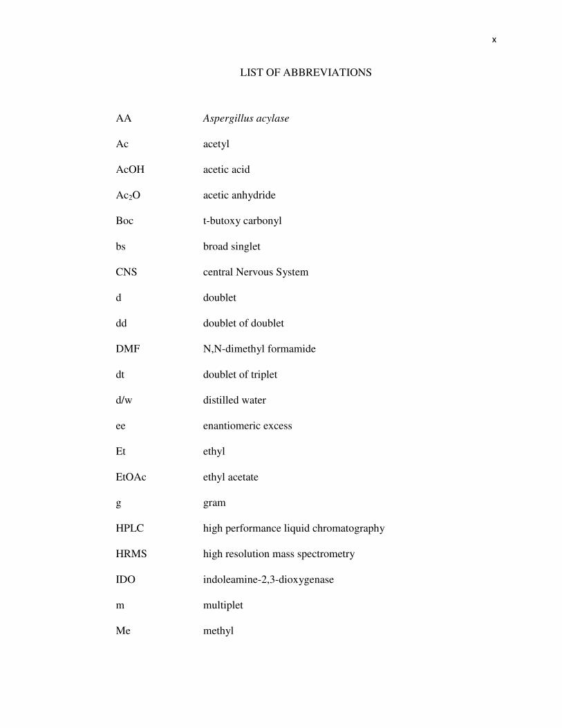
LIST OF ABBREVIATIONS
AA
Ac
AcOH
Ac2O
Boc
bs
CNS
d
dd
DMF
dt
d/w
ee
Et
EtOAc
g
HPLC
HRMS
IDO
m
Me
Aspergillus acylase
acetyl
acetic acid
acetic anhydride
t-butoxy carbonyl
broad singlet
central Nervous System
doublet
doublet of doublet
N,N-dimethyl formamide
doublet of triplet
distilled water
enantiomeric excess
ethyl
ethyl acetate
gram
high performance liquid chromatography
high resolution mass spectrometry
indoleamine-2,3-dioxygenase
multiplet
methyl

xi
mg
mM
µM
mol
MeOH
mins.
ml
NAD+
NADH
NADP+
NADPH
NBS
NCS
nm
NMDA
ns
PMP
Ph
PLP
psi
rac. or DL
RT
milligram
millimolar
micromolar
number of moles
methanol
minutes
milliliter
nicotinamide adenine dinucleotide
reduced nicotinamide adenine dinucleotide
nicotinamide adenine dinucleotide phosphate
reduced nicotinamide adenine dinucleotide phosphate
N-bromosuccinimide
N-chlorosuccinimide
nanometer
N-methyl-D-aspartate
nanosecond
pyridoxamine-5'-phosphate
phenyl
pyridoxal-5'-phosphate
pounds per square inch
racemic mixture
room temperature

xii
RM
t
TDO
TEA
TFA
TRIS
reaction mixture
triplet
tryptophan-2,3-dioxygenase
triethanolamine
trifluoroacetic acid
tris (hydroxymethyl) amino methane
1
CHAPTER 1
INTRODUCTION AND LITERATURE REVIEW
Tryptophan
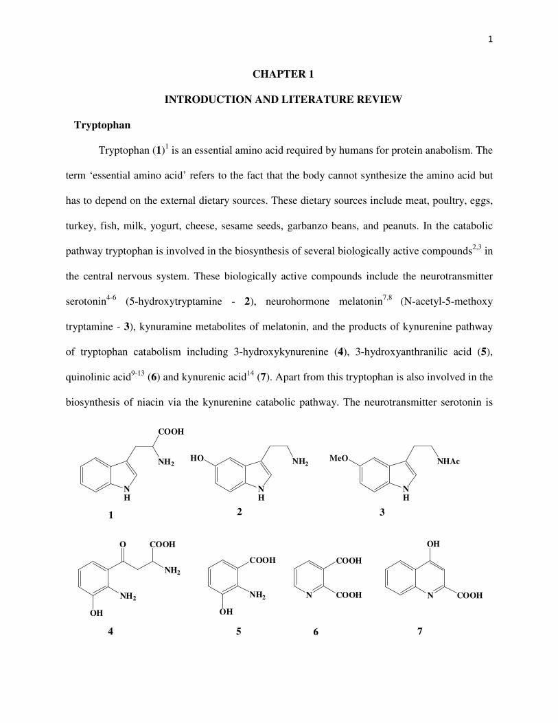
Tryptophan (1)1 is an essential amino acid required by humans for protein anabolism. The
term ‘essential amino acid’ refers to the fact that the body cannot synthesize the amino acid but
has to depend on the external dietary sources. These dietary sources include meat, poultry, eggs,
turkey, fish, milk, yogurt, cheese, sesame seeds, garbanzo beans, and peanuts. In the catabolic
pathway tryptophan is involved in the biosynthesis of several biologically active compounds2,3 in
the central nervous system. These biologically active compounds include the neurotransmitter
serotonin4-6 (5-hydroxytryptamine - 2), neurohormone melatonin7,8 (N-acetyl-5-methoxy
tryptamine - 3), kynuramine metabolites of melatonin, and the products of kynurenine pathway
of tryptophan catabolism including 3-hydroxykynurenine (4), 3-hydroxyanthranilic acid (5),
quinolinic acid9-13 (6) and kynurenic acid14 (7). Apart from this tryptophan is also involved in the
biosynthesis of niacin via the kynurenine catabolic pathway. The neurotransmitter serotonin is
NH
COOH
NH2
1
NH
NH2HO
2
NH
NHAcMeO
3
4 5 6 7
NH2
OH
O
NH2
COOH
NH2
OH
COOH
N
COOH
COOH N
OH
COOH

2
involved in the modulation of mood, anger, emotion and appetite, and is implicated in the control
of several behavioral and physiological functions. The neurohormone melatonin serves as a
biological clock that controls the sleep patterns of the individual. The metabolite quinolinic acid
has been found to have agonist effects on the N-methyl-D-aspartate receptors in the central
nervous system and thus acts as a potent neurotoxin. Thus, different metabolites of tryptophan
play an important role in the central nervous system and in the overall physiology and behavioral
patterns of the organism.
Catabolism of tryptophan
The kynurenine pathway is the primary pathway for the catabolism of the essential amino
acid tryptophan. Out of the different catabolic breakdown pathways for tryptophan leading to the
formation of the bioactive compounds, 99% of the dietary tryptophan that is not used in protein
synthesis is catabolised by the kynurenine pathway15. In the central nervous system before
crossing the blood-brain barrier, approximately 90% of the tryptophan is complexed with plasma
albumin16 and this complex cannot cross the blood brain barrier. However, the free tryptophan17
can cross blood-brain barrier where it is then available for further metabolism in the brain.
In the serotonergic neurons and mast cells of the CNS (Scheme 1) the free tryptophan is
acted upon by the enzyme tryptophan hydroxylase-2 also called tryptophan-5-monooxygenase
(EC: 1.14.16.4). This enzyme uses molecular oxygen and catalyzes the hydroxylation of
tryptophan to 5-hydroxy-L-tryptophan in the presence of the cofactor tetrahydrobiopterin. The 5-
hydroxy-L-tryptophan is then rapidly decarboxylated to serotonin18-20 in the presence of the PLP

3
dependent enzyme dopa decarboxylase, also called aromatic-L-amino acid decarboxylase (EC:
4.1.1.28). The serotonin is then converted into N-acetyl serotonin by the action of arylalkyl
amine-N-acetyl transferase (EC: 2.3.1.87). Finally melatonin21 is obtained by the action of
acetylserotonin-O-methyl transferase (EC: 2.1.1.4) on N-acetyl serotonin.
In the CNS (including the astrocytes, microglia, macrophages, and dendritic cells)
and in the hepatic and non-hepatic tissues (including the lungs, small intestine, and placenta of
mammals such as rabbits, rats, mice, and humans) L-tryptophan is catabolised by the kynurenine
pathway15. The first step in this pathway (Scheme 2) is the oxidative cleavage of the pyrrole ring
of tryptophan by the action of the hemeprotein indoleamine-2,3-dioxygenase (EC: 1.13.11.52) in
the presence of molecular oxygen to give N-formyl-L-kynurenine. However, in the mammalian
liver, the major site for L-tryptophan catabolism, the same reaction is carried out by another
NH
NH3
COO
L-Tryptophan
H
NH
NH21. O2, Tryptophanhydroxylase 2
2. Dopadecarboxylase
HO
Serotonin
1. Arylalkyl amine-N-acetyl transferase
2. Acetylserotonin-O-methyl transferase
NH
NHCOCH3MeO
Melatonin
Scheme 1

4
hemeprotein tryptophan-2,3-dioxygenase (EC: 1.13.11.11). Despite both the enzymes catalyzing
the same reaction using a heme cofactor, there is no significant sequence similarity between IDO
and TDO. Furthermore, it has been found that TDO stereospecifically catabolises only L-
tryptophan, but IDO can catabolise the oxidative cleavage of D-tryptophan, L-tryptophan, as
well as the various indoleamines such as melatonin, serotonin, hence the name IDO22 for the
latter. The N-formyl-L-kynurenine so formed in the first step is then deformylated to L-
kynurenine by an aryl formamidase (EC: 3.5.1.9). L-kynurenine is then hydroxylated by a
flavoenzyme kynurenine-3-monooxygenase (EC: 1.14.13.9) to give 3-hydroxy-L-kynurenine.
Subsequent action of the PLP dependent enzyme kynureninase (EC: 3.7.1.3) on 3-hydroxy-L-
kynurenine results in the cleavage of the β,γ C-C bond to give 3-hydroxyanthranilate and L-
NH
NH3
COO
L-Tryptophan
TDO / IDOCOO
NH3
O
NHCHO
N-formyl-L- kynurenine
Kynurenineformamidase COO
NH3O
NH2
HH
H
L-kynurenine
Kynurenine-3-monooxygenase COO
NH3O
NH2
H
3-hydroxy-L-kynurenine
Kynureninase
OH
−−−− L-alanine
OH
COO
NH2
3-hydroxy anthranilate
N
COO
COOH
Quinolinate
3-hydroxy anthranilate-3,4-dioxygenase
OH
COO
NH2O O
N
COO
Nicotinate
(KMO)
Acetyl CoA
2-amino-3-carboxymuconate semialdehyde
spontaneous
cyclization
- H2OLiver
NAD(P)+
Scheme 2
5
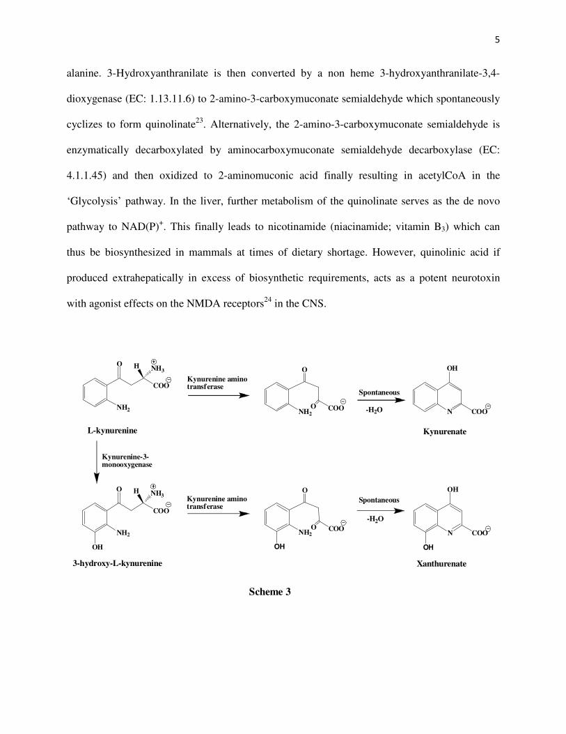
alanine. 3-Hydroxyanthranilate is then converted by a non heme 3-hydroxyanthranilate-3,4-
dioxygenase (EC: 1.13.11.6) to 2-amino-3-carboxymuconate semialdehyde which spontaneously
cyclizes to form quinolinate23. Alternatively, the 2-amino-3-carboxymuconate semialdehyde is
enzymatically decarboxylated by aminocarboxymuconate semialdehyde decarboxylase (EC:
4.1.1.45) and then oxidized to 2-aminomuconic acid finally resulting in acetylCoA in the
‘Glycolysis’ pathway. In the liver, further metabolism of the quinolinate serves as the de novo
pathway to NAD(P)+. This finally leads to nicotinamide (niacinamide; vitamin B3) which can
thus be biosynthesized in mammals at times of dietary shortage. However, quinolinic acid if
produced extrahepatically in excess of biosynthetic requirements, acts as a potent neurotoxin
with agonist effects on the NMDA receptors24 in the CNS.
COO
NH3
O
NH2
H
L-kynurenine
COO
O
NH2
O
Spontaneous
-H2O N
OH
COO
Kynurenate
Kynurenine-3-monooxygenase
COO
NH3O
NH2
H
OH
COO
O
NH2
ON
OH
COO
OHOH
Xanthurenate
Spontaneous
-H2O
Kynurenine aminotransferase
Kynurenine aminotransferase
3-hydroxy-L-kynurenine
Scheme 3
6
In another side biochemical reaction (Scheme 3) of the kynurenine pathway, L-
kynurenine is acted upon by kynurenine-oxoglutarate transaminase to give 4-(2-aminophenyl)-
2,4-dioxobutanoate which spontaneously dehydrates to produce kynurenate. The neuroactive
metabolite kynurenic acid has been found to have antagonist effects14 on the NMDA and α7
nicotinic acetyl choline receptors. Similarly, 3-hydroxy-L-kynurenine is acted upon by
kynurenine-oxoglutarate transaminase to give 4-(2-amino-3-hydroxyphenyl)-2,4-dioxo butanoate
which spontaneously dehydrates to produce xanthurenate.
The enzyme kynureninase
Kynureninase25 or L-kynurenine hydrolase (EC: 3.7.1.3) is a pyridoxal-5'-phosphate
(PLP) dependent enzyme that catalyzes the hydrolytic cleavage of L-kynurenine (Scheme 4) to
NH2
COO
NH3O
NH2
O
O
+COO
NH3
+
-
L-Kynurenine Anthranilate L-Alanine
Kynureninase
ααααγγγγ
Scheme 4
NH2
COO
NH3O
NH2
O
O
+COO
NH3
+
-
3-hydroxy-L-Kynurenine L-Alanine
3-hydroxyKynureninase
ααααγγγγ
OH OH
3-hydroxyanthranilate

7
give anthranilic acid and L-alanine. This is a key enzyme in the kynurenine pathway in the
tryptophan catabolism and catalyzes the unique β,γ-cleavage of aryl substituted γ-keto-α- amino
acids26. The enzyme has been isolated from Pseudomonas fluorescens27, Neurospora crassa
28,29,
rat liver30 and porcine liver31. It has been found that the mammalian liver kynureninase cleaves
3-hydroxy-L-kynurenine about twice as rapidly as it does L-kynurenine26,30 while the bacterial
kynureninase from Pseudomonas fluorescens cleaves L-kynurenine about five times32 as rapidly
as it does 3-hydroxy-L-kynurenine. Thus, L-kynurenine is the preferred substrate for the
pseudomonad enzyme while 3-hydroxy-L-kynurenine is the preferred substrate for the
mammalian liver kynureninase.
The two distinct types of kynureninases33 that have been shown to occur differ in terms
of their kinetic and chemical properties toward kynurenine and 3-hydroxykynurenine and in
terms of their response to PLP. Of these two types, the inducible enzyme is termed kynureninase
and is involved in preferential reaction with L-kynurenine in the aromatic and the quinoline
pathway of tryptophan catabolism. The specific activity of the inducible enzyme depends on the
concentration of tryptophan in the medium to such an extent that almost no inducible activity34 is
observed in the cells not supplemented with L-tryptophan. Thus, the cells utilize L-tryptophan as
the sole source of carbon, nitrogen, and energy for growth. On the other hand, the non-inducible
or the constitutive enzyme is termed 3-hydroxykynureninase and is mainly involved in the
biosynthesis of NAD i.e. the NAD pathway of tryptophan catabolism. The specific activity of
the non-inducible enzyme is independent of the concentration of tryptophan in the growth
medium34. The inducible enzyme has low Km for L-kynurenine while the non-inducible enzyme
has low Km for 3-hydroxy-L-kynurenine. Furthermore, it has been found that the inducible

8
enzyme is reversibly inactivated9 by L-alanine resulting in a transamination reaction to give
pyridoaxmine-5'-phosphate (Scheme 5) and pyruvate from L-alanine. However, the enzyme
activity is restored either by addition of PLP or pyruvic acid in the latter case there being a
reverse transamination between pyridoxamine-5'-phosphate and the added pyruvate to give back
PLP and alanine.
On the other hand the non inducible (or constitutive) enzyme is little or not at all affected
by the presence of L-alanine or other amino acids35a. Even then the rate of the hydrolytic
cleavage reduces with time indicating that the product 3-hydroxyanthranilate inhibits the non
inducible enzyme thereby regulating the enzyme action in the NAD biosynthetic pathway35b.
N
HOOPO3
H
OOC
NH3
Alanine
PLP bound to lysine
N
HOOPO3
HN
H
OOC
N
HOOPO3
HN
H
OOC
N
HOOPO3
H2N
H
OOC
O
+
+
Pyruvate
Pyridoxamine-5'-phosphate
Scheme 5
HNEnz

9
The bacterial cultures that possess the inducible enzyme include Pseudomonas
fluorescens and Bacillus cereus36, Bacillus megaterium
37,38 Acinetobacter calcoaceticus39 and
Xanthomonas pruni40. Among the fungal species, Neurospora crassa, Aspergillus niger, and
Penicillium roqueforti possess both the inducible as well as the constitutive kynureninases while
Rhizopus stolonifer34 possesses only the constitutive enzyme. The kynureninases obtained from
yeast and the livers of mammals like dog, mouse, guinea pig, beef, and human are the
constitutive enzyme26.
Mechanism of kynureninase action
The enzyme kynureninase catalyzes the unique β,γ-cleavage of aryl substituted γ-keto-α-
amino acids in the kynurenine pathway of tryptophan catabolism. The mechanism of
kynureninase has been the subject of considerable interest due to the unique nature of this PLP
dependent reaction. In one of the mechanisms by Dalgliesh et al it was proposed that
kynureninase catalyzes the transamination of kynurenine41 by PLP to give the β-anthraniloyl
pyruvic acid (Scheme 6) which is then hydrolyzed to anthranilic acid and pyruvate or partly
undergoes a spontaneous dehydrative cyclization to give kynurenic acid. The pyruvate in turn
recycles with PMP to give back PLP and alanine. But later another enzyme kynurenine amino
transferase42-44 was shown to be involved in the formation of kynurenic acid.
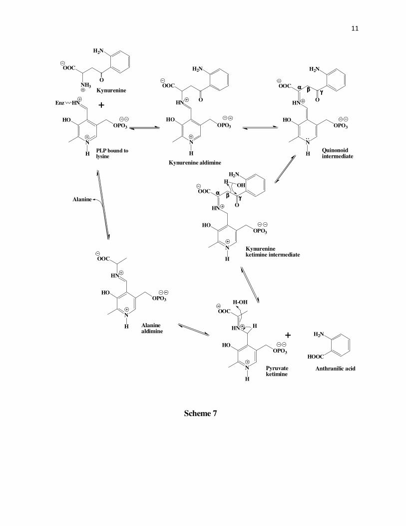
In another mechanism proposed by Braunstein et al45-46 (Scheme 7) the initially formed
Schiff’s base between PLP and kynurenine undergoes a tautomerization followed by hydrolysis
at the γ-carbonyl carbon. This cleaves the β,γ-carbon bond in a way that the β-carbon takes up

10
N
HOOPO3
HN
H
OOC
NH3Kynurenine
PLP bound to lysine
N
HOOPO3
HN
H
OOC
N
HOOPO3
HN
H
OOC
N
HOOPO3
H2N
H
OOC
O
+
+
ββββ-anthraniloyl pyruvate Pyridoxamine-5'-phosphate
Scheme 6
H2N
O
H2N
O
H2N
O
H2N
O
Spontaneous-H2O
Hydrolysis
H2N
HOOC
OOC
O
(PMP)
Pyruvate Anthranilic acid
Alanine
PMP
+
NOOC
OH
Kynurenate
Enz
N
HOOPO3
HN
H
OOC
H2N
O
Ketimine intermediate
Quinonoidintermediate
11
N
HOOPO3
HN
H
OOC
H2N
O
Kynurenineketimine intermediate
N
HOOPO3
HN
H
OOC
NH3Kynurenine
N
HOOPO3
HN
H
OOC
N
HOOPO3
HN
H
OOC
+
+
Scheme 7
H2N
O
H2N
O
H2N
O
HOOC
H2N
Alanine
Kynurenine aldimine
Quinonoidintermediate
HOH
N
HOOPO3
HN
H
OOC
Pyruvateketimine
Anthranilic acid
H-OH
N
HOOPO3
HN
H
OOC
Alaninealdimine
ββββααααγγγγ
Enz
PLP bound tolysine
αααα ββββ γγγγ
H

12
N
HOOPO3
HN
H
OOC
H2N
OH
Kynurenineketimine intermediate
N
HOOPO3
HN
H
OOC
NH3Kynurenine
PLP bound tolysine
N
HOOPO3
HN
H
OOC
N
HOOPO3
HN
H
OOC
+
+
Scheme 8
H2N
O
H2N
O
H2N
O
H2N
Alanine
Kynurenine aldimine
Quinonoidintermediate
N
HOOPO3
HN
H
OOC
αααα-amino acrylatealdimine
Anthranyl anion
N
HOOPO3
HN
H
OOC
Alaninealdimine
ββββ γγγγ
O
ββββ
γγγγ
αααα,ββββ-elimination
αααα
H
H2N
OHC
Anthranaldehyde
Redoxreaction
+
H2N
HOOC
Anthranilic acid
Enz
ααααββββ
γγγγ

13
the σ-electrons of the β,γ-carbon bond to give anthranilic acid and the pyruvate ketimine. The
pyruvate ketimine is then converted into alanine and PLP after tautomerization.
Longenecker et al47 however proposed a slightly different mechanism than the Braunstein
group based on their study of mechanisms of enzymes including serine dehydrase,
tryptophanase, and cysteine desulfhydrase. In this mechanism (Scheme 8) after the initial
formation of the kynurenine ketimine, instead of the β-carbon keeping the electrons of the β,γ-
carbon bond, the γ-carbonyl carbon takes up the electron pair as shown by the tautomerization
process to give the anthranyl anion and the aldimine of α-amino acrylate. The anthranyl anion
can then either before or after stabilization (as anthraldehyde) undergoes a non-enzymatic redox
reaction to give anthranilic acid, and the Schiff’s base of alanine, the latter being eventually
hydrolyzed to alanine with the regeneration of PLP.
In their mechanistic studies on kynureninase from Pseudomonas marginalis Bild and
Morris48 suggested that the β-carbon of kynurenine must be serving as carbanion. This was based
on the formation of 2-amino-4-hydroxy-4-phenyl butanoic acid (Scheme 9) via an aldol type
reaction49 between the incipient alanine and benzaldehyde.
COOH
NH2O
NH2
+
CHO
Kynurenine Benzaldehyde Anthranilicacid
2-amino-4-hydroxy-4-phenyl butanoic acid
COOH
NH2OH
+
COOH
NH2
Scheme 9
14
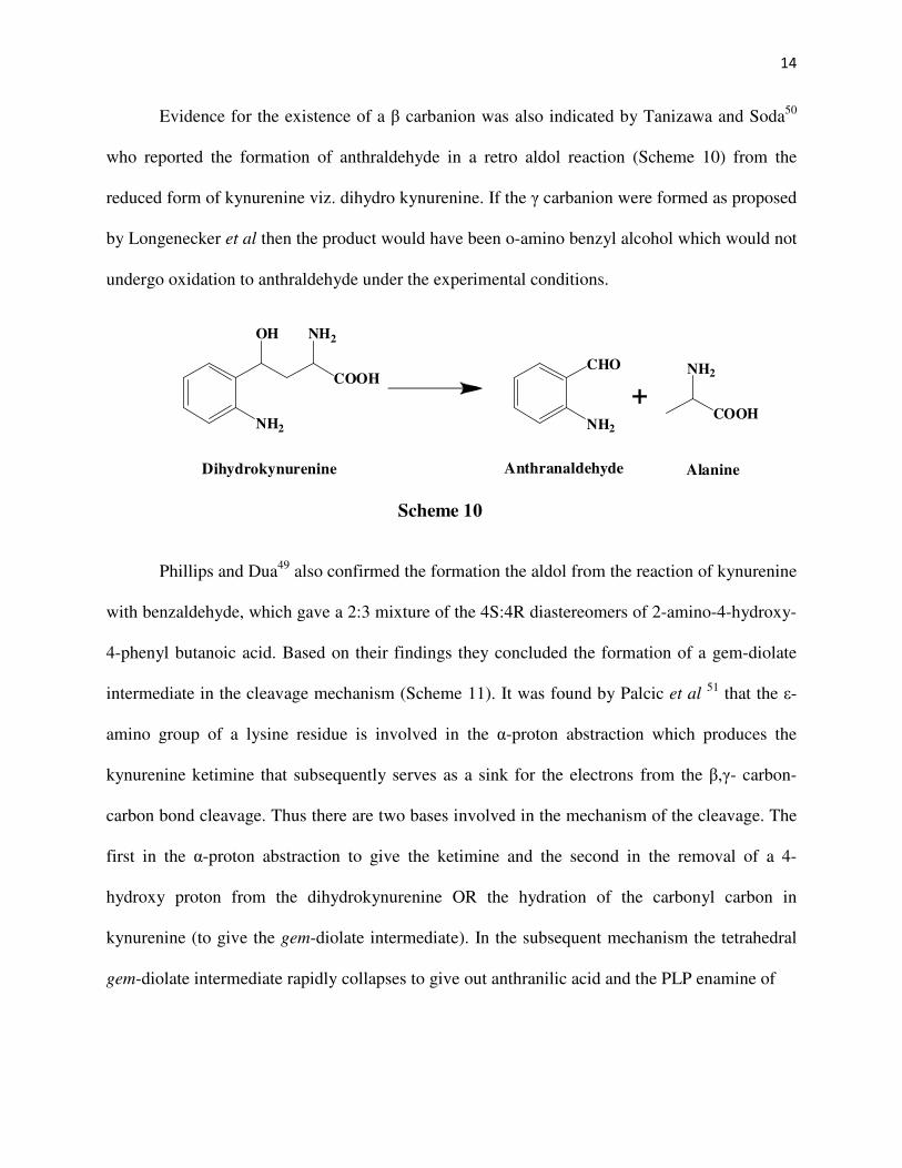
Evidence for the existence of a β carbanion was also indicated by Tanizawa and Soda50
who reported the formation of anthraldehyde in a retro aldol reaction (Scheme 10) from the
reduced form of kynurenine viz. dihydro kynurenine. If the γ carbanion were formed as proposed
by Longenecker et al then the product would have been o-amino benzyl alcohol which would not
undergo oxidation to anthraldehyde under the experimental conditions.
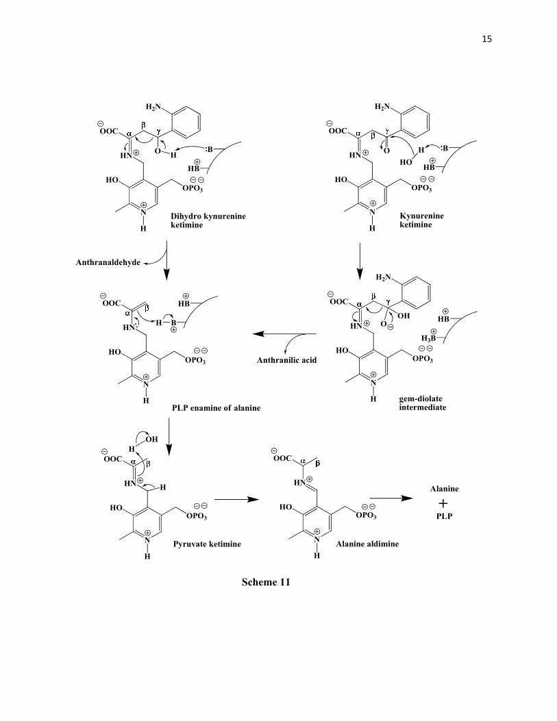
Phillips and Dua49 also confirmed the formation the aldol from the reaction of kynurenine
with benzaldehyde, which gave a 2:3 mixture of the 4S:4R diastereomers of 2-amino-4-hydroxy-
4-phenyl butanoic acid. Based on their findings they concluded the formation of a gem-diolate
intermediate in the cleavage mechanism (Scheme 11). It was found by Palcic et al 51 that the ε-
amino group of a lysine residue is involved in the α-proton abstraction which produces the
kynurenine ketimine that subsequently serves as a sink for the electrons from the β,γ- carbon-
carbon bond cleavage. Thus there are two bases involved in the mechanism of the cleavage. The
first in the α-proton abstraction to give the ketimine and the second in the removal of a 4-
hydroxy proton from the dihydrokynurenine OR the hydration of the carbonyl carbon in
kynurenine (to give the gem-diolate intermediate). In the subsequent mechanism the tetrahedral
gem-diolate intermediate rapidly collapses to give out anthranilic acid and the PLP enamine of
COOH
NH2OH
NH2
Dihydrokynurenine
COOH
NH2CHO
NH2
+
Anthranaldehyde Alanine
Scheme 10
15
N
HOOPO3
HN
H
OOC
H2N
O
Kynurenineketimine
N
HOOPO3
HN
H
OOC
H2N
O
Dihydro kynurenineketimine
H
N
HOOPO3
HN
H
OOC
Pyruvate ketimine
B
HB
N
HOOPO3
HN
H
OOC
HO
H
N
HOOPO3
HN
H
OOC
H2N
OHB
H3B
OH
gem-diolateintermediate
HB
BH
PLP enamine of alanine
B
HB
Anthranaldehyde
Anthranilic acid
H
OH
N
HOOPO3
HN
H
OOC
Alanine aldimine
Alanine
PLP
+
Scheme 11
H

16
alanine. This enamine first takes up a proton at the β-carbon to give the pyruvate ketimine that
accepts a second proton at the α-carbon to give alanine aldimine which finally releases alanine
and the cofactor recyles in the process.
Later Dua and Phillips52 also showed that the sulfone analog of kynurenine viz. S-(2-
aminophenyl)-L-cysteine-S,S-dioxide was a potent inhibitor of kynureninase with a Ki value of
70 nM which is about 300-fold lower than the Km for L-kynurenine. This further supports the
gem-diolate hypothesis. Kinetic isotope effect studies53 by the Phillips group led to the
conclusion that the rate determining step is the deprotonation of the aldehydic carbon of PLP in
the pyruvate ketamine intermediate to give the alanine quinonoid intermediate. Using rapid-
scanning stopped-flow spectrophotometry and rapid chemical quench methods54 the L-
kynurenine quinonoid intermediate, and the pyruvate ketimine intermediate were detected with
L-kynurenine as the substrate. Thus, the mechanism for kynureninase proposed by Phillips et al
is shown in Scheme 12.

17
O C OO
NH 3
K ynu re n in ase .P LP
Ex te rn al aldim ine
(λλλλ m ax = 4 20 n m)
NH 2
L-K ynu re n ine (λλλλ m ax = 3 60 nm )
O COO
N H
NH 2
N
HOO P O 3
H
B
L-K y nur en in e qu in ono id inte rm e diate
(λλλλ m ax = 49 4 nm )
O CO O
NH
N H 2
NH
H OO P O 3
B
H
O C OO
NH
N H 2
N
H O
O P O 3
CO O
N H
NH 2
NH
H OO P O 3
L -K ynu re n ine k etimin e inte rm e diate
( λλλλ m ax = 330 n m )
H O HB
OH O CO O
NH
NH
HO
OPO 3
CO OH
NH 2
An th ra nilic a cid(S )- gem - diolate inter m ed iate
(λλλλ m ax = 3 30 n m )
Retro -Cla isen B
H
CO O
NH
NH
HO
OPO 3
E na m ine inter m ed iate
(λλλλ max = 330 nm )
P yru va te k etim in e in te rm ediate
(λλλλ m ax = 3 30 n m )
H
BCO O
NH
NH
HO
OP O 3
Ala n ine q u inon oid in te rm ed ia te
(λλλλ max = 500 nm )
CO O
NH
NH
H O
OPO 3
B
H
Alan in e aldim ine inter m ed iate
(λλλλ max = 4 20 n m )
L- Ala nin e
H
H
+ K y n urenin a se.P LP Schem e 12

18
Kynurenines
Kynurenine or β-(2-aminobenzoyl)alanine was first discovered by Matsuoka and
Yoshimatsu55 in the urine of rabbits fed large quantities of the amino acid tryptophan. About two
decades later the structure was determined by Butenandt et al.56 In Pseudomonas fluorescens and
some other bacteria kynurenine is a substrate in its reaction with the enzyme kynureninase to
give anthranilic acid and L-alanine in the tryptophan catabolic pathway. As described above, in
eukaryotes a similar substrate viz. 3-hydroxy-L-kynurenine is involved in a similar reaction to
produce L-alanine and 3-hydroxyanthranilic acid (Scheme 4). In animals including humans 3-
hydroxyanthranilic acid serves as a precursor for the biosynthesis of quinolinic acid (Scheme 2).
Excessive levels of quinolinic acid have been implicated in a range of neurological disorders57-58
such as Huntington’s chorea, Lou Gehrig’s disease59, epilepsy60, and AIDS related dementia. An
excessive level of quinolinate has been shown to be present after a stroke and is responsible for
further damage61. Furthermore, the brains of Alzheimer’s patients have also been shown to have
high levels of quinolinate which may be responsible for the progression of the disease62. It has
also been shown that spontaneously hypertensive rats have a significantly higher kynureninase
activity in tissues63 and recently it has been established that there is close link between
hypertension and an allele of the human kynureninase viz. K412E64. Selective inhibitors of 3-
hydroxykynureninase could thus be used as drugs for the treatment of these diseases. Several
structural analogs of kynurenine have been synthesized in the past to check for their inhibitory
activity. The most potent inhibitor of kynureninase reported the date is the S-(2-aminophenyl)-L-
cysteine-S,S-dioxide, 8 with a Ki of 27 nM, some 925-fold lower than the Km of L-kynurenine
(~25 µM)52, 65. The 4-bromo, 9, and the 5-bromo, 10 analogs of S-(2-aminophenyl)-L-cysteine-
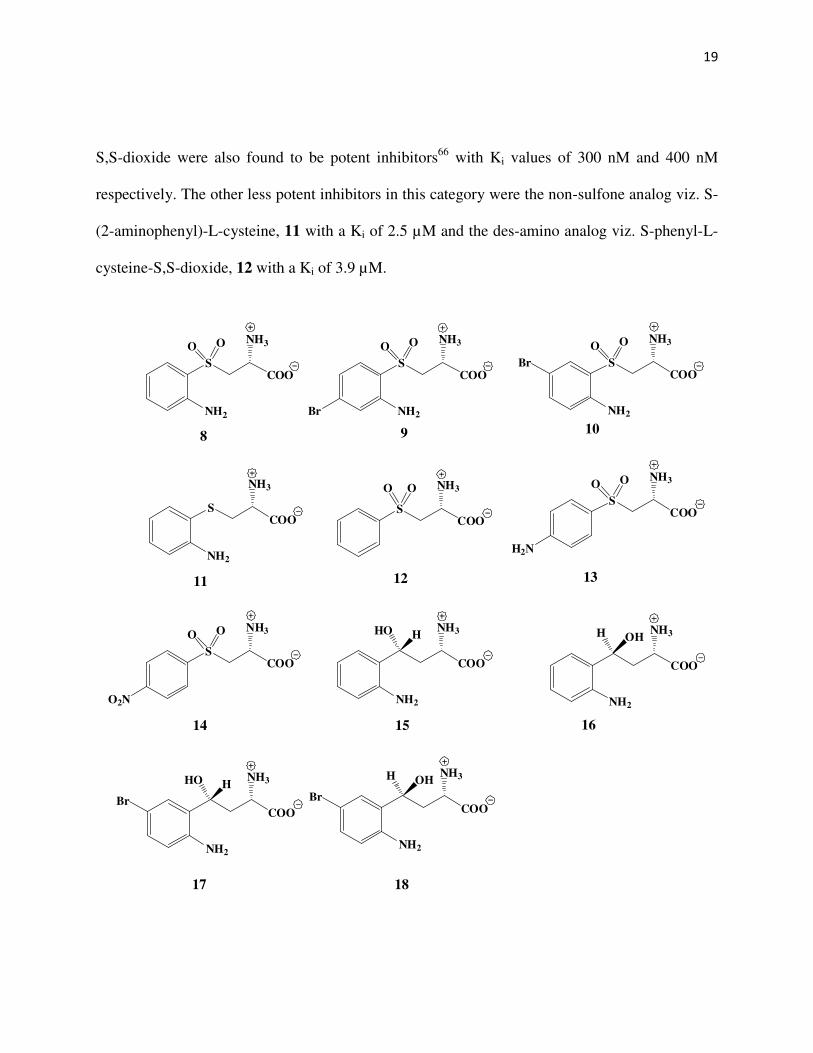
19
S,S-dioxide were also found to be potent inhibitors66 with Ki values of 300 nM and 400 nM
respectively. The other less potent inhibitors in this category were the non-sulfone analog viz. S-
(2-aminophenyl)-L-cysteine, 11 with a Ki of 2.5 µM and the des-amino analog viz. S-phenyl-L-
cysteine-S,S-dioxide, 12 with a Ki of 3.9 µM.
SCOO
NH3
NH2
O O
8 9 10
11 1312
SCOO
NH3
NH2
SCOO
NH3
SCOO
NH3O O
H2N
SCOO
NH3O O
O2N
14
COO
NH3
NH2
HO H
COO
NH3
NH2
H OH
15 16
SCOO
NH3
NH2
O O
SCOO
NH3
NH2
O O
COO
NH3
NH2
HO H
COO
NH3
NH2
H OH
17 18
Br
Br
O O
Br Br

20
Also, the 4-amino and the 4-nitro sulfone analogs viz. S-(4-aminophenyl)-L-cysteine-S,S-
dioxide, 13 and S-(4-nitrophenyl)-L-cysteine-S,S-dioxide, 14 were shown to have competitive
inhibitory activity with Ki values of 8.5 µM, and 12 µM respectively. The diastereomeric 4R, 15
and 4S, 16 dihydro kynurenines, have also been shown by Phillips et al49 to be potent inhibitors
of kynureninase with Ki values of 1.4 µM, and 0.3 µM respectively. The 5-bromo analogs of the
dihydrokynurenines have also been shown by Heiss et al66 to possess good inhibitory activity
with Ki values of 55 nM and 170 nM respectively for the 4R, 17 and the 4S, 18 diastereomers.
In chapter 2 of this dissertation the synthesis of a new class of substrate analogs of
kynurenines has been described.
21
References
1. Matarese, V.; Bernlohr, D. A. J. Biol. Chem. 1988, 263, 14544-14551.
2. Zubay, G.; in Biochemistry, 1988 pp 630-631, MacMillan, New York
3. The Chemical Society, The Alkaloids, 1971 London, Specialist Periodical Report
4. Pihel, K.; Hsieh, S. C.; Jorgenson, J. W.; Wightman, R. M. Biochemistry 1998, 37, 1046-
1052.
5. Purcell, W. M.; Atterwill, C. K. Neurochem. Res. 1995, 20, 521-32.
6. Niacaris, T.; Avery, L. J. Exp. Biol. 2003, 206, 223-231.
7. Altun, A.; Ugur-Altun, B. Int. J. Clin. Pract. 2007, 61, 835-845.
8. Hardeland, R. Endocrine 2005, 27, 119-130.
9. Beal, M. F.; Kowall, N. W.; Ellison, D. W.; Mazurek, M. F.; Swartz, K. J.; Martin, J. B.
Nature 1986, 321, 168-171.
10. Heyes, M. P.; Brew, B. J.; Martin, A.; Price, R. W.; Salazar, A. M.; Sidtis, J. J.; Yergey,
J. A.; Mouradian, M. M.; Sadler, A. E.; Keilp, J.; Rubinow, D.; Markey, S. P. Ann.
Neurol. 1991, 29, 202-209.
11. Saito, K.; Crowley, J. S.; Markey, S. P.; Heyes, M. P. J. Biol. Chem. 1993, 268, 15496-
15503.
12. Carpenedo, R.; Chiarugi, A.; Russi, P.; Lombardi, G.; Carla, V.; Pellicciari, R.; Moroni,
F.; Mattoli, L. Neuroscience 1994, 61, 237-244.
13. Stone, T. W.; Perkins, M. N. Eur. J. Pharmacol. 1981, 72, 411-412.
14. Perkins, M. N.; Stone, T. W. Brain Res. 1982, 247, 184-187.
15. Peters, J. C. Adv. Exp. Med. Biol. 1991, 294, 345-58.
16. McMenamy, R. H. J. Biol. Chem. 1965, 240, 4235-4243.

22
17. Madras, B. K.; Cohen, E. L.; Messing, R.; Munro, H. N.; Wurtman, R. J. Metab. Clin.
Exp. 1974, 23, 1107-1116.
18. Wang, L.; Erlandsen, H.; Haavik, J.; Knappskog, P. M.; Stevens, R. C. Biochemistry
2002, 41, 12569-12574.
19. Martinez, A.; Knappskog, P. M.; Haavik, J. Curr. Med. Chem. 2001, 8, 1077-1091.
20. Donaldson, R. M., Jr.; Gray, S. J.; Letsou, V. G. Lancet 1959, 1959-II, 1002-3.
21. Kopp, N.; Claustrat, B.; Tappaz, M. Neurosci. Lett. 1980, 19, 237-242.
22. Yoshida, R.; Hayaishi, O. Meth. Enzymol. 1987, 142, 188-195.
23. Colabroy, K. L.; Begley, T. P. J. Am. Chem. Soc. 2005, 127, 840-841.
24. Schwarcz, R.; Whetsell, W. O.; Mangano, R. M. Science 1983, 219, 316-318.
25. Braunstein, A. E.; Goryachenkova, E. V.; Paskhina, T. S. Biokhimiya (Moscow) 1949, 14,
163-79.
26. Soda, K.; Tanizawa, K. Adv. Enzymol. Relat. Areas Mol. Biol. 1979, 49, 1-40.
27. Hayaishi, O.; Stanier, R. Y. J. Biol. Chem. 1952, 195, 735-740.
28. Jakoby, W. B.; Bonner, D. M. J. Biol. Chem. 1953, 205, 699-707.
29. Jakoby, W. B.; Bonner, D. M. J. Biol. Chem. 1953, 205, 709-715.
30. Knox, W. G. Biochem. J. 1953, 53, 379.
31. Wiss, O.; Weber, F. Hoppe-Seyler's Z. Physiol. Chem. 1956, 304, 232-40.
32. Hayaishi, O. in A Symposium on Amino Acid Metabolism, W. D. McElroy and H. B.
Glass, Eds. Johns Hopkins Press, Baltimore, 1955, pp. 914-929
33. Turner, J. R.; Drucker, H. Biochem. Biophys. Res. Commun. 1971, 42, 698
34. Shetty, A. S.; Gaertner, F. H. J. Bacteriol. 1975, 122, 235-244.
35. a) Tanizawa, K.; Soda, K. J. Biochem. 1979, 86, 499-508.
23
b) McDermott, C. E; Casciano, D. A.; Gaertner, F. H. Biochem. Biophys. Res. Commun.
1973, 51, 813-818.
36. Prasad, C.; Srinivas.Vr Biochem. J. 1970, 119, 343-&.
37. Tabone, J.; Robert, D. Bull. Soc. Chim. Biol. 1952, 34, 1102-1105.
38. Bouknight, R. R.; Sadoff, H. L. J. Bacteriol. 1975, 121, 70-76.
39. Wheelis, M. L. Arch. Mikrobiol. 1972, 87, 1-&.
40. Brown, A. T.; Wagner, C. J. Bacteriol. 1970, 101, 456-&.
41. Dalgliesh, C. E.; Knox, W. E.; Neuberger, A. Nature 1951, 168, 20-22.
42. Miller, I. L.; Tsuchida, M.; Adelberg, E. A. J. Biol. Chem. 1953, 203, 205-211.
43. Mason, M. J. Biol. Chem. 1954, 211, 839-844.
44. Jakoby, W. B.; Bonner, D. M. J. Biol. Chem. 1956, 221, 689-695.
45. Braunstein, A. E.; Shemyakin, M. M. Biokhimiya 1953, 18, 393-411.
46. Braunstein, A. E. in The Enzymes, Vol. II, 2nd ed. P. D. Boyer, Ed., Academic Press, New
York, 1960, p. 170
47. Longenecker, J. B.; Snell, E. E. J. Biol. Chem. 1955, 213, 229-235.
48. Bild, G. S.; Morris, J. C. Arch. Biochem. Biophys. 1984, 235, 41-47.
49. Phillips, R. S.; Dua, R. K. J. Am. Chem. Soc. 1991, 113, 7385-7388.
50. Tanizawa, K.; Soda, K. J. Biochem. 1979, 86, 1199-1209.
51. Palcic, M. M.; Antoun, M.; Tanizawa, K.; Soda, K.; Floss, H. G. J. Biol. Chem. 1985,
260, 5248-5251.
52. Dua, R. K.; Taylor, E. W.; Phillips, R. S. J. Am. Chem. Soc. 1993, 115, 1264-1270.
53. Koushik, S. V.; Moore, J. A.; Sundararaju, B.; Phillips, R. S. Biochemistry 1998, 37,
1376-1382.

24
54. Phillips, R. S.; Sundararaju, B.; Koushik, S. V. Biochemistry 1998, 37, 8783-8789.
55. Matsuoka, Z.; Yoshimatsu, N. Z. Physiol. Chem. 1925, 143, 206-10.
56. Butenandt, A.; Weidel, W.; Weichert, R.; von Derjugin, W. Hoppe-Seyler's Z. Physiol.
Chem. 1943, 279, 27-43.
57. Schwarcz, R.; Okuno, E.; White, R. J.; Bird, E. D.; Whetsell, W. O. Proc. Natl. Acad.
Sci. U.S.A. 1988, 85, 4079-4081.
58. Mazzari, S.; Aldinio, C.; Beccaro, M.; Toffano, G.; Schwarcz, R. Brain Res. 1986, 380,
309-316.
59. Guillemin, G. J.; Meininger, V.; Brew, B. J. Neurodegener. Dis. 2005, 2, 166-176.
60. Kaminski, R. M.; Zielinska, E. B.; Dekundy, A.; van Luijtelaar, G.; Turski, W. A. Pol. J.
Pharmacol. 2003, 55, 741-746.
61. Stone, T. W. Expert Opin. Investig. Drugs 2001, 10, 633-645.
62. Guillemin, G. J.; Brew, B. J.; Noonan, C. E.; Takikawa, O.; Cullen, K. M. Neuropathol.
Appl. Neurobiol. 2005, 31, 395-404.
63. Mizutani, K.; Sugimoto, K.; Okuda, T.; Katsuya, T.; Miyata, T.; Tanabe, T.; Higaki, J.;
Ogihara, T.; Yamori, Y.; Tsujita, Y.; Tago, N.; Iwai, N. Hypertens. Res. 2002, 25, 135-
140.
64. Zhang, Y.; Zhang, K. X.; He, X.; Yuan, W. T.; Wang, G. L.; Mao, S. Y.; Gao, P. J.;
Huang, W.; Zhu, D. L. Zhonghua Xin Xue Guan Bing Za Zhi 2005, 33, 588-591.
65. Drysdale, M. J.; Reinhard, J. F. Bioorg. Med. Chem. Lett. 1998, 8, 133-138.
66. Heiss, C.; Anderson, J.; Phillips, R. S. Org. Biomol. Chem. 2003, 1, 288-295.
25
CHAPTER 2
SYNTHESIS OF SUBSTRATE ANALOGS OF KYNURENINE
Abstract
The DL-3-bromo, DL-3-chloro, DL-3-fluoro, DL-3-methyl, L-5-bromo, and L-5-chloro
kynurenines have been synthesized. The DL analogs have been synthesized starting from
acrolein. Reaction of acrolein with the diethyl acetamidomalonate anion gives the Michael
adduct1-4 which on treatment with the corresponding 2-halosubstituted phenylhydrazine5-11 yields
a phenylhydrazone derivative2. The different 2-halosubstituted pehnylhydrazones are then
subjected to a Fischer indole cyclization to give the 7-halosubstituted indolylmethylacetamido
malonates12-16. An ozonolysis of these indole compounds give the respective diethyl-2-amino-3-
halobenzoylmethylacetamidomalonates which upon acid hydrolysis produce the racemic 3-halo
substituted kynurenines. The 5-substituted kynurenines17 have been synthesized from L-
tryptophan via first the ozonolysis of the methyl ester of Nα-acetyl-L-tryptophan followed by
TFA hydrolysis and acylation of the intermediate to give the methyl ester of Nα,N-
diacetylkynurenine. This intermediate on bromination or chlorination, followed by acid
hydrolysis produces the respective 5-halosubstituted-L-kynurenines.

26
Experimental methods
Instrumentation
1HNMR ,13CNMR, and 19FNMR spectra were recorded on a Varian 400MHz instrument.
Two different deuterated solvents were used for the different products. Of these, the 2-
substituted phenylhydrazine, the phenylhydrazone derivative, the diethyl-7-substituted
indolylmethylacetamidomalonate, and the 2-amino3-substituted
benzoylmethylacetamidomalonate intermediates were tested in deuterated methanol, while the
ultimate substituted kynurenines were tested in deuterated water containing 1 – 2 % of DCl.
HPLC measurements were carried out on a Spectrasystem P 2000 instrument connected to a UV
6000 detector and controlled by a Dell PC using Chromquest software. A gradient elution was
used consisting of 5 % MeOH, and 95 % 0.1 % aq. acetic acid from 0 – 5 mins. followed by a
programmed increase of MeOH percentage from 5% to 70% over 5 – 20 mins. with a
corresponding decrease of the percentage of 0.1 % aq. acetic acid from 95% to 30% over the
same time period. This is followed by an increase of MeOH percentage to 100% with the
corresponding decrease of the percentage of 0.1% aq. acetic acid to 0% over 20 – 25 mins. And
finally, a programmed return back of the elution system to 5% MeOH, and 95% of 0.1% aq.
acetic acid over the period from 25 – 30 mins. A 100 µM solution of the individual substituted
kynurenines in 1 mM HCl was used for injection. Chiral HPLC of the DL-3-methylkynurenine
was done using a chiral Pro-Cu column (5µ, 4.5 x 250 mm) and a 1 mM aq. CuSO4 solution was
used as the eluant. Elution for both columns was done with a flow rate of 1ml/min. with
detection by absorbance at 254 nm and 370 nm. GCMS of the intermediate compounds was done
on a Shimadzu instrument in Prof. Dr. V. Popik’s lab in the Chemistry Department.

27
Synthesis of 2-chlorophenyl hydrazine6,18-20
Take 10 g of 2-chloroaniline hydrochloride (prepared by dissolving 10 ml of 2-
chloroaniline in 100 ml acetone and adding 14 ml conc. HCl with stirring. Chill the resulting
suspension, filter and wash the white solid with about 15 ml acetone) in 200 ml conc. HCl, stir at
RT for about 15 mins. when a white suspension results. Cool the soln. to -20 ºC, in a dry ice-
acetone bath, add to it an aq. soln. of 5.05 gm of sodium nitrite in 25 ml d/w. (Addition of
sodium nitrite solution is done in such a way that the tip of the
dropping funnel is dipping into the RM via a small tube attached to
the dripping tip of the dropping funnel) Complete the addition in
about 15 mins. and then continue stirring at -20 ºC for about 10 - 15
mins. Then to the same RM while maintaining the temp. at -20 to -
25ºC add a soln. of 27.52 gm of stannous chloride dihydrate in 25 ml
of conc. HCl. Complete the addition in about 45 mins when a thick
precipitate of the hydrazine hydrochloride salt is formed. Allow the
RM to stir at 0 to -10ºC for about 45 mins. Check TLC (Fig. 1) Cool
the suspension to -45 to -50 ºC, for about 15 mins. then filter. Spread
the solid on a petri dish to let it air dry overnight to give 24 g of a crude solid from which the
free base is obtained.
The free base of the 2-chlorophenylhydrazine is released by treatment of the hydrochloride salt
with 2.7 equivalents of NaOH and the free base extracted with ether.
Yield of the free base = 7 g, 81 % , m.p. = 45-46ºC
SM
System: Hexane: EtOAc2ml : 1ml
Detection: uv 254 nmor I2 vapors
SM = Starting materialCo = Mixture spot
Co RM
Fig. 1
28
1HNMR of free base: d-MeOH δ 6.65 (d, 1H), 6.75 (t, 1H), 7.3 (t, 1H), 7.6 (d, 1H) the NH
protons exchanged with the solvent and merged around δ 4.5
13CNMR of free base: d-MeOH δ 108.5, 118.2, 121, 126, 123.2, 140.5
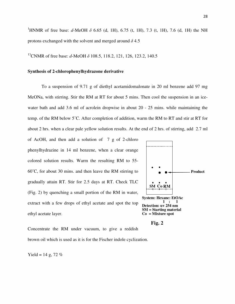
Synthesis of 2-chlorophenylhydrazone derivative
To a suspension of 9.71 g of diethyl acetamidomalonate in 20 ml benzene add 97 mg
MeONa, with stirring. Stir the RM at RT for about 5 mins. Then cool the suspension in an ice-
water bath and add 3.6 ml of acrolein dropwise in about 20 - 25 mins. while maintaining the
temp. of the RM below 5˚C. After completion of addition, warm the RM to RT and stir at RT for
about 2 hrs. when a clear pale yellow solution results. At the end of 2 hrs. of stirring, add 2.7 ml
of AcOH, and then add a solution of 7 g of 2-chloro
phenylhydrazine in 14 ml benzene, when a clear orange
colored solution results. Warm the resulting RM to 55-
60˚C, for about 30 mins. and then leave the RM stirring to
gradually attain RT. Stir for 2.5 days at RT. Check TLC
(Fig. 2) by quenching a small portion of the RM in water,
extract with a few drops of ethyl acetate and spot the top
ethyl acetate layer.
Concentrate the RM under vacuum, to give a reddish
brown oil which is used as it is for the Fischer indole cyclization.
Yield = 14 g, 72 %
SM Co RM
System: Hexane: EtOAc1 : 1
Detection: uv 254 nmSM = Starting materialCo = Mixture spot
Product
Fig. 2
29
1HNMR: d MeOH δ 7.4 (d, 1H), 7.3 (t, 1H), 7.2 (d, 1H), 7.1 (t, 1H), 6.7 (t, 1H), 4.2 (q, 4H),
2.2 (t, 1H), 2.1 (s, 3H), 1.9 (q, 2H), 1.2 (t, 6H)
13CNMR: d MeOH δ 13.4, 20.2, 21.6, 26.9, 62.5, 62.7, 66.5, 116.5, 119.3, 127.8, 129.1, 129.2,
142.4, 168, 171.3
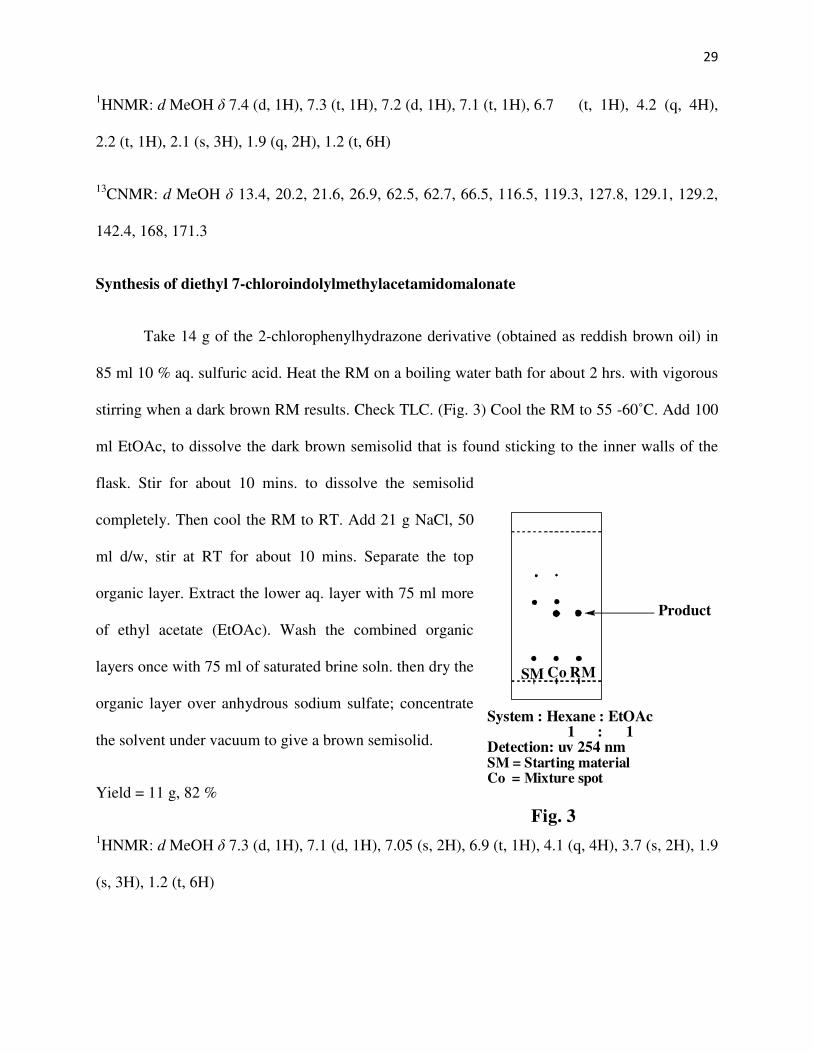
Synthesis of diethyl 7-chloroindolylmethylacetamidomalonate
Take 14 g of the 2-chlorophenylhydrazone derivative (obtained as reddish brown oil) in
85 ml 10 % aq. sulfuric acid. Heat the RM on a boiling water bath for about 2 hrs. with vigorous
stirring when a dark brown RM results. Check TLC. (Fig. 3) Cool the RM to 55 -60˚C. Add 100
ml EtOAc, to dissolve the dark brown semisolid that is found sticking to the inner walls of the
flask. Stir for about 10 mins. to dissolve the semisolid
completely. Then cool the RM to RT. Add 21 g NaCl, 50
ml d/w, stir at RT for about 10 mins. Separate the top
organic layer. Extract the lower aq. layer with 75 ml more
of ethyl acetate (EtOAc). Wash the combined organic
layers once with 75 ml of saturated brine soln. then dry the
organic layer over anhydrous sodium sulfate; concentrate
the solvent under vacuum to give a brown semisolid.
Yield = 11 g, 82 %
1HNMR: d MeOH δ 7.3 (d, 1H), 7.1 (d, 1H), 7.05 (s, 2H), 6.9 (t, 1H), 4.1 (q, 4H), 3.7 (s, 2H), 1.9
(s, 3H), 1.2 (t, 6H)
Fig. 3
SM Co RM
System : Hexane : EtOAc1 : 1
Detection: uv 254 nmSM = Starting materialCo = Mixture spot
Product

30
13CNMR: d MeOH δ 13.3, 19.9, 28.2, 62.5, 68.1, 109.4, 116.7, 117.1, 119.7, 120.8, 125.1, 130.2,
133.4, 167.8, 171.5
Dissolve the resulting brown semisolid in 50 ml MeOH, add 1 g activated charcoal, stir at RT
for about 10 - 15 mins. Filter through Celite, and wash the Celite bed with about 50 ml MeOH.
The dark brown filtrate is used as is for the ozonolysis step.
Synthesis of diethyl 2-amino-3-chlorobenzoylmethylacetamidomalonate and its acid
hydrolysis to give DL-3-chlorokynurenine21
Cool the soln. of diethyl 7-chloroindolylmethylacetamidomalonate (11 g in 100 ml
MeOH) to below -70˚C using a dry ice – acetone bath. Bubble ozone gas (at 0.5 psi) through the
RM for about 90 mins. Check TLC. (Fig. 4) Quench the RM with an aq. soln. of sodium bisulfite
(44 g in 200 ml d/w), when a yellow suspension results.
Stir for about 10 – 15 mins. to allow the RM to attain RT.
Concentrate the solvent MeOH under vacuum, Add 100
ml distilled water (d/w), extract with two 75 ml portions
of EtOAc. Wash the combined organic layers with 75 ml
saturated brine solution. Charcoalize the organic layer,
filter over Celite, dry the filtrate over anhydrous sodium
sulfate, concentrate to remove the solvent and give the
product as a semisolid.
Yield = 5.5 g, 50 %
System: Hexane : EtOAc3ml : 2ml
Detection: uv 254 nmSM = Starting materialCo = Mixture spot
Product
SM Co RM
Fig. 4
31
Recrystallization from 30 ml of 2-propanol gives 4 g of the product as a pale yellow solid with
m.p. of 177 -178ºC.
1HNMR: d-MeOH δ 7.4 (d, 1H), 7.2 (d, 1H), 7.05 (t, 1H), 6.8 (s, 1H), 4.4 (q, 4H), 3.4 (s, 2H),
2.1 (s, 3H), 1.2 (t, 6H)
13CNMR: d-MeOH δ 13.5, 20.1, 36.5, 62.2, 70.1, 116.2, 118.6, 122.1, 124.3, 130.6, 142.1, 170.4,
173.1, 205.3
Take the solid from the previous step in 40 ml of 6N HCl. Reflux on an oil bath for about 4 hrs.
Then cool the RM to RT, concentrate to remove water under vacuum. Take the resulting
semisolid in 20 ml d/w, charcoalize at RT for about 15 mins. Filter through celite, wash the bed
with 5ml d/w. Basify the filtrate to approx. pH 6.5 using 2N NaOH, when a solid precipitates.
Filter the solid racemic 3-chlorokynurenine; wash with about 5 ml d/w. Allow to air dry
overnight.
Weight of product = 1.2 g, 48 %, m.p. = 216 - 218ºC
1HNMR: (1 – 2% DCl – D2O) δ 7.7 (d, 1H), 7.6 (d, 1H), 6.7 (t, 1H), 4.2 (t, 1H), 3.7 (d, 2H)
13CNMR: (1 – 2% DCl – D2O) δ 43.2, 55.4, 119.1, 121.8, 123.3, 125.2, 131.4, 143.6, 172.1,
204.2
Synthesis of 2-fluorophenylhydrazine22,23
Take 10 g of 2-fluoroaniline hydrochloride (prepared by dissolving 10 ml of 2-
fluoroaniline in 100ml acetone and adding 13.5 ml conc. HCl with stirring. Chill the resulting
suspension, filter and wash the white solid with about 15ml acetone) in 200 ml conc. HCl, stir at

32
RT for about 15 mins. when an almost clear solution results. Cool the soln. to -20 ºC, in a dry
ice-acetone bath, add to it an aq. soln. of 5.61 g of sodium nitrite in 28 ml d/w. (Addition of
sodium nitrite solution is done in such a way that the tip of the dropping funnel is dipping into
the RM via a small tube attached to the dripping tip of the dropping funnel) Complete the
addition in about 15 mins. and then continue stirring at -20 ºC for about 10 - 15 mins. Then to the
same RM while maintaining the temp. at -20 to -25ºC add a soln. of 31 gm of stannous chloride
dihydrate in 28 ml of conc. HCl. Complete the addition in about 45 mins. when a thick
precipitate of the hydrazine hydrochloride salt is formed. Allow the RM to stir at 0 to -10 ºC for
about 45 mins. Check TLC (Fig. 1 above) Cool the suspension to -45 to -50 ºC, for about 15
mins. then filter. Spread the solid on a petri dish to let it air dry overnight to give 20 g of a crude
solid from which the free base is obtained.
The free base of 2-fluorophenylhydrazine is released by treating the hydrochloride salt with 2.7
equivalents of NaOH and the free base extracted with ether.
Yield of free base = 6 g, 71 %, m.p. = 44 – 45ºC.
1HNMR of free base: d-MeOH δ 6.55 (d, 1H), 6.67 (t, 1H), 7.2 (t, 1H), 7.5 (d, 1H) the NH
protons exchanged with the solvent and merged around δ 4.5
13CNMR of free base: d-MeOH δ 111.5, 118.2, 123.5, 128.2, 138.4, 146.1
19FNMR of free base: d-MeOH δ -137.5

33
Synthesis of 2-fluorophenylhydrazone derivative
To a suspension of 9.4 g of diethyl acetamidomalonate in 20 ml benzene add 94 mg
MeONa, with stirring. Stir the RM at RT for about 5 mins. Then cool the suspension in an ice-
water bath and add 3.46 ml of acrolein dropwise over 20 - 25 mins, while maintaining the temp.
of the RM below 5˚C. After completion of addition, warm the RM to RT and stir at RT for about
2 hrs. when a clear pale yellow solution results. At the end of 2 hrs. of stirring, add 2.5 ml of
AcOH, and then add a solution of 6 g of 2-fluorophenylhydrazine in 12 ml benzene, when a
clear orange colored solution results. Warm the resulting RM to 55-60˚C, for about 30 mins. and
then leave the RM stirring to gradually attain RT. Stir for about 2.5 days at RT. Check TLC (Fig.
2 above) For TLC check quench a small portion of the RM in water, extract with a few drops of
ethyl acetate and spot the top ethyl acetate layer.
Concentrate the RM under vacuum, to give a reddish brown oil which is used as it is for the
Fischer indole cyclization.
Yield = 11 g, 61 %
1HNMR: d- MeOH δ 7.39 (t, 1H), 7.25 (t, 1H), 6.96 (d, 1H), 7.01 (d, 1H), 6.92 (s, 1H), 6.68 (m,
1H), 4.21 (q, 4H), 2.54 (t, 2H), 2.21 (q, 2H), 2.03 (s, 3H), 1.21 (t, 6H)
13CNMR: d -MeOH δ 13.3, 21.5, 26.8, 29.9, 62.5, 66.5, 118.4, 118.5, 124.6, 134.3, 141.2, 150.1,
168, 171.3
19FNMR: d-MeOH δ -137.9

34
Synthesis of diethyl 7-fluoroindolylmethylacetamidomalonate
Take 11 g of the 2-fluorophenylhydrazone derivative (obtained as reddish brown oil) in
66 ml 10 % aq. sulfuric acid. Heat the RM on a boiling water bath for about 2 hrs. with vigorous
stirring when a dark brown RM results. Check TLC. (Fig. 3 above) Cool the RM to 55 - 60˚C.
Add 75 ml EtOAc, to dissolve the dark brown semisolid that is found sticking to the inner walls
of the flask. Stir for about 10 mins. to dissolve the semisolid completely. Then cool the RM to
RT. Add 16.5 g NaCl, 40 ml d/w, stir at RT for about 10 mins. Separate the top organic layer.
Extract the lower aq. layer with 50 ml more of ethyl acetate (EtOAc). Wash the combined
organic layers once with 50 ml of saturated brine soln. then dry the organic layer over anhydrous
sodium sulfate; concentrate the solvent under vacuum to give a brown semisolid.
Yield = 7.8 g, 74 %
1HNMR: d MeOH δ 7.12 (d, 1H), 7.04 (s, 1H), 6.93 (d, 1H), 6.81 (t, 1H), 4.18 (q, 4H), 3.76 (s,
2H), 1.97 (s, 3H), 1.2 (t, 6H)
13CNMR: d MeOH δ 13.3, 21.6, 28.2, 62.5, 68.1, 105.9, 109.1, 114.2, 119.1, 125.1, 132.4, 142.8,
167.9, 172.4
19FNMR: d-MeOH δ -137.1
Dissolve the resulting brown semisolid in 40 ml MeOH, add activated charcoal, stir at RT for
about 10 - 15 mins. Filter through Celite, and wash the Celite bed with about 24 ml MeOH. The
dark brown filtrate is used as is for the ozonolysis step.

35
Synthesis of diethyl 2-amino-3-fluorobenzoylmethylacetamidomalonate and its acid
hydrolysis to give DL-3-fluorokynurenine
Cool the soln. of diethyl 7-fluoroindolylmethylacetamidomalonate (7.8 g in 64 ml
MeOH) to below -70˚C using a dry ice – acetone bath. Bubble ozone gas (at 0.5 psi) through the
RM for about 90 mins. Check TLC. (Fig. 4 above) Quench the RM with an aq. soln. of sodium
bisulfite (31.2 g in 156 ml d/w), when a yellow suspension results. Stir for about 10 – 15 mins. to
allow the RM attain RT. Concentrate the solvent MeOH under vacuum, Add 70 ml distilled
water (d/w), extract with two 60 ml portions of EtOAc. Wash the combined organic layers with
50 ml saturated brine solution. Charcoalize the organic layer, filter over Celite, dry the filtrate
over anhydrous sodium sulfate, concentrate to remove the solvent and give the product as a
brown oil.
Yield = 3.5 g, 45 %
1HNMR: d-MeOH δ 7.62 (d, 1H), 7.25 (d, 1H), 7.05 (t, 1H), 6.4 (s, 1H), 4.25 (q, 4H), 4.1 (s,
2H), 1.97 (s, 3H), 1.25 (t, 6H)
13CNMR: d-MeOH δ 13.8, 24.2, 36.5, 66.5, 72.5, 119.3, 121.4, 123.2, 127.6, 138.4, 162.5, 169.5,
172.2, 204.1
19FNMR: d-MeOH δ -137.6
Take the oil from the previous step in 32 ml of 6N HCl. Reflux on an oil bath for about 4 hrs.
Then cool the RM to RT, concentrate to remove water under vacuum. Take the resulting
semisolid in 7 ml d/w, charcoalize at RT for about 15 mins. Filter through Celite, wash the bed
with 3 ml d/w. Basify the filtrate to approx. pH 6.5 using 2N NaOH, when a brown solid
36
precipitates. Filter the solid racemic 3-fluorokynurenine; wash with about 2 ml d/w. Allow to air
dry overnight.
Weight of product = 0.65 g, 31 %, m.p. = 205 - 210ºC
1HNMR: (1 – 2% DCl – D2O) δ 7.5 (d, 1H), 7.2 (d, 1H), 7.1 (t, 1H), 4.1 (t, 1H), 3.5 (d, 2H)
13CNMR: (1 – 2% DCl – D2O) δ 46.5, 53.2, 119.2, 121.6, 123.1, 128.2, 140.6, 159.2, 176.5,
204.2
19FNMR: d-MeOH δ -126
Synthesis of 2-methylphenylhydrazine20, 24-30
Add 10 ml of predistilled 2-methylaniline drop wise to 200 ml of conc. HCl, over 20
mins. with stirring. Then stir at RT for about 15 mins. when an almost clear yellow solution
results. Cool the soln. to -20 ºC, in a dry ice-acetone bath, add to it an aq. soln. of 7.73 g of
sodium nitrite in 39 ml d/w. (Addition of sodium nitrite solution done in such a way that the tip
of the dropping funnel is dipping into the RM via a small tube attached to the dripping tip of the
dropping funnel) Complete the addition in about 15 mins. and then continue stirring at -20 ºC for
about 10 - 15 mins. Then to the same RM while maintaining the temp. at -20 to -25 ºC add a
soln. of 42.1 g of stannous chloride dihydrate in 38 ml of conc. HCl. Complete the addition in
about 45 mins. when a thick precipitate of the hydrazine hydrochloride salt is formed. Allow the
RM to stir at 0 to -10 ºC for about 45 mins. Check TLC (Fig. 1 above). Cool the suspension to -
45 to -50 ºC, for about 15 mins. then filter. Spread the solid on a petri dish to let it air dry
overnight to give 22 g of a crude solid from which the free base is obtained.

37
The free base of the 2-methylphenylhydrazine is released only when needed, by treatment of the
hydrochloride salt with 2.7 equivalents of NaOH and the free base extracted with ether.
Yield of free base = 7.5 g, 65 %, m.p. = 45ºC.
1HNMR of free base: d-MeOH δ 7.1 (t, 1H), 6.9 (d, 2H), 6.7 (t, 1H), 2.1 (s, 3H), the NH protons
exchanged with the solvent and merged around δ 4.9
13CNMR of free base: d-MeOH δ 16.1, 110.3, 118.8, 122.3, 126.8, 129.8, 149.2
Synthesis of 2-methylphenylhydrazone derivative
To a suspension of 12.13 g of diethylacetamidomalonate in 24 ml benzene add 121 mg
MeONa, with stirring. Stir the RM at RT for about 5 mins. Then cool the suspension in an ice-
water bath and add 4.5 ml of acrolein dropwise in about 20 - 25 mins. while maintaining the
temp. of the RM below 5˚C. After completion of addition, warm the RM to RT and stir at RT for
about 2 hrs. when a clear pale yellow solution results. At the end of 2 hrs. of stirring, add 3.6 ml
of AcOH, and then add a solution of 7.5 g of 2-methylphenylhydrazine in 15 ml benzene, when
a clear orange colored solution results. Warm the resulting RM to 55-60 ˚C, for about 30 mins.
and then leave the RM stirring to gradually attain RT. Stir for about 2.5 days at RT. Check TLC
(Fig. 2 above) For TLC check quench a small portion of the RM in water, extract with a few
drops of ethyl acetate and spot the top ethyl acetate layer.
Concentrate the RM under vacuum, to give a reddish brown oil which is used as it is for the
Fischer indole cyclization.
Yield = 16 g, 69 %

38
1HNMR: d- MeOH δ 7.31 (d, 1H), 7.23(t, 1H), 7.05 (t, 1H), 6.98 (d, 1H), 6.67 (t, 1H), 4.21 (q,
4H), 2.54 (t, 2H), 2.23 (q, 2H), 2.02 (s, 3H), 1.98(s, 3H), 1.2 (t, 6H)
13CNMR: d -MeOH δ 13.4, 16.6, 21.5, 26.8, 30.1, 62.5, 66.6, 112.5, 118.9, 120.8, 126.7, 130.2,
140.7, 143.9, 168, 171.3
Synthesis of diethyl 7-methylindolylmethylacetamidomalonate31
Take 16 g of the 2-methylphenylhydrazone derivative (obtained as reddish brown oil) in
96 ml 10 % aq. sulfuric acid. Heat the RM on a boiling water bath for about 2 hrs. with vigorous
stirring when a dark brown RM results. Check TLC. (Fig. 3 above) Cool the RM to 55 – 60 ˚C.
Add 100 ml EtOAc, to dissolve the dark brown semisolid that is found sticking to the inner walls
of the flask. Stir for about 10 mins. to dissolve the semisolid completely. Then cool the RM to
RT. Add 24 g NaCl, 60 ml d/w, stir at RT for about 10 mins. Separate the top organic layer.
Extract the lower aq. layer with 75 ml more of EtOAc. Wash the combined organic layers once
with 75 ml of saturated brine soln. then dry the organic layer over anhydrous sodium sulfate;
concentrate the solvent under vacuum to give the product as a brown semisolid.
Yield = 11 g, 72 %
1HNMR: d-MeOH δ 7.22 (d, 1H), 6.97 (s, 1H), 6.91 (d, 1H), 6.87 (t, 1H), 4.18 (q, 4H), 3.76 (s,
2H), 2.45 (s, 3H), 1.95 (s, 3H), 1.22 (t, 6H)
13CNMR: d-MeOH δ 13.3, 15.9, 21.6, 28.3, 62.4, 68.2, 108.4, 115.6, 119, 120.8, 121.8, 123.7,
128.1, 135.9, 167.9, 171.3
39
Dissolve the resulting brown semisolid in 70 ml MeOH, add activated charcoal, stir at RT for
about 10 - 15 mins. Filter through Celite, and wash the Celite bed with about 40 ml MeOH. The
dark brown filtrate is used as is for the ozonolysis step.
Synthesis of diethyl 2-amino-3-methylbenzoylmethylacetamidomalonate and its acid
hydrolysis to give DL-3-methylkynurenine
Cool the soln. of diethyl 7-methylindolylmethylacetamidomalonate (11gm in 110 ml
MeOH) to below -70˚C using a dry ice – acetone bath. Bubble ozone gas (at 0.5 psi) through the
RM for about 90 mins. Check TLC. (Fig. 4 above) Quench the RM with an aq. soln. of sodium
bisulfite (44 g in 220 ml d/w), when a yellow suspension results. Stir for about 10 – 15 mins. to
allow the RM to attain RT. Concentrate the solvent MeOH under vacuum, Add 70 ml distilled
water (d/w), extract with two 75 ml portions of EtOAc. Wash the combined organic layers with
50 ml saturated brine solution. Charcoalize the organic layer, filter over Celite, dry the filtrate
over anhydrous sodium sulfate, concentrate to remove the solvent and give the product as a
semisolid.
Yield = 6.1 g, 55 %
Recrystallization from 42 ml of 2-propanol gives 4.5 g of the product as a pale yellow solid with
m.p. of 183 -185ºC.
1HNMR: d-MeOH δ 7.71 (d, 1H), 7.46 (d, 1H), 7.28 (t, 1H), 4.28 (s, 2H), 4.26 (q, 4H), 2.28 (s,
3H), 1.96 (s, 3H), 1.25 (t, 6H)
13CNMR: d-MeOH δ 13.9, 19.5, 22.9, 36.7, 43.9, 62.9, 63.9, 112, 126.2, 127.9, 136.2, 158.9,
167.2, 169.7, 200.1

40
Take the solid from previous step in 54 ml of 6N HCl. Reflux on an oil bath for about 4 hrs.
Then cool the RM to RT, concentrate to remove water under vacuum. Take the resulting
semisolid in 12 ml d/w, charcoalize at RT for about 15 mins. Filter through Celite, wash the bed
with 8 ml d/w. Basify the filtrate to approx. pH 6.5 using 2N NaOH, when a pale yellow solid
precipitates. Filter the solid racemic 3-methylkynurenine; wash with about 5 ml d/w. Allow to air
dry overnight.
Yield = 2.1 g, 75 %, m.p. = 215 – 217 ºC
1HNMR: (1 – 2% DCl – D2O) δ 7.46 (d, 1H), 7.11 (d, 1H), 6.97 (t, 1H), 4.01 (t, 1H), 3.43 (d,
2H), 1.81 (s, 3H)
13CNMR: (1 – 2% DCl – D2O) δ 16.2, 39.1, 47.2, 126.6, 129.3, 130, 134.3, 138, 142.4, 170.6,
201
Synthesis of 2-bromophenyl hydrazine
Take 10 g of 2-bromoaniline hydrochloride (prepared by dissolving 10 g of 2-
bromoaniline in 100 ml acetone and adding 7.6 ml conc. HCl with stirring. Chill the resulting
suspension, filter and wash the white solid with about 15 ml acetone) in 200 ml conc. HCl, stir at
RT for about 15 mins. when a white suspension results. Cool the RM to -20 ºC, in a dry ice-
acetone bath, add to it an aq. soln. of 4.81 g of sodium nitrite in 24 ml d/w. (Addition of sodium
nitrite solution done in such a way that the tip of the dropping funnel is dipping into the RM via a
small tube attached to the dripping tip of the dropping funnel). Complete the addition in about 15
mins. and then continue stirring at -20 ºC for about 10 - 15 mins. Then to the same RM while
maintaining the temp. at -20 to -25ºC add a soln. of 26.3 g of stannous chloride dihydrate in 24

41
ml of conc. HCl. Complete the addition in about 45 mins. when a thick precipitate of the
hydrazine hydrochloride salt is formed. Allow the RM to stir at 0 to -10 ºC for about 45 mins.
Check TLC (Fig. 1 above) Cool the suspension to -45 to -50 ºC, for about 15 mins. then filter.
Spread the solid on a petri dish to let it air dry overnight to give 27 g of a crude solid from which
the free base is obtained.
The free base of the 2-bromophenylhydrazine is released by treatment of the hydrochloride salt
with 2.7 equivalents of NaOH and the free base extracted with ether.
Yield of free base = 8 g, 89 %, m.p = 44 – 45 ºC
1HNMR of free base: d-MeOH δ 7.33 (d, 1H), 7.19 (t, 1H), 7.01 (d, 1H), 6.61 (t, 1H), the NH
protons exchanged with the solvent and merged around δ 4.9
13CNMR of free base: d-MeOH δ 107.7, 112.6, 119.4, 128.4, 131.2, 148.1
Synthesis of 2-bromophenylhydrazone derivative
To a suspension of 8.46 g of diethyl acetamidomalonate in 17 ml benzene add 84 mg
MeONa, with stirring. Stir the RM at RT for about 5 mins. Then cool the suspension in an ice-
water bath and add 3.2 ml of acrolein drop wise in about 20 - 25 mins. while maintaining the
temp. of the RM below 5˚C. After completion of addition, warm the RM to RT and stir at RT for
about 2 hrs. when a clear pale yellow solution results. At the end of 2 hrs. of stirring, add 2.4 ml
of AcOH, and then add a solution of 8 g of 2-bromophenylhydrazine in 16 ml benzene, when a
clear orange colored solution results. Warm the resulting RM to 55-60˚C, for about 30 mins. and
then leave the RM stirring to gradually attain RT. Stir for 2.5 days at RT. Check TLC (Fig. 2

42
above) For TLC check quench a small portion of the RM in water, extract with a few drops of
ethyl acetate and spot the top ethyl acetate layer.
Concentrate the RM under vacuum, to give a reddish brown oil which is used as it is for the
Fischer indole cyclization.
Yield = 16.5 g, 87 %
1HNMR: d- MeOH δ 7.39 (t, 1H), 7.3(dd, 1H), 7.18 (dd, 1H), 6.65 (t, 1H), 4.21 (q, 4H), 2.61 (t,
2H), 2.21 (q, 2H), 2.03 (s, 3H), 1.21 (t, 6H)
13CNMR: d -MeOH δ 13.4, 21.5, 26.8, 29.7, 62.5, 66.5, 106.1, 114.3, 119.9, 128.3, 132.2, 132.5,
142.5, 167.9, 171.3
Synthesis of diethyl 7-bromoindolylmethylacetamidomalonate
Take 16.5 g of the 2-bromophenylhydrazone derivative (obtained as reddish brown oil) in
99 ml 10 % aq. sulfuric acid. Heat the RM on a boiling water bath for about 2 hrs. with vigorous
stirring when a dark brown RM results. Check TLC. (Fig. 3 above) Cool the RM to 55 - 60˚C.
Add 100 ml EtOAc, to dissolve the dark brown semisolid that is found sticking to the inner walls
of the flask. Stir for about 10 mins. to dissolve the semisolid completely. Then cool the RM to
RT. Add 32 g NaCl, 100 ml d/w, stir at RT for about 10 mins. Separate the top organic layer.
Extract the lower aq. layer with 100 ml more of EtOAc. Wash the combined organic layers once
with 100 ml of saturated brine soln. then dry the organic layer over anhydrous sodium sulfate;
concentrate the solvent under vacuum to give the product as a brown semisolid.
Yield = 14 g, 88 %
43
1HNMR: d-MeOH δ 7.38 (d, 1H), 7.23 (d, 1H), 7.01 (s, 1H), 6.9 (t, 1H), 4.16 (q, 4H), 3.77 (s,
2H), 1.96 (s, 3H), 1.19 (t, 6H)
13CNMR: d-MeOH δ 13.5, 20.1, 28.4, 62.6, 68, 104.7, 109.5, 117.7, 120.2, 124, 125.2, 129.9,
134.9, 167.9, 171.5
Dissolve the resulting brown semisolid in 100 ml MeOH, add activated charcoal, stir at RT for
about 10 - 15 mins. Filter through celite, and wash the celite bed with about 60 ml MeOH. The
dark brown filtrate is used as is for the ozonolysis step.
Synthesis of diethyl 2-amino-3-bromobenzoylmethylacetamidomalonate and its acid
hydrolysis to give DL-3-bromokynurenine
Cool the soln. of diethyl 7-bromoindolylmethylacetamidomalonate (14gm in 140 ml
MeOH) to below -70˚C using a dry ice – acetone bath. Bubble ozone gas (at 0.5 psi) through the
RM for about 90 mins. Check TLC. (Fig. 4 above) Quench the RM with an aq. soln. of sodium
bisulfite (84 g in 420 ml d/w), when a yellow suspension results. Stir for about 10 – 15 mins. to
allow the RM attain RT. Concentrate the solvent MeOH under vacuum, Add 100 ml distilled
water (d/w), extract with two 100 ml portions of EtOAc. Wash the combined organic layers with
75 ml saturated brine solution. Charcolize the organic layer, filter over Celite, dry the filtrate
over anhydrous sodium sulfate, concentrate to remove the solvent and give the product as a
semisolid.
Yield = 7.2 g, 51 %
Recrystallize the semisolid from 50 ml of 2-propanol to give 4.8 g of the product as a pale
yellow solid.

44
1HNMR: d-MeOH δ 8.21 (d, 1H), 7.77 (d, 1H), 7.21 (t, 1H), 4.26 (q, 4H), 4.21 (s, 2H), 2.01 (s,
3H), 1.25 (t, 6H)
13CNMR: d-MeOH δ 13.9, 22.9, 36.7, 42.9, 63, 63.9, 110.1, 126.4, 132.1, 136.4, 158.5, 167.1,
169.9, 201
Take the solid from previous step in 45 ml of 6N HCl. Reflux on an oil bath for about 4 hrs.
Then cool the RM to RT, concentrate to remove water under vacuum. Take the resulting
semisolid in 12 ml d/w, charcoalize at RT for about 15 mins. Filter through Celite, wash the bed
with 8 ml d/w. Basify the filtrate to approx. pH 6.5 using 2N NaOH, when a pale yellow solid
precipitates. Filter the solid racemic 3-bromokynurenine; wash with about 5 ml d/w. Allow to air
dry overnight.
Yield = 1.1 g, 34 %, m.p. = 200 – 205 ºC
1HNMR: (1 – 2% DCl – D2O) δ 7.43 (d, 1H), 7.28 (d, 1H), 6.41 (t, 1H), 4.18 (t, 1H), 3.51 (d,
2H) The compound being impure there are other peaks also seen in the 1HNMR.
Synthesis of the methyl ester of L-tryptophan
Suspend 10 g of L-tryptophan in 100 ml of methanol, add to this suspension, dropwise
and with stirring 10 ml of sulfuric acid over about 10 - 15 minutes. After completion of addition,
stir the RM for about 18 hrs. at RT. Concentrate the MeOH under vacuum, add 100 ml water,
extract with one 50 ml portions of EtOAc. Basify the aq. layer to pH 8 with 6N NaOH, extract
with two 50 ml portions of EtOAc. Wash the combined organic layers with two 75 ml portions of
water, then with one 75 ml portions of satd. aq. sodium bicarbonate soln. Finally wash the

45
organic layer with 75 ml brine, then dry over anhydrous sodium sulphate, concentrate under
vacuum to give a yellow oil.
Yield = 9 g, 84 %
1HNMR: CDCl3 δ 8.92 (s, 1H), 7.85 (d, 1H), 7.54 (d, 1H), 7.32 (s, 1H), 7.28 (t, 2H), 4.53 (t, 1H),
4.05 (s, 3H), 3.85 (d, 2H)
13CNMR: CDCl3 δ 29.2, 49.5, 52.2, 108.4, 110.5, 116.2, 117.5, 119.7, 121.2, 126.2, 135.4, 175.3
Synthesis of methyl ester of Nα-acetyl-L-tryptophan
Dissolve 9 g of the methyl ester of L-tryptophan
(yellow oil) in about 45 ml of THF. Add 8 ml of triethylamine,
and 5 ml of acetic anhydride. Continue stirring the RM at RT
for about 2 hrs. Check TLC. (Fig. 5) Concentrate the THF
under vacuum, add about 50 ml water, stir at RT. A solid
product precipitates; allow the suspension to stir for about 2
hrs. at RT. Filter the solid, wash with about 50 ml water, suck
dry. Allow to air dry overnight.
Yield = 10 g, 93 %, m.p. = 147 -149 ºC
1HNMR: CDCl3 δ 8.93 (s, 1H), 7.83 (d, 1H), 7.56 (d, 1H), 7.38 (s, 1H), 7.31 (t, 2H), 6.67 (d,
1H), 4.62 (t, 1H), 4.12 (s, 3H), 3.86 (d, 2H), 1.96 (s, 3H)
13CNMR: CDCl3 δ 22.1, 30.8, 50.1, 55.2, 109.1, 111.2, 116.9, 118.3, 120.2, 122.5, 127.9, 136.8,
169.3, 171.5
SM
Solvent system: EtOAcDetection: uv 254 nmSM = Starting materialCo = Mixture spot
Co RM
Product
Fig. 5

46
Synthesis of methyl ester of Nα,N-diacetyl-L-kynurenine
Take 10 g of Nα-acetyltryptophan methyl ester in 150 ml methanol, stir to dissolve, cool
to -78 ºC, using a dry ice - acetone bath. Bubble ozone (at 0.5 psi) through the cold RM, for
about 2 hrs. maintaining temperature below at -70 ºC. Check TLC. (Fig. 6) Quench the RM with
an aq. sodium bisulphite solution (prepared by dissolving 40 g of sodium bisulfite in 120 ml
water). Stir for about 10 -15 mins as the RM attains RT. Concentrate the methanol, and add
about 100 ml water. Extract the RM with two 75 ml portions of EtOAc, wash the combined
EtOAc layers with about 75 ml water, followed by
75 ml brine. Dry the organic layer over anhydrous
sodium sulfate; concentrate under vacuum to give a
yellow oil (9 g) which is used as is for the next
TFA hydrolysis step.
Take the oil from the previous step in 180
ml MeOH, add 18 ml trifluoroacetic acid (TFA),
stir overnight at RT. Check TLC (Fig. 7)
Concentrate the RM under vacuum to remove all the solvent MeOH, to give 12 g of a reddish
brown oil. Take the oil in 240 ml chloroform, add 10.5 ml acetic anhydride. Stir the RM at RT
for about 3 hrs. Check TLC (Fig. 7). Wash the RM with two 75 ml portions of aq. saturated
sodium bicarbonate solution followed by 75 ml brine. Dry the organic layer over anhydrous
SM
Detection: uv 254 nmSolvent system: CHCl3 / MeOH
(3 ml) / (9 drops)SM = Starting materialCo = Mixture spot
partially hydrolyzedamine spot
N-formyl spot
Co RM
Fig. 6

47
sodium sulfate, concentrate under vacuum to remove the solvent completely. Take the resulting
oil in about 40 ml n-hexane, scratch the inner walls of the flask with a spatula to induce
crystallization. Filter the solid and wash with about 10 – 15 ml n-hexane, allow to air dry.
Yield = 9.1 g, 78 %, m.p. = 161 - 163ºC
1HNMR: CDCl3 δ 11.45 (s, 1H), 8.71 (d, 1H), 7.91 (d, 1H), 7.45 (t, 1H), 7.25 (t, 1H), 6.54 (d,
1H), 4.75 (t, 1H), 4.02 (s, 3H), 3.95 (m, 2H), 2.12 (s, 3H), 1.97 (s, 3H)
13CNMR: CDCl3 δ 22.1, 24.2, 41.2, 49.4, 53.5, 119.6, 122.3, 123.2, 127.5, 131.2, 135.4, 169.5,
170.2, 171.8, 201.2
SM
Detection: uv 254 nmSolvent system: CHCl3 / MeOH
(3 ml) / (9 drops)SM = Starting materialCo = Mixture spot
SM
Detection: uv 254 nmSolvent system: EtOAcSM = Starting materialCo = Mixture spot
Co RM Co RM
TLC for the acetylation stepTLC for the TFA hydrolysis step
Fig. 7
Nαααα-acetyl
product Nαααα,,,,ΝΝΝΝ-diacetyl
product

48
Synthesis of methyl ester of 5-bromo-Nα,N-diacetyl-L-kynurenine
17
Dissolve 5 g of the methyl ester of Nα,N-diacetylkynurenine in 100 ml acetic acid. Add
7.5 g of anhydrous sodium acetate with stirring and then add dropwise 1.26 ml of liquid bromine
in about 10 mins. After completion of addition a dark brown
RM results but the color of the RM fades after stirring for about
1 hr. at RT. Check TLC (Fig 8) at this point, by quenching a
small portion of the RM in an aq. solution of sodium bisulfite,
and extract with a few drops of EtOAc. Spot the EtOAc layer.
Quench the RM with an aq. solution of sodium bisulfite (10 gm
sodium bisulfite dissolved in 40 ml water). Stir for about 5
mins. and extract with two 50 ml portions of chloroform. Wash
the combined organic layers with 50 ml water, followed by 50 ml of brine. Dry the organic layer
over anhydrous sodium sulfate, concentrate to give 6 g of a semisolid. Recrystallization from 42
ml of MeOH to give the product as pale yellow needles.
Yield = 4.5 g, 72 %, m.p. = 187-189ºC
1HNMR: CDCl3 δ 11.35 (s, 1H), 8.62 (s, 1H), 7.95 (d, 1H), 7.52 (dd, 1H), 6.49 (d, 1H), 4.62 (m,
1H), 4.12 (s, 3H), 3.65 (m, 2H), 2.15 (s, 3H), 1.98 (s, 3H)
13CNMR: CDCl3 δ 22.5, 25.2, 41.3, 52.5, 53.2, 116.3, 117.8, 122.1, 131.3, 136.5, 137.9, 167.7,
168.5, 169.5, 201.6
Product
SM Co RM
System: Ethyl acetateDetection: uv 254 nmSM= Starting materialCo = Mixture spot
Fig. 8
49
Synthesis of 5-bromo-L-kynurenine17
Reflux 4.5 g of the methyl ester of 5-bromo-Nα,N-diacetyl-L-kynurenine in 41 ml of 6N
HCl for about 4 hrs. Concentrate the RM and take the resulting semisolid in 15 ml water,
charcoalize at RT for about 20 mins. Filter through Celite, and wash the Celite with 10 ml water.
Basify the filtrate with 6N NaOH to pH 6.5 when the product precipitates as a pale yellow solid.
Filter, wash the solid with 10 ml water, and allow to air dry overnight.
Yield = 1.9 g, 57 %, m.p. = 225 – 227 ºC. Sp. rotation = -14.6º (c = 0.28 in 1:1 dioxane : water)
1HNMR: (1 – 2% DCl – D2O) δ 8.12 (s, 1H), 7.79 (dd, 1H), 7.22 (d, 1H), 4.28 (t, 1H), 3.65 (d,
2H)
13CNMR: (1 – 2% DCl – D2O) δ 42.3, 54.2, 117.3, 119.8, 121, 131.5, 139.5, 151.2, 171.6, 202.1
Synthesis of methyl ester of 5-chloro-Nα,N-diacetyl-L-kynurenine
Dissolve 2 g of the methyl ester of N,Nα-
diacetylkynurenine in 40 ml acetic acid and stir at RT until
a clear solution results. In a separate Erlenmeyer, dissolve
2 g of N-chloro succinimide in 12 ml of AcOH (which has
been pre-bubbled and saturated with dry HCl gas), and add
the resulting yellowish green solution to the above acetic
acid solution of the methyl ester of N,Nα-
diacetylkynurenine. After completion of addition, stir at
RT for about 1 hr. Check TLC (Fig. 9). For checking the TLC, quench a small portion of the RM
in aq. sodium bisulfite, extract with a few drops of EtOAc, shake well, and spot the EtOAc layer.
Product
SM Co RM
System: Ethyl acetateDetection: uv 254 nmSM= Starting materialCo = Mixture spot
Fig. 9
50
Quench the RM with an aq. solution of sodium bisulfite (6 g dissolved in 24 ml water). Extract
with two 40 ml portions of chloroform. Wash the combined organic layers with 40 ml water and
then with 40 ml brine. Dry the organic layer over anhydrous sodium sulfate; concentrate under
vacuum to give 2.2 g of a semisolid. Recrystallize the semisolid from 20 ml of 2-propanol to
give the product as pale yellow needles.
Yield = 1.6 g, 72 %, m.p = 185- 187ºC
1HNMR: CDCl3 δ 11.32 (s, 1H), 8.12 (d, 1H), 7.98 (s, 1H), 7.78 (dd, 1H), 6.52 (d, 1H), 4.65 (m,
1H), 4.21 (s, 3H), 3.58 (m, 2H), 2.21 (s, 3H), 2.01 (s, 3H)
13CNMR: CDCl3 δ 22.8, 24.8, 40.5, 51.3, 54.6, 121.2, 122.3, 128.5, 129.8, 134.3, 135.6, 168.5,
169.3, 170.1, 200.3
Synthesis of 5-chloro-L-kynurenine
Reflux 1.6 g of the methyl ester of 5-chloro-Nα,N-diacetyl-L-kynurenine in 15 ml of 6N
HCl for about 4 hrs. Concentrate the RM and take the resulting semisolid in 12 ml water,
charcoalize at RT for about 10 mins. Filter through Celite, and wash the Celite with 10 ml water.
Basify the filtrate with 6N NaOH to pH 6.5 when the product precipitates as a pale yellow solid.
Filter, wash the solid with 10 ml water, and allow to air dry overnight.
Yield = 0.7 gm, 61 %, m.p. = 216 – 218 ºC. Sp. rotation= -15.7º (c = 0.28 in 1:1 dioxane : water)
1HNMR: (1 – 2% DCl – D2O) δ 7.75 (s, 1H), 7.35 (dd, 1H), 7.25 (d, 1H), 4.31 (t, 1H), 3.58 (d,
2H)

51
13CNMR: (1 – 2% DCl – D2O) δ 41.8, 53.6, 118.2, 120.1, 125.3, 130.5, 137.8, 150.1, 170.5,
201.8
Results and Discussion
The 3-substituted DL kynurenines have been synthesized starting from the corresponding
2-substituted anilines (Scheme 13). These aniline compounds are first diazotized by the regular
procedures using sodium nitrite in a conc. HCl system to give the corresponding diazonium salts.
Reduction of the diazonium salts with stannous chloride11 gives the 2-substituted
phenylhydrazines6,18-20, 22-30. The 2-substituted phenylhydrazines are stable as hydrochloride
salts, and the free bases are generated only when they are to be used immediately in the
respective further reactions. Furthermore, isolation of the free base of the 2-substituted
phenylhydrazines is done by a solvent extraction method using ether as the extracting solvent.
Use of ethyl acetate should be avoided owing to the enhanced potential of the substituted
phenylhydrazines to attack nucleophilically (the α-effect) on the carbonyl carbon of ethyl acetate
to produce a hydrazide impurity. The free bases forms of the 2-substituted phenylhydrazines are
generated by treating the hydrochloride salt with a strong base like NaOH or KOH. GCMS of the
free base forms of the 2-substituted phenylhydrazines shows about 98% purity and are used as
such for the next step without further purification.
In order to synthesize the phenylhydrazone derivatives from the substituted
phenylhydrazines, the carbonyl compound required for the reaction is first synthesized by a
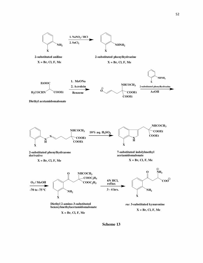
52
NH2
X
1. NaNO2 / HCl
2. SnCl2NHNH2
X
2-substituted aniline 2-substituted phenylhydrazine
NHNH2
X
2-substituted phenylhydrazine
H3COCHN
EtOOC
COOEt
1. MeONa
Benzene
2. Acrolein
O
NHCOCH3
COOEt
COOEt
AcOH
NH
X
N
NHCOCH3
COOEt
COOEt
2-substituted phenylhydrazonederivative
Diethyl acetamidomalonate
10% aq. H2SO4
NH
NHCOCH3
COOEt
COOEt
7-substituted indolylmethylacetamidomalonate
X
O3 / MeOH COOC2H5
COOC2H5
NHCOCH3
X
NH2
6N HCl,ref lux
ONH3
X
NH2
O
COO
rac 3-substituted kynurenine
-70 to -75°C
Diethyl 2-amino-3-substitutedbenzoylmethylacetamidomalonate
3 - 4 hrs.
Scheme 13
X = Br, Cl, F, Me X = Br, Cl, F, Me
X = Br, Cl, F, Me X = Br, Cl, F, Me
X = Br, Cl, F, MeX = Br, Cl, F, Me

53
Michael addition reaction3 between acrolein and the diethylacetamidomalonate anion. The
intermediate aldehyde of this reaction is not isolated and is used as such for its reaction with the
2-substituted phenylhydrazine to give the corresponding phenylhydrazone derivative. GCMS of
the substituted phenylhydrazones shows about 97% purity and are used as such for the next step
without further purification.
The phenylhydrazone derivative is then subjected to an acid catalyzed Fischer indole
cyclization to give the corresponding 7-substituted indolylmethylacetamidomalonates12-16 as
gummy semisolids. Purity check by GCMS show the 7-substituted
indolylmethylacetamidomalonates to be about 95% pure and are used as such for the next
reaction.
Ozonolysis of the 7-substituted indolylmethylacetamidomalonate intermediate in
methanol produces the diethyl 2-amino-3-substitutedbenzoylmethylacetamidomalonate
intermediate as either a brown oil or a semisolid which is crystallized from 2-propanol to give a
yellow solid. Finally, hydrolysis of the diethyl 2-amino-3-substituted
benzoylmethylacetamidomalonate intermediate with 6N HCl produces the corresponding 3-
substituted DL-kynurenine. The racemic compound is isolated from an aq. solution, as a pale
yellow solid by adjusting the pH to approximately 6.5. In the case of 3-fluoro kynurenine the
racemic compound was obtained as a brown solid.
Purity of the racemic 3-chloro (λmax at 262 nm and 365 nm), 3-fluoro (λmax at 258 nm and
360 nm), and the 3-methyl (λmax at 259 nm and 362 nm) kynurenines is about 99.5% by HPLC.
The retention times for these three compounds are 14.7 mins., 8.7 mins., and 13.5 mins.
respectively. The racemic 3-methylkynurenine when run on the Pro-Cu chiral column separated
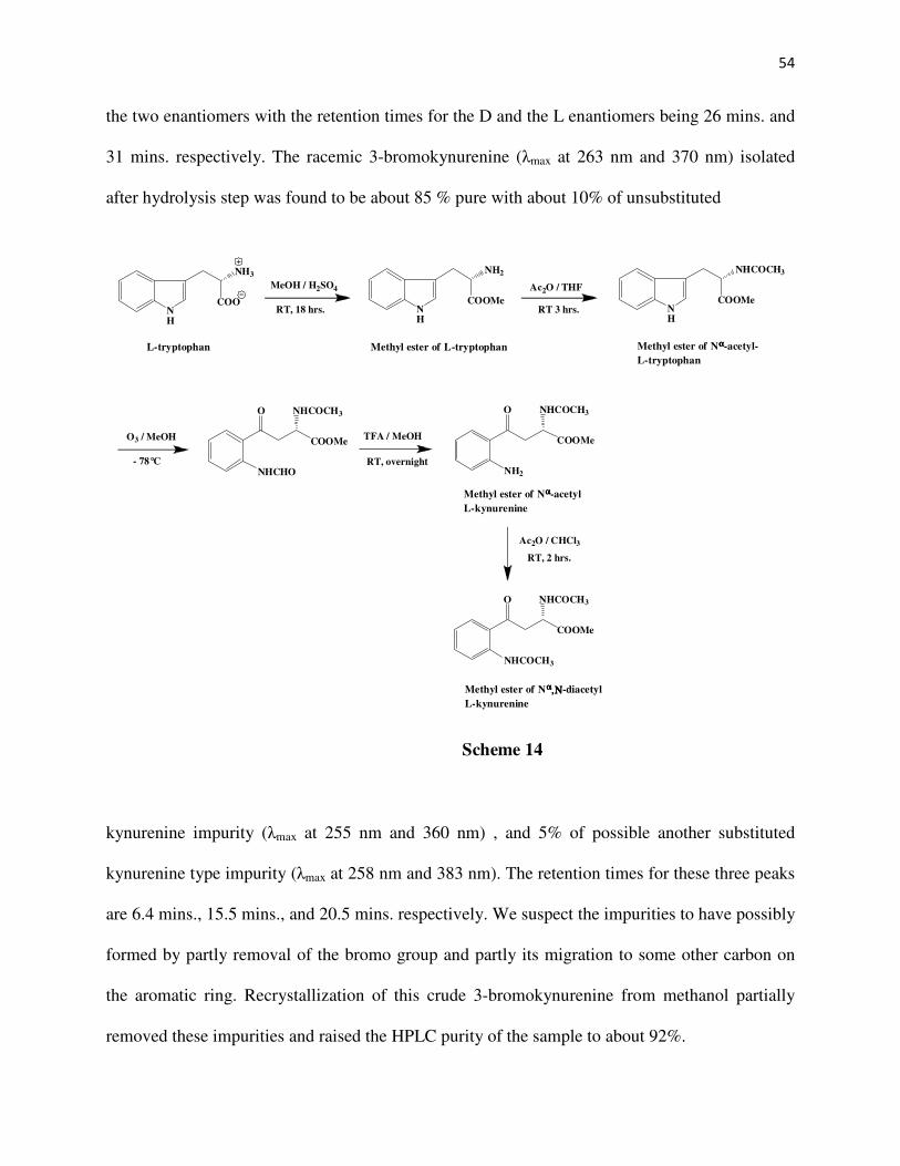
54
the two enantiomers with the retention times for the D and the L enantiomers being 26 mins. and
31 mins. respectively. The racemic 3-bromokynurenine (λmax at 263 nm and 370 nm) isolated
after hydrolysis step was found to be about 85 % pure with about 10% of unsubstituted
kynurenine impurity (λmax at 255 nm and 360 nm) , and 5% of possible another substituted
kynurenine type impurity (λmax at 258 nm and 383 nm). The retention times for these three peaks
are 6.4 mins., 15.5 mins., and 20.5 mins. respectively. We suspect the impurities to have possibly
formed by partly removal of the bromo group and partly its migration to some other carbon on
the aromatic ring. Recrystallization of this crude 3-bromokynurenine from methanol partially
removed these impurities and raised the HPLC purity of the sample to about 92%.
NH
NH2
COOMe
O3 / MeOH
- 78°C
COOMe
NHCHO
O NHCOCH3
TFA / MeOH
RT, overnight
COOMe
O NHCOCH3
NH2
Ac2O / CHCl3
RT, 2 hrs.
COOMe
O NHCOCH3
NHCOCH3
Methyl ester of L-tryptophan
NH
NH3
COONH
NHCOCH3
COOMe
L-tryptophan
MeOH / H2SO4
RT, 18 hrs.
Ac2O / THF
RT 3 hrs.
Methyl ester of Nαααα-acetyl-
L-tryptophan
Methyl ester of Nαααα-acetyl
L-kynurenine
Methyl ester of Nαααα,,,,ΝΝΝΝ-diacetyl
L-kynurenine
Scheme 14
55
For synthesizing the 5-bromo, and 5-chloro L-kynurenines, the intermediate methyl ester
of Nα,N-diacetylkynurenine is first synthesized (Scheme 14). Thus, Fischer esterification of L-
tryptophan with methanol in the presence of sulfuric acid gives the methyl ester of L-tryptophan,
which is about 99% pure by GCMS and used as such for the acylation step. Acylation with acetic
anhydride in THF produces the methyl ester of Nα-acetyl-L-tryptophan which is about 99% pure
by GCMS and used as such for the ozonolysis step. Ozonolysis of the methyl ester of Nα-acetyl-
L-tryptophan, followed by hydrolysis with trifluoroacetic acid, and then acylation with acetic
anhydride, gives the methyl ester of Nα,N-diacetylkynurenine17 as a white solid with 99% purity
by GCMS. Having prepared the methyl ester of Nα,N-diacetylkynurenine, either bromination or
chlorination gives the methyl ester of the corresponding 5-halosubstituted-Nα,N-
diacetylkynurenine which finally upon acid hydrolysis give the 5-halosubstituted-L-kynurenines.
(Scheme 15)
Bromination of the methyl ester of Nα,N-diacetylkynurenine is done with liquid
bromine, and gives regioselectively the methyl ester of 5-bromo-Nα,N-diacetylkynurenine. This
compound upon crystallization from methanol gives the product as pale yellow needles with
99.5% purity by GCMS. Finally hydrolysis of the 5-bromo intermediate with 6N HCl gives the
5-bromo-L-kynurenine as a pale yellow solid. The reported melting point for this solid is 213-
217 ºC17. However, we got a much higher melting point of 225-227 ºC which could be because
of the greater purity of our sample. The greater purity of our sample could be because of the
different isolation method used by us. The HPLC purity was found to be about 99.8%, with the
retention time being 14.4 mins. and λmax of 265 nm and 370 nm.
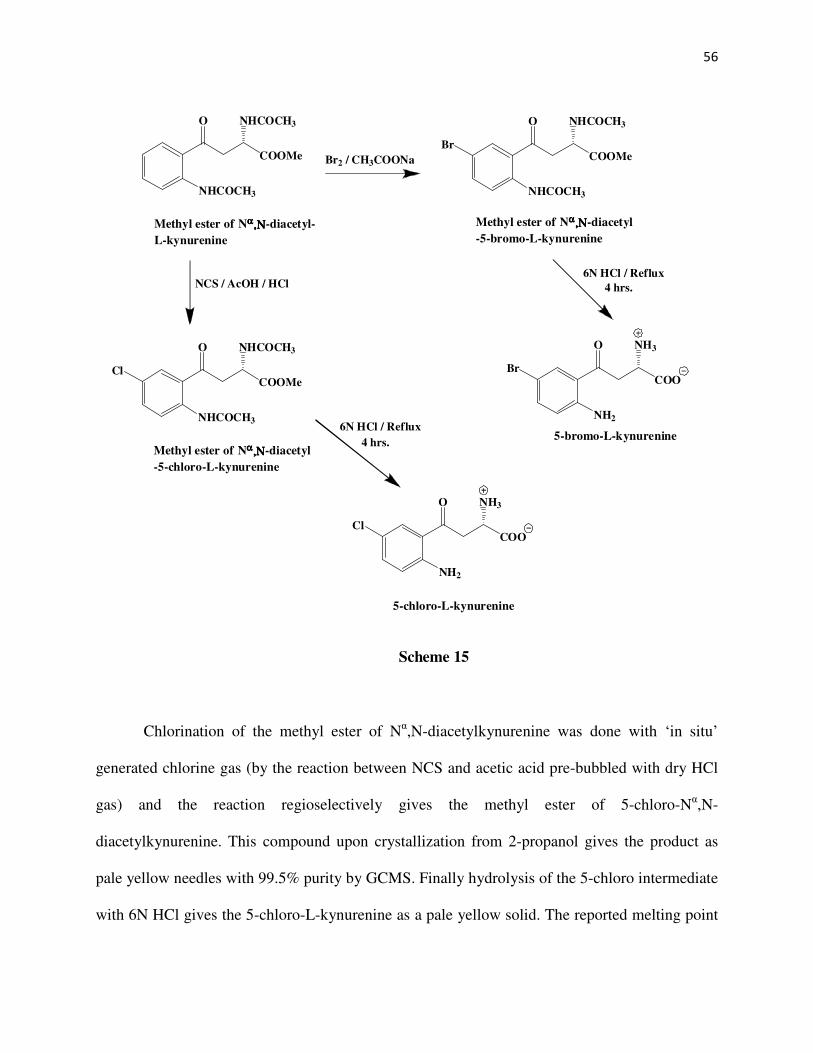
56
Chlorination of the methyl ester of Nα,N-diacetylkynurenine was done with ‘in situ’
generated chlorine gas (by the reaction between NCS and acetic acid pre-bubbled with dry HCl
gas) and the reaction regioselectively gives the methyl ester of 5-chloro-Nα,N-
diacetylkynurenine. This compound upon crystallization from 2-propanol gives the product as
pale yellow needles with 99.5% purity by GCMS. Finally hydrolysis of the 5-chloro intermediate
with 6N HCl gives the 5-chloro-L-kynurenine as a pale yellow solid. The reported melting point
COOMe
O NHCOCH3
NHCOCH3
Methyl ester of Nαααα,,,,ΝΝΝΝ-diacetyl-
L-kynurenine
Br2 / CH3COONa
NCS / AcOH / HCl
COOMe
O NHCOCH3
NHCOCH3
Methyl ester of Nαααα ,,,,ΝΝΝΝ-diacetyl
-5-bromo-L-kynurenine
COOMe
O NHCOCH3
NHCOCH3
Methyl ester of Nαααα ,,,,ΝΝΝΝ-diacetyl
-5-chloro-L-kynurenine
Cl
Br
6N HCl / Reflux
4 hrs.
6N HCl / Reflux
4 hrs.
COO
O NH3
NH2
Cl
5-chloro-L-kynurenine
COO
O NH3
NH2
Br
5-bromo-L-kynurenine
Scheme 15

57
for this solid is 208-211 ºC21. However, we got a much higher melting point of 216-218 ºC which
could be because of the greater purity of our sample. The HPLC purity was found to be about
99.8%, with the retention time being 12.8 mins. and λmax of 260 nm and 370 nm.

58
References
1. Furniss, B. S.; Hannaford, A.J.; Smith Peter, W. G.; Tatchell, A. R. in Vogel’s Textbook
of Practical Organic Chemistry, 5th ed. Longman Scientific & Technical, England, 1989,
pp 757-759.
2. Moe, O. A.; Warner, D. T. J. Am. Chem. Soc. 1948, 70, 2763-2765.
3. Warner, D. T.; Moe, O. A. J. Am. Chem. Soc. 1948, 70, 2765-2767.
4. Warner, D. T.; Moe, O. A. J. Am. Chem. Soc. 1948, 70, 3470-3472.
5. Stevens, F. J.; Higginbotham, D. H. J. Am. Chem. Soc. 1954, 76, 2206-2207.
6. Harden, F. A.; Quinn, R. J.; Scammells, P. J. J. Med. Chem. 1991, 34, 2892-2898.
7. Fugger, J.; Tien, J. M.; Hunsberger, I. M. J. Am. Chem. Soc. 1955, 77, 1843-8.
8. Tien, J. M.; Hunsberger, I. M. J. Am. Chem. Soc. 1955, 77, 6696-8.
9. Tien, J. M.; Hunsberger, I. M. J. Am. Chem. Soc. 1955, 77, 6604-7.
10. Tien, J. M.; Hunsberger, I. M. Chem. Ind. (London) 1955, 119-119.
11. Hunsberger, I. M.; Shaw, E. R.; Fugger, J.; Ketcham, R.; Lednicer, D. J. Org. Chem.
1956, 21, 394-399.
12. Thiruvikraman, S. V.; Sakagami, Y.; Katayama, M.; Marumo, S. Tetrahedron Lett. 1988,
29, 2339-2342.
13. Shiba, T.; Mukunoki, Y.; Akiyama, H. Bull. Chem. Soc. Jpn. 1975, 48, 1902-1906.
14. Vanpee, K. H.; Salcher, O.; Lingens, F. Liebigs Ann. Chem. 1981, 233-239.
15. Rydon, H. N.; Tweddle, J. C. J. Chem. Soc. 1955, 3499-3503.
16. Porter, J.; Dykert, J.; Rivier, J. Int. J. Pept. Protein Res. 1987, 30, 13-21.
17. Heiss, C.; Anderson, J.; Phillips, R. S. Org. Biomol. Chem. 2003, 1, 288-295.
18. Wang, Y.; Liu, Q. Pige Huagong 2002, 19, 23-27.

59
19. Bandgar, B. P.; Uppalla, L. S. J. Chem. Res.,Synop. 1999, 714-715.
20. Nagarajan, K.; Talwalker, P. K.; Kulkarni, C. L.; Venkateswarlu, A.; Prabhu, S. S.;
Nayak, G. V. Indian J. Chem. Sect. B 1984, 23, 1243-1257.
21. Varasi, M.; Della Torre, A.; Heidempergher, F.; Pevarello, P.; Speciale, C.; Guidetti, P.;
Wells, D. R.; Schwarcz, R. Eur. J. Med. Chem. 1996, 31, 11-21.
22. Pegurier, C.; Collart, P.; Danhaive, P.; Defays, S.; Gillard, M.; Gilson, F.; Kogej, T.;
Pasau, P.; Van Houtvin, N.; Van Thuyne, M.; van Keulen, B. Bioorg. Med. Chem. Lett.
2007, 17, 4228-4231.
23. Suschitzky, H. J. Chem. Soc. 1953, 3326-7.
24. Carlin, R. B.; Odioso, R. C. J. Am. Chem. Soc. 1954, 76, 100-104.
25. Bullock, M. W.; Hand, J. J. J. Am. Chem. Soc. 1956, 78, 5852-5854.
26. Bullock, M. W.; Hand, J. J. J. Am. Chem. Soc. 1956, 78, 5854-5857.
27. Lee, An-Rong; Huang, Wen-Hsin; Lin, Tung-Liang; Shih, Kun-Min; Lee, Hsiao-Feng;
Lin, Cheng-I J. Heterocycl. Chem. 1995, 1, 1-12.
28. Bloss; Timberlake J. Org. Chem. 1963, 28, 267-268.
29. Cook; France J. Am. Chem. Soc. 1934, 56, 2225
30. Burkhard.W; Kauffman.T Angew. Chem. Int. Ed. 1967, 6, 84.
31. Bajwa, G. S.; Brown, R. K. Can. J. Chem. 1968, 46, 1827.

60
CHAPTER 3
STEADY STATE KINETICS OF SUBSTRATE ANALOGS FOR HUMAN AND
BACTERIAL KYNURENINASE
Abstract
Different substituted kynurenines have been tested for their substrate activity with human
as well as Pseudomonas fluorescens kynureninase. All the synthesized compounds viz. the 3-
chloro-DL-kynurenine, 3-fluoro-DL-kynurenine, 3-methyl-DL-kynurenine, 5-bromo-L-
kynurenine, and the 5-chloro-L-kynurenine have good substrate activity for both human as well
as Pseudomonas fluorescens kynureninase. For the human enzyme, 3-chloro-DL-kynurenine, 5-
bromo-L- kynurenine, and the 5-chloro-L-kynurenine have closely comparable kcat and kcat/Km
value to that of the natural substrate 3-hydroxykynurenine. And for the bacterial enzyme, 3-
fluoro-DL-kynurenine, 5-bromo-L-kynurenine, and the 5-chloro-L-kynurenine have closely
comparable kcat and kcat/Km value to that of the natural substrate L-kynurenine. Thus, 5-bromo-
L-kynurenine, and the 5-chloro-L-kynurenine seem to be good substrates for both human as well
as bacterial enzyme.

61
Experimental Methods
General
The steady state kinetic measurements were performed on a Varian Cary 1E UV/Visible
spectrophotometer equipped with a Peltier-type 6 x 6 thermoelectric cell block for temperature
control. The instrument was controlled by a PC using software provided by Varian Instruments.
Enzyme assay
Kynureninase activity was measured from the decrease in absorbance at 360 nm (∆ε =
4500 M-1.cm-1)1 upon conversion of kynurenine to anthranilic acid. Similarly the human enzyme
activity was measured from the decrease in absorbance at 370 nm upon conversion of DL-3-
hydroxykynurenine to 3-hydroxyanthranilic acid. The reaction mixtures for these measurements
contained 100 µM of the substrate L-kynurenine or 3-hydroxy- DL-kynurenine in 30 mM of
potassium phosphate buffer pH 8, containing 40 µM of pyridoxal-5'-phosphate at 25 ºC. Kinetic
and scanning kinetic measurements of the L-compounds was done in a similar manner but for the
DL compounds a final concentration of 200 µM of each compound was used for the individual
assay, while keeping the other conditions same.
Kinetic measurements
The scanning kinetic measurements to determine the absorbance change for the 3-
substituted substrate analogs was done using the human enzyme since 3-hydroxykynurenine is
the natural substrate for the human enzyme. The scanning kinetic measurements to determine
the absorbance change for the 5-substituted substrate analogs was done using the Pseudomonas
fluorescens enzyme since 5-substitued kynurenines have been shown to be good substrates2 for

62
the P. fluorescens enzyme. The wavelength range used for the scanning kinetic measurements
was 450 nm – 220 nm and the scan was performed at the rate of 200 nm/min. As shown in Table
3.1 below, different quantities of the human and P. fluorescens enzyme were used for different
substrate analogs with the final volume of 600 µL for each assay solution. These same quantities
of enzyme were used for determining the initial rates of the respective substrate analogs with the
two different enzymes.
Table 3.1
Compound Human enzyme
mg
P. fluorescens enzyme
mg
3-chloro-DL-kynurenine 3.29 x 10⁻³ 1.33 x 10⁻³
3-fluoro-DL-kynurenine 6.58 x 10⁻³ 3.33 x 10⁻⁴
3-methyl-DL-kynurenine 6.58 x 10⁻³ 6.66 x 10⁻⁴
5-bromo-L-kynurenine 1.32 x 10⁻³ 3.33 x 10⁻⁵
5-chloro-L-kynurenine 2.63 x 10⁻³ 3.33 x 10⁻⁵
The initial rates of reaction of 3-chloro-DL-kynurenine were measured at its absorption
maximum, 365 nm (∆ε = 4648 M-1.cm-1). The initial rates of reaction of 3-fluoro-DL-kynurenine
were measured at its absorption maximum, 360 nm (∆ε = 3053 M-1.cm-1). The initial rates of
reaction of 3-methyl-DL-kynurenine were measured at its absorption maximum, 362 nm (∆ε =
3440 M-1.cm-1). The initial rates of reaction of 5-bromo-L-kynurenine were measured at its
absorption maximum, 370 nm (∆ε = 4006 M-1.cm-1). And the initial rates of reaction of 5-chloro-
L-kynurenine were measured at its absorption maximum, 370 nm (∆ε = 4330 M-1.cm-1). The Km,

63
Vmax, and Vmax/Km were determined by fitting of the initial rate data (See Appendix 1 for tables
of raw data) into the equation 1 using the compiled FORTRAN program HYPER of Cleland3 on
a Dell personal computer.
ν = Vmax [S]/(Km + [S]) (1)
The values of Vmax, and Vmax/Km were divided by the respective extinction coefficients and by
the enzyme concentration to give the kcat and kcat/Km values (Tables 3.2 & 3.3)
Results and discussion
A representative scan graph for 5-bromo-L-kynurenine is shown below in Fig. 10 (X-
axis shows the wavelength in nm, Y-axis shows the absorbance) which shows the λmax of 370 nm
for this compound. The other scan graphs are shown in the Appendix section. The reaction
shows clear isosbestic points at 245 nm, 258 nm, 280 nm, and 338 nm indicating that there are
only two species in the reaction viz. 5-bromo-L-kynurenine and the product, 5-bromoanthranilate
as shown below.
COO
NH3
NH2
Br
O
5-bromo-L-kynurenine
Kynureninase
L-alanine5-bromoanthranilate
NH2
Br COO
COO
NH3
+

64
As seen from tables 3.2 and 3.3 the human as well as the P. fluorescens enzyme have
about the same catalytic turnover number for 3-chloro-DL-kynurenine with kcat values of 0.67
sec-1 and 0.71 sec-1 respectively. When compared with the natural substrate (3-
hydroxykynurenine5) 3-chloro-DL-kynurenine is about 5-fold less efficiently cleaved by the
human enzyme but has a 3-fold higher kcat than L-kynurenine for the same enzyme. For the
bacterial enzyme it is found that 3-chloro-DL-kynurenine has a kcat value that is 22-fold lower
than the natural substrate, L-kynurenine, 17-fold lower than 5-bromo-L-kynurenine, 13-fold
lower than 5-chloro-L-kynurenine, and 15-fold lower than 3-bromo-L-kynurenine2 for the same
enzyme. Thus the turnover number with the 3-Cl substituent is much less affected with the
Fig. 10
-0.4
0.1
0.6
1.1
1.6
2.1
220 270 320 370 420

65
human enzyme than with the bacterial enzyme. This is consistent with the preference of the
human enzyme for 3-hydroxy-L-kynurenine.
The kcat/Km for 3-chloro-DL-kynurenine is also about the same i.e. (8.16 ± 1.34) x 103 and (1.05
± 0.075) x 104 for the human and the bacterial enzyme respectively. Considering no interference
by the D enantiomer the value of kcat/Km for 3-chloro-L-kynurenine would be (1.63 ± 0.27) x 104
and (2.1 ± 0.15) x 104 for the human and the bacterial enzyme, respectively. For the human
Table 3.2 Substrate analogs for human enzyme
enzyme this kcat/Km value of (1.63 ± 0.27) x 104 is about 15-fold lower that for the natural
substrate 3-hydroxy-L-kynurenine5 (2.46 x 10⁵) but about 35-fold higher than L-kynurenine for
Compound Km µM Vmax. kcat sec-1 kcat / Km M-1. sec-1
3-chloro-DL-kynurenine
83.4 ± 21 0.018 ± 0.0017 0.67 ± 0.063 (8.16 ± 1.34) x 103
3-fluoro-DL-kynurenine
85.8 ± 21.6 0.0080 ± 0.00079 0.23 ± 0.022 (2.66 ± 0.45) x 103
3-methyl-DL-kynurenine
179 ± 28.3 0.013 ± 0.0010 0.33 ± 0.025 (1.81 ± 0.15) x 103
5-bromo-L-kynurenine
42.2 ± 4.7 0.0058 ± 0.00020 0.63 ± 0.022 (1.51 ± 0.11) x 104
5-chloro-L-kynurenine
43.5 ± 6.3 0.0094 ± 0.00044 0.47 ± 0.022 (1.097 ± 0.12) x 104
L-kynurenine5 495 - 0.23 465
3-hydroxy-DL-kynurenine5 28.3 - 3.5 1.23 x 10⁵

66
the same enzyme. Thus, having a Cl substituent at the 3-position of L-kynurenine increases the
substrate activity for the human enzyme but not as much as that of the natural substrate. For the
bacterial enzyme the kcat/Km value of (2.1 ± 0.15) x 104 is about 29-fold lower than that of the
natural substrate L-kynurenine4, but about 85-fold lower than that of 5-chloro-L-kynurenine, and
a huge 156-fold lower than that of 5-bromo-L-kynurenine, and about 4-fold higher than that of 3-
bromo-L-kynurenine for the same enzyme. Thus, a Cl substituent at 3-position of L-kynurenine
Table 3.3 Substrate analogs for bacterial enzyme
seems to decrease the catalytic efficiency while a substituent at 5-position actually increases the
catalytic efficiency for the bacterial enzyme and even more so with the increased size of the
substituent at the 5-position. To conclude, 3-chloro-DL-kynurenine has about the same substrate
Compound Km µM Vmax. kcat sec-1 kcat / Km M-1. sec-1
3-chloro-DL-kynurenine
70.8 ± 9.2 0.0095 ± 0.00056 0.71 ± 0.042 (1.05 ± 0.075) x 104
3-fluoro-DL-kynurenine
75.5 ± 11.2 0.015 ± 0.0010 6.88 ± 0.46 (8.99 ± 0.78) x 104
3-methyl-DL-kynurenine
69.4 ± 14.8 0.0074 ± 0.00057 1.51 ± 0.12 (2.24 ± 0.33) x 104
5-bromo-L-kynurenine
3.58 ± 1.11 0.0034 ± 0.000198 11.89 ± 0.69 (3.29 ± 0.87) x 106
5-chloro-L-kynurenine
4.90 ± 0.96 0.0027 ± 0.00012 8.73 ± 0.39 (1.78 ± 0.29) x 106
L-kynurenine 25 - 16 6 x 10⁵

67
activity for both the human as well as the bacterial enzyme but not better than the natural
substrate for each of these enzymes.
In the case of 3-fluoro-DL-kynurenine the kcat for the bacterial enzyme is about 34-fold
greater (6.88 sec-1) than that for the human enzyme (0.23 sec-1). For the human enzyme this kcat
value of 0.23 sec-1 is about the same as that of L-kynurenine but about 15-fold lower than that of
the natural substrate 3-hydroxy-L-kynurenine5. Thus, the human enzyme seems to be very
specific about the type of substituent at the 3-position, a larger and polar substituent being the
best. On the other hand, for the bacterial enzyme the kcat value of 6.88 sec-1 is about half that of
the natural substrate L-kynurenine4 but about the same as that of 5-bromo, 3-bromo, and 5-
chloro-L-kynurenines. Thus, the catalytic turnover number of this substrate with the bacterial
enzyme does not seem to be affected as much as with the human enzyme by the presence of a 3-
F substituent. The kcat/Km values for 3-fluoro-DL-kynurenine are (2.66 ± 0.45) x 103 and (8.99 ±
0.78) x 104 respectively for the human and bacterial enzyme. Assuming that the D enantiomer
does not affect the enzyme activity the value of kcat/Km for 3-fluoro-L-kynurenine would be (5.32
± 0.9) x 103 and (1.80 ± 0.16) x 105 for the human and the bacterial enzyme respectively.
Apparently, the kcat/Km value for this substrate is about 34-fold higher with the bacterial enzyme
than with the human enzyme. For the human enzyme this kcat/Km value of (5.32 ± 0.9) x 103 is
about 46-fold lower that for the natural substrate 3-hydroxy-L-kynurenine5 (2.46 x 10⁵) but about
11-fold higher than L-kynurenine for the same enzyme.5 Thus, based on the discussion in the
previous paragraph, it can be said that the human enzyme prefers a 3-Cl over 3-F, both of which
are obviously better than having no substituent on the aromatic ring, but at the same time both of
them are not as good as having a 3-OH substituent because of the capability of the –OH group to
undergo H-bonding as indicated by the crystal structure studies5. For the bacterial enzyme,
68
however, the kcat/Km value of (1.80 ± 0.16) x 105 is just about 3-fold lower that of the natural
substrate L-kynurenine4 (6 x 10⁵) but about 18-fold lower than that of 5-bromo-L-kynurenine,
10-fold lower than that of 5-chloro-L-kynurenine and actually about 33-fold higher than that of
3-bromo-L-kynurenine2. Thus, based on the previous discussion, it can be said that unlike the
human enzyme, the catalytic efficiency of the bacterial enzyme is not affected much by
introducing a 3-F substituent but this catalytic efficiency is not better than having a 5-Br or 5-Cl
substituent although it is better than having a 3-Br substituent. Thus, only a small substituent
such as H or F is optimal for bacterial kynureninase, which is consistent with our recently
proposed model of substrate specificity5.
In the case of 3-methyl-DL-kynurenine, the turnover number with the bacterial enzyme is
5-fold better than with the human enzyme, as seen from the kcat values of 0.33 sec-1 and 1.51 sec-1
for the human and bacterial enzyme, respectively. For the human enzyme, this kcat value of 0.33
sec-1 is about the same as that of L-kynurenine but about 11-fold lower than that of the natural
substrate 3-hydroxy-L-kynurenine5. Thus, based on the previous discussion, it seems like the
turnover ability of the human enzyme is better when having a 3-methyl rather than a 3-F, but not
as good as having a 3-Cl though all three have a lower turnover number than the natural
substrate3-hydroxkynurenine5. On the other hand, with the bacterial enzyme the kcat value of
1.51 sec-1 is about 11-fold lower than that of the natural substrate L-kynurenine4 and about 8-fold
lower that of 5-bromo-L- & 3-bromo-L-kynurenine2, and about 5-fold lower than that of 3-
fluoro-DL- & 5-chloro-L- kynurenines. Thus the turnover number of the bacterial enzyme is less
affected with having a 3-Me than having a 3-Cl, although both of these lower the turnover
number than for the substrate with a 3-F substituent. Considering the kcat/Km values for this
substrate we found these to be (1.81 ± 0.15) x 103 and (2.24 ± 0.33) x 104 for the human and the

69
bacterial enzyme respectively. Thus, the catalytic efficiency of the bacterial enzyme is about 12-
fold better than the human enzyme for this substrate. Assuming that the D enantiomer does not
affect the enzyme activity the value of kcat/Km for 3-methyl-L-kynurenine would be (3.62 ± 0.3)
x 103 and (4.48 ± 0.65) x 104 for the human and the bacterial enzyme, respectively. For the
human enzyme, this kcat/Km value of (3.62 ± 0.3) x 103 is about 68-fold lower that for the natural
substrate 3-hydroxy-L-kynurenine5 (2.46 x 10⁵) but about 8-fold higher than L-kynurenine for
the same enzyme.5 Thus, the catalytic efficiency of the human enzyme is reduced to a greater
extent by having a 3-methyl substituent rather than having a 3-Cl or a 3-F substituent, all of the
three being better than having no substituent on the aromatic ring of kynurenine but not as good
as having a 3-OH substituent, probably because the 3-OH forms an H-bond with Asn-333.5 For
the bacterial enzyme the kcat/Km value of (4.48 ± 0.65) x 104 is about 14-fold lower than that of
the natural substrate L-kynurenine4 (6 x 10⁵) but about 73-fold lower than that of 5-bromo-L-
kynurenine, 40-fold lower than that of 5-chloro-L-kynurenine and actually about 8-fold higher
than that of 3-bromo-L-kynurenine2. Thus, the catalytic efficiency of the bacterial enzyme is
better when having a 3-methyl rather than a 3-Cl, both of them not better than having 3-F, 5-Br
or 5-Cl substituent.
Considering the activity of 5-bromo-L-kynurenine, we have found the kcat values for this
substrate to be 0.63 sec-1 and 11.9 sec-1 for the human and bacterial enzyme respectively.
Although Heiss et al2 have reported a kcat value of 2.1 sec-1 for the bacterial enzyme we got about
6-fold higher kcat of 11.9 sec-1. This could most probably be due to the greater purity of our
substrate as indicated by its much higher melting point than the one reported previously2. For the
human enzyme this kcat value of 0.63 sec-1 is about the 3-fold higher than that of L-kynurenine
but about 6-fold lower than that of the natural substrate 3-hydroxkynurenine5. Thus the catalytic
70
turnover number of the human enzyme is almost equal when having a 5-Br or a 3-Cl substituent
but lowered to a greater extent when having a 3-F or 3-methyl substituent although all of the four
being less active than the natural substrate 3-hydroxykynurenine. With the bacterial enzyme the
kcat value of 11.9 sec-1 is about the same as that of 3-bromo-L-kynurenine2, 5-chloro-L-
kynurenine and the natural substrate L-kynurenine4 but about 8-fold higher than 3-methyl and
16-fold higher than 3-Cl. Thus, the turnover number of the bacterial enzyme does not seem to
lowered as much as when having a 3-methyl or a 3-Cl substituent. Considering the catalytic
efficiency of this substrate for both enzymes we got the kcat/Km values of (1.51 ± 0.11) x 104 and
(3.29 ± 0.87) x 106 for the human and the bacterial enzyme respectively. For the bacterial
enzyme, we got an 18-fold higher kcat/Km value than the value of 1.8 x 10⁵ reported earlier2
which could again be due to the greater purity of our substrate and the enzyme sample. For the
human enzyme this kcat/Km value of (1.51 ± 0.11) x 104 is about 16-fold lower than for the
natural substrate 3-hydroxy-L-kynurenine5 (2.46 x 10⁵) but about 32-fold higher than L-
kynurenine for the same enzyme.5 Thus, the catalytic efficiency with the human enzyme is about
the same when having a 5-Br or a 3-Cl substituent which is better than having a 3-methyl or a 3-
fluoro substituent but all four are not better than having a 3-OH substituent. For the bacterial
enzyme the kcat/Km value of (3.29 ± 0.87) x 106 is about 6-fold higher than that of the natural
substrate L-kynurenine4 (6 x 10⁵), 600-fold higher than that of 3-bromo-L-kynurenine, and about
twice that of 5-chloro-L-kynurenine. Thus, the catalytic efficiency for the bacterial enzyme is
improved by introducing a 5-Br or 5-Cl on the aromatic ring of kynurenine, and these
substituents are hugely better than having a 3-Br substituent.
For our newly synthesized substrate 5-chloro-L-kynurenine the kcat values are 0.47 sec-1
and 8.73 sec-1for the human and the bacterial enzyme respectively. Thus, the turnover number

71
for this substrate with the bacterial enzyme is about 19-fold higher than with the human enzyme.
For the human enzyme this kcat of 0.47 sec-1 is about the same as that of 5-bromo-L-kynurenine
but about twice that of L-kynurenine and actually 7-fold lower than that of the natural substrate
3-hydroxy-L-kynurenine5. Thus, having a 5-Cl substituent has a similar effect on the turnover
number as having a 5-Br or a 3-Cl substituent, and this turnover number is higher than when
having a 3-F or a 3-methyl substituent, but all the five compounds are not better than having a 3-
OH substituent as in the natural substrate. With the bacterial enzyme, the kcat value of 8.73 sec-1
is about the same as that for 3-F, 3-Br or 5-Br substituent, and about half that of the natural
substrate L-kynurenine4 (16 sec-1), but based on the previous discussion this turnover number is
better than when having a 3-Cl or a 3-methyl substituent. Considering the catalytic efficiency
with this substrate we have got kcat/Km values of (1.097 ± 0.12) x 104 and (1.78 ± 0.29) x 106 for
the human and the bacterial enzyme, respectively. For the human enzyme, this kcat/Km value of
(1.10 ± 0.12) x 104 is about 22-fold lower that for the natural substrate 3-hydroxy-L-kynurenine5
(2.46 x 10⁵) but about 24-fold higher than L-kynurenine for the same enzyme.5 Thus, based on
this and the previous discussion the catalytic efficiency of the human enzyme with 5-chloro-L-
kynurenine is lesser than when having a 3-Cl or a 5-Br substituent but better than when having a
3-F or a 3-methyl substituent although all five being not as better as having a 3-OH substituent.
For the bacterial enzyme the kcat/Km value of (1.78 ± 0.29) x 106 is about 3-fold higher than that
of the natural substrate L-kynurenine4 (6 x 10⁵), 324-fold higher than that of 3-bromo-L-
kynurenine, and about half that of 5-bromo-L-kynurenine. Based on this and the previous
discussion a 5-Br or 5-Cl actually has a positive effect on the catalytic efficiency of the bacterial
enzyme, but with the exception of 3-F, the other 3-substituents in the group seem to have a
negative effect on the catalytic efficiency of the enzyme.
72
To conclude, these results are useful in drug design. Thus, substituents in the 5-position
are well tolerated by both enzymes, which is consistent with the X-ray structures5. Furthermore,
a hydroxyl group at 3-position is not absolutely necessary for good activity, as seen for the
human enzyme that the halogens in place of hydroxyl group also work fine.

73
References
1. Kishore, G. M. J. Biol. Chem. 1984, 259, 10669-74.
2. Heiss, C.; Anderson, J.; Phillips, R. S. Org. Biomol. Chem. 2003, 1, 288-295.
3. Cleland, W. W. Meth. Enzymol. 1979, 63, 103-38.
4. Gawandi, V. B.; Liskey, D.; Lima, S.; Phillips, R. S. Biochemistry 2004, 43, 3230-3237.
5. Lima, S.; Kumar, S.; Gawandi, V.; Momany, C.; Phillips, R. S. J. Med. Chem. 2009, 52,
389-396.
74
CHAPTER 4
SYNTHESIS AND STABILITY STUDIES OF CAGED KYNURENINE
Abstract
Kynureninase or L-kynurenine hydrolase, EC 3.7.1.3 is a pyridoxal-5'-phosphate (PLP)
dependent enzyme catalyzing the hydrolytic cleavage of kynurenine to anthranilic acid and L-
alanine. This is the key step in the catabolism of tryptophan in Pseudomonas fluorescens, and
some other bacteria. In eukaryotes a similar enzyme catalyzes the hydrolytic cleavage of 3-
hydroxykynurenine to 3-hydroxyanthranilic acid and L-alanine. Earlier, the mechanism for the
hydrolytic cleavage of kynurenine has been proposed1. The external aldimine and quinonoid
intermediate are formed too rapidly within the dead time of the stopped-flow instrument. We
therefore synthesized a novel caged kynurenine which would release kynurenine ‘in situ’ thereby
allowing the detection of formation and decay of the external aldimine intermediate. This chapter
provides a detailed synthesis of the caged compound, along with its stability studies.

75
Introduction
Caged biomolecules have been known for about three decades now2,3 and have become
increasingly important by virtue of the light initiated release of the biologically active molecule.
In the broader sense, caged compounds are protected photo-labile bioactive substrates that
release the desired substrate upon irradiation with light of a suitable wavelength (> 300 nm).
Some of the caged compounds include protected phosphates, carboxylates, amines, alcohols, and
phenols. Caged compounds find extensive application in investigating molecular processes in
biochemistry and biophysics. Some bioorganic reactions are too rapid to allow for detection of
the intermediate or measure the rate of such reactions. However, by using a caged biomolecule
the bioactive substrate can be released ‘in situ’ only when desired by means of a light flash for a
few nanoseconds duration. Caged compounds thus allow chemists, biologists, and molecular
physiologists to examine the rates of even the fastest biological reactions known. At the same
time, light initiated substrate release also gives good insight into the mechanism of enzyme
catalyzed biological reactions.
Caging chromophores need to satisfy several key properties or attributes4. Among these
are a reasonable absorption in the uv-visible region (> 300nm). This is important as most of the
enzymes are destroyed at shorter wavelengths. Also, there should be a hypsochromic shift of the
absorption spectrum due to the photoproduct so that this absorption does not interfere with the
absorptions of any intermediates in the biological processes being studied. It should be easy to
attach the chromophore to the substrate, and without the introduction of any new stereocenters.
The caged compound as well as the photoproduct should be biologically inert, and should also be
inert or at least benign with respect to other reagents or products. Furthermore, the caged
compound should have good aqueous solubility for biological studies. Also, the photochemical

76
release must be efficient, and the departure of the substrate from the protecting group should be
the primary photochemical process occurring directly from the excited state of the caged
chromophore. Apart from all these the caging chromophore should preserve the chiral integrity
of the substrate (i.e. not convert the substrate into a racemate or other enantiomer) during the
protection or the photolytic deprotection cycle. Some of the chromophores used for caging
bioactive substrates include, 2-nitrobenzyl5-10 and 7-nitroindoline11 derivatives, coumarin-4-
ylmethyl12-19 phototriggers, and the p-hydroxyphenacyl group20-24. Among these the p-
hydroxyphenacyl group is a versatile photoremovable protective group with wide range of
applications in mechanistic bioorganic chemistry. However, this group can only be used to
protect conjugate bases of acids4 such as carboxylic acids, thiols, and phosphates.
In the catabolism of tryptophan in Pseudomonas fluorescens, and some other bacteria,
anthranilic acid and alanine are produced by the hydrolytic cleavage of kynurenine in the
presence of the enzyme kynureninase. The mechanism of the cleavage has been proved by
Phillips and Dua1 and proceeds through the initial formation of an external aldimine and
quinonoid intermediate as shown in Scheme 16. However, the reaction involving the formation
of these intermediates is too rapid for the rate to be measured, and is over within the dead time of
the stopped-flow instrument (ca. 2 milliseconds). Often detection of an intermediate is essential
in proving the mechanism that an enzyme follows. In our attempt to prepare a caged kynurenine,
we hope to release the substrate kynurenine ‘in situ’ which would then undergo the reaction with
kynureninase thereby allowing us to detect the external aldimine intermediate. Future experiment
would involve obtaining a structure of kynureninase “in action” with kynurenine bound in the
active site. For this experiment the caged substrate will be soaked into the crystals of either
human or Pseudomonas fluorescens kynureninase, then the reaction initiated by flash photolysis

77
followed by rapid cooling in a cold nitrogen stream, and immediate collection of the X-ray
diffraction data. Since kynureninase is a drug target enzyme, these structures will be invaluable
in the design of the next generation of more potent and selective inhibitors of kynureninase.
O COO
NH3
Kynureninase.PLP
External aldimine
(λλλλmax = 420 nm)
NH2
O COO
NH
NH2
N
HO
OPO3
H
B
Kynurenine quinonoid intermediate
(λλλλmax = 494 nm)
O COO
NH
NH2
NH
HO
OPO3
H
Kynurenine
Scheme 16

78
Experimental Methods
General
1HNMR and 13CNMR spectra were recorded on a Varian 400MHz instrument in
deuterated DMSO. HPLC measurements were carried out on a Spectrasystem P 2000 instrument
connected to a UV 6000 detector and controlled by a Dell PC using Chromquest software. A
gradient elution was used consisting of 5 % MeOH, and 95 % 0.1 % aq. acetic acid from 0 – 5
mins. followed by a programmed increase of MeOH percentage from 5% to 70% over 5 – 20
mins. with a corresponding decrease of the percentage of 0.1 % aq. acetic acid from 95% to 30%
over the same time period. This is followed by an increase of MeOH percentage to 100% with
the corresponding decrease of the percentage of 0.1% aq. acetic acid to 0% over 20 – 25 mins.
And finally, a programmed return back of the elution system to 5% MeOH, and 95% of 0.1% aq.
acetic acid over the period from 25 – 30 mins. A 100 µM solution of the caged compound in 1
mM HCl was used for injection. The flow rate for the elution was 1 ml/min. with detection by
absorbance at 254 nm and 370 nm.
Synthesis of 2-bromo-4'-hydroxyacetophenone
To a solution of 5 g of 4-hydroxyacetophenone in 50 ml methanol, add portion wise 30 g copper
(II) bromide while maintaining temperature of the reaction mixture below 25ºC. After
completion of addition, stir the reaction mass between 20 -25ºC, for 4 hrs. Check TLC. (Fig. 10)
Concentrate the methanol under vacuum below 25 ºC. Add 100 ml d/w to the residual semisolid,
extract with two 50 ml portions of EtOAc. Wash the combined EtOAc layers first with two 50 ml
satd. NaHCO3 solution, then with two 50 ml water, dry the EtOAc layer over anhydrous sodium
sulfate, and concentrate under vacuum. Take the resulting semisolid in about 25 ml toluene. Stir

79
the suspension at 5 -10 ºC for about 15 minutes, filter the product,
and wash with 15 ml of chilled toluene. Allow to air dry overnight.
Yield = 7.5 g, 96 %, m.p. = 124 -127 ºC
1HNMR: d-MeOH δ 7.81 (d, 2H), 6.78 (d, 2H), 4.42 (s, 2H)
13CNMR: d-MeOH δ 35.3, 111.2, 126.3, 131.2, 159.3, 195.6
Synthesis of 4-hydroxyphenacyl ester of Nα-Boc-L-tryptophan
Add 5 g of p-hydroxyphenacyl bromide to a solution containing 3.5 gm of K2CO3, and
7.1 g of Boc-tryptophan, in 50 ml dry DMF. Stir the resulting suspension at RT for about 3 hrs.
Check TLC (Fig.11). For checking the TLC quench a small portion
of the RM in water, extract with a few drops of EtOAc, and spot the
EtOAc layer. Add 500 ml water to the RM, extract with two 100 ml
portions of EtOAc. Dry the combined organic layer over anhydrous
sodium sulfate; concentrate under vacuum to give 12 gm of pale
yellow oil. Take the oil in about 75 ml of toluene, scratch the inner
walls of the flask to induce crystallization. Chill the resulting
suspension in an ice-water bath for about 10 mins. Filter, and wash
the product with about 35 ml of chilled toluene.
Yield = 10 g, 98 %, m.p = 195 – 198 ºC
1HNMR: CDCl3 δ 7.85 (d, 2H), 7.75 (d, 1H), 7.54 (d, 1H), 7.32 (t, 1H), 7.25 (t, 1H), 7.1 (d, 2H),
7.05 (s, 1H), 5.62 (s, 2H), 4.57 (m, 1H), 3.57 (dd, 2H), 2.95 (s, 9H)
SM
System: Toluene : EtOAc8 : 2
Detection: uv 254 nm
SM = starting materialCo = mixture spot
Co RM
Fig. 10
SM
System: CHCl3 : MeOH9 : 1
Detection: uv 254 nmSM = Starting materialCo = Mixture spot
Co RM
Fig. 11
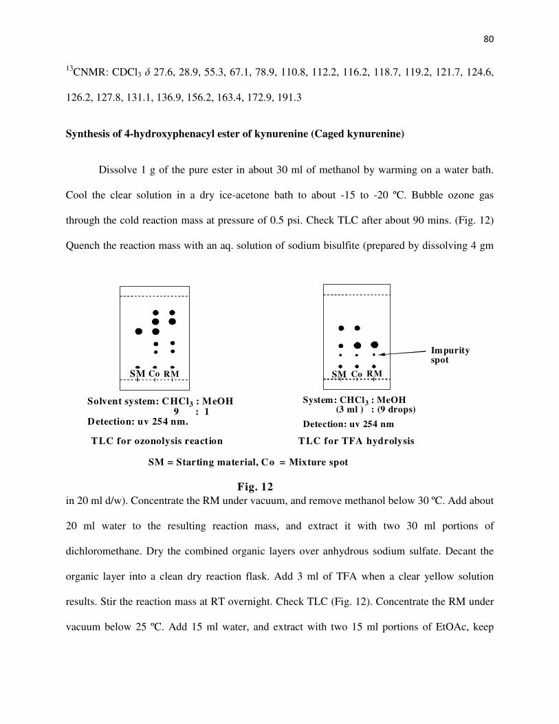
80
13CNMR: CDCl3 δ 27.6, 28.9, 55.3, 67.1, 78.9, 110.8, 112.2, 116.2, 118.7, 119.2, 121.7, 124.6,
126.2, 127.8, 131.1, 136.9, 156.2, 163.4, 172.9, 191.3
Synthesis of 4-hydroxyphenacyl ester of kynurenine (Caged kynurenine)
Dissolve 1 g of the pure ester in about 30 ml of methanol by warming on a water bath.
Cool the clear solution in a dry ice-acetone bath to about -15 to -20 ºC. Bubble ozone gas
through the cold reaction mass at pressure of 0.5 psi. Check TLC after about 90 mins. (Fig. 12)
Quench the reaction mass with an aq. solution of sodium bisulfite (prepared by dissolving 4 gm
in 20 ml d/w). Concentrate the RM under vacuum, and remove methanol below 30 ºC. Add about
20 ml water to the resulting reaction mass, and extract it with two 30 ml portions of
dichloromethane. Dry the combined organic layers over anhydrous sodium sulfate. Decant the
organic layer into a clean dry reaction flask. Add 3 ml of TFA when a clear yellow solution
results. Stir the reaction mass at RT overnight. Check TLC (Fig. 12). Concentrate the RM under
vacuum below 25 ºC. Add 15 ml water, and extract with two 15 ml portions of EtOAc, keep
SM
Solvent system: CHCl3 : MeOH9 : 1
Detection: uv 254 nm.
Co RM
Fig. 12
TLC for ozonolysis reaction
SM
Impurityspot
System: CHCl3 : MeOH(3 ml ) : (9 drops)
Detection: uv 254 nm
Co RM
SM = Starting material, Co = Mixture spot
TLC for TFA hydrolysis

81
aside the aqueous layer. Wash the combined organic layer with 15 ml of approx. 2N HCl,
separate the aqueous layer, and charcoalize it with a pinch of activated charcoal, for about 5
mins. at RT, filter through celite, lyophilize the aqueous layer.
Yield: 0.2 g, 26 %
1HNMR: d-MeOH δ 7.82 (d, 2H), 7.75 (d, 1H), 7.25 (t, 1H), 6.92 (d, 2H), 6.89 (d, 2H), 6.58 (t,
1H), 5.52 (d, 2H), 4.51 (t, 1H), 3.73 (dd, 2H),
13CNMR: d-MeOH δ 38.3, 48.2, 68.1, 115.7, 119.7, 120.6, 121.2, 124.9, 131.1, 131.6, 136,
142.8, 162.1, 169.8, 194.2, 198.5
[α]D = -14.2º (c = 1.3 in water)
HRMS analysis MW = 342.1294, Calculated MW = 342.1216
Results and Discussion
In order to synthesize the caged compound, 2-bromo-4'-hydroxyacetophenone is first
synthesized from 4-hydroxyacetophenone (Scheme 17). The published procedures25-28 for the
synthesis of 2-bromo-4'-hydroxyacetophenone use a variety of reagents like NBS, liquid
bromine, pyridinium hydrobromide perbromide, and copper (II) bromide, with yields of the
product ranging from 58-94%. We however, have an even more efficient procedure (96%)
without the formation of the undesirable side products viz. the 2,2-dibromo-4'-
hydroxyacetophenone compound. The aromatic ring in our method is also less likely to be
brominated due the presence of the deactivating acetyl group as well as the fact that the
bromonium electrophile is weak enough and even more so because we carry out the reaction

82
below 25ºC. The protocol using CuBr2 in a refluxing mixture of CHCl3/EtOAc4 was especially
giving us a mixture of the mono and the dibromo compounds. Using our method the reaction was
done conveniently in MeOH at RT using copper (II) bromide as the brominating agent. An SN2
reaction between the conjugate base of Nα-Boc tryptophan and 2-bromo-4'-hydroxyacetophenone
in a polar aprotic solvent like DMF is carried out to give the 4-hydroxyphenacyl ester of Nα-Boc
tryptophan. This ester upon ozonolysis followed by TFA hydrolysis29 produces the desired caged
kynurenine. The caged compound is isolated by lyophilization as a pale yellow hygroscopic solid
hydrochloride salt.
A photo-Favorskii rearrangement has been proposed for the release of the substrates from
caged biomolecules30,31. According to the mechanism proposed by Givens et al (Scheme 18)
initially the chromophore gets excited to the singlet excited state, followed by a rapid intersystem
crossing4 (kST = 2.7 x 10¹¹ s-1) to quantitatively generate the excited triplet state. In the excited
triplet state the phenolic group then adiabatically loses a proton to the solvent to give phenoxide
anion which then releases the substrate in a rate-limiting step, via the intermediacy of the
spirodienedione. The caged kynurenine has a retention time of about 18 mins. using the above
mentioned gradient elution system. HPLC analysis shows the compound to be about 99% pure.
HRMS analysis shows the molecular weight of 342.1294 with the calculated value being
342.1216. The uv absorption pattern shows three distinct λmax values at 263, 283 and 365 nm.

83
O
HO
4-hydroxy acetophenone
O
HO
Br
2-bromo-4′′′′-hydroxyacetophenone
CuBr2 / MeOH
RT 4 hrs. DMF / K2CO3
RT 3 hrs.
NH
NHCOOtBu
O
OHO
O
NHCHO
O
OH
O
O
NHCOOtBu O
O3 / MeOH
-15°C to -20°C
NH2
O
OH
O
O
NH2 O
TFA / MDCRT overnight
(Caged kynurenine)
NH
COOH
Nαααα-Boc-L-tryptophan
4-hydroxyphenacyl ester of
Nαααα-Boc-L-tryptophan
4-hydroxyphenacyl ester of
Nαααα-Boc-N-formylkynurenine
4-hydroxyphenacylester of kynurenine
Scheme 17
NHCOOtBu
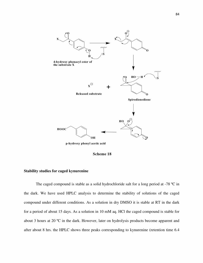
84
Stability studies for caged kynurenine
The caged compound is stable as a solid hydrochloride salt for a long period at -78 ºC in
the dark. We have used HPLC analysis to determine the stability of solutions of the caged
compound under different conditions. As a solution in dry DMSO it is stable at RT in the dark
for a period of about 15 days. As a solution in 10 mM aq. HCl the caged compound is stable for
about 3 hours at 20 ºC in the dark. However, later on hydrolysis products become apparent and
after about 8 hrs. the HPLC shows three peaks corresponding to kynurenine (retention time 6.4
X
O
O
X
O
O
Released substrate
+
O
O
Spirodienedione
HHO S
HO
O
O
HOOC
OH
p-hydroxy phenyl acetic acid
4-hydroxy phenacyl ester ofthe substrate X
SH
Scheme 18
X

85
mins.), 2,4'-dihydroxyacetophenone (retention time 15 mins., λmax 273 nm), and the caged
compound in an approximate ratio of 2:3:5. Based on a similar stability in 1 mM aq. HCl
solution, we irradiated a 100µM aq. acidic solution of the caged compound with a Xenon flash
lamp using a 330 nm filter to check if the photocleavage is initiated. However, HPLC analysis
showed no cleavage of the cage.
The caged compound is found to be extremely unstable in pH 8 phosphate and TEA
buffers and gets almost completely hydrolyzed in about 15 mins. The half life for the caged
compound in these buffer systems was 3.75 mins. and 4.65 mins. respectively. We were
expecting the stability of the caged compound and hence its half life to grow 10-fold in going
from pH 8 to pH 7 and 100-fold in going from pH 8 to pH 6. However, the half life of the caged
compound in pH 7 and pH 6 phosphate buffer was found to be 9.75 mins. and 17.1mins.
respectively. Thus, the stability did not raise considerably in going from pH 8 to pH 6 buffer
system. Nevertheless, the caged compound is stable for up to 3 hrs. in a 1 mM aq. HCl (pH 3)
solution. We then checked the stability of the caged compound in pH 7 Tris buffer containing 55
mM MgCl2 and 25% w/v polyethylene glycol (PEG), the solution used for crystallization of
human kynureninase. We have found that the caged compound is fairly stable in this buffer
system for about 30 mins. but later on hydrolysis products are seen in the HPLC analysis. Based
on this stability we irradiated a 200µM solution of the caged compound with one hundred 1ns
laser pulses and found by HPLC that the cage has been removed completely. In future
experiments this would be the buffer system of choice to study the reaction involving the release
of kynurenine in situ. We were however not able to perform the experiment due to instrumental
problems wherein the enzyme fluorescence skewed the observance of the desired intermediates.

86
References
1. Dua, R. K.; Taylor, E. W.; Phillips, R. S. J. Am. Chem. Soc. 1993, 115, 1264-
1270.
2. Kaplan, J. H.; Forbush, B.; Hoffman, J. F. Biochemistry 1978, 17, 1929-1935.
3. Engels, J.; Schlaeger, E. J. J. Med. Chem. 1977, 20, 907-911.
4. Givens, R. S.; Yousef, A. L. in Dynamic Studies in Biology Goeldner, Maurice;
Givens, R. S. Ed., Wiley-VCH, 2005.
5. Lester, H. A.; Nerbonne, J. M. Annu. Rev. Biophys. Bioeng. 1982, 11, 151-175.
6. Rothman, D. M.; Vazquez, E. M.; Vogel, E. M.; Imperiali, B. Org. Lett. 2002, 4,
2865-2868.
7. Dinkel, C.; Wichmann, O.; Schultz, C. Tetrahedron Lett. 2003, 44, 1153-1155.
8. Shigeri, Y.; Tatsu, Y.; Yumoto, N. Pharmacol. Therapeutics 2001, 91, 85-92.
9. Marriott, G.; Roy, P.; Jacobson, K. In Biophotonics, Pt A 2003; Vol. 360, p 274-
288.
10. Brubaker, M. J.; Dyer, D. H.; Stoddard, B.; Koshland, D. E. Biochemistry 1996,
35, 2854-2864.
11. Canepari, M.; Nelson, L.; Papageorgiou, G.; Corrie, J. E. T.; Ogden, D. J.
Neurosci. Methods 2001, 112, 29-42.
12. Furuta, T.; Torigai, H.; Osawa, T.; Iwamura, M. Chem. Lett. 1993, 1179-1182.
13. Takaoka, K.; Tatsu, Y.; Yumoto, N.; Nakajima, T.; Shimamoto, K. Bioorg. Med.
Chem. Lett. 2003, 13, 965-970.
14. Schoenleber, R. O.; Giese, B. Synlett 2003, 501-504.
15. Suzuki, A. Z.; Watanabe, T.; Kawamoto, M.; Nishiyama, K.; Yamashita, H.; Ishii,
M.; Iwamura, M.; Furuta, T. Org. Lett. 2003, 5, 4867-4870.
87
16. Kaupp, U. B.; Solzin, J.; Hildebrand, E.; Brown, J. E.; Helbig, A.; Hagen, V.;
Beyermann, M.; Pampaloni, F.; Weyand, I. Nat. Cell Biol. 2003, 5, 109-117.
17. Furuta, T.; Takeuchi, H.; Isozaki, M.; Takahashi, Y.; Kanehara, M.; Sugimoto,
M.; Watanabe, T.; Noguchi, K.; Dore, T. M.; Kurahashi, T.; Iwamura, M.; Tsien,
R. Y. Chembiochem 2004, 5, 1119-1128.
18. Nishigaki, T.; Wood, C. D.; Tatsu, Y.; Yumoto, N.; Furuta, T.; Elias, D.; Shiba,
K.; Baba, S. A.; Darszon, A. Dev. Biol. 2004, 272, 376-388.
19. Hagen, V.; Frings, S.; Wiesner, B.; Helm, S.; Kaupp, U. B.; Bendig, J.
Chembiochem 2003, 4, 434-442.
20. Givens, R. S.; Park, C. H. Tetrahedron Lett. 1996, 37, 6259-6262.
21. Givens, R. S.; Weber, J. F. W.; Conrad, P. G.; Orosz, G.; Donahue, S. L.; Thayer,
S. A. J. Am. Chem. Soc. 2000, 122, 2687-2697.
22. Evanko, D. S.; Sul, J.-Y.; Zhang, Q.; Haydon, P. G. Glial & Neuronal Signaling
2004, 397-416.
23. Specht, A.; Loudwig, S.; Peng, L.; Goeldner, M. Tetrahedron Lett. 2002, 43,
8947-8950.
24. Geibel, S.; Barth, A.; Amslinger, S.; Jung, A. H.; Burzik, C.; Clarke, R. J.;
Givens, R. S.; Fendler, K. Biophys. J. 2000, 79, 1346-1357.
25. Gupta, R.; Gupta, M.; Paul, S.; Gupta, R.; Loupy, A. Lett. Org. Chem. 2008, 5,
153-157.
26. Shang, G.; Liu, D.; Allen, S. E.; Yang, Q.; Zhang, X. Chem. Eur. J. 2007, 13,
7780-7784.

88
27. Shen, Y.; Sheng, R.; Zhang, J.; He, Q.; Yang, B.; Hu, Y. Bioorg. Med. Chem.
2008, 16, 7646-7653.
28. Ueda, S.; Fujita, M.; Tamamura, H.; Fujii, N.; Otaka, A. Chembiochem 2005, 6,
1983-1986.
29. Heiss, C.; Anderson, J.; Phillips, R. S. Org. Biomol. Chem. 2003, 1, 288-295.
30. Conrad, P. G.; Givens, R. S.; Hellrung, B.; Rajesh, C. S.; Ramseier, M.; Wirz, J.
J. Am. Chem. Soc. 2000, 122, 9346-9347.
31. Givens, R. S.; Lee, J.-I. J. Photosci. 2003, 10, 37-48.

89
CHAPTER 5
SUMMARY AND CONCLUSIONS
In conclusion, we have prepared some novel substrates and determined their activity for
human as well as Pseudomonas fluorescens kynureninase. We synthesized the 3-chloro-DL-
kynurenine, 3-fluoro-DL-kynurenine, 3-methyl-DL-kynurenine, 5-bromo-L-kynurenine, and 5-
chloro-L-kynurenine and compared the turnover number as well as the catalytic efficiency of
each of these substrates as compared to L-kynurenine and 3-hydroxykynurenine for both the
human and the bacterial enzyme. Thus, for the human enzyme a comparison of the turnover
number for each of these substrates with the natural substrate 3-hydroxykynurenine, shows that
all of them have a lower turnover number than the natural substrate and the turnover number
decreases in the order: 3-OH > 3-Cl > 5-Br > 5-Cl > 3-Me > 3-F. And for the human enzyme a
comparison of the turnover number for each of these substrates with L-kynurenine shows the
turnover number to decrease in the order: 5-Br = 3-Cl > 5-Cl > 3-F = 3-Me = L-kynurenine.
Thus, for the human enzyme having a substituent on the aromatic ring of kynurenine increases
the turnover number but not as much as that of the 3-OH substituent. For the bacterial enzyme a
comparison of the turnover number for each of these substrates with the natural substrate L-
kynurenine the turnover number decreases in the order: L-kynurenine = 3-Br = 5-Br > 3-F = 5-Cl
> 3-Me > 3-Cl. Thus, the turnover number with the bacterial enzyme seems to be unaffected by
the presence of a larger substituent but other substituents have a reduced turnover number than
the natural substrate. From the foregoing discussion it can be concluded that while the bacterial
enzyme seems to be bothered by the presence of a substituent other than 3-Br or 5-Br, the human
enzyme on the other hand prefers to have a substituent on the aromatic ring, as seen by an
improved turnover number than when there is no substituent (L-kynurenine).

90
For the human enzyme a comparison of the catalytic efficiency for each of these
substrates with the natural substrate 3-hydroxykynurenine, shows that the catalytic efficiency is
much lower than that for the natural substrate and it decreases in the order: 3-OH > 3-Cl = 5-Br >
5-Cl > 3-F > 3-Me. Thus, for the human enzyme having a substituent other than –OH seems to
actually reduce the catalytic efficiency of the enzyme for that substrate as compared to the
natural substrate. For the human enzyme a comparison of the catalytic efficiency for each of
these substrates with L-kynurenine shows the decreasing order to be: 5-Br = 3-Cl > 5-Cl > 3-F >
3-Me > L-kynurenine. Thus, for the human enzyme having a substituent on the aromatic ring
actually increases the catalytic efficiency than when there is no substituent (L-kynurenine) but
this improvement in catalytic efficiency is not better than having a 3-OH substituent as in the
natural substrate, presumably because these other groups are not capable of H-bonding like –OH,
which has been recently proved from the crystal structure of the enzyme (Lima. S. et al 2009).
For the bacterial enzyme a comparison of the catalytic efficiency for each of these substrates
with the natural substrate L-kynurenine shows the decreasing order to be: 5-Br > 5-Cl > L-
kynurenine ~ 3-F > 3-Me > 3-Cl > 3-Br. Thus, the catalytic efficiency of the bacterial enzyme is
actually better than the natural substrate when there is a 5-Br or a 5-Cl substituent, but the
catalytic efficiency is lowered by the other substituents. And from the foregoing discussion it can
be concluded that while the catalytic efficiency of the bacterial enzyme seems to be bothered by
the presence of a substituent other than 5-Br or 5-Cl, the human enzyme on the other hand
actually prefers to have a substituent on the aromatic ring, as seen by an improved catalytic
efficiency than when there is no substituent (L-kynurenine).
Also, to conclude we have synthesized a novel caged kynurenine which in future will be
used for the generation of kynurenine ‘in situ’ by a flash photolysis thereby allowing the

91
detection of formation and decay of the external aldimine intermediate in the reaction of
kynureninase. This detection will further prove the mechanism of action followed by the enzyme
kynureninase.

92
APPENDIX
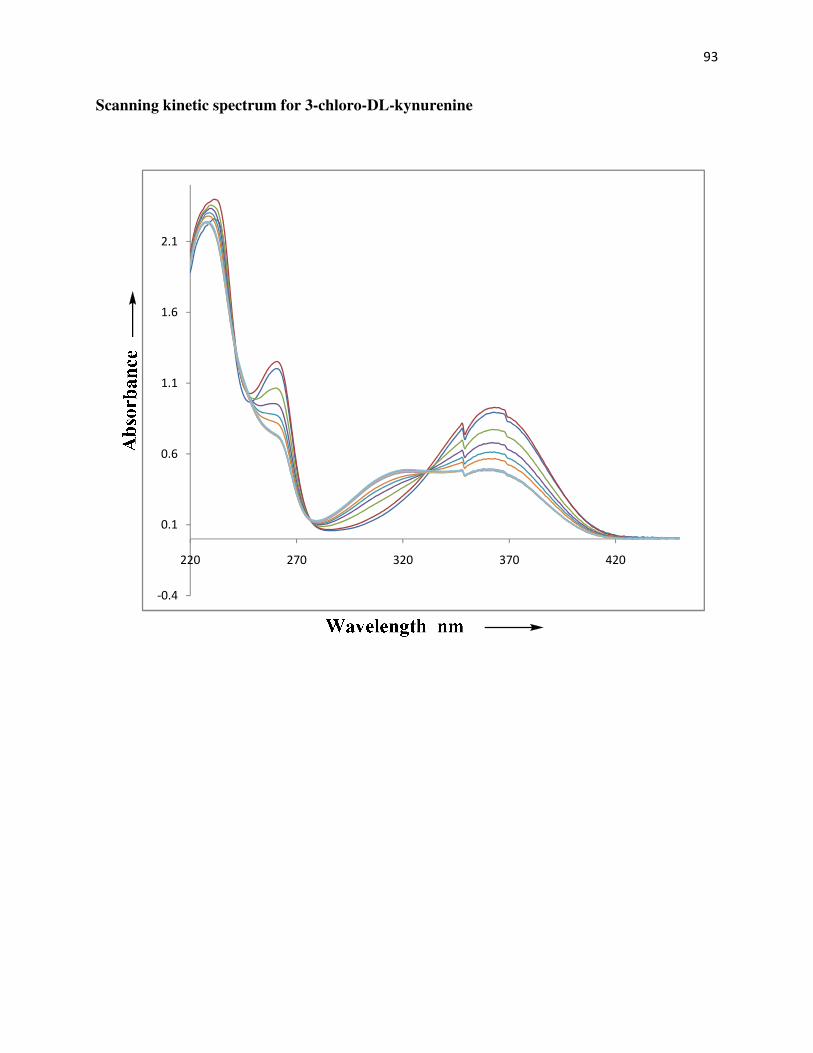
93
Scanning kinetic spectrum for 3-chloro-DL-kynurenine
-0.4
0.1
0.6
1.1
1.6
2.1
220 270 320 370 420

94
Scanning kinetic spectrum for 3-fluoro-DL-kynurenine
-0.4
0.1
0.6
1.1
1.6
2.1
220 270 320 370 420
Scanning kinetic spectrum for 3
for 3-methyl-DL-kynurenine
95

96
Scanning kinetic spectrum for 5-chloro-L-kynurenine
-0.4
0.1
0.6
1.1
1.6
2.1
220 270 320 370 420
97
Rate kinetics data for 3-chloro-DL-kynurenine with human enzyme
Notes:
1. Stock solution of human enzyme used was about 7.9 mg/ml by assay. 5µL of this solution
was diluted to 600µL using ionized water and used the above mentioned volumes of this
enzyme solution for the assay.
2. Wavelength used: 365nm, Cycle repeated every minute for 60 mins., Block temp.: 37ºC
Sr. No.
2mM PLP
( µL)
1M pH8 KPi
( µL)
3mM 3-Cl-DL- KYNU
( µL)
Enzyme
Solution
( µL)
Ionized water
( µL)
Final concn.
of 3-Cl-DL-
KYNU
( µM)
Final Vol. of assay soln.
( µL)
Rate set 1
Rate set 2
1
12
18
4
50
516
20
600
-0.0026
-0.0027
2
12
18
8
50
512
40
600
-0.0048
-0.0054
3
12
18
16
50
504
80
600
-0.0100
-0.0092
4
12
18
32
50
488
160
600
-0.0117
-0.0139
5
12
18
64
50
456
320
600
-0.0123
-0.0146

98
Rate kinetics data for 3-fluoro-DL-kynurenine with human enzyme
Notes:
1. Stock solution of human enzyme used was about 7.9 mg/ml by assay. 5µL of this solution
was diluted to 600µL using ionized water and used the above mentioned volumes of this
enzyme solution for the assay.
2. Wavelength used: 360nm, Cycle repeated every 2 mins. for 120 mins., Block temp.: 37ºC
Sr. No.
2mM PLP
( µL)
1M pH8 KPi
( µL)
3mM 3-F-DL- KYNU
( µL)
Enzyme
Solution
( µL)
Ionized water
( µL)
Final concn. of 3-F-DL- KYNU
( µM)
Final Vol. of assay soln.
( µL)
Rate set 1
Rate set 2
1
12
18
4
100
466
20
600
-0.0012
-0.0012
2
12
18
8
100
462
40
600
-0.0026
-0.0019
3
12
18
16
100
454
80
600
-0.0045
-0.0037
4
12
18
32
100
438
160
600
-0.0062
-0.0054
5
12
18
64
100
406
320
600
-0.0062
-0.0057

99
Rate kinetics data for 3-methyl-DL-kynurenine with human enzyme
Notes:
1. Stock solution of human enzyme used was about 7.9 mg/ml by assay. 5µL of this solution
was diluted to 600µL using ionized water and used the above mentioned volumes of this
enzyme solution for the assay.
2. Wavelength used: 362nm, Cycle repeated every 1.5 mins. for 90 mins., Block temp: 37ºC
Sr. No.
2mM PLP
( µL)
1M pH8 KPi
( µL)
3mM 3-Me-DL- KYNU
( µL)
Enzyme
Solution
( µL)
Ionized water
( µL)
Final concn. of
3-Me-DL-
KYNU
( µM)
Final Vol. of assay soln.
( µL)
Rate set 1
Rate set 2
1
12
18
4
100
466
20
600
-0.0014
-0.0015
2
12
18
8
100
462
40
600
-0.0024
-0.0019
3
12
18
16
100
454
80
600
-0.0044
-0.0039
4
12
18
32
100
438
160
600
-0.0064
-0.0056
5
12
18
64
100
406
320
600
-0.0079
-0.0087

100
Rate kinetics data for 5-bromo-L-kynurenine with human enzyme
Notes:
1. Stock solution of human enzyme used was about 7.9 mg/ml by assay. 5µL of this solution
was diluted to 600µL using ionized water and used the above mentioned volumes of this
enzyme solution for the assay.
2. Wavelength used: 370 nm, Cycle repeated every0.5 mins. for 30 mins., Block temp: 37ºC
Sr. No.
2mM PLP
( µL)
1M pH8 KPi
( µL)
3mM 5-Br-L-
KYNU
( µL)
Enzyme
Solution
( µL)
Ionized water
( µL)
Final concn.
of 5-Br-L-
KYNU
( µM)
Final Vol. of assay soln.
( µL)
Rate set 1
Rate set 2
1
12
18
2
20
548
10
600
-0.0013
-0.0013
2
12
18
4
20
546
20
600
-0.0019
-0.0018
3
12
18
8
20
542
40
600
-0.0028
-0.0026
4
12
18
16
20
534
80
600
-0.0039
-0.0038
5
12
18
32
20
518
160
600
-0.0041
-0.0046
6
12
18
64
20
486
320
600
-0.0050
-0.0055

101
Rate kinetics data for 5-chloro-L-kynurenine with human enzyme
Notes:
1. Stock solution of human enzyme used was about 7.9 mg/ml by assay. 5µL of this solution
was diluted to 600µL using ionized water and used the above mentioned volumes of this
enzyme solution for the assay.
2. Wavelength used: 370 nm, Cycle repeated every0.5 mins. for 30 mins., Block temp: 37ºC
Sr. No.
2mM PLP
( µL)
1M pH8 KPi
( µL)
3mM 5-Cl-L-
KYNU
( µL)
Enzyme
Solution
( µL)
Ionized water
( µL)
Final concn.
of 5-Cl-L-
KYNU
( µM)
Final Vol. of assay soln.
( µL)
Rate set 1
Rate set 2
1
12
18
2
40
528
10
600
-0.0018
-0.0014
2
12
18
4
40
526
20
600
-0.0031
-0.0028
3
12
18
8
40
522
40
600
-0.0048
-0.0043
4
12
18
16
40
514
80
600
-0.0064
-0.0056
5
12
18
32
40
498
160
600
-0.0083
-0.0068
6
12
18
64
40
466
320
600
-0.0086
-0.0076

102
Rate kinetics data for 3-chloro-DL-kynurenine with bacterial enzyme
Notes:
1. Stock solution of bacterial enzyme used was about 19 mg/ml by assay. 2.1µL of this
solution was diluted to 600µL using ionized water. A further ten times dilution of this
solution was done and used the above mentioned volumes of this enzyme solution for the
assay.
2. Wavelength used: 365nm, Cycle repeated every minute for 60 mins., Block temp.: 37ºC
Sr. No.
2mM PLP
( µL)
1M pH8 KPi
( µL)
3mM 3-Cl-DL-
KYNU
( µL)
Enzyme
Solution
( µL)
Ionized water
( µL)
Final concn.
of 3-Cl-DL-
KYNU
( µM)
Final Vol. of
assay soln.
( µL)
Rate set 1
Rate set 2
1
12
18
4
20
546
20
600
-0.0023
-0.0023
2
12
18
8
20
542
40
600
-0.0031
-0.0035
3
12
18
16
20
534
80
600
-0.0049
-0.0051
4
12
18
32
20
518
160
600
-0.0069
-0.0064
5
12
18
64
20
486
320
600
-0.0060
-0.0053

103
Rate kinetics data for 3-fluoro-DL-kynurenine with bacterial enzyme
Notes:
1. Stock solution of bacterial enzyme used was about 19 mg/ml by assay. 2.1µL of this
solution was diluted to 600µL using ionized water. A further ten times dilution of this
solution was done and used the above mentioned volumes of this enzyme solution for the
assay.
2. Wavelength used: 360nm, Cycle repeated every minute for 60 mins., Block temp.: 37ºC
Sr. No.
2mM PLP
( µL)
1M pH8 KPi
( µL)
3mM 3-F-DL-
KYNU
( µL)
Enzyme
Solution
( µL)
Ionized water
( µL)
Final concn. of 3-F-DL- KYNU
( µM)
Final Vol. of assay soln.
( µL)
Rate set 1
Rate set 2
1
12
18
4
50
518
10
600
-0.0015
-0.0016
2
12
18
8
50
516
20
600
-0.0030
-0.0028
3
12
18
16
50
512
40
600
-0.0047
-0.0053
4
12
18
32
50
504
80
600
-0.0082
-0.0081
5
12
18
64
50
488
160
600
-0.0103
-0.0093

104
Rate kinetics data for 3-methyl-DL-kynurenine with bacterial enzyme
Notes:
1. Stock solution of bacterial enzyme used was about 19 mg/ml by assay. 2.1µL of this
solution was diluted to 600µL using ionized water and used the above mentioned
volumes of this enzyme solution for the assay.
2. Wavelength used: 362nm, Cycle repeated every minute for 60 mins., Block temp: 37ºC
Sr. No.
2mM PLP
( µL)
1M pH8 KPi
( µL)
3mM 3-Me-DL- KYNU
( µL)
Enzyme
Solution
( µL)
Ionized water
( µL)
Final concn. of
3-Me-DL-
KYNU
( µM)
Final Vol. of assay soln.
( µL)
Rate set 1
Rate set 2
1
12
18
4
10
556
20
600
-0.0016
-0.0015
2
12
18
8
10
552
40
600
-0.0025
-0.0027
3
12
18
16
10
544
80
600
-0.0041
-0.0042
4
12
18
32
10
528
160
600
-0.0059
-0.0046
5
12
18
64
10
496
320
600
-0.0066
-0.0054

105
Rate kinetics data for 5-bromo-L-kynurenine with bacterial enzyme
Notes:
1. Stock solution of bacterial enzyme used was about 19 mg/ml by assay. 2.1µL of this
solution was diluted to 600µL using ionized water. A further hundred times dilution of
this solution was done and used the above mentioned volume of this enzyme solution for
the assay.
2. Wavelength used: 370 nm, Cycle repeated every 0.33 mins. for 30 mins., Block temp:
37ºC
Sr. No.
2mM PLP
( µL)
1M pH8 KPi
( µL)
3mM 5-Br-L-
KYNU
( µL)
Enzyme
Solution
( µL)
Ionized water
( µL)
Final concn.
of 5-Br-L-
KYNU
( µM)
Final Vol. of assay soln.
( µL)
Rate set 1
Rate set 2
1
12
18
1
50
519
5
600
-0.0022
-0.0017
2
12
18
2
50
518
10
600
-0.0024
-0.0025
3
12
18
4
50
516
20
600
-0.0026
-0.0035
4
12
18
8
50
512
40
600
-0.0030
-0.0028
5
12
18
16
50
504
80
600
-0.0031
-0.0035

106
Rate kinetics data for 5-chloro-L-kynurenine with bacterial enzyme
Notes:
1. Stock solution of bacterial enzyme used was about 19 mg/ml by assay. 2.1µL of this
solution was diluted to 600µL using ionized water. A further hundred times dilution of
this solution was done and used the above mentioned volume of this enzyme solution for
the assay.
2. Wavelength used: 370 nm, Cycle repeated every 0.33 mins. for 30 mins., Block temp:
37ºC.
Sr. No.
2mM PLP
( µL)
1M pH8 KPi
( µL)
3mM 5-Cl-L-
KYNU
( µL)
Enzyme
Solution
( µL)
Ionized water
( µL)
Final concn.
of 5-Cl-L-
KYNU
( µM)
Final Vol. of assay soln.
( µL)
Rate set 1
Rate set 2
1
12
18
1
50
519
5
600
-0.0013
-0.0014
2
12
18
2
50
518
10
600
-0.0016
-0.0020
3
12
18
4
50
516
20
600
-0.0021
-0.0021
4
12
18
8
50
512
40
600
-0.0026
-0.0026
5
12
18
16
50
504
80
600
-0.0023
-0.0025