syncope - rochester, ny€¦ · diagnosis & evaluation test/procedure yield (based on mean time...
TRANSCRIPT

Syncope
A Diagnostic and
Treatment Strategy
Kevin McGrody MD, FACC
November 5, 2015

Syncope Presentation Overview
I. Prevalence & Impact
II. Etiology
III. Diagnosis & Evaluation
IV. Treatment

Prevalence & Impact
More than 1 million patients in the U.S.1
More than 500,000 new patients per year1
1-6% of admissions2,3,4
3% of emergency room visits per year3
1 National Disease and Therapeutic Index on Syncope and Collapse, ICD-9-CM 780.2, IMS America, 1997
2 Blanc J-J, L’her C, Touiza A, et al. Eur Heart J, 2002; 23: 815-820.
3 Day SC, et al, AM J of Med 1982
4 Kapoor W. Evaluation and outcome of patients with syncope. Medicine 1990;69:160-175

Individuals <18 yrs : 15%
Military Population 17- 46 yrs : 20-25%
Individuals 40-59 yrs* : 16-19%
Individuals >70 yrs* : 23%
Prevalence & Impact
*during a 10-year period
Brignole M, Alboni P, Benditt DG, et al. Eur Heart J, 2001; 22: 1256-1306.

Cause Prevalence (Mean) % Prevalence (Range) %
Reflex-mediated:
Vasovagal 18 8-37
Situational 5 1-8
Carotid Sinus 1 0-4
Orthostatic hypotension 8 4-10
Medications 3 1-7
Psychiatric 2 1-7
Neurological 10 3-32
Organic Heart Disease 4 1-8
Cardiac Arrhythmias 14 4-38
Unknown 34 13-41
Etiology
Kapoor W. In Grubb B, Olshansky B (eds) Syncope: Mechanisms and Management. Armonk NY; Futura Publishing Co, Inc: 1998; 1-13.

Etiology
Bariatric surgery • �As bariatric surgery becomes an increasingly popular treatment for
obesity, we have seen an increasing number of patients present after bariatric surgery with new-onset syncope, near-syncope, and lightheadedness.
• Several case series have shown increased symptoms after significant weight loss (mean 55 kg loss)
Pacing Clin Electrophysiol. 2008 Jul;31(7):884-8. doi: 10.1111/j.1540-8159.2008.01103.x. New-onset orthostatic intolerance following bariatric surgery. Billakanty SR, Kligman MD, Kanjwal YM, Kosinski DJ, Maly GT, Karabin B, Grubb BP.

Diagnosis & Evaluation
Primary Evaluation: Determine whether the patient is at increased risk of
death
Identify risk of:
• Underlying structural heart disease
• Myocardial ischemia
• Wolff-Parkinson-White Syndrome
• Genetic disease such as the LQTS, Brugada syndrome,
catecholaminergic polymorphic ventricular tachycardia, ARVD
If none of the above risk then goal is to identify cause and improve
quality of life

Diagnosis & Evaluation
Detailed History & Physical • Document details of events • Assess frequency, severity • Obtain careful family history
Heart disease present? • Physical exam • ECG: long QT, WPW, conduction system disease • Echo: LV function, valve status, HOCM
Follow a diagnostic plan...

Diagnosis & Evaluation
Test/Procedure Yield
(based on mean time to diagnosis of 5.1
months7
History and Physical
(including carotid sinus massage)
49-85% 1, 2
ECG 2-11% 2
Electrophysiology Study without SHD* 11% 3
Electrophysiology Study with SHD 49% 3
Tilt Table Test (without SHD) 11-87% 4, 5
Ambulatory ECG Monitors:
Holter 2% 7
External Loop Recorder
(2-3 weeks duration)
20% 7
Insertable Loop Recorder
(up to 14 months duration)
65-88% 6, 7
Neurological †
(Head CT Scan, Carotid Doppler)
0-4% 4,5,8,9,10
* Structural Heart Disease † MRI not studied
1 Kapoor, et al N Eng J Med, 1983. 2 Kapoor, Am J Med, 1991. 3 Linzer, et al. Ann Int. Med, 1997. 4 Kapoor, Medicine, 1990.
5 Kapoor, JAMA, 1992 6 Krahn, Circulation, 1995 7 Krahn, Cardiology Clinics, 1997. 8 Eagle K,, et al. The Yale J Biol and Medicine. 1983; 56: 1-8.
9 Day S, et al. Am J Med. 1982; 73: 15-23. 10 Stetson P, et al. PACE. 1999; 22 (part II): 782.

Diagnosis & Evaluation
12 Lead ECG
• Normal or Abnormal?
• Acute MI
• Severe Sinus Bradycardia/pause
• AV Block
• Tachyarrhythmia (SVT, VT)
• Preexcitation (WPW), Long QT, Brugada, ARVD
ECHO
• Valvular heart disease
• Pulmonary embolism pulmonary hypertension
• HOCM
• Decreased LV function

Diagnosis & Evaluation
Ischemia evaluation
• If history and physical suggest coronary artery disease then ischemic
work up would be reasonable
• Exercise testing with echocardiographic imaging to determine exercise
related arrhythmia’s and underlying heart disease
11

Diagnosis & Evaluation
Head-up Tilt Test • Unmasks VVS susceptibility • Reproduces symptoms • Patient learns VVS warning
symptoms • Physician is better able to give
prognostic / treatment advice

Diagnosis & Evaluation
Patient history and physical exam
Tilt table test (ACC Consensus Protocol) • Overnight fast • ECG • Blood pressure • Supine and upright • Tilt to 70 degrees 20 min • SL NTG or isoproterenol • Re-tilt 15 min
DG Benditt, Tilt Table Testing, 1996.
60° - 80°

Diagnosis & Evaluation
Conventional EP Testing in Syncope
• Limited utility in syncope evaluation
• Most useful in patients with structural heart disease
• Heart disease……..50-80%
• No Heart disease…18-50%
• Relatively ineffective for assessing bradyarrhythmias
• Essentially we are looking for ventricular arrhythmias and whether the patient would need an ICD
Brignole M, Alboni P, Benditt DG, et al. Eur Heart Journal 2001; 22: 1256-1306.
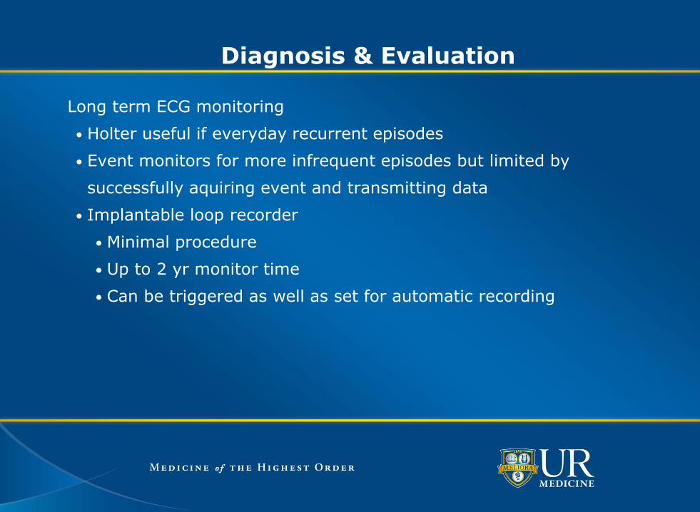
Diagnosis & Evaluation
Long term ECG monitoring
• Holter useful if everyday recurrent episodes
• Event monitors for more infrequent episodes but limited by
successfully aquiring event and transmitting data
• Implantable loop recorder
• Minimal procedure
• Up to 2 yr monitor time
• Can be triggered as well as set for automatic recording

Diagnosis & Evaluation

Treatment
Recognize life threatening causes of syncope
• Structural heart disease
• Corrective surgery (valvular)
• Physical limitations (HOCM)
• Predisposition to malignant fatal arrhythmia
• Antiarrhythmic therapy
• Device therapy
• Ischemic heart disease and revascularization

Treatment
Education
• symptom recognition
• reassurance
• situation avoidance
Tilt-Training
• prescribed upright posture
Pharmacologic Agents
• salt/volume management
• beta-adrenergic blockers
• SSRIs
• vasoconstrictors (e.g., midodrine)
Cardiac Pacemakers

Treatment
VVS: Tilt-Training • Objectives
• Enhance Orthostatic Tolerance • Diminish Excessive Autonomic Reflex Activity • Reduce Syncope Susceptibility / Recurrences
• Technique • Prescribed Periods of Upright Posture • Progressive Increased Duration

Treatment
Tilt-Training: Clinical Outcomes
42 HUT positive (21±13 min) VVS patients
Home training: two 30 minute sessions daily
Outcomes
• 41/42 pts --->45 min asymptomatic HUT
• Clinical follow-up: 15.1±7.8 mos
• 36 pts syncope free
• 4 pts: presyncope
• 1 pt: syncope recurrences
Reybrouck et al. PACE 2000; 23:493-8

Treatment
Pharmacologic Rx
• Salt /Volume
• Salt tablets, ‘sport’ drinks, fludrocortisone. Uncontrolled trials
have shown effectiveness at reducing recurrent syncope
• Beta-adrenergic blockers (? diminish activation of C-fibers)
• 5 of 7 controlled trials no significant benefit,
• POST Trial1 RCT of metoprolol vs placebo in 208 pts. At 1 yr no
difference in syncope free periods between groups
• Disopyramide, Enalapril, Theophylline, Ephedrine: No controlled
trials
1 Sheldon R. POST Trial Late Breaking Clinical Trial HRS 2004

Pharmacologic Rx
• SSRI
• 68 pt with positive HUT without success from other agents
• Randomized to paroxetine 20mg or placebo for one month then
repeat HUT
• Negative HUT: paroxetine 61.8% vs placebo 38.2% p<0.0001
• Follow up after 25 months: paroxetine 17.6% syncope vs placebo
52.9% p<0.0001
• Concluded that paroxetine improved symptoms of VVS
Di Girolamo et al JACC 1999;33:1227-30
Treatment

Treatment
Theodorakis et al �Europace. 2006;8:193–198
Di Girolamo et al JACC 1999;33:1227-30

Pharmacologic Rx
• Vasoconstrictors (e.g., midodrine)
• Several small RCT showing more symptom free days and better
quality of life1,2,3
Treatment
1 Ward CR et al Heart 1998;79:45-9
2 Kaufmann H et al Ann Neurol 2002;52:342-5
3 Perez-Lugones A et al J Cardiovasc Electrophysiol 2001;12:935-8

Midodrine for Neurocardiogenic Syncope
Journal of Cardiovascular Electrophysiology Vol. 12, No. 8, Perez-Lugones, et al. Months
p < 0.001
Sym
pto
m –
Free I
nte
rval
180 160 140 120 100 80 60 40 20 0
100
80
60
40
20
0
Fluid
Midodrine
Treatment

Treatment
Pacing for VVS • VVS with +HUT and cardioinhibitory response a Class IIb indication1
Clinical studies demonstrated benefits of pacing in select VVS patients: • VPS I • VPS II –Phase I • VASIS • SYDIT
1Gregoratos G, et al. ACC/AHA Guidelines for Implantation of Cardiac Pacemakers and Antiarrhythmic Devices. Circulation. 1998; 97: 1325-1335.

Treatment
Connolly S, et al. J Am Coll Cardiol 1999; 33: 16-20.
VPS-I Vasovagal Pacemaker Study I
• 54 patients randomized, prospective, single center
• 27 DDD pacemaker with rate drop response (RDR)
• 27 no pacemaker
• Outcome:
RESULTS
PACEMAKER
(n= 27)
CONTROL
(n=27)
Number of patients w/syncopal
recurrence
6 (22%) 19 (70%)
Mean time to first recurrence (days) 112 54
Relative risk reduction of syncope* 85.4% -
*2p = 0.000022

VPS- I
Connolly S, et al. J Am Coll Cardiol 1999; 33: 16-20.
100 C
um
ula
tive R
isk (
%) 90
80
70
60
50
40
30
20
10
0
15 12 9 6 3 0
Control (No Pacemaker)
2P=0.000022
Pacemaker
Time in Months
VPS-I Vasovagal Pacemaker Study I
Treatment

VPS-II: Phase I Vasovagal Pacemaker Study-II
Study Design:
• 100 patients, randomized, prospective, multicenter
• 50 DDD pacemaker with rate drop response (RDR)
• 50 ODO pacemaker (inactive mode)
• Outcome:
Presented at the 23rd Annual Scientific Sessions of the North American Society of Pacing and Electrophysiology. Late Breaking Clinical Trials, May 11, 2002.
RESULTS
DDD Pacemaker
(n= 50)
ODO Pacemaker
(n= 50)
Number of patients w/syncopal recurrence 16 (32%) 22 (44%)
Relative Risk Reduction* 28.7% -
*P=0.153
Treatment

0.4
0.3
0.2
ODO DDD
P = 0.153 (one-sided)
Number at Risk
0 1 2 3 4 5 6
0.1
0.0
Cu
mu
lati
ve R
isk o
f S
yn
co
pe
Presented at the 23rd Annual Scientific Sessions of the North American Society of Pacing and Electrophysiology. Late Breaking Clinical Trials, May 11, 2002.
VPS-II: Phase I
VPS-II: Phase I Vasovagal Pacemaker Study-II
Treatment

Sutton, R, et al. Circulation. 2000; 102:294-299.
VASIS Vasovagal Syncope International Study
Study Design:
• 42 patients, randomized, prospective, multicenter
• 19 DDI pacemaker (80 bpm) with rate hysteresis (45 bpm)
• 23 no pacemaker
• Outcome:
RESULTS
Pacemaker
(n= 19)
No Pacemaker
(n=23)
Number of patients w/syncopal recurrence 1 (5%) 14 (61%)
Median time to first recurrence (months)* 15 5
*P= 0.0006
Treatment

Pacemaker
No Pacemaker
p=0.0004
Years
% s
yn
co
pe-f
ree
100
80
60
40
20
0 2 3 4 5 6
Sutton, R, et al. Circulation. 2000; 102:294-299.
VASIS Vasovagal Syncope International Study
Treatment

SYDIT Syncope Diagnosis and Treatment Study
Study Design:
• 93 patients randomized, prospective, multicenter
• 46 DDD pacemaker with rate drop response (RDR)
• 47 Atenolol 100 MG/D
• Outcome:
Ammirati F, et al. Circulation. 2001; 104:52-57.
RESULTS
PACED
(n= 46)
DRUG
(n= 47)
Number of patients w/syncopal recurrence* 2 (4%) 12 (25%)
Median time to first recurrence (days) 390 135
*P=0.004
Treatment

Ammirati F, et al. Circulation. 2001; 104:52-57.
SYDIT
pacemaker
% o
f syn
co
pe f
ree p
ts
1.0
Time (days)
100
0.9
0.8
0.7
0.6 20
0 300
400
500
600
700
800
900
1000
0
P = 0.0032
drug
SYDIT Syncope Diagnosis and Treatment Study
Treatment

VVS Pacing Trials Conclusions
• DDD pacing with rate drop response reduces the risk of syncope in
patients with recurrent, refractory, highly-symptomatic,
cardioinhibitory vasovagal syncope
• In other words, patients with symptomatic bracycardia need a
pacemaker
Treatment

Conclusion
Syncope is a common clinical problem often debilitating and recurrent
Initial evaluation should quickly determine if the patient has a high risk of
death
It is often difficult to treat but effective patient education and persistent
appropriate medical or device therapy can be effective in diminishing the
impact on our patients lives

37