symptoms and signs of anterior uveitis · symptoms and signs of anterior uveitis us ophthalmic...
TRANSCRIPT
Section Heading Section sub
© TOUCH MEDICAL MEDIA 2013
33
Uveitis
Anterior uveitis denotes intraocular inflammation that involves the
iris (iritis), anterior part of the ciliary body (anterior cyclitis), or both
(iridocyclitis). Primary site of inflammation, as determined clinically, is the
anterior chamber and/or anterior vitreous.1 The standardization of uveitis
nomenclature (SUN) working group has categorized uveitis according
to the onset, duration, and course of the disease.1 Anterior uveitis can
be of an acute or insidious onset. The duration of anterior uveitis can be
limited (less than or equal to three months) or persistent (more than three
months). Anterior uveitis is also classified based on the disease course:
It is classified as acute anterior uveitis when there is an episode of sudden
onset and limited duration; recurrent anterior uveitis when repeated
episodes occur separated by periods of inactivity for at least three months
without treatment; and chronic anterior uveitis when it persists and
relapses in less than three months after discontinuing treatment. Anterior
uveitis may also be classified as granulomatous or non-granulomatous
according to its clinical appearance, and infectious or non-infectious
according to its etiology. A clear classificaton of uveitis helps clinician
in performing investigations and laboratory tests. Anterior uveitis can
be associated with various systemic diseases and further systemic
physical examination and investigations should be carried out when
required. With a systematic approach, including detailed history, ocular
examination and ancillary investigations, a diagnosis can be established
in up to 70 % of cases.2
Although causes of uveitis differ among the various regions of the world,
anterior uveitis is the most common form of intraocular inflammation at
uveitis centers worldwide.3–6 Anterior uveitis is usually the most easily
managed form of uveitis. However, in some cases it can lead to sight-
threatening and serious complicatons such as glaucoma, cataract, and
cystoid macular edema. Complications can be prevented or managed
earlier, if anterior uveitis is promptly diagnosed and appropriately treated.
Symptoms of Anterior UveitisAs anterior uveitis can present with an acute, chronic, or recurrent form,
the severity of symptoms ranges from no symptoms in chronic disease
to very severe symptoms in acute uveitis. Patients with insidious-onset
chronic anterior uveitis do not have symptoms until the development of
complications that cause blurred vision. Young children with chronic anterior
uveitis are typically asymptomatic.7 By contrast, patients with acute anterior
uveitis associated with the HLA-B27 antigen are severely symptomatic and
start feeling a dull ocular pain even before any evidence of an acute attack
can be detected on clinical examination. Symptoms of acute anterior uveitis
include pain, photophobia, redness, tearing, blurred vision and floaters.
PainThe pain of anterior uveitis usually results from acute inflammaton of the
iris and ciliary body and is most severe when the patient fixates at a near
object, especially a light source. Ciliary and iris sphincter muscle spasm
can cause varying degrees of pain that may be described as a dull aching
type of pain or a throbbing sensation localized to the eye, but may also be a
referred pain that seems to radiate over a larger area served by the
trigeminal nerve. In contrast to the ocular pain associated with scleritis or
papillitis, a nocturnal pain is not specifically reported and pain does not
increase with eye movements.
PhotophobiaPhotophobia is usually caused by ciliary muscle spasm but can also be
associated with pupillary muscle involvement or corneal epithelial edema.
Symptoms and Signs of Anterior Uveitis
Esra Guney, MD1 and Ilknur Tugal-Tutkun, MD2
1. Department of Ophthalmology, Umraniye Training and Research Hospital, Istanbul, 2. Department of Ophthalmology, Istanbul Faculty of Medicine, Istanbul University, Istanbul.
AbstractAnterior uveitis is an inflammation of the uveal tract that involves the iris, anterior part of ciliary body or both. It can be classified as acute or chronic anterior uveitis, according to its clinical course, granulomatous or non-granulomatous anterior uveitis, according to its clinical appearence or infectious or non-infectious anterior uveitis, according to its etiology. It is the commonest form of uveitis and less sight-threatening than posterior segment inflammation, however in some cases it can lead to serious complications such as cataract, glaucoma, and cystoid macular edema. Nevertheless these complications can be prevented if anterior uveitis diagnosed and treated on time. Recognition of ocular findings and diagnostic clues in anterior uveitis is essential in order to identify specific ocular and systemic conditions.
Keywordsanterior uveitis, symptoms of anterior uveitis, signs of anterior uveitis, diagnosis, complications
Disclosure: The authors have no conflicts of interest to declare.
Received: October 23, 2012 Accepted: November 27, 2012 Citation: US Ophthalmic Review, 2013;6(1):33–7
Correspondence: Esra Guney, MD. Umraniye Egitim ve Arastırma Hastanesi, Göz Kliniği, 34764,Umraniye, Istanbul. E: [email protected]
DOI: 10.17925/USOR.2013.06.01.33
Uveitis
US OPHTHALMIC REVIEW
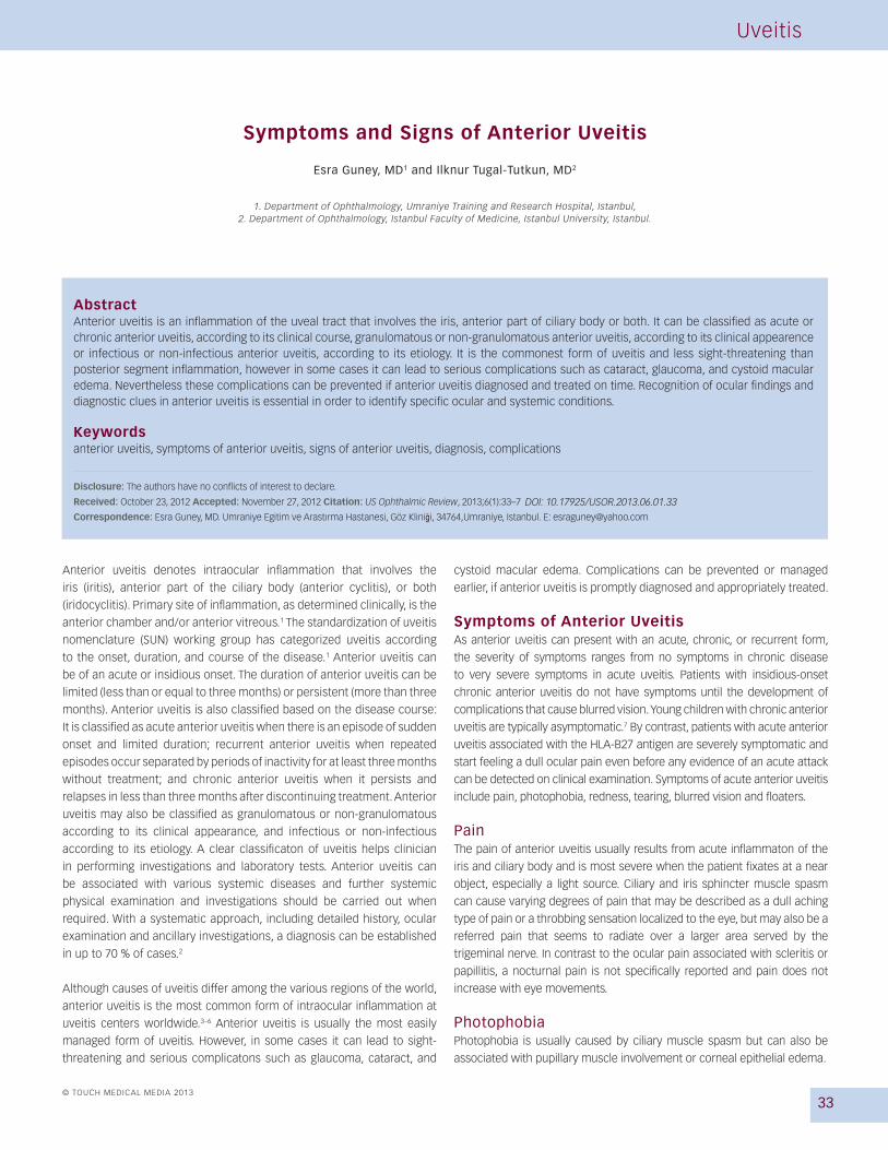
Figure 1: Slit-Lamp Photograph Shows Trace Ciliary Injection in the Right Eye of a Patient with Behçet Uveitis and Hypopyon Formation
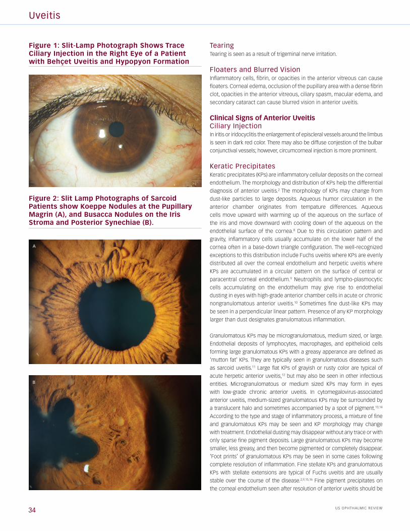
Figure 2: Slit Lamp Photographs of Sarcoid Patients show Koeppe Nodules at the Pupillary Magrin (A), and Busacca Nodules on the Iris Stroma and Posterior Synechiae (B).
A
B
TearingTearing is seen as a result of trigeminal nerve irritation.
Floaters and Blurred VisionInflammatory cells, fibrin, or opacities in the anterior vitreous can cause
floaters. Corneal edema, occlusion of the pupillary area with a dense fibrin
clot, opacities in the anterior vitreous, ciliary spasm, macular edema, and
secondary cataract can cause blurred vision in anterior uveitis.
Clinical Signs of Anterior UveitisCiliary InjectionIn iritis or iridocyclitis the enlargement of episcleral vessels around the limbus
is seen in dark red color. There may also be diffuse conjestion of the bulbar
conjunctival vessels; however, circumcorneal injection is more prominent.
Keratic Precipitates Keratic precipitates (KPs) are inflammatory cellular deposits on the corneal
endothelium. The morphology and distribution of KPs help the differential
diagnosis of anterior uveitis.2 The morphology of KPs may change from
dust-like particles to large deposits. Aqueous humor circulation in the
anterior chamber originates from tempature differences. Aqueous
cells move upward with warming up of the aqueous on the surface of
the iris and move downward with cooling down of the aqueous on the
endothelial surface of the cornea.8 Due to this circulation pattern and
gravity, inflammatory cells usually accumulate on the lower half of the
cornea often in a base-down triangle configuration. The well-recognized
exceptions to this distribution include Fuchs uveitis where KPs are evenly
distributed all over the corneal endothelium and herpetic uveitis where
KPs are accumulated in a circular pattern on the surface of central or
paracentral corneal endothelium.9 Neutrophils and lympho-plasmocytic
cells accumulating on the endothelium may give rise to endothelial
dusting in eyes with high-grade anterior chamber cells in acute or chronic
nongranulomatous anterior uveitis.10 Sometimes fine dust-like KPs may
be seen in a perpendicular linear pattern. Presence of any KP morphology
larger than dust designates granulomatous inflammation.
Granulomatous KPs may be microgranulomatous, medium sized, or large.
Endothelial deposits of lymphocytes, macrophages, and epithelioid cells
forming large granulomatous KPs with a greasy apperance are defined as
‘mutton fat’ KPs. They are typically seen in granulomatous diseases such
as sarcoid uveitis.11 Large flat KPs of grayish or rusty color are typical of
acute herpetic anterior uveitis,12 but may also be seen in other infectious
entities. Microgranulomatous or medium sized KPs may form in eyes
with low-grade chronic anterior uveitis. In cytomegalovirus-associated
anterior uveitis, medium-sized granulomatous KPs may be surrounded by
a translucent halo and sometimes accompanied by a spot of pigment.13,14
According to the type and stage of inflammatory process, a mixture of fine
and granulomatous KPs may be seen and KP morphology may change
with treatment. Endothelial dusting may disappear without any trace or with
only sparse fine pigment deposits. Large granulomatous KPs may become
smaller, less greasy, and then become pigmented or completely disappear.
‘Foot prints’ of granulomatous KPs may be seen in some cases following
complete resolution of inflammation. Fine stellate KPs and granulomatous
KPs with stellate extensions are typical of Fuchs uveitis and are usually
stable over the course of the disease.2,9,15,16 Fine pigment precipitates on
the corneal endothelium seen after resolution of anterior uveitis should be
34
Symptoms and Signs of Anterior Uveitis
US OPHTHALMIC REVIEW 35
differentiated from pigment precipitates associated with pigment dispersion
syndrome, pseudoexfoliation syndrome, or any other condition that may
cause pigment discharge into the anterior chamber. Pigment dispersion
syndrome typically causes a perpendicular line of pigment deposition
defined as a Krukenberg’s spindle.17 There are also recently described
entities of pigment dispersion that may masquerade anterior uveitis.18,19 In
both bilateral acute depigmentation of the iris (BADI) and bilateral acute
transillumination of the iris (BAIT), the finding of corneal endothelial pigment
precipitates is a useful clue to the differential diagnosis.18,19
Anterior Chamber FlareAqueous humor is a transparent liquid in healthy eyes. Breakdown of the
blood-aqueous barrier causes protein exudation into the anterior chamber.
Increased protein concentration of the aqueous humor causes an optical
phenomenon called flare or Tyndall effect.20 The grading of flare in the
anterior chamber helps to assess the severity of anterior uveitis and has
importance in monitoring the patients’ response to therapy. Flare can be
clinically graded on a 0 + to 4 + scale at the slit lamp.21 A beam 1 mm wide
and 3 mm long is used with the highest light intensity and 16 x magnification
at the slit-lamp. The score of 1 + corresponds to faint flare, 2 + corresponds
to moderate flare (iris and lens details clear), 3 + to marked flare (iris
and lens details hazy) and 4 + to intense flare (fibrin or plastic aqueous).
However slit-lamp assessment of flare is subjective and imprecise.
Laser Flare Photometry (LFP) is a quantitative and automated technique
that can measure flare. It is the only objective way to measure
intraocular inflammation.22 It was shown to be most useful in anterior
uveitis.23 Flare measurements by LFP provide precise monitoring of
inflammation and can be used to adjust the management of both acute
and chronic anterior uveitis. LFP flare has been reported to be a more
sensitive parameter than slit-lamp grading of cells in assessing evolution
of HLA-B27-associated acute anterior uveitis.24 Several groups have
reported that LFP flare was the most important inflammatory parameter
in children with Juvenile Idiopathic Arthritis (JIA)-associated chronic
anterior uveitis and high flare values were correlated with poor visual
acuity and a higher prevalence of ocular complications.25–28
Aqueous Cells Anterior chamber cells are primarily lymphocytes in most episodes of
anterior uveitis, but a significant number of neutrophilis may be present early
in the course of disease.8 The intensity of the cellular reaction in the anterior
chamber is graded according to the number of inflammatory cells in the
1 x 3 mm slit-lamp beam. The level 0.5 + corresponds to (1–5) cells,
1 + corresponds to (6–15) cells, 2 + corresponds to (16–25) cells, 3 +
corresponds to (26–50) cells and 4 + corresponds to more than 50 cells.1
When there is an excedingly high amount of leukocytes in the aqueous,
they precipitate with gravity and form an accumulation in the anterior
chamber angle that is referred to as a hypopyon formation. Various
inflammatory, infective and neoplastic conditions can cause a hypopyon.29
Hypopyon is a nonspecific sign, more often seen in HLAB27 related uveitis
and Behçet’s uveitis. Hypopyon in Behçet’s uveitis shifts freely with head
positioning, forms a smooth layer, and dissolves rapidly (see Figure 1). By contrast, in HLA B27 related uveitis, hypopyon is always sticky and
does not move freely with position.29,30 Other noninflammatory particles
in the aqueous humor should be differentiated from inflammatory
cells. Erythrocytes, ghost cells, debris, and pigment particles may be
seen circulating in the anterior chamber in nonuveitic conditions such
as trauma, pigment dispersion syndrome (PDS) and pseudo exfoliation
syndrome (PXS);31,32 but also in uveitis patients with ocular complications
that may result in dispersion of such particles in the anterior chamber.
Iris and Trabecular Meshwork NodulesIris nodules are accumulations of leukocytes on the anterior iris and
they represent granulomatous uveitis. Iris nodules are called Koeppe
nodules when they are seen at the pupillary magrin and Busacca
nodules when they occur on the iris stroma (see Figure 2). Iris nodules
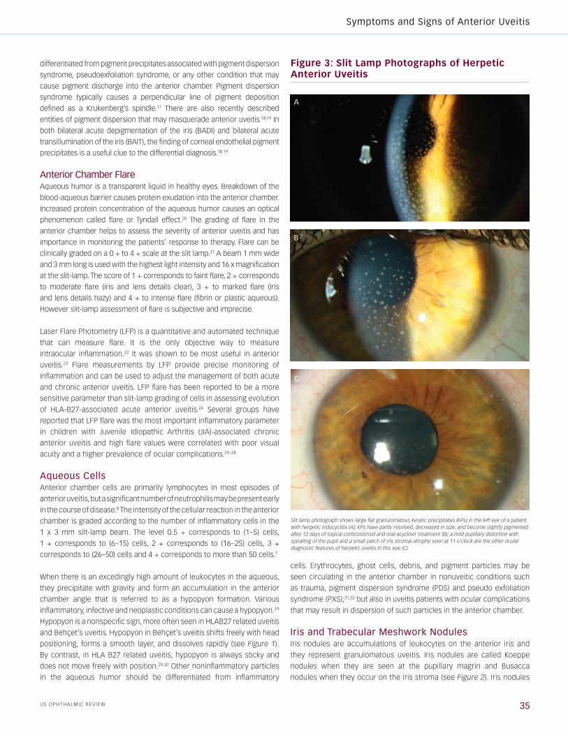
Figure 3: Slit Lamp Photographs of Herpetic Anterior Uveitis
A
B
C
Slit lamp photograph shows large flat granulomatous keratic precipitates (KPs) in the left eye of a patient with herpetic iridocyclitis (A); KPs have partly resolved, decreased in size, and become slightly pigmented after 12 days of topical corticosteroid and oral acyclovir treatment (B); a mild pupillary distortion with spiralling of the pupil and a small patch of iris stromal atrophy seen at 11 o’clock are the other ocular diagnostic features of herpetic uveitis in this eye (C).
Uveitis
US OPHTHALMIC REVIEW36
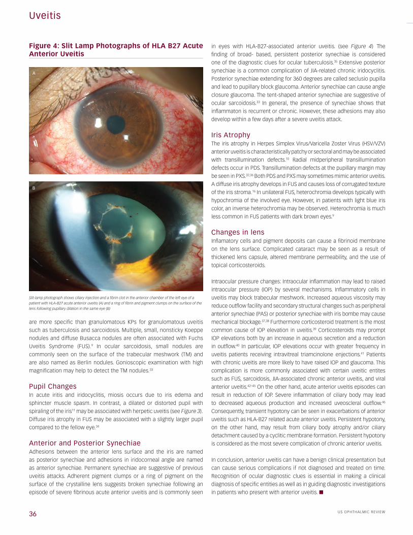
Figure 4: Slit Lamp Photographs of HLA B27 Acute Anterior Uveitis
Slit-lamp photograph shows ciliary injection and a fibrin clot in the anterior chamber of the left eye of a patient with HLA-B27 acute anterior uveitis (A) and a ring of fibrin and pigment clumps on the surface of the lens following pupillary dilation in the same eye (B)
A
B
are more specific than granulomatous KPs for granulomatous uveitis
such as tuberculosis and sarcoidosis. Multiple, small, nonsticky Koeppe
nodules and diffuse Busacca nodules are often associated with Fuchs
Uveitis Syndrome (FUS).9 In ocular sarcoidosis, small nodules are
commonly seen on the surface of the trabecular meshwork (TM) and
are also named as Berlin nodules. Gonioscopic examination with high
magnification may help to detect the TM nodules.33
Pupil Changes In acute iritis and iridocyclitis, miosis occurs due to iris edema and
sphincter muscle spasm. In contrast, a dilated or distorted pupil with
spiraling of the iris11 may be associated with herpetic uveitis (see Figure 3). Diffuse iris atrophy in FUS may be associated with a slightly larger pupil
compared to the fellow eye.34
Anterior and Posterior SynechiaeAdhesions between the anterior lens surface and the iris are named
as posterior synechiae and adhesions in iridocorneal angle are named
as anterior synechiae. Permanent synechiae are suggestive of previous
uveitis attacks. Adherent pigment clumps or a ring of pigment on the
surface of the crystalline lens suggests broken synechiae following an
episode of severe fibrinous acute anterior uveitis and is commonly seen
in eyes with HLA-B27-associated anterior uveitis. (see Figure 4) The
finding of broad- based, persistent posterior synechiae is considered
one of the diagnostic clues for ocular tuberculosis.35 Extensive posterior
synechiae is a common complication of JIA-related chronic iridocyclitis.
Posterior synechiae extending for 360 degrees are called seclusio pupilla
and lead to pupillary block glaucoma. Anterior synechiae can cause angle
closure glaucoma. The tent-shaped anterior synechiae are suggestive of
ocular sarcoidosis.33 In general, the presence of synechiae shows that
inflammaton is recurrent or chronic. However, these adhesions may also
develop within a few days after a severe uveitis attack.
Iris Atrophy The iris atrophy in Herpes Simplex Virus/Varicella Zoster Virus (HSV/VZV)
anterior uveitis is characteristically patchy or sectoral and may be associated
with transillumination defects.10 Radial midperipheral transillumination
defects occur in PDS. Transillumination defects at the pupillary margin may
be seen in PXS.32,36 Both PDS and PXS may sometimes mimic anterior uveitis.
A diffuse iris atrophy develops in FUS and causes loss of corrugated texture
of the iris stroma.16 In unilateral FUS, heterochromia develops typically with
hypochromia of the involved eye. However, in patients with light blue iris
color, an inverse heterochromia may be observed. Heterochromia is much
less common in FUS patients with dark brown eyes.9
Changes in lens Inflamatory cells and pigment deposits can cause a fibrinoid membrane
on the lens surface. Complicated cataract may be seen as a result of
thickened lens capsule, altered membrane permeability, and the use of
topical corticosteroids.
Intraocular pressure changes: Intraocular inflammation may lead to raised
intraocular pressure (IOP) by several mechanisms. Inflammatory cells in
uveitis may block trabecular meshwork. Increased aqueous viscosity may
reduce outflow facility and secondary structural changes such as peripheral
anterior synechiae (PAS) or posterior synechiae with iris bombe may cause
mechanical blockage.37,38 Furthermore corticosteroid treatment is the most
common cause of IOP elevation in uveitis.39 Corticosteroids may prompt
IOP elevations both by an increase in aqueous secretion and a reduction
in outflow.40 In particular, IOP elevations occur with greater frequency in
uveitis patients receiving intravitreal triamcinolone enjections.41 Patients
with chronic uveitis are more likely to have raised IOP and glaucoma. This
complication is more commonly associated with certain uveitic entites
such as FUS, sarcoidosis, JIA-associated chronic anterior uveitis, and viral
anterior uveitis.42–45 On the other hand, acute anterior uveitis episodes can
result in reduction of IOP. Severe inflammation of ciliary body may lead
to decreased aqueous production and increased uveoscleral outflow.46
Consequently, transient hypotony can be seen in exacerbations of anterior
uveitis such as HLA-B27 related acute anterior uveitis. Persistent hypotony,
on the other hand, may result from ciliary body atrophy and/or ciliary
detachment caused by a cyclitic membrane formation. Persistent hypotony
is considered as the most severe complication of chronic anterior uveitis.
In conclusion, anterior uveitis can have a benign clinical presentation but
can cause serious complications if not diagnosed and treated on time.
Recognition of ocular diagnostic clues is essential in making a clinical
diagnosis of specific entities as well as in guiding diagnostic investigations
in patients who present with anterior uveitis. n
Symptoms and Signs of Anterior Uveitis
US OPHTHALMIC REVIEW 37
1. Jabs DA, Nussenblatt RB, Rosenbaum JT, Standardization of uveitis nomenclature for reporting clinical data, results of the first international workshop, Am J Ophthalmol, 2005;140:509–16.
2. Herbort CP, Appraisal work–up and diagnosis of anterior uveitis: a practical approach, Middle East Afr J Ophthalmol, 2009; 16(4):159–67.
3. Kazokoğlu H, Onal S, Tugal–Tutkun I, et al, Demographic and clinical features of uveitis in tertiary centers in turkey, Ophthalmic Epidemiol, 2008;15(5):285–293.
4. Khairallah M, Yahia SB, Ladjimi A, et al., Pattern of uvetis referral centre in tunisia, north africa, Eye, 2007;21(1):33–9.
5. Cimino L, Aldigeri R, Salvarani C, et al., The causes of uveitis in a referral centre of northern italy, Int Ophthalmol, 2010;30:521–9.
6. Yang P, Zhang Z, Zhou H, et al., Clinical Patterns and Char-acteristics of uveitis in a Tertiary center for uveitis in China, Curr Eye Research, 2005;30:943–8.
7. Tugal–Tutkun I, Pediatric Uveitis, J Ophthalmic Vis Res, 2011;6(4):259–69.
8. Whitcup SM, Anterior Uveiti, In: Nussenblatt RB, Whitcup SM (eds), Uveitis Fundamentals and Clinical Practice, Philadel-phia, Pennsylvania, Mosby, 2004;273–87.
9. Tugal–Tutkun I, Guney–Tefekli E, Kamaci Duman F, et al., A Cross–sectional and Longitudinal Study of Fuchs Uveitis Syndrome in Turkish Patients, Am J Ophthalmol, 2009;148(4):510–15.e1.
10. Chang JH, McCluskey PJ, Wakefield D, Acute anterior uveitis and HLA–B27, Surv Ophthalmol, 2005;50:364–88.
11. Herbort CP, Rao NA, Mochizuki M, et al, International criteria for the diagnosis of ocular sarcoidosis: results of the first International Workshop on Ocular Sarcoidosis (IWOS), Ocul Immunol Inflamm, 2009;17:160–9.
12. Tugal–Tutkun I, Otük–Yasar B, Altinkurt E, Clinical features and prognosis of herpetic anterior uveitis: a retrospective study of 111 cases, Int Ophthalmol, 2010;30:559–65.
13. Jap A, Chee SP, Cytomegalovirus–associated anterior segment infection, Exp Rev Ophthalmol, 2011;6:1–12.
14. Chee SP, Jap A, Presumed Fuchs heterochromic iridocyclitis and Posner–Schlossman syndrome: comparison of cyto-megalovirus–positive and negative eyes, Am J Ophthalmol, 2008;146:883–9.
15. Mohamed Q, Zamir E, Update on Fuchs’ uveitis syndrome, Curr Opin Ophthalmol, 2005;16:356–63.
16. Jones NP, Fuchs’ heterochromic uveitis: an update, Surv Ophthalmol, 1993;37:253–72.
17. Scheie HG, Cameron JD, Pigment dispersion syndrome: a clinical study, Br J Ophthalmol, 1981;65:264–9.
18. Tugal–Tutkun I, Araz B, Taskapili M et al., Bilateral acute depigmentation of report of 26 new cases and four–year follow–up of two patients, Ophthalmology, 2009;116:1552–7.
19. Tugal–Tutkun, Onal S, Garip A et al., Bilateral acute iris trans-illumination, Arch Ophthalmol, 2011;129(10):1312–9.
20. Tyndall J, On the blue of the sky, the polarization of the skylight, and on the polarization of light by cloudy matter generally, Philos Mag J,1869;37:384–404.
21. Hogan MJ, Kimura SJ, thygeson P, Signs and symptoms of uveitis. I. Anterior uveitis, Am J Ophthalmol, 1959;47:155–70.
22. Tugal–Tutkun I, Herbort CP, Laser flare photometry: a invasive, objective and quantitative method to measure intraocular inflammation, Int Ophthalmol, 2010;30:453–64.
23. Wakefield D, Herbort CP, Tugal–Tutkun I, et al., Controversies in ocular inflammation and immunology laser flare photom-etry, Ocular Immunol Inflamm, 2010;18(5):334–40.
24. Bernasconi O, Papadia M, Herbort CP, Sensitivity of laser flare photometry compared to slit–lamp cell evaluation in monitoring anterior chamber inflammation in uveitis, Int Ophthalmol, 2010;30:495–500.
25. Davis JL, Dacanay LM, Holland GN, et al., Laser flare photom-etry and complications of uveitis in children, Am J Ophthalmol, 2003;135:763–71.
26. Holland GN, A reconsideration of anterior chamber flare and its clinical relevance for children with chronic anterior uveitis (An American Ophthalmological Society Thesis), Trans Am Ophthalmol Soc, 2007;105: 344–64.
27. Holland GN, Denove CS, Yu F, Chronic anterior uveitis in children: clinical characteristics and complications, Am J Ophthalmol, 2009;147:667–678.
28. Tappeiner C, Heinz C, Roesel M, Heiligenhaus A, Elevated laser flare values correlate with complicated course of anterior uveitis in patients with juvenile idiopathic arthritis, Acta Ophthalmol, 2011;89:e521–7.
29. Ramsay A, Lightman S, Hypopyon uveitis, Surv Ophthalmol, 2001;46:1–18.
30. Tugal–Tutkun I, Behçet’s uveitis, Middle East Afr J Ophthalmol, 2009;16:219–24.
31. Niyadurupola N, Broadway DC, Pigment dispersion syndrome and pigmentary glaucoma: a major review, Clin Experiment Ophthalmol, 2008;36(9):868–82.
32. Ritch R, Schlötzer–Schrehardt U, Exfoliation syndrome, Surv Ophthalmol, 2001;45(4):265–315.
33. Papadia M, Herbort HP, Mochizuki M, Diagnosis of Ocular sarcoidosis, Ocular Immunol Inflamm, 2010;18(6):432–41.
34. Bonfioli AA, Curi AL, Orefice F, Fuchs’s Heterochromic Cyclitis, Semin Ophthalmol, 2005;20(3):143–6.
35. Gupta A, Bansal R, Gupta V, et al., Ocular Signs of Tuberculo-sis Uveitis, Am J Ophthalmol 2010; 149(4) 562–570.
36. Gillies WE, Brooks AM, Clinical features at presentation of anterior pigment dispersion syndrome, Clin Exp Ophthalmol, 2001;29:125–7.
37. Moorthy RS, Mermoud A, Baerveldt G, et al., Glaucoma associated with uveitis, Surv Ophthalmol, 1997;41:361–394,
38. Perets WL, Tomasi TB, Aqueous humor proteins in uveitis, Immunoelectrophoretic and gel diffusion studies on normal and pathological human aqueous humor, Arch Ophthal-mol,1961;65:20–3.
39. Sallam A, Sheth HG, Habot–Wilmer Z, Lightman S, Outcome of raised intraocular pressure in uveitic eyes with and without a corticosteroid–induced hypertensive response, Am J Ophthalmol, 2009;148:207–13.
40. Jacop E, FitzSimon JS, Brubaker RF, Combined corticosteroid and catecholamine stimulation of aqueous humor flow, Ophthalmology, 1996;103:1303–8.
41. Galor A, Margolis R, Brasil OM,et al., Adverse events after intravitreal triamcinolone in patients with or without uveitis, Ophthalmology, 2007;114:1912–1918.
42. Takahashi T, Ohtani S, Miyata K, et al., A clinical evaluation of uveitis associated secondary glaucoma, Jpn J Ophthalmol, 2002;46:556–562.
43. La Hey E, de Vries J, Langerhorst CT, et al., Treatment and prognosis of secondary glaucoma in Fuchs heterochromic cyclitis, Am J Ophthalmol, 1993;116:327–40.
44. Miserocchi E, Waheed NK, Dios E, et al., Visual outcome in herpes simplex virus and varicella zoster virus uveitis: a clinical evaluation and comparison, Ophthalmology, 2002;109(8):1532–7.
45. Jap A, Chee S, Viral anterior uveitis, Curr Opin Ophthalmol, 2011;22:483–488.
46. Toris CB, Pederson JE, Aqueous humor dynamics in experimental iridocyclitis, Invest Ophthalmol Vis Sci, 1987;28(3):477–81.