suture materials and suturing techniques
TRANSCRIPT
DAS_49DDCH_2017
SUTURE MATERIALS &
SUTURINGTECHNIQUES
Content o Definitionso Goals of suturingo Suture characteristicso Armamentarium of suturingo Suture materialso Principles of suturingo Suturing techniqueso Surgical knoto Removal of sutureo Reasons for failure of sutureo Possible complicationso Alternatives to suture
DEFINITIONS: what is suture? Suture is a stich or series of stiches made to secure apposition of the edges of a surgical or traumatic wound.
What is suture materials? Suture materials is an artificial fibers used to keep wound together until they hold themselves by natural which is synthesized & oven into a stronger scar.

GOALS OF SUTURING Wound edge apposition.Provide adequate tension. Maintain hemostasis. Aid in wound healing. Avoid wound infection. Produce aesthetically pleasing
scar by approximating skin edges.

SUTURE CHARACTERISTICS
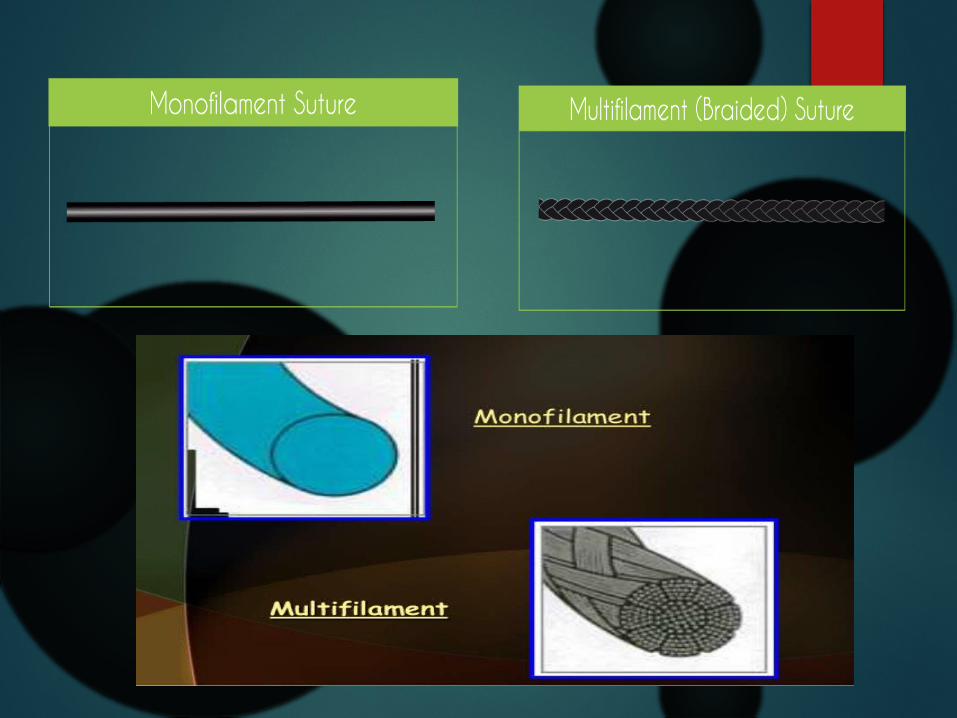
physical structure: Monofilament-
This suture material is smooth & tends to slide through tissues easily.
Difficult to knot. Can be damaged by gripping it with
needle holder or forceps.That can lead to fracture of the suture materials.

SUTURE CHARACTERISTICS
Multifilaments- Easy to knot. Have a greater surface area than
monofilaments. Have a capillary actions where
bacteria may lodge & be responsible for persistent infections.
This material can be coated with silicone in order to make it smooth.

SUTURE CHARACTERISTICS Tensile Strength: It can be expressed as the force required to break it when pulling the two ends apart.It depends upon – Constituent of suture materials. Thickness of suture materials. How it is handled in the tissues.

SUTURE CHARACTERISTICS Absorbability: Suture materials may be absorbable or non-absorbable.This property must be taken into consideration when choosing suture materials for specific wound closures. Oral mucosa & Deep sturcture need to be absorbable suture materials but vascular anastomoses need non-
absorbable suture materials.
Biological Behaviour: It depends upon the constituent of raw materials.

Armamentarium of suturing
Needle holder
A suture needle
Suture material

Needle holder
Parts: Working tip/jaws Hinge joint Shank/body Catch mechanism/ratchet Grip area

Needle holder
How to hold? The needle holder is held with thumb & ring finger through the rings & with the index finger along the length of needle holder to provide stability & control.
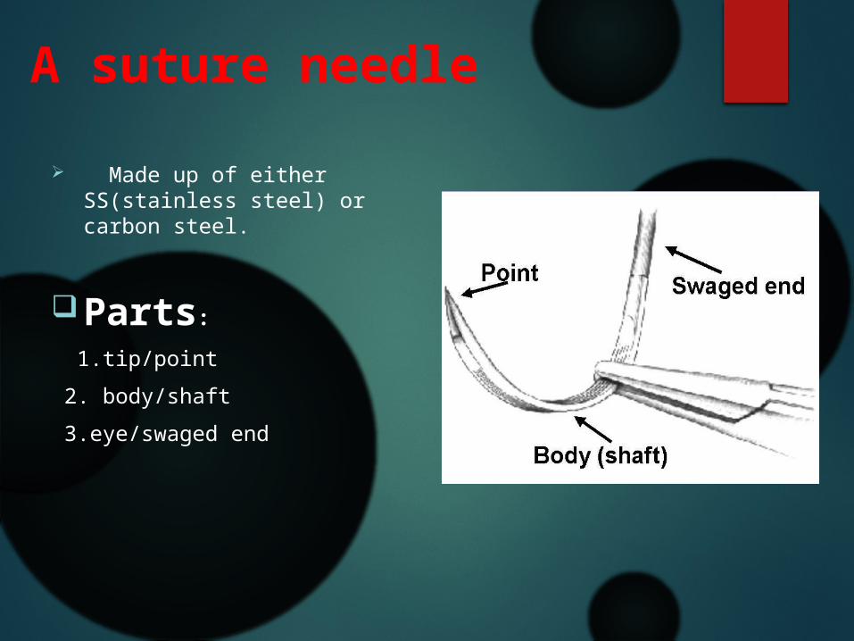
A suture needle Made up of either
SS(stainless steel) or carbon steel.
Parts: 1.tip/point 2. body/shaft 3.eye/swaged end

A suture needle
Shape of needle:

Classification of needle
According to Shape:
1.Straight 2.Curved
According to eye:
1.Eyed needle/Traumatic 2.Eyeless needle/Atraumatic

Classification of needle According to
cutting edge 1.Round body 2.Cutting body-
ConventionalReverse cutting
According to its tip
1.Triangular 2.Round 3.Blunt

Suture materials

Suture materials
Ideal properties: Easy to handle. Predictable behaviour in tissues. Predictable tensile strength. Sterile. Secure knotting ability. Minimal tissue reaction. Non-alergenic,non-carcinogenic,non-shrinkage.
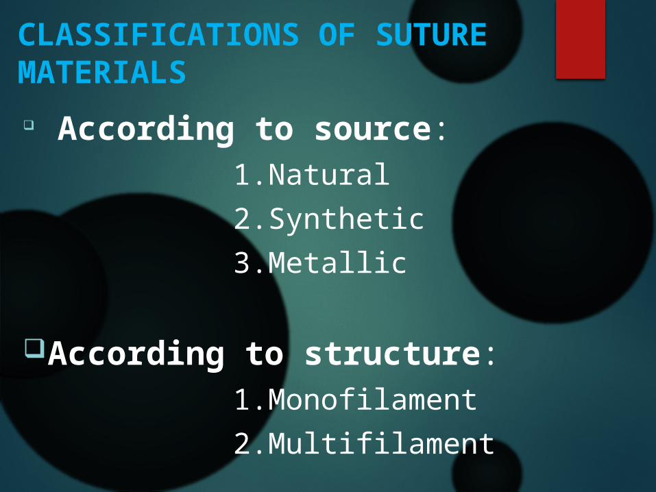
CLASSIFICATIONS OF SUTURE MATERIALS According to source: 1.Natural 2.Synthetic 3.Metallic
According to structure: 1.Monofilament 2.Multifilament
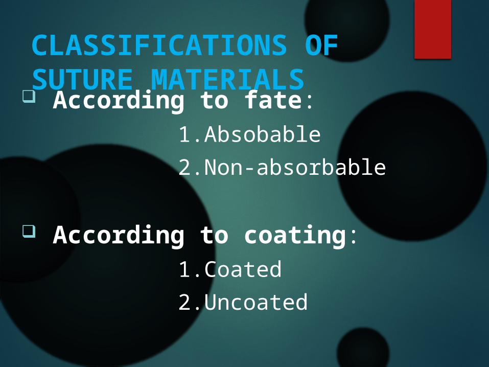
CLASSIFICATIONS OF SUTURE MATERIALS
According to fate: 1.Absobable 2.Non-absorbable
According to coating: 1.Coated 2.Uncoated

Natural
Absorbable
• catgut• Chromic
catgut• Collagen• Fascia lata• Beef tendon
Non-absorbable
• Silk• Silk worm gut• Linen• Cotton• Ramie

Synthetic Absorbable Polyglycolic acid Polyglactic acid Polyglactin(vicryl) Polydioxanone(PDS)
Non-absorbable nylone(polyamide) Polypropyline(Prolene) Polyesters polyethelene
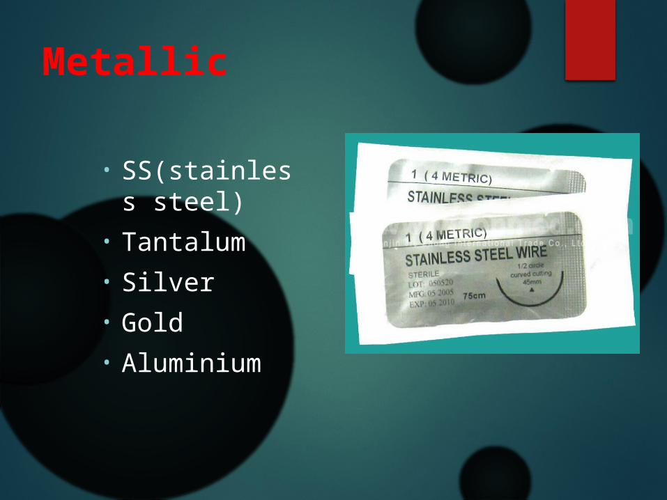
Metallic
• SS(stainless steel)
• Tantalum• Silver• Gold • Aluminium

Monofilament
Absorbable
• Catgut• Chromic
catgut• Vicryl• PDS
Non-absorbable
• Polyoropylene• Polyester• Nylone• Polyvenyleidene
fluoride/PVDF suture

Multifilament
Absorbable
• Vicryl• Polyglycolic
acid
Non-absorbable
• Silk• Cotton• linen

Monofilament vs multifilment
Monofilament Has no capillary action Less infection risk Smooth tissue passage Higher tensile strength More throws required
Multifilament Has capillary action Increased infection risk Less smooth passage Less tensile strength Better knot security
Absorbable vs Non-absorbable
Absorbable Degraded by
enzymes,hydrolysis or phagocytosis
Used to hold the edges in approximation temporarily until the wound is heal
Non-absorbable Encapsulated or walled
off by fibrosis
Used to suture at sites where tensile strength need to be maintained

overview
Selection of suture materials
Condition of the wound. Tissues to be repaired. Tensile strength. Knot holding characteristics. Reaction of surrounding tissues.
Commonly Used Suture Materials
Polypropylene(prolene) It is synthetic ,non-absorbable monofilament suture materials. Polymer of propylene.
Uses: 1.General surgery. 2.Plastic surgery. 3.Cardiovascular surgery. 4.Skin closure.

Advantages: 1.Won’t loose tensile strength over time. 2.Good knot security. 3.Very little tissue reaction. 4.High plasticity. Disadvantage: 1.Stretch when pulled. 2.Loosens when edema subsides.

Commonly Used Suture Materials
Silk: It is natural,non-absorbable multifilament suture materials. Made from the filament spun by silkworm larva. Uses: 1.Opthalmic surgery.
2.General surgery. 3.plastic surgery.

Advantage: 1.Ease of handling. 2.Good knot security. 3.Cost effective. Disadvantage: 1.Very reactive. 2.can’t be used in presence of infection.

Commonly Used Suture Materials Vicryl: It is synthetic & absorbable suture materials. Monofilament/multifilament & coated/uncoated. Available in purple color/undyed. Uses: 1.Intra oral suturing. 2.Gut anastomoses. 3.Vascular ligature. 4.Opthalmic surgery 5. Superficial soft tissue approximation of the skin and mucosa.

Advantage:
1.Minimal tissue reactivity.2.Can be used in infected tissues.3.Stronger than gut:retains strength 3 weeks. Disadvantage:
1.In case of prolong approximation can’t be used.2.Delayed absorption & increased infalmmation.

Uses of different sizes of suture

Biological response to suture materials
The early response is a generalized acute aseptic inflammation involving primarily polymorphonuclear leucocytes.
After few days mononuclear cells fibroblast & histiocytes become evident.
Capillary formation occurs at the end of this initial phase.

Biological response to suture materials
Natural absorbable- Proteolytic degradation. Intense tissue response.
Synthetic absorbable- Hydrolysis. Less intense response.
Non-absorbable- Encapsulation. Acellular response.

Principle of suturing

Principle of suturing The needle should be
grasped at approximately 1/3 of the distance from the eye & 2/3 from point.
The needle should be pierced the tissue perpendicular to its surface.
The needle should be placed equidistant (2-3mm) from the incision line.
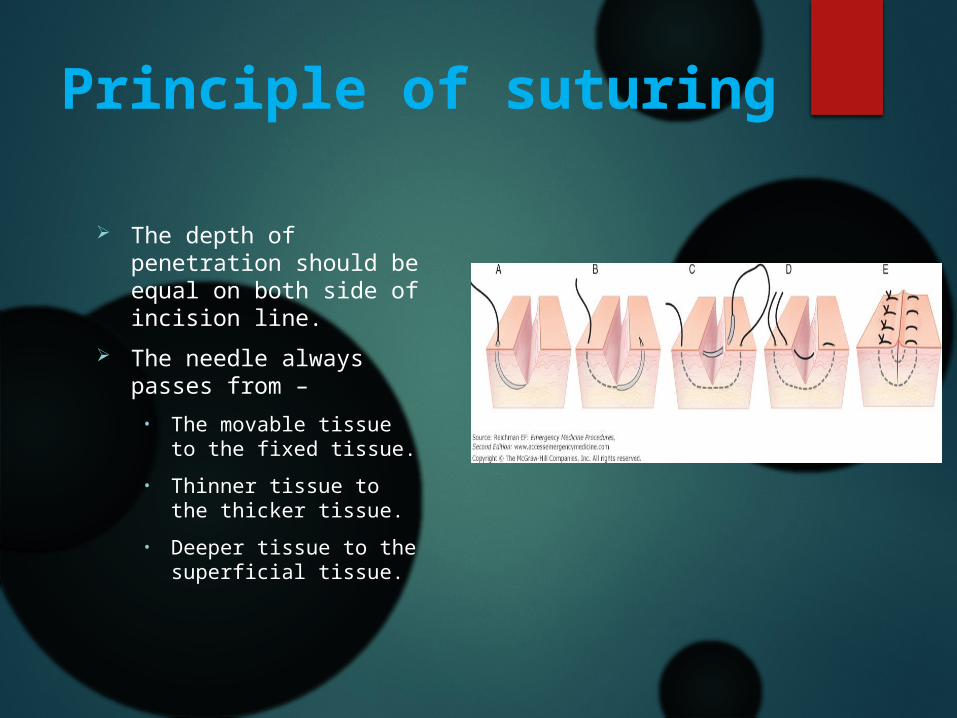
Principle of suturing
The depth of penetration should be equal on both side of incision line.
The needle always passes from –• The movable tissue to
the fixed tissue.• Thinner tissue to the
thicker tissue.• Deeper tissue to the
superficial tissue.

Principle of suturing
The tissue never be closed under tension.
Each suture must be placed 3-4 mm apart from the incision line.
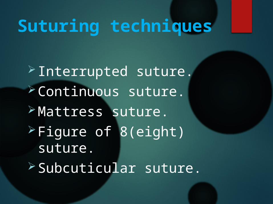
Suturing techniques
Interrupted suture.Continuous suture.Mattress suture.Figure of 8(eight) suture.Subcuticular suture.
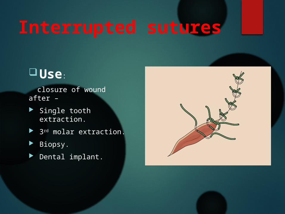
Interrupted sutures
Use: closure of wound after – Single tooth extraction. 3rd molar extraction. Biopsy. Dental implant.
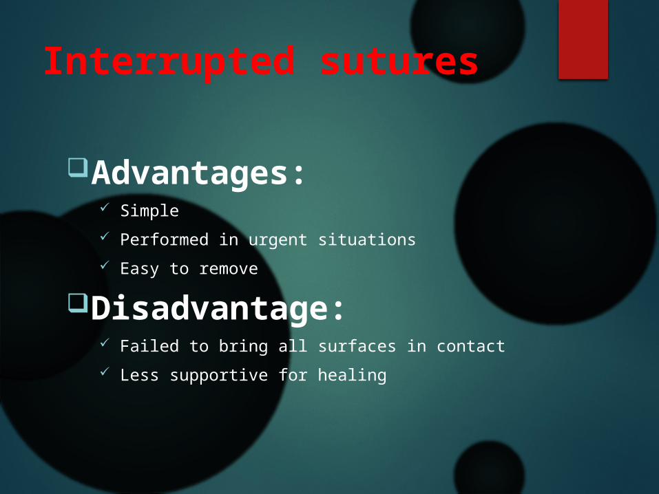
Interrupted sutures
Advantages: Simple Performed in urgent situations Easy to remove
Disadvantage: Failed to bring all surfaces in contact Less supportive for healing

Technique for interrupted sutures Cleansing & debridement. Selection of appropriate suture. Wound margins are accurately opposed. Suture needle is held with the needle holder positioned at least one
third of the length of the suture away from the end of suture attachment.
A ‘bite of skin is taken at a landmarked site. The suture needle is advanced to the depth of the wound margin &
then out through the wound opening. The needle is next inserted via the depth of the wound & rotated up
through the opposite skin margin & the landmarked site for apposition of the wound.

Technique for interrupted sutures
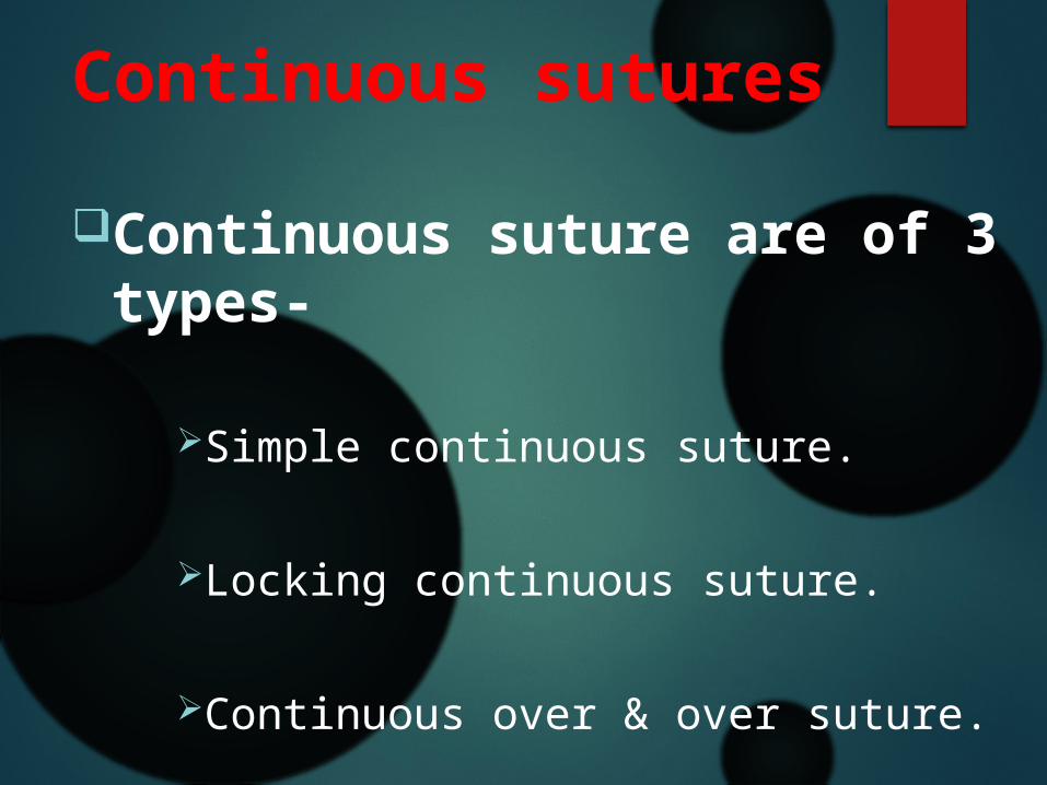
Continuous sutures
Continuous suture are of 3 types-
Simple continuous suture.
Locking continuous suture.
Continuous over & over suture.

Simple continuous sutures
uses:Well approximated wounds with minimal
tension.
Advantage:Rapid technique for closure.Even distribution of tension over the
suture line.
Can be used in swelled up tissues.
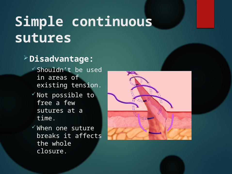
Simple continuous sutures
Disadvantage: Shouldn’t be used
in areas of existing tension.
Not possible to free a few sutures at a time.
When one suture breaks it affects the whole closure.

Technique for simple continuous sutures
The beginning of the simple continuous suture is similar to the simple interrupted suture.
The needle is then reinserted in a continuous fashion such that the suture passes perpendicular to the incision line.
The suture is ended by passing a square knot over the untightened end of the suture.
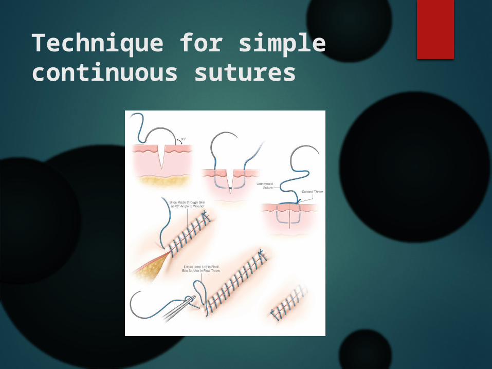
Technique for simple continuous sutures
Locking continuous suturesUses:
Long edentulous areas.Tuberosities/retromolar areas.
Advantage:
Avoid the multiple knot of the interrupted suture.

Locking continuous suturesTechnique:
At first a single interrupted suture is used to make a tie. The needle is next inserted through the underlying surface of the flap. The needle is then passed through the remaining loop of the suture &
the suture is pulled tightly,thus loocking it. This procedure is continued until the final suture is tied off at the
terminal end.

Locking continuous sutures
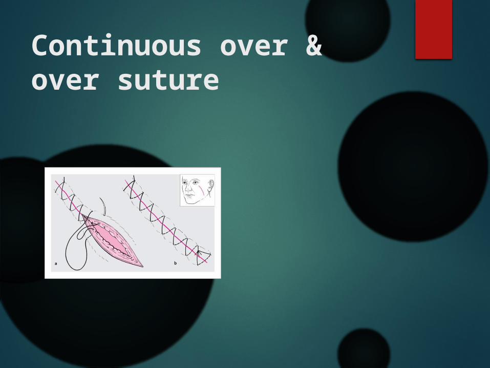
Continuous over & over suture
Initially a simple interrupted suture is placed & the needle is then reinserted in a continuous fashion such that the suture passes perpendicular to the incision line below & obliquely above.
The suture is ended by passing a knot over the untightened end of the suture.
It provides a rapid technique for closure & distribute the tension uniformly over the suture line.
It also offers a more water tight closure.
Continuous over & over suture
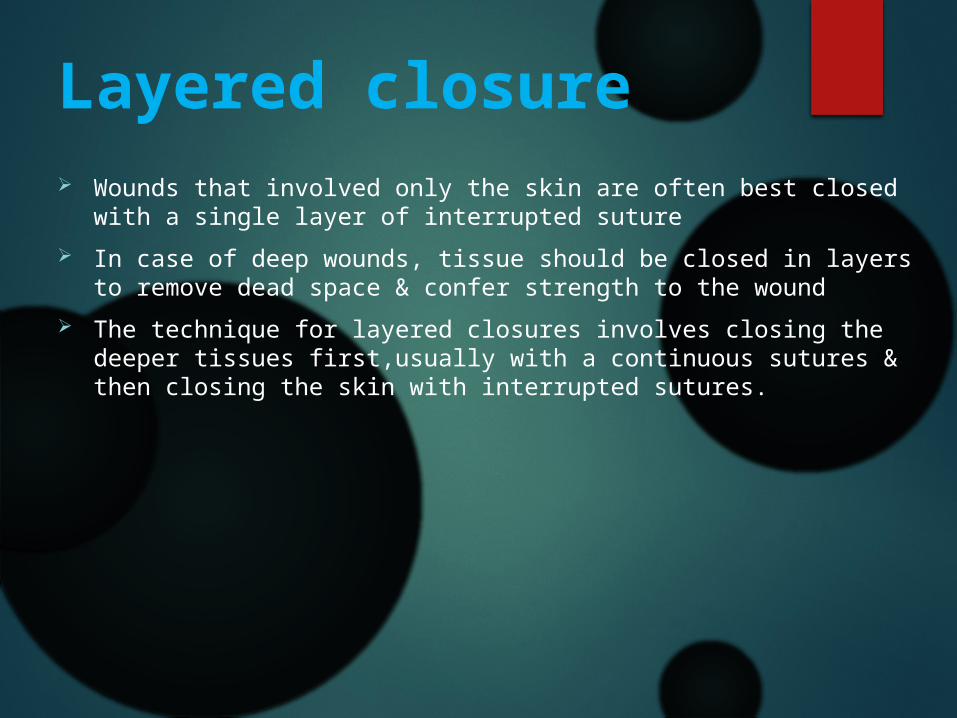
Layered closure Wounds that involved only the skin are often best closed with a
single layer of interrupted suture In case of deep wounds, tissue should be closed in layers to remove
dead space & confer strength to the wound The technique for layered closures involves closing the deeper
tissues first,usually with a continuous sutures & then closing the skin with interrupted sutures.

Layered closure
Mattress suturesThese suture may be –
Horizontal.Vertical.

Horizontal mattress suturesUses:
Intraoral bone grafting. Closure of extraction socket.
Advantage: Provides a broad contact of the wound margin. Provides a water tight closure.
Disadvantage: If improperly used bone necrosis & wound dehiscence may occur due
to limited blood supply.

Horizontal mattress sutures Technique:
The needle is passed from one edge of the incision to another & again from the latter edge to the first edge in a horizontal manner & knot is tied.
The distance of the needle penetration from the incisal line & the depth penetration of the needle is the same for each entry point.
The horizontal distance of the points of penetration on the same side of the flap differs(needle penetration through the surgical flap should be at least 8mm from flap edge)

Horizontal mattress sutures
This procedure is continued till the entire length of the incision & a knot is then tied.

Vertical mattress sutures It is similar to the horizontal mattress
except the depth of penetration, i.e. when the needle is brought back from the second flap to the first,the depth of penetration is more superficial.
vertical mattress sutures Uses:
Closing deep wounds abdomen or hip. Advantage:
decreasing the dead space & providing increased strength.
doesn’t interfere with healing. Disadvantage:
Approximation is difficult.

overview

Figure of 8(eight) suture
Most commocly used for extraction socket closure as well as adaptation of gingival papilla around the tooth.
Technique: The needle first inserted into the outer surface
of the buccal flap & then the lingual flap. Suturing begins on the buccal surface 3-4 mm
from the tip of the papilla.

Figure of 8(eight) suture Then the needle should be inserted in the same fashion at a
horizontal distance & then both ends tied.
Advantage:
Rapid closureDisadvantage:
Due to its orientation ,it is difficult to remove & it leaves a significant amount of suture threads inside the socket.

Figure of 8(eight) suture
Subcuticular suture Usually a running stitch,but can be interrupted Intradermal horizontal bites. Allow suture to remain for a longer period of
time without development of crosshatch scaring.
Uses:Simple,uncomplicated
wound.

Subcuticular suture Advantage:
Excellent cosmetic closure.
No stitch to remove.
Disadvantage: Technically more
difficult to master. Dosen’t hold in
thin skin

Surgical Knots
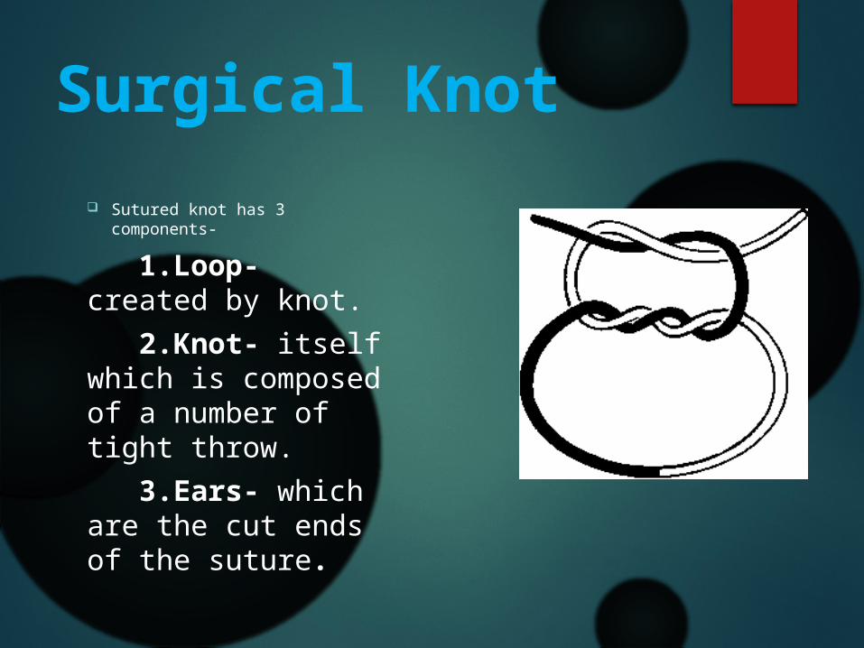
Surgical Knot Sutured knot has 3
components-
1.Loop- created by knot. 2.Knot- itself which is composed of a number of tight throw. 3.Ears- which are the cut ends of the suture.

Principles of knot tying Use the simplest knot that will prevent slippage. Tying the knot as small as possible & cutting the end of the suture
as short as reasonable to minimize foreign body reaction. Avoid friction or sawing. Avoid excessive tension. Tying sutures too tightly to strangulates the tissues. Maintenance of traction at one end of the suture after the first loop
is thrown .

Principles of knot tying Placing the final throw at horizontally as possible to keep the knot
flat. Limiting extra throws to the knot as they don’t add strength to a
properly tied knot.

Different types of knotsecure/square knot.
Surgeons knot.
Granny’s knot/slip knot.

Square knot The first throw is placed
in precise position for the knot,using a double loop.
The second throw is tied using horizontal tension.
Additional 2 throws are desirable.
Totally there should be 4 throws.
Best for catgut,silk,cotton & SS(stainless steel).

Surgeons knot Formed by two throws
on the first tie & one throw in the opposite direction in the second tie .
Recommended for tying polyester suture materials such as vicryl & mersiline.

Granny’s knot A tie in one direction
followed by a tie in the same direction & a third tie in the opposite direction to square the knot & hold it permanently.
Can be used in silk,chromic catgut/plain catgut.

overview
Removal of suture All sutures,being foreign bodies,cause irritation to the tissues &
hence have the potential to cause scarring. Skin sutures are removed as soon as tissue healing allows. Non-absorbable sutures are best removed from the face after a
period of 5-6 days.Tissues such as the scalp may require a longer period(7-10 days).

Removal of suture Face 3-5 days Lip 3-5 days Oral cavity 6-8 days Neck 5-6 days
Scalp 7-10 days Chest 10-14 days Abdomen 10-14 days Leg 10-14 days
Principle of suture removal Suture area is first clean with normal saline. The suture is grasped with non tooth dissecting forceps & lifted
above the epithelial surface. Scissors are then passed through one loop & then transected close
to the surface to avoid dragging contaminated suture materials through tissues.
The suture is then pulled towards the incision line to prevent dehiscence.if suture entrapped in a scab,application of hydrogen per oxide/normal saline is necessary.

Principle of suture removal If pieces of suture left infection may occur.

Reasons for failure of sutures
Breakage Cuts out Knot slips Extruded suture Resorbs too rapidly Removed too early

Possible complications of leaving sutures for many daysSutural abscess.
Scar or stitch mark.
Dermoid cyst.

Alternatives to suture
Name: Staples Tissue adhesives Tape
Disadvantage: Not absolute alternative to mechanical means More tissue reaction

Bibliography 1)Textbook of Oral & Maxillofacial Surgery
Neelima Anil Malik 2)Textbook of Oral & Maxillofacial Surgery
S M Balaji 3)An Introduction of Oral & Maxillofacial Surgery
David A Mitchell4)Contemporary of Oral & Maxillofacial Surgery (5th ed.)
Hupp,Ellis,Tucker5)Oral & Maxillofacial Surgery (Vol-1)
Luskin
6)Principles of Oral & Maxillofacial Surgery(6th ed.) U J Moore
7)Baily & Love’s Short Practice of Surgery(26th ed) Norman S. WIlliams