surgical management of grossly comminuted fractures … mohanty.pdf · surgical management of...
TRANSCRIPT

[1]
SURGICAL MANAGEMENT OF GROSSLY COMMINUTED FRACTURES OF DISTAL POLE OF PATELLA
A STUDY DONE AT NALCO HOSPITAL DAMANJODI, ODISHA
DISSERTATION SUBMITTED TO UNIVESITY OF SEYCHELLES
AMERICAN INSTITUTE OF MEDICINE
IN PARTIAL FULFILLMENT OF THE REQUIREMENTS FOR THE DEGREE
M.Ch (Orthopaedic surgery)
BY
DR. AMARJIT MOHANTY Orthopaedic surgeon
November 2011

[2]
SURGICAL MANAGEMENT OF GROSSLY
COMMINUTED FRACTURES OF
DISTAL POLE OF PATELLA
BY
DR. AMARJIT MOHANTY Orthopaedic surgeon
NALCO HOSPITAL
DAMANJODI

[3]
INTRODUCTION
Patella is the largest sesamoid bone in the body. The quadriceps tendon insert at the
superior pole of the body and the patellar tendon originates form the inferior pole and
inserts into the tibial tuberosity. The articular surface has the thickest layer of cartilage
in the body, upto 5 mm. The patella is of importance for the extension of knee joint.
Fractures of the patella constitute almost 1% of all skeletal injuries, resulting from either
direct or indirect trauma. The anterior subcutaneous location of the patella makes it
vulnerable to direct trauma, such as the knee striking the dash board of an automobile
or from a fall on the anterior knee. These injuries often are comminuted or displaced
and may include chondral injury to the distal femur or patella. Fractures caused by
indirect mechanisms result from a violent contraction of the quadriceps with keen flexed.
These fractures usually are transverse and may be associated with tears of the medial
and lateral retinacular expansions. Most patellar fractures are caused by a
combination of direct and indirect forces. The most significant effects of fracture of the

[4]
patella are loss of continuity of the extensor mechanism of the knee and potential
incongruity of the patello-femoral articulation.
Fractures of the patella can be classified as un-displaced or displaced and sub
classified further according to fracture configuration such as , transverse, oblique,
vertical and comminuted or stellate patellar fractures, which is associated with a
variable amount of displacement. Inability of the patients to extend the affected knee
actively usually indicates a disruption of the extensor mechanism and a torn
retinaculum, which require surgical treatment.
The treatment of comminuted fractures of the patella varies. There are proponents for
patellectomy and those for repair. Often only the distal pole of the patella is
fragmented, leaving a substantial and relatively normal proximal fragment. This
fragment is an important part of the extensor mechanism and should be preserved. To
perform partial patellectomy of distal pole with retention of proximal pole and suturing of
patellar tendon to it seems to be reasonable.

[5]
MATERIALS AND METHODS
In this series only the grossly comminuted fracture of distal pole of patella which were
managed by partial patellectomy with patellar tendon suturing to proximal major
fragment are taken for study.
Over a period of 11 years (2000 to 2011), 22 cases of comminuted fracture of distal
pole of patella were treated in Nalco Hospital, Damanjodi. Out of 22 cases, 16 were
male and 6 were females. The age ranged from 30 years to 50 years; and 14 fractures
were of the right side and 8 of the left side.
At admission aspiration of the knee was done and a compression bandage with knee
back slab applied. Those who had superificial skin injuries, were treated with cleaning
and dressing initially. All patients were kept under antibiotic coverage. All the cases

[6]
were radiologically evaluated with AP, lateral and axial views pre-operatively and were
thoroughly evaluated for medical fitness for surgery. All were operated within one week
to 10 days of admission.
All the 22 cases were managed by partial patellectomy and suturing by stainless steel
wire of patellar tendon to the major proximal fragment transosseously taking care to
avoid a tilt of the fragment so that to prevent its sharp edge erode the patellar grove.
OPERATIVE PROCEDURE
After spinal or general anaesthesia surgery was performed under tourniquet through
standard transverse incision in majority of cases and through longitudinal incision in 6
cases to avoid deep skin wound present over the knee at incision site and the joint was
cleaned thoroughly and loose fragments of bone and cartilage were cleared.

[7]
The intact proximal half of patella was preserved and the comminuted fragments of
distal pole were excised, leaving a small fragment of the distal and anterior part of the
patella buried deep within the tendon to facilitate anchorage. The articular edge of the
proximal fragment was trimmed and smoothen with a rasp. Then two holes were drilled
beginning on the fracture surface of the proximal fragment just anterior to the articular
cartilage in a proximal direction. A No.18 stainless steel wire was passed through the
patellar tendon distal to the small fragment of bone and then inserted its ends through
the holes in the remaining part of the patella. Then the wire was drawn tightly so that
the small fragment of bone in the patellar tendon was evaginated and remained in the
axis at a right angle to its original position and apposed the fractured surface. By
placing the wire suture in a posterior position through the fracture surface, the patellar
tendon was come in contact with the articular edge of the fragment and not its anterior
edge. Thus by this method of wire suturing, the tilt of the proximal fragment was
prevented and its raw surface did not contact the femur. In majority of cases a cerclage
wiring was done for additional protection and early mobilisaiton of patient.

[8]
AFTER TREATMENT
Post operatively, a plaster cylinder cast, ankle to proximal thigh was used. Non-weight
bearing, crutch walking was started the day after the operation. Quadriceps setting
exercise were started after 2 days of the operation. The patients were discharged on
the third or fourth post-op day. At 3 weeks from the operation the patients were
readmitted. The plaster cast and sutures were removed. Knee mobilization quadriceps
strengthening and weight bearing were started. When the patients were sufficiently
adequate in knee function, usually knee bending 90 degrees or more and quadriceps
power around grade four, they were once again discharged and continued on
physiotherapy on an outdoor basis.
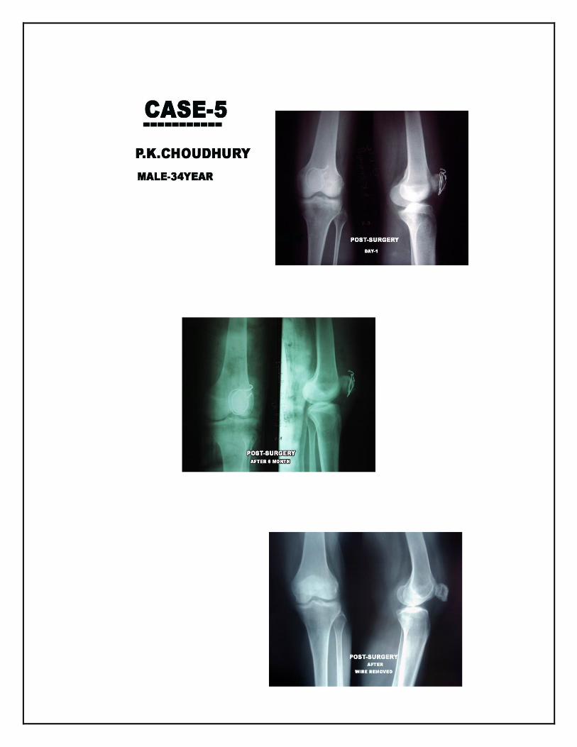
Follow up was continually maintained till the patients had reached maximum recovery
and thereafter at 3 monthly intervals for one year. Wires were removed after 8 months
and within one year under local anesthesia in all of the cases mentioned and advised to
continue physiotherapy.

[9]

[10]

[11]

[12]
[13]

[14]

[15]
OUTCOMES
The results were graded as follows (Table-1)
Out of the 22 cases, 16 were male and 6 were female with an average age of 40 years.
Maximum recovery took place between 6 weeks to one year averaging 5 months, after
removal of the plaster cast and commencement of physiotherapy.
Out of 22 cases, the outcome was excellent in 8, good in 10, fair in 3 and bad in 1.
One of the case aged 48 years with only transosseous suturing walked down
immediate post – op day one without any support felt something pulled out inside the
knee and in consequence he could not regained appreciable extension of knee in
further follow up of one year.
The feeling of subjective weakness persisted in 6 cases. No ossification of tendon was
observed in this series.

[16]
Table-1 : CRITERIA AND GRADING OF OPERATIVE OUTCOMES
Criteria Excellent Good Fair Bad
1. Pain Nil Mild Moderate Severe
2. Wasting Nil < ½” ½ to 1” >1 inch
3. Quadriceps 5 4 + 4 < 4
4. Range of matiod (Rom) of knee
Full No exterior large loss of flexion upto 15 degree
Loss of extension upto 5 degree loss of flexion upto 30 degrees
>5 degree extension lag
>30 degree loss of flexion

[17]
ANALYSIS
The people who are 20 to 50 years old are more vulnerable to fracture the knee cap and
the cases with excellent and good results were of a relatively younger age group.
Men are twice as likely as women to fracture the knee cap, which is reflected in this
series.
Failure was occurred in one case having only transosseous suturing after partial
patellectormy due to powerful contraction of quadriceps which pulled out the wire from
distal fragment. In contrast those who had additional cerclage wiring were more
protected post operatively.
The average follow up is 6 years in the series. However, except in few cases, majority
of patients had no specific complaint and no arthritic changes in knee joints observed.

[18]
DISCUSSION
Opinions differ widely as to the proper treatment of a fractured patella, especially in
reference to patellectomy.
Brooke in 1937 suggested that the patella is inherited phylogenetically and is not a
functional organ. He stated that although the patella is a sesamaid bone, there is no
evidence that it developed in the quadriceps tendon in response to function and rather
the extensor mechanism is more efficient if the patella is excised. Later, however,
Haxton in 1945 and Kaufer in 1971 made complete studies that refute these claims.
They studied the comparative anatomy, human embryology, human anatomy and
experimental anatomy of the patella and also the biomechanical aspects and clinical
results of patellectomy. Haxton stated that anyone who has removed the patella can
certify that the patella actually gives attachment to most of the fibres of the quadriceps

[19]
and patellar tendons and that the bone transmits tension produced by the quadriceps.
In experimental studies of patients with and without patellae he demonstrated that the
power of extension of knee increases as the joint extends, in other words the power
extension is greater with the knee at 30 degree flexion than at 60,90 or 120
degrees.This is true despite the law of Von Schwann,that the tension of muscular
contraction diminishes as muscle fibers shortened. By comparing patients after
patellectomy with normal people, he showed that after patellectomy much of ihis
increase in power as the knee is extended is lost. Since extension is the most important
function of the knee, it must be concluded that patellectomy definitely impairs the
efficiency of the quadriceps mechanism, but this may not be enough to interefere with
ordinary activities.
After platellectomy the effective radius of the patella – quadriceps pull from the center of
the rotation of the knee is shortened, thereby requiring more quadriceps force to
accomplish the same degree of powerful knee extension. The presence of the patella
increases the radius from the centre of rotation of the knee, thereby increasing the

[20]
mechanical advantage of the patella – quadriceps mechanism and making knee
extension more efficient.
Besides this, the patella provides a protective function for the knee and is of cosmetic
value. After total patellectomy, weakness of the quadriceps is due to shortening of the
distance between the axis of movement of the knee and the quadriceps tendon.
Patellectomised knees may require 30% increase in quadriceps power for extension.
Patellectomy decreases quadriceps strength permanently by one-third and reduces joint
stability in half the cases with complaints of giving way. With partial patellectomy, the
continued presence of the pay rope or pulley mechanism and repair of the quadriceps
with reattachment of patellar tendon to the retained bony patella giving a strong hold,
help in good quadriceps function. However, the feeling of subjective weakness, though
there is no demonstrable decrease in quadriceps strength is a known sequelae of
patellectomy, partial or total. This was observed in 6 cases in the present series of
partial patellectomies. Duthie and Hutchison reported pathological ossification may

[21]
develop where the patella was excised which may cause pain and limitation of
movement.
In an experimental study in rabbits, early and severe arthritic changes were recorded in
the femoral condyles after total patellectomy This was not corroborated in human as an
inevitable sequel, especially with avoidance of patellar tilt and consequent patello
femoral arthritis by placing of wire suture correctly in a posterior position through the
fracture surface, the patellar tendon will come in contact principally with the articular
edge of the fragment and not its anterior edge. This procedure was followed in the
present series. To Thomson also goes the credit for the first report on five fractures of
the patella treated by excision of the smaller fragments and capsular repair. Cohn
recommended partial patellectomy whenever possible and showed reduced arthritis as
compared to total patellectomy. Degenerative arthrosis of ageing complicates
evaluation of traumatic arthritis and was not used as a criterion for evaluation of results
in the present series.

[22]
Fixation of the fracture is an alternative. It is however beset with the usual problems of
utilisation of implants. Percutaneous tension band wiring may be used in undisplaced or
slightly displaced closed patella fracture where the gap is less than half a em and the
patient mobilised immediately after the operation. With open tension band wiring, pain
because of skin stretch over the proximal ends of the Kirschner wires with the knee in
flexion, bursa over the K-wires, proximal or distal migration of K-wire, have been
reported. Inability to start early movements because of inability to fix the fractured
patella firmly, especially when comminuted or grossly displaced may compromise
movement of the knee following this intraarticular fracture. Potential incongruities of the
articular surface after fixation may persist with its consequences. Finally, a second
operation for implant removal is required With these drawbacks, partial patellectomy,
whenever possible is a good choice.

[23]
Failure of one case was occurred immediate post – op day which was happened due to
powerful contraction of quadriceps. In view of this failure, an additional cerclage wiring
was given to majority of cases for better protection and early knee movement post
operatively in this series.

[24]
CONCLUSIONS AND RECOMMENDATIONS
Because of the objections to total patellectomy the patella or at least the proximal or
distal half should be tried to save if practical. In simple transverse fractures without
comminution, the patellar fragments are opposed anatomically and internally fixed. If
the distal or proximal pole of the patella is comminuted, the fragments are removed, but
the largest fragment is preserved. When the comminution is extensive and
reconstruction of the articular surface is not possible, complete patellectomy is
preformed.
In addition to transosseous wire suturing, an additional cerclage wiring gives better
protection and early mobilization to patients.

[25]
Lastly, with partial patellectomy, especially of either of the poles, patellar replacement
can still be considered if needed in future. Patients without patella may be at a higher
risk for failure of total knee arthroplasty.

[26]
REFERENCES
1 Haxton HA. The function of the patella and the effects of its excision. Surg Gynec
Obst 1945; 80: 389-395.
2 Einola S, Aho AJ, Kallio P. Patellectomy after fraction long-term follow up results
with special references to functional disability. Acta Orth Scand 1976; 47:441-
447
3 Jensenius H. On the result of excision of the fractured patella Acta Clin Scand 1951;
102:275.
4 Kaufer H. Mechanical function of the patella. J Bone Joint Surg 1971; 53A:551-
560.
5 Lennox IAC, Cobb AG, Knowles J. Long term results of patellectomy, J Bone Joint
Surg Suppl 1, 1992; 74B:112.
6 Bruce J, Walmsley R. Excision of the patella. Some experimental and anatomical
observations. J Bone Joint Surg 1942; 24:311-315.

[27]
7 Thomson JEM. Comminuted fractures of the patella. Treatment of cases presenting
one large fragment and several small fragments. J Bone Joint Surg 1935; 17:431-
434
8 Duthie HL, Hutchinson JR. The results of partial and total excision of the patella. J
Bone Joint Surg 1958; 40B:75-81.
9 Marder RA, Swanson TV, Sharkey NA. Effects of partial patellectomy and
reattachment of the patellar tendon on patellofemoral contact areas and pressures J
Bone Joint Surg 1993; 75A:35-45.
10. Cohn BNE. Total and partial patellectomy. An experimental study Surg Gynec Obst
1944; 79:526-536.
11. Leung PC, Mak KH, Lee SY. Percutaneous tension and wiring a new method of
internal fixation for mildly displaced patella fracture. J Trauma, 1983; 23:62-64.
12. Dudani B, Sancheti KH. Management of fracture patella by tension band wiring. Ind
J Orth 1981; 15:43-48.
13. Larson KR, Crachiola A, Dorey FJ. Total knee arthroplasty in patients after
pateflectomy Clin Orth Rel Res 1991; 264:243-252.