surgical approaches to the shoulder joint in … · buckley r. surgical exposures in or-thopaedics:...
TRANSCRIPT
2916 https://www.journal-imab-bg.org J of IMAB. 2020 Jan-Mar;26(1)
Original article
SURGICAL APPROACHES TO THE SHOULDERJOINT IN UNIPOLAR POST FRACTUREENDOPROSTHESIS REPLACEMENT
Ivaylo MitkovskiOrthopaedics and Traumatology Department, St. Anna General Active TreatmentHospital, Orthopaedics and Traumatology Department, Medical University-Varna, Bulgaria.
Journal of IMAB - Annual Proceeding (Scientific Papers). 2020 Jan-Mar;26(1)Journal of IMABISSN: 1312-773Xhttps://www.journal-imab-bg.org
ABSTRACTThis notification is intended to present our experi-
ence in applying various anatomic landmarks and ap-proaches to the shoulder joint in unipolar post fractureendoprosthesis replacement.
MATERIALS AND METHODS: The period of moni-toring includes the last 5 years. For this period, 35 shoul-der joint aloplastics after a proximal shoulder fracturehave been performed at the Orthopaedics and Traumatol-ogy Department at the Medical University - Varna, St.Anna General Active Treatment Hospital Base. The pa-tients were in the group above 70 years of age; of them,30 were women, and 5 were men. 32 hemiprostheses and3 bipolar prostheses were implanted.
RESULTS: During the unipolar endoprosthesis re-placement, we have used various approaches to the shoul-der joint. To process the results of the arthroplastics per-formed after the proximal humerus fracture, the ConstantShoulder Score method was used with a QuestionnaireCard. This Questionnaire Card investigates the patient’scondition on the fourth week after the intervention. Thelevel of pain, activity and motions in the shoulder are ex-amined. Each one of the indexes gives the respective es-timation, as a result of which the patient’s status is sum-marized. This status varies from “bad” to “excellent”. Theother method used for a statistical result processing wasVAS (Visual analogue scale of pain).
CONCLUSION: Using various approaches to shoul-der joint in aloplastics depends on the reason which hasled to this aloplastics, on the surgeon’s experience andon knowing the anatomic details of the area affected.
Keywords: Humeral Fractures, Shoulder Replace-ment Arthroplasty, Joints, Dislocation Fracture, ShoulderProsthesis, Surgical Approaches,
INTRODUCTIONThe proximal humerus fractures represent 4-5% of
all fractures. These fractures are the third most often lo-cation in elderly patients after the proximal femoral boneand the distal radial bone. Their incidence has increasedsignificantly after the 70's of the 20th century, especiallyin women, according to a Finnish study [1]. Argumentsconclude that if this trend keeps on growing and in thecontext of population ageing, the proximal shoulder bonefractures incidence shall be increased threefold until2030. In 80% of the cases [2], fractures in the proximalhumerus end are slightly dislocated or not dislocated atall. Fragments remain on their places held by the rotatorcuff, joint capsule and the periosteum [3]. Since thesefragments remain stable, the fractures can successfully betreated conservatively. Unfortunately, such type of treat-ment is not applicable to 15% of the patients with unsta-ble fractures, in which a high-energy trauma is responsi-ble for the great dislocation. The trauma type and the dis-location degree are the two causes for often unsatisfac-tory results because of pains, limited motions and loss ofstrength. In order to restore the anatomy close to normal,blood reposition with internal fixation is a method ofchoice for unstable fractures. Surgical treatment, however,is difficult and problematic because of the difficult ac-cess to the fracture and the large fragmentation of pieces.A proximal shoulder bone fracture, which disturbs bloodsupply of the humeral head, may require placement of aprosthesis. In such cases, hemiarthroplastics is a logicalapproach.
MATERIALS AND METHODS:During the endoprosthesis replacement in patients
at the Orthopaedics and Traumatology Clinic, St. AnnaGeneral Active Treatment Hospital, we have used severaldifferent approaches to the shoulder joint.
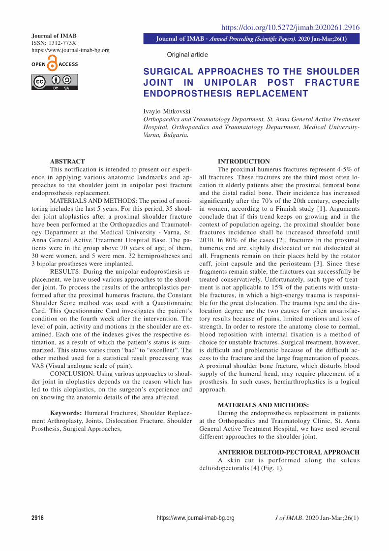
ANTERIOR DELTOID-PECTORAL APPROACHA skin cut is performed along the sulcus
deltoidopectoralis [4] (Fig. 1).
https://doi.org/10.5272/jimab.2020261.2916
J of IMAB. 2020 Jan-Mar;26(1) https://www.journal-imab-bg.org 2917
Fig. 1. Sulcus Deltoidopectoralis – Skin Cut Fig. 3. MusculusDeltoideus and Musculus Pecto-ralis Major
After cutting the skin and subcutaneous tissue, thesulcus between m. deltoideus and m. pectoralis major,filled with fatty tissue, can be seen. There lies v. cefalica,over the anterior edge of m. Deltoideus (Fig. 2).
Fig. 2. Vena Cefalica
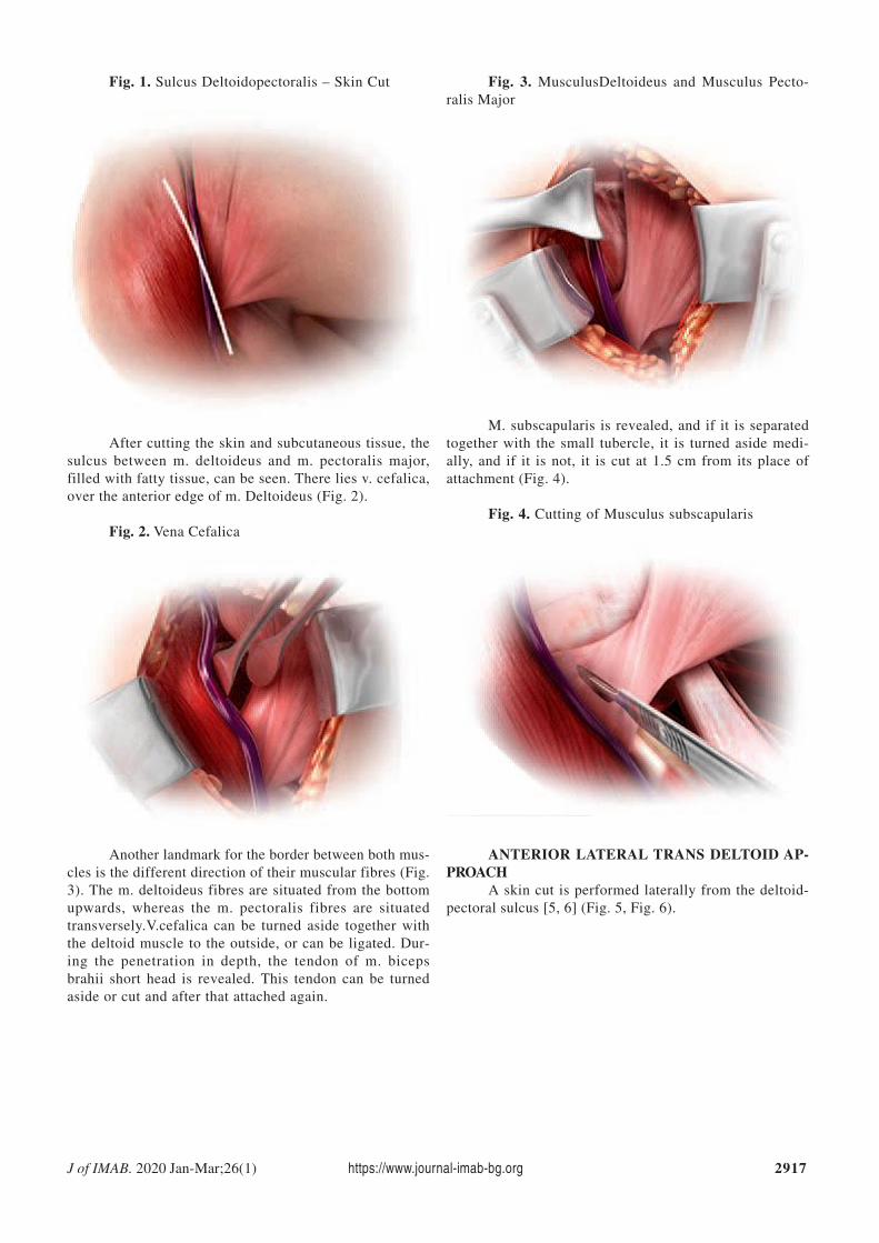
Another landmark for the border between both mus-cles is the different direction of their muscular fibres (Fig.3). The m. deltoideus fibres are situated from the bottomupwards, whereas the m. pectoralis fibres are situatedtransversely.V.cefalica can be turned aside together withthe deltoid muscle to the outside, or can be ligated. Dur-ing the penetration in depth, the tendon of m. bicepsbrahii short head is revealed. This tendon can be turnedaside or cut and after that attached again.
M. subscapularis is revealed, and if it is separatedtogether with the small tubercle, it is turned aside medi-ally, and if it is not, it is cut at 1.5 cm from its place ofattachment (Fig. 4).
Fig. 4. Cutting of Musculus subscapularis
ANTERIOR LATERAL TRANS DELTOID AP-PROACH
A skin cut is performed laterally from the deltoid-pectoral sulcus [5, 6] (Fig. 5, Fig. 6).

2918 https://www.journal-imab-bg.org J of IMAB. 2020 Jan-Mar;26(1)
Fig. 5. Deltoid-Pectoral Sulcus Fig. 7. The Penetration Between the Musculus Del-toideus Without Cutting
Fig. 6. Anterior Lateral Trans Deltoid Approach –Skin Cut
N. musculocutaneous is revealed and is turned asidetogether with the coracobrachial muscle. The penetrationbetween the m. deltoideus is performed without cuttingthem in order to preserve their innervation and preventany possibility of subsequent atrophy (Fig. 7). The nextsteps are identical with those at the anterior deltoid-pec-toral approach.
UPPER LATERAL APPROACHThe skin cut is 10-12 cm, it could be performed
close to the beginning or close to the end of the acromionlateral edge, or in the lateral direction [7] (Fig. 8). Fol-lowing the subcutaneous dissection, anterior and middledeltoid muscular ligaments, located towards the acromionlateral end, are detached, using a rounded dissection [8].
Fig. 8. Upper Lateral Approach
The dissection starts on the acromioclavicular jointlevel, on 5-7 mm after the acromion top, and continuesstraight laterally down along the deltoid muscle. It shouldnot go on more than 4 cm from the deltoid muscle’s ex-ternal part, in order to protect n. axillary, which is locatedbelow the subacromial bursa fold.

J of IMAB. 2020 Jan-Mar;26(1) https://www.journal-imab-bg.org 2919
1. Palvanen M, Kannus P, Niemi S,Parkkari J. Update in theepidemiologyof proximal humeralfractures. Clin Orthop Relat Res. 2006Jan;442:87-92. [PubMed] [Crossref]
2. Takov E, Tivchev P. [The Frac-ture diagnostics and treatment.] Venel(Bulgaria). 1996. [in Bulgarian]
3. Foster RJ, Dixon GL Jr, BachAW, Appleyard RW, Green TM. Inter-nal fixation of fractures and non-un-ions of the humeral shaft. Indicationsand results in a multi-center study. JBone Joint Surg Am. 1985Jul;67(6):857-64. [PubMed]
4. Lädermann A, Lo EY,Schwitzguébel AJ, Yates E. Subscapu-laris and deltoid preserving anteriorapproach for reverse shoulder arthro-
plasty. Orthop Traumatol Surg Res.2016 Nov;102(7):905-908. [PubMed][Crossref]
5. Marinello PG, Amini MH, PeersS, O’Donnell J, Iannotti JP. Reverse to-tal shoulder arthroplasty with com-bined deltoid reconstruction in pa-tients with anterior and/or middle del-toid tears. J Shoulder Elbow Surg.2016 Jun;25(6):936-41. [PubMed][Crossref]
6. Hoppenfeld S, de Boer P,Buckley R. Surgical Exposures in Or-thopaedics: The Anatomic Approach.5th ed. Wolters Kluwer. October 14,2016.
7. Boichev B. [Orthopaedics andTraumatology Surgery.] Sofia: Medi-cina i Fizkultura. 1983; 135-42. [in
When the subacromial bursa is visible, a carefullongitudinal pull away along the extremity length allowsa retractor to be placed in the subacromial space (Fig. 9).Then the anterior deltoid part is released from the acro-mial insertion to the acromioclavicular joint. Detachingthe deltoid from the acromion anterior part may includea small part of the bone, in order to facilitate restorationand to protect the deltoid muscle. After that, the extrem-ity is rotated outwards, and the head is moved onwardsand upward in order to perform a correct positioning. Ifthe biceps is still visible, a tenotomy or a tenodesis hasto be performed [9, 10, 11]. M. subscapularis, m. teres mi-nor and m. infraspinatus are held when visible. A partialdetachment of m. subscapularis could be performed, whenit is hard to obtain a larger dislocation of the humerus.
Fig. 9. Subacromial Bursa
LATERAL TRANS DELTOID APPROACHBasically, at this approach, the n. axcilaris passage
should be taken into consideration. The n.axcilaris goesfrom the back to the front transversely to the muscularfibres to about 5 cm from the acromioclavicular joint andthe beginning of the deltoid muscle. Therefore, the lon-gitudinal separation of muscular fibres should be no morethan 3-4 cm from the muscle beginning (Fig. 10).
Fig. 10. Lateral Trans Deltoid Approach andNervusAxcilaris
CONCLUSION:Using a certain approach to the shoulder joint at
unipolar post fracture endoprosthesis replacement dependson several factors. On the one hand, from the type of theendoprosthesis used - monoblock, bicomponent or reverseprosthesis.The surgeon’s experience and preferences areessential. On the other hand, the available working baseand surgical equipment are also substantial.
The main approaches to the shoulder joint, whichwe use most often in cases of unipolar post fractureendoprosthesis replacement, are the deltoid-pectoral ap-proach and the anterior-lateral trans deltoid approach.
REFERENCES:Bulgarian]
8. Duranthon LD, VandenbusscheE, Goubier JN, Augereau B. [Thesuperolateral approach for shoulderprosthesis]. [in French] Rev ChirOrthop Reparatrice Appar Mot. 2002Jun;88(4):415-9. [PubMed]
9. Hassan S, Patel V. Biceps tenod-esis versus biceps tenotomy for bicepstendinitis without rotator cuff tears. JClin Orthop Trauma. 2019 Mar-Apr;10(2):248-256. [PubMed][Crossref]
10. Dines DM, Tuckman D, DinesJ. Hemiarthroplasty for complex four-part fracture of the proximal humerus:technical considerations and surgicaltechnique. Univ Pennsyl Orthop J.2002; 15:29-36.

2920 https://www.journal-imab-bg.org J of IMAB. 2020 Jan-Mar;26(1)
Addresses for correspondence:Dr. Ivaylo Mitkovski,Medical University – Varna55, Marin Drinov Str., 9002 Varna, Bulgaria,E-mail: [email protected]
Please cite this article as: Mitkovski I. Surgical Approaches to the Shoulder Joint in Unipolar Post Fracture EndoprosthesisReplacement. J of IMAB. 2020 Jan-Mar;26(1):2916-2920. DOI: https://doi.org/10.5272/jimab.2020261.2916
Received: 21/03/2019; Published online: 17/02/2020
11. Hempfing A, Leunig M,Ballmer FT, Hertel R. Surgical land-marks to determine humeral headretrotorsion for hemiarthroplasty infractures. J Shoulder Elbow Surg. 2001Sep-Oct;10(5):460-3. [PubMed][Crossref]