surgical anatomy of nose mah
TRANSCRIPT
Surgical Anatomy of The Nose
M.A.H
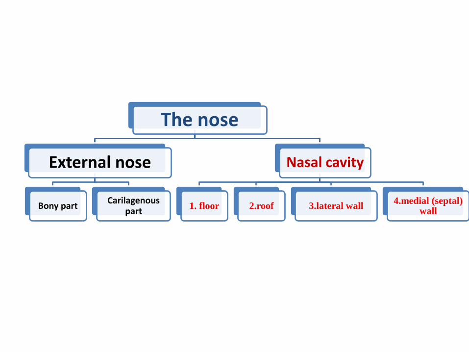
The nose
External nose
Bony partCarilagenous
part
Nasal cavity
1. floor 2.roof 3.lateral wall4.medial (septal)
wall

External nose:-
- pyramidal in shape
- it has root continous with forehead , and apex which is its free edge.
- lateral surface of nose called dorsum nasi, which end below in rounded ala nasi .
- external nose compose of :
- - Bones
- Hyline cartilage
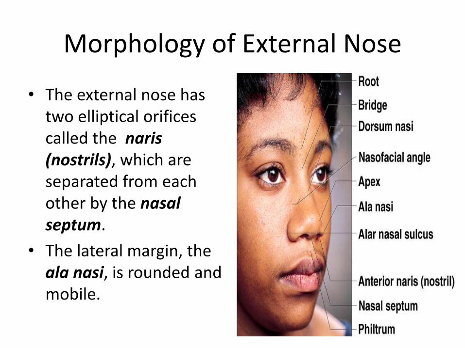
Morphology of External Nose
• The external nose has two elliptical orifices called the naris (nostrils), which are separated from each other by the nasal septum.
• The lateral margin, the ala nasi, is rounded and mobile.
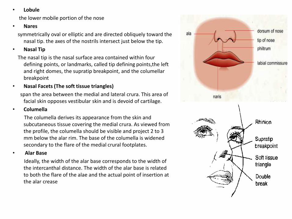
• Lobule
the lower mobile portion of the nose
• Nares
symmetrically oval or elliptic and are directed obliquely toward the nasal tip. the axes of the nostrils intersect just below the tip.
• Nasal Tip
The nasal tip is the nasal surface area contained within four defining points, or landmarks, called tip defining points,the left and right domes, the supratip breakpoint, and the columellarbreakpoint
• Nasal Facets (The soft tissue triangles)
span the area between the medial and lateral crura. This area of facial skin opposes vestibular skin and is devoid of cartilage.
• Columella
The columella derives its appearance from the skin and subcutaneous tissue covering the medial crura. As viewed from the profile, the columella should be visible and project 2 to 3 mm below the alar rim. The base of the columella is widened secondary to the flare of the medial crural footplates.
• Alar Base
Ideally, the width of the alar base corresponds to the width of the intercanthal distance. The width of the alar base is related to both the flare of the alae and the actual point of insertion at the alar crease
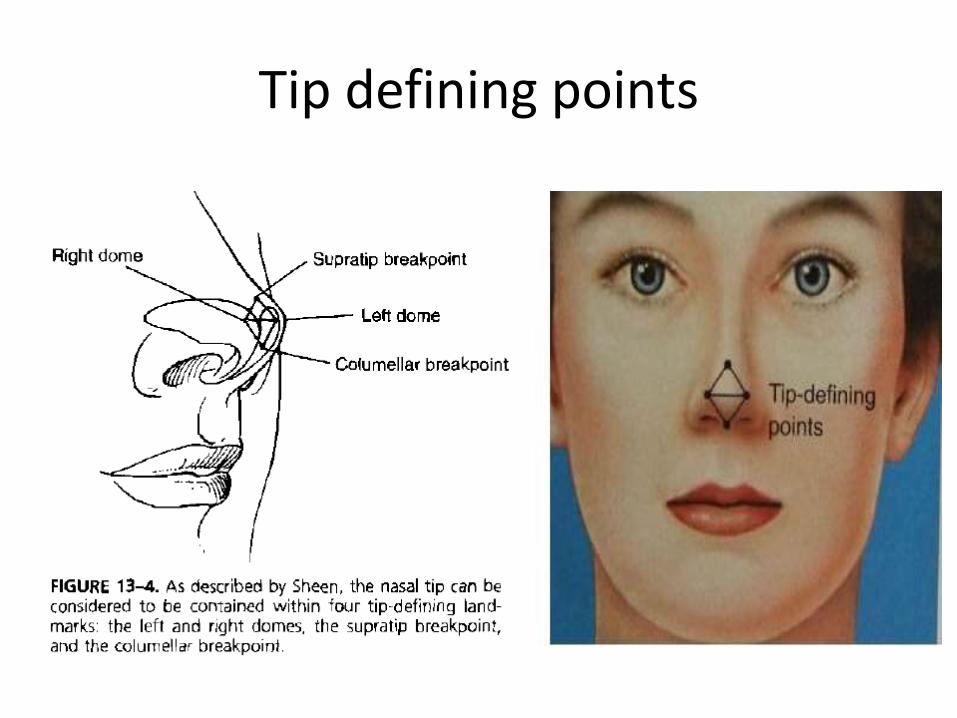
Tip defining points
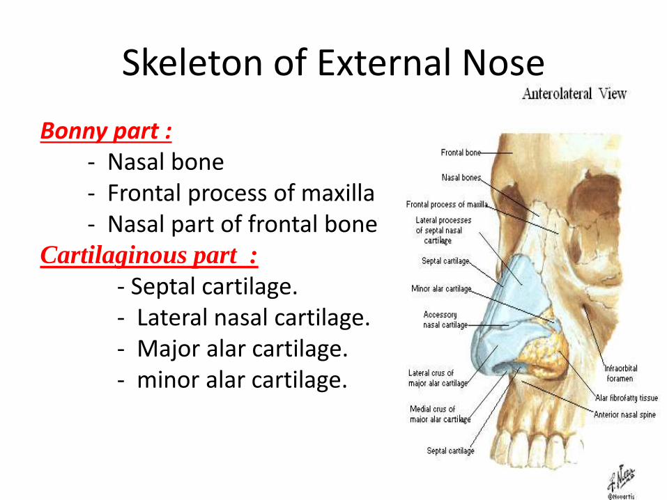
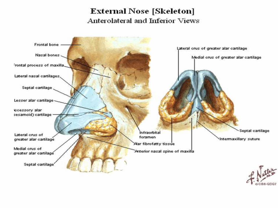
Skeleton of External Nose
Bonny part :- Nasal bone - Frontal process of maxilla - Nasal part of frontal bone
Cartilaginous part :
- Septal cartilage.- Lateral nasal cartilage.- Major alar cartilage.- minor alar cartilage.
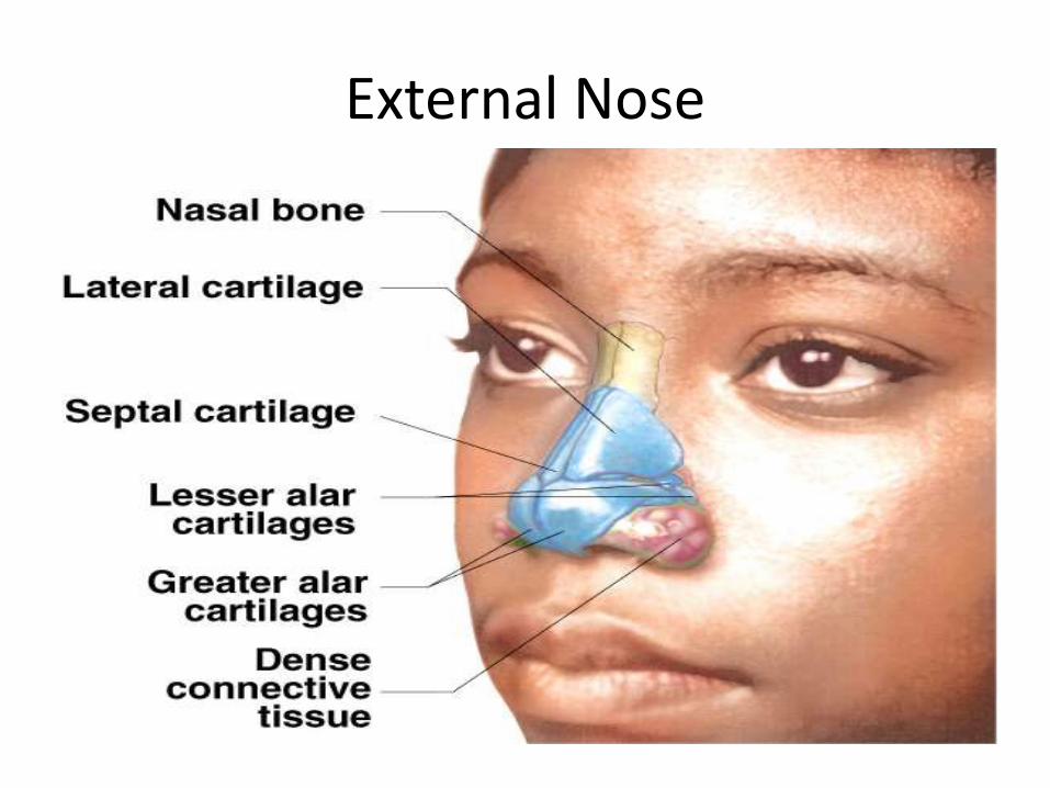
External Nose
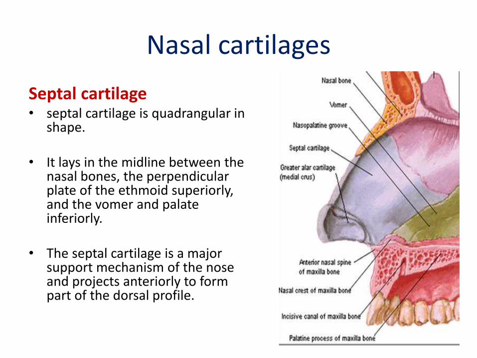
Nasal cartilages
Septal cartilage• septal cartilage is quadrangular in
shape.
• It lays in the midline between the nasal bones, the perpendicular plate of the ethmoid superiorly, and the vomer and palate inferiorly.
• The septal cartilage is a major support mechanism of the nose and projects anteriorly to form part of the dorsal profile.
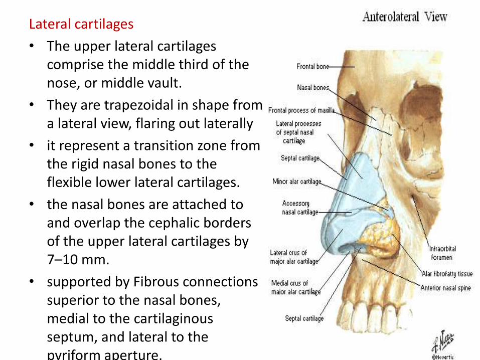
Lateral cartilages
• The upper lateral cartilages comprise the middle third of the nose, or middle vault.
• They are trapezoidal in shape from a lateral view, flaring out laterally
• it represent a transition zone from the rigid nasal bones to the flexible lower lateral cartilages.
• the nasal bones are attached to and overlap the cephalic borders of the upper lateral cartilages by 7–10 mm.
• supported by Fibrous connections superior to the nasal bones, medial to the cartilaginous septum, and lateral to the pyriform aperture.
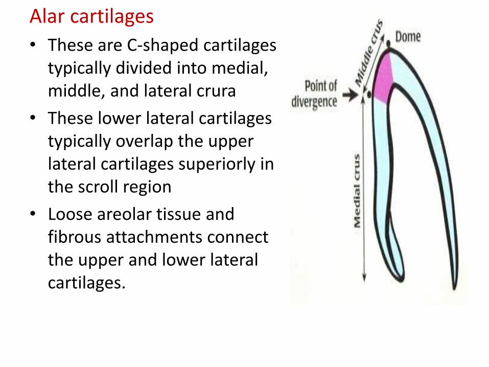
Alar cartilages
• These are C-shaped cartilages typically divided into medial, middle, and lateral crura
• These lower lateral cartilages typically overlap the upper lateral cartilages superiorly in the scroll region
• Loose areolar tissue and fibrous attachments connect the upper and lower lateral cartilages.

• The medial crura form the cartilaginous support for the columella
• Aesthetically, only 2–4 mm of columellar show should be present below the alar margins from a profile View. Anything more than this reflects a “hanging columella” or alar retraction.
• while any smaller distance represents columellar retraction.
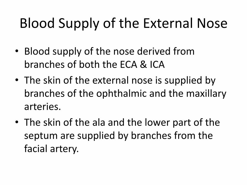
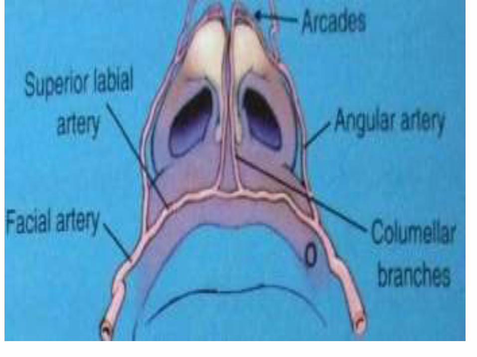
Blood Supply of the External Nose
• Blood supply of the nose derived from branches of both the ECA & ICA
• The skin of the external nose is supplied by branches of the ophthalmic and the maxillary arteries.
• The skin of the ala and the lower part of the septum are supplied by branches from the facial artery.
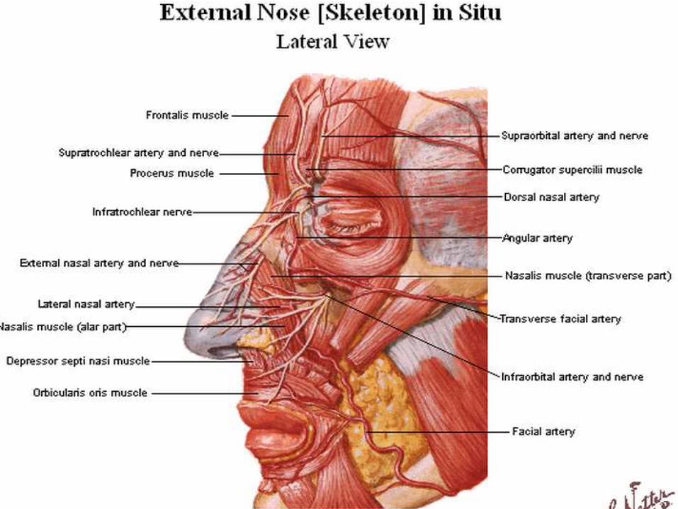
Nerve Supply of the External Nose
• The infratrochlear and external nasal branches of the ophthalmic nerve (CN V) and the infraorbital branch of the maxillary nerve (CN V).
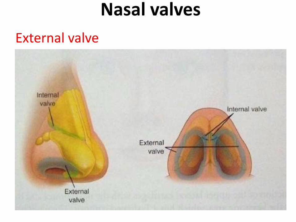
Nasal valves
External valve

Internal valve
The junction of the upper lateral cartilages with the nasal septum forms the internal nasal valve. This valve angle should be between 10 and 15° for adequate nasal airflow
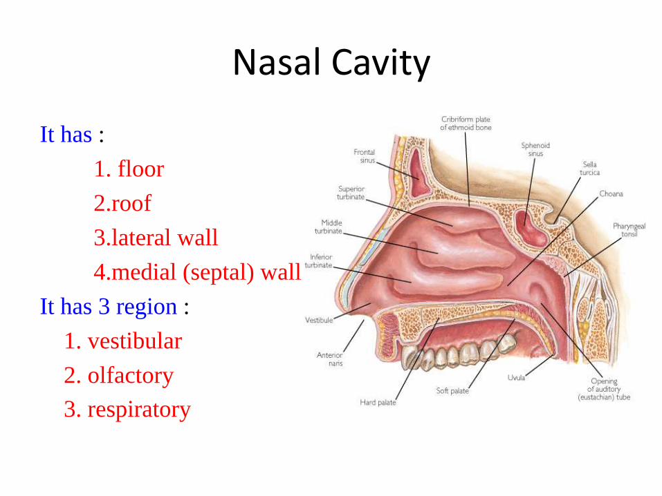
Nasal Cavity
It has :
1. floor
2.roof
3.lateral wall
4.medial (septal) wall
It has 3 region :
1. vestibular
2. olfactory
3. respiratory
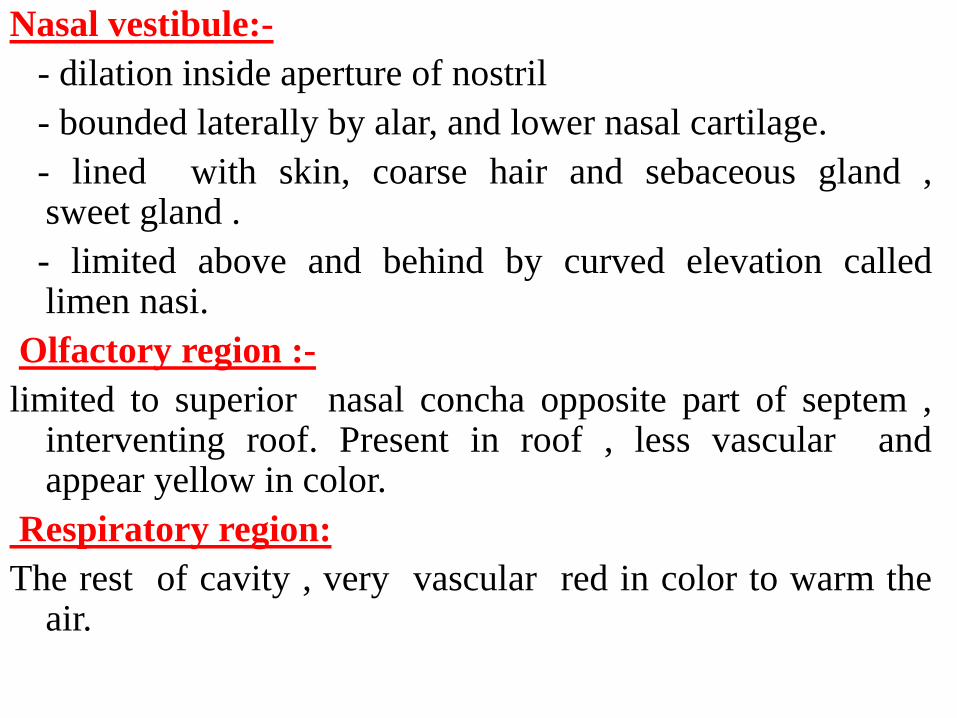
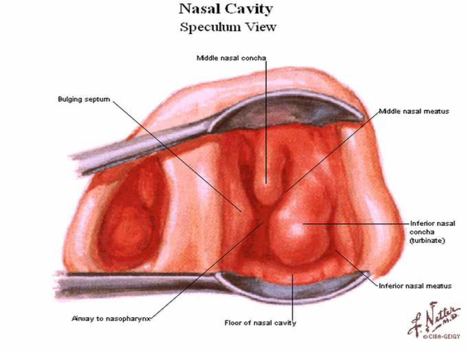
Nasal vestibule:-
- dilation inside aperture of nostril
- bounded laterally by alar, and lower nasal cartilage.
- lined with skin, coarse hair and sebaceous gland ,sweet gland .
- limited above and behind by curved elevation calledlimen nasi.
Olfactory region :-
limited to superior nasal concha opposite part of septem ,interventing roof. Present in roof , less vascular andappear yellow in color.
Respiratory region:
The rest of cavity , very vascular red in color to warm theair.
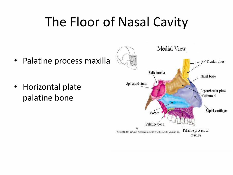
The Floor of Nasal Cavity
• Palatine process maxilla
• Horizontal plate palatine bone

The Roof of Nasal Cavity
• Narrow
• It is formed – anteriorly beneath the bridge
of the nose by the nasal and frontal bones,
– in the middle by the cribriform plate of the ethmoid,
– located beneath the anterior cranial fossa,
– posteriorly by the downward sloping body of the sphenoid
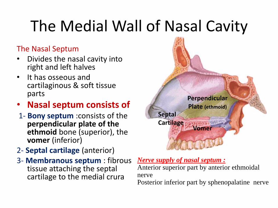
The Medial Wall of Nasal Cavity The Nasal Septum• Divides the nasal cavity into
right and left halves• It has osseous and
cartilaginous & soft tissue parts
• Nasal septum consists of 1- Bony septum :consists of the
perpendicular plate of the ethmoid bone (superior), the vomer (inferior)
2- Septal cartilage (anterior)3- Membranous septum : fibrous
tissue attaching the septal cartilage to the medial crura
Perpendicular Plate (ethmoid)
Septal Cartilage
Vomer
Nerve supply of nasal septum :Anterior superior part by anterior ethmoidalnerve Posterior inferior part by sphenopalatine nerve
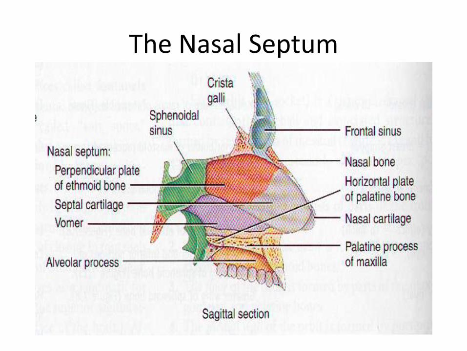
The Nasal Septum
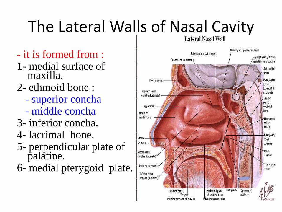
The Lateral Walls of Nasal Cavity
- it is formed from :1- medial surface of
maxilla.2- ethmoid bone :
- superior concha- middle concha
3- inferior concha.4- lacrimal bone.5- perpendicular plate of
palatine.6- medial pterygoid plate.
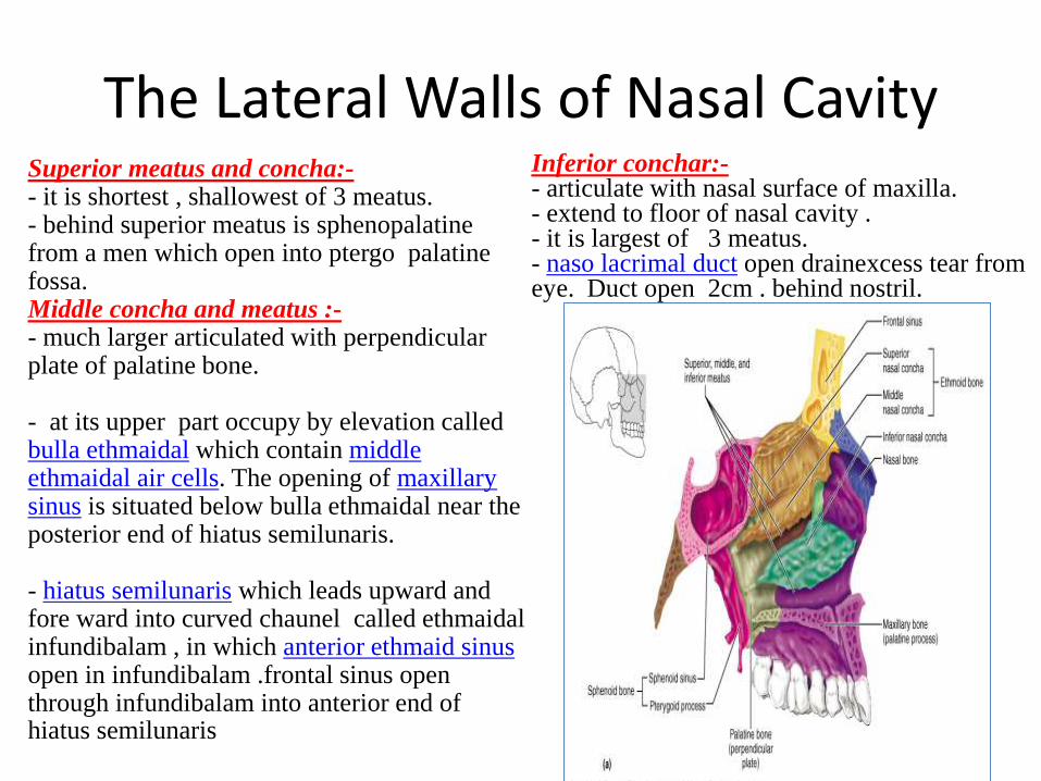
The Lateral Walls of Nasal Cavity Superior meatus and concha:-- it is shortest , shallowest of 3 meatus.- behind superior meatus is sphenopalatinefrom a men which open into ptergo palatine fossa.Middle concha and meatus :-- much larger articulated with perpendicular plate of palatine bone.
- at its upper part occupy by elevation called bulla ethmaidal which contain middle ethmaidal air cells. The opening of maxillary sinus is situated below bulla ethmaidal near the posterior end of hiatus semilunaris.
- hiatus semilunaris which leads upward and fore ward into curved chaunel called ethmaidalinfundibalam , in which anterior ethmaid sinusopen in infundibalam .frontal sinus open through infundibalam into anterior end of hiatus semilunaris
Inferior conchar:-- articulate with nasal surface of maxilla.- extend to floor of nasal cavity .- it is largest of 3 meatus.- naso lacrimal duct open drainexcess tear from eye. Duct open 2cm . behind nostril.
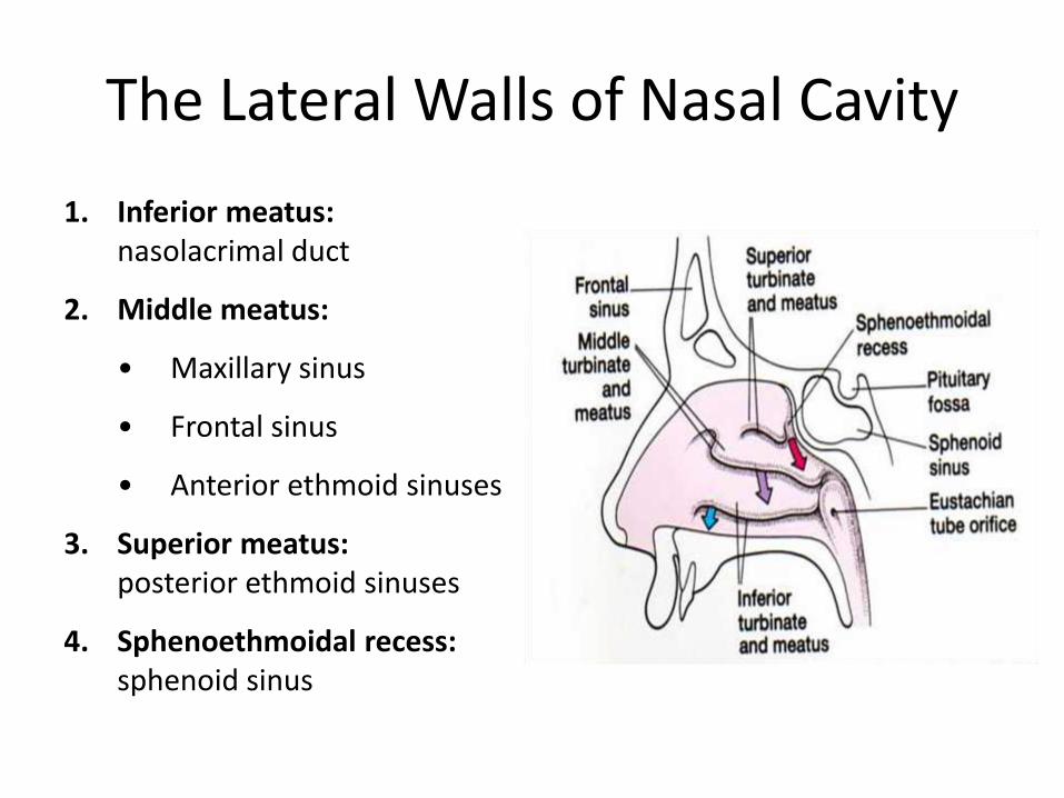
The Lateral Walls of Nasal Cavity
1. Inferior meatus: nasolacrimal duct
2. Middle meatus:
• Maxillary sinus
• Frontal sinus
• Anterior ethmoid sinuses
3. Superior meatus: posterior ethmoid sinuses
4. Sphenoethmoidal recess: sphenoid sinus
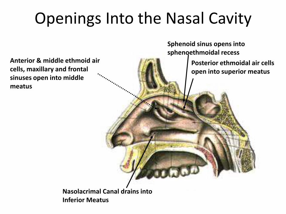
Openings Into the Nasal Cavity
Nasolacrimal Canal drains intoInferior Meatus
Sphenoid sinus opens into sphenoethmoidal recess
Posterior ethmoidal air cells open into superior meatus
Anterior & middle ethmoid air cells, maxillary and frontal sinuses open into middle meatus
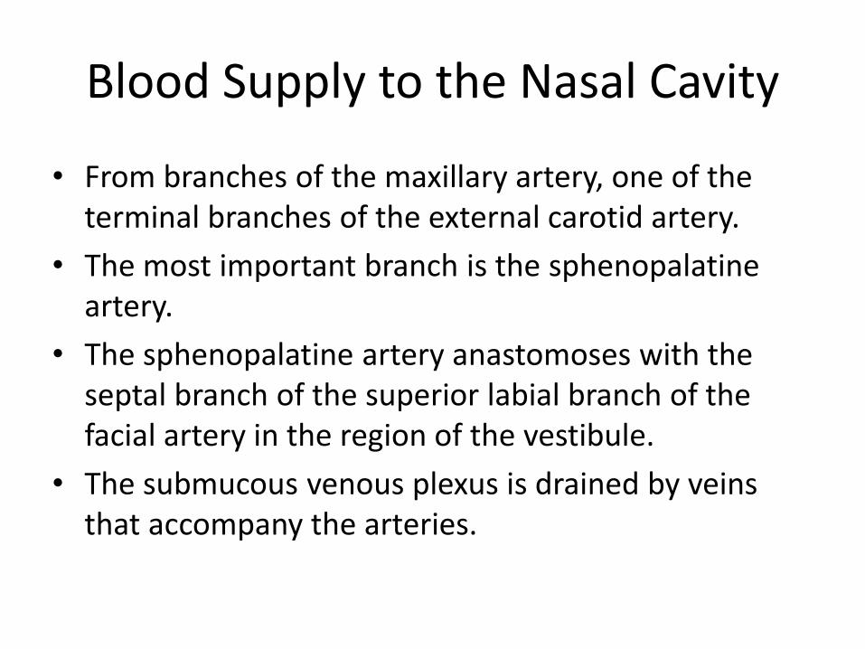
Blood Supply to the Nasal Cavity
• From branches of the maxillary artery, one of the terminal branches of the external carotid artery.
• The most important branch is the sphenopalatineartery.
• The sphenopalatine artery anastomoses with the septal branch of the superior labial branch of the facial artery in the region of the vestibule.
• The submucous venous plexus is drained by veins that accompany the arteries.
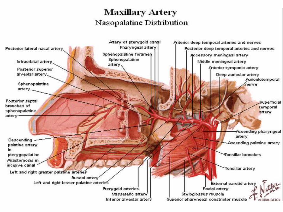
Blood Supply to the Nasal Cavity
Sphenopalatine a.
Maxillary a.
Netter, Frank H., Atlas of Human Anatomy. Ciba-Geigy Corporation, Summit, N.J. 1993. Plate 35.
Nerve Supply of the Nasal Cavity
• The olfactory nerves from the olfactory mucous membrane ascend through the cribriform plate of the ethmoid bone to the olfactory bulbs .
• The nerves of ordinary sensation are branches of the ophthalmic division (V1) and the maxillary division (V2) of the trigeminal nerve.
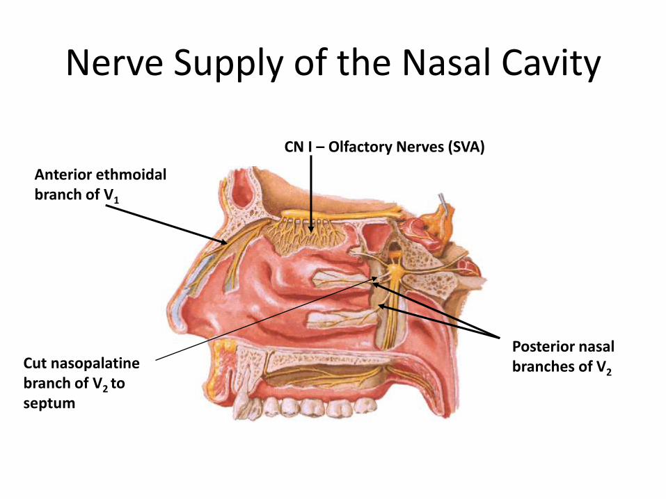
Nerve Supply of the Nasal Cavity
CN I – Olfactory Nerves (SVA)
Anterior ethmoidalbranch of V1
Posterior nasal branches of V2
Cut nasopalatinebranch of V2 to septum
Lymph Drainage of the Nasal Cavity
• The lymph vessels draining the vestibule end in the submandibular nodes.
• The remainder of the nasal cavity is drained by vessels that pass to the upper deep cervical nodes.