surger - coffee break cornercoffeebreakcorner.weebly.com/uploads/5/1/0/5/51059527/...rina —* ball...
TRANSCRIPT

t
fe
SURGER
; [6th edition (201.3)
umbilical
inguinal~r?—<r
epigastrica ^"~3tti
femoral
£,<•• •.*<--- ;, , ,..•'•
tas^^o^
i
a

Pioneer ofSurgeryfor under graduate
# Layers of the anterior abdominal wall are: (from outwards inwards)
1- Skin & S.C. tissue 2- Superficial fascia
3- Abdominal muscles
— 5- Extra-peritoneal fat
4- Fascia transversalis
6- Peritoneum
Superficial fascia:
Hernia -,t-
Below the umbilicus it is divided into 2 layers:
_
1- Superficial fatty layer —•> Camper's fascia
2- Deep membranous layer —> Scarpei's fascia —>
* It is continuous downward with the fascia lata of the thigh
* Transverse line 5cm below & parallel to the inguinal ligament (Holden 's line)
* Surgical importance: prevent the following from descending into the thigh:
1- Femoral hernia
2- Air in surgical emphysema
3- Urine in rupture membranous urethra & extra-peritoneal rupture of U.B.
Line of fusion () scarpie'sfascia & fascia lataof the thigh
L
Abdominal muscles:
5 muscles:
2 paramedian —> rectus abdominis & pyramidalis
3 anterolateral—» External oblique - internal oblique- transversus abdominus
- Symphysis pubis
1 - Rectus abdominis;
** Origin: - Pubic crest
** Direction: vertically upwards
** Segments: 3 or 4 segment
lsl: Xiphistcrnum
3rd: umbilicus 4m: below umbilicus
(These indicate segmental origin ofthe muscle from fusion ofthe lower 6 thoracic mvotoms)
** Insertion: Xiphoid process &5lh,6Ul: 7,h costal cartilages
inc)2 : midway () xiph. &. umbilicus I I7f_ith.
A.F. A.F. ,
i— ' Pioneer ofSurgeryfor under graduate.'•—-Jr " '
is;- 2-
u** Not always present
** Origin: pubic crest
** Insertion: linea albe 1 inch above its origin
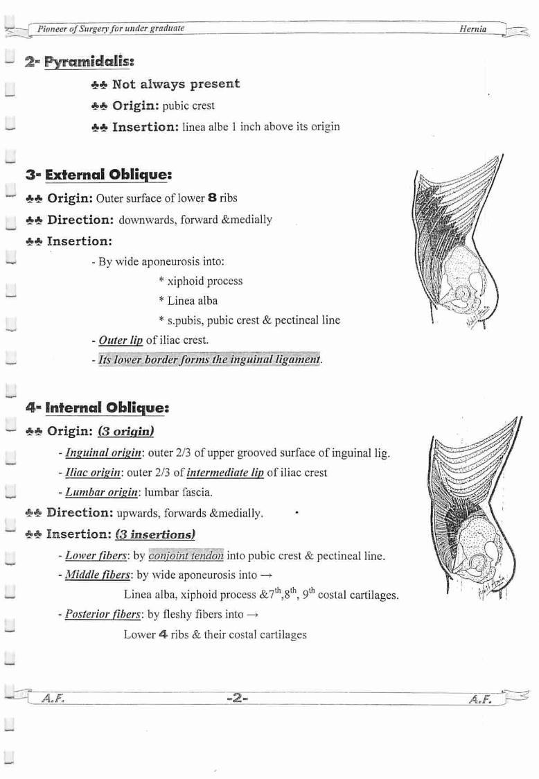
3- External Oblique;
** Origin: Outer surface of lower 8 ribs
** Direction: downwards, forward &medially
** Insertion:
- By wide aponeurosis into:
* xiphoid process
* Linea alba
* s.pubis, pubic crest & pectineal line
- Outer lip of iliac crest.
_
_
_
_
u
u
- Its lower borderforms the inguinal ligament.
4- Internal Oblique;
** Origin: (3 origin)
- Inguinal origin: outer 2/3 of upper grooved surface of inguinal lig.
- Iliac origin: outer 2/3 of intermediate lip of iliac crest
- Lumbar origin: lumbar fascia.
** Direction: upwards, forwards &medially.
** Insertion: (3 insertions)
- Lower fibers: by conjoint tendon into pubic crest & pectineal line.
- Middle fibers: by wide aponeurosis into —*•
Linea alba, xiphoid process &7th,8th, 9th costal cartilages.- Posterior fibers: by fleshy fibers into -»
Lower 4- ribs & their costal cartilages
^CaE -2-
Hernia J&
A.F.~

Pioneer ofSurgeryfor under graduate
U 5- Transversus Abdominus;
_
** Origin: (4 origins)
- Costal: inner surface of lower 6 ribs
- Lumbar: lumbar fascia
- Iliac: ant. 2/3 of inner lip of iliac crest
— - Inguinal: outer 1/3 of upper grooved surface of inguinal ligament
** Direction: Transverse & inwards
** Insertion: (2 insertions)
_ - By wide aponeurosis into xiphoid process & linea alba
- By conjointtendon into pubic crest & pectineal line
— ** Nerve supply of all anterior wall muscles:
Lower 6 thoracic nerves & 1st lumbar nerves
Hernia
U Fascia Transversalis- Thin but strong fasical layer that lines the inner surface of transversus abdominis muscle & is
— separated from peritoneum by fatty tissue
- It is the most important defense mechanism against hernia formation
- The lower part of fascia transversalis is thickened to form the ileo pupic tract which runs justabove & parallel to the inguinal ligament.
Rectus sheatfi
** formed by:
L. * Above a point midway () umbilicus &s.pubis:
- Ant. Wall: 1- ext. oblique aponeurosis •
2- ant.lamina of int. oblique
- Post. Wall: 1- post. Lamina of int. oblique
2- trans. Abd. aponeurosis
_
'S^AT. -3-
Fused with fascia
transversalis* Below a point midway () umbilicus & s. pubis: (arcuate line)
- Ant. Wall: 1- ext. obi. aponeurosis
2- int. obi. Aponeurosis
3- trans. Abdom. Aponeurosis
- Post. Wall: deficient (rectus lies on fascia transversalis)
A, f. •<

I I
i i
L
** Contents:
1- 2 muscles'. 1- rectus abdom
2- 4 vessels:
2- pyramidalis
1- Sup. Epigastart. 2- Sup. Epigast.vein
3-Inf. Epigast. Art. 4-Inf. Epigast. Vein
3- 6 nerves: 1-Lower 5 intercostal ns 2- subcostal ns.
** Applied anatomy:
1- The nerves enter the sheath from its lateral side -• So, the rectus muscle should retractedlaterally in paramedian incision
2-InKocher incision -> muscle cutting ofrectus sheath
3-Rectus muscle can beused in breast reconstruction -»• TRAM
4- In cancer breast affecting the lower inner quadrant lymphatic spread to liver occursthrough lymphatics ofrectus sheath
5- Desmoid tumor -»tumors arise from rectus sheath-* locally malignant tumor
•Araphe formed by interlacing aponeurotic fibers forming rectus sheath.•It extends from xiphoid process to s. pubis•Above umbilicus —>1 cm wide
•Below umbilicus -> it is narrow & difficult to define.
-4- \.F.
"^—J Pioneer ofSurgeryfor under graduate Hernia
_
L
Inguinal ligament
*4» Formation: by lower border ofext. oblique aponeurosis which is thickened & reflected
upon itself post.
ۥ* Extent: from ASIS to pubic tubercle.
** Attachments:
* Its upper grooved surface gives attachment to:
1- Int. oblique (from outer 2/3)
2- Trans. Abd. (from outer 1/3)
* Its lower convex surface is firmly attached to fascia lata of thigh.
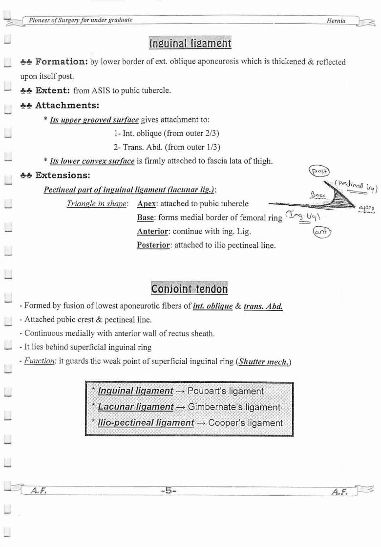
** Extensions:
Pectineal part ofinguinal ligament (lacunar lig.):
Triangle in shape: Apex: attached to pubic tubercle
Base: forms medial border of femoral ring (—*^- U*\\
Anterior: continue with ing. Lig. (onT)
Posterior: attached to ilio pectineal line.
Conjoint tendon• • • •
- Formed by fusion of lowest aponeurotic fibers of/'///. oblique & trans. Abd.
- Attached pubic crest & pectineal line.
- Continuous medially with anterior wall of rectus sheath.
— - It lies behind superficial inguinal ring
- Function: it guards the weak point of superficial inguinal ring (Shutter mech.)
IF.
* Inguinal ligament —* Poupart's ligament
* Lacunar ligament.—* Gimbernate's ligament
* llio-pectineal ligament —> Cooper's ligament
-5- lfT^P^

Pioneer ofSurgeryfor undergraduate Hernia
_
Inguinal Canal
** Definition: an oblique passage in the lower part of ant. Abd. wall.
_ ** Site: just above medial 1/2 of ing. Lig.
** Length: 1.5 inch = 4 cm
** Direction: downward, forward & medially.
** Extent: from deep inguinal ring to superficial inguinal ring.
@ Deep (Internal) inguinal ring":
— - U shaped opening in fascia transversalis
- Situated 1/2 inch above mid point of inguinal ligament lateral to inferior epigastric art.
- Transmits: In male: spermatic cord
/// female: round ligament of uterus.
@ Superficial (external) inguinal ring:
— - Triangular opening in ext. oblique apon.
- Situated 1/2 inch above &lateral to pubic tubercle &medial end of ing. Lig.- Triangle in shape it has:
=apex: directed upward & laterally
=base: directed downward & medially.
— =2 sides called crura: held together by inter crural fibers ofext. oblique aponeurosis- Transmits: In male: spermatic cord & ilio-inguinal nerve
//; female: round ligament of the uterus & ilio-inguinal nerve
-
Mid point of inguinal ligament -> Midway () ASIS &pubic tubercle (Lateral)
Mid inguinal point -+ Midway ( ) ASIS &symphysis pubis (Medial)
=?""—[ Pioneer ofSurgeryfor under graduate
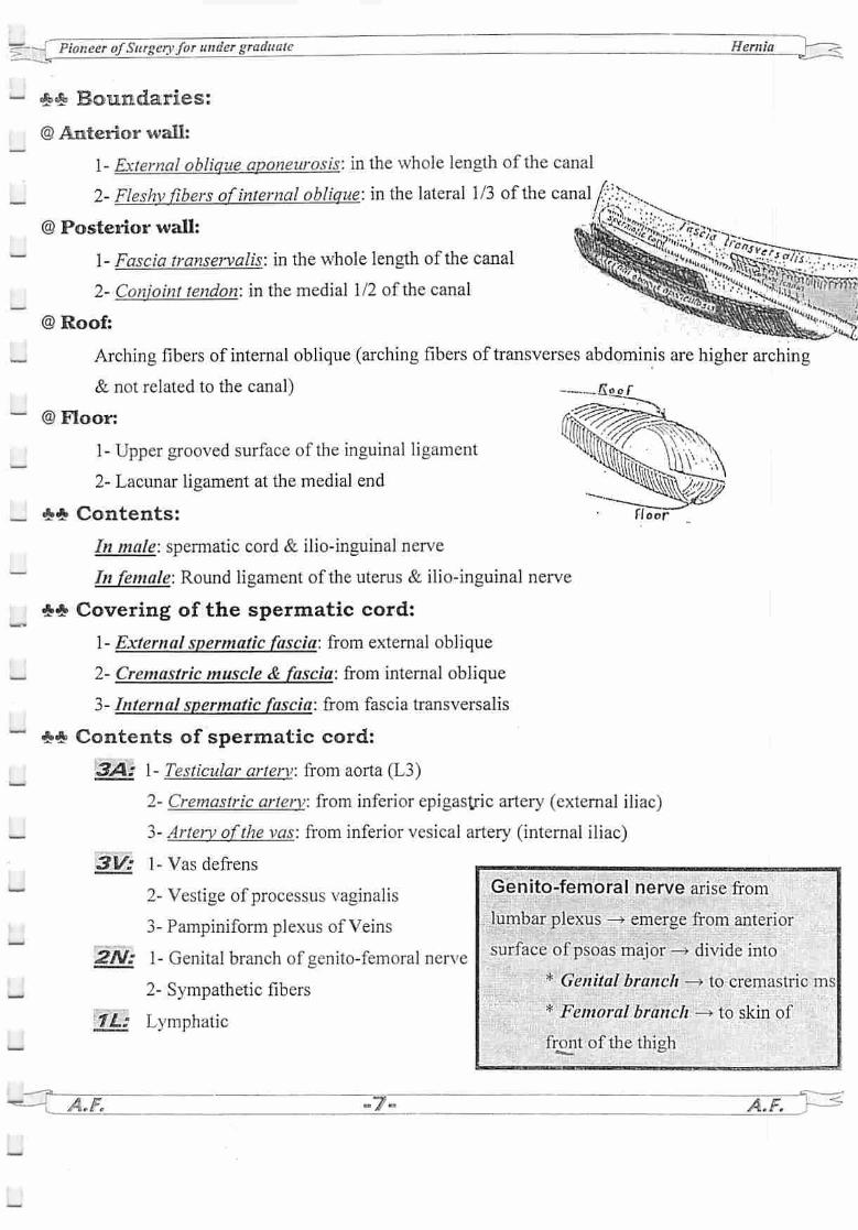
** Boundaries:
@ Anterior wall:
1- External oblique aponeurosis: in the whole length of the canal
2-Fleshy fibers of internal oblique: in the lateral 1/3 of the canal /?.•?>...
@ Posterior wall:
1- Fascia transervalis: in the whole length of the canal
2- Conjoint tendon: in the medial 1/2 of the canal
©Roof:
Arching fibers of internal oblique (arching fibers of transvcrses abdominis are higher arching
& not related to the canal)
@ Floor:
1- Upper grooved surface of the inguinal ligament
2- Lacunar ligament at the medial end
*«*> Contents:
In male: spermatic cord & ilio-inguinal nerve
//; female: Round ligament of the uterus & ilio-inguinal nerve
** Covering of the spermatic cord:
1- External spermatic fascia: from external oblique
2- Cremastric muscle & fascia: from internal oblique
3- Internal spermatic fascia: from fascia transversalis
** Contents of spermatic cord:
3A: 1- Testicular arterv: from aorta (L3)
2- Cremastric artery: from inferior epigasU"ic artery (external iliac)
3- Artery ofthe vas: from inferior vesical artery (internal iliac)
3V: 1-Vasdefrens
Genito-femoral nerve arise from
Soof
floor
Hernia
2N:
2- Vestige of processus vaginalis
3- Pampiniform plexus of Veins
1- Genital branch of genito-femoral nerve
2- Sympathetic fibers
lumbal- plexus —> emerge from anterior
surface of psoas major —> divide into
* Genital branch —> to cremastric ms
* Femoral branch —* to skin of
front of the thigh1L: Lymphatic
i^krtw ^7^ A.F.
27"i Pioneer ofSurgeryfor undergraduate^ Hernia j-
~~ ** Sex difference:
It is wider in ntsile than female so that are more liable to inguinal hernia than female
** Integrity of the canal:
A- anatomical:
1- Obliquity of the canal as the superficial ring &the deep ring don't lie opposite eachother -* So. when Tintra-abdominal pressure -» approximation of anterior & posteriorwall -*
closure of the canal —> flan valve mechanism
2- The superficial ring supported posterior by conjoint tendon & reflected ligament
3- The deep ring supported anterior by fleshy fibers of internal oblique muscles
_ B~ Physiological:
1- Contraction of the external oblique results in approximation of the 2 crura of the
superficial ring -* slit valve mechanism
2- Contraction of the arching fibers ofinternal oblique makes it straight & approximated
to the fibers of the canal like a shutter —• shutter mechanism
3- Contraction of the cremastric muscle helps the spermatic cord to plug the superficial
rina —* ball valve mechanism
~" ** Disturbance of these factors -» predispose to inguinal hernia
— ** Surgical importance of the inguinal canal:
1- Indirect inguinal hernia passes thought it
2- Incomplete descended testis
3- Hydrocele & varicocle
r ## What is the ileo-inguinal nerve? iIleoinguinal nerve arise from JLI & passes between the transvcrses abdominis & internal
oblique then it pierce the internal oblique to pass between it & external oblique to enter the
r
C
inguinal canal from which it escape by passing through the superficial inguinal ring. It
supply the conjoint tendon & skin of the external genitalia & inner aspect of the thigh
** Injury during —* appendectomy.—* paralysis of conjoint tendon —* direct hernia
—> herniotomy —> loss of sensation only
U. « . —M A.F. -8- A.F.

:=*— Pioneer ofSurgeryfor under graduate Hernia

Pioneer ofSurgeryfor under graduate
Inguinal triangle fHesselbaclTs trianglej
** Site: behind the posterior wall ofthe inguinal canal **•* b,,c^> a
** Boundaries:
Medially: lateral border of the rectus abdominis
Laterally: inferior epigastric artery
Inferior: Medial 1/2 of inguinal ligament
Floor: fascia tranversalis & medially the conjoint tendon
** Subdivisions:
By medial umbilical fold (obliterated umbilical artery) —* cross medial to inferior
artery —* dividing A into medial & lateral part
** Surgical importance of the triangle:
1- Direct inguinal hernia passes through it
2- Spigellian hernia at the apex
3- Ogelvi hernia through the conjoint tendon
^=rr
For Oral Exam
# Fascia transversalis analogues: >i>Jjbz/s
1- Iliopubic tract .
2- Iiopectineal ligament
3- transversus abdominus aponeurotic arch
4- fascia transversalis sling
# Umbilical ligament
A- Median umbilical ligament: reminant of urachus
B- 2 Medial umbilical ligament: obliterated umbilical artery
C- 2 Lateral Umbilical Ligament: fold of peritoneum over the inferior
epigastric artery and vein
-» branch from external iliac artery
—> branch from internal mammary artery from
# Inferior epigastric artery
# Superior epigastric arter)
part of subclavian
A.F. -lO-
Hernia
lst
A,F. r^

Pioneer ofSurgeryfor undergraduate Hernia
FEMORAL SHEATH, CANAL & TRIANGLE
Femoral sheath
** Site & Shape: a funnel shaped fascial sleeve enclosing the upper 1.5 inches of the femoral vs.
k** Formation:
- It is formed by downward extension of the abdominal fascia —*
* Posterior wall: formed by ihe fasica iliaca lining the posterior abdominal wall
** Length:
- Lateral wall: vertical (4cm long)
- Medial wall: oblique (1cm long)
** Compartments & contents:
Divided into 3 compartments by 2 antero-lateral septa:
1- Lateral compartment: Femoral artery & femoral branch of genito-femoral nerve
2- Intermediate compartments: Femoral vein
3- Medial compartments: femoral canal
Femoral canal
** Site: The medial compartments of the femoral sheath
*♦ Shape: conical —> wide (above) & narrow (below)
** Size: 1/2 inch long & 1/2 wide at the base
*•* Upper opening: (Femoral ring)
- Shape: oval
- Boundaries:
Anterior: inguinal ligament
Posterior: ilio-pectineal ligament
Lateral: septum separating it from femoral vein
Medially: the sharp edge of lacunar ligament
«£* Lower opening: Open into femoral triangle
&# Contents:
1- Lymph node ol'Cloquet
2- Lymph vessels 3- Small amount of areoalr tissue
femoral arteryfemoral V-
fetaotal canalprelMcatly-
lersnnl Bj"fckicltoi/cli-
%*Anterior wall: formed by the fascia transversalis lining the anterior abdominal walry* ^g/
y=r A.F. A.F.
d
_
-
_
_
_
_
Pioneer ofSurgeryfor undergraduate Hernia
** Function:
1- It act as dead space which accommodates the distention of the femoral vein that occurs in
case of ft venous return during muscular exercise
2- Passage of lymphatics from L.L. to the abdomen
** Femoral septum:
It is condensation of extra-peritoneal C.T. to close the femoral ring
** Sex difference in the femoral canal:
It is wider in female than in male due to wider pelvis & smaller size of the femoral vessels
So, that femoral hernia is commoner in female than in male
** Surgical importance of the femoral canal:
1- Femoral hernia passes through it (retort shaped hernia)
- First it passes downwards through the femoral canal v___>-' fy**^ f/**u*p- Then it passes forwards through the cribriform fascia of the saphenous opening
- Then it passes upwards & laterally
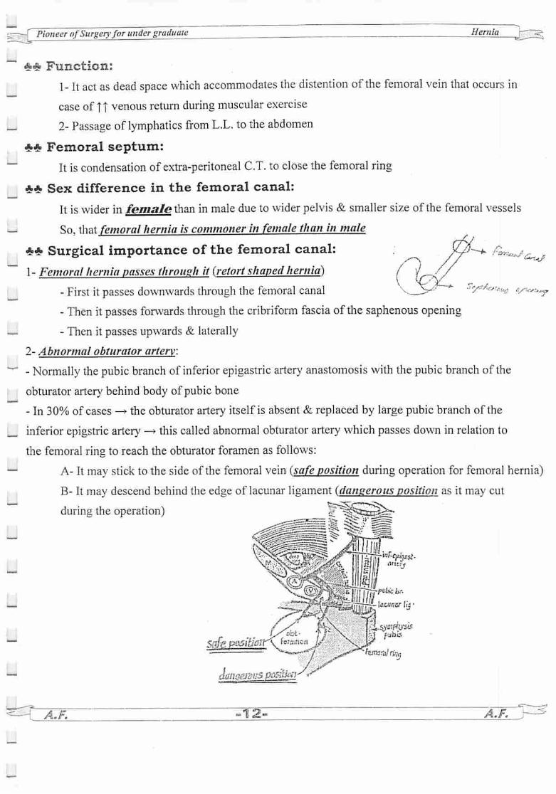
2- Abnormal obturator artery:
- Normally the pubic branch of inferior epigastric artery anastomosis with the pubic branch of the
obturator artery behind body of pubic bone
- In 30% of cases —»the obturator artery itself is absent & replaced by large pubic branch of the
inferior epigstric artery —»this called abnormal obturator artery which passes down in relation to
the femoral ring to reach the obturator foramen as follows:
A- It may stick to the side of the femoral vein (safe position during operation for femoral hernia)
B- It may descend behind the edge of lacunar ligament (dangerous position as it may cut
during the operation)
safe pMiiJo;r\ fwaincti
A.F. tt^t
a
fee ^
VI anty
aOHlBf Iij•
pants
txjraml fine,
&L/f-*- '~w,w•>••-.-:
A.F.

_
_
Pioneer ofSurgeryfor undergraduate
Femoral triangle
** Boundaries:
Base: inguinal ligament
Lateral: medial border of sartorius
Medial: medial border of adductor longus
** Roof: 1- Skin
2- Superficial fascia containing:
a- The upper part of GSV
b- Superficial inguinal L.N. (horizontal group & vertical
c- Superficial inguinal arteries:
- Superficial epigastric artery
- Superficial external pudendal
- Superficial circumflex iliac artery
3- Deep fascia of the thigh contains saphenous opening —• see after
** Floor: from medial to lateral
1-Adductor longus
2- Pectineus
3- Psoas major
4- iliacus
** Contents:
1-Femoral sheath
2- Femoral artery and its branches
3- Femoral vein & its branches
4- Femoral nerve and its branches
5- Deep inguinal L.N.
6- Lateral cutanous nerve of the thigh
7- Femoral branch of genito-femoral nerve
**• Applied anatomy:
D.D. of femoral Aswelling see after page 39
^A.F. 3^
Hernia
group)
A.F. T^
_
_
Pioneer ofSurgeryfor under graduate Hernia
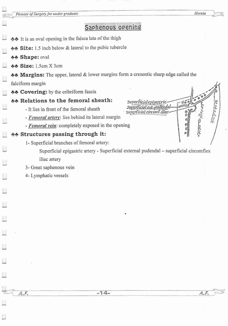
Saphenous opening
** It is an oval opening in the faisca lata of the thigh
** Site: 1.5 inch below & lateral to the pubic tubercle
** Shape: oval
** Size: 1.5cm X 3cm
** Margins: The upper, lateral & lower margins form a cresentic sharp edgecalled the
falciform margin
** Covering: by the cribriform fascia
** Relations to the femoral sheath: rffcialcpijastrlc--It lies in front of the femoral sheath Sejj^U&wgM
zXPCffictal araisnt:iliac
- Femoral artery: lies behind its lateral margin
- Femoral vein: completely exposed in the opening
** Structures passing through it:
1- Superficial branches of femoral artery:
Superficial epigastric artery - Superficial external pudendal - superficial circumflex
iliac artery
3- Great saphenous vein
4- Lymphatic vessels
fej* AJF. •14- A.F.
L

'~~Pionecr ofSurgeryfor under graduate ____ Hernia
ABDOMINAL HERNIAS
** Definition:
Abnormal protrusion of a viscus or part of a viscus, with or without a peritoneal sac through
an opening or a weak part of the abdominal wall
— ** Etiology:
(1) Congenital (Preformed sac):
1- Congenital weakness of abdominal wall
2- Presence of preformed sac as in congenial inguinal hernia (patent processus vaginalis)
(2) Acquired (Pulsion sac)
— a. Weakness ofabdominal wall due to:
1- Congenita! weakness of mesenchvme.
2- Traumatic: post operative as fibrosis weakens muscle
3- Inflammatory: as fibrosis weakens muscle
4- Senility
U 5- Obesity6- Pregnancy
— 7- Nerve injury during operation e.g. ilioinguinal nerve injury during appendectomy —*
paralysis of conjoint tendon —* direct inguinal hernia
b. Increased intra-abdominal pressure:
l - Chronic straining: cough, constipation
2- Abdominal swelling: pregnancies, hepato- splenomegaly, and abdominal tumors.
— ** Pathology:
A. External abdominal hernias:
_ I. Groin hernia: hernia occurring in inguinal region (area 1 inch above & 1 inch below inguinal ligament)
1. Inguinal (direct, indirect) 73%
— 2. Femoral 17%
II. Ventralhernia: hernia occurring in the anterior abdominal wall.
l-Congenital: Omphalocele. Gastroschiasis and infantile umbilical hernia.
bkj- A.F. ~15- A.F. V^
U

Pioneer ofSurgeryfor under graduate . Hernia
— 2- Acquired:
A- Midline: divarication of recti, epigastric, and umbilical, paraumblical hernia
B- Median: supravesical (anterior, posterior, and lateral).
C- Paramedian: Spigelian
3. Incisional
4. Traumatic:
_ III. Rare types: 1.5%
Q: Common hernias
1- .Inguinal hernia —> ]" common type
2- Incisional hernia —* 2" common type
3- Femoral hernia —> 3r commontype
- B. Internal abdominal hernia (inside abdominal cavity):
- Hernia through mesenteric defect
- Hernia through foramen of Winslow
- Hiatus hernia
- Diaphragmatic hernia
# Structure: Hernia consists of:
(1) The defect: Through which the sac bulges out.
(2) The sac: (absent in traumatic hernia)
© © Is the peritoneal pouch, which contains protruded viscus.
© © Is divided into a mouth, neck, body & fundus.
© © The sac may be:
* Pyriform (funicular type of oblique inguinal).
~~ * Hemispherical (direct inguinal-omphalocele major- incisional-true epigastric)
* Rounded (bubonocele-omphalocele minor)
* Retort (femoral).
* Saddle (pantaloon).
* Multilocular (PUH).
(3) Contents:
Any abdominal organ except the pancreas & kidnev (fixed retroperitoneal organ) e.g.
1. Intestine (Enterocele): LIsually the small intestine.
2. Omentum (Eoiplocele or Omentocele).
=H* A.F. -16- AJL
_
_
-
Pioneer ofSurgeryfor undergraduate Hernia
Enterocele Omcntocele
I. Consistency Soft. Doughy.
2. Gurgling During reduction Absent
3. Ease ofreduction. The 1st portion is more The last portion is more
difficult to reduce than last difficult to reduce than 1st due
due to gaseous distension of to edema and congestion of last
loop at neck on reduction. part.
4.Percussion Resonant Dull
5. Auscultation Intestinal sound Silent
3.Aportion of the circumference ofthe intestine = Richter's hernia.
4. Meckel's diverticulum = Littre's hernia.
5. Two loops ofthe bowel (Hernia-in-Er) = Mayd/'s hernia.
6. Aportion ofurinary bladder or a diverticulum ofthe bladder = Cvstocele.
(4) Coverings: derived from layers of abdominal wall through which sac passes.
— (5) Descent: it is the direction of protrusion of the contents.
-—
** Complications:
1. Irreducibility
3. Strangulation
«** Clinical picture:
A- History:
* Age: To choose the proper treatment.
* Occupation: Heavy work predisposes to hernia and to recurrence.
* History ofchronic cough (chronic bronchitis), difficulty (S.E.P., B.N.O., urethral stricture),
chronic constipation —* Treat these first.
* History oftrauma in traumatic hernia
B- Examination:
2. Obstruction
4. Inflammation
I- General:
_
u
* Chest: For chronic disease.
* Abdomen: For anv intra-abdominal swelling.
* P.R.: For S.E.P.
. A.F. ^17^ A.F.
\ i
Pioneer ofSurgeryfor under graduate
II- Local: Swellingwhich has thefollowing criteria:
1- At anatomical site of a hernia with special relation to the surroundings.
2- Give an expansile impulse on coughing.
(The swelling increase in size in all direction when the patient coughs)
3- Increases in size on standing or straining.
4- Decrease in size (or reduced) on lying down unless complicated.
5- Direction of descent & reduction
6- opaque in transillumination
7- Special tests.U
i [
i i
\ i
! I
\ i
I ;
** Investigations:
1- For assessment ofgeneral condition ofpatient: Hb%, creatinine, LFTs
2- For predisposing factors:
a. ChestX-rav if there is chronic cough.
b. I. V.P. if there is straining and difficulty in micturition.
c. Abdominal US if there is abdominal swelling.
3- For contents: Ascending cystography in sliding UB.
4- For complications: Plain x-ray.abdomen in obstructed & strangulated hernia
** Treatment:
1- Treatment ofpredisposing factor
2- Palliative treatment: by using a truss —»all except femoral hernia
Indications:
1. Contraindication for operations e.g. cardiac or pulmonary patient.
2. If the cause cannot be eradicated.
3. Ifpatient refuse operation.
Contraindication: Irreducible hernia.
3- Surgical treatment:
A- Herniotomy—* excision of sac at proper neck.
B- Herniorrhaphy —• repair of the defects
C- Hernioplasty —> strengthen defects by additional layer
A.F.
Hernia
A.F.

Pioneer ofSurgeryfor undergraduate Hernia
[COMPLICATIONS OF HERNIA
(1) Irreducible Hernia
Li £* Definition: Failure ofreduction of the content with no evidence ofother complications
(The best person to reduce hernia is the patient himself—* failins to do —»irreducible)
** Etiology:
(1) Adhesions between contents themselves orbetween contents and sac wall.
(2) Contents inside a hernial sac may TT i*1 size gradually due to edema or suddenly due to
_ straining.
(3) The neck of the sac may become narrow.
~" ** Clinical Picture: The contents fail to return to the abdomen but:
* There is still impulse on cough.
* The hernia is not tense or tender.
_
Irreducible Inflammed Obstruction Strangulation
I-Acutel.O. --
+ +
2- Impulse on
cough
+ *+
(difficult to elicit)
3- Irreducible o- 4* + +
4- Tender -
+
+ +
5- Tense -- -
+
6- Others Constitutional
manifestations•
** Treatment:
A- Taxis:
* Manual reduction of an irreducible hernia (in lsl 4 hours)
*Occasionally taxis results in reduction contents ofhernia sac en mass (still obstructed),
reduction ofstrangulated bowel resulting in generalized peritonitis, so taxis should be
avoided. (NOT DONE NOVO
B- Operative repair: herniorrhaphy
A.F. "^T9-" A.F.

g^jrgf Pioneer ofSurgery for under graduate Hernia
i l
(2) Obstructed HerniaI *•^ 44 Definition:
I Itis an irreducible hernia containing intestine —* its lumen is obstructed from outside or from inside;BUT there is NO interference to blood supply to the intestine.
L. 44 Etiology:1- Obstruction from outside: pressure by narrow neck or adhesions.
*-* 2- Obstruction from inside: occlusion by faeces ^^ihWaWal&e^M) —> the contentofthe bowel indented with the finger like putty.
44 Clinical Picture: See table
Li 44 Treatment:
1. Early: Repeated oily enemata may relieve incarcerated stool.
^ 2. Ifthis fails:* Ifobstruction is due to incarceratedstools -»the hard stools are broken by the fingers
without opening the lumen of the gut
JJ *Ifadhesions are the cause -* they are divided then do hemiorrhaphy
^ (3) Inflammed HerniaLi 44 Etiology: Inflammation ofthe contents (ovary- tubes- appendix) or its covering
44 Clinical Picture: see table
44 Treatment:
U (I) Conservative: General, local, observation.(2) Removal ofoffending focus if indicated (acute appendicitis) & do herniotomy only
— (Herniorrhaphy is not preferred, as the tissues are edematous and friable)
(4) Strangulated Hernia
44 Definition:
[J - An irreducible hernia with interference to the blood supply ofits contents- Gangrene may occur 5-6 hours after onset ofthe 1st symptoms of strangulation.
^©^ A.F. T-^
uPioneer ofSurgery for undergraduate
Li
L!
Li
Li
Li
Li
44 Etiology:
©Predisposing factors:
(1) Sudden expulsionof new contents.
(2)Narrowing of theneck by adhesions inside the sac
(3) Sharp edgeof the defect (femoral & P.U.H.).
(4)Irreducibility, obstruction & inflammation.
@Constricting agents: (In order olJremency)(a) In Femoral hernia: The sharp edge of the lacunar ligament (60%).
(b) In Paraumbilical hernia: Thedefect in linea alba
(c) In Ineuinal hernia: The neck of the sac.
44 Pathology:
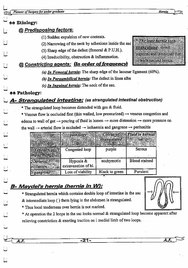
A- Strangulated intestine: (as strangulated intestinal obstruction)
*The strangulated loop becomes distended with gas & fluid.
* Venous flow is occluded first (thin walled, low pressurized) —• venous congestion and
edema towallof gut —• pouring of fluid in lumen —• more distension —• more pressure on
the wall -> arterial flow is occluded -> ischaemia and gangrene —• peritonitisi ,' if% FlmannJiermal
I '
i i,
i i
LI
occluswn.
I
M^ j? -rA%i. i
3-gangriene
Congested loop
Hypoxia &extravasation of bl.
Loss ofviability
Spyt,'jntXi'--'(*W.|,- WW
purple Serous
ecchymotic Blood stained
Black to green Purulent
B- Mavdel's hernia (hernia in W):
* Strangulated hernia which contains double loop of intestine in the sac
& intermediate loop () them lying in theabdomen is strangulated.
* Thus local tenderness over hernia is not marked.
*At operation the 2 loops in the sac looks normal &strangulated loop become apparent afterrelieving constriction &exerting traction on i medial limb oftwo loops.
Y.F. •2T<
Hernia
Pioneer ofSurgeryforunder graduate r, .
^ C- Strangulated Richter's hernia
i ; *Aportion ofthe circumference ofthe intestine is herniated &strangulated —*• Incompleteobstruction —> the patient may pass flatus &faeces —• absolute constipation is delayed until
U paralytic ileus occurs
D- Strangulated omentocelei !
*"" *At first omentum is congested edematous and later itbecome gangrenous invaded with! bacteria infection spread to peritoneum —• peritonitis
* Also there is no vomiting or constipation except very late
u, 44 Complications:
1- Intestinal obstruction.
] 2- Gangrene
3- Peritonitis & toxemia —»• septic shock —»• death
L 44 Clinical Picture:
(1) General:
l- Manifestations ofacute intestinal obstruction (A.I.O.) with strangulation.
^J 2- Absent in strangulated omentum &Littre's hernia &Richter 's hernia(2) Local signs: Cardinal Signs of Strangulation, which are:
•—> 1- There is No expansile impulse on cough.
2- The hernia is irreducible.
3- The hernia is tender.
4-The hernia is tense.
44 Differential Diagnosis:
Irreducible - inflamed - obstruction - strangulation —*• see table before
L 44 Treatment: Emergency operation
1. Hospitalization & antishock measures.
[J 2. Gastric suction through a nasogastric Ryle's tube.
3. Urinary catheter.
wj 4. Antibiotic.
^T A.F. " -ggT -jlfTV^

_
_
Li
U
_
_
_
_
_
Pioneer ofSurgeryfor under graduate Hernia
B- itnmediateOperaponi Once the patient is resuscitated:
1- At first is to expose the fundus of the sac —* open it —* aspirate the fluid inside (if this fluid
escapes into the peritoneal cavity it causes toxemia)
2- Then the constriction ring has to be released
3- Draw the contents out & see:
A- Viable intestine —> return to the peritoneal cavity
B- Omentum —> Excision whether viable or non viable.
C- Non-viable:
* Small intestine —> resection anastomosis
* M$ght colon —» Right hemi colectomy & Ileo-transverse anastmosis
* Transverse or leftcolon —» Exteriorisation resection (Paul Mickulicz operation)
D- Doubtful:
Cover intestine with hot saline packs to abdomen for 10 minutes + administration of
pure 02 ventilation Then, intestine is re- examined viable or not
4- Herniotomy + Closure of wound with a drain put subcutaneous for 48 hours.
Viable loop Gangrenous loop
Inspect/on:
* Luster Present (shining) Absent (dull)
* Color Red lighter Dark or black
* Peristalsis Seen Absent
Palpation:
* Tone Present (firm) Absent (flabby)
* Pulsation Felt in mesentery Absent
Operative Positive (no thrombosis) Negative (thrombosis)
DopplerUS:
Fluid in hernial At 1st. clear, but later on The exudate in the sac
sat it becomes blood blood stained or muddy &
stained has a faeculant odor
IF.
* Herniorrhaphy is better avoided —»tissues being friable, edematous
:: liernioplustv is NEVER attempted—* it always fails due to infection.£
•23-
Pioneer ofSurgeryfor under graduate ~ 71 r~
INGUINAL HERNIA
— ** Classification:
I. Oblique (Indirect) inguinal hernia (60%)
1. Congenital
2. Infantile
3. Adult: Bubonocele- funicular - scrotal type
— II. Direct inguinal hernia (10%)
1. Diffuse type
2. Funicular type.
_ III. Rare inguinal hernia
1. Pantaloon (Dual) hernia.
2. Sliding hernia (3%).
.1. Indirect (Oblique) Inguinal Hernia
— [Commonest type of hernia)
1~ eONGBNITAIp INGUINAL, HERNIA
** Etiology:
The whole processus vaginalis is patent and thus the general peritoneal cavity is connected totunica vaginalis.
** Clinical presentation:
_ - Although it is called congenital, it may not appear until adolescent or adult life
- The testis appears to lie within lower part of the-hernia.
" *<£ Treatment: Age: at the age of3 months unless complicated
A- Herniotomy:
*Through inguinal incision (2-3cm) -» expose the sac ÷ it transversely -* the proximal— part is excised at proper neck & the distal part is either left in situ or removed
* In children below 7 years —> Herniotomy is done without opening the external oblique
apeneurosis i.e. without opening the canal as the 2 inguinal rings are opposite each other.
_ B- Marcev repair:
If the defect is large —> tightening ofthe internal ring without opening the inguinal canal
~'Z A.F. -24- ~AJi

—f Pioneer ofSurgery forunder graduate Hernia
i?~ INFANTj
— £* Etiology:The proximal part ofprocessus vaginalis is patent and tunica vaginalis extends upwardstowards inguinal canal ->2 sacs are found within the cord: j, ta**«r«0 &* c
jj a. Hernial sac. ^2L rt^G, W^*4a •b. Extension of tunica vaginalis upwards in the cord. f^y n
<3~> ADUL>cf IH&UINAE HERNIA
~~ ** Etiology: Due to deficiency of mechanisms that normally prevent herniation —> see anatomy
_ ** Pathology:
Site: In the groin above inguinal ligament (above and medial to pubic tubercle)
Structure: Hernia consists of:
(1) Defect: Stretched deep ring.
(2) Sac:
— a. Bubonocele: Hernia passes through internal ring, stops in inguinal canal.b. Funicular: Hernia passes through internal ring, inguinal canal, external ring &stops at neck of scrotumc. Complete (Scrotal): Hernia passes through internal ring, inguinal canal, external ring and reachesbottom ofscrotum -> Sac not continuous with tunica vaginalis so testis can be felt separate from sac
(3) Contents: See before.
_
(4) Coverings:
In inguinal region
1. Skin.
2. 2 layers of superficial fascia
3. External oblique aponeurosis.
4. Cremasteric muscle & fascia (from internal oblique
muscle).
5. Internal spermatic fascia (from transversalis fascia)
6. Extraperitoneal tissue
In scrotum
l-Skin.
2- Dartos muscle & fascia
3- External spermatic fascia (from external oblique
aponeurosis).
4- Cremaster muscle & fascia (from internal oblique
muscle).
5- Internal spermatic fascia (from transversalis fascia)
6- Extraperitoneal tissue
(S) Descent: Downward, forward and medially
? A.F. "25- -AzL.

r~r{ Pioneer ofSurgery for under graduate
^ ## Clinical Picture:
U @ Type ofpatient:* Sex: Male > Female 20:1 as inguinal canal is wider in male than female.
Li *Side:- Right > left side due to the later descent of right testis.
- The hernia is bilateral in 30% of cases.L
Hernia
^ @ C/O: Intermittent swelling
, a. Increases in size on standing or straining.
b. Decrease in size (or reduced) on lying down unless complicated.
L @ Signs-.* Position of the patient standing looking upward and to the left and expose the lower half of
^ the body and then lying down to reduce itand feel the defecti ( * Swelling which has the following criteria:
1- Shape: pear shaped swelling
Li 2-Site:- Inguinal (Bubonocele) or inguino-scrotal (Funicular & Scrotal) —• by scrotal neck
•—' test (palpate the neck of the scrotum by thumb & index finger)
- Above the inguinal ligament
- Above and medial to the pubic tubercle
'• ' 3. Give an expensile impulse on coughing.
4. Increases in size on standing or straining.
i— 5. Decrease insize on lying down or reduced in certain direction (upward, backward and laterally)
6. Direction of.
Descent: Downwards, forwards & medially,
i ! Reduction: upwards, backwards & laterally
7. Transillumination: Opaque
i^i 8. Special tests:

-f PioneerofSurgeryfor under graduate Hernia
A. Interna/ ring test
-To differentiate an indirect from a direct inguinal or a femoral hernia.
- Patient lies down, hernia is reduced, the thumb obliterates the internal ring. With the thumb in
place, the patient stands and asked to cough.
* Hernia does not appear: Indirect.
* Hernia appears above inguinal ligament: Direct
* Hernia appears below inguinal ligament: Femoral.
B. External ring test
- To differentiate indirect from direct inguinal hernia.
- Reduce the hernia and while me patient is standing invaginate
the skin of the scrotum with the tip of the little finger and enter the
external ring.
- Normally the external ring admits just the tip of the little finger* Ifthe ring is wide: Indirect inguinal hernia. —,
* Ifthe ring is normal: Direct inguinal hernia.
* Ask the patient to cough:
- Impulse on the tip of the little finger: Indirect.
- Impulse on the medial side: Direct.
C. Three fingers tests (Zieman's test):
- To differentiate an indirect from a direct inguinal or a femoral hernia
- While the patient is standing, stand behind the patient and place:
* Index finger over internal ring
* Middle finger over Hassalbach's A. j&k
* Ring finger over saphenous opening.
- Ask patient tocough and note which fingers feels impulse.
* Impulse on tip of index finger: Indirect.
* Impulse on tip of middle finger: Direct.
* Impulse on tip ofring finger: Femoral.
A.F. -27-
How vou can locate the puoic tubercle?•:-.:-. • ' • .
Flex . abduct & laterally rotates the thigh to. fell the tendon ofadductor longus m the upper
y medial side of the thigh -» Follow the tendon to its origin, the 1st bony prominence aboveI
/\tC •
Pioneer ofSurgeryfor under graduateHernia
** Investigations: see before
_ *4> D.D.: from other causes of inguinoscrotal swelling (see male genital system)
** Treatment:
1- Eliminate causes ofraised intra-abdominal pressure first
2- Palliative treatment: Truss (see before)
3- Surgical treatment: The treatment of choice
_. A- Herniotomy
B- Herniorrhaphy
C- Hernioplasty
D- Laparoscopic repair
©Indication ofeachJtype_o£repair:
• -
Herniotomy Herniorrh aphy Hernioplasty
1- Infant & children 1- Old 1-Very old
2- Small defect (< 2cm) 2- Large defect (2-4cm) 2- very large (>4cm)
3- Good musculature 3- weak musculature 3- very weak
4- recurrence after 4- recurrence after
herniotomy herniorrhaphy
&JH^)rMs>i.9..^x@Principle: Transfixion, ligation &excision of the hernial sac at its proper neck \ X/^V-j, " i'i
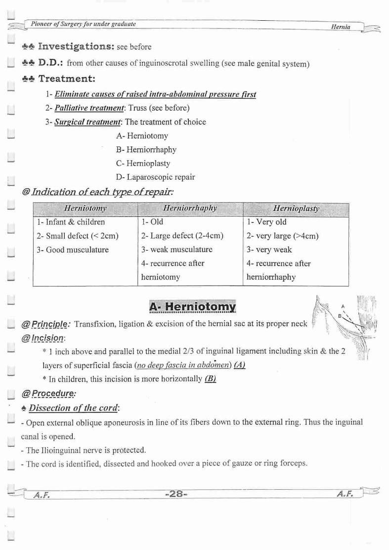
\V< V,":@!ncjsjp_n:
* 1 inch above and parallel to the medial 2/3 of inguinal ligament including skin & the 2
layers of superficial fascia (no deep fascia in abdomen) (A)
* In children, this incision is more horizontally (B)_
@ PC9P£dure:
* Dissection of the cord:
- Open external oblique aponeurosis in line of its fibers down to the external ring. Thus the inguinal
canal is opened.
- The Ilioinguinal nerve is protected.
- The cord is identified, dissected and hooked over a piece of gauze or ring forceps.
A.F. ^28^ A.F.
_
_
_
tef Pioneer ofSurgeryfor under graduate
Aponeurosisof externaloblique muscle
Seine of oubit
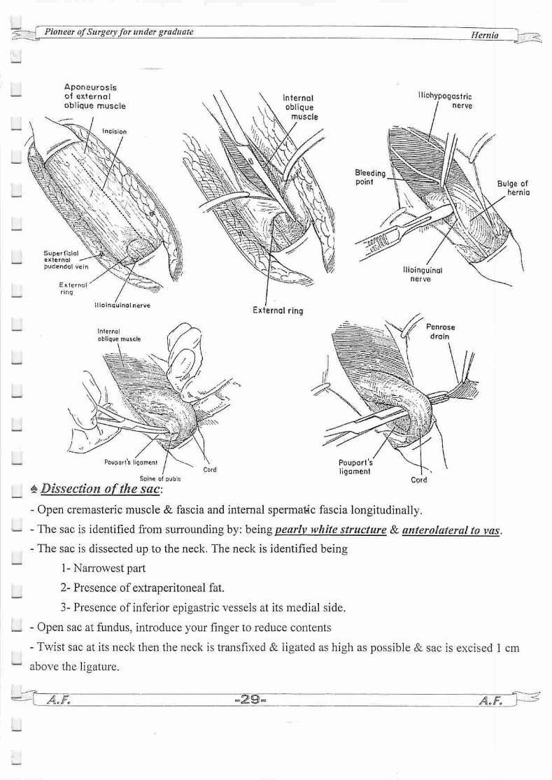
£ Dissection ofthe sac:
Hernia
Iliohypogastricnerve
Cord
Bulge ofhernia
- Open cremasteric muscle & fascia and internal spermatic fascia longitudinally.
- The sac is identified from surrounding by: being pearly white structure & anterolateral to vas.
- The sac is dissected up to the neck. The neck is identified being
1- Narrowest pail
2- Presence of extraperitoneal fat.
3- Presence of inferior epigastric vessels at its medial side.
- Open sac at fundus, introduce your finger to reduce contents
- Twist sac at its neck then the neck is transfixed & ligated as high as possible & sac is excised 1 cm
above the ligature.
A.F. A.F.
Pioneer ofSurgeryfor under graduate Hernia
-
_
- i.
_2-
- i,
2.
Pubic brancholinferior
epigastricarlery
Cremoster
muscle
Dividingcremaster
^-.Herniorrhaphy
Iliopubic bond
Conjoined lendor
Spine ol pubis
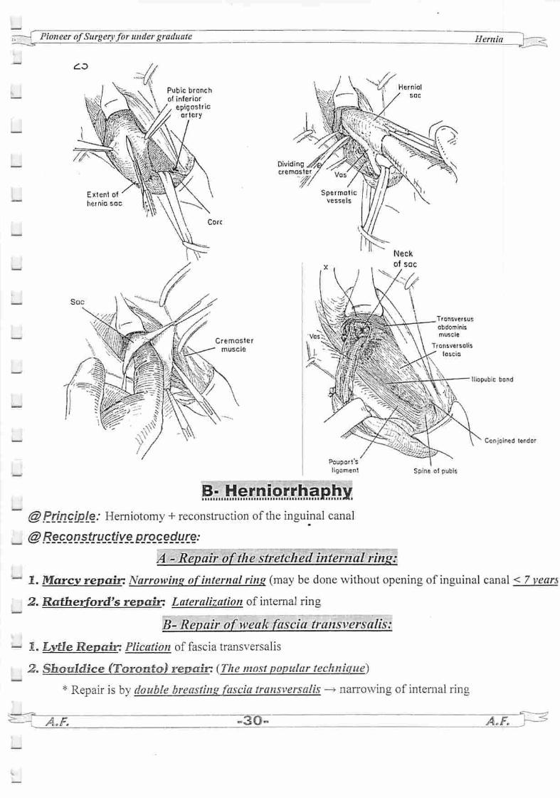
}Principle: Herniotomy + reconstruction of the inguinal canal
\BeSLQnstru_ctjye_ procedure:
A - Repair of the stretched internal ring:
Marcy repair: Narrowing of internal ring (may be done without opening of inguinal canal < 7 years
Ratherford's repair: Lateralization of internal ring
B- Repair of weak fascia transversalis:
Lytle Repair: Plication of fascia transversalis
Sbauldice (Toronto) repair: (The most popular technique)
* Repair is by double breasting fascia transversalis —» narrowing of internal ring
A.F. -30. aO-^
-
Pioneer ofSurgeryfor undergraduate Hernia
*Approximation ofconjoined tendon to inguinal ligament
C- Reconstruction ofthe posterior wall ofthe inguinal canal:
1. Anterior approach:
- 1. Bassini's operation: (triple layer) (the father of herniology 1880)
* Ist the transversalis fascia is opened from the internal inguinal ring to the pubic tubercle
* Then suturing the lower border of transversus abdominis muscle, internal oblique muscle and
fascia transversalis (triple layer) to the fascia transversalis and inguinal ligament behind the cord
* Disadvantages: It is unphysiological i.e.
1- It interferes with shutter mechanism of the internal oblique muscle
2- sutures are under tension —> failed repair
3- suturing ms. Fibers (conjoint) to tendon (ing.lig.) -» delayed repair
* Tanner's slide release incision:
A relaxing incision is done in anterior rectus sheath —* to avoid tension sutures
2. Halsted repair:
Bassini's repair + Repair of the external oblique aponeurosis edge to edge behind the cord
cord becomes subcutaneous —* Cord is liable to trauma.
MC Andrew operation:
Andrew I:
As Halsted repair but with double breasting ofext. oblique aponeurosis
behind the cord —* cord becomes subcutaneous
Andrew II:
U 3.
4.
5.
vL.* -.•<'• ';•,-, fT^Jr-"v; • •• • i
=*=-*s^=
85•_* -/
^vTifTpi"zy^xidfr
V ;•'•••'
Sk
,£As Andrew's I but —* cord isplaced in between the 2 flaps of external oblique aponeurosis
Blood Good Repair: = Use of rectus sheath.
Triangular flap ofthe anterior rectus sheath is turned laterally & is sutured to the inguinal
ligament behind the cord.
Cooper's ligament repair: (McVay repair)
* Suture the transversus abdominis aponeurotic arch to:
Medially —»to cooper's ligament (ilio-pectineal ligament)
Laterally —> ilio pubic tract
fer A.F. 3-U A.F.

-S-— Pioneer ofSurgeryfor under graduate Hernia
— II. Posterior properitoneal approach:
Nyhus repair: (Inguinal canal is not opened)
Transverse muscle cutting incision 4 cm above inguinal lig —> open external oblique muscle
_ —» internal oblique muscle —> trasnversus abdominus —* fascia transversalis —• reach
properitoneal space (NO opening ofthe inguinal canal) —* deal with the hernia —> the
dilated internal ring is narrowed -* suture transversus abdominis aponeurotic arch into ilio
pubic tract behind cord (jjj j*^ McVay repair $} /% & £»)_
_
C- Hernioplasty
@ Pri_ncf]2[e: herniotomy + Reinforcement of the post, wall by using mesh
@ Methods:
1. Lichtenstein tension free repair: (the most accepted repair nowadays)
* A mesh is used to cover the area () conjoint tendon & inguinal ligament —•
- It is sutured to lacunar ligament, inguinal ligament
- The medial edge is sutured to rectus sheath.
- The superior edge is sutured to the internal oblique apeneurosis.
- The lateral edge is split at the internal ring, and the 2 tails are
brought around the cord and hold the cut edses.
Lotoiol ovortap
Ot IOIIB
2. Stoppa repair: (What is the hemi-Stoppa?)
# Indicated in:
1- Recurrent inguinal hernia with disturbed anatomy.
2- Bilateral ineuinal hernia.
# Principal:
Midline lower abdominal incision -* properitoneal space (between the peritoneum
and abdominal walls) —• place very large mesh
D- Laparoscopic repair;MnllllMllllllllllllllllllMlllllllllllAllllMllllltllillllllMIIIIMIII
1. TAPP (Transabdominal preproteneal repair)
2. TEP (Totally extraperitoneal repair)
— •-. A.F. A.F.
"— Pioneer ofSurgeryfor undergraduate Hernia
-
_
•
TyPQS-Pf-PJpsfc Polyproline - Teflon - Dacron - Nylon - PTFE. ' - ' -" . . . . .
Criteria of ideal mesh:
1- Non-absorbable 2- Non-infective 3- Non-oncogenic
4- Inert
7- Monofilament
5- Durable 6-Narrow pores
rosis8- Induce fibr
II. Direct Inguinal Hernia
** Etiology: Always acquired dueto:
(1) Atrophy ofconjoined tendon due to senility.
(2) Paralysis ofconjoined tendon due to injury of ilioinguinal nerve during an appendicectomy
(3) Predisposing factors are chronic cough, straining and heavy work.
** Pathology:
Site: In the groin above inguinal ligament (above and lateral to pubic tubercle)
Structure:
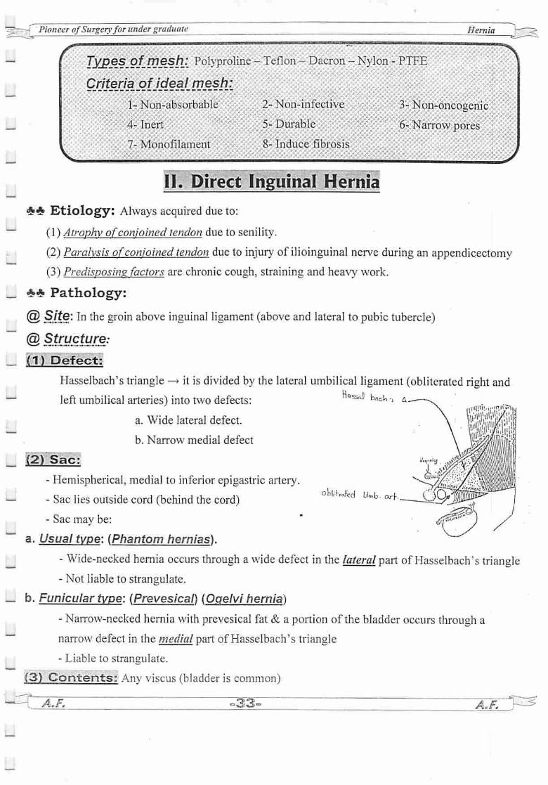
(1) Defect:
Hasselbach's triangle —> it is divided by the lateral umbilical ligament (obliterated right and
left umbilical arteries) into two defects:
a. Wide lateral defect.
b. Narrow medial defect
(2) Sac:
- Hemispherical, medial to inferior epigastric artery.
- Sac lies outside cord (behind the cord)
- Sac may be:
a. Usual type: (Phantom hernias).
- Wide-necked hernia occurs through a wide defect in the lateral part of Hasselbach's triangle
- Not liable to strangulate.
b. Funicular type: (Prevesical) (Ooelvi hernia)
- Narrow-necked hernia with prevesical fat & a portion of the bladder occurs through a
narrow defect in the medial part of Hasselbach's triangle
- Liable to strangulate.
(3) Contents: Any viscus (bladder is common)
? A.F. °33- A.F. V^
NQ**& bncW-,
oU'Ued Umfc, Qrr

Pioneer ofSurgeryfor undergraduate
(4) Coverings:
1. Skin.
2. 2 layers of superficial fascia.
3. External oblique aponeurosis.
4. + Conjoined tendon.
5. Fascia transversalis.
6. Extraperitoneal tissue.
(5) Descent: Bulges directly forwards
*«£ Clinical picture:
Hernia
Oblique inguinal hernia Direct inguinal hernia
1- Incidence 60% 10%
2- Definition The hernial sae passes through
inguinal ring
The hernial sac protrude
through Hasselbach's triangle
3- Covering See before See before
4- Relations:
a- Inferior epigastric art. Lateral to the artery Medial to the artery
b- Cord The sac inside the cord The sac behind the cord
c- Pubic tubercle Medial Lateral
S- Diagnosis
I. Age any age old age
2. Sex Commoner in male
Male : Female = 20 : l
Only in male
(Not in female)
3.Side >in right side, bilateral in 30% Bilateral in 50%.
4.Shape Pyriform or tubular Hemispherical
5.Direction ofdescent Downward, forward & medially Directly forward.
6. Direction ofreduction Upwards, backwards & laterally Backwards
7. Descend into scrotum Can descend Extremely rare
S.Internal ring test Hernia does not descend. Hernia will descend.
9.External ring test a) Wide ring.
b) Impulse on tip of finger
a) Normal.
b) Impulse on medial aspect
of finger.
A.F. ~-5&T A.F. y •<

——- Pioneer ofSurgeryfor under graduate Hernia
_
10. Three fingers test Impulses are perceived by index
which lies over internal ring
Impulses are perceived by-
middle finger which lies over
external ring
6- Compl. Common Uncommon
7- At operation Sac inside cord Sac outside cord
** Treatment:
I. Eliminate causes of raised intra-abdominal pressure first
II. Truss: Small hernias with wide neck in elderly people best treated with a truss
III. Operation
No Herniotomy
1. Herniorrhaphy:
No herniotomy, the hernial sac can be inverted. Repair of weak posterior wall of the canal
can be done by plicating the fascia transversalis with Bassini or shouldice or Mcvay's repair.
2. Hernioplasty: in large defects.
Pantaloon Hernia
(Dual or Saddle-Bag)
# Here there are 2 sacs —* one oblique inguinal hernia & other direct inguinal hernia —* with
inferior epigastric artery separating the two sacs (one sac beins medial & the other lateral to the
artery)
# This condition is a cause of recurrence —> one of the sacs may be missed at the time of operation.
— " Treatment:
Puiling on the neck of the oblique sac, die direct sac is pulled lateral to the inferior epigastric
vessels converting the 2 sacs into one, which is transfixed & excised.
A.F. ^35^ A.F.

-
-.
Pioneer ofSurgeryfor undergraduate Hernia
Sliding Hernia
(Hernia-En-Glissade)
** Definition: Hernia in which the wall of the viscus forms a part of the sac instead of
contents i.e. outside sac. behind it
** Etiology:
As a result ofslipping of the posterior parietal peritoneum on the
underlying retroperitoneal structures, posterior wall of the sac isnot formed of peritoneum alone, but also bya viscus.
** Site: Related to organs fixed to posterior abdominal wall -»
* On right side —* caecum
* On left side —» sigmoid
* On either sides -» urinary bladder (the commonest)
** Clinical Picture: Difficult to diagnose, should be suspected if.
(1) Old male with an incompletely reducible hernia —* residual swelling after reduction
(2) If the bladder is involved, hemia TT in size when patient does not micturate & is U in
aftermicturation. and patient experiences desire of micturation on pressure on the hernia.
** Investigations: As before +
* Cystography —> urinary bladder OR Barium enema —• colon inside hernia.
** Treatment:
** No attempt should be made to dissect the caecum or colon free from the peritoneum —» as it is
liable to: 1- Injury of the viscus —» peritonitis & faecal fistula
2- Interfering with blood supply may occur.
A- Ifsac is small: It is unnecessary to remove sac, reduce sac with contents + hemiorraphy.
B- Ifsac is large:
U-shaped incision is made in the peritoneum & reflected behind the bowel to be sutured
together then reduce the viscus & excision of the remaining part + hemiorraphy.
C- Very large sac: It may need also to open the abdomen through separate incision & pull bowel
back into abdomen & do colopexy then do herniotomy and hemiorraphy as usual.
Hcmlo sac
oilo cacgm
size
l4^ A.F. ^3S^ A.F. V^
-

- Pioneer ofSurgeryforunder graduate Hernia
FEMORAL HERNIA
__ (3rd commonest type ofhernia)
** Definition: External abdominal hernia which passes through femoral ring into femoral canal
** Etiology: Always acquired &never congenital due to TT abdominal pressure (pregnancy)
L ** Pathology:(a) Site: Groin below inguinal ligament (Below & lateral to the pubic tubercle)
Structure: 0£»* /C^ ,•(1) Defect: femoral ring.
(2) Sac: JS^"* *?**»<*** *s»*..7L Retort shaped with a narrow neck —* proceeds downwards through femoral ring —• femoral
canal till apex —* it pushes cribrifom fascia at apex of canal in front of it —» then upward in
front of the inguinal ligament (why?).
__
A.F. -37- A.F.
-
(3) Contents: Omentum, small or large gut, Richter's hernia, sliding bladder
(4) Coverings:
1-Skin
2- Superficial fascia:
- Superficial fatty layer (Camper's fascia)
- Deep membranous layer (Scarpas's fascia)
3- Cribriform fascia (covering saphinous opening).
4- Transversalis fascia derived from the anterior wall of the canal.
5- Fat & lymphoid tissues derived from the femoral septum.
(5) Descent:
* U-shaped course —>
* Downward: through femoral canal then,
* Forward: through saphinous opening then,
_ *Upward: in the SC tissue may overlie inguinal ligament —*
This is because of fusion of superficial fascia of the abdomen (Scarpa's fascia) to
~" the deep fascia of thigh (fascia lata) at a line runs transversely from saphinous
opening laterally (Holden line).
'-r-f Pioneer ofSurgeryfor under graduate
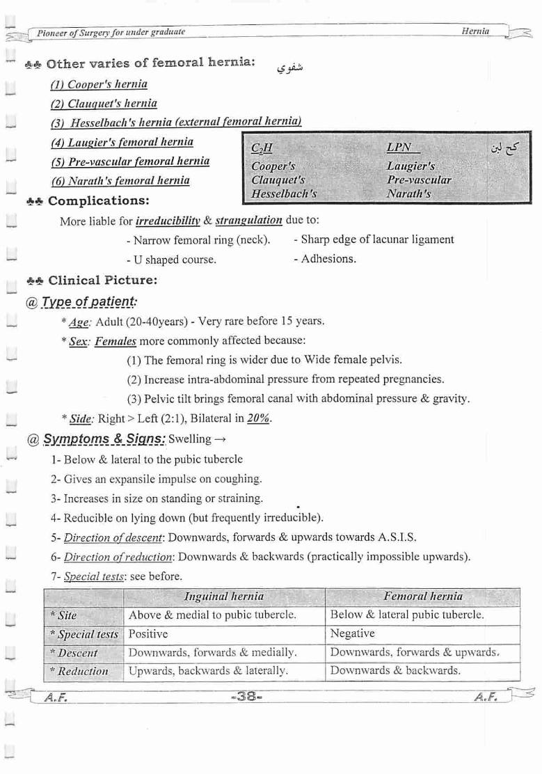
** Other varies of femoral hernia:
(1) Cooper's hernia
(2) Clauauet's hernia
(3) Hesselbach 's hernia (external femoral hernia)
t^yjo
(4) Lausier's femoral hernia
(5) Pre-vascular femoral hernia
(6) Narath 's femoral hernia
** Complications:
More liable for irreducihility & strangulation due to:
- Narrow femoral ring (neck). - Sharp edge of lacunar ligament
- U shaped course. - Adhesions.
** Clinical Picture:
*Age: Adult (20-40years) - Very rare before 15 years.
* Sex: Females more commonly affected because:
(1) The femoral ring is wider due to Wide female pelvis.
(2) Increase intra-abdominal pressure from repeated pregnancies.
(3) Pelvic tilt brings femoral canal with abdominal pressure & gravity.
*Side: Right > Left (2:1), Bilateral in 20%.
@ SyjPJ?t9IP£&-SJQLn.?J Swelling -*1- Below & lateral to the pubic tubercle
2- Gives an expansile impulse on coughing.
3- Increases in size on standing or straining.
4- Reducible on lying down (but frequently irreducible).
5- Direction ofdescent: Downwards, forwards & upwards towards A.S.I.S.
6- Direction ofreduction: Downwards & backwards (practically impossible upwards).
7- Special tests: see before.
Cooper'sClaliquet'sHesselbach's
LPN
Laugier'sPre-vascular
Naraih 's
Hernia
oJ^r
Inguinal hernia Femoral hernia
*Site Above & medial to pubic tubercle. Below & lateral pubic tubercle.
* Special tests Positive Negative
* Descent Downwards, forwards & medially. Downwards, forwards & upwards.
* Reduction Upwards, backwards & laterally. Downwards & backwards.
A.F. 138" S\\r/T0

PioneerofSurgeryfor undergraduate Hernia
^ ** Differential Diagnosis: Swellings in the femoral triangleU (1) Enlarged inguinal lymph nodes: TMcommonesi:
(2) Femoral hemia.
^ (3) Saphena varix:* Very softcompressible swelling.
* Thrill on coughing.
i * Evident varicosities of the lower limb.
(4) Femoral veinthrombosis.
U (5) Aneurysm of femoral artery.(6) Psoas abscess:
^ * has impulse on coughing
* Cross fluctuation of the swelling above & below inguinal ligament.
* Evidence ofPott's disease of spine with psoas spasm.
U (7) Psoas bursa.(8) Ectopic testis
^ (9) Neurofibroma, Lipoma, sebaceous cyst, haemangioma, lymphangioma.
\ ** Treatment:
(1). Low afiproach.(femora
*"* @Incision: 1/2 inch below ¶llel to medial portion ofthe inguinal ligament
i ©Refiair:<m f
- The sac is identified and dissected —> transfixion excision of the neck as high as possible
! - Repair -»• HHg -*• APoupart (inguinal) to Pectineal ligaments -»to close femoral ring
(2) High approach .(Mc Eyedy's approach)l— ® Incision: A vertical incision is made over the femoral hernia & continued above
I | the inguinal ligament.^ @Repair: SS-> Conjoined to Cooper's ligament closing femoral ringLj (3) InjguinaJ approach b^
@ Incision: Like an inguinal hernia incision.
L @RejP.air: *^^M^eWipm Poupart's to Pectineal ligaments.*§!?fiiilllill: Conjoined to Cooper's ligament
^ * WioW&PW&ffiwii; Poupart's to Conjoined to Pectineal ligaments
(4) Mesh.pJup.Xdpne.npwa.days).
~A~F7 °39- ~ A.F.
rr-J Pioneer ofSurgery for under graduate Hernia
U
LI
L
" i^"?s»=e«rKOT?V'.,iWs-V.-- «.0 .-.-•-. -'-- ,.t-' ::->js,;,'*.i:«..- -."3=^^JSJ^^^aas-^ Ka;iK:M „-;-j1"
** Definition: Hernia at & around the umbilicus.
** Five types:
1- Physiological umbilical hernia
2-Exomphalos = Omphalocele
3- Congenital umbilical hernia
4- Infantile Umbilical Hernia
5- Adult Umbilical Hernia:
a- True umbilical hernia (10%)
b- Para-Umbilical Hernia (90%)
(H Physiological Umbilical Hernia
*It occurs atthe 5th week intrauterine life due torapid growth of intra-abdominal organs, sothe
abdominal cavity cannot accommodate the contents.
* The hernia returns to the abdominal cavity by the 10 week.
(11) Exomphalos a Omphalocele! f
^ ** Etiology: It is developmental defect ofabdominal wall with failure ofall or part of midgutto return to coelomic cavity during early foetal life.
to-*
** Pathology:
•—' @ Site: umbilical region.
@ Structure:
# Defect: peri-umbilical abdominal wall.
# Sac: pouch ofperitoneum.
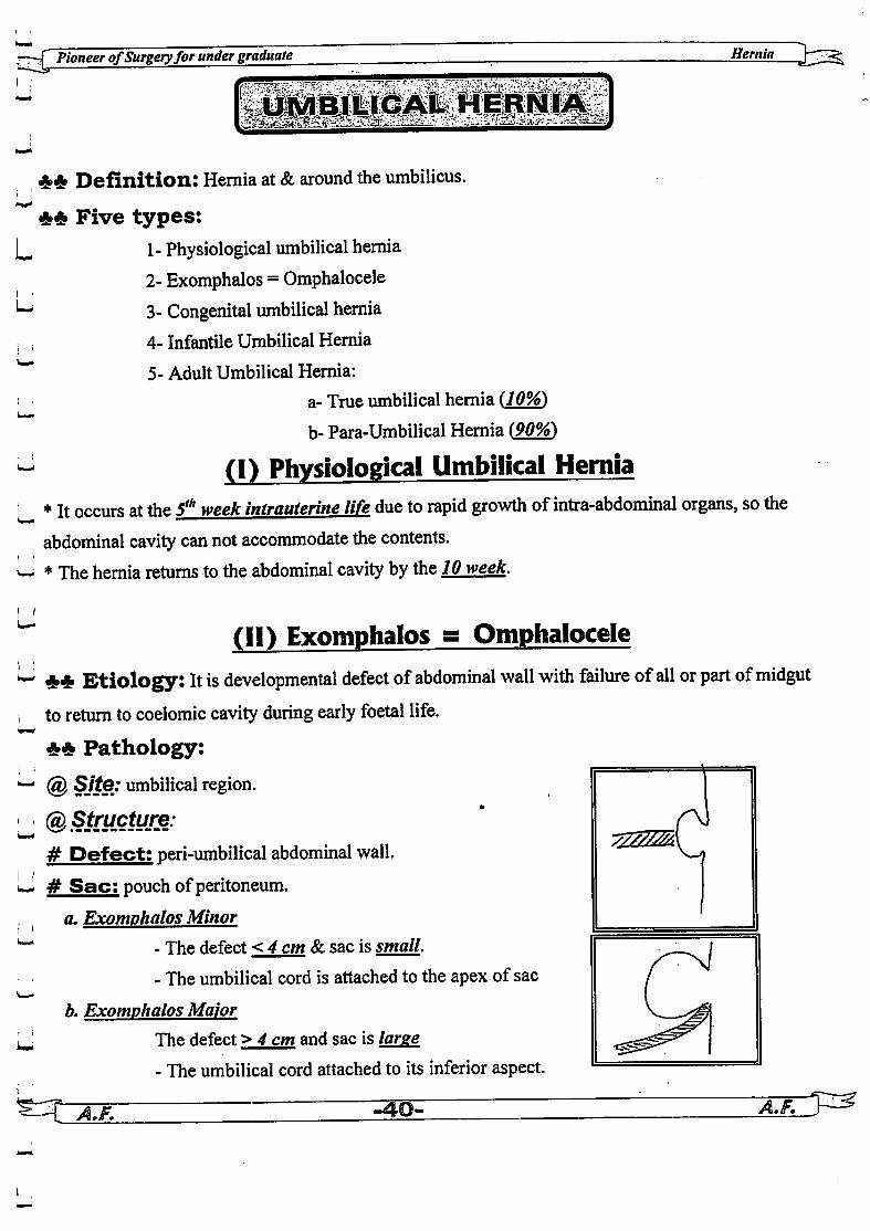
a. Exomphalos Minor
- The defect <4cm & sac is small.
- The umbilical cord is attached to the apex of sac
b. Exomphalos Major
The defect >4 cm and sac is large
- The umbilical cord attached to its inferior aspect.
i i
W-^T1
11
^^5^5*"**'̂

= Pioneer ofSurgeryfor undergraduate Hernia
~- # Contents:
a. Exomphalos Minor: Loop of intestine or Meckel's diverticulum
b. Exonwhalos Major: Small & large intestine & nearly always a portion of liver
L # Covering:When sac remains unruptured -»it is semi-translucent composed of 3 layers although very
— thin (no skin covering):
(1) Outer layer of amniotic membrane.
(2) Middle layer of Wharton's jelly.
(3) Inner layer of peritoneum.
# Descent: Directly forward.
Gastroschiasis:• .'.'• • • • • ' •
t Herniation of the abdominal contents usually only intestine through a small defect (usually'•
< 3 cm) in the anterior abdominal wall to the right side ofumbilical cord -> there are NO
coverings jjjjj NO sac ^0^-^M^a*Associated congenital anomalies especially intestinal atresia or stenosis
* Treatment: Silo followed by repair of the defect.
J
Exomphalos major Gastroschisis
** Complications:
a. Exoniphalas Minor: During ligation of cord, a loop of intestine may be entangled in the ligature.
b. Exonwhalos Major: Rupture of sac & peritonitis.
£-<& Clinical picture: manifested since birth.
-\~AJF. =41- -aZfTT^
u
_
_
_
_
_
u
L
L
-J Pioneer ofSurgeryfor under graduate Hernia
=&& Treatment:
A- Exomphalos Minor:
* It is easy as the defect is small & abdominal cavity is well developed
* It is performed by excision of sac & closure of defect in layers
B- Exomphalos Major:
Operation within the 1st few hoursoflife . otherwise the sac will burst.
Reduction of the contents into small abdominal cavity is difficult & hazardous because trial to
reduce the contents back —• 1- Respiratory embarrassment.
2- Compression on IVC —• interference with VR.
3- Intestinal obstruction.
SO, we try to cover the hernia with either (a) or (b) to avoid risk ofperitonitis.
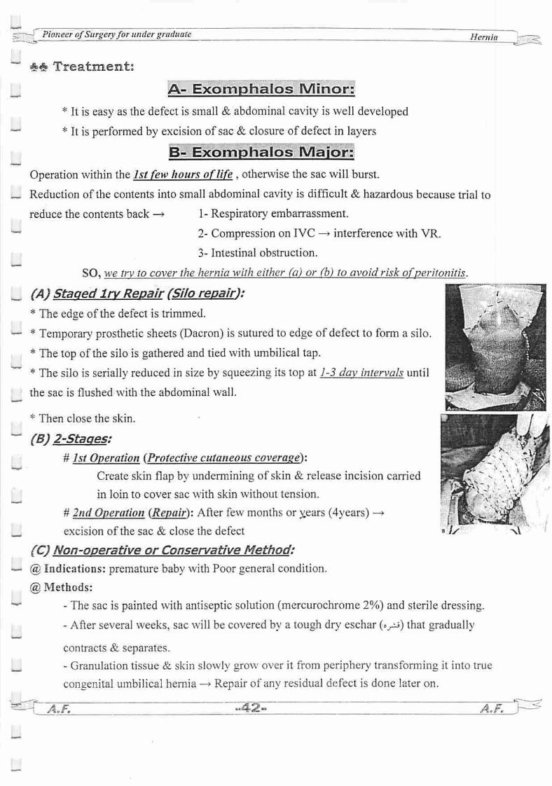
(A) Staged Irv Repair (Silo repair):
* The edge of the defect is trimmed.
* Temporary prosthetic sheets (Dacron) is sutured to edge of defect to form a silo.
* The top of the silo is gathered and tied with umbilical tap.
* The silo is serially reduced in size by squeezing its top at 7-5 day intervals until
the sac is flushed with the abdominal wall.
* Then close the skin.
(B) 2-Staaes:
# 75/ Operation (Protective cutaneous coverage):
Create skin flap by undermining of skin & release incision carried
in loin to cover sac with skin without tension.
# 2nd Operation (Repair): After few months or y.ears (4years) —♦
excision of the sac & close the defect
(C) Non-operative or Conservative Method:@ Indications: premature baby with Poor general condition.
@ Methods:
- The sac is painted with antiseptic solution (mercurochrome 2%) and sterile dressing.
- After several weeks, sac will be covered by a tough dry eschar (»yj) that gradually
contracts & separates.
- Granulation tissue & skin slowly grow over it from periphery transforming it into true
congenital umbilical hernia —* Repair of any residual defect is done later on.
ATT ~^42- A.F. h^

LI ,Pioneer ofSurgeryfor under graduate Hernia
" (111) Congenital umbilical herniaLi It is due to either: (covered bv skin)
1-Epithelization of a smallexamphalos either intrauterine or extrauterine;
2- The umbilical scar fails to form or is weak—• it appears after ligation ofthe cord
L(IV) Infantile Umbilical Hernia
Li** Etiology: Due to weakness ofthe umbilical scar due to:
[J 1- Neonatal sepsis2- Increased abdominalpressure due to crying, phimosis (urethral stricture) & cough
u ** Pathology:
- Defect: in the linea alba at the umbilicus.
- Sac: Small, conical, wide necked (No strangulation).
U - Contents: easily reducible omentum or intestine.
- Coverings: Stretched umbilical scar & extraperitoneal fat.
w - Descent: Directly forward.
. ** Clinical picture:
1- Small reducible conical swelling (pine pong hernia) at umbilical scar which increases in size
L with crying, coughing or straining
2- Such hernia in infants have a great tendency to close spontaneously (>9S%) in the first 2 years
^ of life by contraction ofumbilicus (^e^S^m^^^iaWmMm^mBoimmmmi , $£ Treatment:
(1) Treatment of the causes of raised intra-abdominal pressure first
U (2) Strapping:
* Indications:
'—' 1- Child is less than 2 years
I f 2- Defect is < 2 cmin diameter
3- Hernia is reducible.
The contents are reduced —* the skin and abdominal musculature are pulled together with
l—» adhesive plaster which are applied transversely waiting for spontaneous closure
A.F. "43- A.F.
L , : . __gssrT Pioneer ofSurgery for under graduate Hernia J^F^,
i i
^ f3) Herniorrhaphy:
I * iPSficAtipJl?-1- Child is > 2 years
L 2- Defect is> 2 cm in diameter* Technh[ue:. Crescenteric subumblical incision
•—*
(V) Adult Umbilical Hernia and Para-Umbilical Hernia
*# Definition: hemiapasses through defect in the lineaalba above or below umbilicus
^ «S»* Etiology: increased intra-abdominal pressure.
L ** Pathology:(1) Defect: in the linea alba
w (2) Sac:
i a. Starts small, gradually enlarsim
b. Narrow neck.
U c.Multilocular fundus due to:- Adherence of omentum to the fundus, or
•— - Fibrous septa which are derived from stretched linea alba.
, I d. Site:i. Usually above umbilicus (Supra-umbilical hernia).
I i ii Orraginnally hp.lnw theumbilicus (Infra-umbilical hernia).
iii. Rarely in theumbilical scars (True umbilical hernia).u' (This is because linea alba is wider and weaker above umbilicus than below it)
(3) Contents: Omentum,transverse colon, and small intestine.
(4) Covering: Skin - Superficial fascia - Stretched lineaalba.
! ' (5) Descent: At first, directly forward then downward with sagging
u- $$ Complications:
(1) Irriducibility & strangulation are common due to:
U- 1- Narrow neck. 2- Sharp edge ofthe defect. 3- Omental adhesions.J (2) Dyspepsia is common due to traction on omentum or mesocolon.
(3) Monilial infection of skin () hemia and abdominal wall inold standing cases.i i
^4^ ~~ A.F.
-
_
_
_
_
_
_
_
Pioneer ofSurgeryfor undergraduate
#*> Clinical Picture:
@Ixgeofjoatients: fatty, fertile, forty (35-50 years) females
@ Syp2pt°-m-s.1- A large paraumblical swelling
2- Pain: a. Aching pain by its weight.
b. Dragging pain in epigastrium from traction on stomach & mesentery.
@ Signs:1- Supra, or infra-umbilical swelling giving an expansile impulse on cough.2- It is often irreducible & tender.
3- If the hernia is reducible the sharp edge of the defect is palpable.
4- Should be differentiated from epigastic hernia:
- PUH lies within one inch from umbilicus
- Causes distortion of the umbilicus -» Crescent-shaped
—• If downward —* supraumblical
—* If upward —* infraumblical
** Differential diagnosis: From other causes of umbilical swelling1- Umbilical granuloma. 2- Umbilical adenoma orpolyp.
3- Umbilical enterometrioma. 4- Caput meduosa.
5- lry carcinoma 6- Sister-mary-Joseif nodule7- Vitillointestinal duct cyst 8- Urachal cyst.
** Treatment: Surgery: the ONLY line of treatment
(weieht reduction is beneficial before operation)
A- Small defect: anatomical repair.
B-Moderate size defect: -* Mayo's Operation^ {The standard repair)
(a). Incision: Transverse elliptical incision done over the dome ofthe hemia &enclosing umbilicus
(a). Steps:
* Thesac is opened at neck (due to adhesions at fundus & multiloculations) &
contents are reduced after division of adhesions.
* The sac is excised with overlying skin
*3-5 overlapping transverse mattress sutures are inserted 3.5cm from the edge ofupper flap &O.Scm from lower flap -» the lower flap is drawn under cover ofupper flat?* The remaining free edge ofupper flap is then sutured to anterior rectus
-45-A.F.
Hernia
5 F1-G.B. stone
2- PUH
3- Hiatus hernia
Hs*\ igJ
A.F.
f Pioneer ofSurgeryfor under graduate Hernia
— C- Large defect: —> Hernioplasty
* In-lay to fill the defect
_
L
* On-lay after closure of defect
* Underlay: mesh is sutured to under surface ofthe peritoneum
followed by closure of the defect
EPIGASTRIC HERNIA
** Pathology:
~ (1) Defect:
In the linea alba anywhere between xiphoid process & l inch above umbilicus —* at site of
entrance of blood vessels —> may be single or multiple
_ (2) Sac: 2 types:
* Fatty hernia oflinea alba: (False epigastric hernia)
— It is protrusion of extra-peritoneal fat only through linea alba
* True epigastric hernia: It is protrusion of sac of peritoneum
(3) Contents: The sac either empty or contains a portion of greater omentum
(4) Coverings: Skin & subcutaneous tissue.
(5) Descent: directly forward.
_»* Clinical Picture:
(I) Asymptomatic: Discovered only in the course of routine abdominal examination
(2) Symptomatic
a. Painful nodule due to partial strangulation of fatty contents
b. Referred pain (peptic ulcer cases): Because of dragging on fat of falcifomi ligament and on
peritoneal sac or the contained omentum causes reflex pylorospasm.
@ S/gns:
(1) Small subcutaneous mass in the epigastrium, very similar to lipoma.
(2) It is irreducible and may be tender
— (3) Ifreducible —» the defect in linea alba is palpable
(3) Impulse on cough —* in true epigastric hemia
(4) Umbilicus is normal in shape —»to Diff. () epigastric and PUH
___
«—i A.F. -46- A.F.
Pioneer ofSurgeryfor under graduate5 I Hernia
** Treatment:
(1) Small hernia with no sac: Excision of the fat lobule and repair of the defect in the linea
alba (bv vertical skin incision to deal with multiple hernias).
— (2) Moderate hernia: Mayo's operation as P.U.H
(3) Large hernia (defect > 4 cm): Hernioplasty
_
_
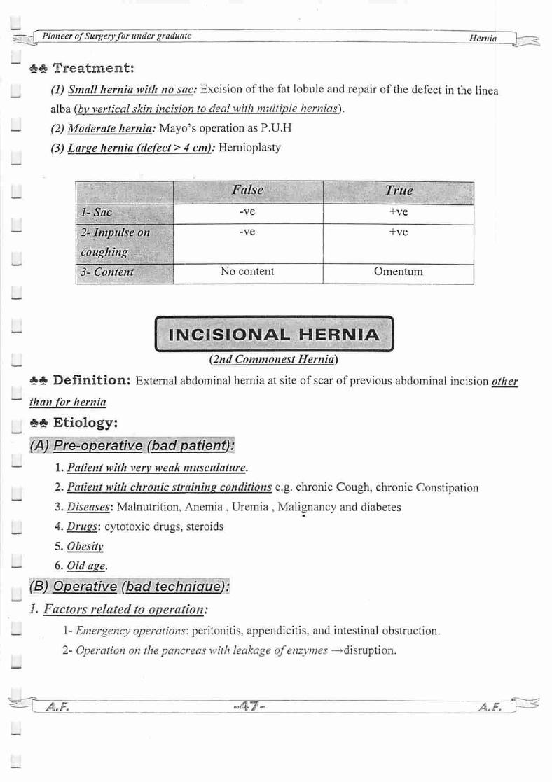
False True
1-Sac -ve +ve
2- Impulse on
coughing
-ve +ve
3- Content No content Omentum
INCISIONAL HERNIA
(2nd Commonest Hernia)
*_» Definition: External abdominal hemia at site of scar of previous abdominal incision other
than for hernia
** Etiology:
(A) Pre-operative (bad patient):
1. Patient with very weak musculature.
2. Patient with chronic straining conditions e.g. chronic Cough, chronic Constipation
3. Diseases: Malnutrition. Anemia , Uremia , Malignancy and diabetes
4. Drugs: cytotoxic drugs, steroids
5. Obesity
6. Old age.
(B) Operative (bad technique):
1. Factors related to operation:
1- Emergency operations: peritonitis, appendicitis, and intestinal obstruction.
2- Operation on thepancreas with leakage ofenzymes —•disruption.
A.F. r-

.=;—-f Pioneer ofSurgeryfor under graduate _^_^_ Hernia "}—^
"~ 2. Factors related to incision:
1- Length of incision: too long incision more than short incisions.
2- Site ofincision: midline incision more than paramedian incisions.
_ 3- Type ofincisions: Muscle cutting than muscle splitting
3. Factors related to closure:
1- Suture material: closure with absorbable more than non-absorbable.
2- Too tight sutures or too loose sutures.
3- Method ofclosure: Continuous sutures more than interrupted sutures.
4- Imperfect homeostasis leads to haematoma and infection.
5- Dead space from inefficient closure leads to seroma and infection.
4. Factors related to the drain:
1- Drain coming out the wound carries a high incidence than drain coming out through
separate stab wound.
_ 2- Prolonged use ofthe drain.
~ (C) Post-operative (bad post operative care):1. Infection ofthe wound—• ft tissue tension & ft collagen breakdown by proteolytic enzymes
2. Liftins heavy weights before 3 months after hemiorraphy.
3. Post-operative chest complications.
4. Post-Operative distension (acute gastric dilatation or ileus).
5. Poor healing power.
6. Dehiscence (burst) ofthe wound.
_
—• <%*
@ «___?•' the site of previous incision.
@ Structure;
* Defect: part or whole of the incision —•common at angles of wound
* Sac: thin with variable size and irregular shape.
* Contents: as before and usually loops of small intestine.
* Coverings: sac always lies beneath the skin, which is stretched & may be atrophic
* Descent: variable according to site —»Large hernia may sag downward.
-L A.F. -48- A.FrV^

L-
1 I
Pioneer ofSurgery for under graduate
*# Clinical Picture:
1-A swelling through scar of an operation which is reducible
2- With an expansile impulse on cough
3- It may contain omentum or intestine.
4- The skin over hemia is stretched and may become atrophic
5- Peristalsis ofunderlying coils of intestine may be seen through it
** Investigations: Fordetection of cause.
4»_» Treatment:
(I) Palliative abdominal belt: If surgery is contraindicated
(II) Operative treatment
1. Anatomical repair:
* Indicated in small defect.
* After excision ofsac, layers ofabdominal wall are closed separately
2. Keel operation:
- The sac is identified & dissected down to the neck. "
- Without opening the sac, it is pushed back in the abdomen by a series of inverting sutures
—• the sac willproject into the abdomen like keel ofa boai-^ Edges of the defectare closed.
3. CattelVs operation: (5 lover repair)
- The sac is dissected & opened, its contents are returned to the abdomen.
- The following layers are closed with non absorbable sutures in the same sequence:
* Is' layer: The neck of the sac is closed from inside
* 2nd layer: The sac is excised 2cm distal to the 1st layer &its cut edges are sutured together* 3 layer: An elliptical incision is made 2cm lateral to the previous suture line in anterior rectus
sheath, & the medial flaps of anterior rectus sheath are approximated by sutures.
* 4th layer: The muscles on either side are approximated bysutures.
* 5th layer: The lateral flaps ofanterior rectus sheath are sutured
i i 4. Hernioplasty: either * In-lay to fill the defectLa*
* On-lay after closure of defect
^ * Underlay: mesh is sutured tounder surface ofthe peritoneum followed byr closure of the defect
^u#tf# »49-
Hernia
ft*tt0
—-f Pioneer ofSurgeryfor under graduate Hernia
BURST ABDOMEN
(Abdominal Dehiscence)
** Definition: Dehiscence of abdominal wound withprolapseof viscera extraperiloneally.
—** Etiology: The same causes ofincisional hemia.
_ ** Pathology:
A- Complete burst:
When all layers of the wound (including skin) give way —> the bowels prolapse through the
skin to exterior.
" B- Incomplete (partial) burst:When the deeper layers ofthe wound only give way with intact skin —> the loops of intestine
prolapse under skin
— ** Complications:
1- Shock, dehydration and electrolyte imbalance.
2- Paralytic ileus and intestinal obstruction.
3- Devitalization of intestinal wall leads to intestinal fistula.
4- Partial burst ends in incisional hemia.
— ** Clinical picture:
1- Serosanguinous discharge from the wound is a warning sign in 50%.
2- Usually between 6,h to 8th day:Complete burst —* the wound gives way and coils of intestine and omentum prolapse through
the skin
— Incomplete burst —* the skin is intact over the intestine
<&* Treatment:
(A) Complete Burst: Emergency operation to replace the bowel &to resuture the wound- All layers ofthe abdominal wall are approximated by through and through sutures as one layer- The abdominal wall may be supported by strips of adhesive plaster encircling anterior 2/3 of the
circumference of the trunk.
(B) Partial Burst:
Treated by abdominal corset (binder), delayed removal of sutures then treat as incisional
hernia
^ZST /i,p. ~5Q- A.F. j -

_
Pioneer ofSurgeryfor under graduate Hernia
Diaphragmatic Hernia
— ** Causes & Types:
(i) Congenital: Failure of fusion of various parts of diaphragm
(1) Eventration:-
_ - Diaphragm consists of sheet of fibrous tissue.
- No symptoms; discovered on routine X-ray.
— (2) EsophageaI hiatus hemia:
- Due to congenital short oesophagus with hiatus hernia
(3) Hernia through Foramen ofMorgagni or Magendie (parasternal hernia):
- Defect through diaphragm between sternal & costal attachments of the diaphragm
- These hernias are situated anteriorly
— - Common on Right side (Pericardium).
(4) Hernia through Foramen ofBochdalek = Pleuro-peritoneal canal- Due to persistence of Pleuro-peritoneal canals which are last parts of diaphragm toclose.
- These hernias are situated posteriorly.
- Common on left side.
_ (5) Hernia through the Dome:
- Junction of muscular & tendinous portion of diaphragm:
— - In right side: the hemia will contain portion of Liver parenchyma.
- In left side: the hernia will contain the fundus of stomach.
— (ii) Acquired•
I-Traumatic: Bullet injury (Thoracoabdominal).
2-Post-operative: Thoracoabdominal operation.
— 3-Esophageal hiatus hernia (3types) &J*"
— ** Clinical picture & Complications:
(1) Strangulation—> Acute intestinal obstruction.
(2) Iflarse:
- Respiratory embarassment
- Lung Collapse on affected side.
-T-^
- Pioneer ofSurgeryfor under graduate
** Investigations:
1- Chest x-ray
2- Abdominal x-ray in erect and supine positions
— ** Treatment: In all cases:
* Excision of sac +
* Plication of diaphragm in eventration OR
* Suturing the diaphragm by overlapping with non-absorbable sutures.
RARE EXTERNAL HERNIAS
Hernia
— 1. Interparietal Hernia (Interstitial hernia):
- Hemia pass () layers of anterior abdominal wall:
I- Properitoneal: Between the peritoneum & transversalis fascia
2- Intermuscular: Between the muscle layers of abdominal wall
3- Proparietal: Between external oblique & the skin
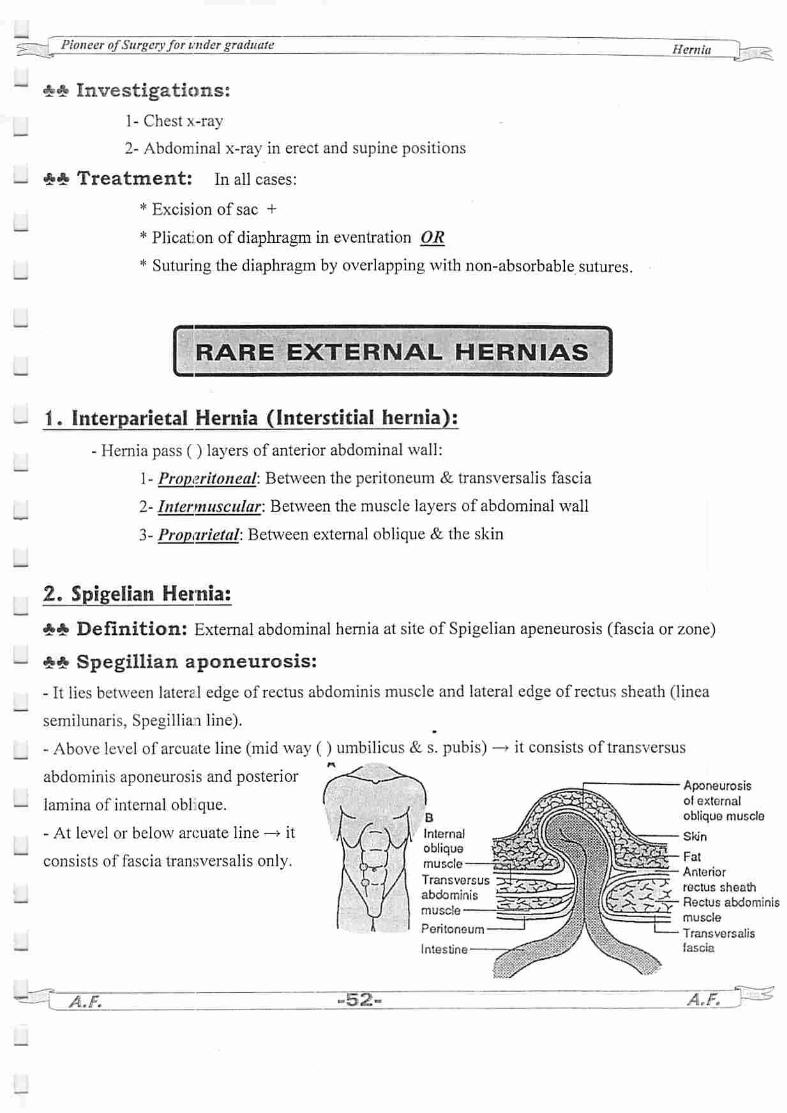
2. Spigelian Hernia:
«£* Definition: External abdominal hemia at site of Spigelian apeneurosis (fascia or zone)
— ** Spegillian aponeurosis:
- It lies between lateral edge of rectus abdominis muscle and lateral edge of rectus sheath (linea
semilunaris. Spegilliai line).
- Above level of arcuate line (mid way () umbilicus & s. pubis) -» it consists of transversus
abdominis aponeurosis and posterior
— lamina of internal obi que.
- At level or below arcuate line —* it
consists of fascia transversalis only.
u*.A.F. -52-
B
Internal
obliquemuscle
Transversus
abdominismuscle
Peritoneum
Intestine—
Aponeurosisof external
oblique muscle
Skin
Fat
Anterior
rectus sheath
*y- Rectus abdominismuscle
Transversalis
Fascia

u r . .gs^ PioneerofSurgery for undergraduate Hemia
3. Lumbar Hernia:
Ll A- Primary lumbar hernia:
1. Inferior lumber hernia through inferior lumbar triangle which is bounded by:
uj Inferior—* crest ofileum jkLaterally -*• external oblique LWIi'ss*, Aa«; ff \ -„l ,,,Medially —>latissimus dorsi / \ ""
2. Superior lumber hernia through superior lumbar triansle which is bounded by:
Li Above ^ 12th rib ^ ^<A>i ; Laterally —»• internal oblique
Medially -*• sacrospinalis Sqcroaf^:^ >/ fa****** c'^i<^Li B- Secondary lumbar hernia:
It is an incisional hemia following lumbar incision in renal surgeryi •'
(J 4. Obturator Hernia: Itoccurs through the obturator foramen —• in the upper medial thigh
( 5. Gluteal Hernia: It occurs through the Wreater sciatic foramen.
6. Sciatic Hernia: It occurs through lesser sciatic foramen -*• A swelling in the buttock
^ 7» Perineal hernia: it occurs through pelvic floor
Lj 8. Divarication of the recti:** Aetiology: stretching of linea alba by a chronically 11 abdominal pressure e.gdrycough.
^ *# C/P: - When the patient strains -» abulge appears between the recti.I \ - Whenthe abdomen is relaxed —• gap is felt between the recti
** Treatment: X / J*-> # Abdominal belt is satisfactory in symptomless cases.
, , # Surgery :
- Vertical mayo repair.
i i - Keel repair.
- Anatomical repair.
Li -Nuttal's repair (with divarication ofrecti below umbilicus) —• divide the 2recti at their insertionthensuture each recti to theperiosteum of the opposite side i.e overlapping their lower parts
^gt~JLF. -53- • AjrV^L_

~*-fFicneer ofSurgeryfor under graduate
Recurrent hernia
_ ** Aetiology:
A- Pre operative causes (badpatient) : as incisional hemia
B- Operative causes (badtechnique) :
incomplete removal of the sac.
Missing a second sac.
_ Inadequate repair - bad choice of repair.
Suture : under tension - absorbable -continuous
— Imperfect haemostasis
C- Postoperative causes : as incisional hernia
** TTT: Correct the cause first
_ I. Conservative tit: in patients with uncorrectable cause —> Truss.
77. Surgical ttt:
— 1- Hernioplasty is the main line of ttt
2- recurrent inguinal hemia in old patient -» high inguinal orchidectomy (i.e orchidectomy &excision ofspermatic cord up to the internal ring with obliteration of inguinal canal
Nyhu s Classification of hernia
Hernia
Typel: Indirect hernia with normal internal rins
— Type M: Indirect hernia with dilated internal rins with intact posterior wall (good musculature)"
_ Type Hf: Posterior wall defect (weak musculature)
TypelUA: Direct hernia—
Type IIIB. Indirect hernia
U Type IIIC: Femoral hemia
Type IV; Recurrent hernia
• —r'"aH. A.F. ~S4- j^f0 j~
_