surface structures ofthe gonococcus
TRANSCRIPT
Brit. J. vener. Dis. (1974) 50, 354
Surface structures of the gonococcus
A. GRIMBLE AND L. R. G. ARMITAGEGuy's Hospital, London SE1 9RT
The fine structures of Neisseria gonorrhoeae, especiallythe surface structures, are of interest at this time,when the organism and the infection it causes arebeing more widely investigated. Ward, Watt, andGlynn (1970) noted the loss of a virulence factor onsubculture of N. gonorrhoeae. Kellogg, Cohen,Norins, Schroeter, and Reising (1968) had previouslydescribed the colonial morphology of the freshpathogenic strains in contrast to the laboratory sub-cultures. They noted that, although the nonpatho-genic laboratory strain (Type 4 colony) sometimesexisted in fresh isolations along with the virulent(Type 1 colony) strain, for the most part the formerappeared to arise in subculture out of the latter.They surmised that the nonpathogenic strain usually*arose from a previously virulent strain. By suitablemethods the pathogenic strain can now be preservedin subculture.The surface appearance of the freshly isolated
micro-organisms seen under the electron microscopediffers from that of the subcultured, laboratorystrains. Most obviously fimbriae, or pill, are to beseen on virulent strains as opposed to subculturedstrains.The surface structures or appendages which may
be seen on bacteria consist of:flagellae, whose functionis primarily that of motility; and fimbriae or pili,which are finer appendages, functioning (dependingupon the type of pilus) either as something imparting'adhesiveness' to the organism, or as a means bywhich metabolites and genetic material are transferredfrom one bacterium to another. The former pili,or common pili, are shorter; the latter sort are longer.The accompanying electron photomicrographs are
presented to show the kinds of surface structureswhich appear in the freshly isolated gonococcus.
Material and methodsA negative staining technique was applied. Studies weremade of strains of N. gonorrhoeae taken direct frompatients, and of cultured strains grown for 12 to 48 hrs on
Received for publication December 11, 1973
ordinary chocolate agar plates and on Thayer-Martinmedium. Two stock laboratory strains from BurroughsWellcome, No. 890 and No. 1000, were also examined.At the time of harvesting, the laboratory strains hadusually completed their log-phase growth, whereas inmost instances the fresh strains had only arrived at themiddle to end of their log-phase growth.The specimens from the patients and harvests from the
plates were obtained with a loop and suspended (using aPasteur pipette) in 5 drops of distilled water. One dropof 3 per cent. potassium phosphotungstate was added.The suspension was transferred without delay to theelectron microscope grids which had been lightlycarbon-coated on a film of polyvinyl formol Formvar(stock solution of 1 per cent. w/v Formvar in chloroform,diluted 1 in 20 in chloroform for use). Prolonged sus-pension of the cocci in distilled water resulted in ruptureof the cell membrane by an osmotic pressure effect, andthis had to be avoided. The difficulty with fresh strainsdirect from patients was the larger quantity of debrisharvested, and the pus, which has to be scanned. Theillustrations provided in the Figures were not taken fromany bacterium seen within a leucocyte.The microscope used was RCA E.M.U3 at 50 KV;
100 KV gave no noticeable improvement in resolution.
Findings and discussion
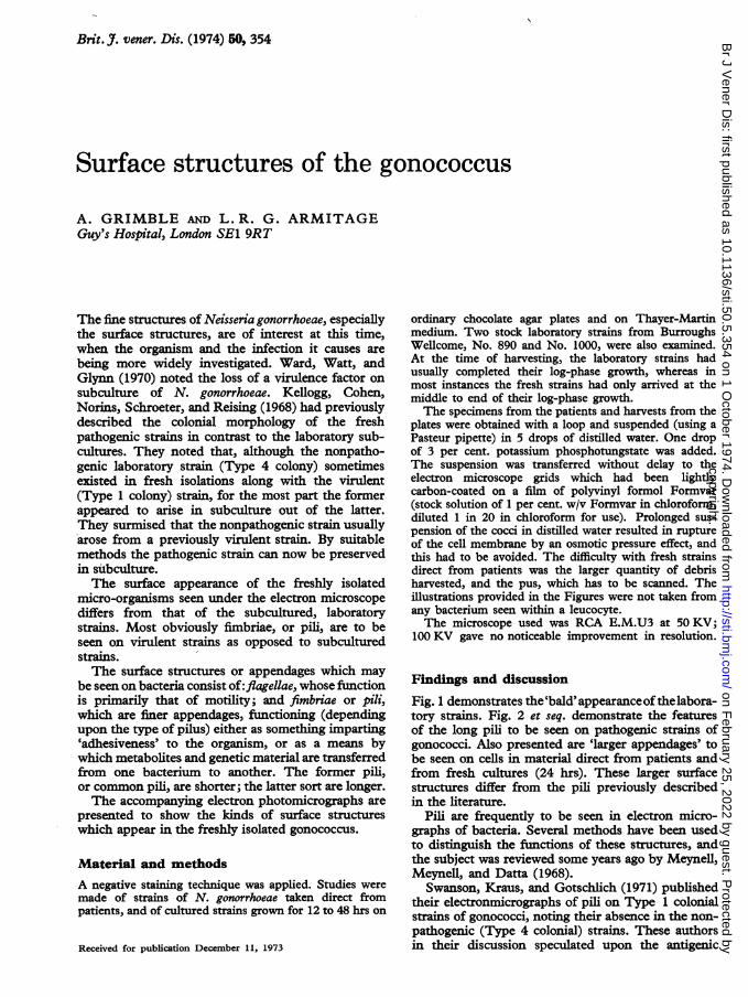
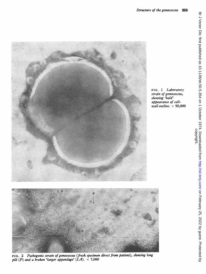
Fig. 1 demonstrates the'bald'appearanceofthelabora-tory strains. Fig. 2 et seq. demonstrate the featuresof the long pili to be seen on pathogenic strains ofgonococci. Also presented are 'larger appendages' tobe seen on cells in material direct from patients andfrom fresh cultures (24 hrs). These larger surfacestructures differ from the pill previously describedin the literature.
Pili are frequently to be seen in electron micro-graphs of bacteria. Several methods have been usedto distinguish the functions of these structures, andthe subject was reviewed some years ago by Meynell,Meynell, and Datta (1968).
Swanson, Kraus, and Gotschlich (1971) publishedtheir electronmicrographs of pili on Type 1 colonialstrains of gonococci, noting their absence in the non-pathogenic (Type 4 colonial) strains. These authorsin their discussion speculated upon the antigenic
copyright. on F
ebruary 25, 2022 by guest. Protected by
http://sti.bmj.com
/B
r J Vener D
is: first published as 10.1136/sti.50.5.354 on 1 October 1974. D
ownloaded from
Structure of the gonococcus 355
* :
lk ~ L
F I G. 1 Laboratorystrain of gonococcus,showing 'bald'appearance of cell-wall outline. x 50,000
I
p
-It
P.10 !.
FIG. 2 Pathogenic strain of gonococcus (fresh specimen direct from patient), showing longpili (P) and a broken 'larger appendage' (LA). x 7,060
a'O...5.... .... .uI
.:4.",. ..: .:: .x ". t
copyright. on F
ebruary 25, 2022 by guest. Protected by
http://sti.bmj.com
/B
r J Vener D
is: first published as 10.1136/sti.50.5.354 on 1 October 1974. D
ownloaded from

356 British Journal of Venereal Diseases
L A
FIG. 3 Pathogenic strain of gonococcus, showing 'plaited' bundle of pili(P) together with a 'larger appendage' (LA). x 14.600
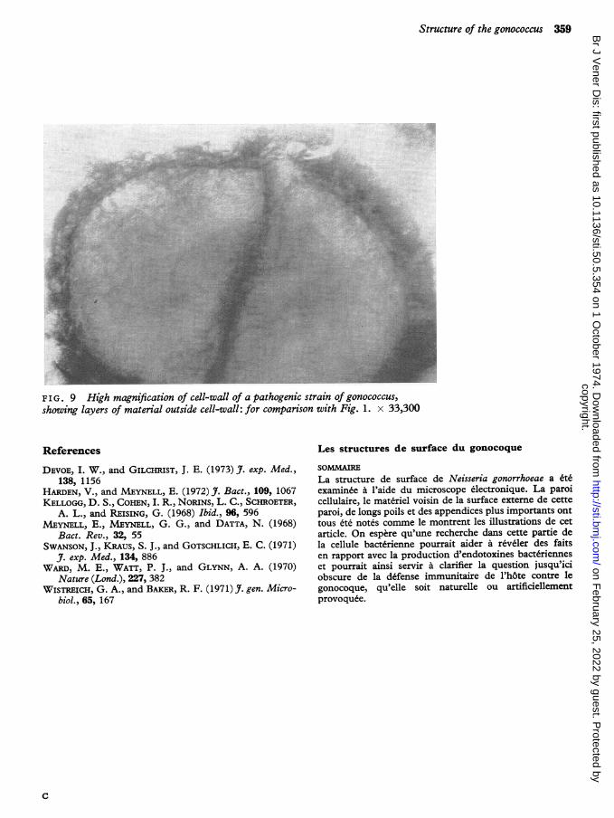
role which these pili might play. The pili were longrather than short and were similar to those shown inFigs 2 and 3. These slim structures (Figs 2 and 3 and,less noticably, in other accompanying Figures)appear in all probability to be related to the long slim'sex pili', F and R specialized pilh (the latter respon-sible for the transference of antibiotic resistance inE. coli) investigated by Harden and Meynell (1972).Moreover, they appear to be in the same morpholo-gical category of surface structure as theones demonstrated on the gonococcus by Swansonand others (1971). There do not appear to be repre-sented, in the photographs of Swanson and others,the shorter 'common pill', whose function is one ofstabilization or adhesiveness, which would enable thegonococcus to stick to the epithelial cell and resistbeing washed away. It was not certain whether ornot these short pili appeared in our material either,although the surface ofpathogenic gonococci appeared'coated' and piliform in contrast with the 'bald'appearance of laboratory strains (compare Fig. 1 withFig. 9).
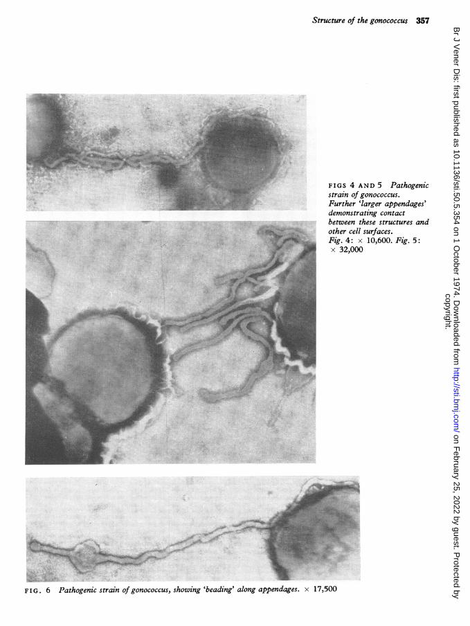
In addition, electronmicrographs of larger append-ages are presented (Figs 3 to 8). They were often tobe seen broken off and lying free in the grid, singlyor in groups. The diameter of these latter surface
appendages is in the region of 20-30 V*m, whereasthe long pili (Figs 2 and 3; and see Swanson andothers, 1971) are about 3 to 7 Fm in diameter. Thelarger appendages are present singly or sparsely onthe cell, or occasionally exist in profusion, over whatappears to be the whole cell surface. At times theymay be seen beginning to appear in a cluster fromthe central fissure of the organism.One feature which was fairly common to these
appendages was the terminal bulb, also to be seenin other pili. Another feature was a 'beading' alongthe length of the appendage itself: these 'beads'occurred singly or in clusters on the appendage(Figs 6 and 7). Occasionally a break was visible in thewall. These larger appendages seemed to makecontact with another bacterial cell (Figs 4 and 5),which might indicate that some metabolic functionwas being fulfilled.Whether these larger tube-like structures are, as
suggested, surface appendages, or whether they arepossibly related in kind to the lipopolysaccharide'surface blebs' described by Devoe and Gilchrist(1973) in Neisseria meningitidis, requires furtherinvestigation. Devoe and Gilchrist observed that theorigin of their 'blebs' was at the outer membranelayer of the cell-wall, the suggested site of endotoxin
copyright. on F
ebruary 25, 2022 by guest. Protected by
http://sti.bmj.com
/B
r J Vener D
is: first published as 10.1136/sti.50.5.354 on 1 October 1974. D
ownloaded from
Structure of the gonococcus 357
FIGS 4 AND 5 Pathogenicstrain of gonococcus.Further 'larger appendages'demonstrating contactbetween these structures andother cell surfaces.Fig.4: x 10,600. Fig. 5:x 32,000
*.
FIG. 6 Pathogenic strain of gonococcus, showing 'beading' along appendages. x 17,500
Awft..i:i,I-3:: ON`
-.B;ju ..
IsLo- .-&&. 14.
W.
I .A
e
tu -:.
.il,
,k-. 1111111111100
.4-. ,- -
copyright. on F
ebruary 25, 2022 by guest. Protected by
http://sti.bmj.com
/B
r J Vener D
is: first published as 10.1136/sti.50.5.354 on 1 October 1974. D
ownloaded from

358 British Journal of Venereal Diseases
I
FIG. 8 Pathogenic strain of gonococcus, showing a'larger appendage' with its point ofjunction with thecell-wall and the appearance of material along theouter side of the cell-wall. x 33,300
FIG. 7 Pathogenic strain of gonococcus, showingmore frequent 'beading' than in Fig. 6. x 14,600
production. Surface material with similar featuresto these so-named 'blebs' is visible in the outer regionsof the cell-wall presented by us at high magnification(Fig. 8).The structure and function of bacterial fimbriae or
pili, as well as of the cell-wall, are receiving muchattention at the present time. These surface areas andappendages are of importance in respect of antigenicroles as well as metabolic and genetic functions orattributes. In the case of the gonococcus, these aspectsare only beginning to receive attention. On thequestion of their antigenic role and their relationshipto pathogenicity, it should be mentioned that pilihave been noted in Neisseria catarrhalis, perflava,and subflava, organisms hitherto considered non-pathogenic (Wistreich and Baker, 1971). It is hopedthat their study in the gonococcus may produce some
of the answers to the problems of antigenicity andcell metabolism posed by this organism.
SummaryThe surface structure of Neisseria gonorrhoeae hasbeen examined by means of electron microscopy. Thecell-wall, material proximal to the external surfaceof the cell-wall, long pili, and larger appendages haveall been noted, and are shown in accompanyingillustrations. It is hoped that investigation of thisregion of the bacterial cell may help to reveal factsconcerned with bacterial endotoxin production andmay thus be of assistance in clarifying the at presentobscure position of the host immune defence againstthe gonococcus, whether naturally or artificiallyinduced.
z:,
copyright. on F
ebruary 25, 2022 by guest. Protected by
http://sti.bmj.com
/B
r J Vener D
is: first published as 10.1136/sti.50.5.354 on 1 October 1974. D
ownloaded from
Structure of the gonococcus 359
FIG . 9 High magnification of cell-wall of a pathogenic strain of gonococcus,showing layers of material outside cell-wall: for comparison with Fig. 1. x 33,300
References
DEVOE, I. W., and GILCHRIST, J. E. (1973) J. exp. Med.,138, 1156
HARDEN, V., and MEYNELL, E. (1972) J. Bact., 109, 1067KELLOGG, D. S., COHEN, I. R., NOIUNS, L. C., SCHROETER,
A. L., and REISING, G. (1968) Ibid., 96, 596MEYNELL, E., MEYNELL, G. G., and DATTA, N. (1968)
Bact. Rev., 32, 55SWANSON, J., KRAUS, S. J., and GOTSCHLICH, E. C. (1971)
J. exp. Med., 134, 886WARD, M. E., WATT, P. J., and GLYNN, A. A. (1970)
Nature (Lond.), 227, 382WISTREICH, G. A., and BAKER, R. F. (1971) J. gen. Micro-
biol., 65, 167
Les structures de surface du gonocoque
SOMMAIRELa structure de surface de Neisseria gonorrhoeae a eexaminee a l'aide du microscope electronique. La paroicellulaire, le materiel voisin de la surface exteme de cetteparoi, de longs poils et des appendices plus importants onttous e notes comme le montrent les illustrations de cetarticle. On espere qu'une recherche dans cette partie dela cellule bacterienne pourrait aider a reveler des faitsen rapport avec la production d'endotoxines bacterienneset pourrait ainsi servir a clarifier la question jusqu'iciobscure de la defense immunitaire de l'h6te contre legonocoque, qu'elle soit naturelle ou artificiellementprovoquee.
c
copyright. on F
ebruary 25, 2022 by guest. Protected by
http://sti.bmj.com
/B
r J Vener D
is: first published as 10.1136/sti.50.5.354 on 1 October 1974. D
ownloaded from