surface force isn the tracheal system of...
TRANSCRIPT

5°7
Surface Forces in the Tracheal System of Insects
By V. B. WIGGLESWORTH{From the Department of Zoology, University of Cambridge)
SUMMARY
The mechanism by which the tracheal system becomes filled with air is reviewed.It is concluded that the fluid contents are actively absorbed by the walls of thesystem. The air often enters from the atmosphere through the spiracles. In someinsects the system fills while the insect is submerged in water or while the spiraclesare closed. It is shown by means of simple models how the tanning of the lining of thelarger tracheae or the secretion of wax over the walls will bring about the liberation ofgas from solution when the fluid is subjected to a very slight negative pressure.
The movements of fluid in the tracheole endings of insects are also reviewed. Theremoval of fluid which takes place during activity, particularly under conditions ofdeficient oxygen supply, is not caused by secretory activity but by the physical forcesproduced by the products of metabolism.
The evidence supports the view that the fluid in the tracheoles is a cell sap whosepassage up the tracheole under the action of capillarity is opposed by the elasticity orswelling pressure of the cytoplasmic sheath of the tracheoles. It is shown by means ofa simple gelatin model how osmotic changes in the surrounding fluid, acting by wayof a cytoplasmic sheath, will bring about the absorption of such a cell sap.
A more exact model, illustrating the greater permeability of the inner wall of thetracheole which the proposed mechanism requires, is provided by the anal papillae ofmosquito larvae. These structures show the same adaptation to a saline medium as ispostulated in the tracheoles of mosquito larvae from salt water.
Alternative mechanisms are discussed. It is suggested that the capillary forces in thetracheoles are probably small. The effects of probable contamination of the tracheolefluid by an oily film (Beament) and the effects of changes in hydrogen-ion concentra-tion on interfacial tensions are illustrated by reference to further models.
INSECTS, for the most part, are of such a size that surface forces are greatenough to move or to support the whole mass of the body. This is parti-
cularly evident in the adaptations to be seen in those insects which live in oron the surface of water (Brocher, 1909) and in the mechanisms of adhesion bymeans of which terrestrial insects hold on to smooth solid surfaces (Gillettand Wigglesworth, 1932).
Surface forces are manifest also in the tracheal system of insects, wheregaseous, solid, and liquid phases meet. They create problems during theinitial filling of the tracheal system with air at the time of hatching from theegg, and they are concerned in the movements of fluid or the maintenance ofgas in the endings of the tracheoles throughout the life of the insect. In boththese regards there are striking differences in different insects and some-times, it would seem, between the different parts of the body in the sameinsect.[Quarterly Journal of Microscopical Science, Vol. 94, part 4, pp. 507-522, December 1953.J
2421.4 L 1
508 Wigglesworth—Surface Forces in the
A good deal of fragmentary information about these matters has accumu-lated during the past 30 years. It is a field in which the phenomena readilylend themselves to interpretation in terms of simple mechanical models. Theobject of this article is to bring together these scattered observations and toformulate the questions that are still unanswered.
THE INITIAL FILLING OF THE TRACHEAL SYSTEM WITH GAS
In most insects the tracheal system is open to the exterior through func-tional spiracles. In some aquatic or parasitic larval forms the spiracles areconcerned only in the moulting of the tracheal linings during ecdysis and thetracheal system is functionally closed. But when we consider the initial rillingof the tracheal system with gas at the time of hatching from the egg there is noreal distinction between the 'open' and the 'closed' system.
In the closed system, of course, gas first makes its appearance while theinsect is still immersed in fluid. That is so in Corethra (Krogh, 1911; v.Frankenberg, 1915), in Chironomids (Pause, 1918), in Ceratopogonids(Keilin, 1924), in Sciara (Keister and Buck, 1949), in Odonata (Calvert, 1898;Tillyard, 1915). But even in insects in which the tracheal system is open, gasmay appear throughout this system while the larva is still bathed in the fluidcontents of the egg. That is so in the blowflies Calliphora (Weismann, 1863)and Lucilia, and in the caterpillar Sitotroga (Sikes and Wigglesworth, 1931).Here the mechanism must be the same as in the closed system.
In many insects the surface of the larva dries while it is still inside the eggand the tracheal system fills, before hatching, from the outside air (as in theflea Ceratophyllus and the mealworm Tenebrio (Sikes and Wigglesworth, 1931).Often the larva at the time of hatching is enclosed in a cuticle which retains alayer of fluid beneath it so that air has access to the tracheae and filling occursonly when this 'embryonic cuticle' is shed soon after hatching (as in the bed-bug Cimex and the louse Polyplax (Sikes and Wigglesworth, 1931)).
But even where filling does not normally occur until the spiracles are incontact with the air, the system may sometimes fill, like the closed system,even when the egg or young larva is kept permanently submerged. That is soin the mealworm provided that the egg receives an adequate supply of oxygen(Sikes and Wigglesworth, 1931). Other insects, such as Ceratophyllus orAedes (Wigglesworth, 19386), have not this ability.
DISAPPEARANCE OF FLUID FROM THE TRACHEAL SYSTEM
It was made clear by the work of Weismann (1863), Keilin (1924), andDavies (1927) that the fluid in the tracheal system is absorbed into the tissues.In the case of the open system this process has been described in detail inAedes aegypti (Wigglesworth, 19386). When the posterior spiracles of theAedes larva come into contact with the atmosphere at the water surface, airenters almost at once and extends along one tracheal trunk and then the otheras the fluid is absorbed. The system is completely filled in 15-30 minutes.
Tracheal System of Insects 509
This absorption is almost certainly the result of active transfer or secretionby the cells of the tracheal system. The evidence is as follows:
(i) It does not take place in the absence of an adequate supply of oxygen.This was shown in the filling of the system of Tenebrio during submergence(Sikes and Wigglesworth, 1931). It was proved in detail by Keister and Buck(1949) who showed that in the larva of Sciara the system (a closed system)does not fill with gas if the oxygen concentration falls much below 0-3 per cent.
(ii) It is inhibited by narcosis. Larvae of Aedes narcotized with chloroformwill rest with the spiracles open at the water surface but no air enters. Evenafter recovery from the narcotic there is a further delay of 10 minutes or sobefore filling begins (Wigglesworth, 19386). These observations suggest notonly that absorption is a secretory phenomenon but that the nervous system,acting perhaps through some hormonal mechanism, is concerned in startingthe process. Filling is also inhibited by low temperature (0° C.) (Keister andBuck, 1949).
(iii) Only for a limited period after hatching is absorption possible. Sub-mergence of newly hatched larvae of the flea Ceratophyllus for several daysprevents the system from filling with gas when the spiracles are later exposedto the air (Sikes and Wigglesworth, 1931). In Aedes larvae, filling of the systemis somewhat delayed after submergence for 2 days; after 3-4 days filling is veryslow and usually incomplete (Wigglesworth, 19386). That is so despite thefact that the tracheal endings remain permeable to the tracheal liquid through-out the life of the insect. In the later larval stages the period during whichabsorption can be effected is much shorter: second-stage larvae preventedfrom coming to the surface for half an hour only, are unable to fill theirtracheal system (Wigglesworth, 1938J).
NATURE OF TRACHEAL LIQUID AND THE SITE OF ABSORPTION
It was earlier suggested that the fluid in the tracheal system in the eggmight be an ultrafiltrate from the body fluids resulting from the expansion ofthe tracheal lumen during development, and that therefore it should becapable of reabsorption by osmosis (Sikes and Wigglesworth, 1931). This,however, is an improbable mechanism. In the embryo, the tracheal fluid iscontinuous with that in the amniotic cavity, which contains proteins and salts(Sikes and Wigglesworth, 1931). The same process occurs at each moult andhere the tracheal fluid is continuous with the moulting fluid below the cuticleand presumably has the same composition. The moulting fluid in Rhodniusis a neutral fluid, apparently free from chloride, with proteins in solution(Wigglesworth, 1933c). In the Cecropia silk moth (Passonneau and Williams,1950) it contains sodium and potassium as well as the breakdown products ofprotein and chitin. Such a fluid could readily be absorbed by an active secre-tory process, though not by simple osmosis.
The moulting fluid is absorbed, shortly before moulting, through thegeneral surface of the cuticle (Wigglesworth, 1933c). The absorption of the
510 Wiggles-worth—Surface Forces in the
tracheal fluid at the time of hatching and during each moult is doubtless acomparable process and probably takes place throughout the system. Thisbelief is supported by certain observations on Aedes in which the maintracheal trunks are connected in each segment by a transverse trachea fromwhich no tracheoles are given off. During filling it is quite common to seecolumns of air enter these transverse branches from both ends simultaneously.
FILLING OF THE CLOSED OR SUBMERGED TRACHEAL SYSTEM: LIBERATION OF
DISSOLVED GASES
There can be little doubt that the removal of fluid from the tracheal systemand its replacement by air is brought about by active absorption. (In theirdiscussion of this problem Keister and Buck (1949) attribute to the presentwriter the belief that an active secretion of gas is responsible; but in the paperin question (Wigglesworth, 19386) it is concluded that 'the fluid present inthe tracheal system is removed . . . by an active absorption'.) It is unlikely thatthere is any fundamental difference between the filling of the open systemfrom the atmosphere and the filling of the closed system from gases in solu-tion. For, as we have seen, certain insects such as Tenebrio, which normallytake in air directly from the atmosphere, are capable of filling when sub-merged.
The chief problem here is the force necessary to rupture a continuous columnof liquid (Keilin, 1924). This force may be very great. The experiments ofDixon (1914) have shown that even with air in solution the cohesion of wateramounts to about 200 atmospheres' pressure. But in order to obtain this highfigure for adhesion and cohesion of water very special precautions had to betaken to remove all grease from the apparatus and to ensure that every partwas thoroughly wetted. If this was not done, free gas appeared when the pres-sure was only slightly reduced; and Dixon admits that air bubbles are com-mon enough in the conducting tissues of the stems of plants although thenegative tension (due to the transpiration force) to which the sap is exposedis only between 5 and 20 atmospheres.
That is so in spite of the fact that the conducting tubes of plants imbibewater and therefore must exert a very strong adhesive power on the sap. Thetracheal intima of insects, on the other hand, has a very low adhesion to water.So far as the general surface of the body is concerned, the cuticle becomescompletely non-wettable by water as the result of the deposition of a crystal-line layer of wax immediately before moulting occurs (Wigglesworth, 1948).The wetting properties of the tracheae are such that it seems highly probablethat the larger branches likewise have a waxy lining. If the deposition of thiswax (or the quinone tanning of the cuticle, which will likewise reduce theadhesion to water) coincides with the development of a small negative pres-sure within the tracheal system, due to a process of active absorption, it isnot surprising that gas should be liberated from solution in the larger tracheae.Once a bubble of gas has formed it will grow rapidly in size as the trachealfluid is absorbed and dissolved gases diffuse into the expanding bubble.

Tracheal System of Insects 511
This process can be illustrated by means of a simple model experiment, amodification of one previously described (Sikes and Wigglesworth, 1931). Abeaker thoroughly freed from grease with chromic acid is partly filled withwater from the cold tap—water, that is, which is supersaturated with air whenbrought to atmospheric pressure. No bubbles of gas are liberated. A slidecovered with paraffin wax is then placed in the beaker, and bubbles of gas aresoon liberated from the supersaturated water, but only on the waxed surface
FIG. 1. Experiments to illustrate the effect of wax in causing the liberation of gas fromsolution. (Explanation in text.)
(fig. 1, A). If the waxed slide is placed in a similar beaker containing distilledwater that has been exposed to the air for some days, no bubbles of gas areset free upon either surface (fig. 1, B). But if now the pressure is lowered by aquarter of an atmosphere, minute bubbles appear upon the waxed surfacewithin a few minutes. At a negative pressure of half an atmosphere thebubbles appear almost at once and grow rapidly in size, but no gas is set freein contact with the clean glass (fig. 1, c).
We are still faced with the problem of why the tracheal system in someinsects (Calliphora (Weismann, 1863), Lucilia, Sitotroga, Tenebrio (Sikes andWigglesworth, 1931), Corethra (v. Frankenberg, 1915), Ceratopogon and Chiro-nomus (Keilin, 1924), Sciara (Keister and Buck, 1949), Odonata (Tillyard,1916)) will fill with gases from solution, whereas in others (Cimex, Polyplax,Ceratophyllus (Sikes and Wigglesworth, 1931), Aedes (Wigglesworth, 19386))it will fill only from the atmosphere. The difference may lie in the nature andwetting properties of the tracheal lining or in the intensity of the absorptiveactivity of the cells.
512 Wigglesworth—Surface Forces in the
It may be recalled that Weismann (1863), who observed the filling of thetracheal system in Calliphora with gases from solution, explained the processas follows: the trachea when first formed has only a potential lumen. Then acuticle is laid down, the lumen expands, and fluid diffuses into it from thetissues. He supposed that at a certain stage the cuticle of the larger trunksbecomes less permeable to fluid; but the lumen goes on enlarging and hencethe space must be filled with gas diffusing from the tissue fluids. Finally,Weismann supposed that the residual fluid is absorbed into the tissues throughthe finest tracheal tubes. Actually the lumen seems to be fully formed beforethe appearance of gas, but the possibility of negative pressure being producedwithin the tracheal system of some insects in this way is worth bearing inmind. (Sikes and Wigglesworth (1931) invoked a somewhat similar process toexplain the appearance within the tracheae of an ultra-filtrate from the tissuesthat could be reabsorbed by osmosis. This idea has since been abandoned infavour of absorption by a secretory process as described above.)
As to the nature of the gas which first appears in the closed tracheal system,a subject which has provoked much discussion, there is no reason to supposethat this will be constant. As already pointed out (Sikes and Wigglesworth,1931) its composition will depend solely upon the partial pressures and theinvasion coefficients of the various gases in solution in the tissue fluids at themoment when filling begins.
EXTENT OF AIR IN THE TRACHEOLES
In the past there has been some doubt as to whether the endings of thetracheoles during life contain liquid or gas. Observations on the living insecthave shown that there is much variation in different insects and in differenttissues in the same insect. In some tracheoles the column of air ends abruptly,long before the tracheole comes to an end. That is so in the tracheoles in thehead and anal papillae of mosquito larvae (Wigglesworth, 1930), the endingson the gut of Aeschna (Wigglesworth, 1930) and Tenebrio larvae (Wiggles-worth, 1931), in many parts of the body in Blattella (Wigglesworth, 1931;Bult, 1939), many of the endings in the abdomen of the flea (Wigglesworth,1931, 1935), the adult mosquito, and the bed-bug Cimex(Wigglesworth, 1931).In other endings the tracheoles contain gas as far as they can be resolved withthe light microscope. That is so in the leg muscles of the flea, in the musclesof the body wall of an active mealworm larva, and apparently in all the organsof Pediculus (Wigglesworth, 1931). Whether those branches which containair as far as they can be resolved do in fact contain gas up to their extremitiesis not certainly known, but it seems likely that this is so.
Measurements made recently on the tracheoles of the epidermis in Rhodnius(Wigglesworth, 1953) show that these spring from the endings of smalltracheae usually i"5-2/x in diameter. At their commencement their internaldiameter varies from 0-9 to o-6 ju,. They run a course of 200-300/u, (with a singlenucleus lying at about 75/J, from their point of origin), diminishing verygradually in diameter and sometimes giving off a single branch, to end at an
Trachea! System of Insects 513
apparent diameter of about o-Zfi or less. Electron microscope studies haveshown that the tracheoles, in which no spiral filament is visible with the lightmicroscope, do in fact have their walls thrown into annular or spiral folds likethe larger tracheae and that they end abruptly in rounded form at a diameterof o-s-o^jLi (Richards and Korda, 1950).
MOVEMENTS OF FLUID IN THE TRACHEOLES : OSMOTIC PRESSURE
In those forms in which the tracheoles contain liquid the extent to which itrises up the tubes is not constant but varies with the metabolism in the tissues:the fluid is absorbed and gas extends farther along the tracheoles after activemuscular contraction, or in the case of tracheoles on the surface of the gut, atthe height of digestion. Removal of fluid is most active during asphyxiacausing violent muscular activity in the absence of oxygen (Wigglesworth,1930, 1931, 1935). A similar removal of fluid is brought about if the trachealendings are exposed to hypertonic solutions of sodium chloride, potassiumlactate, &c. One per cent, sodium chloride or 2 per cent, potassium lactatewill cause complete removal of fluid just like asphyxia.
It was therefore inferred that the fluid was absorbed by osmosis into theblood; that the capillarity of the tracheole was in equilibrium with the osmoticpressure of the blood; and that when the osmotic pressure was increasedlocally by the liberation of unoxidized metabolites into the blood, water wasabsorbed from the tracheal endings so that the meniscus entered finer andfiner tubes until a new equilibrium was established. As the metabolites wereoxidized or removed the local osmotic pressure of the blood was believed tofall and the fluid therefore rose once more in the tracheoles.
Assuming that the fluid in the tracheoles is pure water with a surfacetension equal to that of distilled water, that it wets the wall of the tracheolecompletely, with an angle of contact of o°, and that the barrier between thetracheole lumen and the haemolymph behaves as a simple perfect semi-permeable membrane, it was calculated that the forces in question would justabout balance (Wigglesworth, 1930).
These, however, are very large assumptions and probably no one of themis justified. Indeed, the sole purpose of the calculation was to show 'that thetheory put forward was not unreasonable on physical grounds'.
In discussing above the initial filling of the tracheal system with air we cameto the conclusion that the fluid in the tracheae is of the same nature as theamniotic fluid or moulting fluid: a dilute lymph with which the cells are inosmotic equilibrium. It seems more probable that the fluid in the tracheolesat a later stage in development should also be of this character, than that theyshould contain a fluid that would be virtually distilled water.
The first indication of something of the kind was given by observations onmosquito larvae that had become adapted to salt water. The larva of Aedesaegypti reared in fresh water has an osmotic pressure in the blood equivalentto 0-75-0-9 per cent, sodium chloride. If it is acclimatized to dilute sea water(equivalent to 1-5 per cent. NaCl) the osmotic pressure of the blood rises to

514 Wiggleszvorth—Surface Forces in the
the equivalent of 1 -5 per cent. NaCl, and yet the fluid in the tracheoles of theresting larva rises to its accustomed level and is removed in the usual way uponasphyxiation. Clearly there cannot be a simple balance between osmotic pres-sure in the blood and capillarity (Wigglesworth, 1938a).
COLLOID OSMOTIC PRESSURE OR SWELLING PRESSURE AND THE MOVEMENT OF
FLUID IN THE TRACHEOLES
In the original discussion (Wigglesworth, 1930) the wall of the tracheolewas treated as a simple semi-permeable membrane. But it consists in fact ofa sleeve of cytoplasm surrounding the tracheole wall. We are faced with the
rrzl\z|
" i/l-'i " '
A BFIG. 2. Gelatin model of tracheole. (Explanation in text.)
question whether osmotic changes in the haemolymph bathing such a systemcould bring about the removal from the lumen of the tubes of a fluid withwhich the cytoplasm is in osmotic equilibrium.
We may consider a simple model prepared as follows (fig. 2). A glass tubeof 0-7 cm. internal diameter, open at both ends, is filled with 10 per cent,gelatin in 50 per cent. Ringer's solution (Ringer's solution diluted with anequal volume of water). Before the gelatin has set the end of a capillary pipetteis inserted into the tube so that its end comes to within about 3 mm. from thelower surface of the gelatin. When the gelatin has set the pipette is withdrawn,the cavity (which represents the lumen of the tracheole) is filled with 50 percent. Ringer and the tube is stood upright in the same solution and left over-night for equilibrium to become established. The tube is then stood uprightin 100 per cent. Ringer's solution; and it is found that the solution from thelumen is absorbed. In one experiment the column of fluid in the lumen wasreduced from 13-5 mm. to 2-5 mm. during 24 hours. The capillary lumentapered from 1-5 to 075 mm., so that about 90 per cent, of the fluid had beenabsorbed. In other experiments, the fluid was sometimes completely replacedby air.
Clearly in this model the increased concentration of salt has increased theswelling pressure of the gelatin which has then absorbed the more dilutesolution from the lumen; a process which could be described in terms of theDonnan equilibrium combined perhaps with specific effects of the ions pre-sent in inducing swelling. The model serves the purpose of showing how
Trachea! System of Insects 515
changes in osmotic pressure in the haemolymph might act indirectly throughthe swelling pressure of the cytoplasmic sheath of the tracheole in causing atemporary absorption of a saline solution in the lumen (Wigglesworth, 193 86).
But the wall of the tracheole is a much more complex structure than this.The cytoplasmic sheath is bounded on the outside by a cell membraneseparating it from the haemolymph, and on the inside by the modified cellmembrane which is the wall of the tracheole. The osmotic pressure of thehaemolymph will operate through the outer cell wall upon this cytoplasmicsheath; and the capillarity of the tracheole will operate against the water bind-ing power, 'swelling pressure' or 'colloid osmotic pressure' of the cytoplasm.It is possible therefore to conceive a mechanism by which an increase inosmotic pressure in the haemolymph will cause a temporary shrinkage of thecytoplasmic sheath. The framework or gel of this sheath will thereby be com-pressed. It will tend to expand again by reason of its elasticity (or of its col-loid osmotic pressure). If we suppose that the inner wall of the sheath, thetracheole lining, is more permeable than the outer cell membrane, then fluidwill be absorbed from the lumen of the tracheole.
DIFFERENTIAL PERMEABILITY IN THE ANAL PAPILLAE OF MOSQUITO
LARVAE
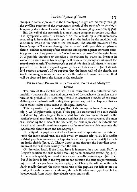
The crux of this mechanism lies in the conception of a differential per-meability between the inner and outer walls of the tracheole. Is such a struc-ture at all probable ? It is scarcely feasible to construct a model of the samedelicacy as a tracheole wall having these properties; but it so happens that anexact model exists ready-made in biological material.
This is provided by the anal papillae of the mosquito larva Aedes aegypti(fig. 3, A) (Wigglesworth, 1933a). These papillae are covered by a thin cuticlelaid down by rather large cells separated from the haemolymph within thepapilla by a cell membrane. It is suggested that the cuticle represents the innerwall bounding the lumen of the tracheole, the cells represent the cytoplasmicsheath, and the cell membrane represents the membrane which separates thecytoplasmic sheath from the haemolymph.
If the tip of the papilla is cut off and immersed in tap water so that this canreach the inner membrane, the cells swell by osmosis (fig. 3, B). If a similarisolated papilla is immersed in hypertonic saline (1 per cent. NaCl) the cellsgradually shrink (fig. 3, c). Clearly water passes through the bounding mem-branes of the cells more readily than the salt.
On the other hand, if the intact larva is immersed in 1 per cent. NaCl orRinger's solution the cells rapidly swell up to an enormous extent (fig. 3, D).If the larva is quickly returned to tap water the cells contract again (fig. 3, E).But if the larva is left in the hypertonic salt solution the cells are permanentlyinjured and the cytoplasm dispersed (fig. 3, F). Clearly the salt enters the cellsfairly readily through the outer membrane of the papillae but fails to pass soreadily through the inner membi'ane; the cells thus become hypertonic to thehaemolymph from which they absorb water and swell.
516 Wigglesworth—Surface Forces in the
These differences in permeability between the outer and inner membranesof the anal papilla are precisely those which are postulated in the tracheoles.Since the lining membrane of the tracheoles is morphologically equivalent tothe outer membrane of the anal papilla it is not unreasonable to suggest thatit may have the same permeability characteristics.
Fie. 3. Anal papilla of mosquito larva as a model to illustrate the permeability properties ofthe tracheoles. A, normal papilla of Aedes aegypti. B, tip of papilla cut off and immersed intap water, c, tip of papilla cut off and immersed in 1 per cent. NaCl. D, papilla of intact larvaimmersed in 1 per cent. NaCl. E, the same restored to tap water. F, the same after prolongedimmersion in 1 per cent. NaCl. The figures also show the changes taking place in the extent
of air in the tracheoles.
If the larva of Aedes aegypti is adapted to live in 1 per cent. NaCl, equili-brium becomes established between the cells of the papillae and the haemo-lymph, and the cells no longer swell when the larva is immersed in this solu-tion (Wigglesworth, 1933a). That is what is believed to happen in the tracheolesof larvae similarly adapted (Wigglesworth, 1938a).
ALTERNATIVE MECHANISMS FOR THE CONTROL OF MOVEMENT OF THETRACHEOLE FLUID
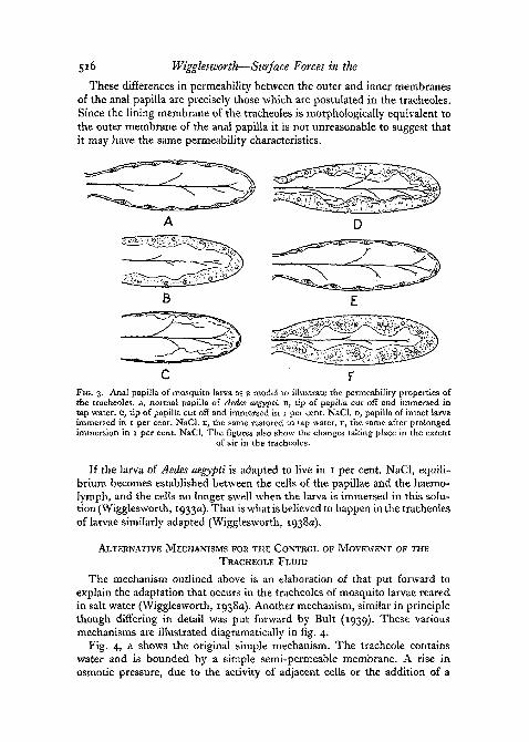
The mechanism outlined above is an elaboration of that put forward toexplain the adaptation that occurs in the tracheoles of mosquito larvae rearedin salt water (Wigglesworth, 1938a). Another mechanism, similar in principlethough differing in detail was put forward by Bult (1939). These variousmechanisms are illustrated diagramatically in fig. 4.
Fig. 4, A shows the original simple mechanism. The tracheole containswater and is bounded by a simple semi-permeable membrane. A rise inosmotic pressure, due to the activity of adjacent cells or the addition of a
Trachea! System of Insects 517
hypertonic solution, draws water from the tracheole until a new balance isestablished between osmotic pressure and capillarity.
Fig. 4, B shows the mechanism described above. The tracheole is surroundedby a cytoplasmic sheath separated from the haemolymph (or from the cyto-plasm in the case of intracellular tracheoles) by a semi-permeable membrane.A rise in osmotic pressure in the haemolymph causes shrinkage of the 'cyto-plasmic sheath and thereby increases the forces of imbibition in that sheath.
FIG. 4. Diagrams to illustrate the various schemes suggested to explain the movement offluid in the tracheoles. A, Wigglesworth (1930); B, Wigglesworth, 1938 and present paper;
C, Bult (1939).
Since the lining membrane of the tracheole is more permeable than the outermembrane of the cytoplasmic sheath, the cytoplasm tends to swell again byabsorbing the cell sap which has been drawn up the tracheole.
Fig. 4, c shows the mechanism proposed by Bult (1939). The tracheole ispictured as coming directly into contact with the cell, and as being separatedfrom it by a membrane of greater permeability than the wall of the tracheoleelsewhere or the surface membrane of the cell itself. The tracheole containsa cell sap which is drawn up the tube by capillarity until it is arrested by theresistance of the cell to collapse—that is, by the force of imbibition. A rise inosmotic pressure outside the cell will extract water from the cell. The elasticrecoil of the cell opposes this shrinkage, and since the membrane separatingthe cell from the tracheole is the more permeable, the fluid from the tracheolewill be absorbed into the cell. (Bult suggests that in normal circumstances theincreased imbibition by the cell is not the result of increased osmotic pressureoutside but is caused directly by oxygen want. This matter will be discussed later.)
In principle this mechanism proposed by Bult is the same as that illus-trated in fig. 4, B, except that the cytoplasm of the cell takes the place of the

518 Wigglesworth—Surface Forces in the
cytoplasmic sheath of the tracheole. This makes it necessary to postulate aspecially permeable region of the tracheole in contact with the cell. At presentthere is no evidence for such an arrangement. Indeed, electron microscopestudies show the tracheoles ending blindly without any change in the wall(Richards and Korda, 1950). But when the tracheoles penetrate within thecells (see fig. 3, D) there is little difference between schemes B and c (fig. 4).
VITAL ACTIVITY AND PHYSICAL FORCES IN THE TRACHEOLE ENDINGS
Throughout this discussion of the movements of fluid in the endings of thetracheoles it has been tacitly assumed that the wall of the tracheole plays apurely passive role, being subject to the physical forces exerted upon it. Butwe have seen that during the initial filling of the tracheal system with air athatching or at moulting the fluid is removed by an active process of secretion.The question arises whether secretory activity may be responsible for themovements of fluid in the functional tracheal system.
It is quite certain that the tracheole is by no means a lifeless structure. Ithas recently been shown (Wigglesworth, 1953) that the tracheole endings willactively migrate considerable distances (perhaps 300-500/x) towards a regionof deficient oxygenation, drawing the tracheae after them. There is no reasontherefore why they should not have retained their secretory function—although, as pointed out above, removal of fluid from the tracheal system ofthe newly moulted insect is not possible if filling is delayed.
There can be little doubt that when the tracheal endings are exposed tohypertonic solutions the extraction of fluid is a passive osmotic phenomenon.This can be brought about by 1 per cent. NaCl, by Ringer's solution osmoti-cally equivalent to 0-9 per cent. NaCl, or by 2 per cent, potassium lactate,which is about equivalent to 1 per cent. NaCl. When unneutralized lactic acidis used a rapid extension of air is followed by an equally rapid retreat. Thiswas attributed to a breakdown in permeability. The same thing happens whenother poisons such as hydrogen cyanide, ammonia, sulphur dioxide, or chlorineare employed, and may perhaps be interpreted as showing that we are con-cerned with the permeability of a membrane which is an inherent part of aliving cell rather than some inert cuticle (Wigglesworth, 1930).
It was originally considered that the removal of fluid which followsasphyxiation is also due to the liberation of osmotically active substances bythe contracting muscles. The reasons for this conclusion were as follows:
(i) Actual activity, particularly muscular activity, is necessary. In mosquitolarvae submersion is much more effective than exposure to hydrogen becauseit causes much greater muscular activity. Pure hydrogen is more effective thanpure carbon dioxide—because the larva is quickly narcotized in carbon dioxideand ceases to move.
(ii) Muscular activity may sometimes over-ride the effects of oxygen supply.If the mosquito larva is exposed in hydrogen the movement of fluid is arrestedwhen the larva comes to rest. If it is then exposed to oxygen it immediately
Tracheal System of Insects 519
becomes active and there may be an extension of air in the tracheoles beforethe larva becomes quiet and the air begins to retreat again.
(iii) Muscular activity in one part of the body may affect the tracheoles else-where. In the larva of Aedes muscular contractions in the head and trunkcause a delayed removal of fluid from the tracheoles in the anal papillae. Andif a larva is asphyxiated by submersion until its tracheoles are filled with air,its haemolymph will cause the removal of fluid from the tracheoles of a restinginsect (Wigglesworth, 1930).
All these observations suggest that the movement of fluid is caused, not byanaerobiosis as such, but by the products of muscular or secretory activityliberated from the cells, particularly when the oxygen supply is deficient. Or,alternatively, that increased imbibition of water by the cells results in increasedosmotic pressure outside them.
In opposition to this view Bult (1939) has suggested that the increasedimbibition by the cells, which according to his view is responsible for absorb-ing fluid through the permeable wall of the tracheole in contact with the cell(fig. 4, c), is the direct result of a low oxidation-reduction potential in thecytoplasm. He suggests that 'it is not at all improbable that . . . the redox-potential might affect the swelling of cell proteins'. He supposes that underconditions of reduced oxygen tension the cell proteins imbibe water and thecells swell. And since the surface of the cell is pictured as being less permeablethan the membrane between the cell and the tracheole (fig. 4, c), fluid isabsorbed preferentially from the lumen of the tracheole.
We have seen that the relation between cell and tracheole as described byBult is improbable. But the fact remains that protoplasmic imbibitionincreased by the mechanism he suggests might well result in an increase inosmotic pressure outside the cell and so a removal of fluid from the tracheolesas outlined above. Or, if the effects of reduced oxygen tension were picturedas occurring actually within the cytoplasmic sheath of the tracheoles, then theywould exert a direct effect on the fluid in the tracheole as already described.
Other indirect effects of this kind, notably the effect of acidity followingoxygen want, in causing increased protoplasmic imbibition were briefly con- •sidered in the original paper (Wigglesworth, 1930). The possibility of suchchanges cannot be disregarded.
THE EFFECTS OF KNOWN CHANGES IN THE PARTIAL PRESSURE OFOXYGEN
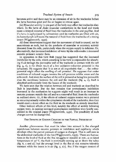
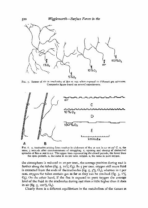
Another phenomenon that must be taken into account is the changedequilibrium between osmotic pressure or imbibition and capillarity whichdevelops when the partial pressure of oxygen is changed. This is well seen inthe abdominal tracheoles of the flea (Wigglesworth, 1935). There are fluctua-tions in the level of fluid as the flea struggles or remains at rest; at a raisedtemperature of 35° C. the fluid is continually moving up or down the tubes(fig. 6, A and B); but the average level in the flea at rest remains tolerablyconstant while the insect is in air (fig. 5, air). But if the oxygen content of
520 Wigglesworth—Surface Forces in the
100%0,
10% 0,
5%0,FIG. 5. Extent of air in tracheoles of flea at rest when exposed to different gas mixtures.
Composite figure based on several experiments.
FIG. 6. A, tracheoles arising from trachea in abdomen of flea at rest in air at 35° C. B, thesame, 5 seconds after commencement of struggling, c, opening and closing of abdominalspiracles of flea at rest in air. The upper lines representing the closed periods, the lower lines
the open periods, D, the same in 10 per cent, oxygen, E, the same in pure oxygen.
the atmosphere is reduced to 10 per cent., the average position during rest isfarther along the tubes (fig. 5, 10% O2). In 5 per cent, oxygen still more fluidis extracted from the ends of the tracheoles (fig. 5, 5% O2), whereas in 1 percent, oxygen the tubes contain gas as far as they can be resolved (fig. 5, 1%O2). On the other hand, if the flea is exposed to pure oxygen the averagelevel of the fluid in the tracheoles during rest rises a little higher than it doesin air (fig. 5, 100% O2).
Clearly there is a different equilibrium in the metabolism of the tissues at
Trachea! System of Insects 521
each partial pressure of oxygen, and one may use this material to provideevidence that there is indeed an increased accumulation of acid metaboliteswhen the partial pressure of oxygen is reduced.
The spiracles in the flea, at rest in air, open and close in a fairly regularrhythm as shown in fig. 6, c. The duration of the closed period is determined(a) by the accumulation of acid metabolites due to oxygen want, and (b) by theaccumulation of carbon dioxide. The duration of the open period is deter-mined by the time taken for the carbon dioxide which has accumulated in thebody during the closed period to diffuse out. The duration of the open periodis therefore largely determined by the duration of the closed period whichprecedes it. That is why the open period is so greatly prolonged in pureoxygen (fig. 6, E) and so much shortened in 5 per cent, oxygen (fig. 6, D). Inother words, when the insect is in pure oxygen, opening is induced by a largeamount of CO2, whereas when the insect is in 5 per cent, oxygen opening isinduced by a very small amount of CO2. It follows, therefore, that the meanlevel of accumulation of acid metabolites must be greater at low partial pres-sures of oxygen than at high—in agreement with the mean level of fluid in thetracheoles. It is therefore reasonable to ascribe the removal of fluid from thetracheoles to these same metabolites.
CAPILLARITY OF THE TRACHEOLES
When it was assumed that the osmotic pressure of the tissue fluids was act-ing in direct opposition to the capillarity of the tracheoles, we had to consideropposing forces of the order of 10 atmospheres. That would indeed be thecapillary force in the tracheoles if they contained pure water and had wallsthat were completely wettable.
According to the arguments set out in this paper we are in fact dealing withforces (colloid osmotic pressure or elasticity in the cytoplasmic sheath of thetracheoles) which are probably only a fraction of an atmosphere.
That presents no difficulty. The tracheole wall is readily wetted by oils(kerosene, &c.) which rapidly fill the system completely. The surface islipophilic as well as hydrophilic; the angle of contact with water is thereforeunlikely to be as small as o°. Moreover, as Dr. J. W. L. Beament has pointedout to me, if the fluid were covered with a film of lipide material its surfacetension would be greatly reduced. The cuticle of the cockroach is coveredwith a mobile grease (Ramsay, 1935) consisting of a wax in a volatile solvent(Beament, 1951) and it seems very probable that this material will spreadthroughout the lining of the tracheal system. In some unpublished observa-tions to which I am kindly permitted to refer, Dr. Beament noted that ina certain clean glass capillary, distilled water rose to a height of 23 cm.,whereas when cockroach grease was added to the distilled water it rose onlyto 10 cm.
It may well be, therefore, that quite small forces are at work in moving thefluid up and down the tracheoles. And it is possible that specific and localvariations in the wetting properties of the tracheole lining may account for the
522 Wigglesworth—Surface Forces in the Trachea! System of Insects
specific and local differences in the extent of air into the tracheoles alreadydescribed.
There remains the possibility that the surface tension of the fluid in con-tact with the walls of the tracheoles may vary under different physiologicalconditions. There are large changes in tension at an oil-water interface followingslight changes in hydrogen-ion concentration. If the oil phase contains carboxylgroups, either free or combined with glycerol, a fall in pH (such as might beexpected to follow muscular contraction in the absence of oxygen) causes arise in interfacial tension (Hartridge and Peters, 1922). This would tend tomake fluid rise in the tracheoles, which is the opposite to what is observed.But if the oil phase contains long-chain amines, a fall in pH within thephysiological range of pH 5 to pH 9 causes a fall in interfacial tension (Peters,
REFERENCESBEAMENT, J. W. L., 1951. Nature, 167, 652.BROCHER, F., 1909. Ann. Biol. Lacustre, 4, 89.BULT, T., 1939. Over de beweging der vloeistof in de trackeolen der insecten. Thesis. Assen.CALVERT, C. P., 1898. Ent. News, 9, 73.DAVIES, W. M., 1927. Quart. J. micr. Sci., 71, 15.DixON, H. H., 1914. Transpiration and the ascent of sap in plants. London (Macmillan).v. FRANKENBERG, G., 1915. Zool. Jahrb., Abt. Physiol., 35, 505.GILLETT, J. D., and WIGGLESWORTH, V. B., 1932. Proc. Roy. Soc. B, i n , 364.HARTRIDGE, H., and PETERS, R. A., 1922. Ibid. A, 101, 348.KEILIN, D., 1924. Proc. Camb. Phil. Soc. (Biol. Sci.), 1, 63.KEISTER, M. L., and BUCK, J. B., 1949. Biol. Bull., 97, 323.KROGH, A., 1911. Skand. Archiv. f. Physiol., 25, 183.PASSONNEAU, J. V., and WILLIAMS, C. M., 1950. Anat. Rec, 108, No. 3, 558.PAUSE, J., 1918. Zool. Jahrb., Abt. Physiol., 36, 339.PETERS, R. A., 1931. Proc. Roy. Soc. A, 133, 140.RAMSAY, J. A., 1935. J. exp. Biol., 12, 373.RICHARDS, A. G., and KORDA, F. H., 1950. Ann. Ent. Soc. Amer., 43, 49.SIKES, E. K., and WIGGLESWORTH, V. B., 1931. Quart. J. micr. Sci., 74, 165.TILLYARD, R. J., 1915. Proc. Linn. Soc. N.S.W., 40, 422.
1916. Ibid., 41, 388.WEISMANN, A., 1863. Z. wiss. Zool., 13, 159.WIGGLESWORTH, V. B., 1930. Proc. Roy. Soc. B, 106, 229.
1931. Ibid., 109, 354.1933a. J. exp. Biol., 10, 1.19336- Ibid., 10, 27.1933c. Quart. J. micr. Sci., 76, 269.1935. Proc. Roy. Soc. B, 118, 397.1938a. J. exp. Biol., 15, 235.19384. Ibid., 15, 248.1948. Biol. Rev., 23, 408.1953- Unpublished observations.