supramolecular nanoparticles for molecular diagnostics and - wiley
TRANSCRIPT

Supramolecular Nanoparticles for MolecularDiagnostics and Therapeutics
Kuan-Ju Chen, Mitch Andre Garcia, Hao Wang, and Hsian-Rong TsengUniversity of California, Los Angeles, CA, USA
1 Introduction 12 Complexity in Biology 13 Size, Surface Chemistry, Charge, and Shape Affect
the Properties of Nanoparticles 24 Limitation of Other Art 25 Supramolecular Chemistry 26 A Supramolecular Approach for Synthesis of
Size-Controllable Nanoparticles 37 Photothermal Effects of Supramolecular Assembled
Gold Nanoparticles 58 SNPs for Targeted Cell Imaging 79 Self-Assembled Nanoparticles for Protein
Detection 710 SNPs for Gene Delivery 811 Nanoparticle-Based Drug Delivery System 1312 Conclusion 14References 14
1 INTRODUCTION
The application of nanoparticles for diagnostics and ther-apeutics offers many advantages over traditional meth-ods and as a result they have attracted diverse communi-ties from chemistry, engineering, and biology to explorethe use of nanoparticles for solving clinically relevantproblems. Nanoparticle-based diagnostics focuses on in
Supramolecular Chemistry: From Molecules to Nanomaterials.Edited by Philip A. Gale and Jonathan W. Steed. 2012 John Wiley & Sons, Ltd. ISBN: 978-0-470-74640-0.
vitro-specific marker detection and/or in vivo imaging thatencompasses magnetic resonance imaging (MRI), positronemission tomography (PET), and computed tomography(CT), while nanoparticle-based therapeutics is concernedwith the delivery of biological molecules such as small-molecule drugs, peptides, proteins, and nucleic acids intocells. A new approach is developed as an emerging tech-nology for nanoparticle-based theranostics.1 Currently, bothnanoparticle diagnostics and therapeutics are actively beingapplied to cancer therapy,2 drug delivery,3 cancer imaging,4
and immunology.5
2 COMPLEXITY IN BIOLOGY
The main challenge for researchers is to design a platformthat is compatible with the complexity of living systems.Cells comprise a plethora of biological molecules and mem-branes that are continuously interacting with each other,and out of this seemingly unpredictable reactive mixturewill come about higher order cellular phenomenon such asthe regulation of DNA transcription or protein synthesis.A nanoparticle must survive in this dynamic in vivo envi-ronment long enough for it to be applied as a diagnosticand therapeutic tool. The nanoparticles enter cells throughan internalization mechanism that presumably involves anendocytosis process, whereby the nanoparticles are uptakeinto the cytosol by endosomes.6 When nanoparticles travelthrough the body, they need to bypass healthy tissues andaccumulate at a tumor site or a specific organ of interest;its final destination will be determined by its chemical andphysical properties.
Please note that you are viewing sample content that may be subject to further alterations before final publication.

2 Nanotechnology
3 SIZE, SURFACE CHEMISTRY,CHARGE, AND SHAPE AFFECTTHE PROPERTIES OFNANOPARTICLES
It is well known that a nanoparticle’s biological activ-ity is a function of size, surface chemistry, charge, andshape.7 The upper size limit of nanoparticles (approxi-mately few hundred nanometers) is bounded by the in vivoformation of granulomas due to the nonspecific phagocy-tosis by hepatic and splenic reticuloendothelial cells.8 Thelower limit (∼10 nm) is determined by the sieving coeffi-cient and permselectivity of the renal capillary walls alongthe glomerulus.9 The surface chemistry can also be modi-fied by incorporating passivation and targeting ligands ontothe surface of the nanoparticle that alleviates nonspecifictrapping in tissues and confers delivery specificity. Thepassivation ligands, that is, polyethyleneglycol (PEG), cansuppress protein adsorption and lengthen retention time.Targeting ligands, such as small molecules, peptides, pro-teins, or antibodies, offer specificity by targeting an over-expressed receptor along the surface of tumor cells. As ananoparticle’s surface charge deviates from neutral, eitherin a positive or in a negative direction, the nanoparticlewill become a target for macrophage scavenging and willresult in increased clearance by the lymph nodes and thespleen. The shape of the nanoparticle has been found toinfluence the mechanism of endocytosis uptake into thecells.10
Molecular buildingblocks
Functionalligands
Variable sizes
A combinatorial librarywith structural/functional diversity
Variable surface chemistry
Variable loadsLoads
Mix
Recognition units
Figure 1 An example of how small molecular building blocks can combine with various functional ligands to form a supramolecularstructure. A load is often incorporated within the nanoparticle to confer therapeutic or diagnostic functionality and may consist of smallmolecules, proteins, or nucleic acids. From these molecular building blocks, a combinatorial library of supramolecular nanoparticles withstructural and functional diversity can be easily generated by varying the mixing ratio of the molecular building blocks, the functionalligands, and the loads.
4 LIMITATION OF OTHER ART
Various approaches in controlling the size and surfacechemistry of nanoparticles have employed the use of lipo-somes,11 polymers,12 and inorganic particles.13 However,the synthesis of these nanoparticles is often time consum-ing and tedious, limiting the ease of generating a diverse setof sizes and unique surface chemistry. Often, the formationof liposomes and inorganic compounds are not compatiblewith biomolecules, which adds additional synthetic steps inorder to attach proteins or DNA to the nanoparticle. Thesescientists approach the synthesis and modification of theirnanoparticles by a semiempirical approach, each generationof nanoparticles is a refined and more optimized versionof their previous. Ideally, a nanoparticle platform shouldprovide a quick method of generating structural diversityin tandem with functional diversity (Figure 1) in order toeasily find the optimal formulation that offers the best bio-logical performance.
5 SUPRAMOLECULAR CHEMISTRY
While conventional chemical synthesis is capable of form-ing/breaking covalent bonds, supramolecular chemistrycombines the twin concepts of self-assembly and molec-ular recognition to easily generate unique nanostructuredmaterials from the noncovalent bonding of the same smallset of molecular building blocks.14–19 The concept of self-assembly has been extensively used to prepare organic
Please note that you are viewing sample content that may be subject to further alterations before final publication.
Supramolecular nanoparticle: diagnostics & therapeutics 3
nanoparticles such as liposomes and nanoscaled vesicles(self-assembly from phospholipids).20 These nanoparticlescan serve as powerful nanocarriers for drug and genedelivery. Self-assembled amphiphilic copolymer’s buildingblocks can also spontaneously form nanoparticles, and theyare currently being used for drug delivery and molecularimaging.21–24 However, the full implementation of “molec-ular recognition” is underutilized, and it could be applied ina more sophisticated synthetic strategy25 to precisely con-trol the properties of nanoparticles.
6 A SUPRAMOLECULAR APPROACHFOR SYNTHESIS OFSIZE-CONTROLLABLENANOPARTICLES
Over the past several decades, there have been significantefforts devoted to exploring the application of nanoparticlesto molecular diagnostics and therapeutics. Several different
types of nanoparticles, for example, nanoshells,26 quantumdots,27, 28 superparamagnetic nanoparticles,29 and polymer-based nanoparticles, have successfully made their waysfrom bench-top to preclinical studies in animals, a fewhave made it to clinical trials in human patients, and only ahandful have become successful commercial products usedin routine clinical practices.2 However, there remains animperious desire for developing novel synthetic approachesin order to make the next generation of nanoparticleswhich will have (i) controllable sizes and morphologies;(ii) low toxicity, compatible immunogenicity, and in vivodegradability; and (iii) proper surface charges and chemistryfor improved physiological stability and longer circulationtime.
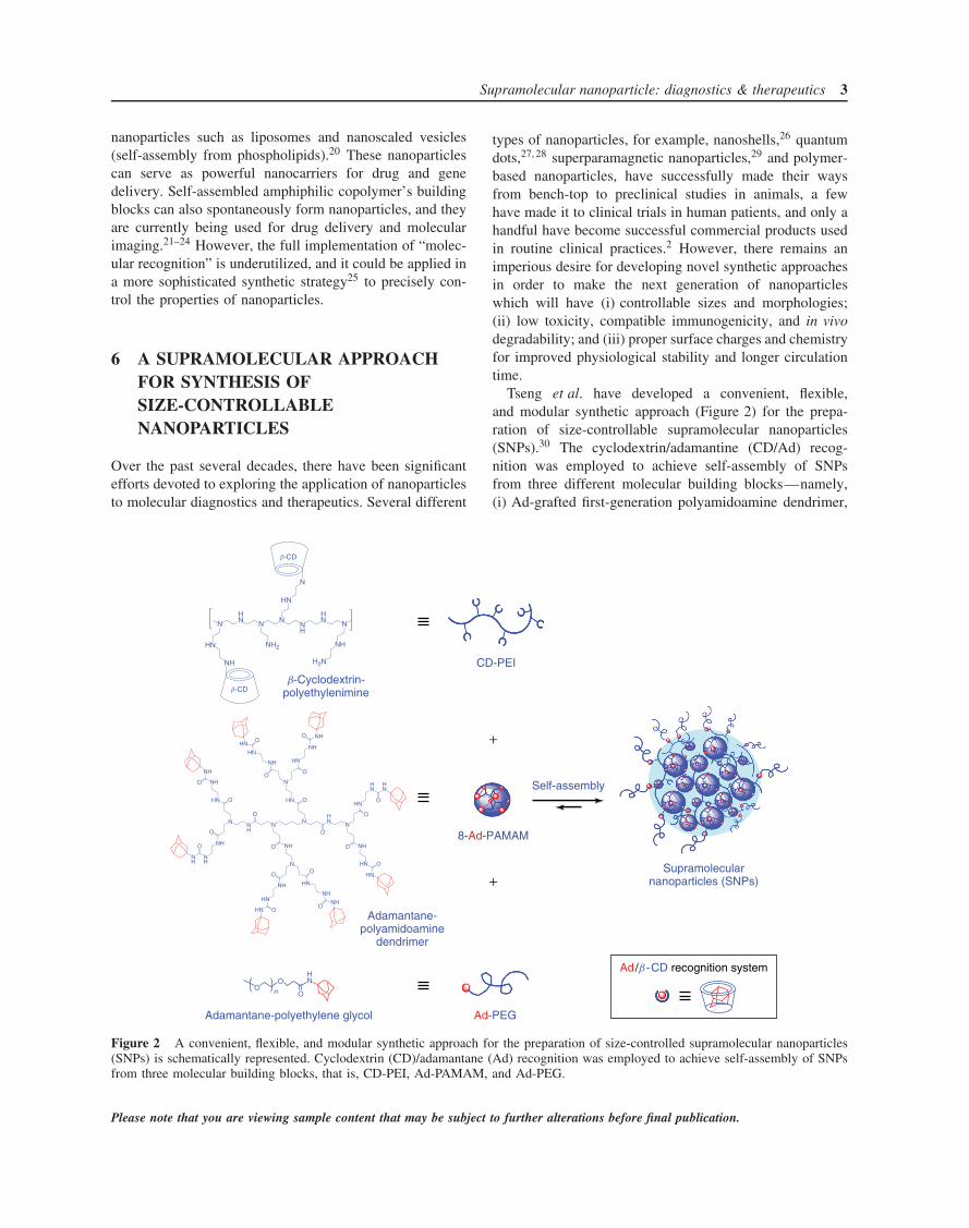
Tseng et al. have developed a convenient, flexible,and modular synthetic approach (Figure 2) for the prepa-ration of size-controllable supramolecular nanoparticles(SNPs).30 The cyclodextrin/adamantine (CD/Ad) recog-nition was employed to achieve self-assembly of SNPsfrom three different molecular building blocks—namely,(i) Ad-grafted first-generation polyamidoamine dendrimer,
CD-PEI
Self-assembly
Supramolecularnanoparticles (SNPs)
8-Ad-PAMAM
Ad-PEG
Ad/b -CD recognition system
Adamantane-polyamidoamine
dendrimer
Adamantane-polyethylene glycol
b-Cyclodextrin-polyethylenimine
NHN N N
NH
HN N
HN
HN
NH
NH2 NH
H2N
N
b-CD
b-CD
NNH
NN
HN
N
O
O
NH
NH
O
NH
O
HNHN
NH
NH
O
O
HN
N
O
HN
NH
NHO
O
NH
HN
HN
O
O
HN
O
HN
O
NH
HN
HN
O
O
NH
N
O
NH
HN
HN O
O
HN
NH
NH
O
O
O nO
O
NH
Figure 2 A convenient, flexible, and modular synthetic approach for the preparation of size-controlled supramolecular nanoparticles(SNPs) is schematically represented. Cyclodextrin (CD)/adamantane (Ad) recognition was employed to achieve self-assembly of SNPsfrom three molecular building blocks, that is, CD-PEI, Ad-PAMAM, and Ad-PEG.
Please note that you are viewing sample content that may be subject to further alterations before final publication.
4 Nanotechnology
8-Ad-PAMAM, (ii) β-CD-grafted branched polyethyleni-mine (MW = 10 kDa), CD-PEI, and (iii) Ad-functionalizedPEG compound (MW = 5 kDa), Ad-PEG.
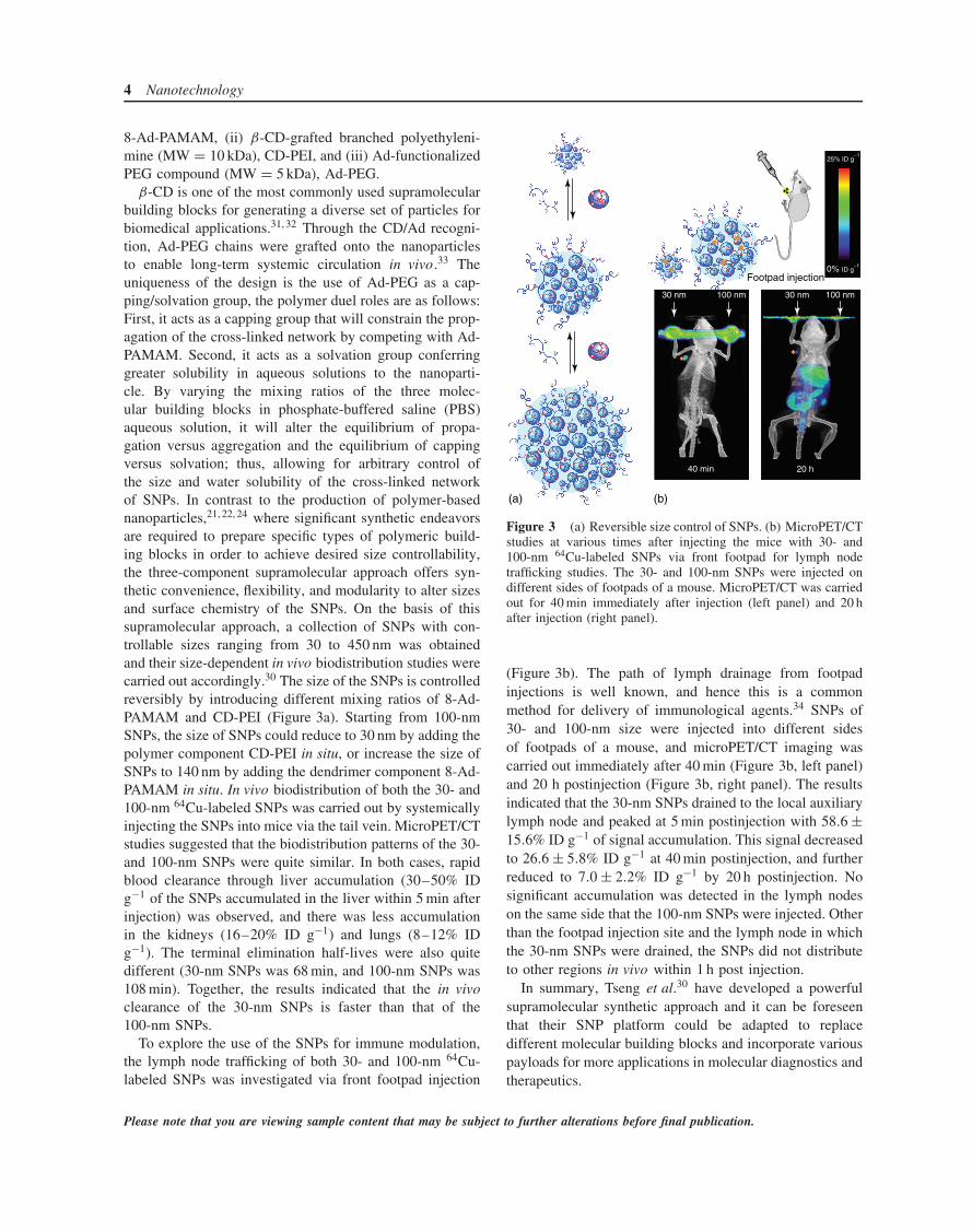
β-CD is one of the most commonly used supramolecularbuilding blocks for generating a diverse set of particles forbiomedical applications.31, 32 Through the CD/Ad recogni-tion, Ad-PEG chains were grafted onto the nanoparticlesto enable long-term systemic circulation in vivo.33 Theuniqueness of the design is the use of Ad-PEG as a cap-ping/solvation group, the polymer duel roles are as follows:First, it acts as a capping group that will constrain the prop-agation of the cross-linked network by competing with Ad-PAMAM. Second, it acts as a solvation group conferringgreater solubility in aqueous solutions to the nanoparti-cle. By varying the mixing ratios of the three molec-ular building blocks in phosphate-buffered saline (PBS)aqueous solution, it will alter the equilibrium of propa-gation versus aggregation and the equilibrium of cappingversus solvation; thus, allowing for arbitrary control ofthe size and water solubility of the cross-linked networkof SNPs. In contrast to the production of polymer-basednanoparticles,21, 22, 24 where significant synthetic endeavorsare required to prepare specific types of polymeric build-ing blocks in order to achieve desired size controllability,the three-component supramolecular approach offers syn-thetic convenience, flexibility, and modularity to alter sizesand surface chemistry of the SNPs. On the basis of thissupramolecular approach, a collection of SNPs with con-trollable sizes ranging from 30 to 450 nm was obtainedand their size-dependent in vivo biodistribution studies werecarried out accordingly.30 The size of the SNPs is controlledreversibly by introducing different mixing ratios of 8-Ad-PAMAM and CD-PEI (Figure 3a). Starting from 100-nmSNPs, the size of SNPs could reduce to 30 nm by adding thepolymer component CD-PEI in situ, or increase the size ofSNPs to 140 nm by adding the dendrimer component 8-Ad-PAMAM in situ. In vivo biodistribution of both the 30- and100-nm 64Cu-labeled SNPs was carried out by systemicallyinjecting the SNPs into mice via the tail vein. MicroPET/CTstudies suggested that the biodistribution patterns of the 30-and 100-nm SNPs were quite similar. In both cases, rapidblood clearance through liver accumulation (30–50% IDg−1 of the SNPs accumulated in the liver within 5 min afterinjection) was observed, and there was less accumulationin the kidneys (16–20% ID g−1) and lungs (8–12% IDg−1). The terminal elimination half-lives were also quitedifferent (30-nm SNPs was 68 min, and 100-nm SNPs was108 min). Together, the results indicated that the in vivoclearance of the 30-nm SNPs is faster than that of the100-nm SNPs.
To explore the use of the SNPs for immune modulation,the lymph node trafficking of both 30- and 100-nm 64Cu-labeled SNPs was investigated via front footpad injection
(a) (b)
Footpad injection
25% ID g−1
0% ID g−1
40 min 20 h
30 nm 100 nm 30 nm 100 nm
Figure 3 (a) Reversible size control of SNPs. (b) MicroPET/CTstudies at various times after injecting the mice with 30- and100-nm 64Cu-labeled SNPs via front footpad for lymph nodetrafficking studies. The 30- and 100-nm SNPs were injected ondifferent sides of footpads of a mouse. MicroPET/CT was carriedout for 40 min immediately after injection (left panel) and 20 hafter injection (right panel).
(Figure 3b). The path of lymph drainage from footpadinjections is well known, and hence this is a commonmethod for delivery of immunological agents.34 SNPs of30- and 100-nm size were injected into different sidesof footpads of a mouse, and microPET/CT imaging wascarried out immediately after 40 min (Figure 3b, left panel)and 20 h postinjection (Figure 3b, right panel). The resultsindicated that the 30-nm SNPs drained to the local auxiliarylymph node and peaked at 5 min postinjection with 58.6 ±15.6% ID g−1 of signal accumulation. This signal decreasedto 26.6 ± 5.8% ID g−1 at 40 min postinjection, and furtherreduced to 7.0 ± 2.2% ID g−1 by 20 h postinjection. Nosignificant accumulation was detected in the lymph nodeson the same side that the 100-nm SNPs were injected. Otherthan the footpad injection site and the lymph node in whichthe 30-nm SNPs were drained, the SNPs did not distributeto other regions in vivo within 1 h post injection.
In summary, Tseng et al.30 have developed a powerfulsupramolecular synthetic approach and it can be foreseenthat their SNP platform could be adapted to replacedifferent molecular building blocks and incorporate variouspayloads for more applications in molecular diagnostics andtherapeutics.
Please note that you are viewing sample content that may be subject to further alterations before final publication.

Supramolecular nanoparticle: diagnostics & therapeutics 5
7 PHOTOTHERMAL EFFECTS OFSUPRAMOLECULAR ASSEMBLEDGOLD NANOPARTICLES
Noble-metal nanostructures have been considered as primecandidate agents for the photothermal treatment of can-cer because of their unique photophysical properties.35–38
In order to generate sufficient energy to damage tumorcells, these nanostructure-based agents are required to havesizes in the range of 10–100 nm.39 However, the rela-tively “large” size of these agents often leads to poor bio-clearance, which can accumulate in the liver, spleen, andkidneys, which is a major obstacle to their in vivo appli-cation.40–42 Alternatively, formatting of aggregates throughself-assembly of the noble-metal nanostructures can be apromising approach.
In this work, Tseng et al. adopted a supramolecularapproach30 to prepare size-controlled Au supramolecularnanoparticles (Au-SNPs) as a new type of photothermalagent from three building blocks: Ad-grafted 2-nm Aucolloids, CD-PEI, and Ad-PEG (Figure 4a). By tuning thedifferent ratio between the molecular building blocks, thatis, Ad-grafted 2-nm Au colloids and CD-PEI, a collectionof size-controllable Au-SNPs was generated with varioussizes ranging from 40 to 118 nm. The size/morphology
of the resulting Au-SNPs collection was characterizedby transmission electron microscope (TEM). The resultsindicate that the Au-SNPs had spherical shapes with anarrow size distribution (Figure 4b bottom).
The photophysical properties of 118-nm Au-SNPs and2-nm Au colloids were studied by UV/vis spectroscopy(Figure 4c). Given the characteristic surface plasmon reso-nance absorption of Au-SNPs and Au colloids (between 500and 530 nm),43 a 532-nm green pulsed laser was chosen totest their photothermal effects and performed laser-inducedmicrobubble-generation studies to monitor the locally accu-mulated heat of individual Au-SNPs (Figure 4d). After test-ing a broad range of energy densities (3–265 mJ cm−2)of a 532-nm pulsed laser with a 6-ns pulse duration, Au-SNPs and Au colloid suspensions in PBS were irradiatedwith different energy ranges. For the 118-nm Au-SNPs,a laser threshold of 32 mJ cm−2 was sufficient for thegeneration of microbubbles on laser irradiation (Figure 4dtop). In contrast, no microbubbles were observed for 2-nm Au colloids even at the maximum laser energy tested(265 mJ cm−2; Figure 4d bottom). The significant enhance-ment of the photothermal effects in 118-nm Au-SNPs canbe attributed to the collective heating effect44 in Au-SNPs.The formation of explosive vapor bubbles on individualAu-SNPs requires an elevated local temperature higher than
CD-PEI
Ad-PEG Ad-PEG-RGDRGD-Au-SNPs
Ad-grafted 2 nm-Au colloid Au-SNPs
Self-assemble
In situligand exchange
(a)
(b) (d)(c)
0.3
0.2
0.1
0.0400 500 600
Wavelength (nm)
Abs
118-nm Au-SNPs2-nm Au colloids
10 nm
2-nm Au colloids /265 mJ cm−2
118-nm Au SNPs/32 mJ cm−2
80 nm
No bubble
Bubbles
+
+
+
Figure 4 (a) Supramolecular synthetic approach for the preparation of size-controlled gold supramolecular nanoparticles (Au-SNPs). Amolecular-recognition system based on Ad and CD was employed to assemble three building blocks: Ad-grafted 2-nm Au colloids, CD-PEI, and Ad-PEG. Ad-PEG-RGD was introduced onto Au-SNPs by in situ ligand exchange to give RGD-Au-SNPs. (b) Transmissionelectron microscopy (TEM) images of Ad-grafted 2-nm Au colloids: the inorganic building blocks of Au-SNPs (top) and a single118-nm Au-SNP obtained by the supramolecular synthetic approach (bottom). (c) UV/vis absorption spectra of 2-nm Au colloids and118-nm Au-SNPs. (d) Time-resolved bright-field micrographs of suspensions of Ad-grafted 2-nm Au colloids (top) and 118-nm Au-SNPs (bottom) during the scanning of a pulsed laser (6 ns, 532 nm; 32 mJ cm−2 for 118-nm Au-SNPs and 265 mJ cm−2 for Ad-grafted2-nm Au colloids).
Please note that you are viewing sample content that may be subject to further alterations before final publication.

6 Nanotechnology
118-nmRGD-Au-SNPs
118-nmRGD-Au-SNPs
RGD-grafted2-nm Au colloids
Cell mixture
(a)(d)
(e)
(f)
(b)
(c)
Before
After
U87
(a
vb3+) U
87 (avb
3 +)M
CF
7 (a
vb
3−)
MC
F7
(avb
3−)
U87
(a
vb
3+)
Figure 5 (a–c) Fluorescence micrographs of U87 cells (αvβ3+, labeled green) treated with 118-nm RGD-Au-SNPs (a), MCF7 cells(αvβ3−, labeled red) treated with 118-nm RGD-Au-SNPs (b), and U87 cells treated with RGD-grafted 2-nm Au colloids (c) afterirradiation with a pulsed laser (6 ns, 120 mJ cm−2). A mask was employed to confine the laser beam to a circular region with a diameterof 1 mm (as indicated by the white dashed circles). (d) Schematically represent a 1 : 1 mixture of U87 and MCF7 cells. (e and f)Fluorescence micrographs of the cell mixture before and after irradiation.
the critical temperature of the liquid medium (374 ◦C forwater).45 It could be hypothesized that upon the formationof microbubbles, the localization of accumulated heat couldfacilitate the thermal disassembly of Au-SNPs into smallerfragments, in a process similar to that observed for 118-nm Au-SNPs when the temperature was above 100 ◦C.46
Thus, most of the Au-SNPs in the solution appeared tohave thermally disassembled into smaller fragments, whichattenuates their photothermal characteristics.
The use of a supramolecular approach enables30, 47 theconvenient incorporation of targeting ligands to providetarget-specific Au-SNPs. Here, arginine–glycine–asparticacid (RGD) peptide was used to recognize tumor cellswith membrane αvβ3 integrin receptors and the result-ing RGD-Au-SNPs were produced by dynamic ligandexchange.47 The 118-nm RGD-Au-SNPs were used alongwith the controls (RGD-grafted 2-nm Au colloids) fortargeted photothermal treatment in 4-well chamber slidescontaining both αvβ3-positive U87 glioblastoma cells andαvβ3-negative MCF7 breast cancer cells. After incuba-tion for 20 min with the agents, the cells in the culture
chambers were exposed to a pulsed laser irradiation (6 ns,120 mJ cm−2). The irradiated cells were kept in an incuba-tor (5% CO2, 378 ◦C) for 2 h, during which time the cellsdamaged by microbubble formation could detach from thesubstrates. The detachment of the cells was observed for theU87 cells treated with RGD-Au-SNPs (Figure 5a). In con-trast, negligible cell detachment was observed for MCF7cells treated with RGD-Au-SNPs (Figure 5b) as well asfor the cells treated with RGD-grafted 2-nm Au colloids(Figure 5c). These results suggest that RGD peptide conferstarget specificity to the Au-SNPs to enable the photother-mal treatment of αvβ3-positive U87 cells. Furthermore, nocell detachment was detected for the U87 cells treated withAu colloids46; this result validated the previous observationthat 2-nm Au colloids exhibit minute photothermal effectsat the given pulsed laser irradiation. To demonstrate theselectivity of RGD-Au-SNPs for target-specific photother-mal treatment, the targeted depletion of αvβ3-positive cellsin a cell mixture containing both αvβ3-positive and αvβ3-negative cell lines was investigated. After treating a 1 : 1cell mixture (Figure 5d), which contains U87 cells (green)
Please note that you are viewing sample content that may be subject to further alterations before final publication.

Supramolecular nanoparticle: diagnostics & therapeutics 7
and MCF7 cells (red) with RGD-Au-SNPs (Figure 5e), themixture was irradiated with a pulsed laser. In the irra-diated region, U87 cells (green) were depleted, and theremaining MCF7 cells (red) were able to be continuouslycultured on the substrates (Figure 5f). In the region out-side the laser footprint, both the positive and the negativecells remained. These results suggest that the photothermaltreatment of RGD-Au-SNPs is highly selective for targetedcells.
In conclusion, size-controlled Au-SNPs have been suc-cessfully demonstrated by supramolecular self-assemblyapproach. The resulting Au-SNPs exhibited significantlyenhanced photothermal effects and were used to demon-strate the targeted photothermal treatment of a subpopula-tion of cancer cells after the incorporation of target-specificligands. It is anticipated that Au-SNPs could serve as apowerful photothermal agent.
8 SNPs FOR TARGETED CELL IMAGING
Synthesis of nanoparticles with narrow size distribu-tions and defined surface chemistry attract more attention,because of their ability to obtain high-quality molecu-lar imaging. In this work, Shen et al.48 demonstrated anew approach for synthesizing uniformly sized SNPs usinga digital microfluidic droplet generator (DMDG), whichenables precise control over the processing parameters. Asa result, high batch-to-batch reproducibility and robust pro-duction of SNPs (Figure 6) with extremely narrow sizedistribution can be generated. By digitally adjusting the vol-ume ratios of the building blocks on the DMDG, a varietyof synthesis parameters were rapidly screened with mini-mum consumption of the reagents. A collection of uniformSNPs with sizes ranging from 35 to 350 nm were obtained.In addition, by incorporating an RGD targeting ligand as anadditional building block, SNPs could be functionalized astargeted nanoparticles with tunable targeting ligand cover-ages. In vitro experiments demonstrated that the sizes and
(a)
(b)
On-chipself-assembly
1: Ad-PAMAM 3: CD-PEI
4: RGD-PEG-Ad
5: RGD-SNPs
2: Ad-PEG
Figure 6 (a) Photograph of the PDMS microfluidic device.(b) Graphical representations of the on-chip approach towardthe preparation of size- and surface-chemistry-controllable RGD-SNPs.
surface properties of the resulting SNPs were correlated totheir cell uptake efficiencies (Figure 7).
9 SELF-ASSEMBLED NANOPARTICLESFOR PROTEIN DETECTION
The presence of certain biomarker proteins and/or irregularprotein concentrations is a sign of cancer and other dis-ease states.49, 50 Sensitive, convenient, and precise protein-sensing methods provide crucial tools for the early diag-nosis of diseases and successful treatment of patients.Recently, the “chemical nose” approach51 was devel-oped, which provides an alternative for the sensing pro-tocols that use exclusive analyte–receptor binding pairsas its basis.52 The strategy for the creation of pro-tein sensors is to use the particle surface for protein
DAPI Cy5 Merge
MCF7 (avb3−)
U87 (avb3+)
Figure 7 Fluorescence microscopy images of 35-nm RGD-SNPs (5% RGD ligand coverage) taken up by U87 (top) and MCF7(bottom) cell lines. The cell nuclei were stained by DAPI; the RGD-SNPs were labeled with Cy5 units.
Please note that you are viewing sample content that may be subject to further alterations before final publication.
8 Nanotechnology
(a)
(b)
A B C D E F G
1
2
3
4
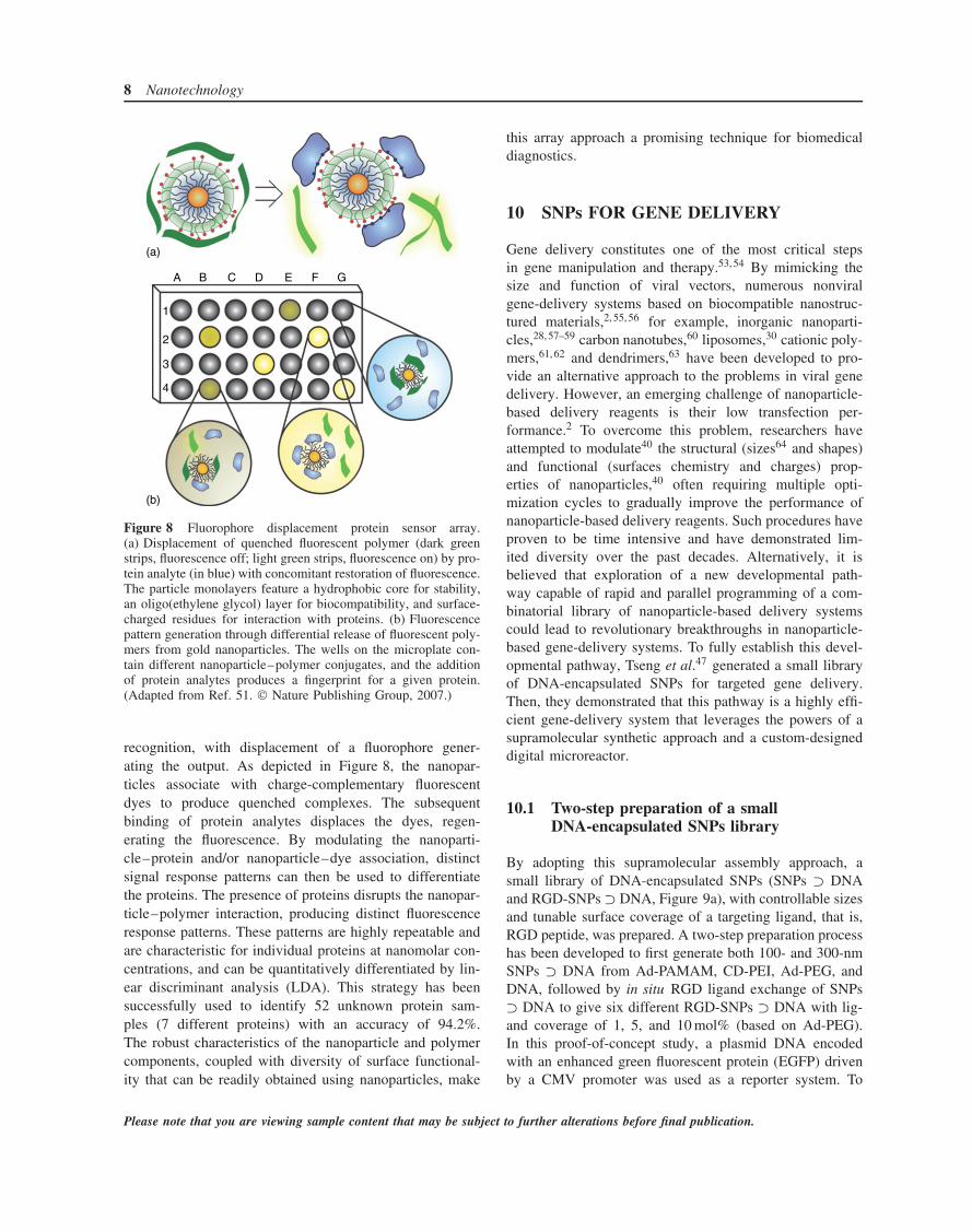
Figure 8 Fluorophore displacement protein sensor array.(a) Displacement of quenched fluorescent polymer (dark greenstrips, fluorescence off; light green strips, fluorescence on) by pro-tein analyte (in blue) with concomitant restoration of fluorescence.The particle monolayers feature a hydrophobic core for stability,an oligo(ethylene glycol) layer for biocompatibility, and surface-charged residues for interaction with proteins. (b) Fluorescencepattern generation through differential release of fluorescent poly-mers from gold nanoparticles. The wells on the microplate con-tain different nanoparticle–polymer conjugates, and the additionof protein analytes produces a fingerprint for a given protein.(Adapted from Ref. 51. Nature Publishing Group, 2007.)
recognition, with displacement of a fluorophore gener-ating the output. As depicted in Figure 8, the nanopar-ticles associate with charge-complementary fluorescentdyes to produce quenched complexes. The subsequentbinding of protein analytes displaces the dyes, regen-erating the fluorescence. By modulating the nanoparti-cle–protein and/or nanoparticle–dye association, distinctsignal response patterns can then be used to differentiatethe proteins. The presence of proteins disrupts the nanopar-ticle–polymer interaction, producing distinct fluorescenceresponse patterns. These patterns are highly repeatable andare characteristic for individual proteins at nanomolar con-centrations, and can be quantitatively differentiated by lin-ear discriminant analysis (LDA). This strategy has beensuccessfully used to identify 52 unknown protein sam-ples (7 different proteins) with an accuracy of 94.2%.The robust characteristics of the nanoparticle and polymercomponents, coupled with diversity of surface functional-ity that can be readily obtained using nanoparticles, make
this array approach a promising technique for biomedicaldiagnostics.
10 SNPs FOR GENE DELIVERY
Gene delivery constitutes one of the most critical stepsin gene manipulation and therapy.53, 54 By mimicking thesize and function of viral vectors, numerous nonviralgene-delivery systems based on biocompatible nanostruc-tured materials,2, 55, 56 for example, inorganic nanoparti-cles,28, 57–59 carbon nanotubes,60 liposomes,30 cationic poly-mers,61, 62 and dendrimers,63 have been developed to pro-vide an alternative approach to the problems in viral genedelivery. However, an emerging challenge of nanoparticle-based delivery reagents is their low transfection per-formance.2 To overcome this problem, researchers haveattempted to modulate40 the structural (sizes64 and shapes)and functional (surfaces chemistry and charges) prop-erties of nanoparticles,40 often requiring multiple opti-mization cycles to gradually improve the performance ofnanoparticle-based delivery reagents. Such procedures haveproven to be time intensive and have demonstrated lim-ited diversity over the past decades. Alternatively, it isbelieved that exploration of a new developmental path-way capable of rapid and parallel programming of a com-binatorial library of nanoparticle-based delivery systemscould lead to revolutionary breakthroughs in nanoparticle-based gene-delivery systems. To fully establish this devel-opmental pathway, Tseng et al.47 generated a small libraryof DNA-encapsulated SNPs for targeted gene delivery.Then, they demonstrated that this pathway is a highly effi-cient gene-delivery system that leverages the powers of asupramolecular synthetic approach and a custom-designeddigital microreactor.
10.1 Two-step preparation of a smallDNA-encapsulated SNPs library
By adopting this supramolecular assembly approach, asmall library of DNA-encapsulated SNPs (SNPs ⊃ DNAand RGD-SNPs ⊃ DNA, Figure 9a), with controllable sizesand tunable surface coverage of a targeting ligand, that is,RGD peptide, was prepared. A two-step preparation processhas been developed to first generate both 100- and 300-nmSNPs ⊃ DNA from Ad-PAMAM, CD-PEI, Ad-PEG, andDNA, followed by in situ RGD ligand exchange of SNPs⊃ DNA to give six different RGD-SNPs ⊃ DNA with lig-and coverage of 1, 5, and 10 mol% (based on Ad-PEG).In this proof-of-concept study, a plasmid DNA encodedwith an enhanced green fluorescent protein (EGFP) drivenby a CMV promoter was used as a reporter system. To
Please note that you are viewing sample content that may be subject to further alterations before final publication.

Supramolecular nanoparticle: diagnostics & therapeutics 9
+
+
+
Ad-PAMAM
Ad-PEG RGD-PEG-Ad
CD-PEI
DNA
300 nmSNPs ⊃ DNA
100 nmSNPs ⊃ DNA
(i) Self-assembly
(ii) In situligand exchange
(ii) In situligand exchange
Targetingligand
5%
5%
1%
1%
10%
10%
(a)
(b)
60
40
20
0
Tra
nsfe
ctio
n ef
ficie
ncy
(%)
RG
D-je
t-P
EI
100-
10%
100-
1%
100-
5%
100-
030
0-10
%
300-
5%30
0-1%
300-
0C
D-P
EI/
Ad-
PE
GC
D-P
EI
DN
A
MCF7
U87
avb
3 high-expressed 3T3
avb
3 low-expressed 3T3
Figure 9 (a) A two-step modular assembly approach for preparation of a small library of DNA-encapsulated supramolecularnanoparticles (SNPs ⊃ DNA and RGD-SNPs ⊃ DNA) with controllable sizes and tunable RGD ligand coverage. (b) EGFP transfectionefficiency of a collection of SNPs ⊃ DNA and RGD-SNPs ⊃ DNA along with control delivery systems for two αvβ3 high-expressedcells (U87 and scraping-collected 3T3 cells) and two αvβ3 low-expressed cells (MCF7 and 0.25% trypsin-treated 3T3 cells). Therepresentative fluorescence micrographs of 57 ± 11% and 9 ± 4% transfection efficiencies observed for 5 mol% RGD-grafted 100-nmRGD-SNPs ⊃ DNA (100-5%)-treated αvβ3 high-expressed 3T3 cells (left image) and 1 mol% RGD-grafted 300-nm RGD-SNPs ⊃DNA (300-1%)-treated αvβ3 low-expressed 3T3 cells (right image).
characterize the sizes, morphologies, and surface chargesof the resulting SNPs ⊃ DNA and RGD-SNPs ⊃ DNA,dynamic light scattering (DLS), TEM, and zeta potentialmeasurements were carried out, respectively. Finally, thegene transfection efficiency and specificity of each SNPs ⊃DNA (100 and 300 nm) and RGD-SNPs ⊃ DNA (100-1%,100-5%, 100-10%, 300-1%, 300-5%, and 300-10%) in the
small library were examined using αvβ3 high-expressed andlow-expressed cells, along with the control delivery sys-tems. The results of average transfection efficiency of gene-delivery vehicles for different cell lines were summarizedin Figure 9(b). First, 100-nm RGD-SNPs ⊃ DNA exhib-ited higher transfection efficiency than 300-nm RGD-SNPs⊃ DNA, DNA complexes based on each of the molecular
Please note that you are viewing sample content that may be subject to further alterations before final publication.

10 Nanotechnology
+
+
+
3: Ad-PAMAM
4: Ad-PEG2: CD-PEI
5: RGD-PEG-Ad
6: TAT-PEG-Ad
1: TAT/RGD-DNA ⊂ SNPs7a: EGFP-DNA7b: FLuc-DNA
On-chipself-assembly
RGD fortargeting
PEG for passivation
TAT for cell membranepenetration
(1a: EGFP)
(1b: FLuc)
Figure 10 Graphical schematic representations of the self-assembly approach for producing a combinatorial library of DNA-encapsulated supramolecular nanoparticles (DNA ⊂ SNPs), in which a broad structural/functional diversity can be programmed intoindividual DNA ⊂ SNPs (1) by systematically altering the mixing ratios of the five functional molecular building blocks, that is, CD-PEI(2), Ad-PAMAM (3), Ad-PEG (4), RGD-PEG-Ad (5), and TAT-PEG-Ad (6), as well as DNA plasmid (7a: enhanced green fluorescentprotein (EGFP) and 7b: firefly luciferase (FLuc)).
building blocks (CD-PEI and CD-PEI/Ad-PEG) and freeplasmid DNA, indicating that the formation of SNPs andtheir sizes play an important role in transfection efficiency.Second, 100-5% RGD-SNPs ⊃ DNA gave the highesttransfection efficiency (57 ± 11% for U87) compared tothose observed for SNPs ⊃ DNA and other targeted RGD-SNPs ⊃ DNA. The reduced transfection efficiency observedfor 100-10% RGD-SNPs ⊃ DNA can be attributed to anexcess amount of free RGD ligand in the culture medium,which compromised the targeted binding of RGD-SNPs ⊃DNA as a result of a competition effect. Moreover, 100-5% RGD-SNPs ⊃ DNA demonstrated outstanding deliveryspecificity with a 2.7-time enhancement of transfection effi-ciency for αvβ3 high-expressed cells (U87) over the αvβ3low-expressed cells (MCF7). These results are comparableto those observed for the commercially available RGD-jet-PEI, which is a well-known selective and efficient transfec-tion reagent for integrin-expressing cell lines. The resultsrevealed that the size and target ligand coverage of RGD-SNPs ⊃ DNA played a critical role in the target-specificgene delivery.
10.2 A SNPs library with programmedstructural and functional diversity
To speed up the exploration of nanoparticle-based genedelivery system, a rapid developmental pathway65 thatleverages the powers of (i) a combinatorial syntheticapproach (Figure 10) on the basis of supramolecular assem-bly30, 66, 67 and (ii) a digital microreactor68–71 (Figure 11)is demonstrated. Unlike the slow, multistep synthesesemployed for producing existing gene-delivery materials,2
this supramolecular method (Figure 10) enables a conve-nient, flexible, and modular method72 for generating acombinatorial library of DNA ⊂ SNPs. A broad struc-tural/functional diversity covering the size variation, surfacechemistry, and DNA loading capacity was programmedinto individual DNA ⊂ SNPs by systematically altering themixing ratios of five functional molecular building blocks(2–6), DNA plasmid [7a: EGFP and 7b: firefly luciferase(FLuc)]. In order to reduce human operational errors, accel-erate handling procedures, enhance experimental fidelity,and achieve economical use of reagents, a digital dual-coremicroreactor (DCM, Figure 11) was designed and imple-mented to allow automated sampling, dilution, metering,and mixing of 2–7, resulting in a combinatorial librarycomposed of 648 different DNA ⊂ SNPs within 2.5 h. Thestructural/functional diversity of the DNA ⊂ SNPs librarycan be translated into diversity in performance by conduct-ing transfection studies of individual DNA ⊂ SNPs in 96-well plates containing mouse fibroblast cells. A small groupof DNA ⊂ SNPs that facilitates high levels of deliveryperformance was identified. Comprehensive characteriza-tions were carried out on these DNA ⊂ SNPs revealingthat improved transfection performance can be attributed tothe defined size, surface chemistry, zeta potential, unifor-mity, and dynamic stability. Compared to the leading genetransfection reagents, such as lipofectamine 2000 and RGD-jet-PEI, the identified 40-nm TAT/RGD-DNA ⊂ SNPs (1)with defined surface chemistry (RGD73–76: an αvβ3 bindingpeptide and TAT,77–79 a cell-penetrating peptide) exhibitedsignificantly improved gene transfection efficiency and lowtoxicity in a number of cancer cell lines and fibroblastcells.
Please note that you are viewing sample content that may be subject to further alterations before final publication.

Supramolecular nanoparticle: diagnostics & therapeutics 11
Flow
Waste outlet
Outlets
Transfectionstudy in
96-well plates
Degasmodules
Valve open
Valve close
PBS PBS7 3
5 PBS
6 PBS
4 2
CIM
AIM
Vacuum
Vacuum
Vacuum
N2
N2
Figure 11 Graphical illustration of a digital dual-core microreactor (DCM). The operation of the circuit was computer controlledusing color-coded pressure-driven valves: red, positive pressure, off/on; yellow, peristaltic pumping; green, vacuum.
4.5 × 10⁴
0.96
0
1.60 1.40 1.20 1.00 0.80 0.60 0.40 0.202: CD-PEI (µM)
6: T
AT-
PE
G-A
d (µ
M)
5: R
GD
-PE
G-A
d(µ
m)
1.201.00
0.800.60
0.400.30
0.200.10
0
6:T
AT
-PE
G-A
d (%
)
5: R
GD
-PE
G-A
d (%
)
5: R
GD
-PE
G-A
d (%
)
2: CD-PEI (µM) 6: TAT-PEG-Ad (%) 2: CD-PEI (µM)
XY-plane YZ-plane XZ-plane
YZXY
XZ
1a: TAT/RGD-DNA ⊂ SNPs
(EGFP)
(a)
(b)
(c)
40 nm
0
Figure 12 (a) A 4D profile of gene transfection performance of TAT/RGD-DNA ⊂ SNPs (EGFP) with variation of CD-PEI (2), TAT,and RGD coverages (648 data points). The XY, YZ, and XZ planes across the best performance were simplified and denoted 2D contourimages. (b and c) TEM and SEM images of the resulting DNA ⊂ SNPs with size of 42 ± 4 nm, which gave the best transfectionperformance (indicated by a red arrow). Scale bar: 100 nm.
Please note that you are viewing sample content that may be subject to further alterations before final publication.
12 Nanotechnology
In proof-of-concept trials, a cytomegalovirus (CMV)promoter-driven EGFP-encoded plasmid DNA was encap-sulated into the DNA ⊂ SNPs. The screening was accom-plished by systematically programming the three variables,that is, eight different concentrations for CD-PEI (2), ninefor RGD-PEG-Ad (5), and nine for TAT-PEG-Ad (6).Using the digital DCM, a combinatory library composedof 648 different DNA ⊂ SNPs was generated within 2.5 hto fill up seven 96-well plates. To ensure the operationfidelity, all the experiments were conducted in triplicate.A 4D gene expression plot (Figure 12a) was employedto summarize the results from the full-scale screening ofthe combinatorial library and the optimal transfection effi-ciency was achieved at CD-PEI (2, 0.7 ± 0.1 µM), RGD-PEG-Ad (5, 0.28 ± 0.04 µM), and TAT-PEG-Ad (6, 0.60 ±0.20 µM).
To characterize the properties of TAT/RGD-DNA ⊂SNPs (1) with the best transfection performance, TEM(Figure 12b) and scanning electron microscopy (SEM,
Table 1 Comparison of TAT/RGD-DNA ⊂ SNPs synthesizedby DCM and conventional pipetting.
Dual-core Pipetting72
microreactor(DCM)
Throughput 250 conditions h−1 <20 conditions h−1
Size distribution Excellent Good(PDIa :<0.05) (PDIa : 0.10–0.20)
Operation error No PossibleReproducibility High Modest
aPDI: The polydispersity index obtained from DLS measurements.
Figure 12c) were carried out to examine the morphologyand sizes of the resulting TAT/RGD-DNA ⊂ SNPs (1).The distinct size of 42 ± 4 nm was observed for thisformulation. It is noticeable that the size distributions ofthe DCM-produced DNA ⊂ SNPs are much narrowerthan those prepared manually (Table 1), and this result
100
80
60
40
20
0
HeL
a
A54
9
U87
MC
F7
3T3
PC
3
IMR
-90
Tra
nsfe
ctio
n ef
ficie
ncy
(%) 50 ng DNA
(a)
100
80
60
Via
bilit
y (%
)
TAT/RGD–DNA ⊂ SNPs
Lipofectamine2000 RGD-jet-PEI
50 ng1000 ng
50 ng1000 ng
50 ng1000 ng(b)
TAT/RGD-DNA ⊂ SNPs
Lipofectamine2000RGD-jet-PEI
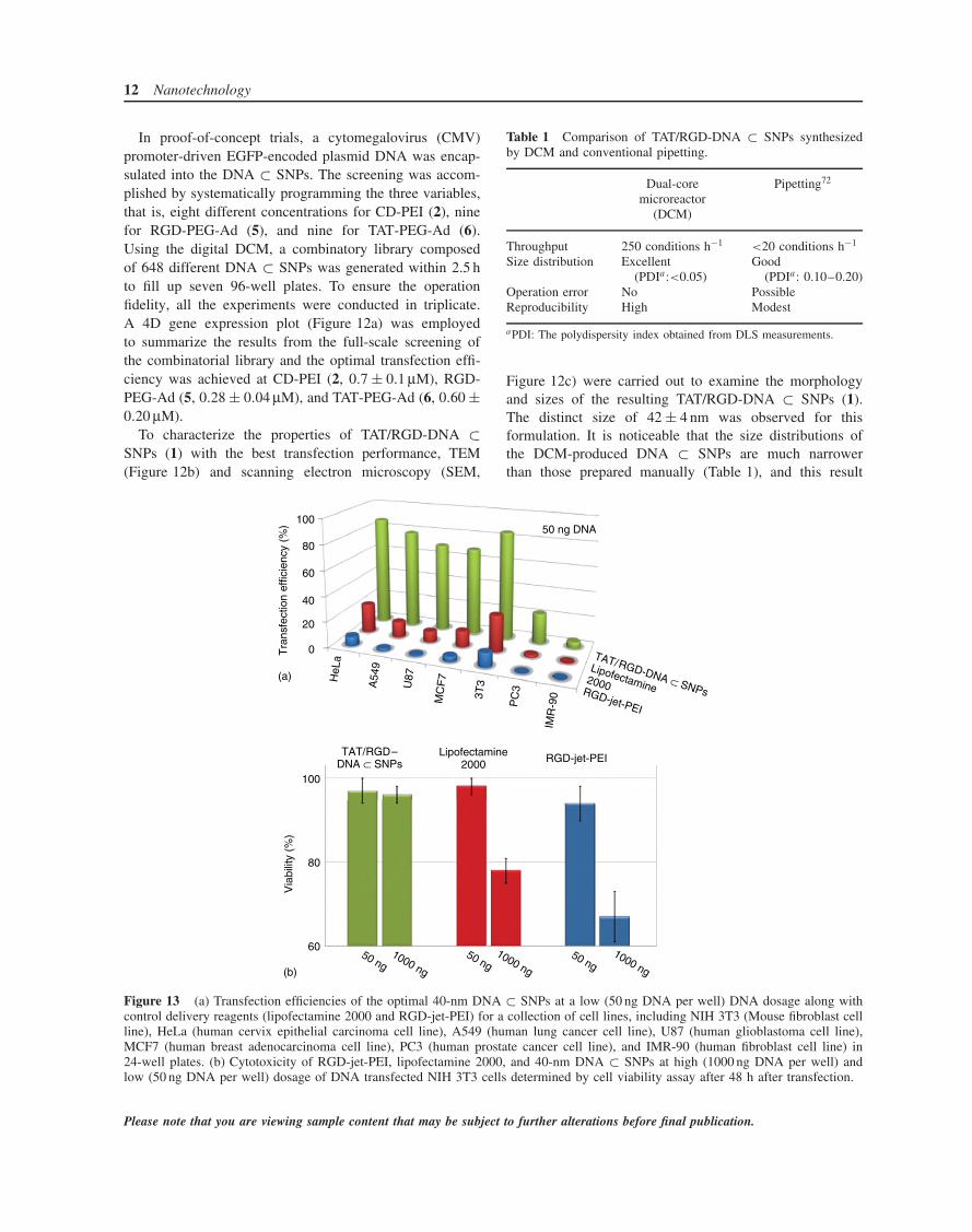
Figure 13 (a) Transfection efficiencies of the optimal 40-nm DNA ⊂ SNPs at a low (50 ng DNA per well) DNA dosage along withcontrol delivery reagents (lipofectamine 2000 and RGD-jet-PEI) for a collection of cell lines, including NIH 3T3 (Mouse fibroblast cellline), HeLa (human cervix epithelial carcinoma cell line), A549 (human lung cancer cell line), U87 (human glioblastoma cell line),MCF7 (human breast adenocarcinoma cell line), PC3 (human prostate cancer cell line), and IMR-90 (human fibroblast cell line) in24-well plates. (b) Cytotoxicity of RGD-jet-PEI, lipofectamine 2000, and 40-nm DNA ⊂ SNPs at high (1000 ng DNA per well) andlow (50 ng DNA per well) dosage of DNA transfected NIH 3T3 cells determined by cell viability assay after 48 h after transfection.
Please note that you are viewing sample content that may be subject to further alterations before final publication.

Supramolecular nanoparticle: diagnostics & therapeutics 13
was confirmed by DLS measurements. The greater sizecontrollability of DNA ⊂ SNPs can be attributed to theprecision and reproducibility of the sampling, metering,and mixing processes in the digital DCM. In addition, theactual surface coverages of RGD-PEG-Ad (5) and TAT-PEG-Ad (6) were estimated. The results indicated that 0.28and 0.60 µM of RGD-PEG-Ad (5) and TAT-PEG-Ad (6)reflect 5 and 9% of surface coverage on the resulting DNA⊂ SNPs, respectively. In short, TAT/RGD-DNA ⊂ SNPs(1) with sizes of 40 and 80 nm, as well as 5% RGD-PEG-Ad (5) and 9% TAT-PEG-Ad (6) coverage72 exhibit optimalcell transfection performance.
To compare the transfection performance of the opti-mal 40-nm TAT/RGD-DNA ⊂ SNPs (1) with the leadingtransfection reagents (i.e., RGD-jet-PEI and lipofectamine2000), the gene transfection studies were carried out in24-well plates with encapsulated-DNA (50 ng per well) byusing a collection of cells, including NIH 3T3 (Mousefibroblast cell line), HeLa (human cervix epithelial car-cinoma cell line), A549 (human lung cancer cell line),U87 (human glioblastoma cell line), MCF7 (human breastadenocarcinoma cell line), PC3 (human prostate cancercell line), and IMR-90 (human fibroblast cell line). Theresults of gene transfection studies (Figure 13a) indicatedthat the 40-nm TAT/RGD-DNA ⊂ SNPs (1) exhibited sig-nificantly improved transfection performance compared tothose observed for RGD-jet-PEI and lipofectamine 2000across the different cancer and fibroblast cell lines at highdosage of DNA (1000 ng DNA per well). Higher than 70%transfection efficiencies were observed across various can-cer cell lines, even for the PC3 cell line that is difficultto be transfected by those two commercial reagents. More-over, the cell viability assay results indicated that the 40-nmTAT/RGD-DNA ⊂ SNPs (1) exhibited negligible cytotox-icity after treatment for 48 h (Figure 13b).
In summary, this pathway can be adopted for the devel-opment of nanoparticle-based vectors capable of deliveringa variety of loads, such as gene, drugs, proteins, and theirmixtures. Tseng’s group is currently exploring the use ofthe DNA ⊂ SNP-based transfection reagents for reprogram-ming of human primary fibroblast cells in order to generateinduced pluripotent stem cells that is crucial in the field ofregenerative medicine.80
10.3 Self-assembled NPs for delivering siRNA inhumans
The development of new therapeutics that engage with anRNAi pathway has opened a new field of drug discoveryand development.81–83 Davis et al.84 have demonstrated thefirst experimental therapeutic to provide targeted deliveryof synthetic, small interfering RNA (siRNA) in humans.
+ +
CDP siRNA
Self-assembly
Ad-PEG
Ad-PEG-Tf
Systemicadministration ofsiRNA in humans
Figure 14 Schematic representation of systemically administer-ing siRNA via a targeted delivery system in human. The tar-geted nanoparticles consist of cyclodextrin-based polymer (CDP),siRNA, adamantane conjugated PEG (Ad-PEG) and targeting lig-and, that is, human transferrin (Tf), conjugated Ad-PEG (Ad-PEG-Tf). (Adapted from Ref. 84. Nature Publishing Group, 2010.)
The targeted NPs consist of (i) a linear, cyclodextrin-basedpolymer, CDP; (ii) a hydrophilic Ad conjugated PEG, Ad-PEG; (iii) a human transferrin (Tf) as a targeting ligandfor binding to transferrin receptors (TfRs) that are typicallyupregulated on cancer cells; and (iv) siRNA (Figure 14).The four components self-assembled to form an NP, whichis then systemically administered intravenously (iv) tomelanoma patients with solid cancers.84, 85 Tumor biopsiesfrom melanoma patients obtained after treatment showthe presence of intracellularly localized NPs in amountsthat correlate with dose levels of the NPs administered.Furthermore, a reduction was found in both the specificmessenger RNA (M2 subunit of ribonucleotide reductase(RRM2)) and the protein (RRM2) levels when compared topredosing tissue. Most notably, they validate that siRNA-mediated mRNA cleavage occurs specifically at the sitepredicted for an RNAi mechanism from a patient whoreceived the highest dose of the nanoparticles. Together,these data demonstrate the first example of dose-dependentaccumulation of targeted nanoparticles in human tumorsand RNAi can occur in humans from a systemicallydelivered siRNA, and that siRNA can be used as a gene-specific therapeutic.84
11 NANOPARTICLE-BASED DRUGDELIVERY SYSTEM
The physicochemical properties of new drugs are impor-tant factors regarding whether they will be successful invivo. However, many potent drug candidates fail in pre-clinical studies because of limited solubility, stability, and
Please note that you are viewing sample content that may be subject to further alterations before final publication.

14 Nanotechnology
H3CONH
NH
H
CONH
OH
H3CO O OH
O OH
OH
O
NH2OH
H3C
O n m
O
N
O ADR
PEG pAsp
Sel
f-as
sem
bly pH-sensitive linker
Drug
Hydrophilic segment Hydrophobic segment
≡
Figure 15 Schematic representation of the self-assembly of pH-sensitive drug-loaded micelles which was synthesized from drug conju-gated amphiphilic block copolymers, that is, poly(ethylene glycol)–poly(aspartate–hydrazone–adriamycin) (PEG–p(Asp–Hyd–ADR)).(Reproduced from Ref. 92. Wiley-VCH, 2004.)
toxicity. Therefore, a number of drug-delivery systems havebeen developed to overcome these transport problems.86–89
Supramolecular chemistry is attracting attention as it offersmethods for assembling different constituents capable ofstructural and dynamic changes into single molecules.90
Kataoka et al. have demonstrated the intracellular local-ization of a pH-sensitive supramolecular assembly thatchanges its structure and fluorescence when activated toinduce mortality of malignant cells.91 The pH-sensitivesupramolecular polymeric NPs are formed by self-assemblyof a drug-loaded amphiphilic block-copolymer (adriamycin(ADR)-block-copolymer) in water (Figure 15).92 With thestimuli in the intracellular environment (pH value), thestructure and/or function of the NPs are changed simulta-neously. Confocal laser scanning microscopy studies wereperformed and the results showed that the NPs are trappedin lysosomes. However, the NPs have been designed torelease their drugs at low pH, and shown to effectivelysuppress the viability of the cancer cells.91
12 CONCLUSION
Over the past decades, various nanoparticles have beenreported to explore their promising applications in the fieldsof material science, biology, and medicine. SNPs haveattracted more attention due to their flexible, convenient,and modular synthetic strategy. It is anticipated that thisapproach will facilitate the development of SNPs formolecular diagnostics and therapeutics in clinical trials.We also foresee that the successful demonstration of thispathway will provide more applications in (i) moleculardiagnostics such as MRI and PET by introducing reportersystems into SNPs, (ii) therapeutics, for example, drug,gene, and protein delivery, and (iii) theranostics, whichincorporates both reporter systems and therapeutic payloadsinside of SNPs. With all the advantages above, we believe
that SNPs could have a potential to pave the way towardpersonalized medicine and translational clinic.
REFERENCES
1. V. I. Shubayev, T. R. Pisanic, and S. H. Jin, Adv. DrugDelivery Rev., 2009, 61, 467.
2. M. E. Davis, Z. Chen, and D. M. Shin, Nat. Rev. DrugDiscov., 2008, 7, 771.
3. J. Shi, A. R. Votruba, O. C. Farokhzad, and R. Langer, NanoLett., 2010, 10, 3223.
4. S. D. Perrault and W. C. W. Chan, Proc. Natl. Acad. Sci.U.S.A., 2010, 107, 11194.
5. A. G.-F. Banu, S. Zolnik, N. Sadrieh, and M. A. Dobrovol-skaia, Endocrinology, 2010, 151, 458.
6. S. Zhang, J. Li, G. Lykotrafitis, et al. Adv. Mater., 2008, 21,419.
7. P. Aggarwala, J. B. Hall, C. B. McLelanda, et al. Adv. DrugDeliv. Rev., 2009, 61, 428.
8. J. Kolosnjaj-Tabi, K. B. Hartman, S. Boudjemaa, et al. ACSNano, 2010, 4, 1481.
9. M. M. Schmidt and K. D. Wittrup, Mol. Cancer Ther., 2009,8, 2861.
10. E. A. Stephanie, S. A. Gratton, P. A. Ropp, et al. Proc. Natl.Acad. Sci. U.S.A., 2008, 105, 11613.
11. F. Sakuraia, R. Inoue, Y. Nishino, et al. J. ControlledRelease, 2000, 66, 255.
12. S. Prabha, W. Z. Zhou, J. Panyam, and V. Labhasetwar, Int.J. Pharm., 2002, 244, 105.
13. M. Sarikaya, C. Tamerler, A. K. Y. Jen, et al. Nat. Mater.,2003, 2, 577.
14. C. D. Meyer, C. S. Joiner, and J. F. Stoddart, Chem. Soc.Rev., 2007, 36, 1705.
15. I. Hwang, W. S. Jeon, H. J. Kim, et al. Angew. Chem. Int.Ed., 2007, 46, 210.
16. M. J. W. Ludden, D. N. Reinhoudt, and J. Huskens, Chem.Soc. Rev., 2006, 35, 1122.
Please note that you are viewing sample content that may be subject to further alterations before final publication.

Supramolecular nanoparticle: diagnostics & therapeutics 15
17. S. Y. Park, A. K. R. Lytton-Jean, B. Lee, et al. Nature,2008, 451, 553.
18. J. F. Stoddart and H. R. Tseng, Proc. Natl. Acad. Sci. U.S.A.,2002, 99, 4797.
19. Y. Liu, H. Wang, P. Liang, and H. Y. Zhang, Angew. Chem.Int. Ed., 2004, 43, 2690.
20. V. P. Torchilin, Nat. Rev. Drug Discov., 2005, 4, 145.
21. G. Sun, J. Xu, A. Hagooly, et al. Adv. Mater., 2007, 19,3157.
22. L. Ferreira, J. M. Karp, L. Nobre, and R. Langer, Cell StemCell, 2008, 3, 136.
23. S. R. Bull, M. O. Guler, R. E. Bras, et al. Nano Lett., 2005,5, 1.
24. P. A. Bertin, J. M. Gibbs, C. K. F. Shen, et al. J. Am. Chem.Soc., 2006, 128, 4168.
25. A. K. Boal, F. Ilhan, J. E. DeRouchey, et al. Nature, 2000,404, 746.
26. C. Loo, A. Lin, L. Hirsch, et al. Technol. Cancer Res. Treat.,2004, 3, 33.
27. X. Gao, Y. Cui, R. M. Levenson, et al. Nat. Biotechnol.,2004, 22, 969.
28. S. M. Nie, Y. Xing, G. J. Kim, and J. W. Simons, Annu.Rev. Biomed. Eng., 2007, 9, 257.
29. Y. W. Jun, J. H. Lee, and J. Cheon, Angew. Chem. Int. Ed.,2008, 47, 5122.
30. H. Wang, S. T. Wang, H. Su, et al. Angew. Chem. Int. Ed.,2009, 48, 4344.
31. G. Wenz, B. H. Han, and A. Muller, Chem. Rev., 2006, 106,782.
32. J. Li and X. J. Loh, Adv. Drug Deliv. Rev., 2008, 60, 1000.
33. D. W. Bartlett, H. Su, I. J. Hildebrandt, et al. Proc. Natl.Acad. Sci. U.S.A., 2007, 104, 15549.
34. L. J. Peek, C. R. Middaugh, and C. Berkland, Adv. DrugDeliv. Rev., 2008, 60, 915.
35. P. K. Jain, X. H. Huang, I. H. El-Sayed, and M. A. El-Sayed, Acc. Chem. Res., 2008, 41, 1578.
36. R. R. Anderson and J. A. Parrish, Science, 1983, 220, 524.
37. K. An and T. Hyeon, Nano Today, 2009, 4, 359.
38. S. Lal, N. K. Grady, J. Kundu, et al. Chem. Soc. Rev., 2008,37, 898.
39. A. R. Lowery, A. M. Gobin, E. S. Day, et al. Clin. CancerRes., 2005, 11, 9097s.
40. S. Mitragotri and J. Lahann, Nat. Mater., 2009, 8, 15.
41. H. S. Choi, W. Liu, P. Misra, et al. Nat. Biotechnol., 2007,25, 1165.
42. A. E. Nel, L. Madler, D. Velegol, et al. Nat. Mater., 2009,8, 543.
43. B. Khlebtsov, V. Zharov, A. Melnikov, et al. Nanotechnol-ogy, 2006, 17, 5167.
44. H. H. Richardson, M. T. Carlson, P. J. Tandler, et al. NanoLett., 2009, 9, 1139.
45. V. Kotaidis and A. Plech, Appl. Phys. Lett., 2005, 87,213102.
46. S. T. Wang, K. J. Chen, T. H. Wu, et al. Angew. Chem. Int.Ed., 2010, 49, 3777.
47. H. Wang, K. J. Chen, S. T. Wang, et al. Chem. Commun.,2010, 46, 1851.
48. K. Liu, H. Wang, K. J. Chen, et al. Nanotechnology, 2010,21, 445603.
49. J. S. Ross and J. A. Fletcher, Stem Cells, 1998, 16, 413.
50. M. J. Daniels, Y. M. Wang, M. Y. Lee, andA. R. Venkitaraman, Science, 2004, 306, 876.
51. C. C. You, O. R. Miranda, B. Gider, et al. Nat. Nanotech-nol., 2007, 2, 318.
52. K. J. Albert, N. S. Lewis, C. L. Schauer, et al. Chem. Rev.,2000, 100, 2595.
53. D. J. Glover, H. J. Lipps, and D. A. Jans, Nat. Rev. Genet.,2005, 6, 299.
54. D. H. Kim and J. J. Rossi, Nat. Rev. Genet., 2007, 8, 173.
55. C. M. Niemeyer, Angew. Chem. Int. Ed., 2001, 40, 4128.
56. M. Ferrari, Nat. Rev. Cancer, 2005, 5, 161.
57. M. De, P. S. Ghosh, and V. M. Rotello, Adv. Mater., 2008,20, 4225.
58. F. Torney, B. G. Trewyn, V. S. Y. Lin, and K. Wang, Nat.Nanotechnol., 2007, 2, 295.
59. N. L. Rosi, D. A. Giljohann, C. S. Thaxton, et al. Science,2006, 312, 1027.
60. Z. Liu, W. B. Cai, L. N. He, et al. Nat. Nanotechnol., 2007,2, 47.
61. H. J. Yu and E. Wagner, Curr. Opin. Mol. Ther., 2009, 11,165.
62. K. A. Woodrow, Y. Cu, C. J. Booth, et al. Nat. Mater.,2009, 8, 526.
63. W. D. Jang, K. M. K. Selim, C. H. Lee, and I. K. Kang,Prog. Polym. Sci., 2009, 34, 1.
64. W. Jiang, B. Y. S. Kim, J. T. Rutka, and W. C. W. Chan,Nat. Nanotechnol., 2008, 3, 145.
65. H. Wang, K. Liu, K.-J. Chen, et al. ACS Nano, 2010, 4,6235.
66. G. A. Silva, C. Czeisler, K. L. Niece, et al. Science, 2004,303, 1352.
67. R. Klajn, M. A. Olson, P. J. Wesson, et al. Nat. Chem.,2009, 1, 733.
68. J. Y. Wang, G. D. Sui, V. P. Mocharla, et al. Angew. Chem.Int. Ed., 2006, 45, 5276.
69. Y. J. Wang, W. Y. Lin, K. Liu, et al. Lab Chip, 2009, 9,2281.
70. C. C. Lee, G. D. Sui, A. Elizarov, et al. Science, 2005, 310,1793.
71. T. S. Park, Z. Galic, A. E. Conway, et al. Stem Cells, 2009,27, 783.
72. H. Wang, K.-J. Chen, S. Wang, et al. Chem. Commun.(Camb.), 2010, 46, 1851.
73. W. B. Cai and X. Y. Chen, Small, 2007, 3, 1840.
74. A. Ishikawa, Y.-M. Zhou, N. Kambe, and Y. Nakayama,Bioconjugate Chem., 2008, 19, 558.
Please note that you are viewing sample content that may be subject to further alterations before final publication.
16 Nanotechnology
75. O. M. Merkel, O. Germershaus, C. K. Wada, et al. Biocon-jugate Chem., 2009, 20, 1270.
76. G. Zuber, M. Dontenwill, and J.-P. Behr, Mol. Pharm., 2009,6, 1544.
77. J. Jung, A. Solanki, K. A. Memoli, et al. Angew. Chem. Int.Ed., 2010, 49, 103.
78. V. P. Torchilin, Pept. Sci., 2008, 90, 604.
79. R. Roy, D. J. Jerry, and S. Thayumanavan, Biomacro-molecules, 2009, 10, 2189.
80. N. Maherali and K. Hochedlinger, Cell Stem Cell, 2008, 3,595.
81. L. D. Kumar and A. R. Clarke, Adv. Drug Deliv. Rev., 2007,59, 87.
82. D. Castanotto and J. J. Rossi, Nature, 2009, 457, 426.
83. P. X. Guo, O. Coban, N. M. Snead, et al. Adv. Drug Deliv.Rev., 2010, 62, 650.
84. M. E. Davis, J. E. Zuckerman, C. H. J. Choi, et al. Nature,2010, 464, 1067.
85. M. E. Davis, Mol. Pharm., 2009, 6, 659.
86. Y. Kakizawa and K. Kataoka, Adv. Drug Deliv. Rev., 2002,54, 203.
87. N. Nishiyama, Y. Nakagishi, Y. Morimoto, et al. J. Con-trolled Release, 2009, 133, 245.
88. R. Liu, Y. Zhang, X. Zhao, et al. J. Am. Chem. Soc., 2010,132, 1500.
89. Z. Liu, A. C. Fan, K. Rakhra, et al. Angew. Chem. Int. Ed.,2009, 48, 7668.
90. J. M. Lehn, Science, 2002, 295, 2400.
91. Y. Bae, S. Fukushima, A. Harada, and K. Kataoka, Angew.Chem. Int. Ed., 2003, 42, 4640.
92. R. Haag, Angew. Chem. Int. Ed., 2004, 43, 278.
Please note that you are viewing sample content that may be subject to further alterations before final publication.