suppression of store-operated calcium entry causes dilated ... · research article suppression of...
TRANSCRIPT

RESEARCH ARTICLE
Suppression of store-operated calcium entry causes dilatedcardiomyopathy of the Drosophila heartCourtney E. Petersen1, Matthew J. Wolf2 and Jeremy T. Smyth3,*
ABSTRACTStore-operated Ca2+ entry (SOCE) is an essential Ca2+ signalingmechanism present in most animal cells. SOCE refers to Ca2+ influxthat is activated by depletion of sarco/endoplasmic reticulum (S/ER)Ca2+ stores. The main components of SOCE are STIM and Orai.STIM proteins function as S/ER Ca2+ sensors, and upon S/ER Ca2+
depletion STIM rearranges to S/ER-plasma membrane junctions andactivates Orai Ca2+ influx channels. Studies have implicated SOCE incardiac hypertrophy pathogenesis, but SOCE’s role in normal heartphysiology remains poorly understood. We therefore analyzed heart-specific SOCE function in Drosophila, a powerful animal modelof cardiac physiology. We show that heart-specific suppression ofStim and Orai in larvae and adults resulted in reduced contractilityconsistent with dilated cardiomyopathy. Myofibers were also highlydisorganized in Stim and Orai RNAi hearts, reflecting possibledecompensation or upregulated stress signaling. Furthermore, weshow that reduced heart function due to SOCE suppressionadversely affected animal viability, as heart specific Stim and OraiRNAi animals exhibited significant delays in post-embryonicdevelopment and adults died earlier than controls. Collectively, ourresults demonstrate that SOCE is essential for physiological heartfunction, and establish Drosophila as an important model forunderstanding the role of SOCE in cardiac pathophysiology.
KEY WORDS: Cardiomyopathy, Cardiac, Store-operated calciumentry, STIM, Orai, Drosophila
INTRODUCTIONCardiomyopathies are a major cause of morbidity and mortalitythroughout the western world, with current treatment optionslimited to palliative pharmacological or invasive therapy (McKennaet al., 2017). The discovery of curative treatments depends on athorough understanding of the molecular mechanisms that governthe onset and progression of cardiac pathophysiology. Significantly,irregularities in cardiomyocyte calcium (Ca2+) homeostasis are amajor contributing factor to cardiomyopathy and heart failurepathogenesis, and targeting Ca2+ signaling mechanisms may
therefore be an important approach to novel therapeuticdevelopment (Abraham and Wolf, 2013; Kranias and Bers, 2007;Limas et al., 1987; MacLeod, 2016 preprint; Tham et al., 2015).
The role of Ca2+ in the process of excitation-contraction (E-C)coupling, which drives cardiomyocyte contractility, is wellestablished. In E-C coupling, membrane depolarization opensL-type voltage gated Ca2+ channels, generating localized Ca2+
elevations that activate ryanodine receptors (RyRs) in thesarcoplasmic reticulum (SR). Release of SR Ca2+ via RyRsresults in a large cytoplasmic Ca2+ pulse that drives acto-myosincontractility (Bers, 2002). In addition to E-C coupling, Ca2+ is alsoan important regulator of cardiomyocyte signaling pathways, suchas those that control differentiation, cell growth and pathologicalremodeling (Vega et al., 2003). Maintenance of Ca2+ homeostasis,including SR Ca2+ stores, is therefore essential to multiple aspects ofcardiomyocyte physiology. Store-operated Ca2+ entry (SOCE) is aprocess that plays a major role in maintaining cellular Ca2+
homeostasis, as it couples the influx of extracellular Ca2+ to thedepletion of sarco/endoplasmic (S/ER) Ca2+ stores. Ca2+ that entersthe cell via SOCE can be pumped back into the S/ER to replenishdepleted stores and restore S/ER Ca2+ homeostasis. Importantly,despite the prominent role for SR Ca2+ stores in cardiomyocytephysiology, the functions of SOCE in cardiomyocytes and overallcardiac physiology are poorly understood.
The main components of the SOCE pathway are STIM(stromal interacting molecule) and Orai. STIM is a single-passtransmembrane protein that serves as an S/ER Ca2+ sensor via itsN-terminal EF-hand domain, and Orai is a SOCE pore formingchannel subunit in the plasma membrane (Putney, 2011, 2018). Inresponse to S/ER Ca2+ store depletion, STIM undergoes a largeconformational change that results in oligomerization and exposureof a cytoplasmic Orai activating domain. Oligomerized STIM thentranslocates to S/ER-plasma membrane junctions where it interactswith and activates Orai to induce Ca2+ influx (Putney, 2018; Smythet al., 2010). In mammals, there are two STIM isoforms, STIM1 andSTIM2, and three Orai isoforms (Orai1–3), with STIM1 and Orai1exhibiting the widest functional distribution across mammalian celland tissue types.
Numerous studies strongly suggest that SOCE contributes to thepathogenesis of pathological cardiac hypertrophy, whereby heartmuscle mass increases in response to stressors such as hypertensionor valve dysfunctions. For example, induction of cardiac hypertrophyby pressure overload results in upregulation of STIM1 and Orai1expression in mouse cardiac tissue, and cardiomyocyte-specificsuppression of STIM1 andOrai1 attenuates the hypertrophic response(Benard et al., 2016; Hulot et al., 2011; Luo et al., 2012; Parks et al.,2016). Similarly, pharmacological induction of cardiac hypertrophyby phenylephrine and endothelin-1 is attenuated by suppression ofSTIM1 or Orai1 in rodent cardiomyocytes (Hulot et al., 2011; Luoet al., 2012; Voelkers et al., 2010). Enhanced SOCE in responseto pathological stimuli likely activates the calcineurin-nuclear factorReceived 11 December 2019; Accepted 7 February 2020
1Graduate Program in Molecular and Cellular Biology, Uniformed ServicesUniversity of the Health Sciences, F. Edward Hebert School of Medicine, Bethesda,MD 20814, USA. 2Division of Cardiovascular Medicine, Department of Medicine,The University of Virginia School of Medicine, Charlottesville, VA 22908, USA.3Department of Anatomy, Physiology, and Genetics, Uniformed Services Universityof the Health Sciences, F. Edward Hebert School of Medicine, Bethesda,MD 20814, USA.
*Author for correspondence ( [email protected])
J.T.S., 0000-0001-7809-2193
This is an Open Access article distributed under the terms of the Creative Commons AttributionLicense (https://creativecommons.org/licenses/by/4.0), which permits unrestricted use,distribution and reproduction in any medium provided that the original work is properly attributed.
1
© 2020. Published by The Company of Biologists Ltd | Biology Open (2020) 9, bio049999. doi:10.1242/bio.049999
BiologyOpen
by guest on June 3, 2020http://bio.biologists.org/Downloaded from
of activated T-cells (NFAT) signaling axis, which is essential forreactivation of developmental gene expression and promotion ofcardiomyocyte growth (Hulot et al., 2011; Molkentin et al., 1998;Schulz and Yutzey, 2004; Wilkins et al., 2004).In light of this strong evidence that enhanced SOCE can drive
pathological responses in cardiomyocytes, an important questionremains: what is the role of SOCE in healthy cardiomyocytes andphysiological heart function? To this end, two independent studieshave shown that cardiomyocyte restricted STIM1 deletion in miceresults in marked left ventricular dilation and reduced ejectionfraction in adult hearts (Collins et al., 2014; Parks et al., 2016).Decreased cardiac function was concomitant with indications of ERstress and changes in cardiomyocyte mitochondrial morphology(Collins et al., 2014), as well as altered contractile Ca2+ transientsand myofibril organization (Parks et al., 2016). Additionally, Orai1suppression in zebrafish embryos resulted in reduced fractionalshortening and severe heart failure (Volkers et al., 2012). Theseresults support the conclusion that SOCE is essential for normalcardiac physiology. Importantly though, the specific cellularprocesses that are regulated by SOCE in cardiomyocytes remainunknown. It is also unclear whether these results reflect fullsuppression of SOCE activity, because functional contributions byother STIM and Orai isoforms cannot be ruled out in thesevertebrate models. The goal of our current study was to address theseimportant questions by testing the role of SOCE in Drosophilamelanogaster heart function, a valuable animal model in whichpowerful genetic tools can be integrated with in vivo analyses ofcardiomyocyte physiology and overall heart function.The Drosophila heart is a muscular tube of cardiomyocytes that
runs along the dorsal midline of the animal. Its primary function isto pump hemolymph, a plasma-like fluid, throughout the body in anopen circulatory system (Rotstein and Paululat, 2016). Importantly,the contractile physiology of the Drosophila heart, includingcardiomyocyte Ca2+ transport mechanisms and sarcomerecomposition, is highly conserved with mammals (Lin et al., 2011;Ocorr et al., 2007), and the genetic and functional bases of manycardiomyopathies can be readily modeled and analyzed in flies(Piazza and Wessells, 2011). Simplified genetics is anotherimportant advantage of Drosophila over other animal models. Inparticular, Drosophila express single isoforms of Stim and Orai,thus precluding complications of functional overlap betweenmultiple STIM and Orai isoforms encountered with vertebratemodels. Taking advantage of the strengths of theDrosophilamodel,we demonstrate that animals with heart-specific suppression of thekey SOCE pathway components, Stim and Orai, exhibit dilatedcardiomyopathy characterized by enlarged end-diastolic and end-systolic dimensions and decreased fractional shortening. Myofibrilswere highly disorganized and loosely spaced in Stim and Oraisuppressed hearts, further consistent with disrupted contractilephysiology. SOCE-suppressed animals also exhibited significantlydelayed post-embryonic development and died earlier than controls,suggesting pathological impairment of cardiac function. Our results,as well as those from other animal models, demonstrate that SOCEhas highly conserved, essential roles in supporting physiologicalheart function, and lay the groundwork for future studies usingDrosophila to mechanistically define SOCE functions in the heart.
RESULTSStim andOrai suppression results in dilated cardiomyopathyDrosophila Stim and Orai loss-of-function mutants fail to growproperly and die as second or third instar larvae (Pathak et al., 2017),limiting their use in analysis of heart function. We therefore used
inducible RNAi to suppress Stim and Orai expression specifically inthe heart and avoid the systemic effects of global SOCE suppression.Efficacy of the RNAi constructs was tested by expressing themubiquitously in the whole animal and analyzing transcript levels andphenotypes. Stim and Orai RNAi driven by the ubiquitous act-GAL4driver suppressed Stim andOraimRNAexpression inwhole first instarlarvae by 72.67±3.18% and 80±2.65% (mean±s.e.m.), respectively,compared to non-targeting RNAi controls, demonstrating potentknockdown of the targeted transcripts (Fig. S1A). Ubiquitousexpression of Stim and Orai RNAi also resulted in reduced growthand larval lethality (Fig. S1B–F) similar to loss-of-function mutants(Pathak et al., 2017), suggesting specific knockdown of the targetedgene products with few to no off-target effects. We also tested severalother publicly available Stim and Orai RNAi Drosophila strains, butfound that they were ineffective at suppressing Stim andOrai transcriptlevels, respectively (data not shown).
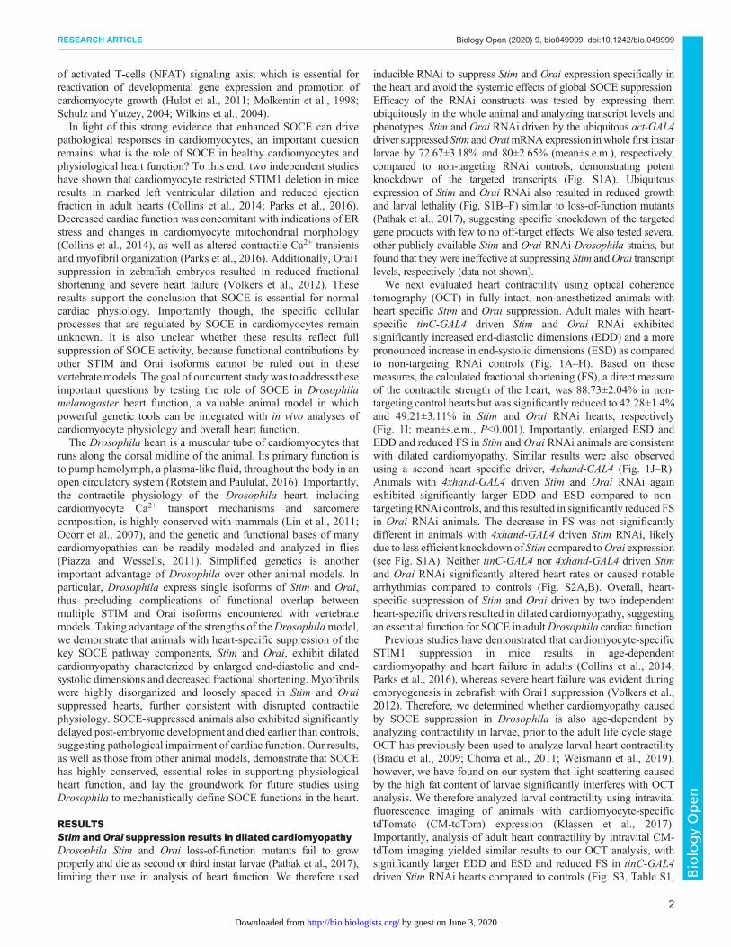
We next evaluated heart contractility using optical coherencetomography (OCT) in fully intact, non-anesthetized animals withheart specific Stim and Orai suppression. Adult males with heart-specific tinC-GAL4 driven Stim and Orai RNAi exhibitedsignificantly increased end-diastolic dimensions (EDD) and a morepronounced increase in end-systolic dimensions (ESD) as comparedto non-targeting RNAi controls (Fig. 1A–H). Based on thesemeasures, the calculated fractional shortening (FS), a direct measureof the contractile strength of the heart, was 88.73±2.04% in non-targeting control hearts but was significantly reduced to 42.28±1.4%and 49.21±3.11% in Stim and Orai RNAi hearts, respectively(Fig. 1I; mean±s.e.m., P<0.001). Importantly, enlarged ESD andEDD and reduced FS in Stim and Orai RNAi animals are consistentwith dilated cardiomyopathy. Similar results were also observedusing a second heart specific driver, 4xhand-GAL4 (Fig. 1J–R).Animals with 4xhand-GAL4 driven Stim and Orai RNAi againexhibited significantly larger EDD and ESD compared to non-targeting RNAi controls, and this resulted in significantly reduced FSin Orai RNAi animals. The decrease in FS was not significantlydifferent in animals with 4xhand-GAL4 driven Stim RNAi, likelydue to less efficient knockdown of Stim compared toOrai expression(see Fig. S1A). Neither tinC-GAL4 nor 4xhand-GAL4 driven Stimand Orai RNAi significantly altered heart rates or caused notablearrhythmias compared to controls (Fig. S2A,B). Overall, heart-specific suppression of Stim and Orai driven by two independentheart-specific drivers resulted in dilated cardiomyopathy, suggestingan essential function for SOCE in adultDrosophila cardiac function.
Previous studies have demonstrated that cardiomyocyte-specificSTIM1 suppression in mice results in age-dependentcardiomyopathy and heart failure in adults (Collins et al., 2014;Parks et al., 2016), whereas severe heart failure was evident duringembryogenesis in zebrafish with Orai1 suppression (Volkers et al.,2012). Therefore, we determined whether cardiomyopathy causedby SOCE suppression in Drosophila is also age-dependent byanalyzing contractility in larvae, prior to the adult life cycle stage.OCT has previously been used to analyze larval heart contractility(Bradu et al., 2009; Choma et al., 2011; Weismann et al., 2019);however, we have found on our system that light scattering causedby the high fat content of larvae significantly interferes with OCTanalysis. We therefore analyzed larval contractility using intravitalfluorescence imaging of animals with cardiomyocyte-specifictdTomato (CM-tdTom) expression (Klassen et al., 2017).Importantly, analysis of adult heart contractility by intravital CM-tdTom imaging yielded similar results to our OCT analysis, withsignificantly larger EDD and ESD and reduced FS in tinC-GAL4driven Stim RNAi hearts compared to controls (Fig. S3, Table S1,
2
RESEARCH ARTICLE Biology Open (2020) 9, bio049999. doi:10.1242/bio.049999
BiologyOpen
by guest on June 3, 2020http://bio.biologists.org/Downloaded from

Movies 1 and 2). Similar to adults, larvae with tinC-GAL4 drivenStim and Orai RNAi exhibited significantly increased EDD andESD compared to non-targeting RNAi controls (Fig. 2A–H;Movies 3 and 4), and this resulted in significantly decreased FS
for Orai, though not for Stim RNAi animals (Fig. 2I). A smallthough significant decrease in heart rate was also observed in Oraibut not Stim RNAi larvae (Fig. S2C). These results suggest thatreduced cardiac output due to SOCE suppression is already evident
Fig. 1. See next page for legend.
3
RESEARCH ARTICLE Biology Open (2020) 9, bio049999. doi:10.1242/bio.049999
BiologyOpen
by guest on June 3, 2020http://bio.biologists.org/Downloaded from
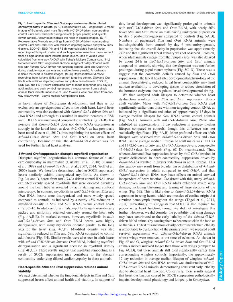
in larval stages of Drosophila development, and thus is notexclusively an age-dependent effect in the adult heart. Larval heartcontractility was also evaluated with 4xhand-GAL4 driven Stim andOrai RNAi and although this resulted in modest increases in ESDand EDD, FS was unchanged compared to controls (Fig. 2J–R). It ispossible that 4xhand-GAL4 does not drive RNAi expression asstrongly in the larval heart as does tinC-GAL4, as has previouslybeen noted (Lee et al., 2017), thus explaining the weaker effects of4xhand-GAL4 driven Stim and Orai RNAi on larval heartcontractility. Because of this, the 4xhand-GAL4 driver was notused for further larval heart analysis.
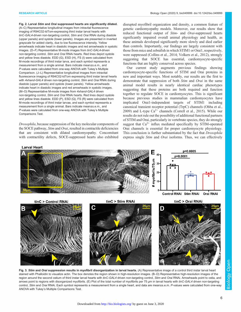
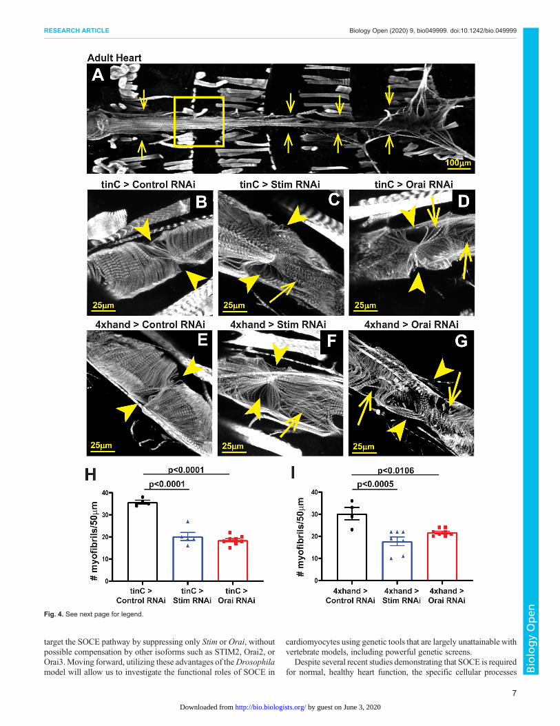
Stim and Orai suppression disrupts myofibril organizationDisrupted myofibril organization is a common feature of dilatedcardiomyopathy in mammalian (Garfinkel et al., 2018; Sussmanet al., 1998) and Drosophila (Ocorr et al., 2007, 2014; Wolf et al.,2006) hearts. We therefore determined whether SOCE-suppressedhearts similarly exhibit disorganized myofibrils. As shown inFig. 3A and B, hearts from tinC-GAL4 driven control RNAi larvaeexhibited evenly spaced myofibrils that were oriented circularlyaround the heart tube as revealed by actin staining and confocalmicroscopy. In contrast, myofibrils in tinC-GAL4-driven Stim andOrai RNAi hearts were disorganized and more widely spacedcompared to controls, as indicated by a nearly 45% reduction inmyofibril density in Stim and Orai RNAi versus control hearts(Fig. 3C–E). In adult control animals, myofibrils again were denselypacked and uniformly oriented circularly around the heart tube(Fig. 4A,B,E). In marked contrast, however, myofibrils in adulttinC-GAL4-driven Stim and Orai RNAi hearts were highlydisorganized, with many myofibrils oriented parallel to the longaxis of the heart (Fig. 4C,D). Myofibril density was alsosignificantly reduced in Stim and Orai RNAi compared to controladult hearts (Fig. 4H). Similar results were also seen in adult heartswith 4xhand-GAL4 driven Stim andOraiRNAi, including myofibrildisorganization and a significant decrease in myofibril density(Fig. 4F,G,I). These results suggest that myofibril remodeling as aresult of SOCE suppression may contribute to the aberrantcontractility underlying dilated cardiomyopathy in these animals.
Heart-specific Stim and Orai suppression reduces animalviabilityWe next determined whether the functional defects in Stim andOraisuppressed hearts affect animal health and viability. In support of
this, larval development was significantly prolonged in animalswith tinC-GAL4-driven Stim and Orai RNAi, with nearly 86%fewer Stim and Orai RNAi animals having undergone pupariationby day 5 post-embryogenesis compared to controls (Fig. 5A,B).Numbers of pupariated Stim and Orai RNAi animals wereindistinguishable from controls by day 6 post-embryogenesis,indicating that the overall delay in pupariation was approximately24 h and that significant larval lethality was not observed. Eclosion,when adult animals emerge from their pupal cases, was also delayedby about 24 h in tinC-GAL4-driven Stim and Orai animalscompared to controls, showing that development was not furtherprolonged during pupal metamorphosis (Fig. 5C–D). These resultssuggest that the contractile deficits caused by Stim and Oraisuppression in the larval heart alter developmental physiology of theanimal. Speculatively, reduced larval heart contractility may limitnutrient availability to developing tissues or reduce circulation ofthe hormone ecdysone that regulates larval developmental timing.
We then analyzed adult lifespan to determine whether heartdysfunction resulting from Stim and Orai suppression affectsadult viability. Males with tinC-GAL4-driven Orai RNAi diedsignificantly earlier than those with non-targeting control RNAi, asindicated by a significant reduction of approximately 13 days inaverage median lifespan for Orai RNAi versus control animals(Fig. 6A,B). Animals with tinC-GAL4-driven Stim RNAi alsoexhibited an approximately 11 day reduction in average medianlifespan compared to controls, though this difference was notstatistically significant (Fig. 6A,B). More profound effects on adultlifespan were observed with 4xhand-GAL4-driven Stim and OraiRNAi, as the average median lifespan of adult males was 11±1.73and 13±2.65 days for Stim andOraiRNAi, respectively, compared to43.64±3.38 days for controls (Fig. 6C–D; mean±s.e.m.). Thus,whereas Stim and Orai suppression driven by tinC-GAL4 resulted ingreater deficiencies in heart contractility, suppression driven by4xhand-GAL4 resulted in greater reductions in adult lifespan. Thisdiscrepancy may result from broader tissue distribution of 4xhand-GAL4 expression in adults compared to tinC-GAL4, and thus4xhand-GAL4-driven RNAi may have effects on animal survivalindependent of heart function. Consistent with this, many 4xhand-GAL4-driven Stim and Orai RNAi adults exhibited severe wingdamage, including blistering and tearing of large sections of thewings (Fig. 6E). This is likely due to 4xhand-GAL4-driven RNAiexpression in wing hearts, which are muscles of cardiac origin thatcirculate hemolymph throughout the wings (Tögel et al., 2013,2008). Interestingly, this suggests that SOCE is also required forproper wing heart function, though we did not investigate thisfurther. However, we did consider the possibility that wing damagemay have contributed to the early lethality of the 4xhand-GAL4-driven RNAi animals by causing them to become stuck in the food oron the vials. To test this andmore clearly determinewhether lethalityis attributable to dysfunction of the primary heart, we repeated adultsurvival experiments with 4xhand-GAL4-driven RNAi animalswhose wings were removed at the time of eclosion. As shown inFig. 6F and G, wingless 4xhand-GAL4-driven Stim and Orai RNAianimals indeed survived longer than those with wings (compare toFig. 6C,D), but these animals still died significantly earlier thancorresponding wingless controls. Importantly, the approximately12-day reduction in average median lifespan of wingless 4xhand-GAL4-driven Stim andOraiRNAi animalswas similar to that of tinC-GAL4-driven RNAi animals, demonstrating consistent early lethalitydue to abnormal heart function. Collectively, these results suggestthat heart dysfunction caused by SOCE suppression pathologicallyimpairs developmental physiology and longevity in Drosophila.
Fig. 1. Heart specific Stim and Orai suppression results in dilatedcardiomyopathy in adults. (A–C) Representative OCT longitudinal B-modeimages of 5-day-old adult male flies with tinC-GAL4 driven non-targetingcontrol, Stim and Orai RNAi during diastole (upper panels) and systole(lower panels). Arrowheads indicate the heart in diastole images. (D–F)Representative M-mode recordings from tinC-GAL4 driven non-targetingcontrol, Stim and Orai RNAi with red lines depicting systole and yellow linesdiastole. EDD (G), ESD (H), and FS (I) were calculated from M-moderecordings of 5-day-old males, and each symbol represents a measurementfrom a single animal. Bars indicate mean±s.e.m., and P-values werecalculated from one-way ANOVA with Tukey’s Multiple Comparison. (J–L)Representative OCT longitudinal B-mode images of 5-day-old adult maleflies with 4xhand-GAL4 driven non-targeting control, Stim and Orai RNAiduring diastole (upper panels) and systole (lower panels). Arrowheadsindicate the heart in diastole images. (M–O) Representative M-moderecordings from 4xhand-GAL4 driven non-targeting control, Stim and OraiRNAi with red lines depicting systole and yellow lines diastole. EDD (P),ESD (Q), and FS (R) were calculated from M-mode recordings of 5-day-oldadult males, and each symbol represents a measurement from a singleanimal. Bars indicate mean±s.e.m., and P-values were calculated from one-way ANOVA with Tukey’s Multiple Comparisons Test.
4
RESEARCH ARTICLE Biology Open (2020) 9, bio049999. doi:10.1242/bio.049999
BiologyOpen
by guest on June 3, 2020http://bio.biologists.org/Downloaded from

DISCUSSIONThe critical importance of proper Ca2+ homeostasis and transportin cardiomyocytes is accentuated by the fact that nearly allcardiomyopathies involve Ca2+ dysregulation. Clear understanding
of the cellular andmolecularmechanisms that regulate cardiomyocyteCa2+ handling is therefore fundamental to unraveling the complexitiesof cardiac pathophysiology. Our results demonstrate that SOCEis an essential component of cardiomyocyte Ca2+ physiology in
Fig. 2. See next page for legend.
5
RESEARCH ARTICLE Biology Open (2020) 9, bio049999. doi:10.1242/bio.049999
BiologyOpen
by guest on June 3, 2020http://bio.biologists.org/Downloaded from
Drosophila, because suppression of the keymolecular components ofthe SOCE pathway, Stim andOrai, resulted in contractile deficienciesthat are consistent with dilated cardiomyopathy. Concomitantwith contractility defects, SOCE-suppressed hearts also exhibited
disrupted myofibril organization and density, a common feature ofgenetic cardiomyopathy models. Moreover, our results show thatreduced functional output of Stim- and Orai-suppressed heartssignificantly impaired overall animal physiology and health, asthese animals developed significantly more slowly and died earlierthan controls. Importantly, our findings are largely consistent withthose frommice and zebrafish inwhich STIM1 orOrai1, respectively,were suppressed (Collins et al., 2014; Volkers et al., 2012), stronglysuggesting that SOCE has essential, cardiomyocyte-specificfunctions that are highly conserved across species.
Our current study augments previous findings showingcardiomyocyte-specific functions of STIM and Orai proteins innew and important ways. Most notably, our results are the first todemonstrate that suppression of both Stim and Orai in the sameanimal model results in nearly identical cardiac phenotypessuggesting that these proteins are both required and functiontogether to regulate SOCE in cardiomyocytes. This is significantbecause previous studies in mammalian cardiomyocytes haveimplicated Orai1-independent targets of STIM1 includingcanonical transient receptor potential (TrpC) channels (Ohba et al.,2009) and L-type Ca2+ channels (Correll et al., 2015). While ourresults do not rule out the possibility of additional functional partnersof STIM and Orai, particularly in vertebrate species, they do stronglysuggest that Ca2+ influx mediated specifically by STIM-operatedOrai channels is essential for proper cardiomyocyte physiology.This conclusion is further substantiated by the fact that Drosophilaexpress single Stim and Orai isoforms. Thus, we can effectively
Fig. 2. Larval Stim and Orai suppressed hearts are significantly dilated.(A–C) Representative longitudinal images from intravital fluorescenceimaging of R94C02-tdTom-expressing third instar larval hearts withtinC-GAL4-driven non-targeting control, Stim and Orai RNAi during diastole(upper panels) and systole (lower panels). Images are presented in invertedgrayscale for added clarity, due to low fluorescence intensity. Yellowarrowheads indicate heart in diastolic images and red arrowheads in systolicimages. (D–F) Representative M-mode images from tinC-GAL4-drivennon-targeting control, Stim and Orai RNAi hearts. Red lines depict systoleand yellow lines diastole. EDD (G), ESD (H), FS (I) were calculated fromM-mode recordings of third instar larva, and each symbol represents ameasurement from a single animal. Bars indicate mean±s.e.m., andP-values were calculated from one-way ANOVA with Tukey’s MultipleComparison. (J–L) Representative longitudinal images from intravitalfluorescence imaging of R94C02-tdTom expressing third instar larval heartswith 4xhand-GAL4 driven non-targeting control, Stim and Orai RNAi duringdiastole (upper panels) and systole (lower panels). Yellow arrowheadsindicate heart in diastolic images and red arrowheads in systolic images.(M–O) Representative M-mode images from 4xhand-GAL4 drivennon-targeting control, Stim and Orai RNAi hearts. Red lines depict systoleand yellow lines diastole. EDD (P), ESD (Q), FS (R) were calculated fromM-mode recordings of third instar larvae, and each symbol represents ameasurement from a single animal. Bars indicate mean±s.e.m., andP-values were calculated from one-way ANOVA with Tukey’s MultipleComparisons Test.
Fig. 3. Stim and Orai suppression results in myofibril disorganization in larval hearts. (A) Representative image of a control third instar larval heartstained with Phalloidin to visualize actin. The box denotes the region shown in high-resolution images. (B–D) Representative high-resolution images of theregion around the second ostium of third instar larval hearts with tinC-GAL4 driven non-targeting control, Stim and Orai RNAi. Arrowheads point to ostia, andarrows point to regions with disorganized myofibrils. (E) Plot of the total number of myofibrils per 75 µm in larval hearts with tinC-GAL4 driven non-targetingcontrol, Stim and Orai RNAi. Each symbol represents a measurement from a single heart, and data are mean±s.e.m. P-values were calculated from one-wayANOVA with Tukey’s Multiple Comparisons Test.
6
RESEARCH ARTICLE Biology Open (2020) 9, bio049999. doi:10.1242/bio.049999
BiologyOpen
by guest on June 3, 2020http://bio.biologists.org/Downloaded from
target the SOCE pathway by suppressing only Stim or Orai, withoutpossible compensation by other isoforms such as STIM2, Orai2, orOrai3.Moving forward, utilizing these advantages of theDrosophilamodel will allow us to investigate the functional roles of SOCE in
cardiomyocytes using genetic tools that are largely unattainable withvertebrate models, including powerful genetic screens.
Despite several recent studies demonstrating that SOCE is requiredfor normal, healthy heart function, the specific cellular processes
Fig. 4. See next page for legend.
7
RESEARCH ARTICLE Biology Open (2020) 9, bio049999. doi:10.1242/bio.049999
BiologyOpen
by guest on June 3, 2020http://bio.biologists.org/Downloaded from

regulated by SOCE in cardiomyocytes have not been clearly defined.Maintenance of SR Ca2+ stores is a seemingly likely function ofSOCE that would broadly impact cardiomyocyte physiology. Forexample, decreased SR Ca2+ content in the absence of SOCE mayreduce RyR-dependent Ca2+ release that drives contraction, andwould be consistent with the severe systolic dysfunction that we andothers have observed in STIM- and Orai-suppressed hearts. In this
case, the myofibril disorganization observed in SOCE-suppressedhearts may be the result of decompensation in response to reducedcontractile force generation (Houser andMargulies, 2003). However,direct analyses of SR Ca2+ store content in STIM- or Orai-depletedcardiomyocytes have yielded conflicting results. For example, siRNAknockdown of STIM1 in freshly isolated neonatal rat ventricularmyocytes led to a significant reduction of the caffeine-releasable SRCa2+ pools (Voelkers et al., 2010),whereas SRCa2+ store content wasunchanged in adult cardiomyocytes isolated from STIM1-deletedmouse hearts compared to controls despite clear indication of dilatedcardiomyopathy (Parks et al., 2016). This suggests that SR Ca2+ storedepletion may not be a direct cause of heart failure when SOCEis suppressed. However, an independent analysis of STIM1deficiency-associated dilated cardiomyopathy in mice demonstratedsignificantly upregulated expression of ER stress response factors(Collins et al., 2014), possibly reflecting mild but chronic Ca2+ storedepletion that may be difficult to measure in acutely isolated cells.Reorganization of cytoarchitecture is a common feature of the ERstress response mechanism, and therefore an important possibility inSOCE-suppressed cardiomyocytes is that ER stress leads tomyofibrildisorganization that contributes to impaired contractility andultimately heart failure.
It is also possible that SOCE has specific signaling functions incardiomyocytes that are required for heart development or tissuehomeostasis. The best characterized signaling function of SOCEacross cell and tissue types involves calcineurin-mediated
Fig. 4. Stim and Orai suppression results in myofibril disorganization inadult hearts. (A) Representative image of an adult control heart (indicatedby arrows) stained with Phalloidin to visualize actin. The box denotes theregion shown in the high-resolution images. (B–D) Representative high-resolution images of the region around the third ostium of 5-day-old adultmale hearts with tinC-GAL4 driven non-targeting control, Stim and OraiRNAi. Arrowheads point to ostia, and arrows point to regions withdisorganized myofibrils. (E–G) Representative high-resolution images of theregion around the third ostium of 5-day-old adult male hearts with 4xhand-GAL4 driven non-targeting control, Stim and Orai RNAi. Arrowheads point toostia, and arrows point to regions with disorganized myofibrils. (H) Plot of thetotal number of myofibrils per 50 µm in adult hearts with tinC-GAL4 drivennon-targeting control, Stim and Orai RNAi. Each symbol represents ameasurement from a single heart, and data are mean±s.e.m. P-values werecalculated from one-way ANOVA with Tukey’s Multiple Comparisons Test.(I) Plot of the total number of myofibrils per 50 µm in adult hearts with4xhand-GAL4 driven non-targeting control, Stim and Orai RNAi. Eachsymbol represents a measurement from a single heart, and data aremean±s.e.m. P-values were calculated from one-way ANOVA with Tukey’sMultiple Comparisons Test.
Fig. 5. Heart specific Stim and Orai suppression delays post-embryonic animal development. (A) Plot of the percent of larvae that pupariated on eachof the indicated days post-embryogenesis for tinC-GAL4 driven Stim, Orai and non-targeting control RNAi. Data are mean±s.e.m. from three independentexperiments with 25–50 animals per experimental group. (B) Comparison of percent pupariated on day 5 post-embryogenesis for tinC-GAL4 driven Stim,Orai and non-targeting control RNAi from three independent replicates. P-values calculated from one-way ANOVA with Tukey’s Multiple Comparisons Test.(C) Plot of the percent of pupae that eclosed on each of the indicated days post-embryogenesis for tinC-GAL4 driven Stim, Orai and non-targeting controlRNAi. Data are mean±s.e.m. from three independent experiments with 25–50 animals per experimental group. (D) Comparison of percent eclosedon day 9 post-embryogenesis for tinC-GAL4 driven Stim, Orai and non-targeting control RNAi from three independent replicates. P-values calculatedfrom one-way ANOVA with Tukey’s Multiple Comparisons Test.
8
RESEARCH ARTICLE Biology Open (2020) 9, bio049999. doi:10.1242/bio.049999
BiologyOpen
by guest on June 3, 2020http://bio.biologists.org/Downloaded from

dephosphorylation and activation of transcription factors such asNFAT (Lacruz and Feske, 2015). Overwhelming evidence hasestablished that upregulated calcineurin-NFAT signaling is anessential mechanism of cardiac hypertrophy pathogenesis (Thamet al., 2015), and SOCE upregulation also results in cardiachypertrophy (Benard et al., 2016; Hulot et al., 2011; Parks et al.,2016) and enhanced calcineurin activity (Luo et al., 2012).
Collectively, these findings suggest that calcineurin is a keycardiomyocyte target of SOCE in the pathological, upregulatedstate. However, whether calcineurin or other signaling factors areessential targets of SOCE in normal heart physiology has not beendetermined. Arguing against a signaling role for SOCE specificallyduring heart development, STIM1-knockout mouse hearts do notexhibit altered phenotypes at early postnatal stages (Collins et al.,
Fig. 6. See next page for legend.
9
RESEARCH ARTICLE Biology Open (2020) 9, bio049999. doi:10.1242/bio.049999
BiologyOpen
by guest on June 3, 2020http://bio.biologists.org/Downloaded from

2014), and we also did not observe effects on Drosophilaembryogenesis with heart-specific Stim or Orai suppression (datanot shown). Additionally, cardiomyopathy in STIM1-knockoutmouse hearts was progressive and age-dependent (Collins et al.,2014), further suggesting that any signaling functions of SOCE arelikely to be homeostatic rather than developmental. Our findings inDrosophila indicate that cardiomyopathy due to Stim and Oraisuppression is already evident in larvae, early in the fly life cycle.This may also reflect post-developmental, homeostatic functions ofSOCE that when suppressed, manifest earlier in flies due to theirfaster lifecycle compared to mice. Clearly, substantial work is stillrequired to identify the specific functional roles of SOCE inphysiological heart function, and genetic tools combinedwith in vivoanalyses of heart physiology and Ca2+ dynamics in Drosophila willallow us to address this in novel and significant ways.Our study is the first to demonstrate the use of intravital
fluorescence imaging of heart contractility in Drosophila larvae, animportant lifecycle stage that involves rapid animal growth andhigh nutritional demands. This is significant because larval heartcontractility has been difficult to analyze using methods commonlyemployed for adults, such as OCT and direct imaging of dissectedpreparations. For example, the high fat content of larvae causes lightscattering that can obscure OCT imaging, though some OCTanalyses of larval heart function have been carried out successfully(Bradu et al., 2009; Choma et al., 2011; Weismann et al., 2019). Inaddition, partial dissections that faithfully preserve heart functioncan be challenging because the larval heart is extremely delicateand poorly attached to the body wall. Thus, intravital fluorescenceimaging of larval hearts is a versatile and widely accessible methodthat is also amenable to longitudinal analysis. We validated ourintravital imaging approach by confirming significantly increasedESD and EDD and decreased FS values in adult Stim RNAi versuscontrol hearts, similar to our results from the more established OCTmethod. However, it was notable that even in controls, ESD andEDD values were substantially larger with intravital imagingcompared to OCT measurements (Table S1). This may be due inpart to better spatial resolution of the heart walls with intravitalfluorescence imaging compared to OCT. Moreover, OCT measuresthe dorsal to ventral movement of the heart walls, whereas intravital
fluorescence imaging measures lateral movement. This importantdistinction may also account for the differences we observedwith the two methods. Importantly, our fluorescence imagingmeasurements of adult heart ESD, EDD and FS were very similar tothose from an earlier study that first described the CM-tdTommodelfor intravital fluorescence imaging (Klassen et al., 2017), furthervalidating the accuracy of our measurements.
Another notable finding of our study was that there were severalkey differences in the results of Stim andOraiRNAi drivenwith tinC-GAL4 compared to 4xhand-GAL4, both of which are commonly usedas heart-specificGAL4 drivers inDrosophila studies.Most strikingly,4xhand-GAL4 but not tinC-GAL4 drivenRNAi resulted in significanteffects on adult wing structure and integrity that were likely causedby defective wing heart function, given that hand but not tinman(the genes from which the respective GAL4 drivers are derived) isexpressed in wing heart myocytes (Tögel et al., 2013, 2008).Importantly, we found that the wing defects caused by 4xhand-GAL4driven Stim and Orai RNAi significantly shortened animal lifespan,independent of effects on contractility of the primary adult heart.This illustrates that caution is warranted when attributing systemicphenotypes, such as premature lethality, to dysfunction of theprimary Drosophila heart when using these GAL4 drivers.
In conclusion, our results demonstrate that SOCE mediated bySTIM and Orai is essential for proper function of the Drosophilaheart, and add to a growing number of studies that collectivelysuggest that SOCE has highly conserved functional roles incardiomyocytes across species. Paradoxically, it is still unknownwhether loss-of-function mutations in Stim1 or Orai1 result in heartdefects or cardiomyopathies in humans. This is likely due to the factthat individuals homozygous for these mutations die fromimmunodeficiency in infancy or early childhood, before adverseheart phenotypes may fully manifest (Rosenberg et al., 2019).Therefore, animal model studies, including powerful geneticscreens and in vivo analyses in Drosophila, will continue to bevital to our understanding of how alterations to SOCE result in orcontribute to devastating cardiomyopathies and heart failure.
MATERIALS AND METHODSFly stocksThe following Drosophila stocks were obtained from the BloomingtonDrosophila Stock Center: mCherry RNAi (35785; non-targeting control),GAL4 RNAi (35783; non-targeting control), Stim RNAi (27263), OraiRNAi (53333), and act-GAL4 (3954). tinC-GAL4 was obtained from DrManfred Frausch (Friedrich Alexander University), and 4xhand-GAL4 wasfrom Dr Zhe Han (George Washington University School of Medicine).CM-tdTom flies were obtained from Dr Rolf Bodmer (Sanford BurnhamPrebys Institute). Flies were maintained on standard cornmeal agar food, andall crosses were carried out at 25°C.
Adult survivalVirgin female tinC-GAL4 or 4xhand-GAL4 flies were crossed with maleRNAi animals, and progeny were raised to adulthood at 25°C. On the day ofeclosion, adult progeny were collected and separated based on sex into vialscontaining up to ten flies per vial, with a total of 20–30 flies per group. Flieswere maintained at 25°C throughout the course of the experiments. Every3 days, vials were checked for dead animals and surviving flies weretransferred into new vials with fresh food. For wingless experiments, wingswere removed immediately following eclosion on the day of adultcollection. Kaplan–Meier survival curves and median lifespan weregenerated using GraphPad Prism.
Developmental timingApproximately 30–40 virgin female tinC-GAL4 or 4xhand-GAL4 animalswere mated with RNAi males for 3 days, at which time animals were
Fig. 6. Heart specific Stim and Orai suppression results in early adultlethality. (A) Representative survival curves in days post-eclosion for adultmales with tinC-GAL4 driven Stim, Orai and non-targeting control RNAi(n=30 animals per group). (B) Plot of the average adult male median lifespan(MLS), calculated from three independent survival curves, for tinC-GAL4driven Stim, Orai and non-targeting control RNAi. Error bars represents.e.m., and P-values were calculated from one-way ANOVA with Tukey’sMultiple Comparisons Test. (C) Representative survival curves in days post-eclosion for adult males with 4xhand-GAL4 driven Stim, Orai and non-targeting control RNAi (n=30 animals per group). (D) Plot of the averageadult MLS, calculated from three independent survival curves, for 4xhand-GAL4 driven Stim, Orai and non-targeting control RNAi. Error bars represents.e.m., and P-values were calculated from one-way ANOVA with Tukey’sMultiple Comparisons Test. (E) Representative images of adult males with4xhand-GAL4 driven Stim, Orai and non-targeting control RNAi on the dayof eclosion. Note the blistered wing on the Stim RNAi animal and theseverely damaged and frayed wings on the Orai RNAi animal (indicated byarrows). (F) Representative survival curves in days post-eclosion for adultmales with 4xhand-GAL4 driven Stim, Orai and non-targeting control RNAi,with their wings removed on day of eclosion (n=30 animals per group).(G) Plot of the average adult MLS, calculated from three independentsurvival curves, for 4xhand-GAL4 driven Stim, Orai and non-targeting controlRNAi animals with their wings removed on the day of eclosion. Error barsrepresent s.e.m., and P-values were calculated from one-way ANOVA withTukey’s Multiple Comparisons Test.
10
RESEARCH ARTICLE Biology Open (2020) 9, bio049999. doi:10.1242/bio.049999
BiologyOpen
by guest on June 3, 2020http://bio.biologists.org/Downloaded from

transferred into egg laying chambers that consisted of a 100 ml plasticbeaker with holes for air exchange affixed over a petri dish containing grapejuice agar (Genesee Scientific). A dollop of yeast paste (active dry yeastmixed with water) was placed in the center of each grape juice agar plateas food. Animals were acclimated in the chambers for 24 h, and thentransferred to new plates for 4 h at 25°C for timed egg laying. Afterremoving the adults, plates with eggs were incubated at 25°C for anadditional 24 h. Hatched larvae were then transferred to vials with standardfly food, with up to 30 larvae per vial, and maintained at 25°C over thecourse of the experiment. Vials were checked each day, and the numbers ofnewly formed pupae and eclosed adults were recorded.
RNA Isolation and RT-qPCRTwenty to 30 first instar larvae were collected in 20 µl cold Trizol andmanually crushed with a pestle. Additional Trizol was then added to a totalvolume of 500 µl, 100 µl chloroform was added, and the aqueous layercontaining extracted RNAwas isolated. Extracted RNAwas further purifiedwith the RNeasy kit (Qiagen) and converted to cDNA using an S1000Thermo Cycler (Bio-Rad) with high capacity cDNA Reverse Transcriptionkit (Thermo Fisher Scientific). Real-time quantitative polymerase chainreaction (RT-qPCR) was performed on a StepOnePlus RT-qPCR machine(Applied Biosystems) with each reaction consisting of triplicate samplescontaining iTaq Universal Probes Supermix (Bio-Rad), pre-validated6-carboxyfluorescein (FAM)-labeled TaqMan probes (Applied Biosystems)against Stim, Orai and RPL32 (housekeeping gene), and template cDNAdiluted per themanufacturer’s instructions. For quantification, triplicate cyclethreshold (Ct) values were averaged and normalized to the RPL32Ct value tocalculate ΔCt. The Δ(ΔCt) was determined by subtracting the control RNAiΔCt value from the experimental ΔCt value, and fold changes expressed as2−Δ(ΔCt). Fold changes are expressed as a percentage of expression comparedto non-targeting RNAi control.
Heart dissection, staining and confocal imagingHearts from third instar larvae or 5-day-old adults were dissected and fixedas previously described (Alayari et al., 2009). In brief, for adults the ventralabdomens and underlying tissues were removed to expose the contractingheart while bathed in oxygenated artificial Drosophila hemolymph [ADH;108 mM NaCl, 5 mM KCl, 2 mM CaCl2, 8 mM MgCl2, 1 mM NaH2PO4,4 mM NaHCO3, 10 mM sucrose, 5 mM trehalose, and 5 mM HEPES(pH 7.1)]. Hearts were then fully relaxed by exchange with fresh ADHcontaining 10 mM EGTA. Larvae were pinned to Sylgard coated dishes attheir anterior and posterior, and a slit was cut along the ventral midline in thepresence of oxygenated ADH. Lateral cuts were then made along the sidesof the animals, and the resulting cuticle flaps were pinned to expose theinternal organs. The gut was removed to expose the beating hearts, whichwere then relaxed with ADH containing 10 mM EGTA. Dissected adult andlarval hearts were both fixed for 20 min at room temperature in PBScontaining 4% paraformaldehyde. Following three 10 min washes in PBScontaining 0.1% Triton X-100 (PBSTx) with gentle rotation, the sampleswere incubated in PBSTx containing 1.0 µM Alexa Fluor 488 Phalloidin(Thermo Fisher Scientific) for 1 h at room temperature with gentle shaking.Samples were again washed three times in PBSTx at room temperature, andmounted on glass slides and coverslips with Vectashield (VectashieldLaboratories) as described previously (Alayari et al., 2009). Samples wereimaged with a Nikon A1R confocal microscope using 10X, 0.45 N.A. and40X, 1.3 NA objectives. Phalloidin was excited with a 488 nm laser.Z-stacks at 1 µm intervals were collected and images are presented asmaximum intensity projections encompassing the whole heart for larvae orthe dorsal half of the heart for adults to avoid the ventral layer of skeletalmuscle. Myofibril density measurements on adults and third instar larvalhearts were taken from the A2 segment of the heart. The number ofmyofibrils per 75 µm in larvae and 50 µm in adults was calculated manually.
Optical coherence tomography (OCT)Adult heart contractility was analyzed using a custom-built OCT apparatusas previously described (Wolf et al., 2006). In brief, 5-day-old males werebriefly anesthetized with CO2, embedded in a soft gel support, and allowedto fully awaken based on body movement. Animals were first imaged in
B-mode in the longitudinal orientation to identify the A1 segment of theheart chamber. They were then imaged transversely in M-mode for 3 s, andmultiple M-modes were recorded for each fly. Animals were then re-imagedin B-mode to ensure proper orientation of the heart chamber. M-modes wereprocessed in ImageJ by referencing to a 150 µm standard. EDD, ESD andheart rate were calculated directly from the processed M-mode traces. FSwas calculated as [(EDD-ESD) / EDD]×100.
Intravital fluorescence microscopyIntravital fluorescence imaging of adult and third instar larval hearts wascarried out using animals that express tdTomato under control of thecardiomyocyte-specific R94C02 enhancer element (Klassen et al., 2017).5-day-old adult males were briefly anesthetized with CO2 and were thenadhered dorsal side down to a glass coverslip with Norland Optical Adhesivethat was then cured with a 48-watt UV LED light source (LKE) for 60 s.Animals were allowed to recover for 10 min prior to imaging. Third instarlarvae were anesthetized by 1-min cold exposure at 4°C and were adheredventral side down to a glass slide with superglue. Larvae were flanked on theglass slide by double-sided tape that was then used to adhere a glass coverslip.The heart of larval and adult animals was imaged through the dorsal cuticle ata rate of 200 frames per second (fps) for 10 s using an ORCA-Flash4.0 V3sCMOS camera (Hamamatsu) on a Nikon Ti2 inverted microscope controlledwith Nikon Elements software. Excitation light at 550 nm was provided by aSpectra-X illuminator (Lumencor) and emission was collected through a 555-635 band-pass filter. To generate M-modes, a 1-pixel wide line was drawnthrough the heart chamber in the A1 segment in adults and the A2 segment inlarvae, and the fluorescence intensity along this line for the full time-coursewas plotted using ImageJ. EDD and ESD were calculated directly from theprocessed M-mode traces by manually measuring the distance between theheart walls at full relaxation and full constriction, respectively. An average offive measurements of EDD and ESD was calculated from each trace. Heartratewas calculated bymanually counting the number of systoles over 10 s. FSwas calculated as [(EDD-ESD)/EDD]×100.
Statistical analysesAll statistical analyses were carried out using GraphPad Prism software.Contractility parameters and myofibril density measurements were analyzedby one-way ANOVA followed by Tukey’s Multiple Comparisons Test.Differences were considered statistically significant at P<0.05.
AcknowledgementsWe thank Drs Georg Vogler and Rold Bodmer at the Sanford Burnham PrebysMedical Institute for providing the flies with heart-specific tdTomato expression forintravital imaging.
Competing interestsThe authors declare no competing or financial interests.
Author contributionsConceptualization: C.E.P., J.T.S.; Methodology: C.E.P., M.J.W., J.T.S.;Investigation: C.E.P., M.J.W., J.T.S.; Writing - original draft: C.E.P., J.T.S.; Writing -review & editing: M.J.W.; Supervision: J.T.S.; Funding acquisition: J.T.S.
FundingThis work was supported by [grant number I80VP000003] from the CollaborativeHealth Initiative Research Program and the National Institutes of Health to C.E.P.and J.T.S., and by the University of Virginia Center for Excellence in CardiovascularGenetics to M.J.W.
Supplementary informationSupplementary information available online athttp://bio.biologists.org/lookup/doi/10.1242/bio.049999.supplemental
ReferencesAbraham, D. M. and Wolf, M. J. (2013). Disruption of sarcoendoplasmic reticulum
calcium ATPase function in Drosophila leads to cardiac dysfunction.PLoSONE 8,e77785. doi:10.1371/journal.pone.0077785
Alayari, N. N., Vogler, G., Taghli-Lamallem, O., Ocorr, K., Bodmer, R.Cammarato, A. (2009). Fluorescent labeling of Drosophila heart structures.J. Vis. Exp. 32, e1423. doi:10.3791/1423
11
RESEARCH ARTICLE Biology Open (2020) 9, bio049999. doi:10.1242/bio.049999
BiologyOpen
by guest on June 3, 2020http://bio.biologists.org/Downloaded from
Benard, L., Oh, J. G., Cacheux, M., Lee, A., Nonnenmacher, M., Matasic, D. S.,Kohlbrenner, E., Kho, C., Pavoine, C., Hajjar, R. J. et al. (2016). Cardiac Stim1silencing impairs adaptive hypertrophy and promotes heart failure throughinactivation of mTORC2/Akt signaling. Circulation 133, 1458-1471. doi:10.1161/CIRCULATIONAHA.115.020678
Bers, D. M. (2002). Cardiac excitation-contraction coupling. Nature 415, 198-205.doi:10.1038/415198a
Bradu, A., Ma, L., Bloor, J. W. and Podoleanu, A. (2009). Dual optical coherencetomography/fluorescence microscopy for monitoring of Drosophila melanogasterlarval heart. J. Biophotonics 2, 380-388. doi:10.1002/jbio.200910021
Choma, M. A., Suter, M. J., Vakoc, B. J., Bouma, B. E. and Tearney, G. J. (2011).Physiological homology between Drosophila melanogaster and vertebratecardiovascular systems. Dis. Model Mech. 4, 411-420. doi:10.1242/dmm.005231
Collins, H. E., He, L., Zou, L., Qu, J., Zhou, L., Litovsky, S. H., Yang, Q., Young,M. E., Marchase, R. B. and Chatham, J. C. (2014). Stromal interaction molecule1 is essential for normal cardiac homeostasis through modulation of ER andmitochondrial function. Am. J. Physiol. Heart Circ. Physiol. 306, H1231-H1239.doi:10.1152/ajpheart.00075.2014
Correll, R. N., Goonasekera, S. A., van Berlo, J. H., Burr, A. R., Accornero, F.,Zhang, H., Makarewich, C. A., York, A. J., Sargent, M. A., Chen, X. et al. (2015).STIM1 elevation in the heart results in aberrant Ca2+ handling andcardiomyopathy. J. Mol. Cell. Cardiol. 87, 38-47. doi:10.1016/j.yjmcc.2015.07.032
Garfinkel, A. C., Seidman, J. G. and Seidman, C. E. (2018). Genetic pathogenesisof hypertrophic and dilated cardiomyopathy. Heart Fail. Clin. 14, 139-146. doi:10.1016/j.hfc.2017.12.004
Houser, S. R. and Margulies, K. B. (2003). Is depressed myocyte contractilitycentrally involved in heart failure? Circ. Res. 92, 350-358. doi:10.1161/01.RES.0000060027.40275.A6
Hulot, J.-S., Fauconnier, J., Ramanujam, D., Chaanine, A., Aubart, F., Sassi, Y.,Merkle, S., Cazorla, O., Ouille, A., Dupuis, M. et al. (2011). Critical role forstromal interaction molecule 1 in cardiac hypertrophy. Circulation 124, 796-805.doi:10.1161/CIRCULATIONAHA.111.031229
Klassen, M. P., Peters, C. J., Zhou, S., Williams, H. H., Jan, L. Y. and Jan, Y. N.(2017). Age-dependent diastolic heart failure in an in vivo Drosophila model. Elife6, e20851. doi:10.7554/eLife.20851.029
Kranias, E. and Bers, D. (2007). Calcium and Cardiomyopathies. Subcell.Biochem. 45, 523-37.
Lacruz, R. S. and Feske, S. (2015). Diseases caused by mutations in ORAI1 andSTIM1. Ann. N. Y. Acad. Sci. 1356, 45-79. doi:10.1111/nyas.12938
Lee, S., Bao, H., Ishikawa, Z., Wang, W. and Lim, H.-Y. (2017). Cardiomyocyteregulation of systemic lipid metabolism by the apolipoprotein B-containinglipoproteins in Drosophila. PLoS Genet. 13, e1006555. doi:10.1371/journal.pgen.1006555
Limas, C. J., Olivari, M. T., Goldenberg, I. F., Levine, T. B., Benditt, D. G. andSimon, A. (1987). Calcium uptake by cardiac sarcoplasmic reticulum in humandilated cardiomyopathy. Cardiovasc. Res. 21, 601-605. doi:10.1093/cvr/21.8.601
Lin, N., Badie, N., Yu, L., Abraham, D., Cheng, H., Bursac, N., Rockman, H. A.andWolf, M. J. (2011). Amethod tomeasuremyocardial calcium handling in adultDrosophila. Circ. Res. 108, 1306-1315. doi:10.1161/CIRCRESAHA.110.238105
Luo, X., Hojayev, B., Jiang, N., Wang, Z. V., Tandan, S., Rakalin, A., Rothermel,B. A., Gillette, T. G. and Hill, J. A. (2012). STIM1-dependent store-operated Ca2+
entry is required for pathological cardiac hypertrophy. J. Mol. Cell. Cardiol. 52,136-147. doi:10.1016/j.yjmcc.2011.11.003
MacLeod, K. T. (2016). Recent advances in understanding cardiac contractility inhealth and disease. F1000Res 5, 1770-1783. doi:10.12688/f1000research.8661.1
McKenna, W. J., Maron, B. J. and Thiene, G. (2017). Classification, epidemiology,and global burden of cardiomyopathies. Circ. Res. 121, 722-730. doi:10.1161/CIRCRESAHA.117.309711
Molkentin, J. D., Lu, J.-R., Antos, C. L., Markham, B., Richardson, J., Robbins,J., Grant, S. R. and Olson, E. N. (1998). A calcineurin-dependent transcriptionalpathway for cardiac hypertrophy. Cell 93, 215-228. doi:10.1016/S0092-8674(00)81573-1
Ocorr, K., Perrin, L., Lim, H.-Y., Qian, L., Wu, X. and Bodmer, R. (2007). Geneticcontrol of heart function and aging in Drosophila. Trends Cardiovasc. Med. 17,177-182. doi:10.1016/j.tcm.2007.04.001
Ocorr, K., Vogler, G. and Bodmer, R. (2014). Methods to assess Drosophila heartdevelopment, function and aging. Methods 68, 265-272. doi:10.1016/j.ymeth.2014.03.031
Ohba, T., Watanabe, H., Murakami, M., Sato, T., Ono, K. and Ito, H. (2009).Essential role of STIM1 in the development of cardiomyocyte hypertrophy.Biochem. Biophys. Res. Commun. 389, 172-176. doi:10.1016/j.bbrc.2009.08.117
Parks, C., Alam, M. A., Sullivan, R. and Mancarella, S. (2016). STIM1-dependentCa(2+) microdomains are required for myofilament remodeling and signaling inthe heart. Sci. Rep. 6, 25372. doi:10.1038/srep25372
Pathak, T., Trivedi, D. and Hasan, G. (2017). CRISPR-Cas-induced mutantsidentify a requirement for dSTIM in larval dopaminergic cells of Drosophilamelanogaster. G3 (Bethesda) 7, 923-933. doi:10.1534/g3.116.038539
Piazza, N. and Wessells, R. J. (2011). Drosophila models of cardiac disease.Prog. Mol. Biol. Transl. Sci. 100, 155-210. doi:10.1016/B978-0-12-384878-9.00005-4
Putney, J. W. (2011). The physiological function of store-operated calcium entry.Neurochem. Res. 36, 1157-1165. doi:10.1007/s11064-010-0383-0
Putney, J. W. (2018). Forms and functions of store-operated calcium entrymediators, STIM and Orai. Adv. Biol. Regul. 68, 88-96. doi:10.1016/j.jbior.2017.11.006
Rosenberg, P., Katz, D. and Bryson, V. (2019). SOCE and STIM1 signaling in theheart: timing and location matter. Cell Calcium 77, 20-28. doi:10.1016/j.ceca.2018.11.008
Rotstein, B. and Paululat, A. (2016). On the morphology of the Drosophila heart.J. Cardiovasc. Dev. Dis. 3, 15. doi:10.3390/jcdd3020015
Schulz, R. A. and Yutzey, K. E. (2004). Calcineurin signaling and NFAT activationin cardiovascular and skeletal muscle development. Dev. Biol. 266, 1-16. doi:10.1016/j.ydbio.2003.10.008
Smyth, J. T., Hwang, S.-Y., Tomita, T., DeHaven, W. I., Mercer, J. C. and Putney,J. W. (2010). Activation and regulation of store-operated calcium entry. J. Cell.Mol. Med. 14, 2337-2349. doi:10.1111/j.1582-4934.2010.01168.x
Sussman, M. A., Welch, S., Cambon, N., Klevitsky, R., Hewett, T. E., Price, R.,Witt, S. A. and Kimball, T. R. (1998). Myofibril degeneration caused bytropomodulin overexpression leads to dilated cardiomyopathy in juvenile mice.J. Clin. Invest. 101, 51-61. doi:10.1172/JCI1167
Tham, Y. K., Bernardo, B. C., Ooi, J. Y. Y., Weeks, K. L. and McMullen, J. R.(2015). Pathophysiology of cardiac hypertrophy and heart failure: signalingpathways and novel therapeutic targets. Arch. Toxicol. 89, 1401-1438. doi:10.1007/s00204-015-1477-x
Togel, M., Pass, G. and Paululat, A. (2008). The Drosophila wing hearts originatefrom pericardial cells and are essential for wing maturation. Dev. Biol. 318, 29-37.doi:10.1016/j.ydbio.2008.02.043
Togel, M., Meyer, H., Lehmacher, C., Heinisch, J. J., Pass, G. and Paululat, A.(2013). The bHLH transcription factor hand is required for proper wing heartformation in Drosophila. Dev. Biol. 381, 446-459. doi:10.1016/j.ydbio.2013.05.027
Vega, R. B., Bassel-Duby, R. and Olson, E. N. (2003). Control of cardiac growthand function by calcineurin signaling. J. Biol. Chem. 278, 36981-36984. doi:10.1074/jbc.R300023200
Voelkers, M., Salz, M., Herzog, N., Frank, D., Dolatabadi, N., Frey, N., Gude, N.,Friedrich, O., Koch, W. J., Katus, H. A. et al. (2010). Orai1 and Stim1 regulatenormal and hypertrophic growth in cardiomyocytes. J. Mol. Cell. Cardiol. 48,1329-1334. doi:10.1016/j.yjmcc.2010.01.020
Volkers, M., Dolatabadi, N., Gude, N., Most, P., Sussman, M. A. and Hassel, D.(2012). Orai1 deficiency leads to heart failure and skeletal myopathy in zebrafish.J. Cell Sci. 125, 287-294. doi:10.1242/jcs.090464
Weismann, C. G., Blice-Baum, A., Tong, T., Li, J., Huang, B. K., Jonas, S. M.,Cammarato, A. and Choma, M. A. (2019). Multi-modal and multiscale imagingapproaches reveal novel cardiovascular pathophysiology in Drosophilamelanogaster. Biol. Open 8, bio044339. doi:10.1242/bio.044339
Wilkins, B. J., Dai, Y.-S., Bueno, O. F., Parsons, S. A., Xu, J., Plank, D. M., Jones,F., Kimball, T. R. and Molkentin, J. D. (2004). Calcineurin/NFAT couplingparticipates in pathological, but not physiological, cardiac hypertrophy. Circ. Res.94, 110-118. doi:10.1161/01.RES.0000109415.17511.18
Wolf, M. J., Amrein, H., Izatt, J. A., Choma, M. A., Reedy, M. C. and Rockman,H. A. (2006). Drosophila as a model for the identification of genes causing adulthuman heart disease. Proc. Natl. Acad. Sci. USA 103, 1394-1399. doi:10.1073/pnas.0507359103
12
RESEARCH ARTICLE Biology Open (2020) 9, bio049999. doi:10.1242/bio.049999
BiologyOpen
by guest on June 3, 2020http://bio.biologists.org/Downloaded from