supportive module 3 basics of diagnosis, treatment and...
TRANSCRIPT


Supportive module 3 "Basics of diagnosis, treatment and prevention of major pulmonary diseases "
Chronic Obstructive Pulmonary Disease
LECTURE IN INTERNAL MEDICINE FOR IV COURSE STUDENTS
M. Yabluchansky, L. Bogun, L. Martymianova, O. Bychkova, N. Lysenko, N. MakienkoV.N. Karazin National University Medical School’ Internal Medicine Dept.

Plan of the Lecture
• Definition
• Epidemiology
• Risk Factors and Etiology
• Mechanisms
• Classification
• Clinical presentation
• Diagnosis
• Treatment
• Prognosis
• Prophylaxis
• Abbreviations
• Diagnostic guidelines
https ://s-media-cache-ak0.pinimg.com/originals/27/8c/c4/278cc463c56cc824d9eb2666e8f3863d.jpg

DefinitionChronic Obstructive Pulmonary Disease (COPD) is a type of lung disease by a decline in lung function over time in which subsets of patients may have dominant features of chronic bronchitis and/or emphysema characterized by long-term airflow obstruction that is not fully reversible with the main symptoms include shortness of breath and cough with sputum production typically worsens over time, some significant extra-pulmonary effects, and important comorbidities which may contribute to the severity of the disease in individual patients.
Chronic bronchitis is defined clinically as the presence of a chronic productive cough for 3 months during each of 2 consecutive years (other causes of cough being excluded).
Emphysema is defined as an abnormal, permanent enlargement of the air spaces distal to the terminal bronchioles, accompanied by destruction of their walls and without obvious fibrosis.
http://www.who.int/respiratory/copd/GOLD_WR_06.pdf en.wikipedia.org/wiki/Chronic_obstructive_pulmonary_diseasemedicine.medscape.com/article/297664-overview?pa=%2FOOPqamIew%2Fp1dAMq%2BJ7KjXjrTEuzO%2FMsNWCIiPMRs0neLeqn1hB%2BszABfchCz uV8SIvl8zjYv73GUyW5rsbWA%3D%3D#showall

Epidemiology• Globally, as of 2010, COPD affected approximately 329 million people
(4.8% of the population)
• In England, an estimated 0.84 million people (of 50 million) have a diagnosis of COPD; this translates into approximately one person in 59 receiving a diagnosis of COPD at some point in their lives
• In the United States approximately 6.3% of the adult population, totaling approximately 15 million people, have been diagnosed with COPD
• The disease affects men and women almost equally, as there has been increased tobacco use among women in the developed world
• COPD is more common in older people
• The number of deaths from COPD in 2012 became the third leading cause of death.
https ://en.wikipedia.org/wiki/Chronic_obstructive_pulmonary_disease#Epidemiology

Epidemiology
http://images.slideplayer.com/16/5124644/slides/slide_13.jpg
COPD - Deaths / 1000 1990 Data projected to 2000.

Epidemiology
http://dm5migu4zj3pb.cloudfront.net/manuscripts/33000/33947/medium/JCI0833947.f2.jpg
Chronic Obstructive Pulmonary Disease Among Adults Aged 18 and Over in the United States, 1998–2009.

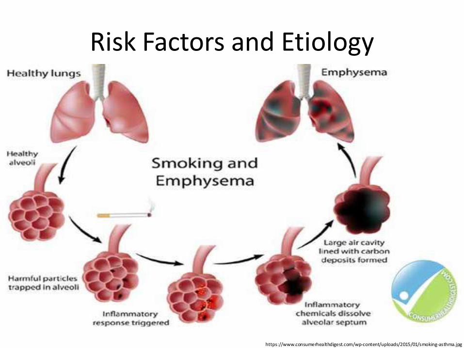
Risk Factors and Etiology • Smoking: smokers are 13 times more likely to die from the COPD
• Air Pollution: indoor and outdoor pollutants
• Occupational Dusts and Chemicals: industrial dust, chemicals, and gases
• Genetics: in rare cases, genetic factors can cause people who have never smoked (a lack of the protein α1-antitrypsin (AAT), Salla disease, etc.)
• Intravenous drug use: the pulmonary vascular damage that results from the insoluble filler contained in methadone or methylphenidate
• Connective tissue disorders: Cutis laxa, Marfan syndrome, Ehlers-Danlos syndrome, Vasculitis syndrome, etc.
• Immunodeficiency syndromes
• Age: incidence COPD increases with age.http://www.healthline.com/health/copd/risk-factors#Overview1
Risk Factors and Etiology
https://www.consumerhealthdigest.com/wp-content/uploads/2015/01/smoking-asthma.jpg

Mechanism General 1
• Pathologic changes in COPD occur in the large (central) airways, the small (peripheral) bronchioles, and the lung parenchyma
• Most cases of COPD are the result of exposure to noxious stimuli, most often cigarette smoke
• The normal inflammatory response is amplified in persons prone to COPD development
• Increased numbers of activated polymorphonuclear leukocytes and macrophages release elastases in a manner that cannot be counteracted effectively by antiproteases, resulting in lung destruction
http://emedicine.medscape.com/article/297664-overview?pa=%2FOOPqamIew%2Fp1dAMq%2BJ7KjXjrTEuzO%2FMsNWCIiPMRs0neLeqn1hB%2BszABfchCzuV8SIvl8zjYv73GUyW5rsbWA%3D%3D#a3

Mechanism General 2
• The primary offender has been found to be human leukocyte elastase, with synergistic roles suggested for proteinase-3 and macrophage-derived matrix metalloproteinases (MMPs), cysteine proteinases, and a plasminogen activator
• Additionally, increased oxidative stress caused by free radicals in cigarette smoke, the oxidants released by phagocytes, and polymorphonuclear leukocytes all may lead to apoptosis or necrosis of exposed cells
• Accelerated aging and autoimmune mechanisms have also been proposed
http://emedicine.medscape.com/article/297664-overview?pa=%2FOOPqamIew%2Fp1dAMq%2BJ7KjXjrTEuzO%2FMsNWCIiPMRs0neLeqn1hB%2BszABfchCzuV8SIvl8zjYv73GUyW5rsbWA%3D%3D#a3

Mechanism General 2
• Cigarette smoke causes neutrophil influx, which is required for the secretion of MMPs; this suggests, that neutrophils and macrophages are required for the development of emphysema
• In addition to macrophages, T lymphocytes, particularly CD8+, play an important role in the pathogenesis of smoking-induced airflow limitation
• The dysregulation of apoptosis and defective clearance of apoptotic cells by macrophages play a prominent role in airway inflammation, particularly in emphysema
• In patients with stable COPD without known cardiovascular disease, there is a high prevalence of microalbuminuria, which is associated with hypoxemia independent of other risk factors.
http://emedicine.medscape.com/article/297664-overview?pa=%2FOOPqamIew%2Fp1dAMq%2BJ7KjXjrTEuzO%2FMsNWCIiPMRs0neLeqn1hB%2BszABfchCzuV8SIvl8zjYv73GUyW5rsbWA%3D%3D#a3

Mechanism Schema 1
Immunology of COPD.http://www.nature.com/nri/journal/v8/n3/images/nri2254-f2.jpg

Mechanism Schema 2
Multifaceted mechanisms in COPD.http://www.nature.com/nri/journal/v8/n3/images/nri2254-f2.jpg

Mechanism Chronic Bronchitis 1
• Mucous gland hyperplasia is the histologic hallmark of chronic bronchitis
• Airway structural changes include atrophy, focal squamous metaplasia, ciliary abnormalities, variable amounts of airway smooth muscle hyperplasia, inflammation, and bronchial wall thickening
• Damage to the endothelium impairs the mucociliary response that clears bacteria and mucus
• Inflammation and secretions provide the obstructive component of chronic bronchitis
• Neutrophilia develops in the airway lumen, and neutrophilic infiltrates accumulate in the submucosa
http://emedicine.medscape.com/article/297664-overview?pa=%2FOOPqamIew%2Fp1dAMq%2BJ7KjXjrTEuzO%2FMsNWCIiPMRs0neLeqn1hB%2BszABfchCzuV8SIvl8zjYv73GUyW5rsbWA%3D%3D#a3

Mechanism Chronic Bronchitis 2
• The respiratory bronchioles display a mononuclear inflammatory process, lumen occlusion by mucus plugging, goblet cell metaplasia, smooth muscle hyperplasia, and distortion due to fibrosis
• These changes, combined with loss of supporting alveolar attachments, cause airflow limitation by allowing airway walls to deform and narrow the airway lumen
• In contrast to emphysema, chronic bronchitis is associated with a relatively undamaged pulmonary capillary bed
• Eventually, hypercapnia and respiratory acidosis develop, leading to pulmonary artery vasoconstriction and cor pulmonale.
• With the ensuing hypoxemia, polycythemia, and increased CO2 retention, these patients have signs of right heart failure and are known as "blue bloaters."
http://emedicine.medscape.com/article/297664-overview?pa=%2FOOPqamIew%2Fp1dAMq%2BJ7KjXjrTEuzO%2FMsNWCIiPMRs0neLeqn1hB%2BszABfchCzuV8SIvl8zjYv73GUyW5rsbWA%3D%3D#a3

Mechanism Schema 3
http://img.tfd.com/MosbyMD/thumb/bronchitis.jpg spiriva.com/img/content/reduced-airflow.jpg

Mechanism Schema 4
http://www.who.int/respiratory/copd/GOLD_WR_06.pdf
Mechanisms Underlying Airflow Limitation in COPD.

Mechanism Emphysema 1
• Emphysema is a diagnosis defined by permanent enlargement of airspaces distal to the terminal bronchioles
• This leads to a dramatic decline in the alveolar surface area available for gas exchange
• Loss of alveoli leads to airflow limitation by 2 mechanisms: 1) loss of the alveolar walls results in a decrease in elastic recoil, which leads to airflow limitation, 2) loss of the alveolar supporting structure leads to airway narrowing, which further limits airflow
• Emphysema has 3 morphologic patterns: centriacinar, panacinar, distal acinar (paraseptal)
http://emedicine.medscape.com/article/297664-overview?pa=%2FOOPqamIew%2Fp1dAMq%2BJ7KjXjrTEuzO%2FMsNWCIiPMRs0neLeqn1hB%2BszABfchCzuV8SIvl8zjYv73GUyW5rsbWA%3D%3D#a3

Mechanism Emphysema 2
• Centriacinar emphysema is characterized by focal destruction limited to the respiratory bronchioles and the central portions of the acini
• Panacinar emphysema involves the entire alveolus distal to the terminal bronchiole
• Distal acinar emphysema is the least common form and involves distal airway structures, alveolar ducts, and sacs
• The gradual destruction of alveolar septae and of the pulmonary capillary bed in emphysema leads to a decreased ability to oxygenate blood
• The body compensates with lowered cardiac output and hyperventilation
• Eventually, patients develop muscle wasting and weight loss and are identified as "pink puffers.“
http://emedicine.medscape.com/article/297664-overview?pa=%2FOOPqamIew%2Fp1dAMq%2BJ7KjXjrTEuzO%2FMsNWCIiPMRs0neLeqn1hB%2BszABfchCzuV8SIvl8zjYv73GUyW5rsbWA%3D%3D#a3

Mechanism Schema 5
http://img.medscapestatic.com/pi/meds/ckb/16/38616tn.jpg media.web.britannica.com/eb-media/04/100104-004-BB90853A.jpg

Classification International Classification of Diseases
https ://www.tsoshop.co.uk/productimages/default.aspx?ISBN=9789241549165&FORMAT=3 http://apps.who.int/classifications/icd10/browse/2016/en#/XI
• X Diseases of the respiratory system
• Chronic lower respiratory diseases(J40-J47)
• J40 Bronchitis, not specified as acute or chronic
• J41 Simple and mucopurulent chronic bronchitis
• J42 Unspecified chronic bronchitis
• J43 Emphysema
• J44 Other chronic obstructive pulmonary disease
• J45 Asthma
• J46 Status asthmaticus
• J47 Bronchiectasis

Classification GOLD staging of severity for COPD, based on the value of FEV1
https ://www.merckmanuals.com/professional/pulmonary-disorders/pneumonia/overview-of-pneumonia
Stage I Mild COPD FEV1/FVC<0.70 FEV1≥ 80% normal
Stage II Moderate COPD
FEV1/FVC<0.70 FEV1 50-79% normal
Stage III Severe COPD FEV1/FVC<0.70 FEV1 30-49% normal
Stage IV Very Severe COPD
FEV1/FVC<0.70 FEV1 <30% normal, or <50% normal with chronic respiratory failure present*
* Usually, this means requiring long-term oxygen therapy.

Classification Clinical GOLD staging of COPD severity
• Stage 0: at risk; chronic cough and sputum production; spirometry is normal
• Stage I: mild COPD; mild airflow limitation (FEV1/FVC) less than 70% but FEV1 80% or more than predicted; usually, but not always, chronic cough and sputum production
• Stage II: moderate COPD; worsening airflow limitation (FEV1 50-79% predicted) and usually progression of symptoms, with shortness of breath, especially on exertion
• Stage III: severe COPD; further worsening of airflow limitation (FEV1 30-50% predicted), increased shortness of breath, and repeated exacerbations
• Stage IV: very severe COPD; severe airflow limitation (FEV1 less than 30% predicted) or the presence of chronic respiratory failure
http://patient.info/doctor/chronic-obstructive-pulmonary-disease-pro

Classification Severity
staging of airflow obstruction according to the American Thoracic Society (ATS)/European Respiratory Society (ERS)
http://www.erswhitebook.org/chapters/chronic-obstructive-pulmonary-disease/
Mild FEV1/VC <5th percentile of predicted and FEV1 ≥70% pred
Moderate FEV1/VC <5th percentile of predicted and FEV1 60–69% pred
Moderately severe
FEV1/VC <5th percentile of predicted and FEV1 50–59% pred
Severe FEV1/VC <5th percentile of predicted and FEV1 35–49% pred
Very severe FEV1/VC <5th percentile of predicted and FEV1 <35% pred
FEV1: forced expiratory volume in 1 second; VC: vital capacity; % pred: % of predicted value; FVC: forced vital capacity.

Symptoms and Signs
• The most important symptoms of COPD are breathlessness on exertion and chronic cough with or without phlegm
• The dyspnea usually worsens over time but is often not present in mild or moderate COPD
• The cough may be dry or productive
• Cough and phlegm often precede dyspnea on exertion by many years
• Other symptoms include wheezing and chest tightness
• As the disease progresses and reaches the severe stages, fatigue, weight loss and anorexia may increase.
http://www.erswhitebook.org/chapters/chronic-obstructive-pulmonary-disease/

Clinical Manifestations 1
• A characteristic of COPD is exacerbations or episodes of acute worsening of the respiratory symptoms.
• The most common causes of exacerbations are viral or bacterial infections
• Increased air pollution also appears to precipitate exacerbations of COPD
• Some patients are particularly prone to exacerbations while others are not
• Two or more exacerbations during the previous year is the most important indicator of a future exacerbation
http://www.erswhitebook.org/chapters/chronic-obstructive-pulmonary-disease/

Clinical Manifestations 2• Exacerbations accelerate the decline in lung function that
characterises COPD, resulting in reduced physical activity, poorer quality of life, and an increased risk of death; they are also responsible for a large proportion of the healthcare costs attributable to COPD
• Patients with COPD often suffer from other diseases (comorbidities)
• The comorbidities may share common risk factors with COPD, in particular cigarette smoking, and may also represent extrapulmonary manifestations or complications of COPD, such as muscle dysfunction due to inactivity
• Comorbidities may be secondary to treatment of COPD; for example, osteoporosis due to oral corticosteroid treatment
http://www.erswhitebook.org/chapters/chronic-obstructive-pulmonary-disease/

Clinical Manifestations 3
• The most common comorbidities in COPD are ischemic heart disease, anxiety and depression, osteoporosis, skeletal muscle dysfunction, gastro-esophageal reflux, anaemia, lung cancer, diabetes and metabolic syndrome
• Comorbidities contribute to the overall severity and manifestations of the disease and can occur in mild, moderate or severe COPD
• The clinical effects of COPD show considerable inter-individual variation, depending on which respiratory symptoms predominate, the frequency of exacerbations, the level and rate of lung function decline and the amount of emphysema, as well as comorbidities.
• Various subtypes of the disease are often termed phenotypes of COPD.
http://www.erswhitebook.org/chapters/chronic-obstructive-pulmonary-disease/

Complications
http://www.healthline.com/health/copd/serious-complications#Complications2
• Pneumonia
• COPD heart failure (Cor pulmonale)
• Lung cancer
• Diabetes
• Dementia
• Arrhythmias
• Osteoporosis
• Respiratory failure.

Diagnosis
• The diagnosis of COPD should be considered in anyone over the age of 35 to 40 who has shortness of breath, a chronic cough, sputum production, or frequent winter colds and a history of exposure to risk factors for the disease
• Spirometry is then used to confirm the diagnosis.
• The formal diagnosis of COPD is made, when the ratio of forced expiratory volume in 1 second over forced vital capacity (FEV1/FVC) is less than 70% of that predicted for a matched control, it is diagnostic for a significant obstructive defect
• Screening those without symptoms is not recommended.
https://en.wikipedia.org/wiki/Chronic_obstructive_pulmonary_disease#Signs_and_symptoms

Diagnosis Spirometry 1
http://www.medscape.org/viewarticle/741196
PEFR = peak expiratory flow rate; TLC = total lung capacity.

Diagnosis Spirometry 2
http://image.slidesharecdn.com/copyofslsqasthmamanagementpptv050811piccomp-141113083910-conversion-gate02/95/spirometry-basics-2-30-638.jpg?cb=1415868230

Diagnosis Spirometry 3
http://a360-wp-uploads.s3.amazonaws.com/wp-content/uploads/rtmagazi/2011/10/10-01_02.jpg

Diagnosis Arterial Blood Gas (ABG) Findings
• ABGs provide the best clues as to acuteness and severity of disease exacerbation
• Patients with mild COPD have mild to moderate hypoxemia without hypercapnia
• As the disease progresses, hypoxemia worsens and hypercapnia may develop, with the latter commonly being observed as the FEV 1 falls below 1 L/s or 30% of the predicted value
• pH usually is near normal; a pH below 7.3 generally indicates acute respiratory compromise
• Chronic respiratory acidosis leads to compensatory metabolic alkalosis.
http://emedicine.medscape.com/article/297664-overview?pa=P4tWSkqAfuEbVp94c%2FW11%2B%2Fk7GZhKH%2B3snXYEMg6JZjYixQ%2FuxvzKUqypCxZhb0X8SIvl8zjYv73GUyW5rsbWA%3D%3D#a1

Diagnosis Frontal And Lateral Chest Radiographs
• Flattening of the diaphragm
• Increased retrosternal air space
• A long, narrow heart shadow
• Rapidly tapering vascular shadows accompanied by hyperlucency of the lungs
• Radiographs in patients with chronic bronchitis show increased bronchovascular markings and cardiomegaly.
http://emedicine.medscape.com/article/297664-overview?pa=P4tWSkqAfuEbVp94c%2FW11%2B%2Fk7GZhKH%2B3snXYEMg6JZjYixQ%2FuxvzKUqypCxZhb0X8SIvl8zjYv73GUyW5rsbWA%3D%3D#a1

Diagnosis Imaging Studies 1
https://images.radiopaedia.org/images/266487/d35992af9e42aba7d71001c13843c3_jumbo.jpg

Diagnosis Imaging Studies 2
http://thoracicandsleep.com.au/wp-content/uploads/2015/10/copd-medical-imaging.jpg

Diagnosis High-Resolution Computed Tomography
• Greater sensitivity than standard chest radiography
• High specificity for diagnosing emphysema (outlined bullae are not always visible on a radiograph)
• May provide an adjunctive means of diagnosing various forms of COPD (e.g., lower lobe disease may suggest alpha1-antitrypsin (AAT) deficiency
• May help the clinician determine whether surgical intervention would benefit the patient.
http://emedicine.medscape.com/article/297664-overview?pa=P4tWSkqAfuEbVp94c%2FW11%2B%2Fk7GZhKH%2B3snXYEMg6JZjYixQ%2FuxvzKUqypCxZhb0X8SIvl8zjYv73GUyW5rsbWA%3D%3D#a1

Diagnosis Imaging Studies 3
https://images.radiopaedia.org/images/266487/d35992af9e42aba7d71001c13843c3_jumbo.jpg

Diagnosis Imaging Studies 4
http://www.radiologyassistant.nl/data/bin/w440/a5097977c9caf7_rb1.jpg

Diagnosis Other Tests
• Hematocrit (patients with polycythemia)
• Serum potassium (diuretics, beta-adrenergic agonists, and theophylline act to lower potassium levels)
• Measure AAT in patients < 40 years or with a family history of emphysema at an early age
• Sputum evaluation (a transformation from mucoid in stable chronic bronchitis to purulent in acute exacerbations)
• Pulse oximetry provides instant feedback on a patient's status
• Electrocardiography can establish that hypoxia is not cardiac
• The distance walked in 6 minutes is a predictor of mortality in patients with moderate COPD
• Right-sided heart catheterization can confirm pulmonary artery hypertension.
http://emedicine.medscape.com/article/297664-overview?pa=P4tWSkqAfuEbVp94c%2FW11%2B%2Fk7GZhKH%2B3snXYEMg6JZjYixQ%2FuxvzKUqypCxZhb0X8SIvl8zjYv73GUyW5rsbWA%3D%3D#a1

Diagnosis Differentiation
http://patient.info/doctor/chronic-obstructive-pulmonary-disease-pro
• Asthma: diagnosed by establishing reversibility or variability of airflow obstruction either by spirometry or peak flow measurements after treatment with a bronchodilator or steroid
• Other diagnoses to consider are congestive heart failure, bronchiectasis, allergic fibrosing alveolitis, pneumoconiosis, asbestosis or other restrictive lung conditions, tuberculosis, lung cancer, obliterative bronchiolitis, bronchopulmonary dysplasia, anaemia or generally poor physical condition.
Management
• Assessment and monitoring of disease, reduction of risk factors, management of stable COPD, management of exacerbations
• The goals of management are to relieve symptoms, prevent disease progression, improve exercise tolerance, improve health status, prevent and treat complications and exacerbations, reduce mortality and prevent or minimize side-effects from treatment
• Important components of management are smoking cessation, medical treatment with bronchodilators as well as inhibitors of inflammation, physical exercise and, in advanced disease, oxygen therapy, influenza vaccination once a year, pneumococcal vaccination once every 5 years
• Pulmonary rehabilitation is important, the most effective component of pulmonary rehabilitation is physical exercise.
http://www.erswhitebook.org/chapters/chronic-obstructive-pulmonary-disease/

Management Approaches to Management by COPD Stage
• Stage I (mild obstruction): short-acting bronchodilator as needed
• Stage II (moderate obstruction): short-acting bronchodilator as needed; long-acting bronchodilator(s); cardiopulmonary rehabilitation
• Stage III (severe obstruction): short-acting bronchodilator as needed; long-acting bronchodilator(s); cardiopulmonary rehabilitation; inhaled glucocorticoids if repeated exacerbations
• Stage IV (very severe obstruction or moderate obstruction with evidence of chronic respiratory failure): short-acting bronchodilator as needed; long-acting bronchodilator(s); cardiopulmonary rehabilitation; inhaled glucocorticoids if repeated exacerbation; long-term oxygen therapy (if criteria met); consider surgical options such as lung volume reduction surgery (LVRS) and lung transplantation.
http://emedicine.medscape.com/article/297664-overview?pa=P4tWSkqAfuEbVp94c%2FW11%2B%2Fk7GZhKH%2B3snXYEMg6JZjYixQ%2FuxvzKUqypCxZhb0X8SIvl8zjYv73GUyW5rsbWA%3D%3D#a1

Management Medications
• Short-acting beta 2 -agonist bronchodilators (e.g., albuterol, metaproterenol, levalbuterol, pirbuterol)
• Long-acting beta 2 -agonist bronchodilators (e.g., salmeterol, formoterol, arformoterol, indacaterol, vilanterol)
• Respiratory anticholinergics (e.g., ipratropium, tiotropium, aclidinium)
• Xanthine derivatives (i.e., theophylline)
• Phosphodiesterase-4 Inhibitors (i.e., roflumilast)
• Inhaled corticosteroids (e.g., fluticasone, budesonide)
• Oral corticosteroids (e.g., prednisone)
• Beta 2 -agonist and anticholinergic combinations (e.g., ipratropium and albuterol, umeclidinium bromide/vilanterol inhaled)
• Beta 2 -agonist and corticosteroid combinations (e.g., budesonide/formoterol, fluticasone and salmeterol).
http://emedicine.medscape.com/article/297664-overview?pa=P4tWSkqAfuEbVp94c%2FW11%2B%2Fk7GZhKH%2B3snXYEMg6JZjYixQ%2FuxvzKUqypCxZhb0X8SIvl8zjYv73GUyW5rsbWA%3D%3D#a1

Management Bronchodilators 1
• Inhaled bronchodilators are the primary medications used and result in a small overall benefit
• There are two major types, β2agonists and anticholinergics; both exist in long-acting and short-acting forms
• Short-acting agents are recommended on an as needed basis
• In those with more severe disease, long-acting agents are recommended
• If long-acting bronchodilators are insufficient, then inhaled corticosteroids are typically added
• With respect to long-acting agents, it is unclear if tiotropium (a long-acting anticholinergic) or long-acting beta agonists (LABAs) are better, and it may be worth trying each and continuing the one that worked best
https://en.wikipedia.org/wiki/Chronic_obstructive_pulmonary_disease#Management

Management Bronchodilators 2
• There are several short-acting β2 agonists available including salbutamol and terbutaline; they provide some relief of symptoms for four to six hours
• Long-acting β2 agonists such as salmeterol and formoterol are often used as maintenance therapy; when used with inhaled steroids they increase the risk of pneumonia
• There are two main anticholinergics used in COPD, ipratropiumand tiotropium; ipratropium is a short-acting agent while tiotropium is long-acting
• Anticholinergics can cause dry mouth and urinary tract symptoms ND are also associated with increased risk of heart disease and stroke
• Aclidinium, another long acting agent which came to market in 2012, has been used as an alternative to tiotropium.
https://en.wikipedia.org/wiki/Chronic_obstructive_pulmonary_disease#Management
Management Corticosteroids
• Corticosteroids are usually used in inhaled form but may also be used as tablets to treat and prevent acute exacerbations
• While inhaled corticosteroids (ICS) have not shown benefit for people with mild COPD, they decrease acute exacerbations in those with either moderate or severe disease
• By themselves ICS have no effect on overall one-year mortality
• When used in combination with a LABAs they may decrease mortality compared to either ICS or LABAs alone
• Inhaled steroids are associated with increased rates of pneumonia
• Long-term treatment with steroid tablets is associated with significant side effects.
https://en.wikipedia.org/wiki/Chronic_obstructive_pulmonary_disease#Management

Management Other medication
• Long-term antibiotics, specifically those from the macrolide class such as erythromycin, reduce the frequency of exacerbations in those who have two or more a year
• This practice may be cost effective in some areas of the world
• Concerns include that of antibiotic resistance and hearing problems with azithromycin
• Methylxanthines such as theophylline generally cause more harm than benefit and thus are usually not recommended, but may be used as a second-line agent in those not controlled by other measures
• Mucolytics may help to reduce exacerbations in some people with chronic bronchitis
• Cough medicines are not recommended.
https://en.wikipedia.org/wiki/Chronic_obstructive_pulmonary_disease#Management

Management Oxygen
• Supplemental oxygen is recommended in those with low oxygen levels at rest (a partial pressure of oxygen of less than 50–55 mmHg or oxygen saturations of less than 88%)
• In those with normal or mildly low oxygen levels, oxygen supplementation may improve shortness of breath
• There is a risk of fires and little benefit when those on oxygen continue to smoke
• During acute exacerbations, many require oxygen therapy; the use of high concentrations of oxygen without taking into account a person's oxygen saturations may lead to increased levels of carbon dioxide and worsened outcomes
• In those at high risk of high carbon dioxide levels, oxygen saturations of 88–92% are recommended, while for those without this risk recommended levels are 94–98%.
https://en.wikipedia.org/wiki/Chronic_obstructive_pulmonary_disease#Management

Management Exacerbations 1
• Acute exacerbations are typically treated by increasing the use of short-acting bronchodilators; this commonly includes a combination of a short-acting inhaled beta agonist and anticholinergic
• These medications can be given either via a metered-dose inhaler with a spacer or via a nebulizer with both appearing to be equally effective
• Nebulization may be easier for those who are more unwell
• Oral corticosteroids improve the chance of recovery and decrease the duration of symptoms; 5 days of steroids work as well as 10 - 14.
• In those with a severe exacerbation, antibiotics improve outcomes ( amoxicillin, doxycycline and azithromycin)
• The FDA recommends against the use of fluoroquinolones when other options are available due to higher risks of serious side effects
https://en.wikipedia.org/wiki/Chronic_obstructive_pulmonary_disease#Management

Management Exacerbations 2
• There is no clear evidence for those with less severe cases
• For those with respiratory failure with acutely raised CO2 levels non-invasive positive pressure ventilation decreases the probability of death or the need of intensive care admission
• Additionally, theophylline may have a role in those who do not respond to other measures
• Fewer than 20% of exacerbations require hospital admission
• In those without acidosis from respiratory failure, home care ("hospital at home") may be able to help avoid some admissions.
https://en.wikipedia.org/wiki/Chronic_obstructive_pulmonary_disease#Management

Management Nebulizers and Spacers
Nebulizer
http://apqualitycare.com/product/nebulizers-spacers/
Spacer

Management Surgery
• For those with very severe disease, surgery is sometimes helpful and may include lung transplantation or lung volume reduction surgery
• Lung volume reduction surgery involves removing the parts of the lung most damaged by emphysema allowing the remaining, relatively good lung to expand and work better
• Volume reduction surgery seems to be particularly effective if emphysema predominantly involves the upper lobe, but the procedure increases the risks of early death and adverse events
• Lung transplantation is sometimes performed for very severe COPD, particularly in younger individuals.
https://en.wikipedia.org/wiki/Chronic_obstructive_pulmonary_disease#Management

Management Pulmonary Rehabilitation
• Patient and family education
• Smoking cessation
• Medical management (including oxygen and immunization)
• Respiratory and chest physiotherapy
• Physical therapy with bronchopulmonary hygiene, exercise, and vocational rehabilitation
• Psychosocial support.
http://emedicine.medscape.com/article/297664-overview?pa=P4tWSkqAfuEbVp94c%2FW11%2B%2Fk7GZhKH%2B3snXYEMg6JZjYixQ%2FuxvzKUqypCxZhb0X8SIvl8zjYv73GUyW5rsbWA%3D%3D#a1
Management Patient Education
• It is important to educate the patient with COPD about the disease and to encourage his or her active participation in therapy
• The 2 most essential points for the patient to understand are as follows:
• The dangers of smoking and the improvement in quality of life attainable with smoking cessation
• The need to seek medical care early during an exacerbation and to not wait until they are in distress.
http://emedicine.medscape.com/article/297664-overview?pa=P4tWSkqAfuEbVp94c%2FW11%2B%2Fk7GZhKH%2B3snXYEMg6JZjYixQ%2FuxvzKUqypCxZhb0X8SIvl8zjYv73GUyW5rsbWA%3D%3D#a1

Prognosis• COPD is a chronic, progressive disease showing great variation in its
natural history
• Spirometry providing data on FEV 1 and FVC is the most common measure of disease progression
• A large cohort of patients with COPD of GOLD stage II+ followed up every 6 months for 3 years showed a mean annual decline in FEV 1 of 33 mL
• An annual decline in FEV 1 >40 mL was seen in 38% of the patients, while 8% showed an average annual increase of 20 mL
• Current smoking and emphysema are related to more rapid decline in FEV 1 .
• Factors that indicate a poor prognosis are frequent exacerbations, respiratory insufficiency, nutritional status and comorbidities.
http://www.erswhitebook.org/chapters/chronic-obstructive-pulmonary-disease/

Prophylaxis• Identification and reduction of exposure to risk factors are important
steps in the prevention and treatment of COPD
• All individuals who smoke should be encouraged to quit regardless of their disease status
• Smokers without COPD should be offered smoking-cessation advice
• Preventing passive smoking in fetal and early life is important to reduce the risk of COPD in adult life
• Reduction of exposure to smoke from indoor biomass combustion, particularly among women and children, is important to reduce the prevalence of COPD
• Prevention of COPD exacerbations is important: influenza and pneumococcal vaccination as well as treatment with inhaled long-acting bronchodilators and inhaled corticosteroids all work to reduce exacerbations and hospitalisations for COPD.
http://www.erswhitebook.org/chapters/chronic-obstructive-pulmonary-disease/

Abbreviations
AAT -protein α1-antitrypsin
ABG - Arterial Blood Gas
COPD - Chronic Obstructive Pulmonary Disease
FEV1 - forced expiratory volume in 1 second
ICS -inhaled corticosteroids
LABAs - long-acting beta agonists

Diagnostic and treatment guidelines
Diagnosis and Management of Stable Chronic Obstructive Pulmonary Disease: A Clinical Practice Guideline Update from the American College of Physicians, American College of Chest Physicians, American Thoracic Society, and European Respiratory Society
Treatment of Stable Chronic Obstructive Pulmonary Disease: the GOLD Guidelines
Chronic obstructive pulmonary disease: Management of chronic obstructive pulmonary disease in adults in primary and secondary care
Chronic obstructive pulmonary disease in over 16s: diagnosis and management
Chronic-Obstructive-Pulmonary-Disease