supporting online material for -...
TRANSCRIPT

www.sciencemag.org/cgi/content/full/335/6069/678/DC1
Supporting Online Material for
Visualizing Long-Term Memory Formation in Two Neurons of the Drosophila Brain
Chun-Chao Chen, Jie-Kai Wu, Hsuan-Wen Lin, Tsung-Pin Pai, Tsai-Feng Fu, Chia-Lin Wu, Tim Tully, Ann-Shyn Chiang*
*To whom correspondence should be addressed. E-mail: [email protected]
Published 10 February 2012, Science 335, 678 (2012) DOI: 10.1126/science.1212735
This PDF file includes:
Materials and Methods Figs. S1 to S13 Table S1 References
Other Supporting Online Material for this manuscript includes the following: (available at www.sciencemag.org/cgi/content/full/335/6069/678/DC1)
Movie S1

1
Supporting Online Material Supporting figure legends Fig. 1. Visualizing and blocking de novo protein synthesis in identified neurons. (A) A single optical section through cell bodies of lateral neurons was taken 4 hours or 12 hours after UV
irradiation. Genotype: per-Gal4/+;UAS-kaede/+. (B) Flies were fed 5% glucose with or without 35mM cycloheximide (CXM) for 1 day before the experiment.
Genotype: per-Gal4/+;UAS-kaede/+. (C) Genotype: per-Gal4/+;UAS-kaede/UAS-ricinCS. (D) For each brain sample, a single optical section through the Kenyon cell body layer was imaged under the
same conditions. Genotypes: 1) Cha-Gal4/+;UAS-kaede/+ and 2) Cha-Gal4/+;UAS-kaede,UAS-ricinCS/+. (E) For each brain sample, a single optical section through the Kenyon cell body layer was imaged under the
same conditions. Genotypes: 1) UAS-kaede/+;OK107-Gal4/+ and 2) UAS-kaede,UAS-ricinCS/+;OK107-Gal4/+.
Fig. 2. Behavioral screen for neurons in which protein synthesis is required for LTM formation. (A) Flies were raised at 18oC. 1-day memory retention was quantified at 18oC. Genotypes: 1) +/+ (wild type), 2)
Cha-Gal4/+, 3) UAS-ricinCS/+ and 4) Cha-Gal4/+;UAS-ricinCS/+. (B) Genotypes: 1) +/+, 2) GMR-Gal4/+;UAS-ricinCS/+, 3) Or83b-Gal4/+;;UAS-ricinCS/+, 4)
GH146-Gal4/+;UAS-ricinCS/+, 5) c316-Gal4/UAS-ricinCS, 6) TH-Gal4/UAS-ricinCS, 7) c247-Gal4/UAS-ricinCS, 8) c305a-Gal4/+;UAS-ricinCS/+, 9) c739-Gal4/+;UAS-ricinCS/+, 10) c772-Gal4/+;UAS-ricinCS/+, 11) E0973-Gal4/+;;UAS-ricinCS/+, 12) G0050-Gal4/+;;UAS-ricinCS/+, 13) UAS-ricinCS/+;OK107-Gal4/+, 14) c217-Gal4/+;UAS-ricinCS/+, 15) c42-Gal4/UAS-ricinCS, 16) c507-Gal4/UAS-ricinCS, 17) Feb170-Gal4/+;UAS-ricinCS/+, 18) P0010-Gal4/+;UAS-ricinCS/+, 19) cer-Gal4/+;UAS-ricinCS/+, 20) repo/UAS-ricinCS, 21) Ddc-Gal4/+;;UAS-ricinCS/+, 22) DVGLUT-Gal4/+;;UAS-ricinCS/+, 23) dTdcII-Gal4/+;UAS-ricinCS/+, 24) Gad-Gal4/+;UAS-ricinCS/+, 25) Trh493-Gal4/UAS-ricinCS, 26) Trh996-Gal4/+;;UAS-ricinCS/+, 27) cry-Gal4/UAS-ricinCS, 28) per-Gal4/+;UAS-ricinCS/+, 29) tim14-27-Gal4/+;UAS-ricinCS/+, 30) tim14-82-Gal4/+;UAS-ricinCS/+, 31) CaMKII-Gal4(X)/+;;UAS-ricinCS/+ and 32) CaMKII-Gal4(III)/UAS-ricinCS.
(C) The experimental procedures were the same as in (A). Genotypes: 1) +/+, 2) Cha-Gal4/+, 3) MB-Gal80/+;UAS-ricinCS/+ and 4) Cha-Gal4/MB-Gal80;UAS-ricinCS/+.
Fig. 3. DAL neurons are required for consolidation and retrieval of LTM. (A) Gal4 expression patterns were labeled by the UAS-mCD8::GFP reporter (green). Some flies were fed with
35mM cycloheximide (+CXM), 1 day before training and again until test trial. Genotypes: 1)

2
Cha-Gal4/UAS-mCD8::GFP;UAS-mCD8::GFP/+, 2) cer-Gal4/UAS-mCD8::GFP;UAS-mCD8::GFP/+, 3) UAS-mCD8::GFP/+;Trh493-Gal4(III)/UAS-mCD8::GFP, 4) Trh996-Gal4/+;UAS-mCD8::GFP/+; UAS-mCD8::GFP/+, 5) UAS-mCD8::GFP/+;cry-Gal4/UAS-mCD8::GFP, 6) CaMKII-Gal4(X)/+; UAS-mCD8::GFP/+;UAS-mCD8::GFP/+ and 7) UAS-mCD8::GFP/+; CaMKII-Gal4(III)/UAS-mCD8::GFP.
(B) Genotypes: 1) Cha-Gal4/UAS-mCD8::GFP;UAS-mCD8::GFP/+ and 2) Cha-Gal4/UAS-mCD8::GFP; cry-Gal80/UAS-mCD8::GFP.
(C) Genotypes: 1) per-Gal4/UAS-mCD8::GFP;UAS-mCD8::GFP/+ and 2) per-Gal4/UAS-mCD8::GFP; cry-Gal80/UAS-mCD8::GFP.
(D) RICINCS was activated immediately after training and then for the 24 hour retention interval. Genotypes: 1) +/+, 2) Cha-Gal4/+, 3) +/UAS-ricinCS, 4) Cha-Gal4/+;UAS-ricinCS/+, 5) Cha-Gal4/+;cry-Gal80/+ and 6) Cha-Gal4/+;cry-Gal80/UAS-ricinCS.
(E) RICINCS was activated immediately after training and then for the 24 hour retention interval. Genotypes: 1) +/+, 2) per-Gal4/+, 3) +/UAS-ricinCS, 4) per-Gal4/+;UAS-ricinCS/+, 5) per-Gal4/+;cry-Gal80/+ and 6) per-Gal4/+;cry-Gal80/UAS-ricinCS.
(F) Genotypes: 1) UAS-mCD8::GFP/+;E0946-Gal4/UAS-mCD8::GFP, 2) G0338-Gal4/+;UAS-mCD8::GFP/+; UAS-mCD8::GFP/+ and 3) G0431-Gal4/UAS-mCD8::GFP;UAS-mCD8::GFP/+.
(G) RICINCS was activated by transferring flies to 30oC immediately after training for 12 hours before evaluating 1-day memory retention. Genotypes: 1) +/+, 2) UAS-ricinCS/+, 3) E0946-Gal4/+, 4) E0946-Gal4/UAS-ricinCS, 5) G0338-Gal4/+, 6) G0338-Gal4/+;;UAS-ricinCS/+, 7) G0431-Gal4/+ and 8) G0431-Gal4/+;UAS-ricinCS/+.
(H) Genotypes: 1) +/+, 2) UAS-shits/+, 3) E0946-Gal4/+, 4) E0946-Gal4/UAS-shits, 5) G0338-Gal4/+, 6) G0338-Gal4/+;;UAS-shits/+, 7) G0431-Gal4/+ and 8) G0431-Gal4/+;UAS-shits/+.
(I) Genotypes: 1) UAS-Dscam::GFP/+; G0431-Gal4/UAS-mKO,UAS-mKO and 2) UAS-syt::GFP/+; G0431-Gal4/UAS-mKO,UAS-mKO.
(J) Genotype: 1) L5275-LexA/+;lexAop-rCD2::GFP/+ and 2) G0431-Gal4/L5275-LexA; UAS-CD4::GFP1-10,lexAop-CD4::GFP11/+.
Fig. 4. RNAi-mediated disruption of specific genes in DAL neurons impairs LTM formation. (A) Genotypes: 1) +/+, 2) G0431-Gal4/+, 3) UAS-perPAS-IR G2/+ and 4) G0431-Gal4/UAS-perPAS-IR G2. (B) Genotypes: 1) +/+, 2) G0431-Gal4/+, 3) UAS-dsNR2;dsNR1/+ and 4) G0431-Gal4/UAS-dsNR2;
UAS-dsNR1/+. (C) Genotypes: 1) +/+, 2) G0431-Gal4/+, 3) UAS-CaMKIIhpn/+ and 4) G0431-Gal4/+;UAS-CaMKIIhpn/+. (D) Genotypes: 1) +/+, 2) G0431-Gal4/+, 3) UAS-teq41/+ and 4) G0431-Gal4/+;UAS-teq41/+. (E) Genotypes: 1) +/+, 2) G0431-Gal4/+, 3) UAS-DdcRNAi/+;;UAS-TrhRNAi/+ and 4) UAS-DdcRNAi/+;
G0431-Gal4/+;UAS-TrhRNAi/+.

3
(F) Genotypes: 1) +/+, 2) G0431-Gal4/+, 3) UAS-perPAS-IR G2/+;tub-Gal80ts/+ and 4) G0431-Gal4/UAS-perPAS-IR G2;tub-Gal80ts/+.
(G) Genotypes: 1) +/+, 2) G0431-Gal4/+;tub-Gal80ts/+, 3) UAS-dsNR2/+;UAS-dsNR1/+ and 4) G0431-Gal4/UAS-dsNR2;UAS-dsNR1/tub-Gal80ts.
(H) Genotypes: 1) +/+, 2) G0431-Gal4/+;tub-Gal80ts/+, 3) UAS-CaMKIIhpn/+ and 4) G0431-Gal4/+;UAS-CaMKIIhpn/tub-Gal80ts.
(I) Genotypes: 1) +/+, 2) G0431-Gal4/+;tub-Gal80ts/+, 3) UAS-teq41/+ and 4) G0431-Gal4/+; UAS-teq41/tub-Gal80ts .
(J) Genotypes: 1) +/+, 2) G0431-Gal4/+;tub-Gal80ts/+, 3) UAS-DdcRNAi/+;;UAS-TrhRNAi/+, and 4) UAS-DdcRNAi/+;G0431-Gal4/+;UAS-TrhRNAi/tub-Gal80ts.
Fig. 5. CREB2 activity in DAL neurons is required for LTM formation. (A) Genotypes: 1) +/+, 2) G0431-Gal4/+, 3) UAS-dcreb2-b/+ and 4) UAS-dcreb2-b/+;G0431-Gal4/+. (B) Genotypes: 1) +/+, 2) G0431-Gal4/+, 3) UAS-creb2RNAi/+ and 4) G0431-Gal4/UAS-creb2RNAi. (C) Genotypes: 1) +/+, 2) G0431-Gal4/+;tub-Gal80ts/+, 3) UAS-dcreb2-b/+ and 4)
UAS-dcreb2-b/+;G0431-Gal4/+;tub-Gal80ts/+. (D) Genotypes: 1) +/+, 2) G0431-Gal4/+;tub-Gal80ts/+, 3) UAS-creb2RNAi/+ and 4)
G0431-Gal4/UAS-creb2RNAi;tub-Gal80ts/+.
Fig. 6. Spaced training-induced transcriptional activities. (A) Genotype: UAS-kaede,UAS-kaede/+;CaMKII-Gal4(III)/UAS-kaede. (B) Genotype: per-Gal4/UAS-kaede,UAS-kaede;UAS-kaede/+. (C) Genotypes: 1) UAS-dcreb2-b/+;tub-Gal80ts/+;CaMKII-Gal4(III)/UAS-kaede and 2)
UAS-dcreb2-b/+;per-Gal4/tub-Gal80ts;UAS-kaede/+. (D) Genotype: Ddc-Gal4/+;UAS-kaede,UAS-kaede/+;UAS-kaede/+. (E) Genotype: UAS-kaede,UAS-kaede/+;cry-Gal4/UAS-kaede. (F) Genotype: UAS-kaede,UAS-kaede/+;Trh493-Gal4/UAS-kaede.

4
fig. S1. Characterization of UAS-ricinCS and UAS-kaede. (A) Three schematic steps for monitoring de novo KAEDE synthesis in a single neuron. In flies raised at 18oC, KAEDE is a green fluorescence protein and RICINCS is inactive (left). After UV irradiation, KAEDE protein is irreversibly photoconverted into red fluorescence proteins (middle). In flies kept at 18oC after photoconversion, RICINCS is inactive and new green KAEDE is synthesized and accumulates with time, while the level of pre-existing red KAEDE remains unchanged. In flies kept at 30oC after photoconversion, activated RICINCS block the synthesis of new proteins. Consequently, new green KAEDE is not synthesized, and only red KAEDE is observed in neurons (right). (B) KAEDE photoconversion. Before UV irradiation (-UV), KAEDE expressed in per neurons emits strong green fluorescence (excited by 488nm laser) and weak red fluorescence (magenta) (excited by 543nm laser). Immediately after UV irradiation (+UV), per>KAEDE switches to emit strong red fluorescence and weak green fluorescence. Scale bar: 10 μm. Genotype: per-Gal4/+;UAS-kaede/+. (C) Effects of constitutive UAS-ricinCS expression on MB development in flies raised at 18oC or 30oC. Five UAS-ricinCS lines (01-05) were obtained from remobilization of a previously generated UAS-ricinCS P element transposon (original) inserted on the third chromosome (43). In OK107-Gal4>UAS-ricinCS flies, leaky activity of RICINCS at low temperature resulted in deformed MBs were noticed in the original strain and new strains of 01-03. Two types of MB defects are noticed: axons crossing the midline (circles and inset) and reduced number of vertical axons (square) compared to those in control flies (OK107/+). In some severe cases, the α’ lobe was missing (arrow). UAS-ricinCS strains (04 and 05) did not show any defects in MB morphology at 18oC but did show severe MB defects at 30oC. UAS-ricinCS strain (04) was used in the rest of this study. Scale bar, 50μm.

5
fig. S2. Expression patterns of Gal4 drivers used for the behavioral screen in Fig. 2. (A) Expression patterns of Cha-Gal4 and Cha-Gal4/MB-Gal80, visualized with the UAS-mCD8::GFP reporter (grayscale). Arrows indicate that the MB lobes are unlabeled in Cha-Gal4/MB-Gal80. Scale bar, 50μm. (B) Expression patterns (green) of Cha-Gal4, cer-Gal4, Ddc-Gal4, Trh493-Gal4, Trh996-Gal4, cry-Gal4, per-Gal4, CaMKII-Gal4(X) and CaMKII-Gal4(III). Brain samples are counterstained with DLG-antibody immunostaining (magenta). Arrows indicate soma of the two DAL neurons. Scale bar, 50μm.

6
fig. S3. Confirmation of LTM impairments with activated RICINCS driven by “Cantonized” Gal4 lines in the experiments balanced with control flies. Inhibition of protein synthesis in cer-Gal4 (A), Ddc-Gal4 (B), Trh493-Gal4 (C), Trh996-Gal4 (D), cry-Gal4 (E), CaMKII-Gal4(X) (F) and CaMKII-Gal4(III) (G) neurons after training impairs 1-day memory after spaced training but not after massed training. 1-day memory after spaced training is normal when RICINCS is inactive (in flies kept at 18oC). 1-day memory after spaced, but not massed, training was impaired, when RICINCS was activated (flies were transferred to 30oC) immediately after training for the entire 24 hours. Values are means + S.E.M. (N = 8-12 experiments; * P< 0.05; ** P< 0.01; *** P< 0.001).

7
fig. S4. per-Gal4 neurons are required for LTM formation. (A) In the per0 mutant, memory retention immediately after single training session (LRN) is mildly impaired, and 1-day memory after spaced training is defective and 1-day memory after massed training is not. Values are means + S.E.M. (N = 8-12 experiments; *, P< 0.05; ***, P< 0.001). Genotypes: 1) +/+ and 2) per0/per0. (B) In tim03, tim04, dClkJrk, and cyc0 mutants, 1-day memory after spaced training is normal. Genotypes: 1) +/+, 2) tim03/tim03, 3) tim04/tim04, 4) dClkjrk/dClkjrk and 5) cyc0/cyc0. (C) Blocking neurotransmission from per neurons during the test trial (retrieval) with UAS-shits at 30oC impairs 1-day memory retention after spaced training but has no effect on memory immediately or 3-hour after single training session. All flies were raised at 18oC until training began. Control flies kept continuously at 18oC exhibit normal 1-day memory after spaced training. Values are means + S.E.M. (N = 12 experiments; *, P< 0.05; ***, P< 0.001). Genotypes: 1) +/+, 2) per-Gal4/+, 3) shits/+ and 4) per-Gal4/+; shits/+. (D) Inhibition of protein synthesis in per-Gal4 neurons at different time windows after training impairs 1-day memory after spaced training but not after massed training. 1-day memory after spaced training is normal when RICINCS is inactive (in flies kept at 18oC). 1-day memory after spaced, but not massed, training was impaired, when RICINCS was activated (flies were transferred to 30oC) immediately after training for the entire 24 hours. If activated RICINCS was restricted to the first 12 hours after training, 1-day memory after spaced training again was impaired. If activated RICINCS was restricted to the last 12 hours after training, however, 1-day memory after spaced training was normal. Values are means + S.E.M. (N = 8-12 experiments; *, P <0.05; ***, P <0.001). Genotypes: 1) +/+, 2) per-Gal4/+, 3) UAS-ricincs/+ and 4) per-Gal4/+;UAS-ricincs/+.
8
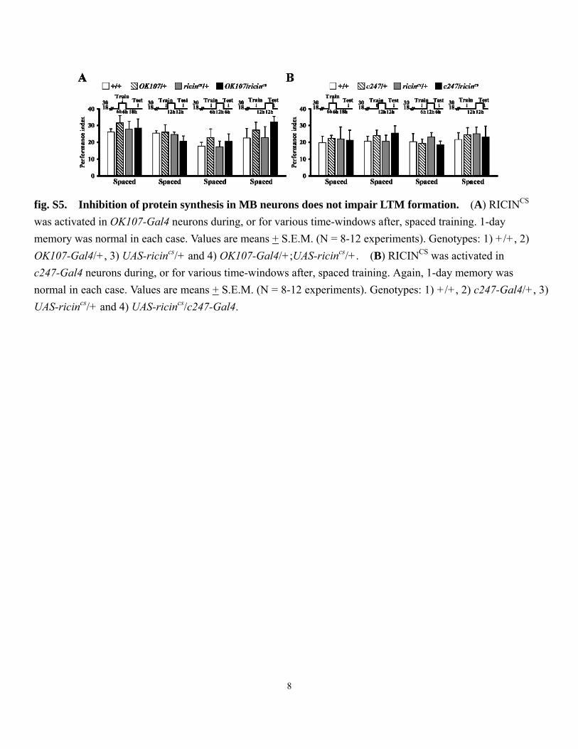
fig. S5. Inhibition of protein synthesis in MB neurons does not impair LTM formation. (A) RICINCS was activated in OK107-Gal4 neurons during, or for various time-windows after, spaced training. 1-day memory was normal in each case. Values are means + S.E.M. (N = 8-12 experiments). Genotypes: 1) +/+, 2) OK107-Gal4/+, 3) UAS-ricincs/+ and 4) OK107-Gal4/+;UAS-ricincs/+. (B) RICINCS was activated in c247-Gal4 neurons during, or for various time-windows after, spaced training. Again, 1-day memory was normal in each case. Values are means + S.E.M. (N = 8-12 experiments). Genotypes: 1) +/+, 2) c247-Gal4/+, 3) UAS-ricincs/+ and 4) UAS-ricincs/c247-Gal4.
9
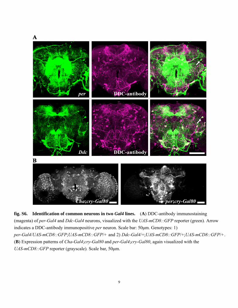
fig. S6. Identification of common neurons in two Gal4 lines. (A) DDC-antibody immunostaining (magenta) of per-Gal4 and Ddc-Gal4 neurons, visualized with the UAS-mCD8::GFP reporter (green). Arrow indicates a DDC-antibody immunopositive per neuron. Scale bar: 50μm. Genotypes: 1) per-Gal4/UAS-mCD8::GFP;UAS-mCD8::GFP/+ and 2) Ddc-Gal4/+;UAS-mCD8::GFP/+;UAS-mCD8::GFP/+. (B) Expression patterns of Cha-Gal4;cry-Gal80 and per-Gal4;cry-Gal80, again visualized with the UAS-mCD8::GFP reporter (grayscale). Scale bar, 50μm.

10
fig. S7. Blocking neurotransmission from DAL neurons does not affect memory retention immediately or 3 hours after single training session. The expression of UAS-shits is driven by three different DAL-specific Gal4 lines, E0946-Gal4, G0338-Gal4 and G0431-Gal4. Flies were kept at 30oC for the test trial (retrieval). All flies were raised at 18oC until training began. Values are means + S.E.M. (N=8 experiments). Genotypes: 1) +/+, 2) UAS-shits/+, 3) E0946-Gal4/+, 4) E0946-Gal4/UAS-shits, 5) G0338-Gal4/+, 6) G0338-Gal4/+;;UAS-shits/+, 7) G0431-Gal4/+ and 8) G0431-Gal4/+;UAS-shits/+.

11
fig. S8. 1-day memory after spaced training is impaired by activated RICINCS expressed in neurons other than DAL neurons. (A) 1-day memory after spaced training was impaired by activated, but not inactivated, RICINCS in cer-Gal4 -expressing neurons with DAL neurons “subtracted” by using cry-Gal80. Flies were kept at 30oC after training for the entire 24 hours to activate RICINCS. Genotype: 1) +/+, 2) cer-Gal4/+, 3) UAS-ricincs/+;cry-Gal80/+ and 4) cer-Gal4/UAS-ricinCS;cry-Gal80/+. (B) Similar subtractions of DAL neurons using cry-Gal80 revealed no impairments in 1-day memory after spaced training for Ddc-Gal4, cry-Gal4, Trh493-Gal4, Trh996-Gal4, CaMKII-Gal4(X) or CaMKII-Gal4(III). Values are means + S.E.M. (N = 8 experiments; **, P< 0.01; ***, P< 0.001).

12
fig. S9. Disruption of genes expressed in DAL neurons impairs 1-day memory after spaced training but not after massed training. (A) Immunostaining (magenta) of PER, dNR1, dNR2, CaMKII, TEQUILA, CRY and octopamine in a DAL neuron, visualized with the UAS-mCD8::GFP reporter (green). Scale bar, 10μm. (B-D) Constitutive disruption of PER in per-Gal4, Ddc-Gal4 or G0338-Gal4 neurons impairs 1-day memory after spaced, but not massed, training. Genotypes: 1) +/+, 2) per-Gal4/+, 3) UAS-perPAS-IR G2/+, 4) per-Gal4/UAS-perPAR-IR G2, 4) Ddc-Gal4/+, 5) Ddc-Gal4/+;UAS-perPAR-IR G2/+, 6) G0338-Gal4/+ and 7) G0338-Gal4/+;UAS-perPAR-IR G2/+. (E) 1-day memory after spaced training remains intact after constitutive disruption of PER in Feb170-Gal4 neurons. Genotypes: 1) +/+, 2) Feb170-Gal4/+, 3) UAS-perPAS-IR G2/+ and 4) Feb170-Gal4/UAS-perPAS-IR G2. (F) Constitutive, but not induced, disruption of CRY in DAL neurons impairs 1-day memory after spaced training but not after massed training. Genotypes: 1) +/+, 2) G0431-Gal4/+, 3) UAS-cryRNAi/+, 4) G0431-Gal4/UAS-cryRNAi, 5) G0431-Gal4/+;tub-Gal80ts/+ and

13
6) G0431-Gal4/UAS-cryRNAi;tub-Gal80ts/+. (G) Constitutive, but not induced, overexpression of CER in DAL neurons impairs 1-day memory after spaced but not after massed training. Genotypes: 1) +/+, 2) G0431-Gal4/+, 3) UAS-cer+/+, 4) G0431-Gal4/UAS-cer+, 5) G0431-Gal4/+;tub-Gal80ts/+ and 6) G0431-Gal4/UAS-cer+;tub-Gal80ts/+. (H) Induced disruption of tyramine β hydroxylase (Tβh) doesn’t impair 1-day memory after spaced training. Genotypes: 1) +/+, 2) G0431-Gal4/+, 3) UAS-TβhRNAi/+, 4) G0431-Gal4/+;tub-Gal80ts/+ and 5) G0431-Gal4/UAS-TβhRNAi;tub-Gal80ts/+. For induced expression, flies were raised at 18oC and then transferred to 30oC for 3 days before training to remove tub-Gal80ts inhibition and allow Gal4-driven expression. In all experiments, values are means + S.E.M. (N = 4-12 experiments; **, P< 0.01; ***, P< 0.001).

14
fig. S10. Induced overexpression of CREB2 repressor in MB neurons does not impair 1-day memory after spaced or massed training. (A-C) Constitutive expression of CREB2 repressor (dcreb2b) in MB

15
neurons impairs 1-day memory after spaced training. In two MB-Gal4 lines (OK107 and c247), immediate memory after single training session also is impaired. Genotypes: 1) +/+, 2) UAS-dcreb2-b/+, 3) OK107-Gal4/+, 4) UAS-dcreb2-b/+;;;OK107-Gal4/+, 5) c247-Gal4/+, 6) UAS-dcreb2-b/+;;c247-Gal4/+, 7) c739-Gal4/+ and 8) UAS-dcreb2-b/+;c739-Gal4/+. (D-F) Constitutive expression of CREB2 repressor produces morphological defects in MB neurons. Arrow indicates missing α lobes; dashed circle indicates fewer fibers in γ lobes; arrowhead indicates some αβ axons crossing the midline. Genotypes: 1) UAS-mCD8::GFP/+; UAS-mCD8::GFP/+;OK107-Gal4/+ and 2) UAS-dcreb2-b/+;UAS-mCD8::GFP/+; UAS-mCD8::GFP/+; OK107-Gal4/+ (D), 1) UAS-mCD8::GFP/+; c247-Gal4/UAS-mCD8::GFP and 2) UAS-dcreb2-b/+; UAS-mCD8::GFP/+;c247-Gal4/UAS-mCD8::GFP (E), 1) UAS-mCD8::GFP/c739-Gal4;UAS-mCD8::GFP/+ and 2) UAS-dcreb2-b/+;UAS-mCD8::GFP/c739-Gal4; UAS-mCD8::GFP/+ (F). (G-I) Induced expression of CREB2 repressor in MB neurons does not impair immediate memory after single training session or 1-day memory after spaced or massed training. Flies were raised and then at 18oC were transferred to 30oC for at least 3 days before training began, to remove tub-Gal80ts inhibition and allow Gal4-mediated transgene expression. Genotypes: 1) +/+, 2) UAS-dcreb2-b/+; tub-Gal80ts/+, 3) OK107-Gal4/+, 4) UAS-dcreb2-b/+;tub-Gal80ts/+;; OK107-Gal4/+, 5) c247-Gal4/+, 6) UAS-dcreb2-b/+;tub-Gal80ts/+;c247-Gal4/+, 7) c739-Gal4/+ and 8) UAS-dcreb2-b/+;tub-Gal80ts/c739. (J-L) Induced expression of CREB2 repressor does not produce MB defects. At 18oC, Gal80 inhibits Gal4 activity and UAS-mCD8::GFP and UAS-dcreb2-b are not expressed. MB morphology is revealed with DLG antibody immunostaining (magenta). After three days at 30oC, GFP and dcreb2-b are expressed and MB morphology remains intact. Genotypes: UAS-dcreb2-b/+;tub-Gal80ts/+;UAS-mCD8::GFP/+; OK107-Gal4/+ (J), UAS-dcreb2-b/+; tub-Gal80ts/+;c247-Gal4/UAS-mCD8::GFP (K), UAS-dcreb2-b/+; c739-Gal4/tub-Gal80ts;UAS-mCD8::GFP/+ (L). (M) When these same flies were raised continuously at 30oC, MB defects were readily apparent as in D-F above. Fiber degeneration was observed in α’β’ (arrow), γ (circle), and αβ (square) lobes. In some cases, fibers may across the midline (arrowhead). (N) In addition to MB neurons, c739-Gal4, but not OK107-Gal4 or c247-Gal4, expressed weakly also in DAL neurons. Scale bar, 50μm. Inset, DAL soma (arrow) weakly labeled by UAS-mCD8::GFP was verified by DDC antibody immunostaining (magenta) while fibers are invisible. Scale bar: 10μm. (O) Induced expression of UAS-mCD8::GFP (green) and UAS-dcreb2-b with the Cha-Gal4 driver after transferring flies to 30oC for three days. GFP expression is not apparent beforehand, when flies are raised at 18oC. Genotypes: UAS-dcreb2-b/+; Cha-Gal4/tub-Gal80ts;UAS-mCD8::GFP/+. (P) Induced expression of UAS-dcreb2-b in Cha neurons impaired 1-day memory after spaced training, but not memory retention immediately after single training session (LRN). Flies raised at 18oC were transferred to 30oC to remove tub-Gal80ts inhibition of Gal4 expression for 3 days. Control flies kept at 18oC for 4 days carrying the same transgenes exhibited normal learning and LTM. Genotypes: 1) +/+, 2) Cha-Gal4/+, 3) UAS-dcreb2-b/+;tub-Gal80ts/+ and 4) UAS-dcreb2-b/+;Cha-Gal4/tub-Gal80ts. In all experiments, values are means + S.E.M. (N = 8-12; ** = P < 0.01; *** = P < 0.001). Except in (N), all brains are counterstained with DLG antibody immunostaining (magenta). Scale bar, 50μm.

16
fig. S11. The spaced training-induced elevation of KAEDE synthesis occurred exclusively in the DAL neurons in per- and CaMKII-Gal4. Living flies were subjected to UV irradiation to convert pre-existing KAEDE into red fluorescence protein just before training. Levels of KAEDE synthesis were quantified 24 hours after spaced or massed training. Whole brain was imaged at two detection sensitivities: high detector gain maximized for the DAL neurons and low detector gain maximized for the MB or the lateral circadian neurons. Except in the DAL neurons, we did not see the spaced training-induced elevation of KAEDE synthesis in any other neurons comparing the 488 channel between massed and spaced training. Arrow: DAL neuron. Scale bar: 50 μm. Genotypes: 1) CaMKII-Gal4(III)/UAS-kaede and 2) per-Gal4/+;UAS-kaede/+.

17
fig. S12. Temporal changes in the elevated CaMKII and per transcriptional activities in the DAL neurons after spaced training. Living flies were subjected to UV irradiation to convert pre-existing KAEDE into red fluorescence protein just before training, 8 hours after training, or 16 hours after training. Levels of KAEDE were quantified every 8 hours after training. A single optical slice through the cell body of a DAL neuron was taken under the same detection condition for all brains. KAEDE synthesis was determined as the ratio of new (green 488nm) to old (red 543nm) KAEDE in the same neuron (%�F/⎯F0). Scale bar: 10 μm. Values are means+ S.E.M. (N=6-16 samples). Genotypes: 1) CaMKII-Gal4(III)/UAS-kaede and 2) per-Gal4/+; UAS-kaede/+.

18
fig. S13. Disruption of an unknown gene expressed in the DAL neurons does not affect 1-day memory after spaced training. (A) 1-day memory after spaced training is normal in heterozygous and in homozygous mutants. Values are means+ S.E.M. (N=8 experiments). Genotypes: 1) +/+, 2) G0431/+ and 3) G0431/G0431. (B) KAEDE synthesis in G0431-Gal4 flies is not induced by spaced or massed training. Values are means+ S.E.M. (N=10-14 samples). Genotype: G0431/UAS-kaede. Scale bar: 10 μm. Living flies were subjected to UV irradiation to convert pre-existing KAEDE into red fluorescence protein just before training. UAS-kaede synthesis was quantified 24 after training. For each brain, a single optical slice through the cell body of a DAL neuron was taken under the same detection conditions. KAEDE synthesis was determined as the ratio of new
(green 488nm) to old (red 543nm) KAEDE within each fly brain (%△F/⎯F0).

19
Table S1. Gal4 expression patterns. The GFP intensity reported by Gal4>UAS-mCD8::GFP;UAS-mCD8::GFP was graded as strong (+++), intermediate (++), weak (+), absence (-) or non-distinguishable (N.D.). APL, anterior paired lateral neurons; DAL, dorsal anterior lateral neuron; DPM, dorsal paired medial neuron; EB, ellipsoid body; MB, mushroom body; OSN, olfactory sensory neuron; PN, projection neuron.

20
Movie S1. Visualizing de novo KAEDE synthesis in per neurons during ZT0-6 and ZT12-18, respectively. Circadian transcriptional activity of per in the lateral neurons reported by de novo synthesis of KAEDE driven by per-Gal4. To reset pre-existing green KAEDE, the brain incubated in DDM2 culture medium was UV irradiated for 20s at ZT0 or ZT12. Changes in KAEDE levels were monitored every 20 minutes with a single optical section through cell bodies of lateral neurons during ZT0-6 (left movie) or ZT12-18 (right movie). Genotype: per-Gal4/+; UAS-kaede/+. driven

21
Materials and Methods
Fly strains. Fly stocks were maintained on standard corn meal/yeast/agar medium at 25 ± 1°C or 18 ± 1°C and 70% relative humidity on a 12h:12h light:dark cycle. The fly lines used were wild-type Canton-S w1118 (iso1CJ), UAS-kaede, UAS-ricinCS (from C. J. O’Kan), UAS-shits, UAS-perPAS-IR G2 (from M. W. Young), UAS-CaMKIIhpn (from S. Kunes), UAS-dcreb2-b (from L. Davis ); Ddc-Gal4, Trh493-Gal4 and Trh996-Gal4 (from J. Hirsh); per-Gal4 and tim-Gal4 (from J. Hall), cry-Gal4 (from J. Blau); cry-Gal80 (from M. Rosbash); MB-Gal80 (from S. Waddell); CaMKII-Gal4(X) and CaMKII-Gal4(III) (from Y. Takamatsu); cer-Gal4, repo-Gal4, UAS-teq41 and UAS-cer+ (from T. Préat); lexAop-rCD2::GFP and UAS-Dscam::GFP (from T. Lee); UAS-CD4::GFP1-10 and lexAop-CD4::GFP11 (from K. Scott); c42, c217, c247, c305a, c316, c507, c739 and c772 (from S. Benzer); UAS-cryRNAi (v105172), UAS-creb2RNAi (v101512), UAS-DdcRNAi (v3330), UAS-TrhRNAi
(v35240) and UAS-TβhRNAi (v51667) (from VDRC). All other flies including E0946 (112097), E0973 (112178), G0338 (12740), G0431 (12837) and P0010 (2023) were derived from DGRC or Bloomington stock centers.
Behavior. Olfactory associative learning was evaluated by training 2- to 3-day-old flies in a T-maze apparatus with a Pavlovian olfactory conditioning procedure (44). Odors used were 3-octanol (OCT) and 4-methylcyclohexanol (MCH). Each experiment consisted of two groups of approximately 100 flies, each of which was conditioned with one of these two odors. Flies were exposed sequentially to two odors which were carried through the training chamber in a current of air (odors were bubbled at 750 ml/min). Flies first were
exposed for 60 s to the conditioned stimulus (CS+), during which time they received the unconditioned
stimulus (US), which consisted of twelve 1.5 s pulses of 60V DC electric shock at 5 s interpulse intervals.
After the presentation of CS+, the chamber was flushed with fresh air for 45 s. Then, flies were exposed for
60 s to the CS-, which was not paired with the US. This procedure constitutes single training session. For
24-hour memory experiments, flies were subjected to ten such training sessions, either massed together without rest or spaced out with a 15 min rest interval. For these multiple training protocols, robotic trainers were used. The test was carried out in the dark in an environment-controlled room at required temperatures and 70% relative humidity. Except in the initial behavioral screen (Fig. 2 and fig. S8B), genetic backgrounds of all fly strains were equilibrated to the “Canton” wild-type background by five or more generations of backcrossing. All genotypes were trained and tested in parallel and rotated between all of the robotic trainers to ensure a balanced experiment. In RICINCS experiments, flies raised at 18oC were transferred to 30oC for 24 hours to allow enough RICINCS expression and then kept under 18oC for another 24 hrs to inactivate RICINCS before experiments. In tub-Gal80ts experiments, flies raised at 18oC were transferred to 30oC for at least 3 days before experiments. For blocking protein synthesis, flies were fed with 35mM cycloheximide 1 day before

22
training until just before the test (1).
To evaluate memory retention immediately after single training session (acquisition), flies were gently tapped into an elevator-like compartment immediately after training. After 90 s, the flies were transported to the choice point of a T maze, in which they were exposed to two converging currents of air (one carrying OCT,
the other MCH) from opposite arms of T maze. Flies were allowed to choose between the CS+and CS-for
120 s, at which time they were trapped inside their respective arms of the T maze (by sliding the elevator out of
register), anesthetized and counted. Flies that chose to avoid the CS+ran into the T maze arm containing the
CS-,while flies that chose to avoid the CS-ran into the T maze arm containing the CS+. To assay 3 hr
memory retention after single training session, trained flies were placed in a food tube in the dark until tested in the T maze apparatus. For each experiment, performance index PI1,2 = (Nnon punished – Npunished)/( Nnon punished + Npunished) was calculated and averaged over these two complimentary experiments, with the final PI = (PI1+PI2)/2. Averaging of the two reciprocal scores eliminates possible bias originating from machine, naïve odor preferences or nonassociative changes in olfaction.
KAEDE measurement. To measure the amount of newly synthesized KAEDE in single neurons, we used the following procedures. (i) Pre-existing KAEDE proteins were photoconverted into red fluorescence proteins by 365-395 nm UV irradiation generated from a 120W mercury lamp. Two methods were used (fig. S1). To monitor circadian synthesis in per neurons, a single fly restricted in a pipette tip was UV irradiated for 5 min through an objective lens (N.A. value 0.5, working distance 2mm). For the behavior assay, approximately 100 flies kept in a clear plastic syringe were directly exposed to UV light at a distance of 5cm for 1 hour. (ii) Individual single neurons expressing KAEDE were directly visualized through an open window of the fly’s head capsule. Living samples were used because the signal to noise ratio of green versus red KAEDE is greatly reduced after chemical fixation. (iii) KAEDE neurons were located in less than 5 seconds by a fast pre-scanning of red KAEDE excited by a 543nm laser to avoid unnecessary fluorescence quenching of green KAEDE during repeated scanning. (iv) A single optical slice through cell bodies at a resolution of 1024x1024 pixels was imaged under a confocal microscope with a 40X C-Apochromat water-immersion objective lens (N.A. value 1.2, working distance 220μm). All brain samples in the experiment were imaged with the same optical setting maximized for green and red KAEDE immediately before and after photoconversion, respectively. (v) In all cases, both green KAEDE excited by a 488 nm laser and red KAEDE excited by 543 nm laser were measured. We found that red KAEDE is very stable with less than 2% decay rate in 24 hours. Using the amount of red KAEDE as an internal standard to calibrate individual variation, we calculated the increasing rate of green KAEDE synthesis after photoconversion with the formula (ΔF)=%(Ft1-⎯Ft0)/⎯Ft0, where

23
F t1 and Ft0 are the ratio of averaged intensities between green (G) and red (R) KAEDE (Gt0/Rt0) immediately after photoconversion (t0) and at a specific later time point (t1), respectively.
Immunohistochemistry. Brains were dissected in phosphate-buffered saline (PBS), heated with a commercial microwave oven in 4% paraformaldehyde on ice for 60 seconds three times, and then in 4% paraformaldehyde with 0.25% Triton X-100 for 60 seconds three times. After being washed in PBS for 10 min at room temperature, brain samples were incubated in PBS containing 2% Triton X-100 and 10% normal goat serum and degassed in a vacuum chamber to expel tracheal air for four cycles (depressurize to -70 mmHg then hold for 10 minutes). Next, brain samples were blocked and penetrated in PBS-T at 4°C overnight and then incubated in PBS-T containing one of the following primary antibodies: (1) 1:500 rat anti-DDC polyclonal antibody (from J. Hirsh), (2) 1:40 mouse 4F3 anti-DLG antibody (Developmental Studies Hybridoma Bank, Univ. of Iowa), (3) 1:5000 rabbit anti-CaMKII polyclonal antibody (from L. C. Griffith), (4) 1:300 guinea pig anti-CRY (from P. E. Hardin), (4) 1:1000 rabbit anti-dNR1 polyclonal antibody (α-85S), (5) 1:500 rabbit anti-dNR2 polyclonal antibody (α-820-1, α-820-2), (6) 1:500 rabbit anti-TEQUILA polyclonal antibody (from T. Préat), (7) 1:10000 rabbit anti-PER polyclonal antibody (from Jeffrey L. Price) and (8) 1:500 rabbit anti-octopamine (from Millipore. AB1799) at 4°C for 1 day. After being washed in PBS-T three times, samples were incubated in PBS-T containing one of the following secondary antibodies: (1) 1:200 biotinylated goat anti-mouse IgG (Molecular Probes), (2) 1:200 biotinylated goat anti-rat IgG, (3) 1:200 biotinylated goat anti-rabbit IgG, and (4) 1:200 biotinylated goat anti-guinea pig IgG at 25°C for 1 day. Next, brain samples were washed and incubated with 1:500 Alexa Fluor 635 streptavidin (Molecular Probes) at 25°C for 1 day. Finally, after extensive washing, immunolabeled brain samples were directly cleared in FocusClear, an aqueous solution that renders biological tissue transparent (45), for 5 min and mounted between two cover slips
separated by a spacer ring of ~200μm in thickness. Sample brains were imaged under a Zeiss LSM 710
confocal microscope with a 40X C-Apochromat water-immersion objective lens (N.A. value 1.2, working distance 220μm).
Statistics. All the raw data were analyzed parametrically with JMP5.1 statistical software. As a result of the nature of their mathematical derivation, performance indexes were distributed normally. Hence, the data were evaluated via one-way ANOVAs. Subsequent pair-wise planned comparisons were adjusted for experiment-wise error (α′), keeping the overall α = 0.05. All data were presented as means+ S.E.M.

24
References
1. T. Tully, T. Préat, S. C. Boynton, M. Del Vecchio, Cell 79, 35 (1994).
2. J. C. Yin et al., Cell 79, 49 (1994).
3. R. L. Davis, Neuron 70, 8 (2011).
4. D. Yu, D. B. Akalal, R. L. Davis, Neuron 52, 845 (2006).
5. M. Heisenberg, Nat. Rev. Neurosci. 4, 266 (2003).
6. B. Gerber, H. Tanimoto, M. Heisenberg, Curr. Opin. Neurobiol. 14, 737 (2004).
7. D. B. Akalal, D. Yu, R. L. Davis, J. Neurosci. 30, 16699 (2010).
8. D. B. Akalal, D. Yu, R. L. Davis, J. Neurosci. 31, 5643 (2011).
9. R. Ando, H. Hama, M. Yamamoto-Hino, H. Mizuno, A. Miyawaki, Proc. Natl. Acad. Sci. U.S.A. 99, 12651 (2002).
10. H. Mizuno et al., Mol. Cell 12, 1051 (2003).
11. P. E. Hardin, J. C. Hall, M. Rosbash, Nature 343, 536 (1990).
12. Y. Endo, K. Mitsui, M. Motizuki, K. Tsurugi, J. Biol. Chem. 262, 5908 (1987).
13. Y. Endo, K. Tsurugi, J. Biol. Chem. 262, 8128 (1987).
14. K. G. Moffat, J. H. Gould, H. K. Smith, C. J. O’Kane, Development 114, 681 (1992).
15. A. S. Chiang et al., Curr. Biol. 21, 1 (2011).
16. T. Riemensperger, T. Völler, P. Stock, E. Buchner, A. Fiala, Curr. Biol. 15, 1953 (2005).
17. A. Claridge-Chang et al., Cell 139, 405 (2009).
18. Y. Aso et al., Curr. Biol. 20, 1445 (2010).
19. S. I. Ashraf, A. L. McLoon, S. M. Sclarsic, S. Kunes, Cell 124, 191 (2006).
20. A. Pascual, T. Préat, Science 294, 1115 (2001).
21. C. L. Wu et al., Nat. Neurosci. 10, 1578 (2007).
22. M. P. Belvin, H. Zhou, J. C. Yin, Neuron 22, 777 (1999).
23. T. Sakai, T. Tamura, T. Kitamoto, Y. Kidokoro, Proc. Natl. Acad. Sci. U.S.A. 101, 16058 (2004).
24. J. Dubnau, L. Grady, T. Kitamoto, T. Tully, Nature 411, 476 (2001).
25. S. E. McGuire, P. T. Le, R. L. Davis, Science 293, 1330 (2001).

25
26. S. Xia et al., Curr. Biol. 15, 603 (2005).
27. H. H. Lin, J. S. Lai, A. L. Chin, Y. C. Chen, A. S. Chiang, Cell 128, 1205 (2007).
28. M. D. Gordon, K. Scott, Neuron 61, 373 (2009).
29. S. Martinek, M. W. Young, Genetics 156, 1717 (2000).
30. S. E. McGuire, P. T. Le, A. J. Osborn, K. Matsumoto, R. L. Davis, Science 302, 1765 (2003).
31. G. Didelot et al., Science 313, 851 (2006).
32. F. V. Bolduc, K. Bell, H. Cox, K. S. Broadie, T. Tully, Nat. Neurosci. 11, 1143 (2008).
33. G. Isabel, A. Pascual, T. Préat, Science 304, 1024 (2004).
34. J. Séjourné et al., Nat. Neurosci. 14, 903 (2011).
35. D. Comas, F. Petit, T. Préat, Nature 430, 460 (2004).
36. A. Pascual, K. L. Huang, J. Neveu, T. Préat,. Nature 427, 605 (2004).
37. J. Dubnau et al., Curr. Biol. 13, 286 (2003).
38. R. Bourtchuladze et al.,. Cell 79, 59 (1994).
39. A. C. Keene, S. Waddell, Nat. Rev. Neurosci. 8, 341 (2007).
40. X. Liu, R. L. Davis, Nat. Neurosci. 12, 53 (2009).
41. C. L. Wu et al., Curr. Biol. 21, 848 (2011).
42. J. L. Pitman et al., Curr. Biol. 21, 855 (2011).
43. K. G. Moffat, J. H. Gould, H. K. Smith, C. J. O’Kane, Development 114, 681 (1992).
44. T. Tully, W. G. Quinn, J. Comp. Physiol. A Neuroethol. Sens. Neural Behav. Physiol. 157, 263 (1985).
45. A. S. Chiang et al., J. Comp. Neurol. 440, 1 (2001).