supporting online material for - chemistry at winthrop...
TRANSCRIPT

www.sciencemag.org/cgi/content/full/333/6046/1151/DC1
Supporting Online Material for
Expanding the Genetic Code of Escherichia coli with Phosphoserine
Hee-Sung Park, Michael J. Hohn, Takuya Umehara, Li-Tao Guo, Edith M. Osborne, Jack Benner, Christopher J. Noren,* Jesse Rinehart,* Dieter Söll*
*To whom correspondence should be addressed. E-mail: [email protected] (D.S.);
[email protected] (C.J.N.); [email protected] (J.R.)
Published 26 August 2011, Science 333, 1151 (2011) DOI: 10.1126/science.1207203
This PDF file includes:
Materials and Methods Figs. S1 and S2 Table S1 References (27–37)

2
Materials and Methods
Construction of strains. To prevent possible enzymatic dephosphorylation of O-phospho-L-serine
(Sep) in vivo, the gene encoding phosphoserine phosphatase (serB), which catalyzes the last step
in serine biosynthesis, was deleted from Escherichia coli strains Top10 and BL21. Markerless gene
deletions were carried out using a λ-red and FLP recombinase-based gene knockout strategy as
described earlier (27). E. coli strains Top10∆serB and BL21ΔserB were used for EF-Tu library
construction and MEK1 expression experiments.
Construction of plasmids. To construct plasmid pSepT, the full-length gene encoding tRNASep
was constructed from overlapping oligonucleotides and ligated immediately downstream of the lpp
promoter in pTECH (28) using EcoRI and BamHI restriction sites. pCysT, encoding the wild type
tRNACys gene from Methanocaldococcus jannaschii was constructed in the same way.
pKD was derived from pKK223-3 (Pharmacia). The ampicillin (Amp) resistance gene was
replaced with a kanamycin (Kan) resistance gene by combining two PCR products generated from
pKK223-3 and pET28a. The following PCR primers were used: PKF (5’-TGC AGCA ATG CGG
CCG CTT TCA CCG TCA TCA CCG AAA C-3’) and PKR (5’-GGG ACG CTA GCA AAC AAA AAG
AGT TTG TAG AA-3’) for pKK223-3 amplification and PKNF (5’-GGG ACG CTA GCT TTT CTC
TGG TCC CGC CGC AT-3’) and PKNR (5’-TGC GCA ATG CGG CCG CGG TGG CAC TTT TCG
GGG AAA T-3’) for KanR gene amplification.
The original multiple cloning site (MCS; NcoI-EcoRI-SacI) was modified by adding an additional
ribosome binding site (RBS) and a second MCS (NdeI-BamHI-SalI-HindIII), thus enabling
simultaneous protein expression from two genes, both under the control of the same tac promoter.
The Methanococcus maripaludis SepRS gene was cloned into pKD using NcoI and SacI sites to
produce pKD-SepRS. The E. coli EF-Tu gene (tufB) was ligated into pKD-SepRS using BamHI and
SalI sites resulting in pKD-SepRS-EFTu. The M. maripaludis pscS gene encoding SepCysS was
cloned into pKD-SepRS using BamHI and SalI sites to produce pKD-SepRS-SepCysS and the M.
maripaludis CysRS gene was cloned into pKD using EcoRI and SalI to yield pKD-CysRS.
pCAT112TAG-SepT was created from pACYC184. The gene encoding chloramphenicol
acetyltransferae (CAT) was modified by quickchange mutagenesis to introduce an amber stop
codon at position Asp112 (29). The resulting plasmid was PCR amplified using primers PBLAF (5’-
TGC GCA ATG CGG CCG CCC GTA GCG CCG ATG GTA GTG T-3’) and PBLAR (5’-ACA CGG AGA
TCT CTA AAG TAT ATA TGA GTA AAC-3’) and ligated with a PCR product containing a tRNASep
expression cassette from pSepT, created with primers TSEPF (5’-GCA TGC GCC GCC AGC TGT
TGC CCG TCT CGC-3’) and TSEPR (5’-GCA TAG ATC TTC AGC TGG CGA AAG GGG GAT G-
3’).
Plasmid pCcdB was created by adding a ccdB gene under the control of a lac promoter into
pTECH (28) using NotI and BglII sites (30). Two amber stop codons were introduced at positions
13 and 44 based on the crystal structure and mutagenesis study of the CcdB protein (31, 32).
pMYO127TAG-SepT was constructed by cloning a codon-optimized and C-terminally His6-
tagged sperm whale myoglobin gene under the control of the lpp promoter between NotI and BglII
in pSepT. An amber stop codon was introduced to the myoglobin gene at position Asp127 by
quickchange mutagenesis. The nucleotide sequence of the codon-optimized myoglobin gene is as
follows:
3
ATGGTTCTGTCTGAAGGTGAATGGCAGCTGGTTCTGCACGTTTGGGCTAAAGTTGAAGCTGAC
GTTGCTGGTCACGGTCAGGACATCCTGATCCGTCTGTTCAAATCTCACCCGGAAACCCTGGA
AAAATTCGACCGTTTCAAACACCTGAAAACCGAAGCTGAAATGAAGGCTTCTGAAGACCTGAA
AAAACACGGTGTTACCGTTCTGACCGCTCTGGGTGCTATCCTGAAGAAAAAGGGTCACCACG
AAGCTGAACTGAAACCGCTGGCTCAGTCTCACGCTACCAAACACAAAATCCCGATCAAATACC
TGGAGTTCATCTCTGAAGCTATCATCCACGTTCTGCACTCTCGTCATCCGGGTAACTTCGGTG
CTGACGCTCAGGGTGCTATGAACAAAGCTCTGGAACTGTTCCGTAAAGACATCGCTGCTAAAT
ACAAAGAACTGGGTTACCAGGGTGGTTCTGGTCATCACCATCACCATCACTAA
Plasmid pL11C-SepT encodes tRNASep and the C-terminal domain of the ribosomal protein L11
under control of lpp promoters. Part of the rplK gene was PCR amplified from genomic E. coli DNA
using primers L11C-F (5’-GGA ATT CCA TAT GAC CAA GAC CCC GCC GGC AGC AGT T-3’)
and L11C-R (5’-AGG CGC GCC TTA GTC CTC CAC TAC-3’). The PCR product was digested with
NdeI and AscI and was ligated into NdeI and AscI digested pMYO127TAG-SepT to replace the
myoglobin gene.
To construct pMAL-EFTu and pMAL-EFSep E. coli tufB, or the gene encoding EF-Sep,
respectively, were cloned between the NdeI and BamHI sites in the pET20b plasmid (Novaven) to
add a C-terminal His6 tag. This fusion construct was then PCR-amplified using primers adding MfeI
and PstI restriction sites. The PCR product was cloned in-frame between EcoRI and PstI in pMAL-
c2x (New England Biolabs) to add an N-terminal maltose binding protein (MBP) tag.
pET15-ERK2 encodes N-terminally His6-tagged mitogen-activated protein kinase (Erk2) under
the control of a T7 promoter. The human Erk2 gene was PCR amplified from plasmid BC017832
(ATCC) using primers ERK2-F (5’-GGA ATT CCA TAT GGC GGC GGC GGC GGC G-3’) and
ERK2-R (5’-CCG CTC GAG TTA AGA TCT GTA TCC TGG-3’). The PCR product was cloned
between NdeI and XhoI in vector pET15b (Novagen).
pET20-MBPMEK1 encodes a fusion protein consisting of human MEK1 with an N-terminal
maltose binding protein (MBP) tag and a C-terminal His6-tag. The gene encoding human MEK1
which was codon-optimized for E. coli and custom-synthesized in vitro (Genscript), was cloned
between EcoRI and PstI into pMAL-c2x (New England Biolabs). The resulting MBP-MEK1 fusion
construct was then amplified with primers ET20MEKF (5’-AAG GAA ATT AAT GAA AAT CGA AGA
AGG TAA-3’) and ET20MEKR (5’-CTA GAG GAT CCG GCG CGC-3’) adding AseI and BamHI
restriction sites, and the PCR product was ligated between NdeI and BamHI into pET20b.
Nucleotide sequence of codon-optimized MEK1:
ATGCCGAAGAAGAAACCGACCCCGATCCAGCTGAACCCGGCTCCGGACGGTTCTGCTGTTAA
CGGCACCTCTTCTGCTGAAACCAACCTGGAAGCTCTGCAAAAGAAACTGGAAGAACTGGAAC
TGGACGAACAGCAGCGTAAACGTCTGGAAGCGTTCCTGACCCAGAAACAGAAAGTTGGTGAA
CTGAAAGACGACGACTTCGAAAAAATCTCTGAACTGGGTGCTGGTAACGGTGGTGTTGTTTTC
AAAGTTTCTCACAAACCGTCCGGTCTGGTTATGGCTCGTAAACTGATCCACCTGGAAATCAAA
CCGGCTATCCGTAACCAGATCATCCGTGAACTGCAAGTTCTGCACGAATGCAACTCTCCGTAC
ATCGTTGGTTTCTACGGTGCTTTCTACTCTGACGGTGAAATCTCTATCTGCATGGAACACATG
GACGGTGGTTCTCTGGACCAGGTTCTGAAAAAAGCTGGTCGTATCCCGGAACAGATCCTGGG
TAAAGTTTCTATCGCTGTTATCAAAGGTCTGACCTACCTGCGTGAAAAACACAAAATCATGCAC
CGTGACGTTAAACCGTCTAACATCCTGGTTAACTCTCGTGGTGAAATCAAACTGTGCGACTTC
GGTGTTTCTGGTCAGCTGATCGACTCTATGGCTAACTCTTTCGTTGGCACCCGTTCTTACATG
4
TCTCCGGAACGTCTGCAAGGCACCCACTACTCTGTTCAGTCTGACATCTGGTCTATGGGTCTG
TCTCTGGTTGAAATGGCTGTTGGTCGTTACCCGATCCCGCCGCCGGACGCTAAAGAACTGGA
ACTGATGTTCGGTTGCCAGGTTGAAGGTGACGCTGCTGAAACCCCGCCGCGTCCGCGTACTC
CGGGTCGTCCGCTGTCTTCTTACGGTATGGACTCTCGTCCGCCGATGGCTATCTTCGAACTG
CTGGACTACATCGTTAACGAACCGCCGCCGAAACTGCCGTCTGGTGTTTTCTCTCTGGAGTTC
CAGGACTTCGTTAACAAATGCCTGATCAAAAACCCGGCTGAACGTGCTGACCTGAAACAGCTG
ATGGTTCACGCTTTCATCAAACGTTCTGACGCTGAAGAAGTTGACTTCGCTGGTTGGCTGTGC
TCTACCATCGGTCTGAACCAGCCGTCTACCCCGACCCACGCTGCTGGTGTGGCAGCCGCAG
CTGCGCATCATCACCACCATCACTAA
pCG-MBPMEK1SS was generated by the ligation of three PCR products. One PCR product
was derived from pGFIB (33) using primers GFIB-F (5’-ATA AGA ATG CGG CCG CGC CGC AGC
CGA ACG ACC GAG-3’) and GFIB-R (5’-CTA GCT AGC GTC TGA CGC TCA GTG GAA CG-3’).
The second PCR product was generated from pCDFDuet-1 (Novagen) using primers CDF-F (5’-
CTA GCT AGC TCA CTC GGT CGC TAC GCT-3’) and CDF-R (5’-ATA AGA ATG CGG CCG CTG
AAA TCT AGA GCG GTT CAG-3’). Both PCR products were digested with NheI and NotI and
ligated to form plasmid pCG. The third PCR product, encoding an expression cassette for MBP-
MEK1-His6 under the control of T7 promoter and T7 terminator, was generated from pET20-
MBPMEK1 using primers ETCDGFF (5’- AAA AGG CGC CGC CAG CCT AGC CGG GTC CTC
AAC G-3’) and ETCDGFR (5’- AAC TGC AGC CAA TCC GGA TAT AGT TC-3’). This PCR product
was cloned between the NarI and PstI sites of pCG.
The codon for Ser 222 in MEK1 was then replaced by a GAA codon (encoding Glu) using
Quickchange mutagenesis (Stratagene). In the same way, codon Ser 218 was either changed to
GAA to generate pCG-MBPMEK1EE, or to an amber stop codon, resulting in pCG-MBPMEK1XE.
In pCG-MBPMEK1XS only the codon for Ser218 was changed to UAG and in pCG-MBPMEK1XX
both codons for Ser218 and Ser222 were changed to amber.
Library construction and selection of Sep-tRNA specific EF-Tu. Six residues, His67, Asp216,
Glu217, Phe219, Thr229, and Asn274, located in the amino acid binding pocket of the E. coli
elongation factor EF-Tu were selected for randomization based on the crystal structure of the E.
coli EF-Tu:Phe-tRNAPhe complex (protein data base accession number 1OB2). Multiple rounds of
overlap PCR were carried out to incorporate random codons (NNK) at these positions (5) by using
the following primers: 67XF, 5’-GT ATC ACC ATC AAC ACT TCT NNK GTT GAA TAC GAC ACC
CCG-3’; H67R, 5’-AGA AGT GTT GAT GGT GAT AC-3’; 216XF, 5’-CCG TTC CTG CTG CCG
ATC NNK NNK GTA NNK TCC ATC TCC GGT CGT GGT-3’; 216R, 5’-GAT CGG CAG CAG GAA
CGG-3’; 229XF, 5’-GGT CGT GGT ACC GTT GTT NNK GGT CGT GTA GAA CGC GG-3’; 229R,
5’-AAC AAC GGT ACC ACG ACC-3’; 274XF, 5’-GAA GGC CGT GCT GGT GAG NNK GTA GGT
GTT CTG CTG CG-3’; 274R, 5’-CTC ACC AGC ACG GCC TTC-3’. The final PCR products were
purified and digested with BamHI and SalI, and ligated into pKD-SepRS to generate the EF-Tu
library. The ligated vectors were transformed into E. coli Top10∆serB containing pCAT112-SepT to
generate a library of 3×10⁸ mutants. The unbiased mutation of the library was confirmed by
selecting twenty random clones and sequencing each mutant tufB insert.
The mutant EF-Tu library was subjected to a first round of selection, in which clones sup-
pressing the amber stop codon in the CAT gene can survive on LB plates supplemented with 10
5
µg/ml tetracycline (Tc), 25 µg/ml Kan, 50 µg/ml chloramphenicol (Cm), 2 mM Sep, and 0.05 mM
isopropyl-β-D-thiogalactopyranoside (IPTG). After 48h incubation at 30°C, a pool of ca. 104
colonies was collected from the plates for plasmid preparation. The pKD-SepRS-EFTu plasmids
were separated from the reporter plasmid by agarose gel electrophosis and isolated using the
Qiagen gel purification kit.
To assure that UAG codon suppression in the CAT gene was dependent on the presence of
EF-Sep and Sep-tRNASep and did not arise from a hypothetical insertion of a canonical amino acid
at this site, EF-Tu mutants were tested in a negative selection experiment in the absence of
tRNASep. The pKD-SepRS-EFTu plasmids from the first round of positive selection were
transformed into E. coli Top10ΔserB cells harboring pCcdB. Suppression of both UAG codons in
the pCcdB-encoded ccdB gene will result in expression of the toxic ccdB gene product and will
therefore eliminate EF-Tu mutants potentially mediating UAG read through with an aminoacylated
non-suppressor tRNA. Transformants were plated onto LB agar supplemented with 25 µg/ml Kan,
25 µg/ml Cm, and 0.1 mM IPTG. After 48h incubation at 30°C, twenty individual clones were
picked and subjected to plasmid purification to isolate pKD-SepRS-EFTu as described above.
Recovered pKD-SepRS-EFTu plasmids were transformed into E. coli Top10∆serB containing
pCAT112-SepT for a third round of selection which was carried out under the same conditions as
the first. This time, individual colonies were isolated from agar plates and clones were tested for
their ability to grow on Cm over a concentration range from 5 to 100 µg/ml. Total plasmid was
purified from isolates showing strong Cm resistance, and pKD-SepRS-EFTu plasmids were
subjected to sequencing.
To confirm that the observed Cm resistance is dependent on the presence of both mutant EF-
Tu and SepRS, EF-Tu mutant genes were excised from their plasmids, recloned into pKD, and
retransformed into E. coli Top10ΔserB containing pCAT112-SepT. Cells were then tested for Cm
resistance as described above.
Protein expression and purification
Expression and purification of M. maripaludis SepRS and CysRS. SepRS and CysRS
were produced in E. coli and purified as described elsewhere (5).
Expression and purification of EF-Tu and EF-Sep. pMAL-EFTu or pMAL-EFSep were
transformed into E. coli BL21 (DE3) codon plus (Stratagene). A pre-culture was used to inoculate
1000 ml of LB broth with 100 µg/ml of Amp, 34 µg/ml Cm, 5052 solution, and phosphate buffer for
autoinduction as described previously (34). The cells were grown for 6 h at 37°C and continued at
20°C for 18 h.
The cells were pelleted and lysed by shaking for 20 min. in BugBuster (Novagen) reagent
supplemented with 50 mM Tris–HCl (pH 7.6), 60 mM NH4Cl, 7 mM MgCl2, 14.3 mM 2-mercapto-
ethanol, 50 µM GDP, 10% glycerol, 25 U ml-1 Benzonase, 1 mg ml-1 lysozyme, and Protease
inhibitor cocktail (Roche).
The extract was clarified by ultracentrifugation and applied to a Ni2+-NTA resin (Qiagen) and
purified according to the manufacturer's instructions.
The eluted enzymes were dialyzed into 20 mM HEPES–KOH (pH 7.0), 40 mM KCl, 1 mM
MgCl2, 5 mM DTT, 50 µM GDP, and 30% glycerol. SDS–PAGE electrophoresis followed by
staining with Coomassie blue revealed greater than 95% purity.
6
Expression and purification of Myoglobin. To express mutant myoglobin, pKD-SepRS-EF-
Sep and pKD-SepRS were transformed into E. coli Top10ΔserB containing pMYO127TAG-SepT.
E. coli Top10ΔserB with pMYO, encoding the wild type myoglobin gene was used as a control.
Cultures were grown in LB medium supplemented with 2 mM Sep. When A600 reached 0.6 protein
expression was induced with 0.05mM IPTG for 12h at 25°C. The cells were harvested,
resuspended in lysis buffer (50mM Tris-HCl (pH 7.8), 300mM NaCl, 14.3 mM 2-mercaptoethanol)
supplemented with protease inhibitor cocktail (Roche), and subjected to sonication. The lysate was
centrifuged at 10,000×g for 30 min and the supernatant was applied to Ni2+-NTA agarose (Qiagen)
purification according to the manufacturer’s instruction. The yield of purified myoglobin was 2mg/L
of culture.
Expression and purification of MEK1. To express MEK1 (as a maltose binding protein
fusion-protein) E. coli BL21∆serB was transformed with plasmids pKD-SepRS-EFSep,
pCAT112TAG-SepT, and pCG-MBPMEK1SS, pCG-MBPMEK1EE, pCG-MBPMEK1XE, pCG-
MBPMEK1XS, or pCG-MBPMEK1XX, respectively. Plasmid pCAT112TAG-SepT was replaced by
pL11C-SepT in the strain used to produce MBP-MEK1(Sep218,Ser222)-His6 for mass
spectrometry analysis.
Cells were grown at 30°C in 1 liter of LB supplemented with 100 µg/ml of Amp, 50 µg/ml Kan,
12 µg/ml Tc, 2mM Sep, 5052 solution, and phosphate buffer for autoinduction as described
previously (34). When A600 reached 0.6, temperature was changed to 16°C and incubation
continued for 18h. After harvesting, cells were lysed in 20 ml BugBuster reagent containing 50 mM
Tris-HCl (pH 7.8), 500 mM NaCl, 0.5 mM EGTA, 0.5 mM EDTA, 14.3 mM 2-mercapto-ethanol,
10% glycerol, 0.03% Brij-35, protease inhibitors, 25 U ml-1 Benzonase, and 1 mg ml-1 lysozyme.
The lysate was clarified by ultracentrifugation, and applied to a 0.4 ml Ni2+-NTA agarose column.
The column was washed with 15 ml wash buffer (50 mM Tris-HCl (pH 7.8), 150 mM NaCl, 0.5 mM
EGTA, 0.5 mM EDTA, 14.3 mM 2-mercaptoethanol, 10% glycerol, 0.03% Brij-35, and 20 mM
imidazole). Proteins were eluted in 0.8 ml of wash buffer supplemented with 300 mM imidazole,
dialyzed against 50 mM Tris-HCl (pH 7.8), 150 mM NaCl, 0.1 mM EGTA, 5 mM DTT, 30% glycerol,
and 0.03% Brij-35, and stored at -20°C. Purified proteins were analyzed by SDS-PAGE. Western
blot analysis was carried out using monoclonal antibodies directed against the phosphorylated
active site of MEK1 (anti-P-MEK1/2 (S217/221; Cell Signaling Technology; Cat# 9154) or against
the maltose binding protein domain (anti-MBP; New England Biolabs; Cat# E8038).
Expression and purification of Erk2. E. coli BL21 (DE3) codon plus cells were transformed
with pET15-ERK2 and grown at 37°C in 1 liter LB broth supplemented with 100 µg/ml Amp and 34
µg/ml Cm. When the cultures reached A600 of 0.6, 0.2 mM IPTG was added and expression was
induced for 19 h at 16°C.
Cell lysis, Ni2+ purification, and dialysis of Erk2 were carried out as described for MEK1. Erk2
was 99% pure, as judged by Coomassie brilliant blue staining after SDS-PAGE.
Preparation and aminoacylation of tRNA. Total tRNA from E. coli Top10 or from E. coli Top10
complemented with pCysT or pSepT, respectively, was purified by standard procedures and
acylated with [14C]Sep by M. maripaludis SepRS as described previously (5). We used in vivo
synthesized tRNA for this experiment to ensure that nucleoside modifications introduced into tRNA
by E. coli modifying enzymes do not affect tRNA recognition by SepRS. M. jannaschii tRNACys
7
contains m1G37 when isolated from M. jannaschii (35). Since the E. coli methylase TrmD is known
to methylate G37 of archaeal tRNAPro (36), we assume that the in vivo expressed tRNASep also
carries the m1G37 modification. In vitro transcript of M. jannaschii tRNACys was prepared and
acylated with [14C]Sep or [35S]Cys using recombinant M. maripaludis SepRS or CysRS as
described previously (5). M. jannaschii tRNACys transcript was chosen for these experiments
because of the poor folding properties of in vitro transcribed M. maripaludis tRNACys (5).
EF-Tu hydrolysis protection assays. To assay hydrolysis protection of acylated tRNACys by EF-
Tu, Mmp tRNACys in vitro transcripts acylated with [14C]Sep or [35S]Cys, respectively, were
phenol/chloroform extracted, and the aqueous phase was passed over Sephadex G25 Microspin
columns (GE Healthcare) equilibrated with water. Protection of aminoacylated tRNA by EF-Tu was
assayed as described earlier with slight modifications (14). Briefly, EF-Tu or EF-Sep (both purified
as maltose binding protein fusion proteins) were activated for 20 min. at 37°C in buffer containing
100 mM Tris-HCl (pH 8.2), 120 mM NH4Cl, 7 mM MgCl2, 5 mM DTT, 5 mM phosphoenolpyruvate,
1.5 mM GTP, and 0.12 µg/µl pyruvate kinase. Hydrolysis of 2 µM [14C]Sep-tRNACys was then
monitored at 25°C in the presence of 40 µM EF-Tu (wt), EF-Sep, or BSA, respectively. Aliquots
were taken from the reaction mix at indicated time points and spotted on 3MM filter discs
presoaked with 10% trichloroacetic acid. Filters were washed with 5% trichloroacetic acid, dried,
and radioactivity was measured by liquid scintillation counting.
MEK activity assays. Recombinant MEK1 variants were assayed (as maltose binding protein
(MBP) fusion-proteins) by a slightly modified established method (37). Briefly, in a first reaction,
various amounts (2.5 – 5000 ng) of recombinant MBP-MEK1 variants were used to phosphorylate
(and activate) bacterially expressed MAP kinase (Erk2) for 15 min. at 30°C in 35 µl kinase assay
buffer containing 12 mM MOPS pH 7.2, 20 mM MgCl2, 3 mM EGTA, 15 mM β-glycerol phosphate,
0.6 mM DTT, 140 µM ATP, and 1 µg Erk2.
After 15 min, a 5 µl aliquot was transferred to a second reaction in which activated Erk2
phosphorylates myelin basic protein (MBP; 570 µg ml-1) in kinase assay buffer in the presence of
[γ-32P]ATP. After 15 min. incubation at 30°C 25 µl aliquots were transferred onto p81 phospho-
cellulose filters (Whatman). The filters were washed three times with 180 mM phosphoric acid and
then rinsed with acetone. Phosphorylation was quantitated by scintillation counting and the specific
activity of MEK1 was calculated from the amount of [32P]phosphate incorporated into MBP.
MS TOF Analysis of Intact Myoglobin. Myoglobin-His6 and Myoglobin-His6 (Asp127Sep) proteins
were subjected to chromatography on a PLRP-S reverse phase column (0.5 x 150 mm, 5 m
particles using an Agilent 1100 LC system at a flow rate of 10 l/min with 15 % ACN with 0.1 %
Formic acid. The column was developed with a linear gradient over 12 minutes to 100 % ACN with
0.1 % Formic acid and the effluent was directly introduced by electrospray ionization to an Agilent
6210 TOF mass spectrometer. Protein peaks were selected, extracted and deconvoluted using
Bioconfirm A 2.0 software.

8
Ion Trap MS/MS and Data Analysis of Trypsin-Digested Myoglobin. Myoglobin-His6 and
Myoglobin-His6 (Asp127Sep) were digested overnight with trypsin and subjected to analyses on an
Agilent 6330 Ion Trap mass spectrometer with an integrated ChipCube C18 nano-column
electrospray ionization. The digests were injected (8 l) and the C18 chip (75 m x 150 mm) with
integrated 40 l trap was developed at 400 l/min using a gradient from 5% ACN to 50 % ACN with
0.1% Formic acid over 45 min. The four largest ions were selected for fragmentation by collision-
induced-disassociation and excluded after two occurrences for 30 seconds. The ion trap was
scanned at a rate of 25,000 Da/sec and a mass range of 150-1800 Da. Files were searched using
Spectrum Mill version 3.03 with a Swissprot database that had two Myoglobin sequences
concatenated to it. Phosphorylated S and T, and oxidation of M were variable modifications.
Peptide and fragment mass tolerance were 1.5 and 0.7 Da, respectively, with 2 missed cleavages.
LC and MS/MS conditions for Multiple Reaction Monitoring (MRM). Purified MEK1 proteins
were separated by SDS-PAGE, visualized with Comassie stain, excised, washed in 50%
acetonitrile (ACN)/50 mM NH4HCO3, crushed, and digested at 37°C in a 20 µg/ml trypsin
(Promega) solution in 10 mM NH4HCO3. Digested peptides in solution were dried and dissolved in
3 µl of 70% formic acid (FA), and then diluted to 10 µl with 0.1% TFA. Peptides for MRM were
synthesized at the KECK peptide synthesis facility at Yale. The human MEK peptide
LCDFGVSGQLIDS*MANSFVGTR (*phospho-Ser; YPED peptide ID, SOL14075) was synthesized
to permit the development of a specific method for quantitative MRM. Crude synthetic peptides
were direct infused at a concentration of ~10 pmol/µl and Collision Energy and Declustering
Potentials of the transitions were optimized. LC-MRM was performed on an ABI 5500 QTRAP triple
quadrupole mass spectrometer inter-faced with a Waters nanoAcquity UPLC system running
Analyst 1.5 software. Peptides were resolved for MRM (LC step) by loading 4 µl of sample onto a
Symmetry C18 nanoAcquity trapping column (180µm x 20mm 5µm) with 100% water at 15 µl per
minute for 1 minute. After trapping, peptides were resolved on a BEH130 C18 nanoAcquity column
(75µmx50mm 1.7 µm) with a 30 minute, 2-40% ACN/0.1% FA linear gradient (0.5 µl/min flow rate).
MRM scanning was carried out with 18 transitions and a cycle time of 1.44 seconds with a 40
millisecond dwell time per transition. An MRM Initiated Detection and Sequencing (MIDAS) was
performed. The IDA method consisted of the most intense peak using rolling collision energy. The
target ions were excluded after 3 occurrences for 30 seconds. The EPI scan had a scan rate of
20,000 Da/sec with a sum of 3 scans and mass range of 100-1000 Da and a cycle time of 1.4
msec. Files were searched using Mascot version 2.3 with the Swissprot database (08/2010)
selected (human taxonomic restriction,). Phosphorylated S and T, and propionamide C were
variable modifications. Peptide and fragment mass tolerance is 0.6 Da, with 1 missed cleavage.
Quantification was performed using MultiQuant 2.0.
To demonstrate the incorporation of Sep at position 218 we used our MRM assay that was
designed to detect an intact tryptic phosphopeptide ion (m/z 823.4+3) derived from MBP-
MEK1(Sep218,Ser222) and 4 fragment ions produced by collision-induced dissociation of this

9
intact phosphopeptide (Figure 4B and Table S1). The MRM method included an Information
Dependent Acquisition (IDA) step that triggered a full MS/MS scan once the 823.4+3 ion, and
associated fragment ions, were detected. The IDA MS/MS spectrum confirmed the incorporation of
Sep at position 218 and Ser at 222 in MBP-MEK1 (Sep218, Ser222) (Fig. 4B). We acknowledge
the efforts of C. Colangelo, T. Abbott and M. Berberich at the W.M. Keck Biotechnology Resource
Laboratory at Yale.
10
Supporting Figures
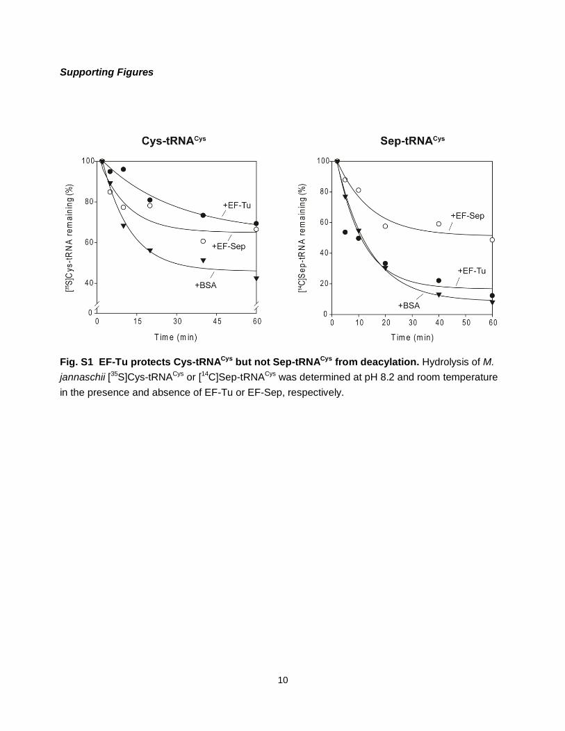
Fig. S1 EF-Tu protects Cys-tRNACys but not Sep-tRNACys from deacylation. Hydrolysis of M.
jannaschii [35S]Cys-tRNACys or [14C]Sep-tRNACys was determined at pH 8.2 and room temperature
in the presence and absence of EF-Tu or EF-Sep, respectively.

11
Fig. S2 Site-specific insertion of Sep in response to a TAG codon in the myoglobin gene
requires tRNASep, SepRS and EF-Sep. (A) Wild-type Myoglobin-His6 (lane 1) and Myoglobin-His6
(Asp127Sep) produced in the presence of Sep (2mM), SepRS and tRNASep, with or without EF-Sep
(lanes 2 and 3, respectively). Proteins were purified by affinity chromatography on Ni2+-NTA and
separated by SDS-PAGE.
(B) Electrospray ionization mass spectrometry of purified Myoglobin-His6 (left) and purified
Myoglobin-His6 (Asp127Sep) (right) was performed as described in the methods. The found and
expected masses are as follows: Myoglobin-His6 (Found= 18354.22; expected = 18354.20 Da);
Myoglobin-His6 (Asp127Sep) (Found= 18405.73 Da; expected= 18406.17 Da). The mass
difference between the two proteins 51.51 Da compares well with the expected mass difference of
51.97 Da.
(C) A representative spectrum from a doubly charged peptide-ion from a trypsin digest of E. coli
produced phospho-myoglobin protein separated using an Agilent C18 reverse phase Chip (0.75

12
mm x150 mm) and fragmented by CID in an Agilent 6330 ion trap mass spectrometer with
integrated Nano-Electrospray ionization. The observed b and y ions are indicated. Multiple spectra
were identified as described that matched the peptide HPGDFGAsAQGAMNK where s was
consistent with a phosphoserine residue. The precursor peptide had an m/z of 784.5 at eluted at
10.05 min. and was identified by Spectrum Mill (Agilent Technologies). Y ions are indicated in red,
b ions in blue and neutral loss fragments are indicated as well as the charge state of each.
Supporting Table
Table S1: Peptide information for MRM
peptide precursor/product ion CE DP
LC*DFGVSGQLIDSPMANSFVGTR 823.4
(3+)/ 333.2
(1+)[y3] 30.85 160.9
LC*DFGVSGQLIDSPMANSFVGTR 823.4
(3+)/ 666.35
(1+)[y6] 38.26 160.9
LC*DFGVSGQLIDSPMANSFVGTR 823.4
(3+)/ 780.4
(1+)[y7] 38.62 160.9
LC*DFGVSGQLIDSPMANSFVGTR 823.4
(3+)/ 851.4
(1+)[y8] 38.12 160.9
SP, phosphoserine; C*, propionamide; CE, Collision energy; DP, Dilution Potential

13
References and Notes 1. F. Lipmann, The bonding of phosphoric acid in phosphoproteins. I. Note: Isolation of some
phosphoric amino acids (Serin phosphoric acid) out of casein. Biochem. Z. 262, 3 (1933).
2. J. V. Olsen et al., Global, in vivo, and site-specific phosphorylation dynamics in signaling networks. Cell 127, 635 (2006).
3. G. Manning, D. B. Whyte, R. Martinez, T. Hunter, S. Sudarsanam, The protein kinase complement of the human genome. Science 298, 1912 (2002).
4. A. Sauerwald et al., RNA-dependent cysteine biosynthesis in archaea. Science 307, 1969 (2005).
5. M. J. Hohn, H. S. Park, P. O’Donoghue, M. Schnitzbauer, D. Söll, Emergence of the universal genetic code imprinted in an RNA record. Proc. Natl. Acad. Sci. U.S.A. 103, 18095 (2006).
6. S. Kamtekar et al., Toward understanding phosphoseryl-tRNACys formation: The crystal structure of Methanococcus maripaludis phosphoseryl-tRNA synthetase. Proc. Natl. Acad. Sci. U.S.A. 104, 2620 (2007).
7. R. Fukunaga, S. Yokoyama, Structural insights into the first step of RNA-dependent cysteine biosynthesis in archaea. Nat. Struct. Mol. Biol. 14, 272 (2007).
8. C. C. Liu, P. G. Schultz, Adding new chemistries to the genetic code. Annu. Rev. Biochem. 79, 413 (2010).
9. B. L. Wanner, W. W. Metcalf, Molecular genetic studies of a 10.9-kb operon in Escherichia coli for phosphonate uptake and biodegradation. FEMS Microbiol. Lett. 79, 133 (1992).
10. F. J. LaRiviere, A. D. Wolfson, O. C. Uhlenbeck, Uniform binding of aminoacyl-tRNAs to elongation factor Tu by thermodynamic compensation. Science 294, 165 (2001).
11. D. M. Rothman et al., Caged phosphoproteins. J. Am. Chem. Soc. 127, 846 (2005).
12. T. Dale, L. E. Sanderson, O. C. Uhlenbeck, The affinity of elongation factor Tu for an aminoacyl-tRNA is modulated by the esterified amino acid. Biochemistry 43, 6159 (2004).
13. J. Eargle, A. A. Black, A. Sethi, L. G. Trabuco, Z. Luthey-Schulten, Dynamics of Recognition between tRNA and elongation factor Tu. J. Mol. Biol. 377, 1382 (2008).
14. J. Ling et al., Pathogenic mechanism of a human mitochondrial tRNAPhe mutation associated with myoclonic epilepsy with ragged red fibers syndrome. Proc. Natl. Acad. Sci. U.S.A. 104, 15299 (2007).
15. Y. Doi, T. Ohtsuki, Y. Shimizu, T. Ueda, M. Sisido, Elongation factor Tu mutants expand amino acid tolerance of protein biosynthesis system. J. Am. Chem. Soc. 129, 14458 (2007).
16. T. Ohtsuki, H. Yamamoto, Y. Doi, M. Sisido, Use of EF-Tu mutants for determining and improving aminoacylation efficiency and for purifying aminoacyl tRNAs with non-natural amino acids. J. Biochem. 148, 239 (2010).
17. P. Nissen et al., Crystal structure of the ternary complex of Phe-tRNAPhe, EF-Tu, and a GTP analog. Science 270, 1464 (1995).
18. J. S. Sebolt-Leopold, R. Herrera, Targeting the mitogen-activated protein kinase cascade to treat cancer. Nat. Rev. Cancer 4, 937 (2004).
14
19. D. R. Alessi et al., Identification of the sites in MAP kinase kinase-1 phosphorylated by p74raf-1. EMBO J. 13, 1610 (1994).
20. J. M. Schrader, S. J. Chapman, O. C. Uhlenbeck, Tuning the affinity of aminoacyl-tRNA to elongation factor Tu for optimal decoding. Proc. Natl. Acad. Sci. U.S.A. 108, 5215 (2011).
21. S. Yoshizawa, A. Böck, The many levels of control on bacterial selenoprotein synthesis. Biochim. Biophys. Acta 1790, 1404 (2009).
22. K. Nozawa et al., Pyrrolysyl-tRNA synthetase-tRNAPyl structure reveals the molecular basis of orthogonality. Nature 457, 1163 (2009).
23. T. Mukai et al., Codon reassignment in the Escherichia coli genetic code. Nucleic Acids Res. 38, 8188 (2010).
24. F. J. Isaacs et al., Precise manipulation of chromosomes in vivo enables genome-wide codon replacement. Science 333, 348 (2011).
25. J. D. Scott, T. Pawson, Cell signaling in space and time: where proteins come together and when they’re apart. Science 326, 1220 (2009).
26. M. B. Yaffe et al., The structural basis for 14-3-3:phosphopeptide binding specificity. Cell 91, 961 (1997).
27. K. A. Datsenko, B. L. Wanner, One-step inactivation of chromosomal genes in Escherichia coli K-12 using PCR products. Proc. Natl. Acad. Sci. U.S.A. 97, 6640 (2000).
28. S. Bunjun et al., A dual-specificity aminoacyl-tRNA synthetase in the deep-rooted eukaryote Giardia lamblia. Proc. Natl. Acad. Sci. U.S.A. 97, 12997 (2000).
29. L. Wang, A. Brock, B. Herberich, P. G. Schultz, Expanding the genetic code of Escherichia coli. Science 292, 498 (2001).
30. P. Bernard, P. Gabarit, E. M. Bahassi, M. Couturier, Positive-selection vectors using the F plasmid ccdB killer gene. Gene 148, 71 (1994).
31. K. Bajaj, P. Chakrabarti, R. Varadarajan, Mutagenesis-based definitions and probes of residue burial in proteins. Proc. Natl. Acad. Sci. U.S.A. 102, 16221 (2005).
32. M. H. Dao-Thi et al., Molecular basis of gyrase poisoning by the addiction toxin CcdB. J. Mol. Biol. 348, 1091 (2005).
33. J. Normanly, R. C. Ogden, S. J. Horvath, J. Abelson, Changing the identity of a transfer RNA. Nature 321, 213 (1986).
34. F. W. Studier, Protein production by auto-induction in high density shaking cultures. Protein Expr. Purif. 41, 207 (2005).
35. C. M. Zhang, C. Liu, S. Slater, Y. M. Hou, Aminoacylation of tRNA with phosphoserine for synthesis of cysteinyl-tRNACys. Nat. Struct. Mol. Biol. 15, 507 (2008).
36. T. Christian, Y. M. Hou, Distinct determinants of tRNA recognition by the TrmD and Trm5 methyl transferases. J. Mol. Biol. 373, 623 (2007).
37. S. Traverse, N. Gomez, H. Paterson, C. Marshall, P. Cohen, Sustained activation of the mitogen-activated protein (MAP) kinase cascade may be required for differentiation of

15
PC12 cells. Comparison of the effects of nerve growth factor and epidermal growth factor. Biochem. J. 288, 351 (1992).