supporting information synthesis and self-assembly of
TRANSCRIPT

Supporting Information
Synthesis and self-assembly of “tree-like” amphiphilic glycopolypeptides
Colin Bonduelle,a Jin Huang,b Emmanuel Ibarboure,a Andreas Heise,*b and Sebastien
Lecommandoux*a
a Université de Bordeaux/IPB, ENSCBP, 16 avenue Pey Berland, 33607 Pessac Cedex, France. CNRS, Laboratoire de Chimie des Polymères Organiques (UMR5629), Pessac,
E-mail: [email protected] b School of Chemical Sciences, Dublin City University, Dublin 9, Ireland.Technische Universiteit Eindhoven, Den Dolech 2, P.O. Box 513,
5600 MB Eindhoven, The Netherlands. E-mail: [email protected]
Materials All chemicals were purchased from Sigma-Aldrich and used as received unless otherwise noted. γ-benzyl-L-glutamate and DL-propargylglycine were supplied by Bachem. Anhydrous DMF, DMSO, ethyl acetate, THF, methanol were used directly from the bottle under an inert and dry atmosphere. Sodium cyanoborohydride (≥ 95%), N,N, N’,N’,N” pentamethyldiethylenetriamine (PMDETA, 99%), copper bromide (99,995%), 3-chloropropylamine hydrochloride (98%), sodium azide (99%) were used as received under inert atmosphere. Bathophenanthroline disulfonic acid disodium salt hydrate (BPDS, 98%) was purchased from Alfa Aesar and used as received. Dextran T10 (Mn = 6600 g/mol, Mw/Mn=1.35) was purchased from Amersham and hyaluronan (Mw = 5140 g/mol, Mw/Mn = 1.41) from Life core company. γ-benzyl-L-glutamate NCA, propargylglycine NCA and poly(γ-benzyl-L-glutamate–b–DL-propargylglycine) was synthesized following a literature procedure.1 1-azido-3-aminopropane was synthesized following a literature procedure.2 The spectroscopic data were in agreement with literature data.
Instrumentation.
1H NMR spectra were recorded at room temperature with a Bruker Avance 400 (400 MHz). Infrared measurements were performed on a Bruker Tensor 27 spectrometer using the attenuated total reflection (ATR) method. The molar mass of dextran, hyaluronan, α-azido dextran and α-azido hyaluronan were determined by SEC equipped with two PL aquagel OH 30 and OH 40 columns (25 x 300 mm), a Wyatt Optilab rEX differential refractometer and with NaNO3 0.1 M as eluent (0.5 mL.min-1). The retention times of tree-like glycopeptides were determined by PL-GPC 50 plus Integrated GPC from Polymer laboratories-Varian having UV and RI detectors, columns oven at 80°C, integrated on-line degasser and equipped with two PLgel 5µm MIXED-D columns (75 x 300 mm). DMSO HPLC grade SCHARLAU with 1g.L-1 of LiBr was used as eluent (0.6 mL.min-1) Transmission Electron Microscopy (TEM) images were recorded on a Hitachi H7650 microscope working at 80 kV. Samples were prepared by spraying a 1g/L solution of the block copolymer onto a copper grid (200 mesh coated with carbon) using a homemade spray tool and negatively stained with 1% uranyl acetate. Dynamic Light Scattering (DLS) was used to obtain the average size of the particles by using a Malvern ZetaSizer NanoZS instrument. Atomic Force Microscopy (AFM) images were recorded in air with a Nanoscope III in dry Tapping mode. AFM measurements were performed at room temperature using a Veeco Dimension Icon AFM system equipped with a Nanoscope V controller. Both topographic and phase images of nanoparticles
Electronic Supplementary Material (ESI) for Chemical CommunicationsThis journal is © The Royal Society of Chemistry 2012
were obtained in tapping mode using rectangular silicon cantilever (AC 160-TS, Atomic Force, Germany) with a spring constant of 42 N m–1, a resonance frequency lying in the 290–320 kHz range, and a radius of curvature of less than 10 nm. The scan rates were in a range of 0.6 to 0.8 Hz. Samples were prepared by adsorption onto freshly cleaved HOPG (High Oriented Pyrolitic Graphite) or Mica from solutions at 1g.l-1 and allowed to dry overnight at room temperature. Measurements of height, width were taken using the section analysis tool provided with the AFM software (Nanoscope Analysis V1.20 from Bruker).
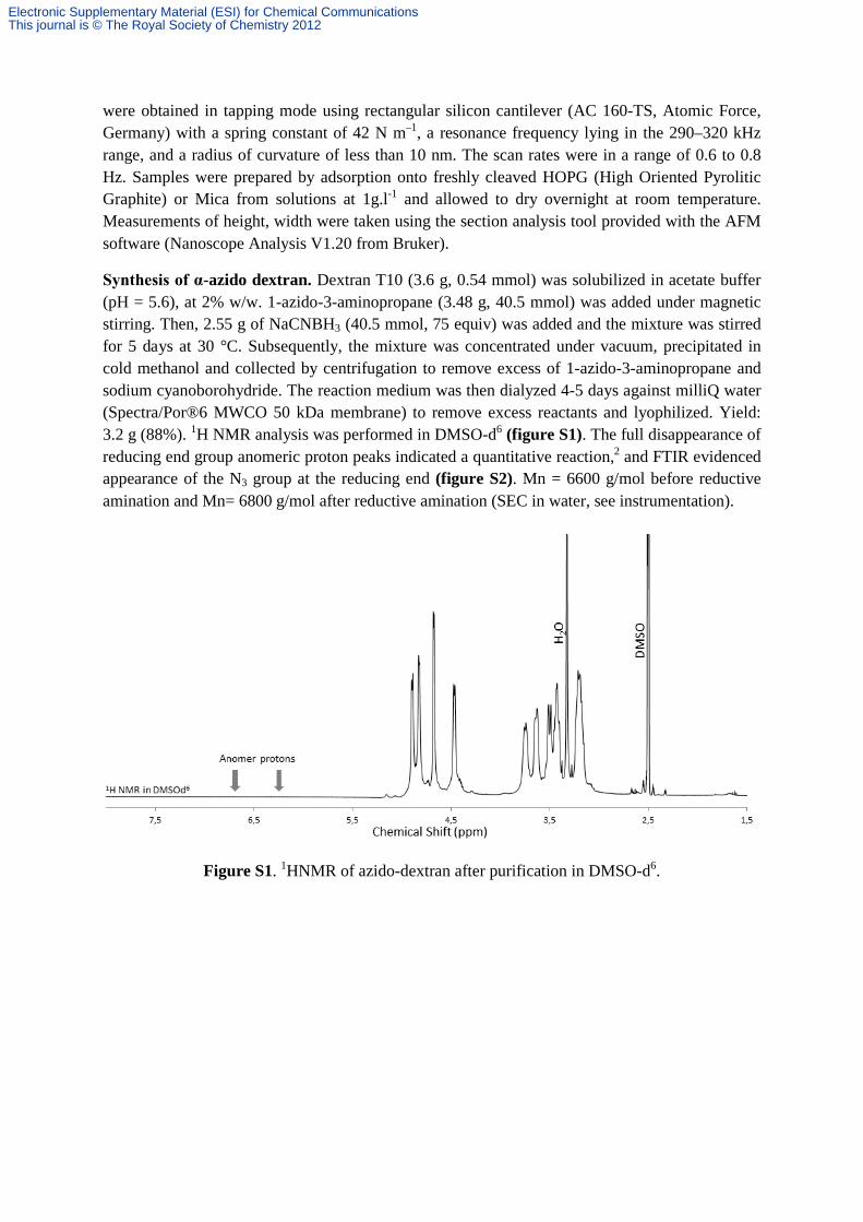
Synthesis of α-azido dextran. Dextran T10 (3.6 g, 0.54 mmol) was solubilized in acetate buffer (pH = 5.6), at 2% w/w. 1-azido-3-aminopropane (3.48 g, 40.5 mmol) was added under magnetic stirring. Then, 2.55 g of NaCNBH3 (40.5 mmol, 75 equiv) was added and the mixture was stirred for 5 days at 30 °C. Subsequently, the mixture was concentrated under vacuum, precipitated in cold methanol and collected by centrifugation to remove excess of 1-azido-3-aminopropane and sodium cyanoborohydride. The reaction medium was then dialyzed 4-5 days against milliQ water (Spectra/Por®6 MWCO 50 kDa membrane) to remove excess reactants and lyophilized. Yield: 3.2 g (88%). 1H NMR analysis was performed in DMSO-d6 (figure S1). The full disappearance of reducing end group anomeric proton peaks indicated a quantitative reaction,2 and FTIR evidenced appearance of the N3 group at the reducing end (figure S2). Mn = 6600 g/mol before reductive amination and Mn= 6800 g/mol after reductive amination (SEC in water, see instrumentation).
Figure S1. 1HNMR of azido-dextran after purification in DMSO-d6.
Electronic Supplementary Material (ESI) for Chemical CommunicationsThis journal is © The Royal Society of Chemistry 2012

Figure S2. FTIR of azido-dextran after purification.
Synthesis of α-azido hyaluronan. Hyaluronan (2 g, 0.55 mmol) was solubilized in acetate buffer (pH = 5.6), at 2% w/w. 1-azido-3-aminopropane (3.55 g, 41.2 mmol) was added under magnetic stirring. Then, 2.6 g of NaCNBH3 (41.2 mmol, 75 equiv) was added and the mixture was stirred for 5 days at 50 °C. Subsequently, the mixture was concentrated under vacuum, precipitated in cold methanol and collected by centrifugation to remove excess of 1-azido-3-aminopropane and sodium cyanoborohydride. The reaction medium was then dialyzed 4-5 days against milliQ water (Spectra/Por®6 MWCO 50 kDa membrane) and lyophilized. Yield: 1.8 g (90%).1H NMR analysis was performed in DMSO-d6 (figure S3). The full disappearance of reducing end group anomeric proton peaks indicated a quantitative reaction,3 and FTIR evidenced appearance of the N3 group at the reducing end (figure S4). Mn = 3600 g/mol before reductive amination and Mn= 3900 g/mol after reductive amination (SEC in water, see instrumentation). For the CuAAc reaction, azido-hyaluronan was first acidified by adding HCl (pH between 2 and 3) so as to be fully soluble in DMSO, the solvent used for the reaction.
Electronic Supplementary Material (ESI) for Chemical CommunicationsThis journal is © The Royal Society of Chemistry 2012

Figure S3. 1HNMR of azido-hyaluronan after acidification in DMSO-d6.
Figure S4. FTIR of azido-hyaluronan after acidification.
Synthesis of poly(γ-benzyl-L-glutamate–grafted–DL-dextran propargylglycine). Poly(γ-benzyl-L-glutamate–block–DL-propargylglycine) (80 mg, ca. 0.04 mmol of alkyne units), α-azido-dextran (500 mg, 0.08 mmol, 2 equiv. to alkyne groups) and PMDETA (16 μL, 78 mmol, 2 equiv. to alkyne groups) were dissolved in 20 mL of anhydrous DMSO in the Schlenk tube. CuBr (11 mg, 78 mmol, 2 equiv. to alkynes groups) was then added and the Schlenk tube was placed in an oil bath at 30 °C for 2 day under inert atmosphere. The reaction medium was then dialyzed 4-5 days against milliQ water (Spectra/Por®6 MWCO 50 kDa membrane), containing EDTA the first 2 days, to remove excess dextran and lyophilized (yield = 299 mg, 91%).1H NMR analysis was
Electronic Supplementary Material (ESI) for Chemical CommunicationsThis journal is © The Royal Society of Chemistry 2012

performed in DMSO-d6 (figure 1 manuscript) as well as 13C NMR (figure S5). FTIR evidenced disappearance of the N3 peak at 2200 cm-1, thus confirming that excess azido-dextran was removed upon dialysis (figure S6). The amide I and amide II bands at 1653 cm-1 and 1545 cm-1 respectively were characteristic for an α-helical secondary structure4 (figure S6). Coupling of dextran was also confirmed by SEC in DMSO (1% LiBr, see instrumentation) where a significant increase of the retention time was observed by UV/RI detection (figure S7).
Figure S5. 13CNMR of poly(γ-benzyl-L-glutamate–grafted–DL-dextran propargylglycine) in DMSO-d6
Electronic Supplementary Material (ESI) for Chemical CommunicationsThis journal is © The Royal Society of Chemistry 2012

Figure S6. FTIR of poly(γ-benzyl-L-glutamate–grafted–DL-dextran propargylglycine); zoom shows carbonyl peaks of the peptide backbone.
Electronic Supplementary Material (ESI) for Chemical CommunicationsThis journal is © The Royal Society of Chemistry 2012

Figure S7. SEC traces of the copolypeptides before and after CuAAc (elution in DMSO containing 1% LiBr). a) IR detection; b) UV detection.
Synthesis of poly(γ-benzyl-L-glutamate–grafted–DL-hyaluronan propargylglycine). Poly(γ-benzyl-L-glutamate–block–DL-propargylglycine) (290 mg, ca. 0.15 mmol of alkyne units), α-azido-hyaluronan (1.2 g, 0.3 mmol, 2 equiv. to alkyne groups) and BPDS (153 mg, 0.28 mmol) were dissolved in 15 mL of anhydrous DMSO in the Schlenk tube. CuBr (41 mg, 0.28 mmol) was then added and the Schlenk tube was placed in an oil bath at 30 °C for 2 day under nitrogen atmosphere. The reaction medium was then dialyzed 4-5 days against milliQ water (Spectra/Por®6 MWCO 50 kDa membrane) containing EDTA the first 2 days, to remove excess of hyaluronan and then lyophilized (yield = 790 mg, 88%). 1H NMR analysis was performed in DMSO-d6 (figure 1 manuscript) as well as 13C NMR (figure S8). FTIR evidenced disappearance of the N3 peak at 2200 cm-1, thus confirming that excess azido-hyaluronan was removed upon dialysis (figure S9). The amide I and amide II bands at 1656 cm-1 and 1547 cm-1 respectively were characteristic for an α-helical secondary structure (figure S9). Coupling of hyaluronan was also confirmed by SEC in DMSO (1% LiBr, see instrumentation) where a significant increase of the retention time was observed by UV/RI detection (figure S7).
Electronic Supplementary Material (ESI) for Chemical CommunicationsThis journal is © The Royal Society of Chemistry 2012
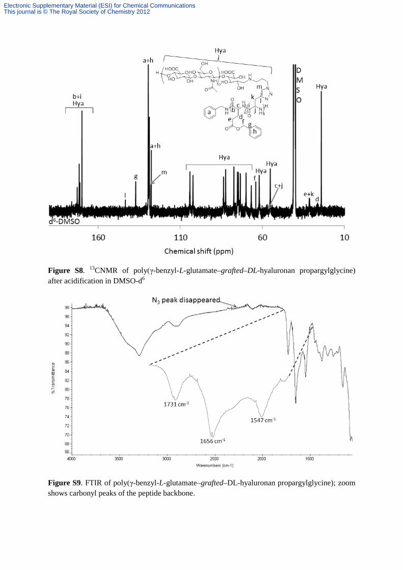
Figure S8. 13CNMR of poly(γ-benzyl-L-glutamate–grafted–DL-hyaluronan propargylglycine) after acidification in DMSO-d6
Figure S9. FTIR of poly(γ-benzyl-L-glutamate–grafted–DL-hyaluronan propargylglycine); zoom shows carbonyl peaks of the peptide backbone.
Electronic Supplementary Material (ESI) for Chemical CommunicationsThis journal is © The Royal Society of Chemistry 2012

Nanoprecipitation. 1 mg/mL of our dextran based tree-like material provided an aqueous solution without macroscopic aggregation after warming a copolymer solution at 60°C during 2 hours. Self-assembly by direct hydration was even better for hyaluronan based tree-like material. Such tree-like glycopolypeptide, at 1 mg/mL, produced an aqueous solution without macroscopic aggregation after few seconds at room temperature. 1H NMR analysis was performed in D2O after hydration (figure S10) verifying PBLG confinement and nanoparticles formation.
Figure S10. 1H NMR (D2O) after direct hydration of dextran-grafted-PBLG (up) or of hyaluronan-grafted-PBLG (down)
Electronic Supplementary Material (ESI) for Chemical CommunicationsThis journal is © The Royal Society of Chemistry 2012
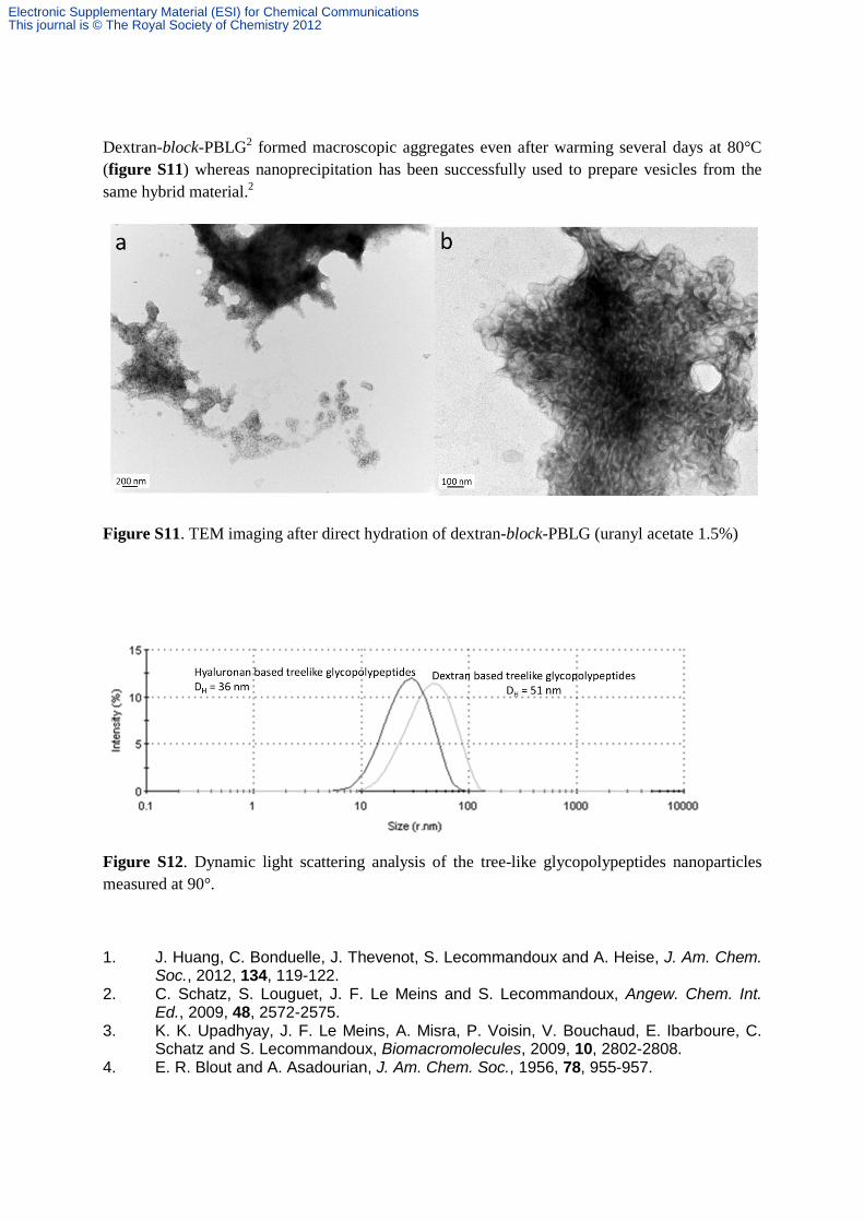
Dextran-block-PBLG2 formed macroscopic aggregates even after warming several days at 80°C (figure S11) whereas nanoprecipitation has been successfully used to prepare vesicles from the same hybrid material.2
Figure S11. TEM imaging after direct hydration of dextran-block-PBLG (uranyl acetate 1.5%)
Figure S12. Dynamic light scattering analysis of the tree-like glycopolypeptides nanoparticles measured at 90°.
1. J. Huang, C. Bonduelle, J. Thevenot, S. Lecommandoux and A. Heise, J. Am. Chem. Soc., 2012, 134, 119-122.
2. C. Schatz, S. Louguet, J. F. Le Meins and S. Lecommandoux, Angew. Chem. Int. Ed., 2009, 48, 2572-2575.
3. K. K. Upadhyay, J. F. Le Meins, A. Misra, P. Voisin, V. Bouchaud, E. Ibarboure, C. Schatz and S. Lecommandoux, Biomacromolecules, 2009, 10, 2802-2808.
4. E. R. Blout and A. Asadourian, J. Am. Chem. Soc., 1956, 78, 955-957.
Electronic Supplementary Material (ESI) for Chemical CommunicationsThis journal is © The Royal Society of Chemistry 2012