supporting information - pnas · supporting information ... (of ppt vs. input) ... according to the...
TRANSCRIPT

Supporting InformationBlanchard et al. 10.1073/pnas.1416321111SI Materials and MethodsFly Lines. Myb interacting protein (Mip) 130, Mip1301-36 (MMBcore component), mip1301-723, mip1301-723; mybmh107, mip12067,mip12067-21-6 were previously developed in our laboratory (1, 2).L(3)mbtgm76 (lethal 3 malignant brain tumor)/tm3 (3) was a kindgift from the Lehman laboratory (Skirball Institute, New York).Df(2L)e2f2329 (transcription repressor)/Df(2L)e2f2329; P[e2f2−;mpp6+]/+/Cyo and e2f276Q.1/Cyo (4) were kind gifts from theLipsick and Duronio laboratories (Stanford University Schoolof Medicine, Stanford, CA and University of North Carolina,Chapel Hill, NC, respectively). Gfp::piwi (transposon regulator)/cyo line (5) was a kind gift from the Hannon and Aravin labora-tories (Cold Spring Harbor Laboratory, New York and CaliforniaInstitute of Technology, Pasadena, CA, respectively). Cyo/sco,tm3/cxd, and tm3 actin::gfp were obtained from our fly stock.
Microscopy. Six to ten third instar larvae were dissected at roomtemperature on 1× PBS, and their salivary glands or brains wereextracted by using fine tip dissection forceps. After removal ofexcess fat, the glands and brains were placed on a precleanedglass slide with 12 μL of PBS with 1:1,000 dilution of Dapi (4′,6-diamidino-2-phenylindole) and covered with a 24 × 24 mmprecleaned coverslip. Larval brains and some salivary glands werethen sealed with rubber cement and directly imaged (unsquashedsamples). Some salivary glands (squashed samples) were furtherprocessed as follows: the excess liquid was carefully removed withKim wipes and the slides were repeatedly tapped lightly with therubber end of a pencil. The slides were then sealed using rubbercement and then expeditiously imaged fresh. When differentiatingYFP from GFP fluorophores (Fig. 2A and Fig. S5), a Leica TCSSP2 AOBS confocal microscope was used to acquire stacks ofimages that were deconvoluted using the commercial softwareHuygens Professional. The remaining samples were imaged usinga Zeiss AxioObserverZ1_RSS compound fluorescent microscope.FISH and IHC samples were imaged using a Zeiss LSM710confocal RSS microscope.
Immunohistochemistry and FISH. The protocols used for all IHCstaining and FISH were obtained from previously published work(6, 7). The following modifications were introduced to the pro-tocols: Once fixed, salivary glands were squashed by placing a no.1 24 × 24 mm cover glass over the slides, inverting them overpaper towels, and pressing down vigorously with two thumbs onthe glass. All FISH probes were generated by cloning the 5′UTRplus the N terminus of the desired genes into a commercialplasmid (Invitrogen Topo-TA). Sequenced plasmids were thenamplified and the desired regions were restriction digested and gelextracted. DNA was quantified and then labeled using a DIG-Nick Translation mix containing digoxigenin-11-dUTP (Roche;11745816910) according to the manufacturer’s protocol. The fourprobes used were pooled into the following hybridyzation mix:25% (vol/vol) pooled probe, 4× SSC, 1× Denhardt’s, 0.4 mg/mLsonicated salmon sperm, 50% (vol/vol) formamide. The mix wasboiled and stored at −20 °C until use, when it was reboiled andadded directly to the slides. After FISH hybridization and beforeIHC slides were immunolabeled, samples were incubated for 1 hat room temperature in 1× blocking buffer [2.5% (wt/vol) nonfatmilk, 0.5% BSA, 0.8% Nonidet P-40, 0.1% Tween 20, 1× Tris S(12 mM Trizma Base, 180 mM NaCl, pH 7.2)]. Slides were thenincubated overnight at 4 °C in blocking buffer containing primaryantibodies (1:20–1:150 dilutions) or serum from L(3)mbt immu-nized rabbit (1:50 dilution).
The following antibodies were used: affinity purified rabbitanti-Mip40, anti-Mip120, anti-Mip130, anti-Caf1/p55, and antiMyb (8), monoclonal anti-Di-methyl H4K20 (1:5 for IHC)anti-CycA (Santa Cruz Biotechnologies; 1:2,000 dilution forWestern blot), serum from rabbits immunized with a GST::L(3)mbtN terminus fusion protein (1:50 for IHC and FISH), andrabbit anti-dmPiwi which was a kind gift from the Hannonlaboratory. The secondary antibodies used were: goat anti-rabbit::Horseradish peroxidase (Sigma-Aldrich 170-6515),Sheep anti-Digoxigenin (Roche 1333089), Alexa fluor goat anti-rabbit 488nm (Molecular probes A11008), Alexa fluor goat anti-rabbit 568 (Molecular probes A11036), Alexa fluor 488nm donkeyanti-sheep (Molecular probes A11015), and Alexa fluor 555nmdonkey anti-sheep (Molecular probes A21436).
Chromatin Immunoprecipitation. These experiments were per-formed on S2 cells (including the immunoprecipitation them-selves and the qPCR readout) according to protocols describedpreviously (9, 10). qPCR experiments were done in triplicate andnormalized to Actin 5C as endogenous control, the mock ChIP(NS) was normalized to a value of 1. The sequence of the primersets used is available upon request.
Salivary qPCR. Fifty salivary glands were dissected for each experi-ment and placed in RNAlater RNA stabilization buffer (Qiagen).Total RNA was then extracted according to the manufacturer’sprotocol, and 1 μg was reverse transcribed using SuperScript IIIReverse Transcriptase (Invitrogen; 18080-093), with conditions setfor a single reverse transcription reaction per mRNA. The cDNAthus generated was then chloroform extracted and resuspended indeionized H2O. qPCR reactions were performed using commercialqPCR mix (Maxima SYBR Green/ROX qPCR Master mix) andPlatinum Taq DNA polymerase (Invitrogen; 10966-018) on anApplied Biosystems 7300 Real-Time PCR cycler and the resultswere analyzed with Applied Biosystems software. All qPCR re-actions were done in triplicate, and most experiments were donetwice, the results presented representing an average. Actin 5Cprimers were used as endogenous control. The primer sequencesused can be provided upon request.
GST Pull Downs. L(3)mbt was cloned into the pGEX-6P to achieveexpression of GST::L(3)mbt on bacteria. The resulting plasmidswere transformed into aBL-21 strain of bacteria inducedwith 1mMisopropyl-beta-D-thiogalactopyranoside for 4 h at 30 °C. Purifica-tion was done according to standard laboratory protocol. The cellswere centrifuged for 10 min at 8,000 × g. The pellets were re-suspended in buffer A [500 mM Tris·HCl, pH 8.0, 100 mM KCl,1MNaCl, 0.05%Nonidet P-40, 1mMEGTApH8.0, 1mMEDTApH 8.0, 0.5 mM DTT, 1 mM PMSF, and 1× protease inhibitor(Roche Complete)]. After freeze-thawing, the resuspended cellswere sonicated on ice five times for 30 s each and then centrifugedat 20,000 × g. The supernatants were incubated for 120 min withglutathione Sepharose 4 fast flow (GE Healthcare/Amersham;17-5132-01) previously equilibrated with buffer A. The beads werethen washed five times with buffer A and stored until required in40% (vol/vol) glycerol at −20 °C. The bound beads were equili-brated with IP buffer (25 mMHepes, 150 mM KCl, 1 mM EDTA,1 mM EGTA, 10% (vol/vol) glycerol, 1 mM DTT, 0.4 mM PMSF1 mM NaMBS, 0.5% Nonidet P-40) and then incubated with invitro translated MMB proteins for 120 min. These were generatedby cloning the cDNAs of the different MMB components into apCS2+ plasmid generating constructs that were then used as tem-
Blanchard et al. www.pnas.org/cgi/content/short/1416321111 1 of 6

plates for a commercial rabbit reticulocite IVT kit, using themanufacturer’s instructions (Promega; TNT Quick Coupled Tran-scription/Translation system L2080). The beads were washed fivetimes with IP buffer (the salt concentration of the buffer was in-creased to 1MKCl in certain cases as indicated above), boiled, andthen developed byWestern blot with the corresponding antibodies.
Data Analysis.A previously published dataset product of ChIP-Sequsing anti-L(3)mbt antibody (11) was mapped against the piwigenomic locus using Bowtie 0.12.5 and the Drosophila genome
version Dme_r5.32 (12). The reads obtained were analyzed andgraphed using the integrated genome browser (IGB) (13) andthe University of California Santa Cruz genome browser (14).The previously published peaks at an FDR of 0.5 (10) werecompared with those obtained by ModEncode dataset (15) usingbedtools (16). The published ChIP-chip smoothed data (10)were used to call peaks with a log2 ratio (of ppt vs. input) greaterthat 0.5. These peaks were compared with the Richter dataset(Peaks FDR 0.5), subtracting those regions not present in theChIP-chip map.
1. Beall EL, Bell M, Georlette D, Botchan MR (2004) Dm-myb mutant lethality in Dro-sophila is dependent upon mip130: Positive and negative regulation of DNA repli-cation. Genes Dev 18(14):1667–1680.
2. Beall EL, et al. (2007) Discovery of tMAC: A Drosophila testis-specific meiotic arrestcomplex paralogous to Myb-Muv B. Genes Dev 21(8):904–919.
3. Yohn CB, Pusateri L, Barbosa V, Lehmann R (2003) l(3)malignant brain tumor andthree novel genes are required for Drosophila germ-cell formation. Genetics 165(4):1889–1900.
4. Cayirlioglu P, Bonnette PC, Dickson MR, Duronio RJ (2001) Drosophila E2f2 promotesthe conversion from genomic DNA replication to gene amplification in ovarian folliclecells. Development 128(24):5085–5098.
5. Le Thomas A, et al. (2013) Piwi induces piRNA-guided transcriptional silencing andestablishment of a repressive chromatin state. Gene Dev 27(4):390–399.
6. Ivaldi MS, Karam CS, Corces VG (2007) Phosphorylation of histone H3 at Ser10 facil-itates RNA polymerase II release from promoter-proximal pausing in Drosophila.Genes Dev 21(21):2818–2831.
7. Brandt T, Corces VG (2008) The Lawc protein is required for proper transcription byRNA polymerase II in Drosophila. Mol Genet Genomics 280(5):385–396.
8. Lewis PW, et al. (2004) Identification of a Drosophila Myb-E2F2/RBF transcriptionalrepressor complex. Genes Dev 18(23):2929–2940.
9. Andrulis ED, Guzmán E, Döring P, Werner J, Lis JT (2000) High-resolution localizationof Drosophila Spt5 and Spt6 at heat shock genes in vivo: roles in promoter proximalpausing and transcription elongation. Genes Dev 14(20):2635–2649.
10. Georlette D, et al. (2007) Genomic profiling and expression studies reveal both pos-itive and negative activities for the Drosophila Myb MuvB/dREAM complex in pro-liferating cells. Genes Dev 21(22):2880–2896.
11. Richter C, Oktaba K, Steinmann J, Müller J, Knoblich JA (2011) The tumour suppressorL(3)mbt inhibits neuroepithelial proliferation and acts on insulator elements. Nat CellBiol 13(9):1029–1039.
12. Langmead B, Trapnell C, Pop M, Salzberg SL (2009) Ultrafast and memory-efficientalignment of short DNA sequences to the human genome. Genome Biol 10(3):R25.
13. Nicol JW, Helt GA, Blanchard SG, Jr, Raja A, Loraine AE (2009) The Integrated GenomeBrowser: Free software for distribution and exploration of genome-scale datasets.Bioinformatics 25(20):2730–2731.
14. Kent WJ, et al. (2002) The human genome browser at UCSC. Genome Res 12(6):996–1006.
15. Roy S, et al.; modENCODE Consortium (2010) Identification of functional elementsand regulatory circuits by Drosophila modENCODE. Science 330(6012):1787–1797.
16. Quinlan AR, Hall IM (2010) BEDTools: A flexible suite of utilities for comparing ge-nomic features. Bioinformatics 26(6):841–842.
Fig. S1. Genome-wide comparison of the cytolocalization of lethal 3 malignant brain tumor [L(3)mbt] and mono- and dimethylated histones. Graphicalrepresentation of the Richter L(3)mbt brain ChIP-Seq (green) (1) and a subset of the modENCODE mono- and dimethylated histone Chip-Seq dataset tracks (2)(color coded as labeled, according to the Venn diagrams in Fig. 1A on the wint complex locus on chromosome 2L. The identity of the tracks are listed on theLeft. Peaks are depicted as color coded bands below the corresponding tracks.
1. Langmead B, Trapnell C, Pop M, Salzberg SL (2009) Ultrafast and memory-efficient alignment of short DNA sequences to the human genome. Genome Biol 10(3):R25.2. Kharchenko PV, et al. (2011) Comprehensive analysis of the chromatin landscape in Drosophila melanogaster. Nature 471(7339):480–485.
Blanchard et al. www.pnas.org/cgi/content/short/1416321111 2 of 6

Fig. S2. Cytolocalization of L(3)mbt and H4K20 dimethyl on Drosophila polytene chromosomes by immunohistochemistry (IHC). (A) Immunohistochemicalstaining of wild-type and l(3)mbt putative null mutant polytene chromosomes. Polytene chromosome spreads from wild type (Upper) or l(3)mbtgm76 putativedefective mutants (Lower) third instar larvae were stained with Dapi (shown in green) and then incubated with serum from rabbits immunized with the first(N-terminal) 441 amino acids of L(3)mbt fused to GST, and then stained with anti-rabbit fluorescently labeled secondary antibody (shown in red). In all panels,the white arrow indicates the centromeric heterochromatic. (B) Cytolocalization of L(3)mbt and H4K20 dimethyl on Drosophila melanogaster polytenechromosomes. A polytene chromosome spread from wild-type third instar larvae was stained with Dapi (in blue), rabbit serum anti-L(3)mbt (green), andmonoclonal antibodies anti-H4K20 dimethyl (red). (Top Left) anti-L(3)mbt. (Top Right) Dapi. (Middle Left) Anti-H4K20 dimethyl. (Middle Right) Anti-L(3)mbt +Dapi. (Bottom Left) Anti-H4K20 dimethyl + anti-L(3)mbt. (Bottom Right) Dapi + anti H4K20 dimethyl. In all panels, the white arrow points to the centromericheterochromatin. B, Inset is an amplification of the region highlighted by a dotted rectangle (Bottom Left). Red arrows indicate H4K20 dimethyl only bandsand green arrows indicate L(3)mbt only bands.
Blanchard et al. www.pnas.org/cgi/content/short/1416321111 3 of 6
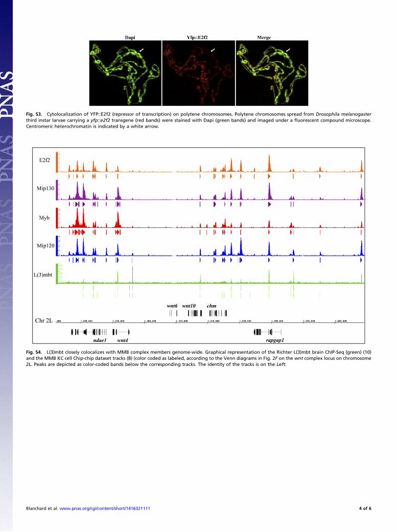
Fig. S3. Cytolocalization of YFP::E2f2 (repressor of transcription) on polytene chromosomes. Polytene chromosomes spread from Drosophila melanogasterthird instar larvae carrying a yfp::e2f2 transgene (red bands) were stained with Dapi (green bands) and imaged under a fluorescent compound microscope.Centromeric heterochromatin is indicated by a white arrow.
Fig. S4. L(3)mbt closely colocalizes with MMB complex members genome-wide. Graphical representation of the Richter L(3)mbt brain ChIP-Seq (green) (10)and the MMB KC cell Chip-chip dataset tracks (8) (color coded as labeled, according to the Venn diagrams in Fig. 2F on the wnt complex locus on chromosome2L. Peaks are depicted as color-coded bands below the corresponding tracks. The identity of the tracks is on the Left.
Blanchard et al. www.pnas.org/cgi/content/short/1416321111 4 of 6

Fig. S5. Testing the wavelength overlap of YFP::E2f2 and GFP::Myb by confocal microscopy (A) Polytene Chromosome spreads from third instar larvae ex-pressing YFP::E2f2 alone (red bands), stained with Dapi (blue bands), were tested for wavelength overlap in the GFP channel (Right, green bands). (B) PolyteneChromosome spreads from third instar larvae expressing GFP::Myb alone (green bands) stained with Dapi (blue bands), were tested for wavelength overlap inthe YFP channel (Right, red bands).
Blanchard et al. www.pnas.org/cgi/content/short/1416321111 5 of 6

Fig. S6. Colocalization of YFP::E2f2 and Cerulean::Mip120. Polytene chromosome spreads from salivary glands of Dm third instar larvae coexpressing Cerulan::Mip120(green bands) and YFP::E2f2 (red bands) imaged by fluorescent compound microscopy.
Fig. S7. Effect of deletion of MMB components on TDtomato::L(3)mbt. TDtomato::L(3)MBT (red bands) cytolocalization on polytene chromosomes stainedwith Dapi (green bands) from larvae either WT (Top row) or homozygous mutant for the following MMB complex members: mip1301-36 (null mutant, Middlerow). and e2f276q.1 (null mutant, Bottom row).
Blanchard et al. www.pnas.org/cgi/content/short/1416321111 6 of 6