supporting information for: containing anthracene … reaction was performed using a modified...
TRANSCRIPT

1
Supporting information for:
A new family of zinc metal-organic framework polymorphs containing anthracene tetracarboxylates**
Kristina Konstas,a Kim F. Taupitz,a David R. Turner, b Danielle F. Kennedy* and Matthew R. Hill*
Electronic Supplementary Material (ESI) for CrystEngComm.This journal is © The Royal Society of Chemistry 2014

2
Supporting Information
Table of Contents
Section 1 Synthetic Procedure S1
Section 2 General Characterisation Information S2
Section 3 Data Analysis S3
3
Section 1 Synthetic Procedure S1
Synthesis of anthracene-9,10-diboronic acid
This reaction was performed using a modified procedure from Kim et. al.1 To a pre-dried and evacuated Schlenk flask, 9,10-dibromoanthracene (30.00 g, 89.28 mmol, 1.0 eq) was added and the material was evacuated and backfilled with argon ( cycle x 3). Under an argon atmosphere, dry THF (1 L) was added and the solution was cooled to -78 °C. To this, n-BuLi (1.6 M, 180 mL, 288 mmol, 3.2 eq) was added and stirred at -78 °C for 2.5 h . Triethyl borate (44.4 mL, 259 mmol, 2.9 eq) was added and the reaction stirred for an additional 2 h at -78 °C until it warmed up to room temperature and stirred overnight. After the reaction finished, it was acidified with diluted HCl solution, extracted with Et2O (50 mL, x 3), the combined organic phases were washed with water (50 mL, x 4) and dried over MgSO4. After removing the solvent, the residue was re-dissolved in acetone (60 mL) and added to dichloromethane (50 mL) to give a precipitate which was filtered, washed and dried in vacuo to give a white powder (8.082 g, 30.38 mmol, 34%). The compound was stored in the freezer until further use.
1H-NMR (400 MHz, DMSO-d6, δ): 8.66 (s, 4 H, B(OH)2), 7.93 (q, 4 H, H-1), 7.42 (q, 4 H, H-2). EI+-MS: 266.1.
Synthesis of dimethyl-5-bromo-isophthalate
5-Bromoisophthalic acid (25.85 g, 105.9 mmol, 1.0 eq) was dissolved in methanol (400 mL) and concentrated H2SO4 (15 mL) was added. The reaction was stirred overnight under reflux conditions. After removing the solvent, the precipitate was redissolved in ethyl acetate (250 mL) and the organic phase was washed with water (50 mL, x 5), dried over MgSO4, evaporated and dried in vacuo to give a white coloured compound (27.21 g, 100.0 mmol, 94%).
1H-NMR (400 MHz, CDCl3, δ): 8.58 (t, 1 H, H-2), 8.33 (d, 2 H, H-4, H-6), 3.93 (s, 6 H, OMe).
Synthesis of 5,5’-(9,10-Anthracenediyl)bis(1,3-benzenedimethoxy-carbonyl)
This reaction was performed using a modified procedure from Ma et. al.2 A pre-dried and evacuated Schlenk flask was charged with anthracene-9,10-diboronic acid (0.5008 g, 1.883 mmol, 1.00 eq), dimethyl-5-bromo-isophthalate (1.707 g, 6.253 mmol, 3.3 eq) and Pd(PPh3)4 (25.0 mg, 0.0216 mmol, 0.01 eq) which were pumped under vacuum for 2.5 h. Then, degassed THF (70 mL) and degassed 2 M Na2CO3 (20 mL) were added and heated to reflux under argon atmosphere for 43 h. Water (100 mL) and dichloromethane (150 mL) were added to the reaction mixture and the phases were separated. The water phase was extracted with DCM (4 x 75 mL) and the combined organic phase dried over MgSO4, filtered and the solvent was removed in vacuo. The brown crude product was purified by recrystallisation from acetone to give a yellow solid (0.5587 g, 1.048 mmol, 56%).
1H-NMR (400 MHz, CDCl3, δ): 8.92 (s, 2 H, H-2), 8.37 (s, 4 H, H-4), 7.56 (m, 4 H, H-1’), 7.37 (m, 4 H, H-2’). EI+-MS: 562.1.1 S.-K. Kim, B. Yang, Y. Ma, J.-H. Lee and J.-W. Park, J. Mater. Chem., 2008, 18, 3376.2 S. Ma, D. Sun, J. Simmons, C. Collier, D. Yuan and H.-C. Zhou, J. Am. Chem. Soc, 2008, 130, 1012.
4
Synthesis of 5,5’-(9,10-anthracenediyl)di-isophthalic acid (H4adip)
This reaction was performed using a modified protocol of Ma et. al.2 5,5’-(9,10-anthracenediyl)bis(1,3-benzenedimethoxycarbonyl) (800 mg, 1.423 mmol, 1 eq) was suspended in THF (65 mL) and KOH aqueous solution 2 M, 15 mL) was added. The mixture was stirred overnight at room temperature. THF was evaporated and dilute HCl (2 M) was added to the remaining aqueous solution until it became acidic. The precipitate was filtered off and washed several times with water and CHCl3 to give a yellow solid (0.6728 g, 1.3296 mmol, 93%).
1H-NMR (400 MHz, DMSO, δ): 13.45 (s, 4 H, COOH), 8.70 (m, 2 H, H-2), 8.13 (m, 4 H, H-4), 7.54 (m, 4 H, H-1’), 7.47 (m, 4 H, H-2’). EI+-MS: 506.1.
General procedure for the synthesis of ZnADIP
ZnADIP crystals were synthesized by in situ. ligand transformation in a solvothermal approach. A mixture of Zn(NO3)2.6H2O and 5,5’-(9,10-Anthracenediyl)di-isophthalic acid (H4ADIP) in DMF were sealed in a pyrex tube and dissolved by sonication. Fluoroboric acid was added and the tubes were heated for two days according to the prescribed temperature. The resulting crystals were washed with DMF before further analysis. See table 1 for details.
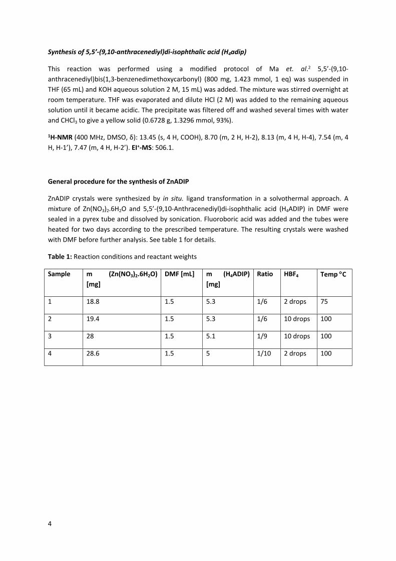
Table 1: Reaction conditions and reactant weights
Sample m (Zn(NO3)2.6H2O) [mg]
DMF [mL] m (H4ADIP) [mg]
Ratio HBF4 Temp C
1 18.8 1.5 5.3 1/6 2 drops 75
2 19.4 1.5 5.3 1/6 10 drops 100
3 28 1.5 5.1 1/9 10 drops 100
4 28.6 1.5 5 1/10 2 drops 100
5
Section 2 General Characterisation Information S2
S2.1 General NMR Experimental
Approximately 10 mg of the organic compound were dissolved in 0.5 ml deuterated solvent. Spectra were recorded using a Bruker Av400 operating with a 9.4 T magnet and 400.13 MHz 1H frequency. It is equipped with a 5 mm 1H-, 19F-, 13C-, 31P- QNP probe with z-gradient and a 60-holder sample changer. It is running with Bruker BioSpin’s ICON-NMR software. Spectra were analysed using MestReNova Lite Version 5.2.5.
S2.2 Mass Spectroscopy
Positive and negative ion electro-spray mass spectra were acquired with a VG Platform mass spectrometer using a cone voltage of 50V and the source was maintained at 80 °C. The used solvent was methanol with a flow rate of 0.04 mL/min.
S2.3 Low Pressure Gas Sorption
Gas adsorption isotherms for pressures in the range of 0-1.2 bar were measured by a volumetric
method using a Micromeritics ASAP 2420 instrument. Freshly prepared sample was transferred to a
pre-dried and weighed analysis tube which was stoppered with a Transeal cap. The sample was
evacuated and activated at 200 C under dynamic vacuum at 10-6 Torr for 24h. Accurate sample
masses were calculated using degassed samples. Gas adsorption measurements were performed
using ultra-high purity Ar, He, H2, CO2 and CH4 gas.
S2.4 Single-Crystal X-Ray Crystallography
Data for Zn1 and Zn3 were collected using the MX1 beamline at the Australian Synchrotron, operating at 17.4 KeV (λ = 0.7107 Å). Data collections were controlled using the Blu Ice software,3 with indexing and data reduction conducted using the XDS package.4 Data for Zn2 was collected using an Oxford Xcalibur diffractometer equipped with Cu-Kα radiation (λ = 1.5418 Å). Data collection and reduction was conducted using the CrysAlisPro software.5
3 T. M. McPhillips, S. E. McPhillips, H. J. Chiu, A. E. Cohen, A. M. Deacon, P. J. Ellis, E. Garman, A. Gonzalez, N. K. Sauter, R. P. Phizackerley, S. M. Soltis, P. Kuhn, J. Synchrotron Radiat., 2002, 9, 401 –406.4 W. Kabsch, J. Appl. Crystallogr., 1993, 26, 795 –800.
6
S2.5 Powder X-ray Crystallography
A Bruker D8 Advance X-ray Diffractometer operating under CuK radiation (40kV, 40mA) equipped with a LynxEye silicon strip detector was employed to obtain the XRD patterns. The samples were scanned over the 2θ range 1° to 30° with a step size of 0.02° 2θ and a count time of 0.8 seconds per step. 52 sensor strips of the LynxEye detector gave equivalent count times of 41.6 seconds per step. Analyses were performed on the collected XRD data using the Bruker XRD search match program EVA™.
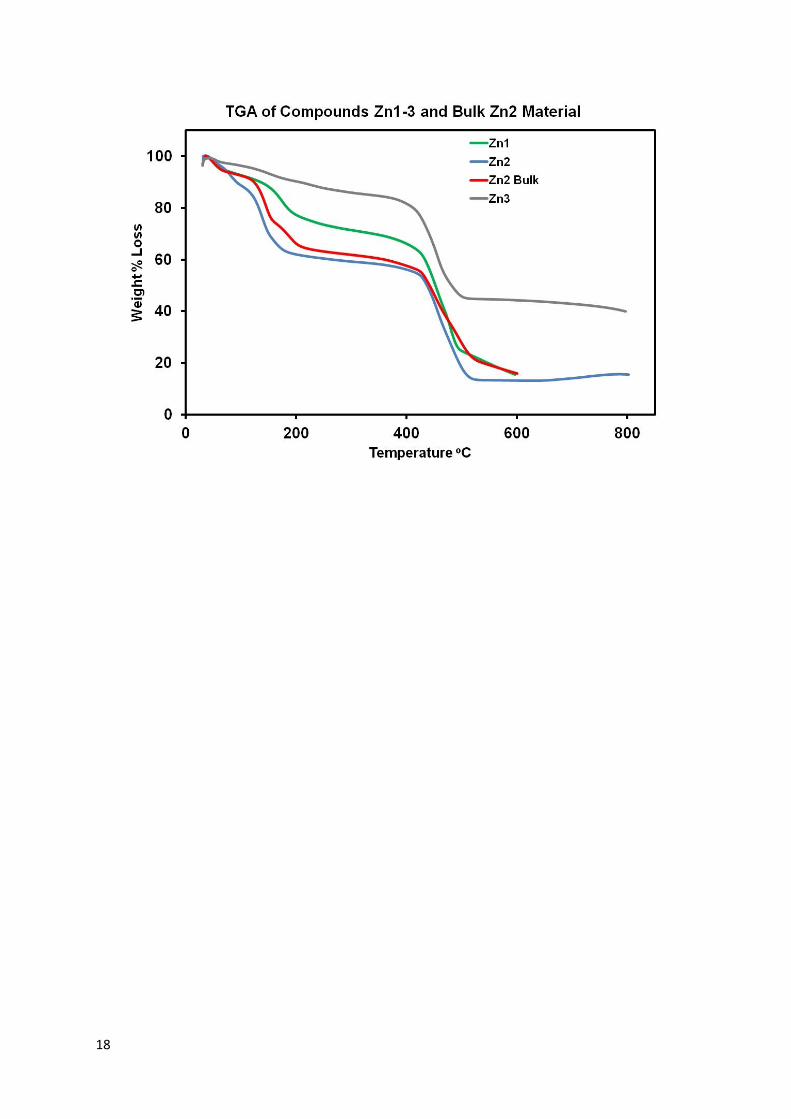
S2.6 Thermal Gravimetric Analysis
A pre-weighed sample of crystals were analysed on a PerkinElmer Pyris 1 Thermal Gravimetric Analyser within a temperature range from 25 C to 800 C and a heating ramp of 10 C per minute after a 10 minute nitrogen gas flow at 25 C.
5 CrysAlisPro, Oxford Diffraction Ltd., Version 1.171.34.36.
7
Section 3 Data Analysis S3
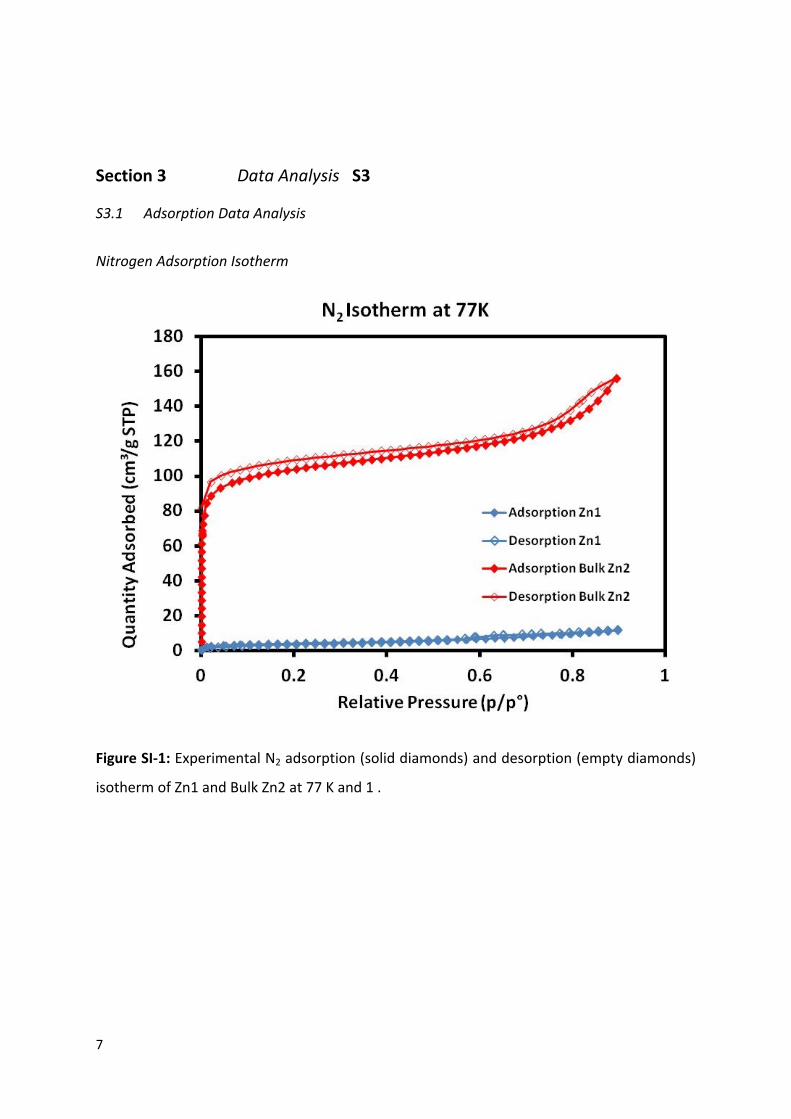
S3.1 Adsorption Data Analysis
Nitrogen Adsorption Isotherm
Figure SI-1: Experimental N2 adsorption (solid diamonds) and desorption (empty diamonds)
isotherm of Zn1 and Bulk Zn2 at 77 K and 1 .

8
Hydrogen Adsorption Isotherm
Methane Adsorption Isotherm

9

10
Carbon Dioxide Adsorption Isotherm
11
S3.2 X-ray Analysis
Single Crystal X-ray Diffraction Analysis
All structures were solved by direct methods using SHELXS-97 and refined against F2 using SHELXL-2013.6 The program X-Seed was used as a graphical interface.7 All non-hydrogen atoms were refined using an anisotropic model, except for instances of disordered solvent (see supporting information for full details). Hydrogen atoms attached to carbon were placed in idealised X-ray positions and refined using a riding model.
Data have been deposited with the Cambridge Crystallographic Data Centre and are available from www.ccdc.cam.ac.uk/deposit.
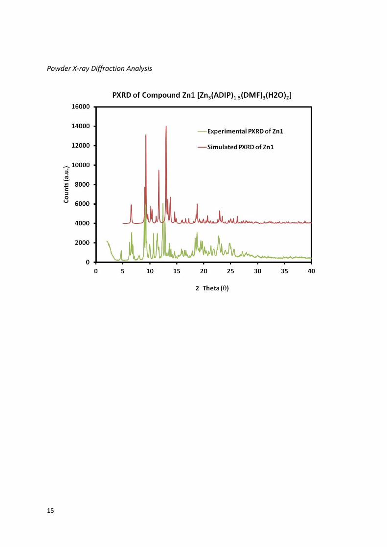
Structure Zn1 [Zn3(ADIP)1.5(DMF)3(H2O)2]·3DMF
Data was treated using the SQUEEZE routine of PLATON8 due to the presence of significant void space containing residual electron density that could not be modelled. A total void space of 2436 Å3 in the unit cell containing a total of 474 e- (the full SQUEEZE output is available in the CIF file). Solvent assignment was made on the basis of calculated electron count and theromogravimetric analysis. Structure Zn1 has 3 DMF solvent molecules included per formula unit. The squeeze shows more solvent than in the TGA measurement which may be due to some solvent lost prior to the experiment.
Hydrogen atoms of the aqua ligands could not be located from the Fourier difference map and are therefore not included in the model, but have been included in the given formula unit. The DMF ligands were refined using distance and angle restraints using isotropic models due to the presence of disorder in these sites that could not be accurately modelled, complicated by the mirror plane passing through the metal and one of the DMF ligands. The DMF that resides across the mirror plane shows some disorder, although this could not be fully resolved - one of the methyl groups is modelled as 50:50 on either side of the mirror with the other lying on the mirror. A residual peak of 1.5 e- lies close to the Zn3 site (that attached to the DMF ligands) which could not be sensibly assigned due to a steric clash with other ligands around this site and it is possible that there is additional, unresolvable disorder around this location.
The O7/C20/O8 carboxylate group is disordered between a chelating and monodentate coordination mode. This disorder was modelled with a restraint imposing co-planarity of the atoms in the ‘B’ component and no further restraints. The relative occupancy of the two components was refined freely to ca. 2:1 (chelating:monodentate). Disorder further along the molecule (i.e. the ring to which the carboxylate is bonded) could not be modelled.
6 G. M. Sheldrick, Acta Crystallogr. Sect. A, 2008, 64, 112 –122.7 L. J. Barbour, J. Supramol. Chem., 2001, 1, 189 –191.8 A .L. Spek, Acta Cryst. 2009, D65, 148-155.
12
Large anisotropic parameters are associated with some of the atoms that lie on the mirror planes, suggesting some disorder of these ligands that could not be modelled with the average site of these atoms imposed by crystallographic symmetry.
Structure Zn2 [Zn2(ADIP)(DMF)0.67(H2O)0.33]·6DMF
Data was treated using the SQUEEZE routine of PLATON due to the presence of significant void space containing residual electron density that could not be modelled. A single void space of 10319 Å3 was located in the unit cell containing a total of 4422 e-. Solvent assignment was made on the basis of calculated electron count and theromogravimetric analysis. Structure Zn2 has 6 DMF solvent molecules included per formula unit. The squeeze shows more solvent than in the TGA measurement which may be due to some solvent lost prior to the experiment.
Modelling of coordinated and uncoordinated solvent was complicated by the high symmetry of the structure. Each zinc paddlewheel has two, symmetry-related terminal solvent sites that appear to be 2/3 H2O and 1/3 DMF. The 1/3 DMF is disorder over two positions which were located from the Fourier difference map and refined using distance and angle restraints using an isotropic model (note, the full occupancy oxygen site was modelled anisotropically). The methyl groups of the DMF ligands were refined with their Uiso values riding on that of the nitrogen to which they are attached due to their low site occupancy. The DMF ligands are required to be 1/3 occupancy due to their orientation around a 3-fold axis meaning that only one can be present at any given time with the remaining two sites around the axis being occupied by aqua ligands. A non-coordinated DMF site was located in a small pore. Although full occupancy, its location on a 3-fold axis means that it is present 2/3 of the time for each zinc paddlewheel. It appears that one C-N bond lies along the 3-fold axis with the remaining methyl group coinciding with the position of a carbonyl carbon atom from another orientation of the disorder. Given the overlap of these positions and the high symmetry, the atoms were assigned at the correct occupancies but hydrogen atoms could not be attached to the model.
Hydrogen atoms of the coordinated and uncoordinated solvent could not be located from the Fourier difference map and are therefore not included in the model, but have been included in the given formula unit.
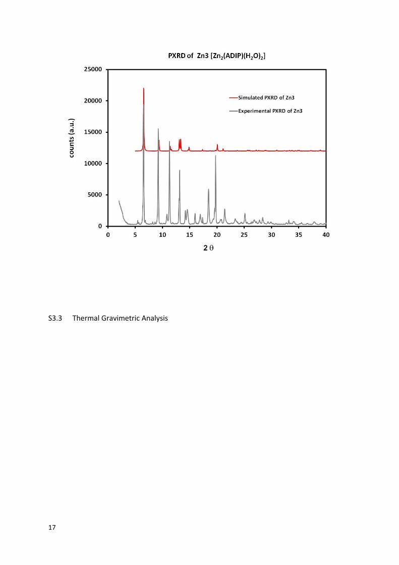
Structure Zn3 [Zn2(ADIP)(H2O)2] ·1.5DMF
Data was treated using the SQUEEZE routine of PLATON due to the presence of significant void space containing residual electron density that could not be modelled. The calculations found three identical voids of 1700 Å3 in the unit cell containing 607 e- each. Solvent assignment was made on the basis of calculated electron count and theromogravimetric analysis. Structure Zn3 has 1.5 DMF solvent molecules included per formula unit. The squeeze shows more solvent than in the TGA measurement which may be due to some solvent lost prior to the experiment.
Hydrogen atoms of the aqua ligands could not be located from the Fourier difference map and are therefore not included in the model, but have been included in the given formula unit.
13
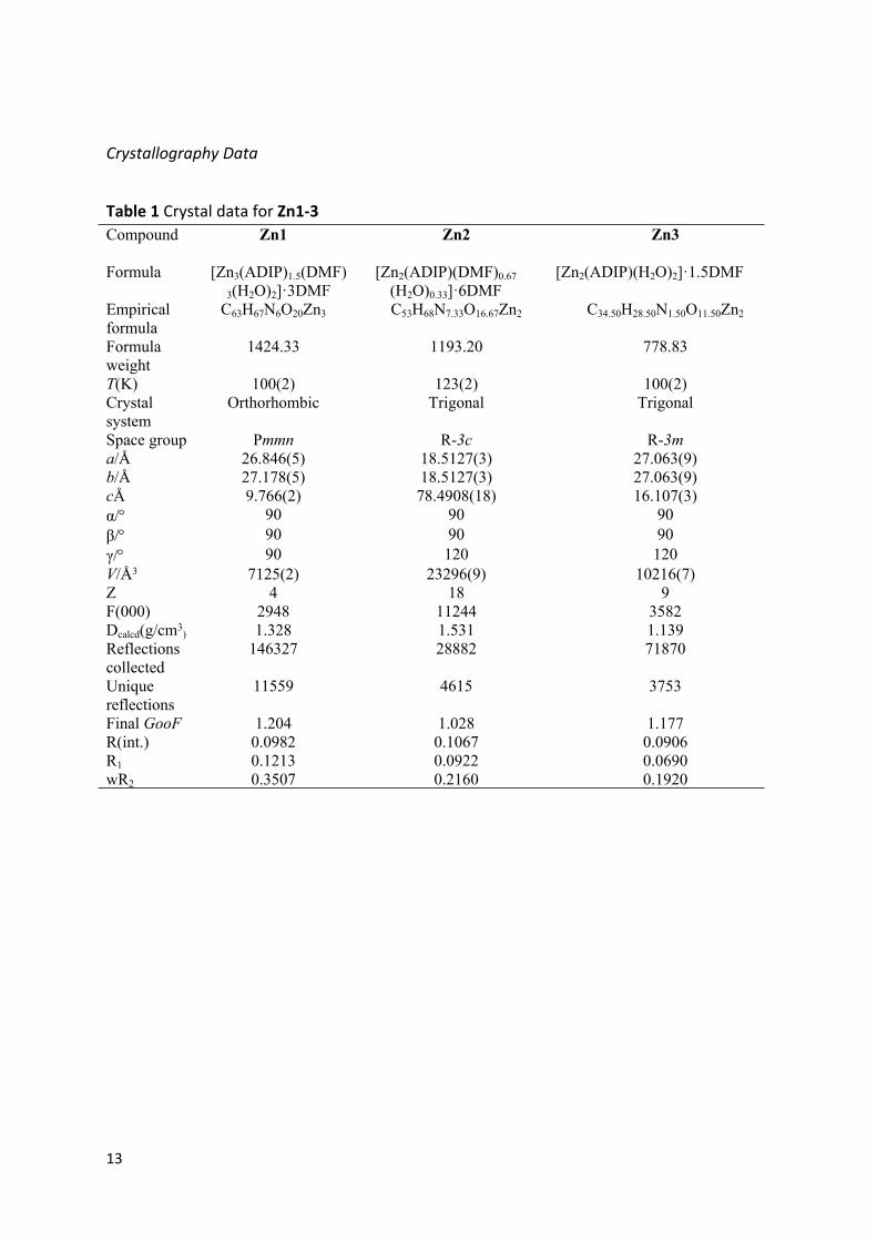
Crystallography Data
Table 1 Crystal data for Zn1-3Compound Zn1 Zn2 Zn3
Formula [Zn3(ADIP)1.5(DMF)3(H2O)2]·3DMF
[Zn2(ADIP)(DMF)0.67(H2O)0.33]·6DMF
[Zn2(ADIP)(H2O)2]·1.5DMF
Empirical formula
C63H67N6O20Zn3 C53H68N7.33O16.67Zn2 C34.50H28.50N1.50O11.50Zn2
Formula weight
1424.33 1193.20 778.83
T(K) 100(2) 123(2) 100(2)Crystal system
Orthorhombic Trigonal Trigonal
Space group Pmmn R-3c R-3ma/Å 26.846(5) 18.5127(3) 27.063(9)b/Å 27.178(5) 18.5127(3) 27.063(9)cÅ 9.766(2) 78.4908(18) 16.107(3)α/ 90 90 90β/ 90 90 90γ/ 90 120 120V/Å3 7125(2) 23296(9) 10216(7)Z 4 18 9F(000) 2948 11244 3582Dcalcd(g/cm3
) 1.328 1.531 1.139Reflections collected
146327 28882 71870
Unique reflections
11559 4615 3753
Final GooF 1.204 1.028 1.177R(int.) 0.0982 0.1067 0.0906R1 0.1213 0.0922 0.0690wR2 0.3507 0.2160 0.1920

14
Table 2 Selected bond lengths (Å) and angles () for Zn1-3Compound Zn1Zn1‒O2 2.075(8) Zn1‒O6 1.870(6) Zn1‒O9 1.90(1)Zn1‒O10 2.17(1) Zn2‒O1 1.962(7) Zn2‒O5 1.940(6)Zn2‒O3 1.989(9) Zn2‒O4 2.35(2) Zn3‒O8B 2.10(2)Zn3‒O12 2.17(1) Zn3‒O13 1.99(1) Zn1‒Zn2 3.487O2‒Zn1‒O6 100.1(3) O2‒Zn1‒O9 93.7(4) O2‒Zn1‒O10 177.3(5)O6‒Zn1‒O9 114.8(4) O6‒Zn1‒O10 81.1(5) O6‒Zn1‒O6(i) 124.6(3)O9‒Zn1‒O10 83.6(6) O1‒Zn2‒O5 107.5(3) O1‒Zn2‒O3 139.4(4)O1‒Zn2‒O4 85.3(4) O5‒Zn2‒O5(ii) 116.1(2) O5‒Zn2‒O3 93.2(4)O5‒Zn2‒O4 117.3(4) O8B‒Zn3‒O12 91.2(5) O8B‒Zn3‒O13 102.2(6)O8B‒Zn3‒O12(iii) 165.3(5) O13‒Zn3‒O12 89.1(5)Symmetry codes: (i) x,1/2-y, z (ii) x, ½ -y, z; (iii) 3/2-x, y, z Compound Zn2Zn1‒O4 2.050(4) Zn1‒O5 1.978(4) Zn1‒O1(i) 2.041(6)Zn1‒O3(ii) 2.010(4) Zn1‒O2(iii) 2.017(3) Zn1‒Zn1(iv) 3.003O4‒Zn1‒O1 148.7(2) O4‒Zn1‒O3 86.7(2) O4‒Zn1‒O2 88.0(2)O5‒Zn1‒O1 104.1(2) O5‒Zn1‒O3 97.1(2) O5‒Zn1‒O2 94.8(2)O1‒Zn1‒O3 89.8(2) O1‒Zn1‒O2 89.0(2) O4‒Zn1‒O5 107.3(2)Symmetry codes: (i) 1-x,1x+-y, ½-z (ii) x-y, -y, ½-z; (iii) 1-x+y, 1-xz; (iv) x, y, zCompound Zn3Zn1‒O1 2.021(2) Zn1‒O7 1.970(4) Zn1‒O2(i) 2.014(5)Zn1‒O1(ii) 2.021(5) Zn1‒Zn1(iii) 3.033 O1‒Zn1‒O7 101.1(2)O1‒Zn1‒O2 84.8(2) O1‒Zn1‒O2(i) 157.6(1) O1‒Zn1‒O1(ii) 90.7(1)O7‒Zn1‒O2 101.3(2) O7‒Zn1‒O1 101.1(2) O2‒Zn1‒O2(i) 91.1(2)Symmetry codes: (i) 1/3-x, 2/3-y, 2/3-z (ii) –x+y, y, z (iii) x, y, z
15
Powder X-ray Diffraction Analysis

16
17
S3.3 Thermal Gravimetric Analysis
18