supplementary table 1: primers for asic...
TRANSCRIPT
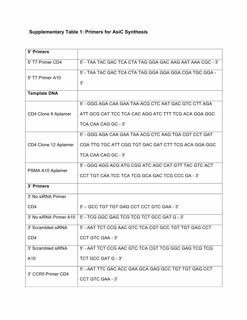
Supplementary Table 1: Primers for AsiC Synthesis
5’ Primers
5' T7 Primer CD4 5' - TAA TAC GAC TCA CTA TAG GGA GAC AAG AAT AAA CGC - 3'
5' T7 Primer A10 5' - TAA TAC GAC TCA CTA TAG GGA GGA GGA CGA TGC GGA -
3'
Template DNA
CD4 Clone 9 Aptamer
5' - GGG AGA CAA GAA TAA ACG CTC AAT GAC GTC CTT AGA
ATT GCG CAT TCC TCA CAC AGG ATC TTT TCG ACA GGA GGC
TCA CAA CAG GC - 3'
CD4 Clone 12 Aptamer
5' - GGG AGA CAA GAA TAA ACG CTC AAG TGA CGT CCT GAT
CGA TTG TGC ATT CGG TGT GAC GAT CTT TCG ACA GGA GGC
TCA CAA CAG GC - 3'
PSMA A10 Aptamer 5' - GGG AGG ACG ATG CGG ATC AGC CAT GTT TAC GTC ACT
CCT TGT CAA TCC TCA TCG GCA GAC TCG CCC GA - 3'
3’ Primers
3' No siRNA Primer
CD4 5' – GCC TGT TGT GAG CCT CCT GTC GAA - 3'
3' No siRNA Primer A10 5' - TCG GGC GAG TCG TCG TCT GCC GAT G - 3'
3' Scrambled siRNA
CD4
5' - AAT TCT CCG AAC GTC TCA CGT GCC TGT TGT GAG CCT
CCT GTC GAA - 3'
3' Scrambled siRNA
A10
5' - AAT TCT CCG AAC GTC TCA CGT TCG GGC GAG TCG TCG
TCT GCC GAT G - 3'
3' CCR5 Primer CD4 5' - AAT TTC GAC ACC GAA GCA GAG GCC TGT TGT GAG CCT
CCT GTC GAA - 3'

3' CCR5 Primer A10 5' - AAT TTC GAC ACC GAA GCA GAG TCG GGC GAG TCG TCG
TCT GCC GAT G - 3'
3' lamin Primer CD4 5' - AAT GTT CTT CTG GAA GTC CAG GCC TGT TGT GAG CCT
CCT GTC GAA - 3'
3' lamin Primer A10 5' - AAT GTT CTT CTG GAA GTC CAG TCG GGC GAG TCG TCG
TCT GCC GAT G - 3'
3' gag Primer CD4 5' - AAC CTG TCT CTC AGT ACA ATC GCC TGT TGT GAG CCT
CCT GTC GAA - 3'
3' gag Primer A10 5' - AAC CTG TCT CTC AGT ACA ATC TCG GGC GAG TCG TCG
TCT GCC GAT G - 3'
3' vif Primer CD4 5' - AAG GGA TGT GTA CTT CTG AAC GCC TGT TGT GAG CCT
CCT GTC GAA - 3'
3' vif Primer A10 5' - AAG GGA TGT GTA CTT CTG AAC TCG GGC GAG TCG TCG
TCT GCC GAT G - 3'
3’ EG5 Primer 5’- AAA TTG TCT TCA GGT CTT CAG GCC TGT TGT GAG CCT CCT
GTC GAA - 3'
3’ CD45 Primer 5’ – AAT GCT CTG AAA TTC AGC CAG GCC TGT TGT GAG CCT
CCT GTC GAA - 3’
3’ Luciferase Primer
5’-AAT CGA AGT ACT CAG CGT AAG GCC TGT TGTGAGCCTCCT
GTCGAA -3'
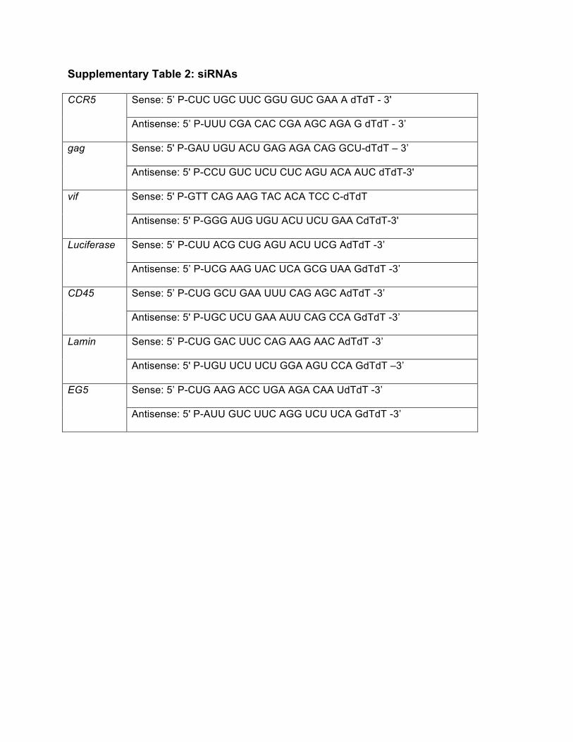
Supplementary Table 2: siRNAs
Sense: 5’ P-CUC UGC UUC GGU GUC GAA A dTdT - 3' CCR5
Antisense: 5’ P-UUU CGA CAC CGA AGC AGA G dTdT - 3’
Sense: 5' P-GAU UGU ACU GAG AGA CAG GCU-dTdT – 3’ gag
Antisense: 5' P-CCU GUC UCU CUC AGU ACA AUC dTdT-3'
Sense: 5' P-GTT CAG AAG TAC ACA TCC C-dTdT vif
Antisense: 5' P-GGG AUG UGU ACU UCU GAA CdTdT-3'
Sense: 5’ P-CUU ACG CUG AGU ACU UCG AdTdT -3’ Luciferase
Antisense: 5’ P-UCG AAG UAC UCA GCG UAA GdTdT -3’
Sense: 5’ P-CUG GCU GAA UUU CAG AGC AdTdT -3’ CD45
Antisense: 5' P-UGC UCU GAA AUU CAG CCA GdTdT -3’
Sense: 5’ P-CUG GAC UUC CAG AAG AAC AdTdT -3’ Lamin
Antisense: 5' P-UGU UCU UCU GGA AGU CCA GdTdT –3’
Sense: 5’ P-CUG AAG ACC UGA AGA CAA UdTdT -3’ EG5
Antisense: 5' P-AUU GUC UUC AGG UCU UCA GdTdT -3’
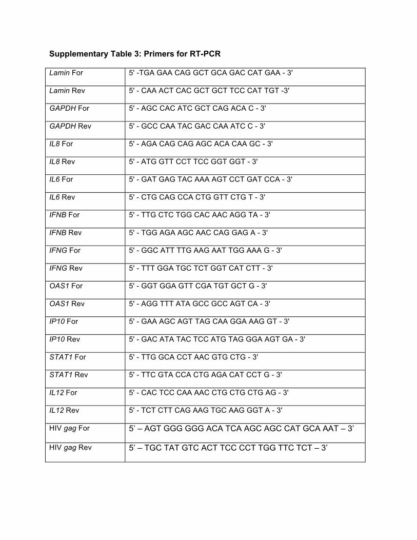
Supplementary Table 3: Primers for RT-PCR
Lamin For 5' -TGA GAA CAG GCT GCA GAC CAT GAA - 3'
Lamin Rev 5' - CAA ACT CAC GCT GCT TCC CAT TGT -3'
GAPDH For 5' - AGC CAC ATC GCT CAG ACA C - 3'
GAPDH Rev 5' - GCC CAA TAC GAC CAA ATC C - 3'
IL8 For 5' - AGA CAG CAG AGC ACA CAA GC - 3'
IL8 Rev 5' - ATG GTT CCT TCC GGT GGT - 3'
IL6 For 5' - GAT GAG TAC AAA AGT CCT GAT CCA - 3'
IL6 Rev 5' - CTG CAG CCA CTG GTT CTG T - 3'
IFNB For 5' - TTG CTC TGG CAC AAC AGG TA - 3'
IFNB Rev 5' - TGG AGA AGC AAC CAG GAG A - 3'
IFNG For 5' - GGC ATT TTG AAG AAT TGG AAA G - 3'
IFNG Rev 5' - TTT GGA TGC TCT GGT CAT CTT - 3'
OAS1 For 5' - GGT GGA GTT CGA TGT GCT G - 3'
OAS1 Rev 5' - AGG TTT ATA GCC GCC AGT CA - 3'
IP10 For 5' - GAA AGC AGT TAG CAA GGA AAG GT - 3'
IP10 Rev 5' - GAC ATA TAC TCC ATG TAG GGA AGT GA - 3'
STAT1 For 5' - TTG GCA CCT AAC GTG CTG - 3'
STAT1 Rev 5' - TTC GTA CCA CTG AGA CAT CCT G - 3'
IL12 For 5' - CAC TCC CAA AAC CTG CTG CTG AG - 3'
IL12 Rev 5' - TCT CTT CAG AAG TGC AAG GGT A - 3'
HIV gag For 5’ – AGT GGG GGG ACA TCA AGC AGC CAT GCA AAT – 3’
HIV gag Rev 5’ – TGC TAT GTC ACT TCC CCT TGG TTC TCT – 3’
Supplementary Figures
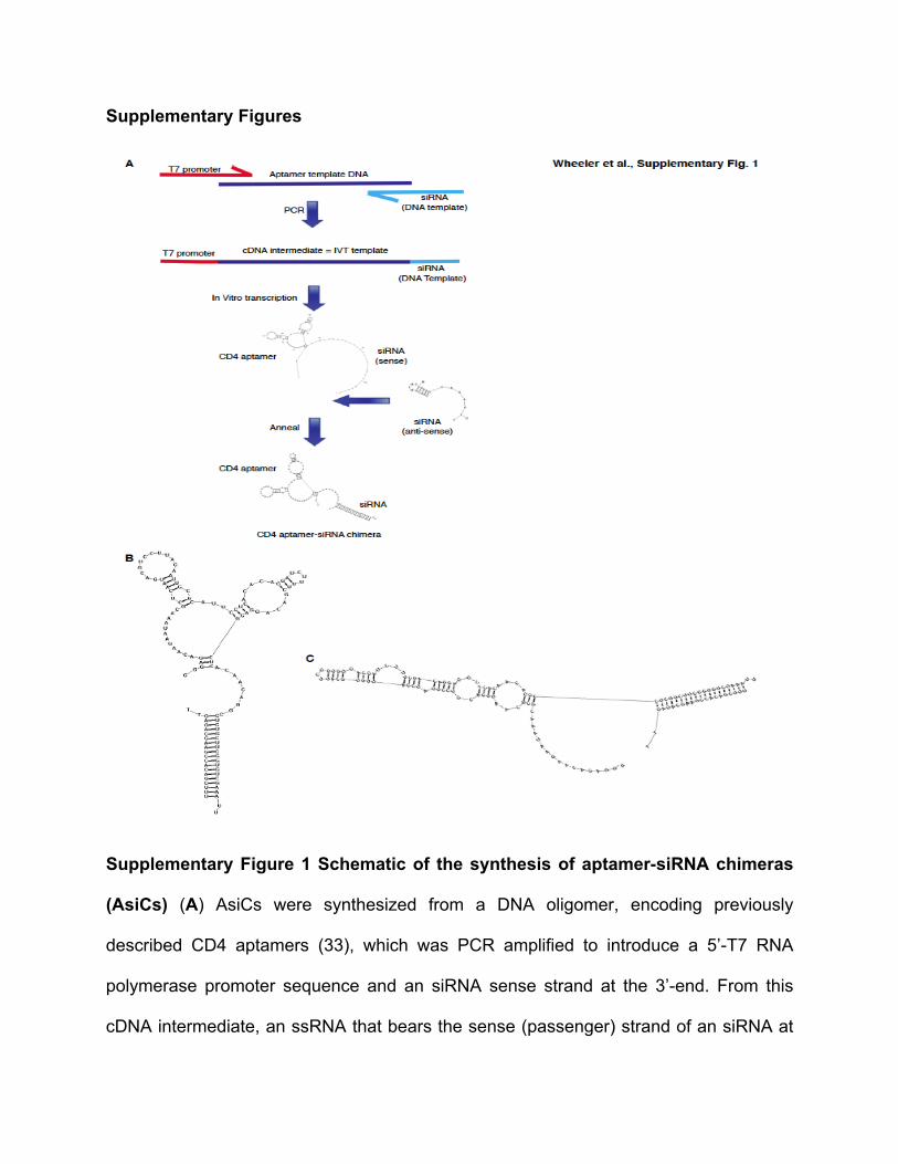
Supplementary Figure 1 Schematic of the synthesis of aptamer-siRNA chimeras
(AsiCs) (A) AsiCs were synthesized from a DNA oligomer, encoding previously
described CD4 aptamers (33), which was PCR amplified to introduce a 5’-T7 RNA
polymerase promoter sequence and an siRNA sense strand at the 3’-end. From this
cDNA intermediate, an ssRNA that bears the sense (passenger) strand of an siRNA at
its 3’-end was generated by in vitro transcription (IVT). This ssRNA was annealed to a
commercially synthesized anti-sense siRNA strand. Shown here are the predicted
secondary structures for two chimeras, each containing a specific CD4-aptamer clone,
(clone 9 (B) and clone 12 (C) from (33)), linked to a CCR5-siRNA duplex at their 3’-end.
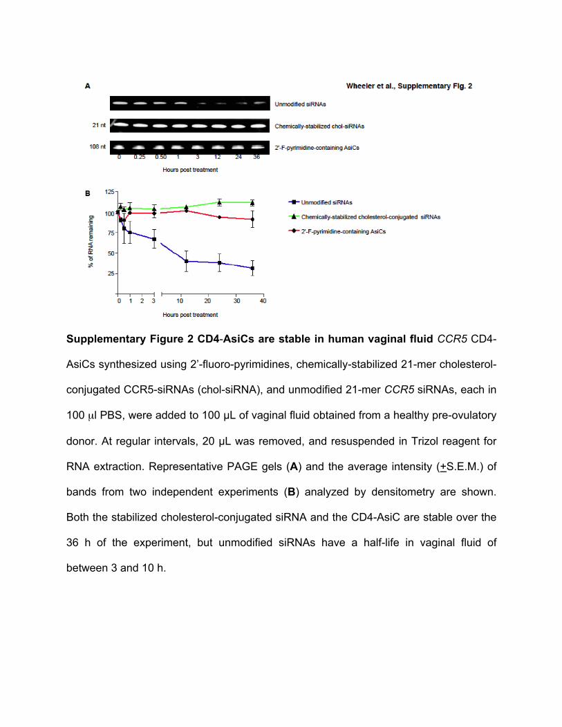
Supplementary Figure 2 CD4-AsiCs are stable in human vaginal fluid CCR5 CD4-
AsiCs synthesized using 2’-fluoro-pyrimidines, chemically-stabilized 21-mer cholesterol-
conjugated CCR5-siRNAs (chol-siRNA), and unmodified 21-mer CCR5 siRNAs, each in
100 µl PBS, were added to 100 µL of vaginal fluid obtained from a healthy pre-ovulatory
donor. At regular intervals, 20 µL was removed, and resuspended in Trizol reagent for
RNA extraction. Representative PAGE gels (A) and the average intensity (+S.E.M.) of
bands from two independent experiments (B) analyzed by densitometry are shown.
Both the stabilized cholesterol-conjugated siRNA and the CD4-AsiC are stable over the
36 h of the experiment, but unmodified siRNAs have a half-life in vaginal fluid of
between 3 and 10 h.
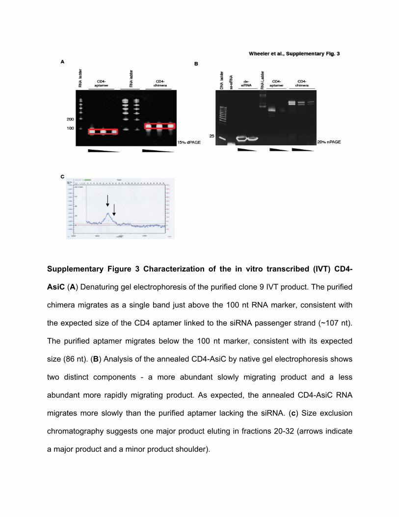
Supplementary Figure 3 Characterization of the in vitro transcribed (IVT) CD4-
AsiC (A) Denaturing gel electrophoresis of the purified clone 9 IVT product. The purified
chimera migrates as a single band just above the 100 nt RNA marker, consistent with
the expected size of the CD4 aptamer linked to the siRNA passenger strand (~107 nt).
The purified aptamer migrates below the 100 nt marker, consistent with its expected
size (86 nt). (B) Analysis of the annealed CD4-AsiC by native gel electrophoresis shows
two distinct components - a more abundant slowly migrating product and a less
abundant more rapidly migrating product. As expected, the annealed CD4-AsiC RNA
migrates more slowly than the purified aptamer lacking the siRNA. (c) Size exclusion
chromatography suggests one major product eluting in fractions 20-32 (arrows indicate
a major product and a minor product shoulder).
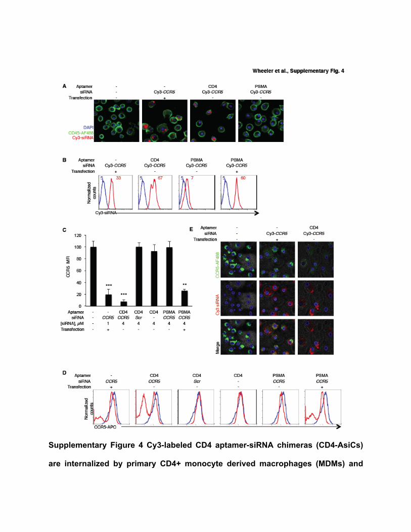
Supplementary Figure 4 Cy3-labeled CD4 aptamer-siRNA chimeras (CD4-AsiCs)
are internalized by primary CD4+ monocyte derived macrophages (MDMs) and
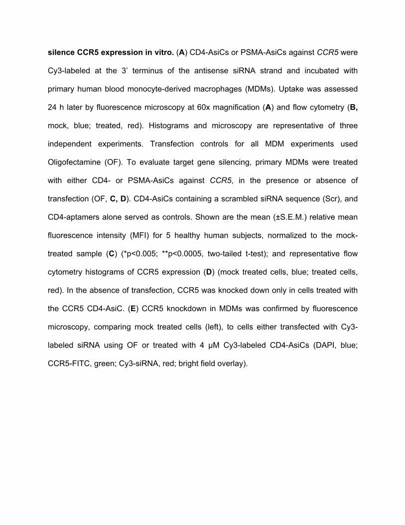
silence CCR5 expression in vitro. (A) CD4-AsiCs or PSMA-AsiCs against CCR5 were
Cy3-labeled at the 3’ terminus of the antisense siRNA strand and incubated with
primary human blood monocyte-derived macrophages (MDMs). Uptake was assessed
24 h later by fluorescence microscopy at 60x magnification (A) and flow cytometry (B,
mock, blue; treated, red). Histograms and microscopy are representative of three
independent experiments. Transfection controls for all MDM experiments used
Oligofectamine (OF). To evaluate target gene silencing, primary MDMs were treated
with either CD4- or PSMA-AsiCs against CCR5, in the presence or absence of
transfection (OF, C, D). CD4-AsiCs containing a scrambled siRNA sequence (Scr), and
CD4-aptamers alone served as controls. Shown are the mean (±S.E.M.) relative mean
fluorescence intensity (MFI) for 5 healthy human subjects, normalized to the mock-
treated sample (C) (*p<0.005; **p<0.0005, two-tailed t-test); and representative flow
cytometry histograms of CCR5 expression (D) (mock treated cells, blue; treated cells,
red). In the absence of transfection, CCR5 was knocked down only in cells treated with
the CCR5 CD4-AsiC. (E) CCR5 knockdown in MDMs was confirmed by fluorescence
microscopy, comparing mock treated cells (left), to cells either transfected with Cy3-
labeled siRNA using OF or treated with 4 µM Cy3-labeled CD4-AsiCs (DAPI, blue;
CCR5-FITC, green; Cy3-siRNA, red; bright field overlay).
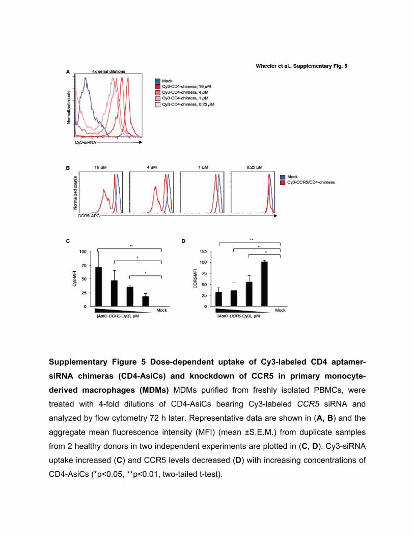
Supplementary Figure 5 Dose-dependent uptake of Cy3-labeled CD4 aptamer-
siRNA chimeras (CD4-AsiCs) and knockdown of CCR5 in primary monocyte-derived macrophages (MDMs) MDMs purified from freshly isolated PBMCs, were
treated with 4-fold dilutions of CD4-AsiCs bearing Cy3-labeled CCR5 siRNA and
analyzed by flow cytometry 72 h later. Representative data are shown in (A, B) and the
aggregate mean fluorescence intensity (MFI) (mean ±S.E.M.) from duplicate samples
from 2 healthy donors in two independent experiments are plotted in (C, D). Cy3-siRNA
uptake increased (C) and CCR5 levels decreased (D) with increasing concentrations of
CD4-AsiCs (*p<0.05, **p<0.01, two-tailed t-test).
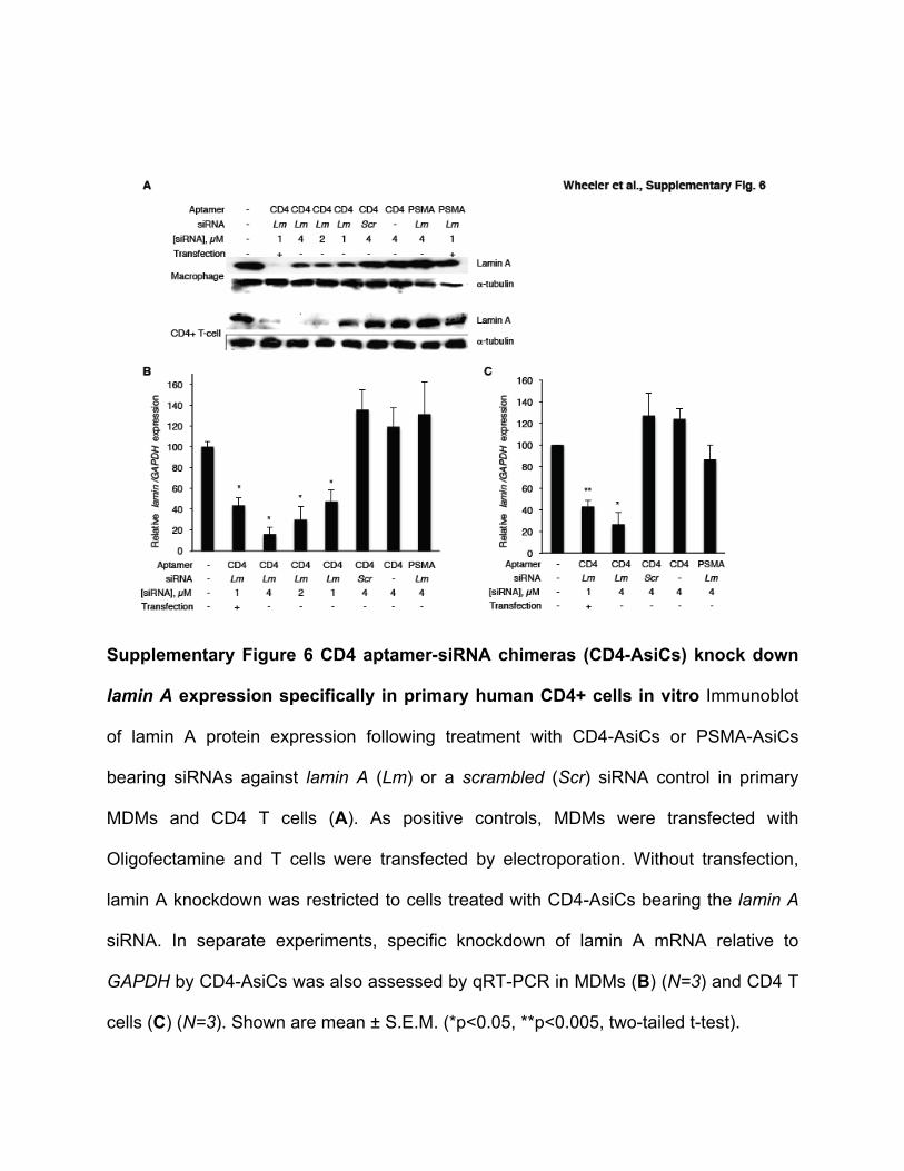
Supplementary Figure 6 CD4 aptamer-siRNA chimeras (CD4-AsiCs) knock down
lamin A expression specifically in primary human CD4+ cells in vitro Immunoblot
of lamin A protein expression following treatment with CD4-AsiCs or PSMA-AsiCs
bearing siRNAs against lamin A (Lm) or a scrambled (Scr) siRNA control in primary
MDMs and CD4 T cells (A). As positive controls, MDMs were transfected with
Oligofectamine and T cells were transfected by electroporation. Without transfection,
lamin A knockdown was restricted to cells treated with CD4-AsiCs bearing the lamin A
siRNA. In separate experiments, specific knockdown of lamin A mRNA relative to
GAPDH by CD4-AsiCs was also assessed by qRT-PCR in MDMs (B) (N=3) and CD4 T
cells (C) (N=3). Shown are mean ± S.E.M. (*p<0.05, **p<0.005, two-tailed t-test).
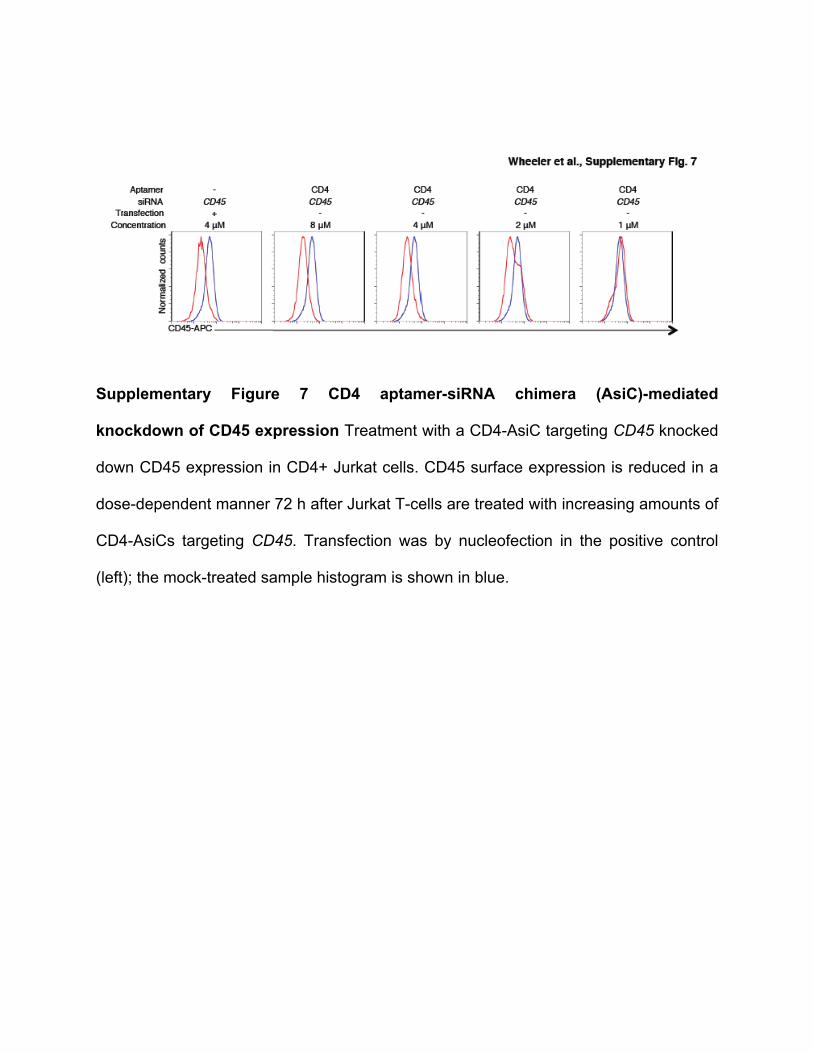
Supplementary Figure 7 CD4 aptamer-siRNA chimera (AsiC)-mediated
knockdown of CD45 expression Treatment with a CD4-AsiC targeting CD45 knocked
down CD45 expression in CD4+ Jurkat cells. CD45 surface expression is reduced in a
dose-dependent manner 72 h after Jurkat T-cells are treated with increasing amounts of
CD4-AsiCs targeting CD45. Transfection was by nucleofection in the positive control
(left); the mock-treated sample histogram is shown in blue.
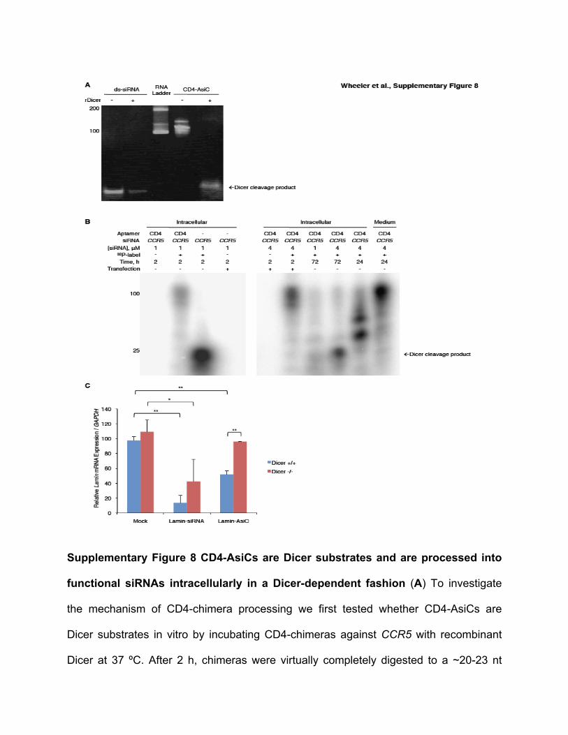
Supplementary Figure 8 CD4-AsiCs are Dicer substrates and are processed into
functional siRNAs intracellularly in a Dicer-dependent fashion (A) To investigate
the mechanism of CD4-chimera processing we first tested whether CD4-AsiCs are
Dicer substrates in vitro by incubating CD4-chimeras against CCR5 with recombinant
Dicer at 37 ºC. After 2 h, chimeras were virtually completely digested to a ~20-23 nt

siRNA duplex that migrated like a commercially synthesized CCR5-siRNA. (B)
Intracellular processing was demonstrated by treating primary CD4+ T cells with CD4-
AsiCs bearing CCR5-siRNAs labeled with 32P at their 5’-end. T cells were incubated for
24 or 72 h with radiolabeled chimeras. Total RNA was harvested by TRIZOL extraction
and the same total number of counts was loaded onto a native polyacrylamide gel.
Nucleofection of 5’-end-labeled chimeras and commercially-synthesized siRNA
duplexes served both as controls and size standards. While some cleavage was seen at
24 h post treatment, after 72 h of treatment, the 32P labeled chimera isolated from cell
lysates was ~21-23 nt in length, suggesting that these fragments are processed in
primary CD4+ cells into siRNA-sized duplexes. (C) To evaluate the functional
dependence of CD4-AsiC-mediated silencing on intracellular Dicer expression we
evaluated target gene silencing of lipofectamine-transfected CD4-chimeras in either
wild-type (WT) or Dicer-/- HCT-116 cells (35). Silencing of lamin A by CD4-AsiCs was
only observed in WT cells, whereas gene knockdown by transfected lamin A siRNAs
was not differentially affected by Dicer expression. Thus, CD4-AsiCs are Dicer
substrates and are processed in primary cells to release functional siRNA duplexes in a
Dicer-dependent manner.
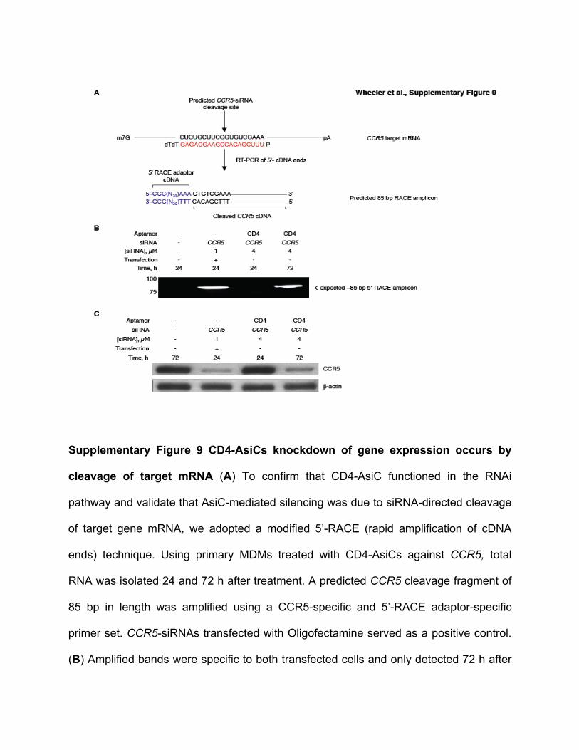
Supplementary Figure 9 CD4-AsiCs knockdown of gene expression occurs by
cleavage of target mRNA (A) To confirm that CD4-AsiC functioned in the RNAi
pathway and validate that AsiC-mediated silencing was due to siRNA-directed cleavage
of target gene mRNA, we adopted a modified 5’-RACE (rapid amplification of cDNA
ends) technique. Using primary MDMs treated with CD4-AsiCs against CCR5, total
RNA was isolated 24 and 72 h after treatment. A predicted CCR5 cleavage fragment of
85 bp in length was amplified using a CCR5-specific and 5’-RACE adaptor-specific
primer set. CCR5-siRNAs transfected with Oligofectamine served as a positive control.
(B) Amplified bands were specific to both transfected cells and only detected 72 h after
incubation with the CD4-chimera. Sequencing of the amplified fragments confirmed that
cleavage occurred 10 nt from the 5’-end of the CCR5 antisense strand, as predicted.
(C) Functional silencing of target CCR5 protein was confirmed by immunoblot using
protein isolated form the same TRIZOL extraction. These data, together with
Supplementary Figure 8, suggest that CD4-AsiCs are processed by Dicer to release
functional siRNA duplexes that direct target mRNA cleavage via the RNAi pathway.
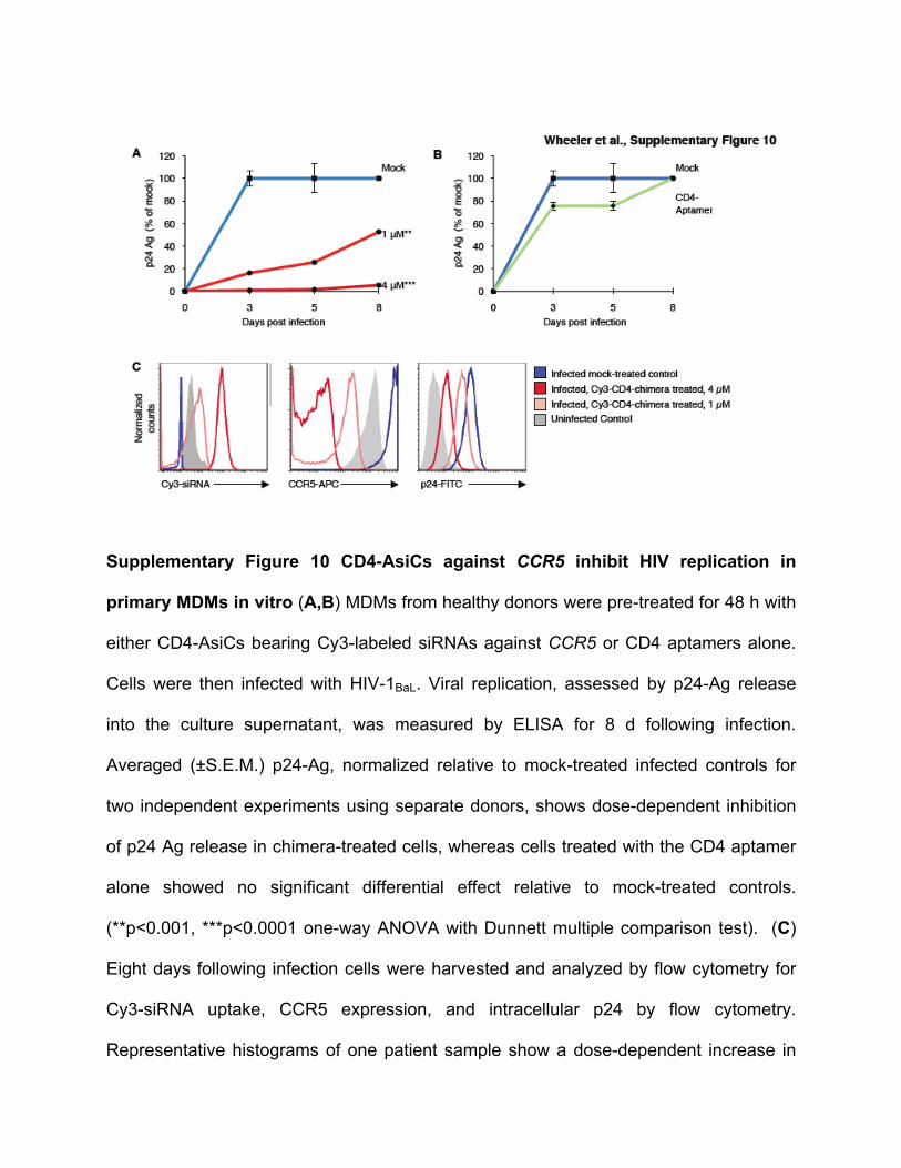
Supplementary Figure 10 CD4-AsiCs against CCR5 inhibit HIV replication in
primary MDMs in vitro (A,B) MDMs from healthy donors were pre-treated for 48 h with
either CD4-AsiCs bearing Cy3-labeled siRNAs against CCR5 or CD4 aptamers alone.
Cells were then infected with HIV-1BaL. Viral replication, assessed by p24-Ag release
into the culture supernatant, was measured by ELISA for 8 d following infection.
Averaged (±S.E.M.) p24-Ag, normalized relative to mock-treated infected controls for
two independent experiments using separate donors, shows dose-dependent inhibition
of p24 Ag release in chimera-treated cells, whereas cells treated with the CD4 aptamer
alone showed no significant differential effect relative to mock-treated controls.
(**p<0.001, ***p<0.0001 one-way ANOVA with Dunnett multiple comparison test). (C)
Eight days following infection cells were harvested and analyzed by flow cytometry for
Cy3-siRNA uptake, CCR5 expression, and intracellular p24 by flow cytometry.
Representative histograms of one patient sample show a dose-dependent increase in
Cy3-siRNA uptake and decrease in both CCR5 expression and HIV infection measured
by intracellular p24 staining.
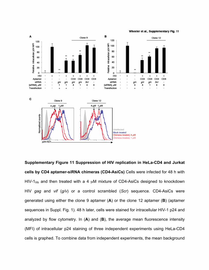
Supplementary Figure 11 Suppression of HIV replication in HeLa-CD4 and Jurkat
cells by CD4 aptamer-siRNA chimeras (CD4-AsiCs) Cells were infected for 48 h with
HIV-1IIIb and then treated with a 4 µM mixture of CD4-AsiCs designed to knockdown
HIV gag and vif (g/v) or a control scrambled (Scr) sequence. CD4-AsiCs were
generated using either the clone 9 aptamer (A) or the clone 12 aptamer (B) (aptamer
sequences in Suppl. Fig. 1). 48 h later, cells were stained for intracellular HIV-1 p24 and
analyzed by flow cytometry. In (A) and (B), the average mean fluorescence intensity
(MFI) of intracellular p24 staining of three independent experiments using HeLa-CD4
cells is graphed. To combine data from independent experiments, the mean background
fluorescence of uninfected control cells was subtracted and the signal was then
normalized to the mean value of HIV-infected mock-treated control cells. (Shown are
mean ± S.E.M.; *p<0.05; **p<0.005, two-tailed t-test). In (C) Jurkat cells were treated 48
h after HIV-1IIIb infection with an equimolar mixture of either clone 9 (left) or clone 12
(right) CD4-AsiCs encoding gag and vif siRNAs. Intracellular p24 staining was
measured 48 h later by flow cytometry. The blue histogram represents p24 staining of
HIV-infected mock-treated cells and the gray histogram represents uninfected controls.
In both cell lines suppression of HIV replication by the CD4-AsiCs increased with dose.
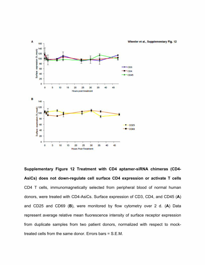
Supplementary Figure 12 Treatment with CD4 aptamer-siRNA chimeras (CD4-
AsiCs) does not down-regulate cell surface CD4 expression or activate T cells
CD4 T cells, immunomagnetically selected from peripheral blood of normal human
donors, were treated with CD4-AsiCs. Surface expression of CD3, CD4, and CD45 (A)
and CD25 and CD69 (B), were monitored by flow cytometry over 2 d. (A) Data
represent average relative mean fluorescence intensity of surface receptor expression
from duplicate samples from two patient donors, normalized with respect to mock-
treated cells from the same donor. Errors bars = S.E.M.
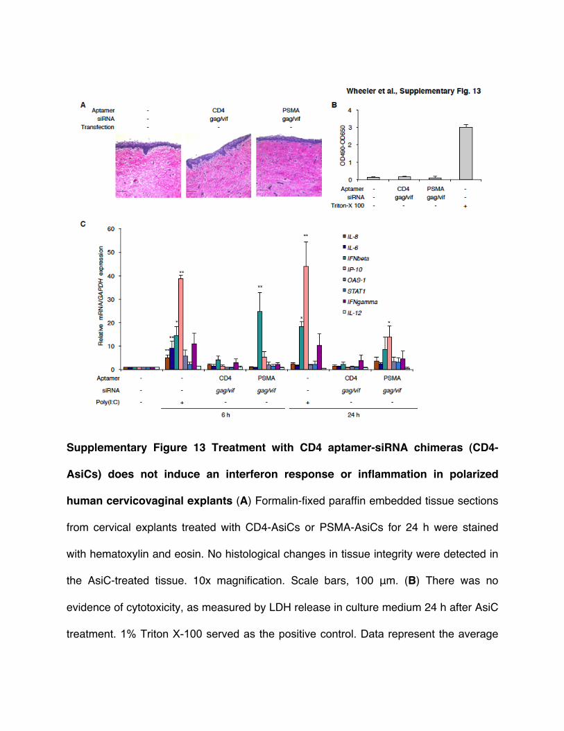
Supplementary Figure 13 Treatment with CD4 aptamer-siRNA chimeras (CD4-
AsiCs) does not induce an interferon response or inflammation in polarized
human cervicovaginal explants (A) Formalin-fixed paraffin embedded tissue sections
from cervical explants treated with CD4-AsiCs or PSMA-AsiCs for 24 h were stained
with hematoxylin and eosin. No histological changes in tissue integrity were detected in
the AsiC-treated tissue. 10x magnification. Scale bars, 100 μm. (B) There was no
evidence of cytotoxicity, as measured by LDH release in culture medium 24 h after AsiC
treatment. 1% Triton X-100 served as the positive control. Data represent the average
(+ S.E.M.) from quadruplicate biological replicates in one representative experiment of
three independent experiments. (C) qRT-PCR was used to measure mRNA expression
in cervical tissue explants 6 and 24 h after treatment with CD4-AsiCs or PSMA-AsiCs.
Data were normalized to GAPDH mRNA. Cytokine and interferon responsive genes that
might be triggered by innate immune RNA receptors were evaluated. Data represent
mean (+ S.E.M.) from at least three independent experiments (*p<0.05; **p<0.01, one
way ANOVA with Dunnett multiple comparison test).
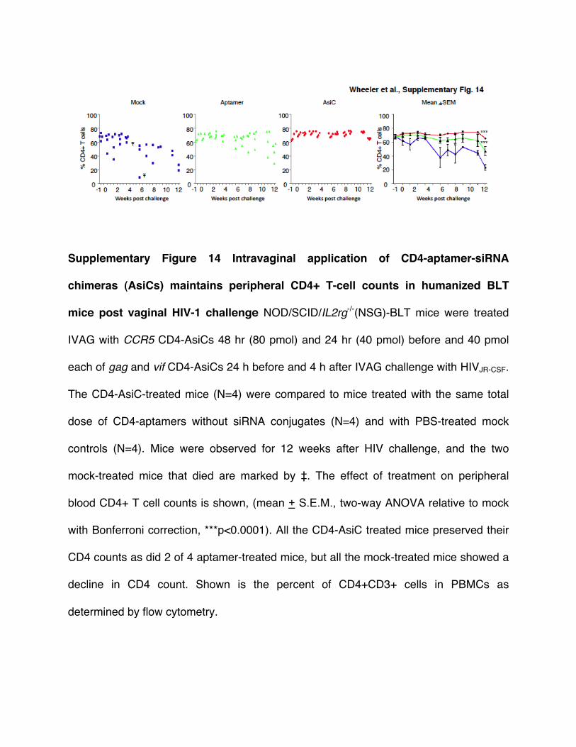
Supplementary Figure 14 Intravaginal application of CD4-aptamer-siRNA
chimeras (AsiCs) maintains peripheral CD4+ T-cell counts in humanized BLT
mice post vaginal HIV-1 challenge NOD/SCID/IL2rg-/-(NSG)-BLT mice were treated
IVAG with CCR5 CD4-AsiCs 48 hr (80 pmol) and 24 hr (40 pmol) before and 40 pmol
each of gag and vif CD4-AsiCs 24 h before and 4 h after IVAG challenge with HIVJR-CSF.
The CD4-AsiC-treated mice (N=4) were compared to mice treated with the same total
dose of CD4-aptamers without siRNA conjugates (N=4) and with PBS-treated mock
controls (N=4). Mice were observed for 12 weeks after HIV challenge, and the two
mock-treated mice that died are marked by ‡. The effect of treatment on peripheral
blood CD4+ T cell counts is shown, (mean + S.E.M., two-way ANOVA relative to mock
with Bonferroni correction, ***p<0.0001). All the CD4-AsiC treated mice preserved their
CD4 counts as did 2 of 4 aptamer-treated mice, but all the mock-treated mice showed a
decline in CD4 count. Shown is the percent of CD4+CD3+ cells in PBMCs as
determined by flow cytometry.