supplementary information - · pdf file1/21 supporting materials for porphysome nanovesicles...
TRANSCRIPT
1/21
Supporting materials for Porphysome Nanovesicles Generated by Porphyrin Bilayers Jonathan F. Lovell, Cheng S. Jin, Elizabeth Huynh, Honglin Jin, Chulhong Kim, John L. Rubinstein, Warren C. W. Chan, Weiguo Cao, Lihong V. Wang and Gang Zheng*
*email:[email protected].
SUPPORTING MATERIALS CONTENTS
Supplementary Methods Supplementary Figure S1 Generation of 30 nm porphysomes. Supplementary Figure S2 Absorbance of porphyrin-lipid in organic solvent. Supplementary Figure S3 Porphyrin-lipid lacks a conventional transition temperature. Supplementary Figure S4 Photoacoustic detection of porphysomes. Supplementary Figure S5 Distinct spectral responses of blood and porphysomes in vivo. Supplementary Figure S6 Photoacoustic mapping of multiple lymph nodes using porphysomes. Supplementary Figure S7 Low fluorescence background of porphysomes. Supplementary Figure S8 Colocalization of porphysomes in early endosomes and lysosomes. Supplementary Figure S9 Biodistribution and blood clearance of porphysomes.
Supplementary Methods
Synthesis of pyropheophorbide-lipid
In a standard reaction, 100 nmol of 1-palmitoyl-2-hydroxy-sn-glycero-3-phosphocholine (Avanti Polar
Lipids), 50 nmol pyropheophorbide (prepared from Spirulina Pacifica algae as described previously;
Zheng et al., Bioconj. Chem., 2002, 13-392), 50 nmol of 1-ethyl-3-(3-dimethylaminopropyl)
carbodiimide (Sigma), 25 nmol of 4-(dimethylamino) pyridine (Sigma) and 50 µL of N,N-
diisopropylethylamine (Sigma) were combined in 10 mL of anhydrous dichloromethane. The reaction
mixture was stirred at room temperature under argon in the dark for 48 hours. The solvent was evaporated
and the residue was subjected to thin layer chromatography purification (20 x 20 cm pre-coated silica gel
plate with fluorescent indicator, 1.5 mm in thickness). Chloroform-methanol-glacial acetic acid-water
65:25:8:2 (volume ratio) was used as the solvent. The major band with Rf=0.4 was isolated from the plate
and eluted giving a final yield of 45%. Recently, we found that improved purification could be achieved
by using diol modified silica (Sorbtech) and eluting the product with 8% methanol in DCM after washing
out impurities with 2% and 5% methanol in DCM. The pyropheophrobide-lipid was then dried under
Porphysome nanovesicles generated by porphyrin bilayers for use as multimodal biophotonic contrast agents
Jonathan F. Lovell, Cheng S. Jin, Elizabeth Huynh, Honglin Jin, Chulhong Kim, John L. Rubinstein, Warren C. W. Chan, Weiguo Cao, Lihong V. Wang and Gang Zheng*
*e-mail: [email protected]
SUPPLEMENTARY INFORMATIONdoi: 10.1038/nmat2986
nature materials | www.nature.com/naturematerials 1
© 2011 Macmillan Publishers Limited. All rights reserved.
2/21
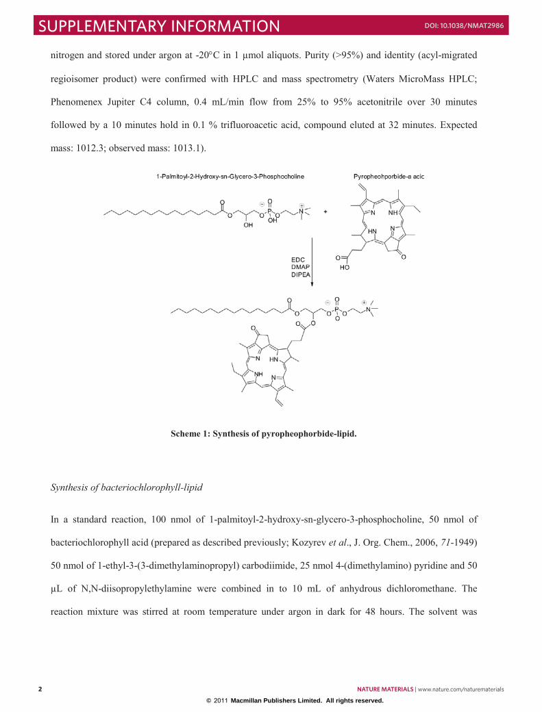
nitrogen and stored under argon at -20C in 1 µmol aliquots. Purity (>95%) and identity (acyl-migrated
regioisomer product) were confirmed with HPLC and mass spectrometry (Waters MicroMass HPLC;
Phenomenex Jupiter C4 column, 0.4 mL/min flow from 25% to 95% acetonitrile over 30 minutes
followed by a 10 minutes hold in 0.1 % trifluoroacetic acid, compound eluted at 32 minutes. Expected
mass: 1012.3; observed mass: 1013.1).
Scheme 1: Synthesis of pyropheophorbide-lipid.
Synthesis of bacteriochlorophyll-lipid
In a standard reaction, 100 nmol of 1-palmitoyl-2-hydroxy-sn-glycero-3-phosphocholine, 50 nmol of
bacteriochlorophyll acid (prepared as described previously; Kozyrev et al., J. Org. Chem., 2006, 71-1949)
50 nmol of 1-ethyl-3-(3-dimethylaminopropyl) carbodiimide, 25 nmol 4-(dimethylamino) pyridine and 50
µL of N,N-diisopropylethylamine were combined in to 10 mL of anhydrous dichloromethane. The
reaction mixture was stirred at room temperature under argon in dark for 48 hours. The solvent was
2 nature materials | www.nature.com/naturematerials
SUPPLEMENTARY INFORMATION doi: 10.1038/nmat2986
© 2011 Macmillan Publishers Limited. All rights reserved.

3/21
evaporated and the residue was subjected to thin layer chromatography plate purification (20 x 20 cm pre-
coated silica gel TLC plate with fluorescence indicator, 1.5 mm in thickness). Chloroform-methanol-
glacial acetic acid-water 65:25:8:2 (volume ratio) was used as the developing system. The final product
was obtained in 38% yield with Rf=0.4. The final product spontaneously oxidized to yield an oxidized
derivative of bacteriochlorophyll-lipid, which was verified by mass spectrometry and the expected
structure is shown in Scheme 2. After purification, the lipid was aliquoted, dried and stored under argon at
-20 C. The purity (>95%) and identity (acyl-migrated regioisomer product) were confirmed by analytical
HPLC and mass spectrometry. (Same protocol as pyropheophorbide-lipid. Compound eluted at 32
minutes. Expected mass: 1104.3; observed mass: 1104.8).
Scheme 2: Synthesis of bacteriochlorophyll-lipid.
nature materials | www.nature.com/naturematerials 3
SUPPLEMENTARY INFORMATIONdoi: 10.1038/nmat2986
© 2011 Macmillan Publishers Limited. All rights reserved.

4/21
Generation of metallic pyropheophorbide-lipid
To generate porphyrin-lipid conjugates with a chelated metal, 10 fold excess free zinc acetate (Bioshop
Canada) was incubated with pyro-lipid in methanol for 1 hour at room temperature under argon. The
same protocol was repeated with copper acetate and palladium acetate. Free metal was removed with 5
butanol/water extractions. The metal porphyrin-lipid was then aliquoted, dried and stored under argon at -
20 C. The stable metal incorporation, purity (>95%) and identity of the porphyrin lipids were confirmed
by HPLC and mass spectrometry (elution time 31 min; expected mass: 1075.6; observed mass: 1075.0).
Scheme 3: Generation of metallic pyro-lipid. This approach was possible for several metals, including zinc,
copper and palladium.
Formation of porphysomes
Porphyrin-lipid films were prepared in 12 mm x 75 mm borosilicate test tubes (Fisher Scientific) by
combining 95 molar % porphyrin-lipid with 5 molar % distearoyl-sn-glycero-3-phosphoethanolamine-N-
methoxy(polyethylene glycol) (PEG-2000-PE, Avanti Polar Lipids) in chloroform. For folate conjugated
porphysomes, 4 molar % PEG-2000-PE was supplemented with 1 molar % 1,2-distearoyl-sn-glycero-3-
4 nature materials | www.nature.com/naturematerials
SUPPLEMENTARY INFORMATION doi: 10.1038/nmat2986
© 2011 Macmillan Publishers Limited. All rights reserved.

5/21
phosphoethanolamine-N-folate(polyethylene glycol) (Folate-PEG-PE, Avanti Polar Lipids) in
chloroform. Films were dried under a stream of nitrogen gas and further dried under vacuum for 1 hour.
The lipid film was stored at -20° C under argon until hydration with phosphate PBS (150 mM NaCl, 10
mM phosphate, pH 7.4) and was then subjected to five freeze-thaw cycles, by freezing the test tube in
liquid nitrogen and thawing it in water heated to 65° C. The porphysome suspension was extruded 15
times using a Mini-Extruder (Avanti Polar Lipids) through a 100 nm pore size polycarbonate membrane
(Avanti Polar Lipids) at 65° C. Porphysomes were usually formed at 1 or 0.5 mg/mL combined
porphyrin-lipid and PEG-lipid concentration. Final porphysome concentration was assessed after their
extrusion by measuring the absorption of a dilute sample in methanol (Bioshop Canada) and using
extinction coefficients of 97,000 M-1cm-1 at 410 nm for pyropheophorbide-lipid and 37,000 M-1cm-1 at
750 nm for bacteriochlorophyll-lipid and assuming 83,000 porphyrin-lipids per 100 nm porphysome
containing 95% porphyrin-lipid24. Usually 1-2 µL of porphysomes were diluted in 1 mL of methanol for
the measurement. Porphysomes were stored at 4° C under argon until use. For the large scale
porphysomes used for in vivo toxicity assessment, porphyrin-lipid was combined with PEG-lipid in a 50
mL round bottom flask and the organic solvent was evaporated under reduced pressure. The flask was
then hydrated with approximately 10 mL of PBS (for ~75 mg lipid) and the solution was subjected to 5
freeze-thaw cycles. Porphysomes were then formed by sonicating the flask at 55 C for 1 hour.
Porphysomes were then filtered through a 0.2 µm filter (Acrodisc filter, Pall) and concentrated with a
centrifugal conical tube concentrator with 100 kDa membrane pore size (Millipore). Final size (125 nm)
was assessed by dynamic light scattering and concentration was determined by absorption. To form small
30 nm porphysomes, a pure porphyrin-lipid film was generated with 0.1 mg porphyrin-lipid and dried
under nitrogen and vacuum. The film was rehydrated with 200 µL of water and was sonicated for 10
minutes at 55 C.
Characterization of size and shape of porphysomes
nature materials | www.nature.com/naturematerials 5
SUPPLEMENTARY INFORMATIONdoi: 10.1038/nmat2986
© 2011 Macmillan Publishers Limited. All rights reserved.
6/21
Liposome and porphysome size was measured using a Malvern Nanosizer ZS90 (Malvern Instruments).
Liposome and porphysome solutions were diluted to 6 µg/mL in PBS and three measurements were
performed with 15 runs each and the results averaged. Electron microscopy specimens were prepared by
incubating 0.05 mg/mL pyropheophorbide porphysomes (5% PEG-lipid, 95% pyro-lipid) on glow
discharged carbon coated grids for 2 minutes, rinsing three times with milli-Q water and staining with 2%
uranyl acetate. Samples were then visualized with a Tecnai F20 electron microscope (FEI Company)
operating at 200 kV and images were recorded with a Tietz F114 CCD (TVIPS).
Characterization of porphysome self-quenching
Porphysomes and liposomes were formed by first creating separate stock solutions of porphyrin-lipid, egg
yolk phosphatidylcholine (EYPC) and cholesterol in chloroform. Free pyrophephorbide was dissolved in
methanol. These constituents were combined at the indicated molar ratios (with a constant EYPC:CHOL
ratio, and increasing amounts of pyro-lipid or free pyrophephorbide) in separate test tubes. The organic
solvent was then evaporated under a nitrogen stream and trace organic solvent was removed by drying the
films under vacuum. The separate lipid films containing all the indicated components were then hydrated
with PBS, freeze-thawed and extruded as described above. Emission spectra were recorded with a
Fluoromax fluorometer (Horiba Jobin Yvon) using 2 nm slit widths. Porphysome solutions were diluted
to 0.02 µg/mL in PBS and those containing free pyropheophorbide or pyropheophorbide-lipid were
excited at 420 nm and emission was measured and integrated from 600 nm to 750 nm. Background
subtraction of an equal concentration of 100 nm egg phosphatidyl choline:cholesterol (3:2) liposomes was
performed. NBD liposomes were formed in the same manner as porphysomes, but by replacing the
porphyrin-lipid with 1-palmitoyl-2-{12-[(7-nitro-2-1,3-benzoxadiazol-4-yl)amino]lauroyl}-sn-glycero-3-
phosphocholine (Avanti Polar Lipids). NBD liposomes were excited at 470 nm and emission was
measured and integrated from 500 nm to 600 nm. The fluorescence self-quenching FDET/F0 of each sample
was determined by ratio of the integrated fluorescence emission in the presence or absence of 0.5% Triton
X-100 (Bioshop) over four measurements from separate preparations.
6 nature materials | www.nature.com/naturematerials
SUPPLEMENTARY INFORMATION doi: 10.1038/nmat2986
© 2011 Macmillan Publishers Limited. All rights reserved.

7/21
Resonance light scattering of porphysomes
Pyropheophorbide porphysomes and gold nanorods (40 nm length by 15 nm width, estimated ε680 =
3.5×10⁹ M-1cm-1, based on Orendorff and Murphy, 2006, J. Phys Chem. B., 110-3990) kindly provided by
the Kumacheva lab, University of Toronto) were adjusted to the same absorbance at 680 nm of 0.067 in
PBS. Excitation and emission were then set to the same wavelength using 1 nm slit widths and scanned
from 400 nm to 700 nm. After blank subtraction, the resonance scatter of the two samples was divided.
Similar results were obtained with commercial 650 nm wavelength nanorods (Nanopartz).
Differential scanning calorimetry
Differential scanning calorimetry was performed on 5 mg/mL samples of 1,2-dimyristoyl-sn-glycero-3-
phosphocholine (DMPC), hydrogenated soy phosphatidyl choline (HSPC) and pyropheophorbide
porphysomes using a 6100 Nano Differential Scanning Calorimeter (Calorimetry Sciences Corporation).
Samples were prepared by forming 5 mg lipid films, rehydrating in 1 mL of PBS and sonicating at 60°C
for 15 minutes. Samples were degassed in a vacuum for 30 minutes prior to measurement and scanned at
a rate of 1°C/min. PBS was used as the reference and one heating and cooling cycle as the baseline. For
each sample, three cooling and heating scans from 5°C to 95°C were performed and the results averaged
to determine the phase transition temperature of the lipid.
Photothermal properties of porphysomes
5 µL drops were placed on a piece of parafilm. All solutions were measured in PBS, with liposomes and
porphysomes normalized to 0.5 mg/mL concentration. Porphysomes and gold nanorods were also
normalized to an optical density at 680 nm of 0.8. Samples were irradiated with a 673 nm diode laser with
150 mW output and the temperature equilibrated within 60 seconds. Surface temperature was then imaged
using a temperature calibrated infrared camera (Mikroshot).
Characterization of photoacoustic properties of porphysomes
nature materials | www.nature.com/naturematerials 7
SUPPLEMENTARY INFORMATIONdoi: 10.1038/nmat2986
© 2011 Macmillan Publishers Limited. All rights reserved.

8/21
Photoacoustic measurements were carried out using a Ti:Saphire tunable laser setup with a ultrasound
transducer as previously described (see Cho et al., J. Phys. Chem., 2009, 113-9023). The light fluence
was less than 7 mJ/cm2 for photoacoustic measurement, within the ANSI limit. The axial and transverse
resolutions of the system were 150 μm and 590 μm, respectively. By measuring the arrival times of
generated photoacoustic signals, one-dimensional depth-resolved images (called A-lines) were acquired.
Additional raster scanning along two transverse directions provided the three-dimensional images. The
acquired volumetric data was processed in a form: a maximum amplitude projection - a projection of the
maximum photoacoustic signal along each A-line onto the corresponding plane. Measurements were
carried out at 760 nm using bacteriochlorophyll porphysomes in PBS solution. For structural dependent
studies, the photoacoustic signal of porphysomes was compared to porphysomes that had been lysed with
0.5% Triton X-100.
Animal experiments were performed in compliance with Washington University guidelines. In vivo
lymphatic mapping with porphysomes was performed using Sprague-Dawley rats (~200 g) and a 100µL
injection of 9 nM bacteriochlorophyll porphysomes on the left forepaw. The region of interest was shaved
prior to injection and photoacoustic measurements. After 2.5 hours, animals were sacrificed and first
draining lymph node photoacoustic signal was confirmed ex-vivo (data not shown). Data shown is
representative of 3 experiments.
Fluorescence activation of porphysomes with KB cells
KB cells were cultured in folate negative RPMI 1640 media (Invitrogen) with 10% FBS and seeded in an
8 well glass chamber (Lab-tek Chamber Coverglass, Nunc) with 30,000 cells in 200 µL media per well
two days prior to imaging. Cells were incubated with pyropheophobide porphysomes (30 pM porphysome
concentration) for 3 hours at 37° C in the media without serum and imaged with confocal microscopy
(Olympus FluoView 1000) using 633 nm laser excitation. The porphysome containing media was not
removed prior to imaging and 0.5 µL of 5 mg/mL Hoechst 33258 stain (Sigma) was added to visualize
8 nature materials | www.nature.com/naturematerials
SUPPLEMENTARY INFORMATION doi: 10.1038/nmat2986
© 2011 Macmillan Publishers Limited. All rights reserved.
9/21
cell nuclei using 405 nm laser excitation. Data shown is representative of over 10 experiments, and
specific folate mediated uptake was also confirmed by flow cytometry (data not shown). For
colocalization studies, cells were also incubated with Alexa 488 transferrin (Invitrogen) or lysotracker
(Invitrogen), as well as Hoechst 33258 prior to live-cell confocal microscopy. Cell viability was assessed
by incubating porphysomes overnight with KB cells in media lacking serum. 20 µL of MTT solution (3-
(4,5-dimethylthiazol-2-yl)-2,5-diphenyltetrazolium bromide, Invitrogen, 5 mg/mL) was then added to
each well and the plate was incubated with cells 1 for hour. Media was replaced with 150 µL of 70%
isoproponal in 0.1 M HCl, shaken for 20 minutes and absorbance was measured at 570 nm to determine
viability relative to an untreated control. Animal experiments were performed in compliance with
University Health Network guidelines. 3x106 KB cells were inoculated subcutaneously in nude mice and
the xenograft grew for 2-3 weeks. Mice (weighing approximately 30 g) were then injected via tail vein
with bacteriochlorophyll porphysomes (7.5 pmol). Imaging was performed using a Maestro imaging
system (CRI) using a 710 to 760 nm bandpass excitation filter and an 800 nm longpass emission filter
with 2 second exposure time. Data shown is representative of 3 experiments.
Biodegradation of porphysomes
Pyropheophorbide porphysomes (with pyropheophorbide-lipid concentration of 400 µM) were incubated
with 200 U lipase (from Rhizomucor miehei, Sigma) for 24 hours at 37° C in PBS containing 0.5% Triton
X-100 and 10 mM CaCl2. The solution was then subjected to HPLC-MS analysis as described for
porphyrin-lipid purification and absorption was analyzed at 400 nm. Following previously described
methods33, 100 µM pyropheophorbide was then incubated in 0.25% Triton X-100 with 25 units of
horseradish peroxidase (type II, Sigma), 250 µM of hydrogen peroxide and 500 µM 2,4-dichlorophenol
(Sigma), and absorption loss at 700 nm was monitored. After 1 hour, another 250 µM hydrogen peroxide
was added and the reaction was monitored for another hour.
Toxicity, biodistribution and blood clearance of porphysomes
nature materials | www.nature.com/naturematerials 9
SUPPLEMENTARY INFORMATIONdoi: 10.1038/nmat2986
© 2011 Macmillan Publishers Limited. All rights reserved.
10/21
Animal experiments were performed in accordance with University Health Network guidelines. 6 week
male BALB/c mice were obtained from Charles River. Blood was sampled from the saphenous vein
approximately 6 hours before porphysome or saline injection. Blood was subjected to the Mammalian
Liver Profile tests (Abaxis), and MASCOT hematology profiling (Drew Scientific) according to
manufacturer protocol. The total bilirubin value for the Liver Profile test was excluded since several
readings gave errors. Mice were injected via tail vein with porphysomes (1000 mg/kg) or an equal volume
of PBS. Over a two week period, mice were observed for behavioral changes and weight was monitored.
Mice were then sacrificed, after cardiac puncture to obtain blood for analysis. Mice carcasses were placed
in a 10% formalin solution and sent to Ontario Veterinary College (Guelph, Ontario) for histopathology
analysis. Tissues examined included: trachea, esophagus, thyroid gland, thymus, heart, lungs, liver,
kidneys, spleen, small intestine, cecum, colon, urinary bladder, prostate, seminal vesicles, testes,
epididymus, skin, femur, bone marrow, skeletal muscle, head, eyes, ears, and brain.
For biodistribution, female nude mice (~23 g) bearing KB tumors were injected with porphysomes (with
5% PEG-lipid; with or without 30 molar % cholesterol) containing 100 nmol pyro-lipid (n=5 in each
group). Mice were sacrificed 24 hours post-injection and organs were collected. 30 mg of tissue were
weighed and homogenized in 1 mL PBS on ice for 2 minutes. Triton X-100 was added to a final
concentration of 1 %, and the mixtures were vortexed for 2 minutes and then centrifuged at 13,200 rpm
for 15 minutes (5415D Microcentrifuge, Eppendorf). Fluorescence of the supernatant was then measured
(excitation, 410 nm; emission, 675 nm; slit width, 5nm), and the % of injected dose per gram of tissue
was calculated based on a standard curve to calibrate pyro-lipid concentration.
For blood clearance, female nude mice (~20 g) were injected with regular porphysomes (95% pyro-lipid,
5% PEG-lipid) or cholesterol porphysomes (65% pyro-lipid, 5% PEG-lipid, 30% cholesterol) via tail vein
based on an injection dose of 100 nmol pyro-lipid (n=4 for each group). Blood was sampled from the
saphenous vein using a 25 gauge needle to puncture the vein and heparinized capillirary tubes (Fisher) to
collect the blood up to 72 hours post-injection, and centrifuged at 3000 rpm (5415D Microcentrifuge,
10 nature materials | www.nature.com/naturematerials
SUPPLEMENTARY INFORMATION doi: 10.1038/nmat2986
© 2011 Macmillan Publishers Limited. All rights reserved.

11/21
Eppendorf) for 10 minutes to isolate plasma. The porphysome concentrations were measured based on the
fluorescence (excitation, 410 nm; emission, 675 nm; slit width, 5nm). The logarithm values of plasma
concentrations were plotted as a function of time, showing that it is a one compartment model. GraphPad
Prism was used for data analysis for the best-fit line and half-life.
Fluorophore and drug loading of porphysomes
To encapsulate 5(6)carboxyfluorescein (Anaspec), a 1 mg porphysome film with or without 30 molar %
cholesterol was hydrated with 250 mM carboxyfluorescein, 10 mM Tris pH 8 (pH was adjusted with
sodium hydroxide). After freeze-thaw and extrusion, free carboxyfluorescein was removed by gel
filtration using a PD-10 column (GE Healthcare) equilibrated with PBS. 300 µL fractions were collected
and a 20 µL aliquot of each fraction was added to a 300 µL solution of 0.5% Triton X-100 and 10 mM
Tris pH 8. Fluorescence of the fractions was then analyzed with a SpectraMax fluorometer (Molecular
Devices) by measuring the porphyrin fluorescence with 415 nm excitation and 685 nm emission, and
measuring the carboxyfluorescein fluorescence with 485 nm excitation and 525 nm emission. Relative
carboxyfluorescein incorporation was determined by first summing the total carboxyfluorescein
fluorescence in the excluded, porphysome-containing fractions. Fluorescence measurements of the
different types of porphysomes were performed at the same time. Carboxyfluorescein incorporation was
then determined (relative to the non-cholesterol porphysomes) by dividing the carboxyfluorescein
fluorescence of the cholesterol-containing porphysomes by the non-cholesterol containing porphysomes.
To incorporate doxorubicin, a 0.45 mg/mL (0.78 mM) solution of doxorubicin hydrochloride (Sigma
Aldrich) with 0.078 mM NaOH was loaded into porphysomes with or without 50 molar % cholesterol. A
1 mg film was hydrated with 1 ml 155mM ammonium sulfate pH 5.5 and subject to freeze-thaw cycles
and extrusion. Free ammonium sulfate was removed by gel filtration using a PD-10 column (GE
Healthcare) equilibrated with PBS and the porphysome containing fractions were collected in 2 mL. A
500 µL aliquot was incubated with doxorubicin (25% of the pyro-lipid concentration) for 2 hours at 37°
C. Following incubation, free doxorubicin was removed by gel filtration using a PD-10 column
nature materials | www.nature.com/naturematerials 11
SUPPLEMENTARY INFORMATIONdoi: 10.1038/nmat2986
© 2011 Macmillan Publishers Limited. All rights reserved.

12/21
equilibrated with PBS. 95 300µl fractions were collected and for each fraction, a 20µL aliquot was added
to 280 µL 0.5% Triton X-100. Porphyrin and doxorubicin fluorescence in each fraction were then
measured with a SpectraMax fluorometer (Molecular Devices) using wavelengths of 420 nm excitation
and 680 nm emission for the porphyrin and 485 nm excitation and 595 nm emission for doxorubicin.
Doxorubucin incorporation was determined by dividing the sum of the doxorubicin fluorescence in the
excluded, porphysome-containing fractions by the sum of doxorubicin fluorescence from all collected
fractions.
Photothermal therapy using porphysomes
KB tumors were generated in female nude mice by injecting 2×10⁶ cells into the right flank of female
nude mice (~23 g). When tumor volumes reached 4-5 mm, 42 mg/kg of porphysomes containing 30 molar
% cholesterol were injected via tail vein. 24 hours later, mice were anesthetized with 2% (v/v) isofluorane
and tumors were irradiated with a 658 nm laser (Orion, Laserglow Technologies). Laser output at 660 nm
was measured as 750 mW and the spot size was 5 mm by 8 mm. Tumor temperatures were recorded with
an infrared camera (Mikroshot). For one week following treatment, all mice received enrofloxacin (0.25
mg/mL) in their drinking water. Tumor volume was measured daily and mice were sacrificed once tumor
size reached 10 mm.
12 nature materials | www.nature.com/naturematerials
SUPPLEMENTARY INFORMATION doi: 10.1038/nmat2986
© 2011 Macmillan Publishers Limited. All rights reserved.

13/21
SUPPLEMENTARY FIGURES
Supplementary Figure S1 Generation of 30 nm porphysomes. Dynamic light scattering measurements
show that pyropheophorbide-lipid that was rehydrated and sonicated in water (red) generated small, 30
nm porphysomes. Porphysomes that were created through extrusion through a 100 nm polycarbonate
membrane were larger in size (blue).
nature materials | www.nature.com/naturematerials 13
SUPPLEMENTARY INFORMATIONdoi: 10.1038/nmat2986
© 2011 Macmillan Publishers Limited. All rights reserved.

14/21
Supplementary Figure S2 Optical extinction of porphyrin-lipid subunits in organic and aqueous solvent.
The absorbance is shown for the indicated porphyrin-lipid in methanol (black). For reference, the
absorbance of porphysomes (composed of the porphyrin-lipid incorporated into 100 nm porphysomes
measured in PBS) is also shown in gray.
14 nature materials | www.nature.com/naturematerials
SUPPLEMENTARY INFORMATION doi: 10.1038/nmat2986
© 2011 Macmillan Publishers Limited. All rights reserved.

15/21
Supplementary Figure S3 Porphyrin-lipid lacks a conventional transition temperature. Differential
scanning calorimetry revealed that while hydrogenated soy phosphatidyl choline and dimiristyol
phosphatidyl choline have clear transition temperatures, pyropheophorbide-lipid does not. The
calorimetry was performed in PBS with a lipid concentration of 5 mg/mL.
nature materials | www.nature.com/naturematerials 15
SUPPLEMENTARY INFORMATIONdoi: 10.1038/nmat2986
© 2011 Macmillan Publishers Limited. All rights reserved.
16/21
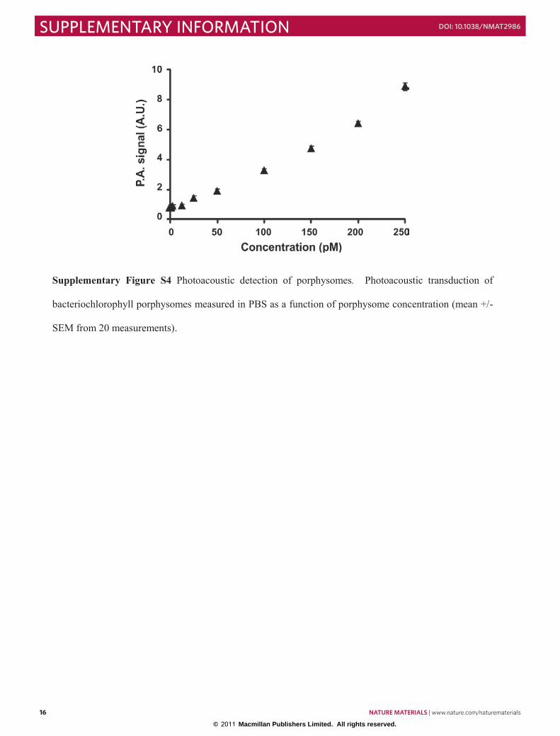
Supplementary Figure S4 Photoacoustic detection of porphysomes. Photoacoustic transduction of
bacteriochlorophyll porphysomes measured in PBS as a function of porphysome concentration (mean +/-
SEM from 20 measurements).
16 nature materials | www.nature.com/naturematerials
SUPPLEMENTARY INFORMATION doi: 10.1038/nmat2986
© 2011 Macmillan Publishers Limited. All rights reserved.

17/21
Supplementary Figure S5 Distinct spectral responses of blood and porphysomes in vivo. Normalized
photoacoustic response for the indicated portion of the image shown in the inset. The in vivo
porphysomes that accumulated in the lymph node have the same spectral response as porphysomes in
solution placed in tubing and measured in vitro.
nature materials | www.nature.com/naturematerials 17
SUPPLEMENTARY INFORMATIONdoi: 10.1038/nmat2986
© 2011 Macmillan Publishers Limited. All rights reserved.

18/21
Supplementary Figure S6 Photoacoustic mapping of multiple lymph nodes using porphysomes.
Secondary and tertiary lymph nodes became detectable in a rat with intradermal injection of 2.3 pmol of
porphysomes. Yellow arrow indicates inflowing lymph vessel. Red and blue arrows indicate the first
draining and subsequent lymph nodes, respectively. 5 mm scale bar is indicated.
18 nature materials | www.nature.com/naturematerials
SUPPLEMENTARY INFORMATION doi: 10.1038/nmat2986
© 2011 Macmillan Publishers Limited. All rights reserved.
19/21
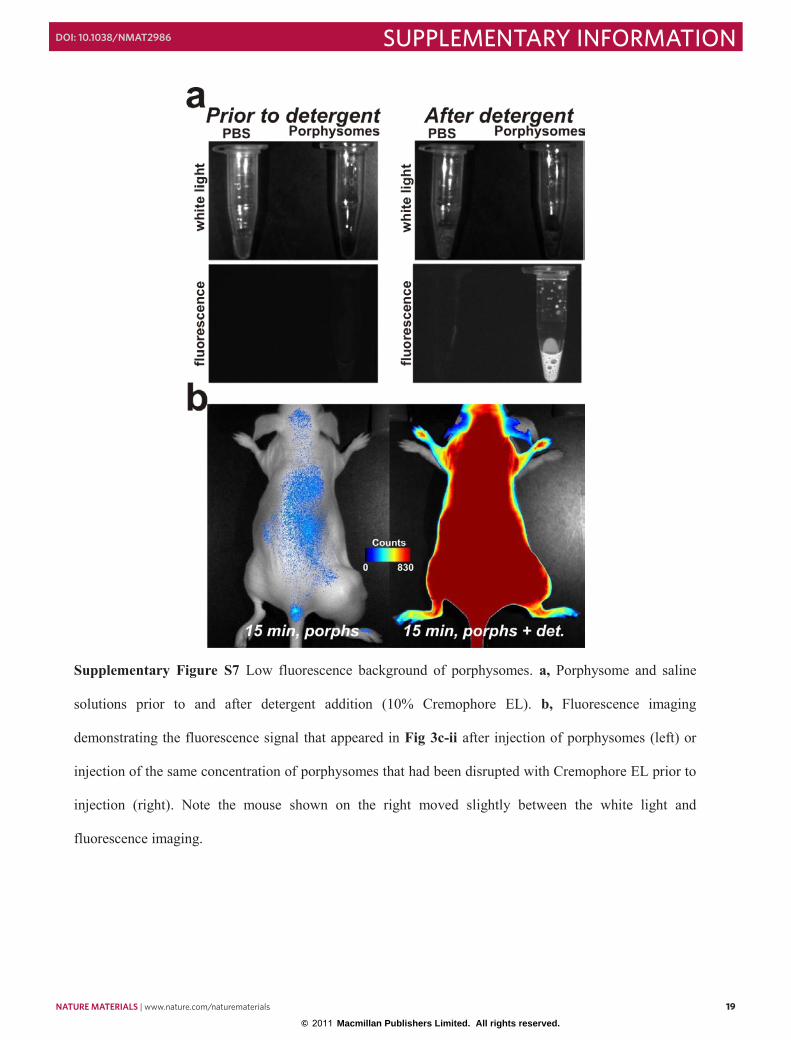
Supplementary Figure S7 Low fluorescence background of porphysomes. a, Porphysome and saline
solutions prior to and after detergent addition (10% Cremophore EL). b, Fluorescence imaging
demonstrating the fluorescence signal that appeared in Fig 3c-ii after injection of porphysomes (left) or
injection of the same concentration of porphysomes that had been disrupted with Cremophore EL prior to
injection (right). Note the mouse shown on the right moved slightly between the white light and
fluorescence imaging.
nature materials | www.nature.com/naturematerials 19
SUPPLEMENTARY INFORMATIONdoi: 10.1038/nmat2986
© 2011 Macmillan Publishers Limited. All rights reserved.

20/21
Supplementary Figure S8 Colocalization of porphysomes in early endosomes and lysosomes. a, KB
cells were co-incubated with porphysomes containing 1 molar % folate-PEG-lipid, and Alexa 488
transferrin for 3 hours prior to live cell confocal microscopy. Channels are colored as indicated. b, KB
cells were incubated with porphysomes containing 1 molar % folate-PEG-lipid for 3 hours, then with
lysotracker for 30 minutes prior to confocal imaging. Channels are colored as indicated.
20 nature materials | www.nature.com/naturematerials
SUPPLEMENTARY INFORMATION doi: 10.1038/nmat2986
© 2011 Macmillan Publishers Limited. All rights reserved.
21/21
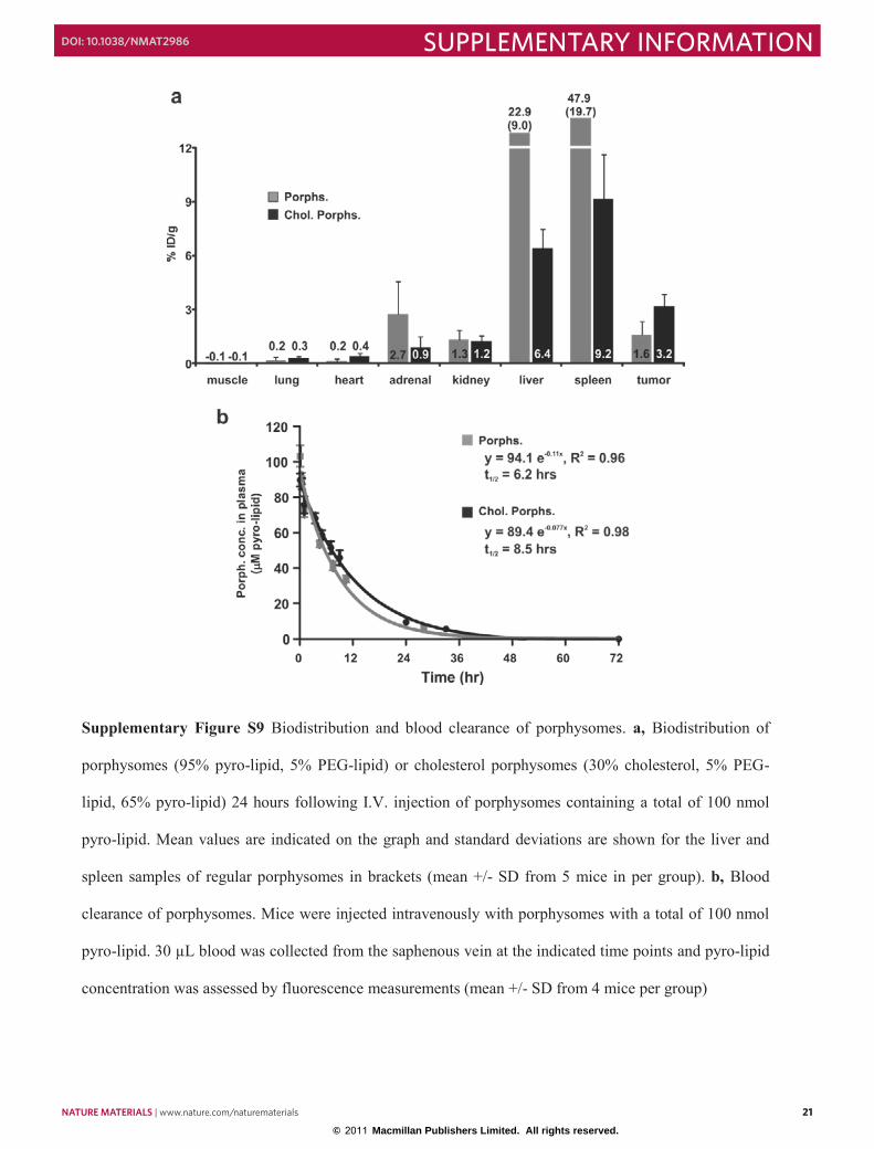
Supplementary Figure S9 Biodistribution and blood clearance of porphysomes. a, Biodistribution of
porphysomes (95% pyro-lipid, 5% PEG-lipid) or cholesterol porphysomes (30% cholesterol, 5% PEG-
lipid, 65% pyro-lipid) 24 hours following I.V. injection of porphysomes containing a total of 100 nmol
pyro-lipid. Mean values are indicated on the graph and standard deviations are shown for the liver and
spleen samples of regular porphysomes in brackets (mean +/- SD from 5 mice in per group). b, Blood
clearance of porphysomes. Mice were injected intravenously with porphysomes with a total of 100 nmol
pyro-lipid. 30 µL blood was collected from the saphenous vein at the indicated time points and pyro-lipid
concentration was assessed by fluorescence measurements (mean +/- SD from 4 mice per group)
nature materials | www.nature.com/naturematerials 21
SUPPLEMENTARY INFORMATIONdoi: 10.1038/nmat2986
© 2011 Macmillan Publishers Limited. All rights reserved.