supplementary information - proceedings of … nada abdelmagid3, mathias uhlén1, tim waterboer5,...
TRANSCRIPT

1
SUPPLEMENTARY INFORMATION
Anoctamin 2 identified as an autoimmune target in multiple sclerosis
Burcu Ayoglu1*, Nicholas Mitsios2, Ingrid Kockum3, Mohsen Khademi3, Arash Zandian1, Ronald Sjöberg1, Björn Forsström1, Johan Bredenberg2, Izaura Lima Bomfim3, Erik Holmgren4, Hans Grönlund4, André Ortlieb Guerreiro-Cacais3, Nada Abdelmagid3, Mathias Uhlén1, Tim Waterboer5, Lars Alfredsson6,7, Jan Mulder2, Jochen M. Schwenk1, Tomas Olsson3†, Peter Nilsson1†
1 Affinity Proteomics, SciLifeLab, School of Biotechnology, KTH – Royal Institute of Technology, Stockholm, Sweden.2 Affinity Proteomics, SciLifeLab, Department of Neuroscience, Karolinska Institute, Stockholm, Sweden.3 Neuroimmunology Unit, Department of Clinical Neuroscience, Karolinska Institute, Stockholm, Sweden 4 Therapeutic Immune Design Unit, Department of Clinical Neuroscience, Karolinska Institute, Stockholm, Sweden 5 Infection and Cancer Program, German Cancer Research Center (DKFZ), Heidelberg, Germany 6 Institute of Environmental Medicine, Karolinska Institute, Stockholm, Sweden 7 Centre for Occupational and Environmental Medicine, Stockholm County Council, Stockholm, Sweden
* To whom correspondence may be addressed: [email protected]
† Shared senior authors
Abbreviations ANO2−Anoctamin 2 (or TMEM16B−transmembrane protein 16 B); AP−Attributable proportion due to interaction; AU−Arbitrary units; CIS−Clinically isolated syndrome; CSF−Cerebrospinal fluid; EBNA−Epstein-Barr virus nuclear antigen; FDR−False discovery rate; GPR62− G protein-coupled receptor 62; HLA−human leukocyte antigen; IFNB1−Interferon beta 1; IgG−Immunoglobulin G; KIR4.1−Potassium inwardly-rectifying channel subfamily J member 10 (KCNJ10); MHC−Major histocompatibility complex; MFI−Median fluorescence intensity; MOG−Myelin oligodendrocyte glycoprotein; MS−Multiple sclerosis; OND−Other neurological diseases; OR−Odds ratio; PGAM5−Phosphoglycerate mutase family member 5; PPMS−Primary progressive multiple sclerosis; RRMS−Relapsing remitting multiple sclerosis; RRrel−RRMS with relapse; RRrem−RRMS with remission; SPMS−Secondary progressive multiple sclerosis; SRSF7− serine/arginine-rich splicing factor

2
TABLE OF CONTENTS
Abbreviations.....................................................................................................................................1Supplementary materials and methods........................................................................................4
Antigens and generation of bead-based antigen arrays.....................................................................4Assays on bead-based antigen arrays.....................................................................................................5Generation of and assays on planar antigen arrays for confirmatory analysis............................5Independent replication assays for detection of ANO2 autoantibodies..........................................6Western blot validation of ANO2 antibody...........................................................................................6Immunofluorescence histochemistry on brain sections......................................................................7
Immunofluorescence..................................................................................................................................................7Slide scanning microscopy.......................................................................................................................................8
Bioinformatics tools and databases.........................................................................................................8Supplementary results......................................................................................................................8
Study and experimental design................................................................................................................8Overview of reactivity profiles of MS cases vs. controls.....................................................................9Identification of additional potential autoimmune targets in MS....................................................9Interaction between MS-related HLA risk alleles and plasma reactivity against GPR62 and PGAM5.......................................................................................................................................................10Overview of publicly available transcriptome data for ANO2, GPR62 and PGAM5...............10Reactivity against literature-based targets..........................................................................................11
Reactivity profiles for viral EBNA-1 antigen..................................................................................................11Reactivity profiles for KIR4.1..............................................................................................................................11Reactivity profiles for other literature-based targets..................................................................................11
Supplementary discussion points................................................................................................12About the structure of ANO2 protein..................................................................................................12About plasma autoantibody reactivity against KIR4.1....................................................................12About plasma autoantibody reactivity against IFNB-1....................................................................13
Supplementary figures and tables..............................................................................................14Figure S1. Overview of the antigen array content and gene ontology (GO) enrichment analysis for the represented targets.....................................................................................................................................14Figure S2. Volcano plot for comparison of the overall plasma antibody reactivity between MS cases and controls....................................................................................................................................................16Figure S3. Representation of ANO2 and the protein fragments used in the study............................17Figure S4. Plasma reactivity profile of ANO2 fragment-A in the discovery study...........................18Figure S5. Plasma reactivity profile of ANO2 fragment-A across all sample sub-groups..............19Figure S6. Profile of two different plasma samples which revealed the highest MFI values for ANO2 fragment-A on two different array platforms...................................................................................20Figure S7. Correlation scatterplots for replicated and independent plasma reactivity dataset for ANO2 protein fragments of different lengths expressed by different E. coli strains in two independent laboratories........................................................................................................................................21Figure S8. Correlation scatterplots for the sample donor age and plasma reactivity against ANO2 fragment-A.................................................................................................................................................................22Figure S9. Autoantibody reactivity against ANO2 fragment-B...............................................................23Figure S10. Mapping of reactivity against ANO2 fragment-A on peptide level................................24Figure S11. Sequence similarity search results for the protein fragments representing ANO2 fragment-A, GPR62 and PGAM5.......................................................................................................................26Figure S12. The predicted B-cell epitope within ANO2 fragment-A.....................................................27Figure S13. Western blot validation of ANO2 antibody used for immunostaining studies on brain tissue............................................................................................................................................................................28Figure S14. Interaction between different combinations of ANO2 or GPR62 autoantibody reactivity, HLA-DRB1*15 positivity and increased levels of EBNA-1 antibody reactivity or absence of HLA-A*02...........................................................................................................................................29Figure S15. Autoantibody reactivity against the protein fragment of PGAM5..................................30

3
Figure S16. Autoantibody reactivity against the protein fragment of GPR62....................................31Figure S17. Representation of the converged model of ANO2...............................................................32Figure S18. Adjusted ORs with 95% confidence intervals for the risk of developing MS for GPR62 autoantibody positivity and different combinations of HLA-DRB*15 and HLA-A*02 status............................................................................................................................................................................33Figure S19. Tissue-specific transcriptome data available for ANO2 in four different publicly available data portals..............................................................................................................................................35Figure S20. Transcriptome data for PGAM5 and GPR62 expression in different cell classes of the brain......................................................................................................................................................................36Figure S21. Antibody reactivity against the viral protein EBNA-1........................................................37Figure S22. Autoantibody reactivity against protein fragments of KIR4.1.........................................38Figure S23. Autoantibody reactivity against the protein fragment of IFNB1.....................................39Table S1. List of protein targets and related fragments revealing differential antibody reactivity in plasma of MS cases............................................................................................................................................40Table S2. Adjusted ORs with 95% CIs of developing MS for GPR62 autoantibody positivity and different combinations HLA-DRB1*15 and HLA-A*02 status..............................................................41
References........................................................................................................................................42

4
Supplementary materials and methods
Antigens and generation of bead-based antigen arrays
A total of 367 protein fragments were used in this study, which were produced within
the Human Protein Atlas routine workflow as previously described (1). In brief, using a
whole-genome bioinformatics approach, antigens of 80-150 amino acid residues were
designed in silico based on the principle of lowest sequence similarity to other human
proteins, avoiding transmembrane and signal peptide regions. The antigens were then
produced in E. coli Rosetta DE3 strain as fusion protein fragments with an N-terminal dual
affinity tag (His6-ABP) consisting of a hexahistidyl (His6) tag and an albumin binding protein
(ABP).
Besides the protein fragments generated within the Human Protein Atlas, the antigen
set included full-length proteins for the viral protein EBNA-1 (Tebu-Bio) and tissue
transglutaminase (tTG) (Tebu-Bio). In addition, the extracellular [region 87-117] of the
KIR4.1 protein was produced as a shorter version of the peptide comprising [region 83-120],
which was identified as the main target region by Srivastava et al. (2). For this, the KIR4.1
[region 87-117] was designed to be in the active site of thioredoxin (TrxA) of Mycoplasma
mycoides to give a constrained loop mimicking the KIR4.1 loop in the cytoplasmic membrane.
The TrxA was also designed to include a strech of residues for in vivo biotinylation and a His-
tag. The whole region was cloned into a pBAD vector for controlled expression using
arabinose as an inducer. This plasmid was used together with a plasmid for expression of
BirA for obtaining efficient in vivo biotinylation. The recombinant protein produced in E. coli
BL21-AI strain was purified using IMAC under denaturing conditions (2-6 M urea) and
analyzed for biotinylation using streptavidin magnetic beads (GE-Healthcare).
Protein fragments were diluted to 80 µg/ml and coupled to 1x10⁶ carboxylated
magnetic beads per ID (MagPlex-C, Luminex Corp.) as recently described (3). Besides the
antigens, a four-point dilution series of human IgG (Sigma) and Fc fragment of human IgG
(Jackson ImmunoResearch) was included in the array for optimizing the dilution rate for the
secondary anti-human IgG antibody and a four-point dilution series of goat anti-human IgG
(Jackson ImmunoResearch) was included to provide a positive control for incubation with
plasma samples, all three antibodies within a concentration range of 5-80 µg/ml. His6-ABP
tag and a protein-free buffer were included as negative assay controls.
Immobilization of the protein fragments was confirmed by incubation of the bead-
based array with a chicken anti-His6-ABP IgY (Agrisera) at 1 µg/ml, followed by detection
with an R-phycoerythrin (R-PE) conjugated anti-chicken IgY antibody at 1 µg/ml.

5
Immobilization of the biotinylated KIR4.1 peptide on neutravidin-coated beads and the
dilution series of assay control antibodies including human IgG, human IgG-Fc fragment and
goat anti-human IgG were confirmed by detection with R-PE conjugated streptavidin (Life
Technologies), R-PE conjugated Fcγ-specific goat anti-human IgG F(ab')2 fragment (Jackson
ImmunoResearch) and Alexa Fluor 555 conjugated rabbit anti-goat IgG, respectively, all at
0.01 µg/ml.
Assays on bead-based antigen arrays
Assay conditions were applied as recently described(3), with minor changes including
an assay format based on 384-well plates (Greiner Bio-One), sample dilution rate for the
plasma samples and the type and dilution rate for the secondary anti-human IgG detection
antibody. For this, plasma sample dilution rates of 1:50, 100, 1:250 and 1:500 were first co-
evaluated with three different secondary anti human-IgG detection antibodies including two
R-PE conjugated Fcγ-specific goat anti-human IgG F(ab')2 fragments (Jackson
ImmunoResearch and Life Technologies) and an R-PE conjugated goat anti-human IgG
(H+L) F(ab')2 fragment (Jackson ImmunoResearch) at dilution rates of 1:500, 1:750 and
1:1000.
Based on the optimized conditions, a plasma sample dilution rate of 1:150 in an assay
buffer consisting of 3% (w/v) BSA (Sigma) - 5% (w/v) blotting-grade nonfat dry milk
(BioRad) in 0.1% PBS-Tween supplemented with 160 µg/ml His6-ABP was applied and R-
PE conjugated Fcγ-specific goat anti-human IgG F(ab')2 fragment (Jackson
ImmunoResearch) at a dilution rate of 1:750 (1.3 µg/ml) was used for detection of bound
human IgG to the final bead array of 384 analytes. All measurements were done on the same
FlexMap3D instrument (Luminex Corp.). At least 100 events per bead identity were counted
and binding events were displayed as median fluorescence intensity (MFI) values.
Generation of and assays on planar antigen arrays for confirmatory analysis
Using previously described procedures (3, 4), planar antigen arrays with 21,120
human protein fragments, encompassing 16,728 unique antigens and representing 12,412
unique Ensembl Gene IDs were generated. The slides, each containing 21,120 features, were
utilized for profiling of plasma samples from two individuals, which revealed the highest
reactivity for ANO2 fragment-A [region 79-167] in the bead-based autoantibody profiling
assays. For this, the plasma sample dilution rate and assay buffer compositions were applied
as described for bead-based arrays. Bound human IgG on the planar array antigens was

6
detected by DyLight 649 conjugated conjugated Fcγ-specific goat anti-human IgG F(ab')2
fragment (Jackson ImmunoResearch).
Independent replication assays for detection of ANO2 autoantibodies
Serum samples were shipped on dry ice to the German Cancer Research Center
(DKFZ, Heidelberg, Germany) and stored at −20°C until analysis. Testing was performed by
laboratory staff, who was blinded to the case–control status of the subjects as previously
described (5). Briefly, recombinant ANO2 proteins were constructed by gene synthesis
(Eurofins Genomics, Ebersberg, Germany) in a modified pGEX-4T-3 vector (GE Healthcare,
Freiburg, Germany), expressed in E. coli BL21 or BL21 Rosetta (Novagen-Merck, Darmstadt,
Germany) as double fusion proteins containing an N-terminal Glutathione-S-Transferase
(GST) domain and a C-terminal tag peptide derived from the large T antigen of simian virus
40. Recombinant GST-ANO2-tag fusion proteins from cleared bacterial lysates were loaded
on spectrally distinct glutathione-casein-coupled fluorescence-labelled polystyrene beads
(SeroMap, Luminex, Austin, Texas) and affinity-purified in a one-step procedure. Sera were
diluted 1:50 in a serum pre-incubation buffer containing 1 mg/ml casein and 2 mg/ml total
lysate protein from bacteria overexpressing GST-tag without intervening sequences to block
binding of antibodies directed against residual bacterial proteins, GST and the tag peptide. To
suppress unspecific binding of antibodies to the beads themselves, the serum pre-incubation
buffer was supplemented with 0.5% w/v polyvinylalcohol, 0.8% w/v polyvinylpyrrolidone
and 2.5% v/v Superchemiblock (Millipore, Billerica, MA, USA)(6). A monoclonal antibody
directed against the C-terminal tag peptide verified the binding of the GST-ANO2-tag fusion
proteins to the beads sets(7). The color-coded beads sets loaded with different antigens were
mixed and incubated in 96 well plates with an equal volume of the serum dilutions.
Antibodies bound to the beads via the GST-ANO2-tag fusion proteins were stained with
biotinylated goat anti-human IgA, IgM, IgG (Dianova, Hamburg, Germany) and the reporter
conjugate R-phycoerythrin-labelled streptavidin. Measurements were done on an LX200
instrument (Luminex Corp.). At least 100 events per bead identity were counted and binding
events were displayed as median fluorescence intensity (MFI) values.
Western blot validation of ANO2 antibody
Cell and tissue extracts were homogenized in lysis buffer (5mM Tris-HCl pH 7.4,
0.5mM EDTA, 0.1M NaCl, 1% beta-octyl-glucoside, 0.5% Triton X-100) containing protease
inhibitors. Samples were incubated for 30 min at 4oC and centrifuged at 14,000xg for 30min

7
at 4oC. Equal amounts of cell homogenates were analyzed by SDS-polyacrylamide
electrophoresis on a 4-15% Mini-PROTEAN TGX Precast Gel (Bio-Rad) for 1h at 180V.
After transferring onto PVDF membranes using a Trans-Blot Turbo Transfer System (Bio-
Rad), proteins were blocked in 5% nonfat dry milk in Tris-buffered saline solution pH 7.4
containing 1% Tween-20 (TBST) overnight at 4°C. Membranes were then incubated with the
primary antibody (polyclonal antibody raised against a 19 amino acid long peptide from near
the N-terminal region of human ANO2, Origene, Catalog Nr. TA316534, diluted 1:400) in the
same blocking solution as above for 2h at room temperature. After several washes in TBST,
membranes were probed with swine anti-rabbit horseradish peroxidase (HRP)-conjugated
secondary antibody (Jackson ImmunoResearch, diluted 1:5,000) for 1h at RT. Following
several more washes, immunoreactivity was visualized using the Clarity Western ECL
Substrate Kit (Bio-Rad). Image acquisition and analysis were performed on a ChemiDoc MP
imaging system (Bio-Rad).
Immunofluorescence histochemistry on brain sections
Immunofluorescence
Multiple fluorescence immunohistochemistry was performed on 7 µm-thick human
cortical sections from MS cases, which were cut from paraffin-embedded blocks on a sliding
microtome and mounted onto glass slides coated with 3-aminopropyltriethoxysilane (Sigma).
Sections containing human cortical and hippocampal cores (Æ 2mm) from individuals
without clinical signs of neuropsychiatric disease served as controls. All sections were stained
on an automated Leica Bond RX system. Briefly, sections were deparaffinized (Bond Dewax
solution AR9222), rehydrated and treated for 40 min in an EDTA based pH 9.0 solution
(Bond Epitope Retrieval solution 2 AR9640) to unmask the antigens. Slides were
subsequently incubated in normal donkey serum for 30 min, followed by addition of the
primary antibody cocktail mix containing the macrophage marker CD68 (AB955, ABCAM),
astrocyte marker glial fibrillary acidic protein GFAP (MAB3402X, Millipore) and
ANO2 (TA316534, Origene) diluted (1:400, 1:2000 and 1:400, respectively) in Bond Primary
antibody diluent (AR9352) for 8 h at room temperature. Sections were washed 3 x 15 min in
PBS and incubated for 90 min at room temperature with secondary antibody cocktail mix
(FITC-anti-mouse, Cy3.5-anti-rabbit and Cy5-anti-goat) diluted 1:200 in 0.2M PB. Slides
were washed 3 x 15 min in PBS and upon incubation for 20 min in 1% Sudan Black solution
in 70% ethanol to quench autofluorescence, slides were mounted in DAPI-containing
mounting medium (P-36931, Life Technologies).

8
Slide scanning microscopy
Fluorescence microscope images were acquired on a Vslide slide scanning microscope
(MetaSystems, Alltlussheim, Germany) equipped with a CoolCube 2 camera, 2.5x, 5x, 10x
and 20x objectives and filter sets for DAPI (EX350/50 - EM470/40), FITC (EX493/16 –
EM527/30), Cy3 (EX546/10 – EM580/30), Cy3.5 (EX581/10 – EM617/40) and Cy5
(EX630/20 – 647/long pass). Whole microscope slides were scanned at 2.5x and tissue was
detected based on the DAPI signal. After generating a position map all tissue covered areas
where scanned using 20x primary objective. Individual field of view images were stitched to
generate a large 4-channel fluorescence image of the entire specimen with microscopic
resolution.
Bioinformatics tools and databases
Sequence similarity searches were performed using NCBI BLASTP algorithm and
scored with BLOSUM-80 matrix for protein fragments of ANO2 fragment-A, GPR62,
PGAM5 and with PAM-30 matrix for the short region of ANO2 identified on peptide arrays.
Protein visualizations, including predicted transmembrane topology annotated from
UniProtKB database and custom regions of interest represented by the utilized protein
fragments were made using the online tool Protter(8). The functional annotation tool of
DAVID database (9) was used to identify enriched Gene Ontology (GO) terms for the content
of the antigen array, where Benjamini-Hochberg FDR-adjusted p-values<0.01 were
considered statistically significant. BepiPred tool available in the Immune Epitope Database
(10) was used for predicting the position of a continuous B-cell epitope in ANO2 fragment-A,
which uses a combination of hidden Markov model and a propensity scale method(11). The
residues with scores above a threshold of 0.75 were predicted to be part of an epitope.
Supplementary results
Study and experimental design
Previously, we discovered and technically verified autoantibody reactivity profiles
associated with MS by screening plasma on a very large panel of human antigens(3) and
described 51 targets that were differential in their reactivity frequency among different MS
subtypes and controls with OND. These targets were now evaluated for verification in an
independent and larger cohort, which consisted of 1,063 MS cases and 1,106 population-
based, non-MS controls. In addition to these 51 antigens, which were now represented by 115

9
protein fragments to cover additional parts of these target proteins, we included 57 other
internally identified targets which were represented by 121 protein fragments. We also
assembled a set of 96 protein fragments representing 66 proteins, which have been previously
identified as autoimmune targets in the context of MS (e.g. MOG) in addition to other targets,
which have been more recently proposed using various untargeted proteomics approaches,
such as KIR4.1(2). Besides this, protein fragments with sequence similarity to KIR4.1 and
other KCNJ family proteins were also included in the target set, which were represented by 35
protein fragments (Figure S1A). A global GO annotation enrichment analysis of this selection
was performed and revealed statistically significant enrichment for terms related to molecular
function, biological process and cellular compartment, e.g. “potassium channel activity” and
“potassium transport. Other enriched GO terms were “nucleotide binding” and “neuron
projection” (Figure S1B).
Overview of reactivity profiles of MS cases vs. controls
We performed analysis using the bead-based arrays on 1,063 MS cases and 1,106
controls to determine IgG reactivity against 384 antigens, representing 196 unique proteins.
Out of this, five antigens revealed an FDR-adjusted Wilcoxon rank-sum test p-value < 0.01.
These antigens included anoctamin 2 (ANO2), Epstein–Barr virus nuclear antigen 1 (EBNA-
1), phosphoglycerate mutase family member 5 (PGAM5), G protein-coupled receptor 62
(GPR62), serine/arginine-rich splicing factor 7 (SRSF7). Among these ANO2, GPR62 and
PGAM5 had been included as follow-up to our previous discovery study. Differences
revealed for interferon beta 1 (IFNB1), G elongation factor, mitochondrial 2 (GFM2), large
tumor suppressor kinase 2 (LATS2) were yet statistically less significant with p-values < 0.05
(Table S1 and Figure S2).
Identification of additional potential autoimmune targets in MS
In addition to ANO2, the group of antigens for which the most significant differences
were revealed between MS cases and controls included PGAM5 [region 165-246]
representing an intracellular region of the protein (Figure S15A) and GPR62 [region 296-
367] representing the intracellular C-terminus of the protein (Figure S16A). As for ANO2,
these antigens were proposed by our previous discovery study.
Compared to ANO2, the fold changes between MS cases and controls were relatively
lower for these two antigens, yet the differences in MFI values were statistically significant
both for GPR62 (Wilcoxon rank-sum test p-value=1.4x10⁻⁴) (Figure S16C) and PGAM5

10
(Wilcoxon rank-sum test p-value=2.1x10⁻⁵) fragments (Figure S15C). The reactivity profiles
for these antigens over all samples are shown in Figure S15B and Figure S16B.
Interaction between MS-related HLA risk alleles and plasma reactivity against GPR62
and PGAM5
GPR62 IgG positivity (above median + 3xSD among controls) was associated with
MS (OR 2.14, 95% CI of 1.40-3.29, p-value <0.003). We also identified interaction both with
carriage of HLA-DRB1*15, AP 0.56 (95% CI of 0.17-0.98) and HLA-A*02, AP 0.51 (95%
CI of 0.12-0.90) (Figure S18). We did not observe any interaction between GRP62 positivity
and increased EBNA1 IgG levels. We have previously seen interaction between HLA-
DRB1*15 and HLA-A*02 with regard to risk of MS(12) and therefore investigated
interaction between GRP62 positivity when stratifying for these HLA exposures. There was a
significant interaction between GRP62 positivity and HLA-DRB1*15 among A*02 negative
but not positive individuals (Table S2A). No interaction between HLA-A*02 and GRP62 was
observed when stratifying for HLA-DRB1*15, this is likely due to lack of power (Table S2B).
The interaction between GRP62 IgG positivity, carriage of the risk factor HLA-DRB1*15,
and A*02 is illustrated in Figure S14B.
No significant interactions were observed between PGAM5 positivity and HLA-
DRB1*15, A*02 or increased levels of EBNA1 IgG.
Overview of publicly available transcriptome data for ANO2, GPR62 and PGAM5
Tissue-specific transcriptome data available for ANO2 in two different publicly
available data portals, the Human Protein Atlas and the GTEx portal, revealed increased
expression of ANO2 in testis tissue (Figure S19A-D), whereas the BioGPS portal provided
data for increased expression of ANO2 in the pineal gland (epiphysis) in the brain (Figure
S19B). A data portal dedicated for transcriptome of the purified cell classes of the brain
revealed relatively increased expression of ANO2 in oligodendrocyte precursor cells (Figure
S19C). For PGAM5, this portal reported mixed expression in all cell classes of the brain
(Figure S20A), whereas the data for GPR62 revealed a remarkably enriched expression in
myelinating oligodendrocytes, followed by newly formed oligodendrocytes (Figure S20B).

11
Reactivity against literature-based targets
Reactivity profiles for viral EBNA-1 antigen
Epstein-Barr virus (EBV) is a human virus known to infect more than 90% of the
adult population worldwide, and it has been implicated as a potential contributor to the
pathogenesis of MS(13). The antigen array utilized in this study included the protein EBNA-1,
which is a protein expressed in all latency types of EBV infection. Comparison of distribution
of MFI values in MS cases and controls revealed a statistically significant difference
(Wilcoxon rank-sum test p-value=2.7x10⁻¹⁶) (Figure S21A). There were no statistically
significant differences within male and female MS cases and controls (Figure S21B) and no
correlation was revealed between age and MFI values for EBNA-1 within control or MS case
groups (Figure S21C-D).
Reactivity profiles for KIR4.1
The glial inwardly rectifying ion channel protein KIR4.1, in particular the
extracellular loop consisting of [region 83-120], was recently identified by Srivastava et al. as
a potential serum target of autoantibodies in the context of MS(2). The antigen array utilized
in our assay included two protein fragments representing this extracellular loop region of
KIR4.1: fragment-A [region 87-117] and fragment-B [region 89-117]. In addition to these,
fragment-C represented the intracellular C-terminal [region 310-379] of KIR4.1 (Figure
S22A). Our analysis revealed no difference between the MS cases and controls for any of the
three protein fragments representing KIR4.1 (Figure S22B-D).
Reactivity profiles for other literature-based targets
Our antigen set included various other targets identified previously by other studies
using both unbiased proteomic approaches or investigating pre-selected collections of
potentially MS-related proteins. Among these literature-based additions, differences in
reactivity were found for a protein fragment representing [region 75-111] of SRSF7, the
serine/arginine-rich splicing factor 7 (Figure S2, Table S1). This protein was previously
described by Beyer et al. as a potential autoimmune target, where autoantibody reactivity in
CSF samples from 15 RRMS patients were compared to 15 controls using a commercially
available protein microarray technology (14). Our analysis in plasma samples revealed a
moderate reactivity against SRSF7 in cases and controls.
Besides, we also investigated reactivity against interferon beta 1 (IFNB-1), an
immunomodulatory protein and drug used for treatment of MS patients with a relapsing
course. It is known that a subset of IFNB-1 treated MS patients might develop antibodies
against IFNB-1 and thereby receive a reduced therapeutic efficacy(15). In our assay we

12
included a protein fragment representing [region 24-101] of IFNB-1 to investigate the
presence of antibodies against IFNB-1 in plasma. Significantly more MS cases showed
IFNB1 reactivity compared to controls (Figure S23A). Dissecting the case groups into IFNB-
1 treated and untreated cases showed distinct antibody reactivity against the protein fragment
only in IFNB-1 treated MS cases (Figure S23B). Further drug-related classification of the
MS case group revealed an IFNB-1 reactivity in particular in Betaseron (IFNB-1b type drug;
also known as Betaferon) treated patients, while reactivity among patients treated with
Avonex and Rebif (both IFNB-1a type drugs), was lower (Figure S23C).
Supplementary discussion points
About the structure of ANO2 protein
The very recently reported experimental structure of ANO1 (PDB ID: 4WIS) is the
only known 3-D structure within the anoctamin protein family(16). In this structure, ten trans-
membrane helices are observed, as opposed to only eight predicted. Since the membrane
bound portions share ~70% sequence homology across the anoctamin family members, and
since the confidence of the predicted intracellular N-terminal portion, i.e. [region 1-365], is
~50%, the autoantibody-binding site may also be on the extracellular domain. Despite the
dissimilarities in the sequence, [region 136-147] of ANO2, which is the region where we
mapped plasma reactivity on a peptide level, highly resembles [region 322-329] in the crystal
structure of ANO1, which is located on the extracellular side of ANO1. It is also possible that
the site is facing the channel cleft allowing the antibody to protrude down the channel cleft
and bind to the predicted epitope. Indeed, the channel entrance is ~15 Å while the vestibule is
widened to ~30 Å. Alignment of the fragment-A [region 79-167] on the experimental
structure shows that both the channel facing (Figure S17B-C) and extracellular surface
exposed autoantibody binding (Figure S17D-E) are plausible, even if the overall non-
membrane architecture may differ since the modeled fragment shows low sequence homology
to any known 3-D structures. However, the staged modeling protocol does not heavily rely on
existing templates but rather accounts for physical interactions, meaning that one can expect
some structural changes when including solvent and membrane-bilayers or a larger portion of
the protein.
About plasma autoantibody reactivity against KIR4.1
The antigen set utilized in this study included three recombinant protein fragments
representing the inward rectifying potassium channel KIR4.1, which was recently suggested

13
as a potential autoimmune target within MS(2). Here, we demonstrated a low presence of
KIR4.1 fragment-specific antibodies in plasma with no discrimination between cases and
controls (Figure S22), with a similar outcome in two other recent studies which investigated
KIR4.1 reactivity (17, 18). The discrepancies between studies on this protein might be due to
different methodologies, different antigen preparations and expression system and thus
differential status of posttranslational modifications such as glycosylation, which highlight the
importance of the potential role of posttranslational modifications in autoantibody binding
(19).
About plasma autoantibody reactivity against IFNB-1
In this study, we also investigated the reactivity towards IFNB-1 in plasma, which is
used as a drug for treatment of MS patients. It is known that a subset of IFNB-1 treated MS
patients, varying between 2-45% depending on the specific drug and dose regimen, develop
antibodies against IFNB-1 and therefore experience a reduced therapeutic benefit(20, 21).
Using a protein fragment representing IFNB-1, we could identify reactivity exclusively in
plasma of MS patients treated with an IFNB-1b type drug (Figure S23), which is known to be
more immunogenic than the IFNB-1a type drug(15). This demonstrated not only the utility of
our assay setup for an efficient detection of treatment-related neutralizing antibodies to IFNB-
1, but also the utility of protein fragments for autoantibody profiling, which are generated
within the Human Protein Atlas as most unique representations of their full-length versions.

14
Supplementary figures and tables
Figure S1. Overview of the antigen array content and gene ontology (GO) enrichment analysis for the represented targets. (A) The pie charts summarize the number of protein targets, selection criteria and number of protein fragments included in the bead array representing these targets. In addition to the 51 follow-up targets represented by 115 protein fragments, the target set included 57 other internally identified targets represented by 121 protein fragments. Furthermore, a set of 96 protein fragments representing 66 proteins, which have been suggested in literature as autoimmune targets in the context of MS were included, as well as more recently proposed targets, such as KIR4.1. Besides KIR4.1, protein
A
Proteins and antibodies as assay controls
Other proposed targets than KCNJ family proteins in literature
KCNJ family proteins and related targets
Other internally identified targets
51 Follow-up targets and antigens for their additional epitopes
B
115
121
35
96
17
384
51
5722
66
196
Antigen SetProtein Set
0 10 20 30 40 50 60
Follow-up targets (n=108)
Literature-based targets (n=88)
All targets (n=196)
potassium channel complexvoltage-gated potassium channel complex
microtubule cytoskeletonneuron projection
glucose metabolic processmonosaccharide catabolic process
hexose catabolic processglycolysis
glucose catabolic processmonovalent inorganic cation transport
protein polymerizationpotassium ion transport
metal ion transmembrane transporter activitynucleotide binding
cation channel activityion channel activity
passive transmembrane transporter activitysubstrate specific channel activity
alkali metal ion bindingvoltage-gated ion channel activity
potassium channel activityligand-gated ion channel activity
voltage-gated cation channel activitypotassium ion binding
inward rectifier potassium channel activity
MOLECULAR
FUNCTION
BIOLOGICAL
PROCESS
CELLULAR
COMPARTMENT
Number of targets annotated to the over-represented GO term
2.2 x 10⁻¹⁹
1.2 x 10⁻⁸
6.8 x 10⁻⁸
6.8 x 10⁻⁸
1.1 x 10⁻⁷
2.3 x 10⁻⁷
1.9 x 10⁻⁶
3.8 x 10⁻⁵
5.4 x 10⁻⁵
7.8 x 10⁻⁵
7.4 x 10⁻⁵ 2.0 x 10⁻⁴
5.0 x 10⁻⁴
8.1 x 10⁻³
8.1 x 10⁻³
6.7 x 10⁻³
6.1 x 10⁻³
1.8 x 10⁻⁵
1.0 x 10⁻⁴
2.4 x 10⁻⁴
2.0 x 10⁻⁴
4.7 x 10⁻⁴
5.0 x 10⁻⁴
5.0 x 10⁻⁴
6.0 x 10⁻⁴

15
fragments with sequence similarity to KIR4.1 and other KCNJ family proteins were also included, which were represented by 35 protein fragments. (B) GO annotation enrichment analysis was performed for both the entire set of targets and for the subset of targets consisting of only literature-based targets or only follow-up targets. Significantly over-represented GO terms for molecular function, biological process and cellular compartment and the number of targets annotated to these over-represented GO terms are shown in the barplot. The reported FDR-adjusted p-values are shown only for the significant enrichment of the respective terms within the entire set of targets. There were no significantly enriched GO terms within the subset of only follow-up targets, whereas the subset of literature-based targets contributed to the enrichment of various terms related to “ion channel activity” in the entire target set.

16
Figure S2. Volcano plot for comparison of the overall plasma antibody reactivity between MS cases and controls. On x-axis the fold change, defined as log2 of mean of MFI values in all MS cases (n=1,063) over mean of MFI values in all controls (n=1,106) for each antigen and on y-axis –log10 of the FDR-adjusted Wilcoxon rank-sum test p-values are shown. Differences in MFI values for reactivity against the antigens annotated in black revealed FDR-adjusted Wilcoxon rank-sum test p-values <0.05.
0
5
10
15
−3 −2 −1 0 1 2 3Fold Change
log2(mean MS/mean CONT)
−log
10 (F
DR
−cor
rect
ed W
ilcox
on p
−val
ue)
p−value=0.05
●●
●
●
●
●● ●
ANO2EBNA1
PGAM5
GPR62
SRSF7
IFNB1GFM2 LATS2

17
Figure S3. Representation of ANO2 and the protein fragments used in the study. Representation of ANO2, also known as TMEM16B, with its eight putative transmembrane regions and the large N-terminal region and the C-terminus predicted to be present in the cytoplasmic space. Two protein fragments, fragment-A representing the N-terminal [region 79-167] and fragment-B representing C-terminus [region 932-1003] of ANO2 were included in the array-based antibody profiling assays.
PTMs
ANO2 fragment-A [region 79-167]
ANO2 fragment-B [region 932-1003]
Variants

18
Figure S4. Plasma reactivity profile of ANO2 fragment-A in the discovery study. (A) The barplot represents the MFI values for plasma reactivity against ANO2 fragment-A within 18 PPMS, 46 SPMS, 43 RRrel, 67 RRrem, 39 CIS and 114 controls with ONDs, which were analyzed in the untargeted discovery stage. (B) The barplot represents the positive reactivity percentages for ANO2 fragment-A within the different MS subtypes and controls with ONDs, where in particular the difference in positive reactivity percentages between the RRrem cases and controls with ONDs revealed a statistically significant Fisher’s exact test p-value. (C) The barplot represents the positive reactivity percentages for ANO2 fragment-A within all the MS cases and controls with ONDs with the respective Fisher’s exact test p-value. The plasma samples utilized in this previous discovery study(3) were collected during routine neurological diagnostic work-up as part of an in-house biobank at Karolinska University Hospital, Neurology Clinic, Stockholm, Sweden and all enrolled participants in that cohort were different from those enrolled in the EIMS cohort utilized in the presented study.
0
5000
10000
15000
Reactivity against ANO2 [region 79-167]
(Discovery Study Ayoglu et al. Mol Cell Proteomics, 2013)
PPMS
n=18
MF
I [A
U]
SPMS
n=46
RR-Rel
n=43
RR-Rem
n=67
CIS
n=39
OND
n=114
A
B
0
5
10
15
20
25
30
PP
MS
SP
MS
RR
rel
RR
rem
CIS
ON
D
Po
sitiv
e R
ea
ctivity [
%]
Fisher’s exactp-value=0.02
C Fisher’s exactp-value=0.006
0
5
10
15
20
25
30
ON
D
All M
S
Po
sitiv
e R
ea
ctivity [
%]
19
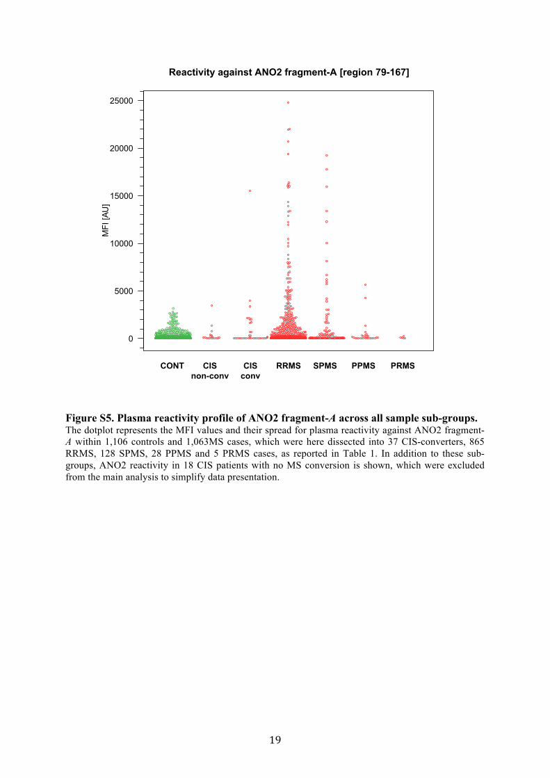
Figure S5. Plasma reactivity profile of ANO2 fragment-A across all sample sub-groups. The dotplot represents the MFI values and their spread for plasma reactivity against ANO2 fragment-A within 1,106 controls and 1,063MS cases, which were here dissected into 37 CIS-converters, 865 RRMS, 128 SPMS, 28 PPMS and 5 PRMS cases, as reported in Table 1. In addition to these sub-groups, ANO2 reactivity in 18 CIS patients with no MS conversion is shown, which were excluded from the main analysis to simplify data presentation.
MFI
[AU
]
CONT CIS
non-conv
CIS
conv
RRMS SPMS PPMS PRMS
0
5000
10000
15000
20000
25000
Reactivity against ANO2 fragment-A [region 79-167]

20
Figure S6. Profile of two different plasma samples which revealed the highest MFI values for ANO2 fragment-A on two different array platforms. (A-B) The barplots represent the MFI values across all antigens included in the bead array for two different plasma samples, which revealed the highest MFI values for ANO2 fragment-A [region 79-167] across the entire cohort. (Data points for a total of eleven positive control bead IDs of the assay were omitted in the barplots) (C-D) The barplots represent the MFI values in a further analysis where these two samples were analyzed on in-house generated planar microarrays with randomly selected 21,120 protein fragments representing 16,728 unique antigens and 12,412 unique proteins. (Data points for a total of 132 positive and negative control spots were omitted in the barplots)

21
Figure S7. Correlation scatterplots for replicated and independent plasma reactivity dataset for ANO2 protein fragments of different lengths expressed by different E. coli strains in two independent laboratories. Scatterplots to the left of the figure diagonal represent the pairwise correlation scatterplots for MFI values obtained for plasma reactivity in MS cases (n=152) and controls (n=39) against ANO2 protein fragment-A [region 79-167] and ANO2 protein fragment [region 1-365] expressed by two different E. coli strains either within the Human Protein Atlas or at an independent laboratory (German Cancer Research Center (DKFZ), Heidelberg, Germany). Corresponding Spearman’s rho values for each pairwise correlation are reported to the right of the figure diagonal.
ANO2 [region 79-167]
BL21 strainProtein expression: DKFZ
Plasma analysis: DKFZ
0.82 0.84 0.80
●
●●
●●
●●●
●
●
●●
●
●
●
●
●
●
●
●● ●
●
●
●
●
●
●●
●
●
●●●
●
●
●●
●
●●
●
●
●
●
●●
●
●
●●
●
●
●
●
● ●
●
●
●
● ●●
●
●
●
●
●●
●
●
●
●●
● ●
●
●●
●
●●
●
●
●
●●
●
●
●
●
●
●
●●●
●●
●●
●● ●
●●
●
●●●●
●
●
●
●●
●
●●
●●
●
●
●●●●
●
●
●
●●●●●●
●●●
●
●
●●
●●
●
●
●
●
●
●
●
●
●
●
●
●
●●
●
●
●
●●
●●
●●
●
●
●
● ●
●
●
●
●
●
●
●
●
●
●
●●
●
●
●
●●
●
●
ANO2 [region 1-365]
BL21 strainProtein expression: DKFZ
Plasma analysis: DKFZ
0.93 0.75
●
●
●●
●
●
●
●
●
●●
●●
●●
●
●
●
●●●
●● ●●
●●
●
● ●
●
●
●●
●●
●
●
●
● ●
●
●
●
●●
●
●
●●
●
●
●
●
●●
●●●
●
●
●
●
●
●
●
●
●●
●
●●
●●
●
●
●
●●
●
●●●
●
●
●
●
●
●
●
●
●
●
●●●
●●
●●
●
●●
●●
●
●●●●●
●
●
●●
●
●●
●●●
●
●●●●
●
●
●
●●
●●●●
●
●●●
●
●●●●
●
●
●
●
●●
●
●
●●●
●
●●
●
●
●
●
●
●●
●
●
●
●
●
●
●
●
●
● ●
●
●
●
●
●
●
●
●
●
●
●
●●
●
●
●
●
●●
●
●
●
●
●
● ●
●●
●●
●
●
●
●●●
● ●●●
●●
●
● ●
●
●
●●
● ●
●
●
●
●●
●
●
●
●●
●
●
●●
●
●
●
●
●●
●● ●
●
●
●
●
●
●
●
●
●●
●
●●
●●
●
●
●
●●
●
●●●
●
●
●
●
●
●
●
●
●
●
●●●
●●
●●
●
●●
●●
●
●●●●●
●
●
●●
●
●●
●●●
●
●●●●
●
●
●
●●
●●●●
●
●●●
●
●●●●
●
●
●
●
●●
●
●
●● ●
●
●●
●
●
●
●
●
●●
●
●
●
●
●
●
●
●
●
●●
●
●
●
●
●
●
●
●
●
●
●
●●
●
●
0.76
1
10
100
1000
10
50
500
5000
●●● ●● ● ●●●●● ●●●● ●●● ●●● ●● ● ●●● ●● ●●●●●●● ●●●● ●●● ●●
●
● ●●● ●●● ● ●● ●●●●● ●● ● ●● ●● ●● ● ●●●● ●●●●
●●●●●● ●●●● ●●●● ●●●●●●●●● ●●●● ●●●●●●● ●●● ●●● ●
●● ●●●●● ●●●●●●●●●●● ●●●●●●
●●●● ●●●● ●●●●
●●●● ●●● ● ●● ●● ●●● ● ● ●●
●
●● ●●●●
●●
●
●●
●
● ●●
● ●● ● ●●●●● ● ●●●●● ● ●● ●●●● ●●● ●●●● ●● ● ●●● ● ●●● ●●● ●● ●
●
● ●● ●●●● ● ●●●● ● ●●●●● ●● ●●●● ● ●●●●● ●●●
● ●●● ● ●●● ●● ●● ●● ●●● ●●●●●●●●●● ●●●
● ●●● ● ●● ●●● ●●● ●●●●● ●● ●●●●●●
● ●● ●● ●●●●
●●● ●● ●● ●●● ●●
●●● ● ●●● ●●●● ●●●●● ● ●●
●
● ●● ●●
●
●●
●
●●
●
● ●●
1 5
50
500
5000
●●●● ●● ●●● ●● ●●●● ● ●● ●●● ●●●● ●●● ●●● ●●●●● ● ●● ●●● ●● ●
●
● ●●● ●●● ● ●●●●● ●● ●● ● ●● ●●●● ●●●●● ● ● ●●
● ●●● ● ●●● ●● ●● ●● ● ●● ●●● ●●● ● ●●● ●●●●●●● ●●● ●●●●
●● ●●●●● ●● ●● ●●●●● ●● ●● ●●●●
●●● ● ●●● ●●●●●
●●● ● ●● ● ●●●● ●●●● ● ● ●●
●
● ●● ●●
●
●●
●
●●
●
● ●●
ANO2 [region 79-167]
Rosetta strainProtein expression:Human
Protein Atlas
Plasma analysis: KTH
0.82 0.84 0.80
0.93 0.75
0.76
1
5
50
500
5000
100
1000
10000
10000
10
100
1000
1
5
50
500
5000
10
100
1000
1000
100
101 5
50
500
5000
1000
100
10
ANO2 [region 1-365]
Rosetta strainProtein expression: DKFZ
Plasma analysis: DKFZ
CONT MS
●●
MFI
[AU
]
MFI [AU]
Spearman’s RhoS
pearman’s R
ho

22
Figure S8. Correlation scatterplots for the sample donor age and plasma reactivity against ANO2 fragment-A. (A-B) The scatterplots represent the relation between sample donor age and MFI values for plasma reactivity against ANO2 fragment-A within all MS cases and controls, respectively. Neither of the relations revealed a correlation between sample donor age and reactivity against ANO2 fragment-A.
MS
Spearman's Rho= −0.01
Age
MFI
[AU
]
510
20 30 40 50 60 70
CONT
Spearman's Rho= −0.05
Age
MFI
[AU
]
20 30 40 50 60 70
50100
5001000
500010000
510
50100
5001000
500010000
Correlation between reactivity against
ANO2 [region 79-167] and sample donor age
A
B

23
Figure S9. Autoantibody reactivity against ANO2 fragment-B. (A) The dotplot represents the MFI values and their spread for plasma reactivity against ANO2 fragment-B representing the C-terminus [region 932-1003] of the protein in 1,106 controls and 1,063 MS cases.

24
Figure S10. Mapping of reactivity against ANO2 fragment-A on peptide level. (A-B) The lineplots represent the MFI values for reactivity against 15-mer and 20-mer overlapping peptides, respectively, representing ANO2 fragment-A [region 79-167]. Each green or red line corresponds to an individual plasma sample of a control (n=178) or MS case (n=185), respectively. (C-D) The barplots represent the average of MFI values for reactivity against the 15-mer and 20-mer overlapping peptides, respectively, across 182 controls (green bars) and 196 MS cases (red bars). Differences in plasma reactivity between MS cases and controls were observed mainly for two overlapping 15-mers and a 20-mer, which all share the 12 amino acid residues long peptide stretch HAGGPGDIELGP.
MFI [AU]
0 5000 10000 15000 0 5000 10000 15000
EPVSLEARLSRMHFH
SLEARLSRMHFHDSQ
ARLSRMHFHDSQRKV
SRMHFHDSQRKVDYV
HFHDSQRKVDYVLAY
DSQRKVDYVLAYHYR
RKVDYVLAYHYRKRG
DYVLAYHYRKRGVHL
LAYHYRKRGVHLAQG
HYRKRGVHLAQGFPG
KRGVHLAQGFPGHSL
VHLAQGFPGHSLAIV
AQGFPGHSLAIVSNG
FPGHSLAIVSNGETG
HSLAIVSNGETGKEP
AIVSNGETGKEPHAG
SNGETGKEPHAGGPG
ETGKEPHAGGPGDIE
KEPHAGGPGDIELGP
HAGGPGDIELGPLDA
GPGDIELGPLDALEE
DIELGPLDALEEERK
LGPLDALEEERKEQR
LDALEEERKEQREEF
LEEERKEQREEFEHN
ERKEQREEFEHNLM
0 5000 10000 15000
EPVSLEARLSRMHFHDSQRK
RMHFHDSQRKVDYVLAYHYR
VDYVLAYHYRKRGVHLAQGF
KRGVHLAQGFPGHSLAIVSN
PGHSLAIVSNGETGKEPHAG
GETGKEPHAGGPGDIELGPL
GPGDIELGPLDALEEERKEQ
DALEEERKEQREEFEHNLM
0 5000 10000 15000
CONT MS
●●
MFI [AU]
Reactivity against 15-mer peptides
representing ANO2 [region 79-167]
Reactivity against 20-mer peptides
representing ANO2 [region 79-167]
EPVSLEARLSRMHFH
SLEARLSRMHFHDSQ
ARLSRMHFHDSQRKV
SRMHFHDSQRKVDYV
HFHDSQRKVDYVLAY
DSQRKVDYVLAYHYR
RKVDYVLAYHYRKRG
DYVLAYHYRKRGVHL
LAYHYRKRGVHLAQG
HYRKRGVHLAQGFPG
KRGVHLAQGFPGHSL
VHLAQGFPGHSLAIV
AQGFPGHSLAIVSNG
FPGHSLAIVSNGETG
HSLAIVSNGETGKEP
AIVSNGETGKEPHAG
SNGETGKEPHAGGPG
ETGKEPHAGGPGDIE
KEPHAGGPGDIELGP
HAGGPGDIELGPLDA
GPGDIELGPLDALEE
DIELGPLDALEEERK
LGPLDALEEERKEQR
LDALEEERKEQREEF
LEEERKEQREEFEHN
ERKEQREEFEHNLM
0
500
1000
1500
2000
2500
Mean o
f M
FI
[AU
] in
sa
mp
le g
rou
p
EPVSLEARLSRMHFHDSQRK
RMHFHDSQRKVDYVLAYHYR
VDYVLAYHYRKRGVHLAQGF
KRGVHLAQGFPGHSLAIVSN
PGHSLAIVSNGETGKEPHAG
GETGKEPHAGGPGDIELGPL
GPGDIELGPLDALEEERKEQ
DALEEERKEQREEFEHNLM
Me
an
of
MF
I [A
U]
in s
am
ple
gro
up
A B
C D
0
500
1000
1500
2000
2500
3000
3500
●●
●●
●●
●●
●●
●●
●●
●●
●●
●●
●●
●●
●●
●●
●
●
●●
●●
●●
●●
●●
●●
●●
●●
●●
●●
●●
●
●
●●
●●
●●
●●
●●
●●
●●
●●
●●
●●
●●
●●
●●
●●
●●
●●
●●
●●
●●
●●
●●
●●
●●
●●
●●
●●
●●
●●
●●
●●
●●
●●
●●
●●
●●
●●
●●
●●
●●
●●
●●
●●
●●
●●
●●
●●
●●
●●
●●
●●
●●
●●
●●
●●
●●
●●
●●
●●
●●
●●
●●
●●
●●
●●
●●
●●
●●
●●
●●
●●
●●
●●
●●
●●
●●
●●
●●
●●
●●
●●
●●
●●
●●
●●
●●
●●
●●
●●
●●
●●
●
●●
●●
●●
●●
●●
●●
●●
●●
●●
●●
●●
●●
●●
●●
●●
●●
●●
●●
●●
●●
●●
●●
●●
●●
●●
●●
●●
●●
●●
●●
●
●
●●
●●
●●
●●
●●
●●
●●
●●
●●
●●
●●
●●
●●
●
●
●
●
●●
●●
●●
●●
●●
●●
●●
●●
●●
●●
●
●
●●
●●
●●
●●
●
●●
●●
●●
●●
●●
●●
●●
●●
●●
●●
●●
●●
●●
●●
●●
●●
●●
●●
●●
●●
●●
●●
●●
●●
●●
●●
●●
●●
●●
●●
●●
●●
●●
●●
●●
●●
●●
●●
●●
●●
●●
●●
●●
●●
●●
●●
●●
●●
●●
●●
●●
●●
●●
●●
●●
●●
●●
●●
●●
●●
●●
●●
●●
●●
●●
●●
●●
●●
●●
●●
●●
●●
●●
●●
●●
●●
●●
●●
●●
●
●●
●●
●●
●●
●●
●●
●●
●●
●●
●●
●●
●●
●●
●●
●●
●●
●●
●●
●●
●●
●●
●●
●●
●●
●●
●●
●●
●●
●●
●●
●●
●
●
●●
●●
●●
●●
●●
●●
●●
●●
●●
●●
●●
●●
●●
●●
●●
●●
●●
●●
●●
●●
●●
●●
●●
●●
●●
●●
●●
●●
●●
●●
●●
●●
●●
●●
●●
●●
●●
●●
●●
●●
●●
●●
●●
●●
●●
●●
●●
●●
●●
●●
●●
●●
●●
●●
●●
●●
●●
●●
●●
●●
●●
●●
●●
●●
●●
●●
●●
●●
●●
●●
●●
●●
●●
●●
●●
●●
●●
●●
●●
●●
●●
●●
●●
●●
●●
●●
●●
●●
●●
●●
●●
●●
●●
●●
●●
●●
●●
●●
●●
●●
●●
●●
●●
●●
●●
●●
●●
●●
●●
●●
●●
●●
●●
●●
●
●
●●
●●
●●
●●
●●
●●
●●
●●
●●
●●
●●
●●
●●
●●
●●
●●
●●
●●
●●
●●
●●
●●
●●
●●
●●
●●
●●
●●
●●
●●
●●
●●
●●
●●
●●
●●
●●
●●
●●
●●
●●
●●
●●
●●
●●
●●
●●
●●
●●
●●
●●
●●
●●
●●
●●
●●
●
●
●●
●●
●●
●●
●●
●●
●●
●●
●●
●●
●●
●
●
●●
●●
●●
●●
●●
●●
●●
●●
●●
●●
●●
●●
●●
●●
●●
●●
●●
●●
●●
●●
●●
●●
●●
●●
●●
●●
●●
●●
●●
●●
●●
●
●●
●●
●●
●●
●●
●●
●●
●●
●●
●●
●●
●●
●●
●●
●●
●●
●●
●●
●●
●●
●●
●●
●●
●●
●●
●●
●●
●●
●●
●●
●●
●●
●●
●●
●●
●●
●●
●●
●●
●●
●●
●●
●●
●●
●●
●●
●●
●●
●●
●●
●●
●●
●●
●●
●●
●●
●●
●●
●●
●●
●●
●●
●●
●●
●●
●●
●●
●●
●●
●●
●●
●●
●●
●●
●●
●●
●●
●●
●●
●●
●●
●●
●●
●●
●
●●
●●
●●
●●
●●
●●
●●
●●
●●
●●
●●
●●
●●
●●
●●
●●
●●
●●
●●
●●
●●
●●
●●
●●
●●
●
●
●●
●●
●●
●●
●●
●●
●●
●●
●●
●●
●●
●●
●●
●●
●●
●●
●●
●●
●●
●●
●●
●●
●●
●●
●●
●●
●●
●●
●●
●●
●●
●●
●●
●●
●●
●●
●●
●●
●●
●●
●●
●●
●●
●●
●●
●●
●●
●●
●●
●●
●●
●●
●●
●●
●●
●●
●●
●●
●●
●●
●●
●●
●●
●●
●●
●●
●●
●●
●●
●●
●●
●●
●●
●●
●●
●●
●●
●●
●●
●●
●●
●●
●●
●●
●●
●●
●●
●●
●●
●●
●●
●●
●●
●●
●●
●●
●●
●●
●●
●●
●●
●●
●●
●●
●●
●●
●●
●●
●●
●●
●●
●●
●●
●
●
●●
●●
●●
●●
●●
●●
●●
●
●
●●
●●
●●
●●
●●
●●
●●
●
●
●●
●●
●●
●●
●●
●●
●●
●●
●●
●●
●●
●●
●●
●●
●●
●●
●●
●●
●●
●●
●●
●●
●●
●●
●●
●●
●●
●
●
●●
●●
●●
●
●
●●
●●
●●
●●
●●
●●
●●
●●
●●
●●
●●
●
●
●●
●●
●●
●●
●●
●●
●●
●●
●●
●●
●●
●●
●●
●●
●●
●●
●●
●●
●●
●●
●●
●●
●●
●●
●●
●●
●●
●●
●●
●●
●●
●
●
●●
●●
●●
●
●
●●
●●
●●
●●
●●
●●
●●
●●
●●
●●
●●
●●
●●
●●
●●
●●
●●
●●
●●
●●
●●
●●
●●
●●
●●
●●
●●
●●
●●
●●
●●
●●
●●
●●
●●
●●
●●
●●
●●
●●
●●
●●
●●
●●
●●
●●
●●
●●
●●
●●
●●
●●
●●
●●
●●
●●
●●
●
●
●●
●●
●●
●●
●●
●●
●●
●●
●●
●
●
●●
●
●
●●
●●
●●
●●
●●
●●
●●
●●
●●
●
●
●●
●●
●●
●●
●●
●●
●●
●●
●●
●●
●●
●●
●●
●●
●●
●●
●●
●●
●●
●●
●●
●●
●●
●●
●●
●●
●●
●●
●●
●●
●●
●●
●●
●●
●●
●
●
●●
●●
●
●●
●●
●●
●●
●●
●●
●●
●●
●●
●●
●●
●●
●●
●
●●
●●
●●
●●
●●
●●
●●
●●
●●
●●
●●
●●
●●
●●
●●
●●
●●
●●
●●
●
●
●●
●●
●●
●
●
●●
●
●●
●
●
●
●●
●●
●●
●●
●●
●●
●●
●
●●
●●
●●
●●
●●
●●
●●
●●
●●
●●
●●
●●
●●
●●
●●
●●
●●
●●
●●
●●
●
●●
●●
●●
●●
●●
●●
●●
●●
●●
●●
●●
●●
●●
●●
●●
●●
●●
●●
●●
●
●
●●
●●
●●
●
●
●●
●●
●●
●●
●●
●●
●●
●●
●●
●●
●●
●●
●●
●●
●●
●●
●
●●
●●
●●
●●
●●
●●
●●
●●
●●
●●
●●
●●
●●
●●
●●
●
●●
●●
●●
●●
●●
●●
●●
●●
●●
●●
●●
●●
●●
●●
●●
●●
●●
●●
●●
●
●
●
●●
●●
●●
●
●●
●●
●●
●●
●●
●●
●●
●●
●●
●●
●●
●●
●●
●●
●●
●
●
●●
●●
●●
●●
●●
●●
●●
●●
●●
●●
●●
●●
●●
●●
●●
●●
●●
●●
●●
●
●
●●
●●
●
●
●
●
●●
●●
●●
●●
●●
●●
●●
●●
●●
●●
●●
●●
●●
●●
●●
●●
●●
●●
●●
●●
●●
●●
●●
●
●
●●
●●
●●
●●
●●
●●
●●
●●
●●
●●
●●
●
●
●●
●●
●●
●●
●●
●●
●●
●
●
●●
●●
●●
●●
●●
●●
●●
●●
●●
●●
●●
●●
●●
●●
●●
●
●
●●
●●
●●
●●
●
●●
●●
●●
●
●●
●●
●●
●
●
●●
●●
●●
●●
●●
●●
●●
●●
●●
●●
●●
●●
●●
●●
●●
●●
●●
●●
●●
●●
●●
●●
●●
●●
●●
●
●
●●
●
●
●●
●●
●●
●●
●●
●●
●●
●
●
●●
●
●
●●
●
●
●●
●●
●●
●●
●●
●●
●●
●●
●●
●●
●●
●●
●●
●●
●●
●
●
●●
●●
●●
●●
●●
●●
●●
●●
●●
●●
●●
●
●
●●
●●
●●
●●
●●
●●
●●
●●
●●
●●
●●
●●
●●
●●
●●
●●
●●
●●
●●
●●
●●
●●
●●
●●
●●
●●
●●
●●
●●
●●
●●
●●
●●
●●
●●
●●
●●
●●
●●
●●
●●
●●
●●
●●
●●
●●
●●
●
●
●●
●●
●●
●●
●●
●●
●●
●●
●●
●●
●●
●●
●●
●●
●●
●●
●●
●●
●●
●●
●●
●●
●●
●●
●●
●●
●●
●●
●●
●●
●●
●●
●●
●●
●●
●
●
●●
●●
●●
●●
●●
●●
●●
●●
●●
●●
●●
●●
●●
●●
●●
●●
●●
●●
●●
●●
●●
●●
●●
●●
●●
●●
●●
●●
●●
●●
●●
●
●
●●
●●
●●
●●
●●
●●
●●
●●
●●
●●
●●
●●
●●
●●
●●
●
●
●●
●●
●●
●●
●●
●●
●●
●●
●●
●●
●●
●●
●●
●●
●●
●●
●●
●●
●●
●●
●●
●●
●●
●●
●●
●●
●●
●●
●●
●●
●●
●●
●●
●●
●●
●●
●●
●●
●●
●
●
●●
●●
●●
●●
●●
●●
●●
●●
●●
●●
●●
●●
●●
●●
●●
●●
●●
●●
●●
●●
●●
●●
●●
●●
●●
●●
●●
●●
●●
●●
●●
●●
●●
●●
●●
●●
●●
●●
●●
●●
●●
●●
●●
●●
●●
●●
●●
●●
●●
●●
●●
●●
●●
●●
●●
●●
●●
●●
●●
●●
●●
●●
●●
●●
●●
●●
●●
●●
●●
●●
●●
●●
●
●●
●●
●●
●●
●●
●●
●●
●●
●●
●●
●●
●●
●●
●●
●●
●●
●●
●●
●●
●●
●●
●●
●●
●●
●●
●●
●●
●●
●●
●●
●●
●●
●●
●●
●●
●●
●●
●●
●●
●●
●●
●●
●●
●●
●●
●●
●●
●●
●●
●●
●●
●●
●●
●●
●●
●●
●●
●●
●●
●●
●●
●●
●●
●●
●●
●●
●●
●●
●●
●●
●●
●●
●
●●
●●
●●
●●
●●
●●
●●
●●
●●
●●
●●
●●
●●
●●
●●
●●
●●
●●
●●
●●
●●
●●
●●
●●
●●
●●
●●
●●
●●
●●
●●
●●
●●
●●
●●
●●
●●
●●
●●
●●
●●
●●
●●
●●
●●
●●
●●
●●
●●
●●
●●
●●
●●
●●
●●
●●
●●
●●
●●
●●
●●
●●
●●
●●
●●
●●
●●
●●
●●
●●
●
●●
●●
●●
●●
●●
●●
●●
●●
●●
●●
●●
●●
●●
●●
●●
●●
●●
●●
●●
●●
●●
●●
●●
●●
●●
●●
●●
●●
●●
●●
●●
●●
●●
●●
●●
●●
●●
●●
●●
●●
●●
●●
●●
●●
●●
●●
●●
●●
●●
●●
●●
●●
●●
●●
●●
●●
●●
●●
●●
●●
●●
●●
●●
●●
●●
●●
●●
●●
●●
●●
●●
●●
●●
●●
●●
●●
●●
●●
●●
●●
●●
●●
●●
●●
●●
●●
●●
●●
●●
●●
●●
●●
●●
●●
●●
●●
●●
●●
●●
●●
●
●
●●
●●
●●
●●
●●
●●
●●
●●
●●
●●
●●
●●
●●
●●
●●
●●
●●
●●
●●
●●
●●
●●
●●
●●
●●
●●
●●
●●
●
●
●●
●●
●●
●●
●●
●●
●●
●●
●●
●●
●●
●●
●●
●
●
●●
●●
●●
●●
●●
●●
●●
●●
●●
●●
●●
●●
●●
●●
●●
●●
●●
●●
●●
●●
●●
●●
●●
●●
●●
●
●●
●●
●●
●●
●●
●●
●●
●●
●●
●●
●●
●●
●●
●●
●●
●●
●●
●●
●●
●●
●●
●●
●●
●●
●●
●●
●●
●●
●●
●●
●●
●●
●●
●●
●●
●●
●●
●●
●●
●●
●●
●●
●●
●●
●●
●●
●●
●●
●●
●●
●●
●●
●●
●●
●●
●●
●●
●●
●●
●●
●●
●●
●●
●●
●●
●●
●●
●●
●●
●●
●●
●●
●●
●●
●●
●●
●●
●●
●●
●●
●●
●●
●●
●●
●●
●●
●●
●●
●●
●●
●
●
●
●●
●●
●●
●●
●●
●
●
●●
●●
●●
●●
●●
●●
●
●
●●
●●
●●
●●
●●
●●
●
●
●●
●●
●●
●●
●●
●●
●●
●●
●●
●●
●●
●●
●●
●●
●●
●●
●●
●●
●●
●●
●●
●●
●●
●●
●●
●●
●●
●●
●
●●
●
●
●●
●●
●●
●
●
●●
●●
●●
●●
●●
●
●
●
●
●●
●●
●●
●●
●●
●●
●●
●●
●●
●●
●●
●●
●●
●●
●●
●●
●●
●●
●●
●●
●●
●●
●●
●●
●●
●●
●●
●●
●●
●●
●●
●●
●●
●●
●●
●●
●●
●●
●●
●●
●●
●●
●●
●●
●●
●●
●●
●●
●●
●
●
●●
●●
●●
●●
●
●●
●●
●●
●●
●●
●●
●●
●●
●●
●●
●●
●●
●●
●●
●●
●●
●●
●●
●●
●●
●●
●●
●●
●●
●●
●●
●●
●
●●
●●
●●
●●
●●
●●
●●
●●
●●
●●
●●
●●
●●
●●
●●
●●
●●
●●
●●
●●
●●
●●
●●
●●
●●
●●
●●
●●
●●
●●
●●
●●
●●
●●
●●
●●
●●
●●
●●
●●
●●
●●
●●
●●
●●
●●
●●
●●
●●
●●
●●
●●
●●
●●
●●
●●
●●
●●
●●
●●
●●
●●
●●
●●
●●
●●
●●
●●
●●
●●
●●
●●
●●
●●
●●
●●
●●
●●
●●
●●
●●
●●
●●
●●
●●
●●
●●
●●
●●
●●
●●
●●
●●
●●
●●
●●
●●
●●
●●
●●
●●
●●
●●
●●
●●
●●
●●
●●
●●
●●
●●
●●
●●
●●
●●
●
●
●●
●●
●●
●●
●●
●●
●●
●●
●●
●●
●●
●●
●●
●●
●●
●●
●●
●●
●●
●●
●●
●●
●●
●●
●●
●●
●●
●●
●●
●●
●●
●●
●●
●●
●●
●●
●●
●●
●●
●●
●●
●●
●●
●●
●●
●●
●●
●●
●●
●●
●●
●●
●●
●●
●●
●●
●●
●●
●●
●●
●●
●●
●●
●●
●●
●●
●●
●●
●●
●●
●●
●●
●●
●●
●●
●●
●●
●●
●●
●●
●●
●●
●●
●●
●
●●
●●
●●
●●
●●
●
●
●●
●●
●●
●●
●●
●●
●●
●●
●●
●●
●●
●●
●●
●●
●●
●●
●●
●●
●●
●●
●●
●●
●●
●●
●●
●●
●●
●●
●●
●●
●●
●●
●●
●●
●●
●●
●●
●●
●●
●●
●●
●●
●●
●●
●●
●●
●●
●●
●●
●●
●●
●●
●●
●●
●●
●●
●●
●●
●●
●●
●●
●●
●●
●●
●●
●●
●●
●●
●●
●●
●●
●●
●●
●●
●●
●●
●●
●●
●●
●●
●●
●●
●●
●●
●●
●●
●●
●●
●●
●●
●
●
●●
●●
●●
●●
●●
●●
●●
●●
●●
●●
●●
●●
●●
●●
●●
●●
●●
●●
●●
●●
●●
●●
●●
●●
●●
●●
●●
●●
●●
●●
●●
●●
●
●●
●●
●●
●●
●●
●●
●●
●●
●●
●●
●●
●●
●●
●●
●●
●●
●●
●●
●●
●●
●●
●●
●●
●●
●●
●●
●●
●●
●●
●●
●●
●●
●●
●●
●
●
●●
●●
●●
●●
●●
●●
●●
●●
●●
●●
●●
●●
●●
●●
●●
●●
●●
●●
●●
●●
●●
●●
●●
●●
●●
●●
●●
●●
●●
●●
●●
●●
●●
●●
●●
●●
●●
●●
●●
●●
●●
●●
●●
●●
●●
●●
●●
●●
●●
●●
●●
●●
●●
●●
●●
●●
●●
●●
●●
●●
●●
●●
●●
●●
●●
●●
●●
●●
●●
●●
●●
●●
●●
●●
●●
●●
●●
●●
●●
●●
●●
●●
●●
●●
●●
●●
●●
●●
●●
●●
●●
●●
●●
●●
●●
●●
●●
●●
●●
●●
●●
●●
●●
●●
●●
●●
●●
●●
●●
●●
●●
●●
●●
●●
●●
●●
●●
●●
●●
●●
●●
●●
●●
●●
●●
●●
●●
●●
●●
●●
●●
●●
●●
●●
●●
●●
●●
●●
●●
●●
●●
●●
●●
●●
●●
●●
●●
●●
●●
●
●●
●●
●●
●●
●●
●●
●●
●●
●●
●●
●●
●●
●●
●●
●●
●●
●●
●●
●●
●●
●●
●●
●●
●●
●●
●●
●●
●●
●●
●●
●●
●●
●●
●●
●●
●●
●●
●●
●●
●●
●●
●●
●●
●●
●●
●●
●●
●●
●●
●●
●●
●●
●●
●●
●●
●●
●●
●●
●●
●●
●●
●●
●●
●●
●●
●●
●●
●●
●●
●●
●●
●●
●●
●
●●
●●
●●
●●
●●
●●
●●
●●
●●
●●
●
●●
●●
●●
●
●
●●
●●
●●
●●
●●
●●
●●
●●
●●
●●
●●
●●
●●
●●
●●
●●
●●
●●
●●
●●
●●
●●
●●
●●
●●
●●
●●
●●
●●
●●
●●
●●
●●
●●
●●
●●
●●
●●
●●
●●
●●
●●
●●
●●
●●
●●
●●
●●
●●
●●
●●
●●
●●
●●
●●
●●
●●
●●
●●
●●
●●
●●
●●
●●
●●
●●
●●
●●
●●
●●
●●
●●
●●
●●
●●
●●
●●
●●
●●
●●
●●
●●
●●
●●
●●
●●
●●
●●
●●
●●
●●
●●
●●
●●
●●
●●
●●
●●
●●
●●
●●
●●
●●
●●
●●
●●
●●
●●
●●
●●
●●
●●
●●
●●
●●
●●
●●
●●
●●
●●
●●
●●
●●
●●
●●
●●
●
●●
●●
●●
●●
●●
●●
●●
●●
●●
●●
●●
●●
●●
●●
●●
●●
●●
●●
●●
●●
●●
●●
●●
●●
●●
●●
●●
●●
●●
●●
●●
●●
●●
●●
●●
●●
●●
●●
●●
●●
●●
●●
●●
●●
●●
●●
●●
●●
●●
●●
●●
●●
●●
●●
●●
●●
●●
●●
●
●●
●●
●●
●●
●●
●●
●●
●●
●●
●●
●●
●●
●●
●●
●●
●●
●●
●●
●●
●●
●●
●●
●●
●●
●●
●●
●●
●●
●●
●●
●●
●●
●●
●●
●●
●●
●●
●●
●●
●●
●●
●●
●●
●●
●●
●●
●●
●●
●●
●●
●●
●●
●●
●●
●●
●●
●●
●●
●●
●●
●●
●●
●●
●●
●●
●●
●●
●●
●●
●●
●●
●●
●●
●●
●●
●●
●●
●●
●●
●●
●●
●●
●●
●●
●●
●●
●●
●●
●●
●●
●●
●●
●●
●●
●●
●●
●●
●●
●●
●●
●●
●●
●●
●●
●●
●●
●●
●●
●●
●●
●●
●●
●●
●●
●●
●●
●●
●●
●●
●●
●●
●●
●●
●●
●●
●●
●●
●●
●●
●●
●●
●●
●●
●
●●
●●
●●
●●
●●
●●
●●
●●
●●
●●
●●
●●
●●
●●
●●
●●
●●
●●
●●
●●
●
●
●●
●●
●●
●●
●●
●●
●●
●●
●●
●●
●●
●●
●●
●●
●●
●●
●●
●●
●●
●●
●●
●●
●●
●●
●●
●●
●●
●●
●●
●●
●●
●●
●●
●●
●●
●●
●●
●●
●●
●●
●●
●●
●●
●●
●●
●●
●●
●●
●●
●●
●●
●●
●●
●●
●●
●●
●●
●●
●●
●●
●●
●●
●●
●●
●●
●●
●●
●●
●●
●●
●●
●●
●●
●●
●●
●●
●●
●●
●
●
●●
●●
●●
●●
●●
●●
●●
●●
●●
●●
●●
●●
●●
●●
●●
●●
●●
●●
●●
●●
●●
●●
●●
●●
●●
●●
●●
●●
●●
●●
●●
●●
●●
●●
●●
●●
●●
●●
●●
●●
●●
●●
●●
●●
●●
●●
●●
●●
●●
●●
●●
●●
●●
●●
●●
●●
●●
●●
●
●
●●
●●
●●
●●
●●
●●
●●
●●
●●
●●
●●
●●
●●
●●
●●
●●
●●
●●
●●
●●
●●
●●
●●
●●
●●
●●
●●
●●
●●
●●
●●
●●
●●
●●
●●
●●
●●
●●
●●
●●
●●
●●
●●
●●
●●
●●
●●
●●
●●
●●
●●
●●
●●
●●
●●
●●
●●
●●
●●
●●
●●
●●
●●
●●
●●
●●
●●
●●
●●
●●
●●
●●
●●
●●
●●
●●
●●
●●
●●
●●
●●
●●
●●
●●
●●
●●
●●
●●
●●
●●
●
●
●●
●●
●●
●●
●●
●●
●●
●●
●●
●●
●●
●●
●●
●●
●●
●●
●●
●●
●●
●●
●●
●●
●●
●●
●●
●●
●●
●●
●●
●●
●●
●●
●●
●
●
●●
●●
●●
●●
●●
●●
●●
●●
●●
●●
●●
●●
●●
●●
●●
●●
●●
●●
●●
●●
●●
●●
●●
●●
●●
●●
●●
●●
●●
●●
●●
●●
●●
●●
●●
●●
●●
●●
●●
●●
●●
●●
●●
●●
●●
●●
●●
●●
●●
●●
●●
●●
●●
●●
●●
●●
●●
●●
●●
●●
●●
●●
●●
●●
●●
●●
●●
●●
●●
●
●
●●
●●
●●
●●
●●
●●
●●
●●
●●
●●
●●
●●
●
●
●●
●●
●●
●●
●●
●●
●●
●●
●●
●●
●●
●●
●●
●●
●●
●●
●●
●●
●●
●●
●●
●●
●●
●●
●●
●●
●●
●●
●●
●●
●●
●●
●●
●●
●●
●●
●●
●●
●●
●●
●●
●●
●●
●●
●●
●●
●●
●●
●●
●●
●●
●●
●
●●
●●
●●
●●
●●
●●
●●
●●
●●
●●
●●
●
●
●●
●●
●●
●●
●●
●●
●●
●●
●●
●●
●●
●●
●●
●●
●●
●●
●●
●●
●●
●●
●●
●●
●●
●●
●●
●●
●●
●●
●●
●●
●●
●●
●●
●●
●●
●●
●●
●●
●●
●●
●●
●●
●●
●●
●●
●●
●●
●●
●●
●●
●●
●●
●●
●●
●●
●●
●●
●●
●●
●●
●●
●●
●●
●●
●●
●●
●●
●●
●●
●●
●●
●●
●●
●●
●●
●●
●●
●●
●●
●●
●●
●●
●●
●●
●●
●●
●●
●●
●●
●●
●●
●●
●●
●●
●●
●●
●●
●●
●●
●●
●●
●●
●●
●●
●●
●●
●●
●●
●●
●●
●●
●●
●●
●●
●●
●●
●●
●●
●●
●●
●●
●●
●●
●●
●●
●●
●●
●●
●●
●●
●●
●●
●●
●●
●●
●●
●●
●●
●●
●●
●●
●●
●●
●●
●●
●●
●●
●●
●●
●●
●●
●●
●●
●●
●●
●●
●●
●●
●●
●●
●●
●●
●●
●●
●●
●●
●●
●●
●●
●●
●●
●●
●●
●●
●●
●●
●
●
●●
●●
●●
●●
●●
●●
●●
●●
●●
●●
●●
●●
●●
●●
●●
●●
●●
●●
●●
●●
●●
●●
●●
●●
●●
●●
●●
●●
●●
●●
●●
●●
●●
●●
●●
●●
●●
●●
●●
●●
●●
●●
●●
●●
●●
●●
●●
●●
●●
●●
●●
●●
●●
●●
●●
●●
●●
●●
●
●
●●
●
●●
●●
●●
●●
●●
●●
●●
●●
●●
●●
●●
●●
●●
●●
●●
●●
●●
●●
●●
●●
●●
●●
●●
●●
●●
●●
●●
●●
●●
●●
●●
●●
●●
●●
●●
●●
●●
●●
●●
●●
●●
●●
●●
●●
●●
●●
●●
●●
●●
●●
●●
●●
●●
●●
●●
●●
●●
●●
●●
●●
●●
●●
●●
●●
●●
●●
●●
●●
●●
●●
●●
●●
●●
●●
●
●●
●●
●●
●●
●●
●●
●●
●●
●●
●●
●●
●●
●●
●●
●●
●●
●●
●●
●●
●●
●
●●
●●
●●
●●
●●
●●
●●
●●
●●
●●
●●
●●
●●
●●
●●
●●
●●
●●
●●
●●
●●
●●
●●
●●
●●
●●
●
●
●
●●
●●
●●
●●
●●
●●
●●
●●
●●
●●
●●
●●
●●
●●
●●
●●
●●
●●
●●
●●
●●
●●
●●
●●
●●
●●
●●
●●
●●
●●
●●
●●
●●
●●
●●
●●
●●
●
●●
●●
●●
●●
●●
●●
●●
●●
●●
●●
●●
●●
●●
●●
●●
●●
●●
●●
●●
●●
●●
●●
●●
●●
●●
●●
●●
●●
●●
●●
●●
●●
●●
●●
●●
●●
●●
●●
●●
●●
●●
●●
●●
●●
●●
●●
●●
●●
●●
●●
●●
●●
●●
●●
●●
●●
●●
●●
●●
●●
●●
●●
●●
●●
●
●
●●
●●
●●
●●
●●
●●
●●
●●
●●
●●
●●
●●
●●
●●
●●
●●
●●
●●
●●
●
●●
●●
●●
●●
●●
●●
●●
●●
●●
●●
●●
●●
●●
●●
●●
●●
●●
●●
●
●
●●
●●
●●
●●
●●
●●
●●
●●
●●
●●
●●
●●
●●
●●
●●
●●
●●
●●
●●
●●
●●
●●
●●
●●
●●
●●
●●
●●
●●
●●
●●
●
●
●●
●●
●●
●●
●●
●●
●●
●●
●●
●●
●●
●●
●●
●●
●●
●●
●●
●●
●●
●●
●●
●●
●●
●●
●●
●●
●●
●●
●●
●●
●●
●●
●●
●●
●●
●●
●●
●●
●●
●●
●●
●●
●●
●●
●●
●●
●●
●●
●●
●●
●●
●●
●●
●●
●●
●●
●●
●●
●●
●●
●●
●●
●●
●●
●●
●●
●●
●●
●●
●●
●●
●●
●●
●●
●●
●●
●●
●●
●●
●●
●●
●●
●●
●●
●●
●●
●●
●●
●●
●●
●●
●●
●●
●●
●●
●●
●●
●●
●●
●●
●●
●●
●●
●●
●●
●●
●●
●●
●●
●●
●●
●●
●●
●●
●●
●●
●●
●●
●●
●●
●●
●●
●●
●●
●●
●●
●●
●●
●●
●●
●●
●●
●●
●●
●●
●●
●●
●●
●●
●●
●●
●●
●
●●
●●
●●
●●
●●
●●
●●
●●
●●
●●
●●
●●
●●
●●
●●
●●
●●
●●
●●
●●
●●
●●
●●
●●
●●
●●
●●
●●
●●
●●
●●
●●
●●
●●
●●
●●
●●
●●
●●
●●
●●
●●
●●
●●
●●
●●
●●
●●
●●
●●
●●
●●
●●
●●
●●
●●
●
●●
●●
●●
●●
●●
●●
●●
●●
●●
●●
●●
●●
●●
●●
●●
●●
●●
●●
●●
●●
●●
●●
●●
●●
●●
●●
●●
●●
●●
●●
●●
●●
●●
●●
●●
●●
●●
●●
●●
●●
●●
●●
●●
●●
●●
●●
●●
●●
●●
●●
●●
●●
●●
●●
●●
●●
●●
●
●
●●
●●
●●
●●
●●
●●
●●
●●
●●
●●
●●
●●
●●
●●
●●
●
●
●
●●
●●
●●
●●
●●
●●
●●
●●
●●
●●
●●
●●
●●
●●
●●
●●
●●
●●
●●
●●
●●
●●
●●
●●
●●
●●
●●
●●
●●
●●
●●
●●
●●
●●
●●
●●
●●
●●
●
●●
●●
●●
●●
●●
●●
●●
●●
●●
●●
●●
●●
●●
●●
●●
●●
●●
●●
●●
●●
●●
●●
●●
●●
●●
●●
●●
●●
●●
●●
●●
●●
●●
●●
●●
●●
●●
●●
●●
●●
●●
●●
●●
●●
●●
●●
●●
●●
●●
●●
●●
●●
●●
●●
●●
●●
●●
●●
●●
●●
●●
●●
●●
●●
●●
●●
●●
●●
●●
●●
●●
●●
●●
●●
●●
●●
●●
●
●
●●
●●
●●
●●
●●
●●
●●
●●
●●
●●
●●
●●
●
●
●●
●●
●●
●●
●●
●●
●●
●●
●●
●●
●●
●●
●●
●●
●●
●●
●
●
●●
●●
●●
●●
●●
●●
●●
●●
●●
●●
●●
●●
●●
●●
●●
●●
●●
●●
●●
●●
●
●
●●
●●
●●
●●
●●
●●
●●
●●
●●
●
●●
●●
●●
●●
●●
●●
●●
●●
●●
●●
●●
●●
●●
●●
●●
●●
●●
●●
●●
●●
●●
●●
●●
●●
●●
●●
●●
●●
●●
●●
●●
●●
●●
●●
●●
●●
●●
●●
●●
●●
●●
●●
●●
●●
●●
●●
●●
●●
●●
●●
●●
●●
●●
●●
●●
●●
●●
●●
●●
●●
●●
●●
●●
●
●
●●
●●
●●
●●
●●
●●
●●
●●
●●
●●
●●
●●
●●
●●
●●
●●
●●
●●
●●
●●
●●
●●
●●
●●
●●
●●
●●
●●
●●
●●
●●
●●
●●
●●
●●
●●
●●
●●
●●
●●
●●
●●
●●
●●
●●
●●
●●
●●
●●
●●
●●
●●
●●
●●
●●
●●
●●
●●
●●
●●
●●
●●
●●
●●
●●
●●
●●
●●
●●
●●
●●
●●
●●
●●
●●
●●
●●
●●
●●
●●
●●
●●
●●
●
●●
●●
●●
●●
●●
●●
●●
●
●
●
●●
●●
●●
●●
●●
●●
●●
●●
●●
●●
●●
●●
●●
●●
●●
●●
●●
●●
●●
●●
●●
●●
●●
●●
●●
●●
●●
●●
●●
●●
●●
●●
●●
●●
●●
●●
●●
●●
●●
●●
●●
●●
●●
●●
●●
●●
●●
●●
●●
●●
●●
●●
●●
●●
●●
●●
●●
●●
●●
●●
●●
●●
●●
●●
●●
●●
●●
●●
●●
●●
●●
●●
●●
●●
●●
●●
●●
●●
●●
●●
●●
●●
●●
●●
●●
●●
●●
●●
●●
●
●●
●●
●●
●●
●●
●●
●●
●●
●●
●●
●●
●●
●●
●●
●●
●●
●●
●●
●●
●●
●●
●●
●●
●●
●●
●●
●●
●●
●●
●●
●●
●●
●●
●●
●●
●●
●●
●●
●●
●●
●●
●●
●●
●●
●●
●●
●●
●●
●●
●●
●●
●●
●●
●●
●●
●●
●●
●●
●●
●●
●●
●●
●●
●●
●●
●●
●●
●●
●●
●●
●●
●●
●●
●●
●●
●●
●●
●●
●●
●●
●●
●●
●●
●●
●●
●●
●●
●●
●●
●●
●●
●●
●●
●●
●●
●●
●●
●●
●●
●●
●●
●●
●●
●●
●
●●
●●
●●
●●
●●
●●
●●
●●
●●
●●
●●
●●
●●
●●
●●
●●
●●
●●
●●
●●
●●
●●
●●
●●
●●
●●
●
●●
●●
●●
●●
●●
●●
●●
●●
●●
●●
●●
●●
●
●
●●
●●
●●
●●
●●
●●
●●
●●
●●
●●
●●
●●
●●
●●
●●
●●
●●
●●
●●
●●
●●
●●
●●
●●
●●
●●
●●
●●
●●
●●
●●
●●
●●
●●
●●
●●
●●
●
●●
●●
●●
●●
●●
●●
●●
●●
●●
●●
●●
●●
●●
●●
●●
●●
●●
●●
●●
●●
●●
●●
●●
●●
●●
●●
●●
●●
●●
●●
●●
●●
●●
●●
●●
●●
●●
●●
●●
●●
●●
●●
●●
●●
●●
●●
●●
●●
●●
●●
●●
●●
●●
●●
●●
●●
●●
●●
●
●●
●●
●●
●●
●●
●●
●
●
●
●●
●●
●●
●●
●●
●●
●●
●●
●●
●●
●●
●●
●
●
●●
●●
●●
●●
●●
●●
●●
●●
●●
●●
●●
●●
●●
●●
●●
●●
●●
●●
●●
●●
●●
●●
●●
●●
●●
●●
●●
●●
●●
●●
●●
●●
●●
●●
●●
●●
●●
●●
●●
●●
●●
●●
●●
●●
●●
●●
●●
●●
●●
●●
●●
●
●
●
●●
●●
●●
●●
●●
●●
●●
●●
●●
●●
●●
●●
●●
●●
●●
●●
●●
●●
●●
●●
●●
●●
●●
●●
●●
●●
●●
●●
●●
●●
●●
●●
●●
●●
●●
●●
●●
●●
●
●
●
●●
●●
●●
●●
●●
●●
●●
●●
●●
●●
●●
●●
●●
●●
●●
●●
●●
●●
●●
●●
●●
●●
●●
●●
●●
●●
●●
●●
●●
●●
●●
●●
●●
●●
●●
●●
●●
●
●●
●●
●●
●●
●●
●●
●●
●●
●●
●●
●●
●●
●●
●●
●●
●●
●●
●●
●●
●●
●●
●●
●●
●●
●●
●●
●●
●●
●●
●●
●●
●●
●●
●●
●●
●●
●●
●●
●●
●●
●●
●●
●●
●●
●●
●●
●●
●●
●●
●●
●●
●●
●●
●●
●●
●●
●●
●●
●●
●●
●●
●●
●●
●●
●●
●●
●●
●●
●●
●●
●●
●●
●●
●●
●●
●●
●●
●●
●●
●●
●●
●●
●●
●●
●●
●●
●●
●●
●●
●●
●●
●●
●●
●●
●●
●●
●●
●●
●●
●●
●●
●●
●●
●●
●●
●●
●●
●●
●●
●●
●●
●●
●●
●●
●●
●●
●
●
●
●●
●●
●●
●●
●●
●●
●●
●●
●●
●●
●●
●●
●●
●●
●●
●●
●●
●●
●●
●●
●●
●●
●●
●●
●●
●
●●
●●
●●
●●
●●
●●
●●
●●
●●
●●
●●
●●
●●
●●
●●
●●
●●
●●
●
●
●●
●●
●●
●●
●●
●●
●●
●●
●●
●●
●●
●●
●●
●●
●●
●●
●●
●●
●●
●●
●●
●●
●●
●●
●●
●●
●●
●●
●●
●●
●●
●●
●
●
●
●●
●●
●●
●
●
●●
●●
●●
●●
●●
●
●●
●●
●●
●●
●●
●●
●●
●●
●●
●●
●●
●●
●●
●●
●●
●●
●●
●●
●●
●●
●●
●●
●●
●●
●●
●●
●●
●●
●●
●
●●
●●
●●
●●
●●
●●
●●
●●
●●
●●
●●
●●
●●
●●
●●
●●
●●
●●
●
●●
●●
●●
●●
●●
●●
●●
●●
●●
●●
●●
●●
●●
●●
●●
●●
●●
●●
●●
●●
●
●●
●●
●●
●●
●●
●●
●●
●●
●●
●●
●●
●●
●●
●●
●●
●●
●●
●●
●●
●●
●●
●●
●●
●●
●●
●●
●●
●●
●●
●●
●●
●
●●
●●
●●
●●
●●
●●
●●
●●
●●
●●
●●
●●
●
●
●
●●
●●
●●
●●
●●
●●
●●
●●
●●
●●
●●
●●
●●
●
●●
●●
●●
●●
●●
●●
●●
●●
●●
●●
●●
●●
●●
●●
●●
●●
●●
●●
●●
●●
●●
●●
●●
●●
●●
●
●●
●●
●●
●●
●●
●●
●
●●
●●
●●
●●
●●
●●
●●
●●
●●
●●
●●
●
●
●●
●●
●●
●●
●●
●●
●●
●●
●●
●●
●●
●●
●●
●●
●●
●●
●●
●●
●●
●●
●●
●●
●●
●●
●●
●●
●●
●●
●●
●●
●●
●●
●●
●●
●●
●●
●●
●●
●●
●●
●
●●
●●
●●
●●
●●
●●
●●
●●
●●
●●
●●
●●
●●
●●
●●
●●
●●
●●
●●
●●
●●
●●
●●
●●
●●
●●
●●
●●
●●
●●
●●
●●
●
●●
●●
●●
●●
●●
●●
●●
●●
●●
●●
●●
●
●
●●
●●
●●
●●
●●
●●
●●
●
●
●●
●●
●●
●●
●●
●
●
●●
●●
●●
●●
●●
●●
●●
●●
●●
●●
●●
●●
●●
●●
●●
●●
●●
●●
●●
●●
●●
●●
●●
●●
●●
●●
●●
●●
●●
●●
●●
●●
●●
●●
●●
●●
●●
●●
●●
●●
●●
●●
●●
●●
●●
●●
●●
●●
●●
●●
●●
●●
●●
●●
●●
●●
●●
●●
●●
●●
●●
●●
●●
●●
●●
●●
●●
●●
●●
●●
●●
●●
●●
●●
●●
●●
●●
●●
●●
●●
●●
●●
●●
●●
●●
●●
●●
●●
●●
●●
●●
●●
●●
●●
●●
●●
●●
●●
●●
●●
●●
●●
●●
●●
●●
●●
●●
●●
●●
●●
●●
●●
●●
●
●●
●●
●●
●●
●●
●●
●●
●●
●●
●●
●●
●●
●●
●●
●●
●●
●●
●●
●●
●●
●
●●
●
●
●●
●●
●●
●●
●●
●
●
●●
●●
●●
●●
●●
●●
●●
●●
●●
●●
●●
●●
●●
●●
●●
●●
●●
●●
●●
●●
●●
●●
●●
●●
●●
●
●
●●
●●
●●
●●
●●
●●
●●
●●
●●
●●
●●
●●
●●
●●
●●
●●
●●
●●
●●
●●
●●
●●
●●
●●
●●
●●
●●
●●
●●
●●
●●
●●
●●
●●
●●
●●
●●
●●
●●
●●
●●
●●
●●
●●
●●
●●
●●
●●
●●
●●
●●
●●
●●
●●
●●
●●
●●
●●
●●
●●
●●
●●
●●
●●
●●
●●
●●
●●
●●
●●
●●
●●
●●
●●
●●
●●
●●
●●
●●
●●
●●
●●
●●
●●
●
●
●
●●
●●
●●
●●
●●
●●
●●
●●
●●
●●
●●
●●
●●
●●
●●
●●
●●
●●
●
●●
●●
●●
●●
●●
●●
●
●
●●
●●
●●
●●
●●
●●
●●
●●
●●
●●
●●
●●
●●
●●
●●
●●
●●
●●
●●
●●
●●
●●
●●
●●
●●
●●
●●
●●
●●
●●
●●
●●
●●
●●
●●
●●
●●
●●
●●
●●
●●
●●
●●
●●
●●
●●
●●
●●
●●
●●
●●
●●
●●
●●
●●
●●
●●
●●
●●
●●
●●
●●
●●
●●
●●
●●
●●
●●
●●
●●
●●
●●
●●
●●
●●
●●
●●
●
●
●
●●
●●
●●
●●
●●
●●
●●
●●
●●
●●
●●
●●
●●
●●
●●
●●
●●
●●
●●
●●
●●
●●
●●
●●
●●
●●
●●
●●
●●
●●
●●
●●
●●
●●
●●
●●
●●
●●
●●
●●
●●
●●
●●
●●
●●
●●
●●
●●
●●
●●
●●
●●
●●
●●
●●
●●
●●
●●
●
●●
●●
●●
●●
●●
●●
●●
●●
●●
●●
●●
●●
●●
●●
●●
●●
●●
●●
●●
●●
●●
●●
●●
●●
●●
●●
●●
●●
●●
●●
●●
●●
●●
●●
●●
●●
●●
●●
●●
●●
●●
●●
●●
●●
●●
●●
●●
●●
●●
●●
●●
●●
●●
●●
●●
●●
●●
●●
●●
●●
●●
●●
●●
●●
●●
●●
●●
●●
●●
●●
●●
●●
●●
●●
●●
●●
●●
●●
●●
●●
●●
●●
●●
●●
●●
●●
●●
●●
●●
●●
●●
●●
●●
●●
●●
●●
●●
●●
●●
●●
●●
●●
●●
●●
●●
●●
●●
●●
●●
●●
●●
●●
●●
●●
●●
●●
●●
●●
●●
●●
●●
●●
●●
●●
●●
●●
●●
●●
●●
●●
●●
●●
●●
●●
●●
●●
●●
●●
●●
●

25
ANO2 fragment-A [region 79-167]
Similarity in human Similarity in bacteria & viruses
GPR62 [region 296-367]
Similarity in human Similarity in bacteria & viruses
* no outcome for alignment for bacterial proteins
PGAM5 [region 165-256]
Similarity in human Similarity in bacteria & viruses
ANO2 fragment-A [region 136-147]
Similarity in human Similarity in bacteria & viruses

26
Figure S11. Sequence similarity search results for the protein fragments representing ANO2 fragment-A, GPR62 and PGAM5. Sequence similarity searches performed using NCBI BLASTP tool revealed that the sequences of protein fragments representing ANO2 fragment-A [region 79-167] or [region 136-147], GPR62, and PGAM5 have no significant similarity to other viral or bacterial proteins. GPR62 and PGAM5 have no significant similarity to other human proteins and ANO2 fragment-A [region 79-167] shows a low degree of similarity to the human protein ANO1.

27
Figure S12. The predicted B-cell epitope within ANO2 fragment-A. The plot represents the scores calculated by the BepiPred tool in the Immune Epitope Database for each residue position in ANO2 fragment-A [region 79-167]. The residues with scores above the threshold 0.75 are predicted to be part of a continuous B-cell epitope and colored in yellow. The peptide stretch HAGGPGDIELGP representing ANO2 fragment-A [region 136-147] and which was identified on peptide arrays overlaps with the peptide representing ANO2 fragment-A [region 129-145] predicted by the BepiPred tool.
ANO2[153-160]
ANO2[129-145]

28
Figure S13. Western blot validation of ANO2 antibody used for immunostaining studies on brain tissue. Antibody validation using positive and negative control cell lysates, together with those from a range of various cell lines (U2OS, HACAT, MCF7, SCLC214) (A) and tissue lysate from rat brain (B), confirmed detection of ANO2 protein only in the over-expressing cell lysate (indicated as “+ve”) and tissue lysates from cortex and septum (indicated as "CTX" and “SEP”, respectively), revealing a band close to 114 kDa, the predicted MW of ANO2 protein.
+ve-ve HACATU2OS MCF7 SCLC214A B
CTX SEP
ANO2 (114 kDa)

29
Figure S14. Interaction between different combinations of ANO2 or GPR62 autoantibody reactivity, HLA-DRB1*15 positivity and increased levels of EBNA-1 antibody reactivity or absence of HLA-A*02. (A) ORs with 95% confidence intervals for different combinations of ANO2 IgG positivity, HLA-DRB1*15 positivity and increased levels of EBNA-1 IgG. Increased EBNA-1 IgG was defined as levels above the median among controls. ORs were calculated relative to the group without risk factors for all three exposures. The analysis is crude without correction for any factors. (B) ORs with 95% confidence intervals for different combinations of GRP62 IgG positivity, HLA-DRB1*15 positivity and absence of HLA-A*02. ORs are calculated relative to the group without risk factors for all three exposures. The analysis is crude without correction for any factors.
Nr. of MS
Nr. of CONT
p-value 3x10⁻⁴ 8x10⁻⁶ 2x10-16-
180
419
24
17
280
381
50
9
EBNA-1 (-)
DRB1*15 (-)
EBNA-1 (+)
DRB1*15 (-)
EBNA-1 (-)
DRB1*15 (+)
EBNA-1 (+)
DRB1*15 (+)
132
112
26
5
306
159
65
4
1x10⁻¹⁰ 2x10⁻⁹ 2x10-16 2x10-16
OR
35
30
25
20
15
10
5
0
40
A
35
30
25
20
15
10
5
0
Nr. of MS
Nr. of CONT
p-value n.s. 2x10-12 5x10⁻⁵-
162
418
13
18
341
381
18
9
A*02 (+)
DRB1*15 (-)
A*02 (-)
DRB1*15 (-)
A*02 (+)
DRB1*15 (+)
A*02 (-)
DRB1*15 (+)
208
148
15
5
285
125
21
2
2x10-16 2x10⁻⁵ 2x10-16 4x10⁻¹⁰
OR
ANO2 (-)
ANO2 (+)
GPR62 (-)
GPR62 (+)
B
70
75
70
75

30
Figure S15. Autoantibody reactivity against the protein fragment of PGAM5. (A) Representation of the single-pass membrane protein PGAM5 with an extracellular N-terminus and an intracellular C-terminus. A protein fragment representing [region 165-246] of PGAM5 was included in the array-based antibody profiling assays. (B) The barplot represents the MFI values for plasma reactivity against PGAM5 [region 165-246] within 1,106 controls and 1,063 MS cases. (C) The dotplot represents the MFI values and their spread for plasma reactivity against PGAM5 [region 165-246] in controls and MS cases.
MFI
[AU
]
0
1000
2000
3000
4000
5000
APTMsPGAM5 fragment [region 165-246]
0
1000
2000
3000
4000
5000
6000Reactivity against PGAM5 fragment [region 165−246]
MFI
[AU
]C
CONT MS
FDR−adjusted p−value(Wilcoxon)= 2.1x10⁻⁵
B
CONT
n=1106
Rel-Rem MS
n=902
Prog MS
n=161

31
Figure S16. Autoantibody reactivity against the protein fragment of GPR62. (A) Representation of GPR62 with its seven transmembrane regions, three extracellular and three intracellular, an extracellular N-terminus and a C-terminus in the cytoplasmic space. A protein fragment representing the C-terminus of GPR62 [region 296-367] was included in the array-based antibody profiling assays. (B) The barplot represents the MFI values for plasma reactivity against GPR62 [region 296-367] within 1,106 controls and 1,063 MS cases. (C) The dotplot represents the MFI values and their spread for plasma reactivity against GPR62 [region 296-367] in controls and MS cases.
MFI
[AU
]
5000
A
Reactivity against GPR62 fragment [region 296-367]
C
CONT MS
FDR−adjusted p−value(Wilcoxon)= 1.4x10⁻⁴
B
CONT
n=1106
Rel-Rem MS
n=902
Prog MS
n=161
10000
15000
0
PTMsGPR62 fragment [region 296-367]
Variants
0
5000
10000
15000
MFI
[AU
]

32
Figure S17. Representation of the converged model of ANO2. (A) Computed 3-D structure of ANO2 fragment-A [region 79-167] with the peptide stretch representing [region 136-147] identified on peptide arrays indicated in red. (B) Top and (C) side views of the overall best-fit alignment of ANO2 fragment-A [region 79-167] on the recently reported crystal structure of ANO1 (PDB ID: 4WIS)(16). (D) Top and (E) side views of the local best-fit alignment for the peptide stretch representing ANO2 fragment-A [region 136-147] identified on peptide arrays on the crystal structure of ANO1. The figures were generated with PyMol.

33
Figure S18. Adjusted ORs with 95% confidence intervals for the risk of developing MS for GPR62 autoantibody positivity and different combinations of HLA-DRB*15 and HLA-A*02 status. Departure from additivity for pairs of risk factors were tested by calculating attributable proportion due to interaction (AP) using logistic regression analysis. The analysis was adjusted for age (groups consisted of 12-24, 25-34, 35-44 and above 45 years of age, 25-34 year olds were used as reference), gender and ancestry (from Sweden, Norway, Denmark, Iceland or not). In this cohort DRB1*15 was positively associated with MS (OR 2.90, 95%CI 2.41-3.49) while A*02 was negatively associated with MS (OR 0.53 95%CI 0.39-0.63). (A) Interaction between GPR62 IgG positivity and presence of HLA-DRB1*15. (B) Interaction between GPR62 IgG positivity and presence of HLA-A*02.
Interaction between DRB1*15 and GPR62 status
20
15
10
0
AP(95%CI)=0.56 (0.17-0.94)
p-valueAP
=5x10⁻³
A
BInteraction between A*02 and GPR62 status
15
10
0
AP(95%CI)=0.51 (0.12-0.90)
p-valueAP
=2x10⁻²
OR
Nr. of MS
Nr. of CONT
p-value 4x10-22x10-16 5x10⁻⁷-
503
799
31
27
493
273
36
7
GPR62 (-)
A*02 (+)
GPR62 (+)
A*02 (+)
GPR62 (-)
A*02 (-)
GPR62 (+)
A*02 (-)
Nr. of MS
Nr. of CONT
p-value 4x10⁻² 5x10-12 2x10⁻⁶-
370
566
28
23
626
506
39
11
5
GPR62 (-)
DRB1*15 (-)
GPR62 (+)
DRB1*15 (-)
GPR62 (-)
DRB1*15 (+)
GPR62 (+)
DRB1*15 (+)
5
OR

34
AAdipose.tissueAdrenal.gland
AppendixBone.marrow
Bone.marrow.cancerBreast
Breast.cancerCerebral.cortex
ColonColorectal.cancer
DuodenumEndocrinepancreasEndometrial.cancer
EsophagusExocrinepancreas
Fallopian.tubeGallbladder
GliomaHeart.muscle
KidneyLiver
Liver.cancerLung
Lung.cancerLymph.nodeMelanoma
MelanomametastasisOvarian.cancer
OvaryPancreas
Pancreatic.cancerPlacentaProstate
Prostate.cancerRenal.cancerSalivary.gland
SkinSkin.cancer
Small.intestineSpleen
StomachTeratoma
TestisTestis.cancerThyroid.gland
Urinary.bladderUrothelial.cancer
Uterus
0 5 10 15
RNA−Seq Class: Mixed highAbundance: Medium
RNA−Seq FPKM B
D
C

35
Figure S19. Tissue-specific transcriptome data available for ANO2 in four different publicly available data portals. Data available for RNA expression of ANO2 in various human tissue types by (A) the Human Protein Atlas portal(22) (www.proteinatlas.org), (B) BioGPS portal(23) (www.biogps.org), (C) the RNA-Seq transcriptome database of purified cell classes of the brain(24) (http://web.stanford.edu/group/barres_lab/brain_rnaseq.html), and (D) the GTEx Portal(25) (www.gtexportal.org).

36
Figure S20. Transcriptome data for PGAM5 and GPR62 expression in different cell classes of the brain. Data available for RNA expression of PGAM5 (A) and GPR62 (B) in the RNA-Seq transcriptome database of purified cell classes of the brain(24).

37
Figure S21. Antibody reactivity against the viral protein EBNA-1. (A) The dotplot represents the MFI values and their spread for plasma reactivity against EBNA-1 within 1,106 controls and 1,063 MS cases. (B) The dotplot represents the MFI values and their spread for plasma reactivity against EBNA-1 within male and female controls and MS cases, where the differences were found statistically non-significant. (C-D) The scatterplots represent the relation between sample donor age and EBNA-1 antibody reactivity within MS cases and controls, where neither of the relations revealed a correlation between age and EBNA-1 antibody levels.
MFI
[AU
]
Reactivity against the
viral protein EBNA-1
FDR−adjusted p−value(Wilcoxon)= 2.7x10⁻¹⁶
0
5000
10000
15000
20000
CONT MS
MFI
[AU
]
0
5000
10000
15000
20000
CONT
Female
CONT
Male
MS
Female
MS
Male
non-significant non-significant
A
B
MS
Spearman's Rho= 0.02
Age20 30 40 50 60 70
HC
Spearman's Rho= 0.13
Age
50
20 30 40 50 60 70
C
D
Correlation between EBNA-1 reactivity
and sample donor age
100
1000
200
500
2000
5000
10000
20000
50
100
1000
200
500
2000
5000
10000
20000
MFI
[AU
]M
FI [A
U]

38
Figure S22. Autoantibody reactivity against protein fragments of KIR4.1. (A) Representation of KIR4.1 with its two putative transmembrane regions, two extracellular loops between [region 90-114] and [region 134-142] and the N-terminal and large C-terminal regions predicted to be present in the cytoplasmic space. Three protein fragments, fragment-A and fragment-B representing [region 89-117] and [region 87-117] of the extracellular loop of KIR4.1, and fragment-C representing the C-terminus of KIR4.1 [region 310-379] respectively, were included in the array-based autoantibody profiling assays. (B-C) The dotplots represent the MFI values and their spread for plasma reactivity against [region 89-117] and [region 87-117] of the extracellular loop of KIR4.1, respectively, within 1,106 controls and 1,063 MS cases. (D) The dotplot represents the MFI values and their spread for plasma reactivity against the C-terminus of KIR4.1 [region 310-379] within 1,106 controls and 1,063 MS cases.
KIR4.1 fragment-C [region 310-379]
KIR4.1 fragment-A [region 89-117]Variants
KIR4.1 fragment-B [region 87-117]
A
D Reactivity againstKIR4.1 [region 310-379]
B CReactivity againstKIR4.1 [region 89-117]
Reactivity againstKIR4.1 [region 87-117]
MFI
[AU
]
FDR−adjusted p−value (Wilcoxon)= 0.11
0
5000
10000
15000
20000
MFI
[AU
]
FDR−adjusted p−value (Wilcoxon)= 0.74
0
500
1000
1500
2000
2500
3000
3500
MFI
[AU
]
FDR−adjusted p−value (Wilcoxon)= 0.99
0
5000
10000
15000
20000
25000
CONT MSCONT MS CONT MS

39
Figure S23. Autoantibody reactivity against the protein fragment of IFNB1. (A) The dotplot represent the MFI values and their spread for plasma reactivity against IFNB1 [region 24-191] within 1,106 controls and 1,063 MS cases. (B) The dotplot represents the MFI values and their spread for plasma reactivity against IFNB1 [region 24-101] within MS cases treated with IFNB1-type drugs (including Betaseron, Avonex and Rebif) and within MS cases which were either untreated or treated with other type of drugs. (C) The dotplot represents the MFI values and their spread for plasma reactivity against IFNB1 [region 24-101] within MS cases dissected further into various treatment categories and drugs. In particular those MS cases treated with Betaseron (also known as Betaferon), which is an interferon beta-1b type drug revealed presence of binding antibodies against IFNB-1 whereas MS cases with binding antibodies against IFNB-1 were rare among those treated with Avonex and Rebif, which are both interferon beta-1a type drugs.
A
MFI
[AU
]
Reactivity against IFNB1 [region 24-101]
0
2000
4000
6000
8000
FDR−adjusted p−value(Wilcoxon)=3.9x10⁻²
CONT MS
MFI
[AU
]
0
2000
4000
6000
8000
Untreated
or
non-IFNB
treatment
IFNB
treatment
BM
FI [A
U]
0
2000
4000
6000
8000
C
Avonex
Beta
sero
n
Copaxone
IVIG
Mitoxantrone
Rebif
Solu
-Medro
l
Oth
er dru
gs
Tysabri
Untreate
d

40
Table S1. List of protein targets and related fragments revealing differential antibody reactivity in plasma of MS cases.
Prot
ein
Prot
ein
frag
men
t
[Am
ino
acid
reg
ion]
Uni
prot
ID
Fold
cha
nge
*
p-va
lue
(W
ilcox
on) *
*
FDR
-adj
uste
d p-
valu
e (W
ilcox
on) *
*
Rea
ctiv
ity in
CO
NT
[%
] #
Rea
ctiv
ity in
MS
[%] #
p-va
lue
(Fis
her'
s Exa
ct)
##
FDR
-adj
uste
d p-
valu
e (F
ishe
r's E
xact
) ##
ANO2 [79-167] Q9NQ90 5.4 3.9x10⁻¹⁹ 1.5x10⁻¹⁶ 3.2 15.5 6.1x10⁻²⁴ 4.3x10⁻²²
EBNA1 n.a.§ P03211 1.2 1.4x10⁻¹⁸ 2.7x10⁻¹⁶ n.a.* n.a.* n.a.* n.a.*
PGAM5 [165-246] Q96HS1 1.4 1.6x10-07 2.1x10-05 3.2 6.0 1.9x10-03 5.8x10-02
GPR62 [296-367] Q9BZJ7 1.7 1.4x10-05 1.3x10-03 3.1 6.3 4.7x10-04 2.6x10-02
SRSF7 [75-111] Q16629 1.5 7.2x10-05 5.5x10-03 1.5 3.1 2.1x10-02 1.6x10-01
IFNB1 [24-101] P01574 2.7 6.2x10-04 3.9x10-02 1.9 8.6 9.0x10⁻¹³ 1.7x10⁻¹⁰
LATS2 [521-590] Q9NRM7 1.4 9.2x10-04 4.8x10-02 1.5 2.9 2.6x10-02 1.7x10-01
GFM2 [365-439] Q969S9 1.2 9.9x10-04 4.8x10-02 1.4 2.3 2.0x10-01 5.1x10-01
§ Not applicable * Fold change defined as mean of MFI(MS group) / mean of MFI(CONT group) ** p-value based on difference in fold change # Reactivity defined based on an arbitrary and antigen-specific threshold of median plus 3xSD of MFI values obtained for the CONT group ## p-value based on difference in reactivity percentage

41
Table S2. Adjusted ORs with 95% CIs of developing MS for GPR62 autoantibody positivity and different combinations HLA-DRB1*15 and HLA-A*02 status.
A. Interaction between GRP62 positivity and HLA-DRB1*15 positivity HLA-DRB1*15 (–) HLA-DRB1*15 (+) Case Cont. OR (95%CI) p-value Case Cont. OR (95%CI) p-value AP (95%CI) p-valueAP All cases and controls GPR62 (–) 503 799 1.0 (-) - 493 273 2.9 (2.4-3.5) 2x10-16 0.6 (0.2-1.0) 5x10⁻³ GPR62 (+) 31 27 1.8 (1.1-3.1) 4x10⁻² 36 7 8.4 (3.7-19.0) 5x10⁻⁷ HLA-A*02 positive GPR62 (–) 162 418 1.0 (-) - 208 148 3.7 (2.8-4.9) 2x10-16 0.5 (-0.1-1.0) n.s. GPR62 (+) 13 18 1.8 (1.1-3.9) n.s. 15 5 8.4 (3.0-23.5) 6x10⁻⁵ HLA-A*02 negative GPR62 (–) 341 381 1.0 (-) - 285 125 2.6 (2.0-3.3) 2x10-12 0.5 (0.1-1.2) 3x10⁻² GPR62 (+) 18 9 2.4 (1.0-5.7) 5x10⁻² 21 2 10.9 (2.5-47.4) 2x10⁻³
B. Interaction between GPR62 positivity and HLA-A*02 negativity HLA-A*02 (+) HLA-A*02 (–) Case Cont. OR (95%CI) p-value Case Cont. OR (95%CI) p-value AP (95%CI) p-valueAP All cases and controls GPR62 (–) 370 566 1.0 (-) - 626 506 1.9 (1.6-2.3) 5x10-12 0.5 (0.1-0.9) 2x10⁻² GPR62 (+) 28 23 1.9 (1.1-3.3) 4x10⁻² 39 11 5.7 (2.8-11.5) 2x10⁻⁶ HLA-DRB1*15 (–) GPR62 (–) 162 418 1.0 (-) - 341 381 2.3 (1.8-2.9) 2x10-11 0.4 (-0.1-1.0) n.s. GPR62 (+) 13 18 1.9 (0.9-3.9) n.s. 18 9 5.6 (2.4-13.3) 1x10⁻⁴ HLA-DRB1*15 (+) GPR62 (–) 208 148 1.0 (-) - 285 125 1.7 (1.2-2.3) 2x10⁻³ 0.6 (-0.1-1.3) n.s. GPR62 (+) 15 5 2.3 (0.8-6.4) n.s. 21 2 6.9 (1.6-30.4) 2x10⁻² OR=Odds ratio, 95%CI= 95 % confidence interval, AP= attributable proportion due to interaction, p-valueAP= p-value for departure from additivity, n.s.=non-significant

42
References
1. Tegel H, Tourle S, Ottosson J, & Persson A (2010) Increased levels of recombinant
human proteins with the Escherichia coli strain Rosetta(DE3). Protein Expr Purif
69(2):159-167.
2. Srivastava R, et al. (2012) Potassium channel KIR4.1 as an immune target in multiple
sclerosis. N Engl J Med 367(2):115-123.
3. Ayoglu B, et al. (2013) Autoantibody profiling in multiple sclerosis using arrays of
human protein fragments. Molecular & cellular proteomics : MCP 12(9):2657-2672.
4. Sjoberg R, et al. (2012) Validation of affinity reagents using antigen microarrays. New
biotechnology 29(5):555-563.
5. Waterboer T, et al. (2005) Multiplex human papillomavirus serology based on in situ-
purified glutathione s-transferase fusion proteins. Clinical chemistry 51(10):1845-
1853.
6. Waterboer T, Sehr P, & Pawlita M (2006) Suppression of non-specific binding in
serological Luminex assays. Journal of immunological methods 309(1-2):200-204.
7. Sehr P, Zumbach K, & Pawlita M (2001) A generic capture ELISA for recombinant
proteins fused to glutathione S-transferase: validation for HPV serology. Journal of
immunological methods 253(1-2):153-162.
8. Omasits U, Ahrens CH, Muller S, & Wollscheid B (2014) Protter: interactive protein
feature visualization and integration with experimental proteomic data. Bioinformatics
30(6):884-886.
9. Huang DW, Sherman BT, & Lempicki RA (2008) Systematic and integrative analysis
of large gene lists using DAVID bioinformatics resources. Nat. Protocols 4(1):44-57.
10. Vita R, et al. (2010) The immune epitope database 2.0. Nucleic acids research
38(Database issue):D854-862.
11. Larsen JEP, Lund O, & Nielsen M (2006) Improved method for predicting linear B-
cell epitopes. Immunome Research 2:2-2.
12. Hedstrom AK, et al. (2011) Smoking and two human leukocyte antigen genes interact
to increase the risk for multiple sclerosis. Brain 134(Pt 3):653-664.
13. Lunemann JD, Kamradt T, Martin R, & Munz C (2007) Epstein-barr virus:
environmental trigger of multiple sclerosis? Journal of virology 81(13):6777-6784.
14. Beyer NH, Lueking A, Kowald A, Frederiksen JL, & Heegaard NH (2012)
Investigation of autoantibody profiles for cerebrospinal fluid biomarker discovery in
patients with relapsing-remitting multiple sclerosis. Journal of neuroimmunology
242(1-2):26-32.

43
15. Hemmer B, Stuve O, Kieseier B, Schellekens H, & Hartung HP (2005) Immune
response to immunotherapy: the role of neutralising antibodies to interferon beta in the
treatment of multiple sclerosis. Lancet neurology 4(7):403-412.
16. Brunner JD, Lim NK, Schenck S, Duerst A, & Dutzler R (2014) X-ray structure of a
calcium-activated TMEM16 lipid scramblase. Nature 516(7530):207-212.
17. Nerrant E, et al. (2014) Lack of confirmation of anti-inward rectifying potassium
channel 4.1 antibodies as reliable markers of multiple sclerosis. Multiple sclerosis.
18. Brickshawana A, et al. (2014) Investigation of the KIR4.1 potassium channel as a
putative antigen in patients with multiple sclerosis: a comparative study. Lancet
neurology 13(8):795-806.
19. Hemmer B (2015) Antibodies to the inward rectifying potassium channel 4.1 in
multiple sclerosis: different methodologies--conflicting results? Multiple sclerosis.
20. Giovannoni G, Munschauer FE, 3rd, & Deisenhammer F (2002) Neutralising
antibodies to interferon beta during the treatment of multiple sclerosis. Journal of
neurology, neurosurgery, and psychiatry 73(5):465-469.
21. Bagnato F, Riva M, & Antonelli G (2006) Neutralising antibodies to IFN-beta in
patients with multiple sclerosis. Expert opinion on biological therapy 6(8):773-785.
22. Uhlen M, et al. (2015) Proteomics. Tissue-based map of the human proteome. Science
347(6220):1260419.
23. Wu C, Macleod I, & Su AI (2013) BioGPS and MyGene.info: organizing online,
gene-centric information. Nucleic acids research 41(Database issue):D561-565.
24. Zhang Y, et al. (2014) An RNA-sequencing transcriptome and splicing database of
glia, neurons, and vascular cells of the cerebral cortex. The Journal of neuroscience :
the official journal of the Society for Neuroscience 34(36):11929-11947.
25. Consortium GT (2013) The Genotype-Tissue Expression (GTEx) project. Nature
genetics 45(6):580-585.